Embed Size (px)
Citation preview
1
The extracellular component of a transport metabolon:
Extracellular loop 4 of the human AE1 Cl-/HCO3- exchanger
binds carbonic anhydrase IV
Deborah Sterling, Bernardo V. Alvarez and Joseph R. Casey†
CIHR Membrane Protein Research Group, Department of Physiology and
Department of Biochemistry, University of Alberta Edmonton, Alberta,
Canada T6G 2H7
†To whom correspondence should be addressed. J. R. C. is a Senior Scholar of theAlberta Heritage Foundation for Medical Research:Department of PhysiologyUniversity of AlbertaEdmonton, AlbertaCanada T6G 2H7Phone: (780) 492-7203Fax: (780) 492-8915Email: [email protected]: HTTP://www.ualberta.ca/~ekarpins/casey.html
A preliminary version of this work was published previously in abstract form (1)Running Title: Extracellular interaction of AE1 and CAIV
Copyright 2002 by The American Society for Biochemistry and Molecular Biology, Inc.
JBC Papers in Press. Published on May 6, 2002 as Manuscript M202562200 by guest on July 13, 2018
http://ww
w.jbc.org/
Dow
nloaded from

2
ABREVIATIONS
AE, anion exchanger; BCECF-AM, 2’,7’–bis(2-carboxyethyl)–5(6)-carboxyfluorescein-
acetoxymethyl ester; CA, carbonic anhydrase; EC, extracellular loop; ECL, enhanced
chemiluminescence; GST, glutathione-S-transferase; GST-AE1EC3, fusion of third
extracellular loop of AE1 to GST; GST-AE1EC4, fusion of fourth extracellular loop of
AE1 to GST; HEK, human embryonic kidney; NBC, sodium/bicarbonate co-
transporter; pH i, intracellular pH; PMSF, phenylmethylsulfonyl fluoride; SDS, sodium
dodecyl sulfate; SDS-PAGE, SDS-polyacrylamide gel electrophoresis; TLCK, N-p-tosyl-
L-lysine chloromethyl ketone; TPCK, N-tosyl-L-phenylalanine.
by guest on July 13, 2018http://w
ww
.jbc.org/D
ownloaded from
3
ABSTRACT
Cytosolic carbonic anhydrase II (CAII) and the cytoplasmic C-terminal tails of
chloride/bicarbonate anion exchange (AE) proteins associate to form a bicarbonate
transport metabolon, which maximizes the bicarbonate transport rate. To determine if
cell surface-anchored carbonic anhydrase IV (CAIV) interacts with AE proteins to
accelerate the bicarbonate transport rate, AE1-mediated bicarbonate transport was
monitored in transfected HEK293 cells. Expression of the inactive CAII V143Y mutant
blocked the interaction between endogenous cytosolic CAII and AE1, AE2 and AE3 and
inhibited their transport activity (53% ± 3, 49% ± 10 and 35% ± 1 inhibition,
respectively). However, in the presence of V143Y CAII, expression of CAIV restored
full functional activity to AE1, AE2 and AE3, (AE1, 101% ± 3, AE2, 85% ± 5, AE3, 108% ±
1). In Triton X-100 extracts of transfected HEK293 cells, resolved by sucrose gradient
ultracentrifugation, CAIV recruitment to the position of AE1 suggested a physical
interaction between CAIV and AE1. Gel overlay assays showed a specific interaction
between CAIV and AE1, AE2 and AE3. GST pull-down assays revealed that the
interaction between CAIV and AE1 occurs on the large fourth extracellular loop of AE1.
We conclude that AE1 and CAIV interact on extracellular loop 4 of AE1, forming the
extracellular component of a bicarbonate transport metabolon, which accelerates the
rate of AE-mediated bicarbonate transport.
by guest on July 13, 2018http://w
ww
.jbc.org/D
ownloaded from

4
INTRODUCTION
Carbonic anhydrases (CA) are a family of zinc metalloenzymes (EC 4.2.1.1) that
catalyze the rapid hydration/dehydration of CO2/HCO3-. Bicarbonate transport
proteins are closely associated functionally with CA and together they eliminate the
metabolic waste, CO2, from the body. There are 14 mammalian isoforms of CA
identified to date, varying in catalytic activity and tissue distribution (2-4). CAII, found
predominantly in red blood cells, has been shown not only to bind to proteins of the AE
family of Cl-/HCO3- anion exchange proteins, but also to potentiate their transport
activity by formation of a transport metabolon (5-8). A metabolon is a complex of
proteins involved in a metabolic pathway that allows metabolites to move rapidly from
one active site to the next (9,10). The physical association of CAII with AE localizes the
site of substrate (HCO3-) production to the transport site, thus creating a transport
metabolon. The CA/AE complex may also accelerate bicarbonate flux in part because
of the increased CAII activity found upon interaction with its binding site on AE (11).
The AE family of proteins is comprised of AE1, AE2, and AE3 (12-15). The
recently cloned AE4, although termed AE, shares little similarity with the other
members of the AE family and is in fact more similar to the sodium/bicarbonate co-
transporters (NBC) (16). AE1 is expressed abundantly in erythrocytes and a truncated
form is also present in the kidney and heart (17,18). AE2 is almost ubiquitous, while
AE3 expression is restricted to the brain, heart and retina (13,15,19,20).
Unlike cytosolic CAII, CAIV is anchored to the extracellular surface of the
plasma membrane by a glycosylphosphatidylinositol (GPI) anchor, thus reversibly
hydrating CO2 in the extracellular space (21). Northern blots, immunoblots and
immunohistochemical analysis, along with functional studies have localized CAIV
by guest on July 13, 2018http://w
ww
.jbc.org/D
ownloaded from

5
expression to the heart, lung, kidney, brain, retina and erythrocyte (22-31), all of which
express AE proteins. CAIV hydrates CO2 with a catalytic activity of 8x10⁵ sec-1, which is
comparable to CAII (>106s-1) (32). The two CA isoforms differ in their susceptibility to
sulphonamide inhibitors, such as acetazolamide, with CAIV having an affinity up to 65
fold less than CAII (32). Furthermore, CAIV is unique in that it contains two disulfide
bonds that contribute to its stability in 5% SDS, a concentration of denaturant that
inactivates CAII (33).
The wide tissue distribution of AE proteins is mirrored by the broad expression
of CA isoforms throughout the body. While some tissues express only one CA isoform,
other tissues express multiple isoforms. The extracellular CAIV isoform is expressed in
the heart, but there is no evidence for cytosolic CAII (22,34). Human erythrocytes
express CAI, CAII and CAIV (5,31). The kidney, which avidly reabsorbs up to 500 g of
NaHCO3/day, expresses both membrane bound CAIV, and cytosolic CAII (24-27).
CAII localizes to the cytosol of cells of renal tubules and collecting ducts, where it is
important for the acidification of urine (25), while membrane bound CAIV localizes to
the apical surface of cortical collecting ducts and α-intercalated cells (35). CAIV plays a
major role in bicarbonate reabsorption by the kidney (36) as well as modulating the pH
in the tubule lumen (37). CAIV is also found on the surface of pulmonary endothelial
cells (23) and in the endothelial cells of an ocular capillary bed, where its presence
suggests it may be the target for CA inhibitors that are used in the treatment of
glaucoma (30). Despite general knowledge of co-localization of carbonic anhydrases
and bicarbonate transporters, precise structural interrelationships have remained
largely unknown. The physiological importance of bicarbonate metabolism and
transport led us to investigate the physical and functional relationship between AE
proteins and CAIV.
by guest on July 13, 2018http://w
ww
.jbc.org/D
ownloaded from

6
In this study we found a functional interaction between AE proteins and CAIV.
Expression of CAIV had no effect on the bicarbonate transport rate in cells expressing
AE1 and cytosolic CAII, since CAII maximizes the bicarbonate flux under these
conditions. It was not possible to use inhibitors to block CAII function since any
membrane-permeable CA inhibitor would access both extracellular CAIV and
intracellular CAII. Thus we used a dominant-negative form of CAII to selectively
neutralize the stimulatory effect of cytosolic CAII on AE transport activity and thereby
examine the role of CAIV in AE-mediated bicarbonate transport activity. We found that
like CAII, CAIV also accelerates AE-mediated bicarbonate transport activity. On the
basis of co-migration on sucrose gradients, overlay assays and GST pull-down assays
we conclude that there is a physical association between extracellular CAIV and the
integral membrane transport protein, AE1. The interaction occurs on the fourth
extracellular loop of AE1. Taken together, CAIV and AE functionally and physically
interact to form the extracellular component of a bicarbonate transport metabolon,
which potentiates AE-mediated bicarbonate transport.
by guest on July 13, 2018http://w
ww
.jbc.org/D
ownloaded from

7
MATERIALS AND METHODS
Materials - ECL chemiluminescent reagent, donkey anti-rabbit IgG conjugated to
horseradish peroxidase and Hyperfilm were from Amersham-Pharmacia Biotech
(Quebec, Canada). Poly-L Lysine and nigericin were from Sigma-Aldrich Canada
(Oakville, Canada). Molecular Probes BCECF-AM was from Cedarlane Laboratories
Ltd. (Ontario, Canada). Glass coverslips were from Fisher Scientific Products (Nepean,
Canada). Jackson Immuno Research Laboratories rabbit anti-goat conjugated to
horseradish peroxidase was from BioCan Scientific (Mississauga, Canada).
Molecular biology - An expression construct for the rabbit CAIV protein was received as
a generous gift from George Schwartz (35) and Carol Fierke provided the V143Y CAII
cDNA (38). Expression constructs for AE and CA proteins have been described
previously (8,39-41). Plasmid DNA for transfections was prepared using Qiagen
columns (Qiagen Inc., Mississauga, Canada).
Protein Expression - AE and CA proteins were expressed by transient transfection of
HEK293 cells (42), using the calcium phosphate method (43). Cells were grown at 37 oC
in an air/CO2 (19:1) environment in Dulbecco’s modified Eagle media (DMEM)
supplemented with 5% (v/v) fetal bovine serum and 5% (v/v) calf serum.
GST-fusion protein construction and purification - Bacterial expression constructs encoding
GST-fusion proteins consisting of the cDNA for glutathione-S-transferase fused to either
cDNA corresponding to the third (amino acids 560-584 - 5’-
by guest on July 13, 2018http://w
ww
.jbc.org/D
ownloaded from

8
IFQDYPLQESYAPVVMKPKPQGPVP-3’) or fourth (amino acids 643-677 -
5’TYTQKLSVPDGLKVSNSSARGWVIHPLGLYNHFPK-3’) extracellular loop of rat AE1
were constructed. Using rat AE1 as a template, the forward and reverse primers 5’-
CGCGGATCCTGATTTTCCAGGACTACCCGCTAC-3’ and 5’-
CGCGGATCCTCAGGGCACGGGGCCCTGAGGTTT-3’ respectively, introduced a
BamH1 site at both ends of the amplified third loop. The PCR product was digested
with BamH1 and ligated into the pGEX-5X-P expression vector (Pharmacia Biotech)
digested in the same way to produce the construct GST-AE1EC3. The forward and
reverse primers 5’-CGCGGATCCTGACCTACACGCAGAAACTCTCG-3’ and 5’-
CGCGGATCCTCACTTGGGGAAATGGTTATACAG-3’ respectively, were used in the
same manner to produce the fourth extracellular loop product (GST-AE1EC4). The
constructs, GST-AE1EC3 and GST-AE1EC4, were verified by sequencing with a
Beckman Instruments CEQ2000 DNA sequencer and plasmid DNA was purified using
Qiagen columns. The GST-AE1EC constructs were transformed into E. coli BL21 and a
single colony used to inoculate 50 ml LB media. Following overnight growth at 37 oC
with shaking, this culture was used to inoculate 1.2 liters LB media (5 ml/200 ml). The
culture was grown at 37 oC with shaking until the A600 was 0.6-1.0.
Isopropylthiogalactoside (1 mM final) was added and growth allowed to continue for 2-
6 hours. The culture was then centrifuged at 10,000 x g, 10 min and bacterial pellets
resuspended in cold PBS (140 mM NaCl, 3 mM KCl, 6.5 mM Na2HPO4, 1.5 mM KH2PO4)
containing protease inhibitors (complete mini protease inhibitor cocktail, Roche
Applied Science). Suspended cells were disrupted by sonication (4 x 1 min) and
inoculated with Triton X-100 to a final concentration of 1% (v/v) with slow stirring for
30 min. Following centrifugation (15,000 x g, 10 min) the supernatant was transferred
to glutathione Sepharose 4B (50% slurry equilibrated with PBS, Pharmacia Biotech) and
by guest on July 13, 2018http://w
ww
.jbc.org/D
ownloaded from
9
allowed to incubate at room temperature with gentle agitation for 1-2 hours. The
sample was centrifuged (500 x g, 5 min) and the pellet washed three times with PBS.
The fusion proteins were eluted with glutathione buffer (10 mM reduced glutathione in
50 mM Tris-HCl, pH 8.0).
Immunodetection - HEK293 cells, grown in 60 mm tissue culture dishes, were transiently
transfected with a construct, pJRC9 (39), to induce expression of AE1 anion exchange
protein, as described above. Cells were also co-transfected with either pJRC36 or pDS14
(8) to induce expression of human wild-type and mutant CAII, respectively and also
with a construct to express rabbit CAIV (35). Plasmids pBSL103 (40) and pJRC31 (41)
encoded mouse AE2 and rat AE3cardiac, respectively. Two days post-transfection, cells
were washed with PBS and lysates of the whole tissue culture cells were prepared by
addition of 200 µl SDS-PAGE sample buffer (20% (v/v) glycerol, 2% (v/v) 2-
mercaptoethanol, 4% (w/v) SDS, 1% (w/v) Bromophenol Blue, 150 mM Tris, pH 6.8)
containing, 0.1 mM PMSF, 0.2 mM TPCK, 0.1 mM TLCK and 2 mM EDTA. Prior to
analysis, samples were heated to 65 oC for 5 min and sheared through a 26-gauge needle
(Becton Dickinson). Insoluble material was then sedimented by centrifugation at 16000
x g for 10 min. Samples were resolved by SDS-PAGE on 8 or 12.5% acrylamide gels
(44). Proteins were transferred to PVDF membranes by electrophoresis for 1 h at 100 V
at room temperature, in buffer composed of 20% (v/v) methanol, 25 mM Tris and 192
mM glycine (45). PVDF membranes were blocked by incubation for 1 h in TBST-M
buffer (TBST buffer (0.1% (v/v) Tween-20, 137 mM NaCl, 20 mM Tris, pH 7.5)
containing 5% (w/v) nonfat dry milk) and then incubated overnight in 10 ml TBST-M
containing either 3 µl 1658 rabbit anti-AE1 polyclonal antibody (46), 3 µl sheep anti-
by guest on July 13, 2018http://w
ww
.jbc.org/D
ownloaded from
10
human CAII antibody (Serotec) or 3 µl goat anti-rabbit CAIV antibody (35). After
washing with TBST buffer, blots were incubated for 1 h with 10 ml of TBST-M
containing 1:3000 diluted donkey anti-rabbit IgG conjugated to horseradish peroxidase.
Anti-CAII immunoblots were incubated with 1:3000 diluted donkey anti-sheep IgG
conjugated to horseradish peroxidase and anti-CAIV immunoblots were incubated with
1:3000 diluted rabbit anti-goat IgG conjugated to horseradish peroxidase. After
washing with TBST buffer, blots were visualized and quantified using ECL reagent and
a Kodak Image Station 440CF.
Anion exchange activity assay - Anion exchange activity was monitored using a
fluorescence assay described previously (41). Briefly, HEK293 cells grown on poly-L-
lysine coated coverslips were transiently transfected. Two days post-transfection,
coverslips were rinsed in serum free DMEM and incubated in 4 ml serum-free media
containing 2 µM BCECF-AM (37 °C, 15 min). Coverslips were then mounted in a
fluorescence cuvette and perfused alternately with Ringer’s buffer (5 mM glucose, 5
mM potassium gluconate, 1 mM calcium gluconate, 1 mM MgSO4, 2.5 mM NaH2PO4, 25
mM NaHCO3, 10 mM HEPES, pH 7.4), containing either 140 mM NaCl or 140 mM
sodium gluconate and bubbled with air/CO2 (19:1). Fluorescence was monitored using
a Photon Technologies International RCR fluorimeter at excitation wavelengths 440 and
502.5 nm and emission wavelength 528.7 nm. Following calibration using the high
potassium nigericin technique (47) at three pH values between 6.5 and 7.5, fluorescence
ratios were converted to pHi. Rates of change of pHi were determined by linear
regression (Kaleidagraph software) of the initial HCO3- efflux/influx and converted to
rates of H+ equivalent flux across the plasma membrane according to the equation: JH+
=
by guest on July 13, 2018http://w
ww
.jbc.org/D
ownloaded from

11
βtotal x ∆pHi (48), where as determined previously βtotal = 57.5 mM (41). In all cases the
transport activity of sham transfected cells was subtracted from the total rate to ensure
that these rates consist only of AE transport activity.
Sucrose Density Ultracentrifugation - HEK293 cells were transfected as described
previously with cDNA encoding either AE1 or CAIV or co-transfected with both
cDNAs. The method used to isolate glysosphingolipid enriched lipid rafts was a
modified version of the Brown and Rose protocol (49). Two days post-transfection cells
were incubated on ice in 2 ml extraction buffer (140 mM NaCl, 1% (v/v) Triton X-100,
25 mM HEPES, pH 7.5) with the protease inhibitors described above) for 10 minutes.
Samples were treated with 10 strokes in a Dounce homogenizer and centrifuged at 1,000
x g for 5 min. Then samples were made up to 4% sucrose by addition of an equal
volume of 8% sucrose in extraction buffer without Triton X-100 and overlaid on 8 ml 5-
30% continuous sucrose gradients in which sucrose had also been dissolved in
extraction buffer without Triton X-100. Gradients were centrifuged in a Beckman SW41
rotor at 200,000 x g for 16-24 hours at 4 oC and then 1 ml fractions were removed. A
sample of each fraction was prepared for SDS-PAGE analysis by addition of an equal
volume of 2 x SDS-PAGE sample buffer. Samples of each fraction were resolved by
SDS-PAGE on 8 or 12.5% acrylamide gels. Immunoblots were prepared and protein
detected as described above.
Gel Overlay assays - HEK293 cells grown in 60 mm culture dishes were transiently
transfected individually with cDNA encoding AE1, AE2, AE3 or CAIV, as described
above. Two days post-transfection cells expressing an AE protein were solubilized in
by guest on July 13, 2018http://w
ww
.jbc.org/D
ownloaded from

12
SDS-PAGE sample buffer and cells expressing CAIV were solubilized in 200 µl 0.2%
(w/v) SDS supplemented with protease inhibitors. Samples were sheared and
centrifuged as described above. Immunoblots of lysates of cells transfected with AE
cDNA were prepared as described above. Immunoblots were blocked for 3 hours with
10% TBST-M then incubated overnight in 1% TBST-M containing 200 µl of the cell lysate
prepared from CAIV transfected cells. Immunoblots were then washed 4 x 15 minutes
in TBST and then probed for CAIV as described previously.
GST pull-down assays - GST fusion proteins of the third and fourth extracellular loop of
AE1 were used in a GST pull-down assay. Briefly, 0.2 pmol of either GST alone, GST-
AE1EC3 or GST-AE1EC4 were bound to 25 µl glutathione Sepharose resin in 1.3 ml of
solubilization buffer (1% (v/v) Igepal, 5 mM EDTA, 0.15 M NaCl, 0.5% (w/v)
deoxycholate, 10 mM Tris, pH 7.5, supplemented with protease inhibitors (mini-
complete, Roche Applied Science)). Cell lysates of sham or CAIV- transfected cells were
prepared by solubilization of cells (60 mm dish of cells) in 200 µl solubilization buffer.
Lysates were applied to the resin and incubated overnight at 4 oC with rocking.
Samples were centrifuged, 1000 x g for 5 min, and the supernatant removed. The resin
was washed three times with PBS and samples eluted by heating at 75 oC for 10 minutes
in SDS-PAGE sample buffer. Samples were resolved by SDS-PAGE on 12.5%
polyacrylamide gels, transferred to a PVDF membrane and probed for CAIV as
described above.
by guest on July 13, 2018http://w
ww
.jbc.org/D
ownloaded from

13
Statistical analysis - Values are expressed ± standard error of measurement. Statistical
significance was determined using a Student’s paired t-test with p<0.05 considered
significant.
by guest on July 13, 2018http://w
ww
.jbc.org/D
ownloaded from

14
RESULTS
Expression of AE1 and CA in HEK293 cells - For functional assays, proteins were
expressed in HEK293 cells. This cell line expresses endogenous CAII (8), yet
undetectable levels of AE protein (13). All cDNAs were inserted into either the
pcDNA3.1 or pRBG4 (40) vector, which place them under the control of the
cytomegalovirus early gene promoter. Cells were transiently co-transfected with
cDNAs encoding AE1, the functionally inactive mutant V143Y CAII, and CAIV. Figure
1 indicates that transient co-transfection of HEK293 cells with cDNAs encoding AE1,
CAII and CAIV results in expression of all three proteins. Cells transfected with vector
alone showed no immunoreactivity with AE or CAIV antibodies but did indicate the
presence of endogenous CAII at a level approximately 20-fold lower than in CAII-
transfected cells (not shown). CAIV on immunoblots frequently appeared as two
bands. The source of this doublet is not clear but the difference in size is consistent with
either two different glycosylated forms (35), or from partial protein processing, leaving
an uncleaved transmembrane anchor on the protein.
Cl-/HCO3- exchange activity - To measure anion exchange activity, transiently transfected
cells were grown on coverslips and loaded with BCECF-AM, a pH sensitive fluorescent
dye. The coverslips were placed in a fluorescence cuvette and perfused alternately with
chloride-containing and chloride-free Ringer’s buffer. In chloride-free Ringer’s buffer,
chloride leaves the cell and bicarbonate enters resulting in cell alkalinisation. In
chloride-containing Ringer’s buffer, the opposite happens, with chloride entering the
cell in exchange for bicarbonate, leading to cell acidification. Following appropriate
calibration using the high potassium nigericin technique (47), changes in fluorescence of
by guest on July 13, 2018http://w
ww
.jbc.org/D
ownloaded from
15
BCECF provide an indirect measure of changes in intracellular pH associated with
chloride bicarbonate exchange activity.
To determine the effect of CAIV on AE transport activity we co-transfected
HEK293 cells individually with AE1, AE2 or AE3 and CAIV cDNAs. Co-expression of
AE proteins with CAIV had no effect on the AE-mediated bicarbonate transport activity
(data not shown). An effect of CAIV may not have been evident because HEK293 cells
endogenously express sufficient CAII to maximize AE transport activity (8). To
separate any effect CAIV might have on AE transport activity from that of CAII, we
over-expressed a functionally inactive V143Y CAII mutant (38). Transfection of
HEK293 cells with V143Y CAII resulted in expression 20 fold over endogenous CAII
levels (not shown). V143Y CAII acts in a dominant-negative manner to displace
functional wild-type CAII from cellular binding sites, thus reducing AE transport
activity by blockage of the functional AE/CAII metabolon (8).
Figure 2 shows that expression of V143Y CAII substantially reduced AE1
transport activity (53 ± 3% inhibition). Strikingly, addition of CAIV to AE1 and V143Y
CAII fully rescued transport activity of AE1, restoring the bicarbonate transport rate to
the same level as cells expressing AE1 and wild-type CAII alone (Fig. 2). The CAIV-
induced rescue of AE1 transport activity indicates a functional interaction between AE1
and CAIV. This result implies that the AE1 bicarbonate transport rate can be
maximized by an interaction with either CAII or CAIV. To determine whether the
rescue of AE1 transport activity by CAIV was dependent on CAIV catalytic activity, we
compared the transport activity of cells expressing AE1, V143Y CAII and CAIV before
and after incubation with the CA inhibitor acetazolamide (Fig. 2). Acetazolamide is a
membrane-permeant inhibitor of both CAII and CAIV that has no direct effect on anion
exchange activity (50,51). The presence of 100 µM acetazolamide abolished the CAIV-
by guest on July 13, 2018http://w
ww
.jbc.org/D
ownloaded from

16
induced rescue of AE1 transport activity (50 ± 1% inhibition) (Fig. 2D). This indicates
that the rescue of AE1 transport activity by CAIV was dependent on the catalytic
activity of CAIV. The co-expression of V143Y CAII also reduced transport activity of
AE2 and AE3 (49 ± 10% and 35 ± 1% inhibition respectively) (Fig. 3). Figure 3 also
demonstrates that co-expression of CAIV with V143Y CAII rescued AE2 and AE3
transport activity to full capacity (85 ± 5% and 108 ± 1% respectively), which indicates a
functional interaction with CAIV.
Sucrose Density Centrifugation - CAIV resides on the extracellular surface of cells,
anchored via a GPI linkage and has been localized to lipid rafts in the plasma
membrane (21). Cold solubilization of membranes with Triton X-100 leaves lipid rafts
intact while solubilizing the rest of the membrane (49). Subsequent sucrose density
centrifugation allows separation of proteins according to density. We used this
technique to investigate the possibility of a physical interaction between CAIV and AE1.
HEK293 cells transiently transfected with either AE1 or CAIV or with both AE1 and
CAIV were treated with Triton X-100 and lysates overlaid onto 5-30% continuous
sucrose gradients. Following 16-24 h ultracentrifugation, fractions were collected and
the relative amount of AE1 and CAIV in each fraction was measured. Figure 4 shows
that when expressed alone, CAIV is found predominantly in fractions 3 and 4, but when
AE1 is expressed alone, AE1 is found predominantly in fraction 7. However, when AE1
and CAIV are co-expressed, AE1 remains predominantly in fractions 7/8 while the
CAIV shifts to fraction 7. The AE1-dependant shift of CAIV suggests a physical
interaction between AE1 and CAIV.
by guest on July 13, 2018http://w
ww
.jbc.org/D
ownloaded from
17
CAIV overlay assay - The interaction between CAIV and AE was further investigated
with a blot overlay assay. Cell lysates of HEK293 cells expressing one of AE1, AE2 or
AE3 were resolved by SDS-PAGE and transferred to a PVDF membrane. Membranes
were incubated overnight with a cell lysate of HEK293 cells, expressing CAIV.
Immunoblots were then probed with anti-CAIV antibody. Figure 5 shows that CAIV
was present at positions corresponding to the migration positions of the AE proteins.
No bands were observed in samples from untransfected HEK293 cells and there are no
immunoreactive bands common to all lanes. Thus the bands observed represent a
specific interaction of CAIV with only the AE protein present in each lane. This data
suggests that there is a physical interaction between CAIV and the AE1, AE2 and AE3
anion exchange proteins.
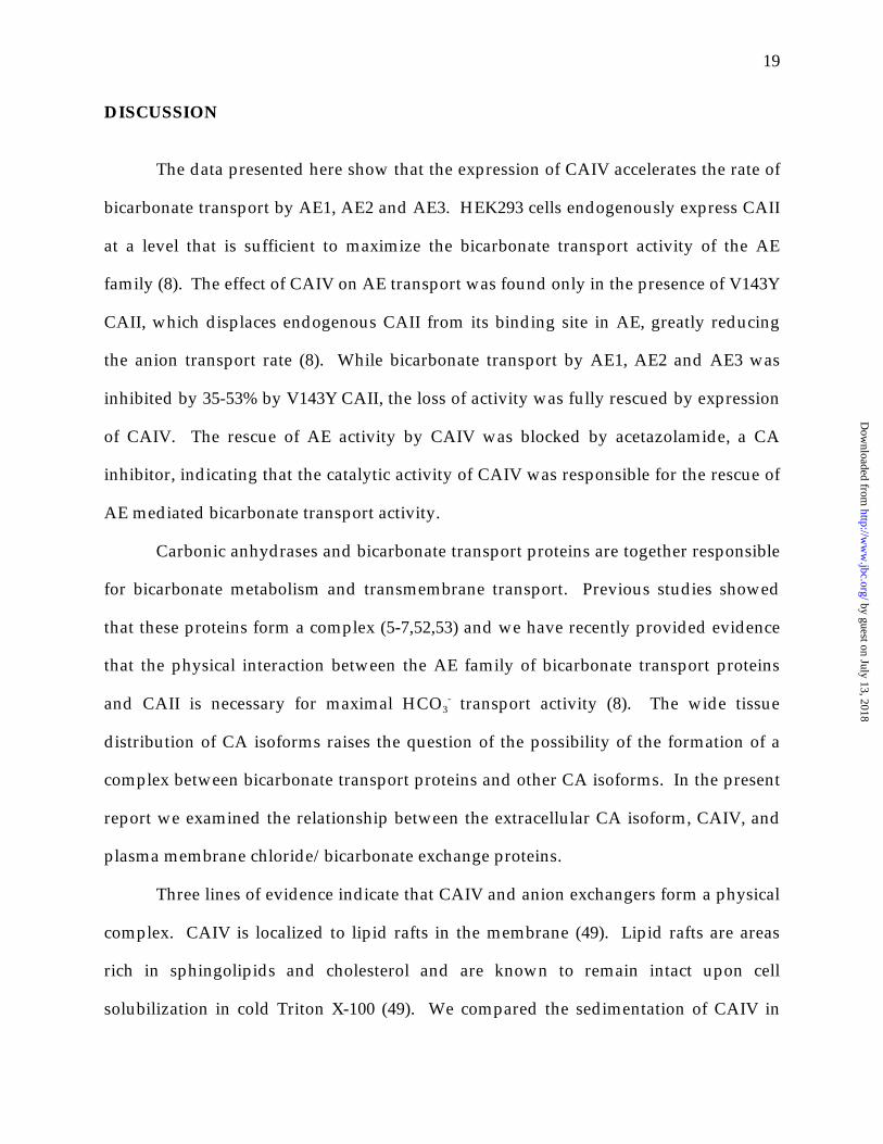
GST pull-down assays of AE1 extracellular loops - To localize the site of AE1 interaction
with CAIV, we reasoned that GPI-anchored CAIV must interact with the extracellular
portion of AE1. The most likely candidates for a CAIV binding site are the largest
extracellular loops of AE1, EC3 and EC4 (Fig. 6). GST fusion proteins of the third and
fourth extracellular loops of AE1 were used in a GST pull-down assay. GST alone, GST-
AE1EC3 and GST-AE1EC4 were immobilized on glutathione Sepharose resin and cell
lysates of either sham-transfected or CAIV-transfected cells were applied. After
washing, proteins were eluted in SDS-PAGE sample buffer resolved on 8%
polyacrylamide gels by SDS-PAGE electrophoresis, transferred to PVDF membranes
and probed for CAIV. In figure 7 bands were evident only in samples incubated with
CAIV-containing lysates. Thus all bands observed result from CAIV, either full length
or possibly shorter proteolytic fragments. GST alone pulled down a small amount of
CAIV, as did GST-AE1EC3 (Fig. 7). Clearly GST-AE1EC4 brought down the most CAIV
by guest on July 13, 2018http://w
ww
.jbc.org/D
ownloaded from
18
(Fig. 7). Indeed, densitometry revealed that GST-AE1EC4 pulled down approximately
ten fold more CAIV than did GST alone or GST-AE1EC3. This demonstrates that CAIV
binds specifically to the fourth extracellular loop of AE1.
by guest on July 13, 2018http://w
ww
.jbc.org/D
ownloaded from
19
DISCUSSION
The data presented here show that the expression of CAIV accelerates the rate of
bicarbonate transport by AE1, AE2 and AE3. HEK293 cells endogenously express CAII
at a level that is sufficient to maximize the bicarbonate transport activity of the AE
family (8). The effect of CAIV on AE transport was found only in the presence of V143Y
CAII, which displaces endogenous CAII from its binding site in AE, greatly reducing
the anion transport rate (8). While bicarbonate transport by AE1, AE2 and AE3 was
inhibited by 35-53% by V143Y CAII, the loss of activity was fully rescued by expression
of CAIV. The rescue of AE activity by CAIV was blocked by acetazolamide, a CA
inhibitor, indicating that the catalytic activity of CAIV was responsible for the rescue of
AE mediated bicarbonate transport activity.
Carbonic anhydrases and bicarbonate transport proteins are together responsible
for bicarbonate metabolism and transmembrane transport. Previous studies showed
that these proteins form a complex (5-7,52,53) and we have recently provided evidence
that the physical interaction between the AE family of bicarbonate transport proteins
and CAII is necessary for maximal HCO3- transport activity (8). The wide tissue
distribution of CA isoforms raises the question of the possibility of the formation of a
complex between bicarbonate transport proteins and other CA isoforms. In the present
report we examined the relationship between the extracellular CA isoform, CAIV, and
plasma membrane chloride/bicarbonate exchange proteins.
Three lines of evidence indicate that CAIV and anion exchangers form a physical
complex. CAIV is localized to lipid rafts in the membrane (49). Lipid rafts are areas
rich in sphingolipids and cholesterol and are known to remain intact upon cell
solubilization in cold Triton X-100 (49). We compared the sedimentation of CAIV in
by guest on July 13, 2018http://w
ww
.jbc.org/D
ownloaded from

20
sucrose gradients in the absence and presence of AE1. In the presence of AE1, the
sedimentation of CAIV shifted from the less dense fractions, where it is found when
expressed alone, and to the denser fractions where AE1 was localized. This result
suggests that AE1 and CAIV physically interact and that AE1 pulls CAIV out of lipid
rafts. In a second approach, AE1, AE2 and AE3 expressed in HEK293 cells were able to
interact with CAIV from cell lysates of HEK293 cells expressing CAIV in gel overlay
assays.
The third and most definitive evidence of a CAIV:AE interaction came from GST
pull-down assays. As CAIV is linked to the extracellular surface of the cell we reasoned
that the AE:CAIV interaction occurred at one of the larger extracellular loops of AE1.
We investigated the extracellular loops between transmembrane segments 5 and 6
(EC3) and TM 7 and 8 (EC4). GST fusion proteins of the individual loops (GST-
AE1EC3, GST-AE1EC4) were constructed. These GST fusion proteins and control GST
alone were immobilized on glutathione Sepharose resin. Lysates prepared from
HEK293 cells transfected with CAIV cDNA or sham-transfected were incubated with
the GST protein/glutathione Sepharose resin complexes. CAIV associated with the
resin was detected on immunoblots. The presence of a band corresponding to the
molecular weight of CAIV appeared only when lysates from CAIV-transfected cells
were applied to immobilized GST-AE1EC4 (Fig. 6). This suggests that CAIV binds
specifically to the fourth extracellular loop of AE1.
On the basis of these three lines of evidence we conclude that CAIV forms a
complex with AE1, AE2 and AE3. The simplest explanation for our observation is that
CAIV directly interacts with AE1, AE2 and AE3. We cannot rule out the possibility that
another protein is required to mediate the AE:CAIV interaction. However, the
requirement of an intermediary protein is highly unlikely since CAII interacts directly
by guest on July 13, 2018http://w
ww
.jbc.org/D
ownloaded from

21
with AE1-AE3 (8) and any intermediary protein would have to be endogenously
expressed in HEK293 cells. The increase of AE1, AE2 and AE3 bicarbonate transport
activity caused by CAIV likely requires a direct interaction between CAIV and AE;
localization of CAIV to the same membrane may not be sufficient to enhance
bicarbonate transport rate.
The identification of EC4 as the binding site for CAIV is interesting in a number
of ways. Studies of AE1 topology suggest that EC4 is the largest extracellular loop
(46,54), and therefore might be expected to form an extracellular binding site. That EC4
forms an accessible extracellular region is demonstrated by the four blood group
antigens, the Wright antigen (E658K)(55), Moa (R656H), Hga (R646Q) and Swa (R646Q)
(56), which are found in EC4 (Fig. 6). The Wright antigen is formed by a complex
between the highly glycosylated single transmembrane protein glycophorin A, and AE1
(55). Thus there is precedent for an interaction between EC4 and the extracellular
moiety of an erythrocyte protein.
A study of the AE1 region from the glycosylation site at N642 (Fig. 6) through
transmembrane segment 8 suggested that the region S643-L655 had a folded structure
that was inaccessible to hydrophilic reagents, while the region R656-I661 had an open
structure with maximum accessibility at R656 (46). Taken together we propose that
CAIV interacts with AE1 somewhere in the R656-I661 region. Interestingly this region
has been suggested to form the outer vestibule that funnels anions to and from the
transport site (46). Localization of CAIV to EC4 would therefore place the enzyme as
close as possible to the extracellular aspect of the anion transport site.
The structure of AE2 and AE3 differs from AE1 in that AE2 and AE3 are
glycosylated on EC3 rather than EC4 and EC3 is larger than EC4 in AE2 and AE3 (57).
It is therefore not clear whether AE2 and AE3 interact with CAIV in the homologous
by guest on July 13, 2018http://w
ww
.jbc.org/D
ownloaded from

22
loop region or not. Nevertheless the CAIV-mediated rescue of AE2 and AE3
bicarbonate transport activity in the presence of V143Y CAII indicates a functional
interaction between CAIV and AE2 and AE3, which is also likely paralleled by a
physical interaction.
We have previously characterized the first example of a transport metabolon by
defining the importance of the physical and functional interaction between AEs and
CAII (8). The present study provides evidence that the extracellular-anchored enzyme
CAIV is the extracellular component of the bicarbonate transport metabolon. The
presence of intracellular CAII and extracellular CAIV catalytic activity in the cell and
the fact that both enzymes can potentiate the bicarbonate transport activity of AE1
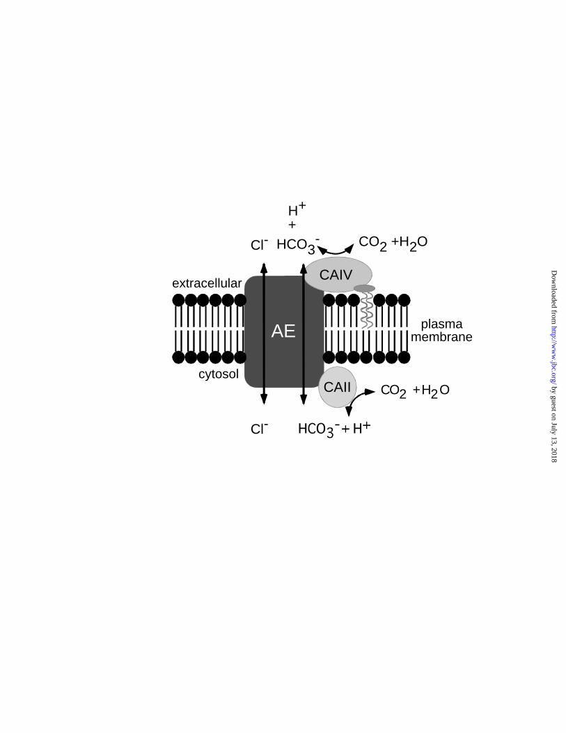
provides the cell with a “push-pull” mechanism for bicarbonate transport (figure 8).
CA-mediated production of HCO3- on the one side of the membrane will provide
“push” for transport by AE and CA-mediated conversion to CO2 on the other side
provides “pull”, by minimization of the [HCO3-] at the trans transport side. This push-
pull mechanism, established by having CA catalytic activity on both sides of the plasma
membrane, accelerates AE-mediated bicarbonate transport as shown in this study.
Although the heart does not express any cytosolic CA, it expresses two
extracellular CA isoforms, one of which is known to be CAIV (22). The heart also
expresses AE1, AE2 and AE3 (19,58,59), which were all shown to require interaction
with CAII for maximal transport activity to be achieved (8). Our results show that
extracellular CAIV can functionally replace CAII. Thus, despite the absence of CAII in
cardiomyocytes, these bicarbonate transporters would be expected to be able to
function at their maximum rate.
The kidney expresses an N-terminally truncated variant of AE1 (kAE1).
Although it is generally agreed that kAE1 localizes to the basolateral surface of α-
by guest on July 13, 2018http://w
ww
.jbc.org/D
ownloaded from

23
intercalated cells (60), there is also one report that kAE1 is found at the apical surface of
β-intercalated cells (61). AE2 is found in the basolateral surface of many portions of the
kidney (62). CAIV has been reported to be in both apical and basolateral surfaces of
proximal tubule (35,63) and basolateral surface of thick ascending limb (64), but others
report that CAIV is only found apically in the kidney (65). Therefore, AE1 and AE2 co-
localize with CAIV in some renal cells.
CAII deficiency is an autosomal recessive condition characterized by renal
tubular acidosis and osteopetrosis (66). Despite gross abnormalities associated with the
absence of CAII, CA activity in erythrocytes is adequate and patients have sufficient
CO2 transport capacity (67). The findings in the present paper, along with the recent
detection of CAIV expression and activity in human erythrocytes (31) explain the ability
of erythrocytes lacking CAII to sufficiently accommodate CO2 metabolism. Normal
bone reabsorption requires basolateral AE2 in osteoclasts. CAII deficiency inactivates
AE2, leading to osteopetrosis. Since CAIV is not expressed in osteoclasts, it cannot
compensate for loss of CAII. Renal tubular acidosis results from a failure to reabsorb
bicarbonate from the renal tubular lumen. While it is possible that CAIV and AE
interact in the kidney, the presence of functional CAIV in CAII deficient patients is not
sufficient to prevent renal tubular acidosis.
This study presents several lines of evidence indicating a physical interaction of
extracellular CAIV with the Cl-/HCO3- transport protein. We have also demonstrated
that the presence CAIV catalytic activity accelerates the movement of bicarbonate across
the plasma membrane by AE1, AE2 and AE3. The results described here demonstrate
that CAIV is the extracellular component of a bicarbonate transport metabolon, formed
along with an anion exchange protein and intracellular CAII. When co-expressed, CAII
and CAIV contribute to the function of AE, providing a “push-pull” mechanism for
by guest on July 13, 2018http://w
ww
.jbc.org/D
ownloaded from

24
bicarbonate movement across the plasma membrane. The potentiation of AE transport
activity by CAIV in the erythrocyte may provide an explanation for the observation that
CAII deficient patients retain the capacity for normal CO2 metabolism and transport. It
also suggests a potential mode of regulation of bicarbonate transport and raises the
possibility that modulation of CAIV:AE interaction, perhaps with antibodies or
peptides, could be a viable therapeutic approach to alter bicarbonate transport.
by guest on July 13, 2018http://w
ww
.jbc.org/D
ownloaded from

25
ACKNOWLEDGEMENTS
We would like to express our gratitude to Dr. George Schwartz for his very kind
donation of CAIV cDNA and the anti-CAIV antibody and to Dr. Carol Fierke for the
V143Y CAII cDNA. This work was funded by the Heart and Stroke Foundation of
Canada (HSF). Deborah Sterling holds studentship trainee awards from the HSF and
the Alberta Heritage Foundation for Medical Research (AHFMR). Bernardo Alvarez
holds a postdoctoral fellowship from AHFMR.
by guest on July 13, 2018http://w
ww
.jbc.org/D
ownloaded from
26
REFERENCES
1. Sterling, D., Alvarez, B. V., and Casey, J. R. (2002) Biophysical Journal 82, 570.
2. Mori, K., Ogawa, Y., Ebihara, K., Tamura, N., Tashiro, K., Kuwahara, T., Mukoyama,
M., Sugawara, A., Ozaki, S., Tanaka, I., and Nakao, K. (1999) J Biol Chem 274, 15701-
15705.
3. Kivela, A., Parkkila, S., Saarnio, J., Karttunen, T. J., Kivela, J., Parkkila, A. K., Waheed,
A., Sly, W. S., Grubb, J. H., Shah, G., Tureci, O., and Rajaniemi, H. (2000) Am J Pathol
156, 577-584.
4. Tureci, O., Sahin, U., Vollmar, E., Siemer, S., Gottert, E., Seitz, G., Parkkila, A. K.,
Shah, G. N., Grubb, J. H., Pfreundschuh, M., and Sly, W. S. (1998) Proc Natl Acad Sci U
S A 95, 7608-7613.
5. Vince, J. W., and Reithmeier, R. A. F. (1998) J Biol Chem 273, 28430-28437.
6. Vince, J. W., and Reithmeier, R. A. F. (2000) Biochemistry 39, 5527-5533.
7. Vince, J. W., Carlsson, U., and Reithmeier, R. A. F. (2000) Biochemistry 39, 13344-
13349.
8. Sterling, D., Reithmeier, R. A., and Casey, J. R. (2001) J Biol Chem 276, 47886-47894.
9. Srere, P. A. (1985) Trends Biochem Sci 10, 109-110.
10. Srere, P. A. (1987) Annu Rev Biochem 56, 89-124.
11. Scozzafava, A., and Supuran, C. T. (2002) Bioorganic and Medicinal Chemistry Letters.
12. Grinstein, S., Ship, S., and Rothstein, A. (1979) Biochim. Biophys. Acta 507, 294-304.
13. Kopito, R. R., Lee, B. S., Simmons, D. M., Lindsey, A. E., Morgans, C. W., and
Schneider, K. (1989) Cell 59, 927-937.
14. Kudrycki, K. E., Newman, P. R., and Schull, G. E. (1990) J. Biol. Chem. 265, 462-471.
15. Alper, S. L., Kopito, R. R., Libresco, S. M., and Lodish, H. F. (1988) J. Biol. Chem. 263,
17092-17099.
16. Tsuganezawa, H., Kobayashi, K., Iyori, M., Araki, T., Koizumi, A., Watanabe, S.,
Kaneko, A., Fukao, T., Monkawa, T., Yoshida, T., Kim, D. K., Kanai, Y., Endou, H.,
Hayashi, M., and Saruta, T. (2001) J Biol Chem 276, 8180-8189.
17. Kollert-Jons, A., Wagner, S., Hubner, S., Appelhans, H., and Drenckhahn, D. (1993) Am
J Physiol 265, F813-821.
by guest on July 13, 2018http://w
ww
.jbc.org/D
ownloaded from

27
18. Richards, S. M., Jaconi, M. E., Vassort, G., and Puceat, M. (1999) J Cell Sci 112, 1519-
1528.
19. Linn, S. C., Kudrycki, K. E., and Schull, G. E. (1992) J. Biol. Chem 267, 7927-7935
20. Kobayashi, S., Morgans, C. W., Casey, J. R., and Kopito, R. R. (1994) J. Neurosci. 14,
6266-6279.
21. Waheed, A., Zhu, X. L., Sly, W. S., Wetzel, P., and Gros, G. (1992) Arch Biochem
Biophys 294, 550-556.
22. Sender, S., Decker, B., Fenske, C. D., Sly, W. S., Carter, N. D., and Gros, G. (1998) J
Histochem Cytochem 46, 855-861.
23. Whitney, P. L., and Briggle, T. V. (1982) J Biol Chem 257, 12056-12059.
24. Brion, L. P., Zavilowitz, B. J., Suarez, C., and Schwartz, G. J. (1994) Am J Physiol 266,
F185-195.
25. Sly, W. S., Whyte, M. P., Krupin, T., and Sundaram, V. (1985) Pediatr Res 19, 1033-
1036.
26. Wistrand, P. J., and Knuuttila, K. G. (1989) Kidney Int 35, 851-859.
27. McKinley, D. N., and Whitney, P. L. (1976) Biochim Biophys Acta 445, 780-790.
28. Tong, C. K., Brion, L. P., Suarez, C., and Chesler, M. (2000) J Neurosci 20, 8247-8253.
29. Ghandour, M. S., Langley, O. K., Zhu, X. L., Waheed, A., and Sly, W. S. (1992) Proc
Natl Acad Sci U S A 89, 6823-6827.
30. Hageman, G. S., Zhu, X. L., Waheed, A., and Sly, W. S. (1991) Proc Natl Acad Sci U S
A 88, 2716-2720.
31. Wistrand, P. J., Carter, N. D., Conroy, C. W., and Mahieu, I. (1999) Acta Physiol Scand
165, 211-218.
32. Baird, T. T., Jr., Waheed, A., Okuyama, T., Sly, W. S., and Fierke, C. A. (1997)
Biochemistry 36, 2669-2678.
33. Okuyama, T., Sato, S., Zhu, X. L., Waheed, A., and Sly, W. S. (1992) Proc Natl Acad Sci
U S A 89, 1315-1319.
34. Geers, C., Krüger, D., Siffert, W., Schmid, A., Bruns, W., and Gros, G. (1992) Biochem.
J. 282, 165-171.
35. Schwartz, G. J., Kittelberger, A. M., Barnhart, D. A., and Vijayakumar, S. (2000) Am J
Physiol Renal Physiol 278, F894-904.
by guest on July 13, 2018http://w
ww
.jbc.org/D
ownloaded from

28
36. Maren, T. H., and Conroy, C. W. (1993) J Biol Chem 268, 26233-26239.
37. Brechue, W. F., Kinne-Saffran, E., Kinne, R. K., and Maren, T. H. (1991) Biochim
Biophys Acta 1066, 201-207.
38. Fierke, C. A., Calderone, T. L., and Krebs, J. F. (1991) Biochemistry 30, 11054-11063.
39. Casey, J. R., Ding, Y., and Kopito, R. R. (1995) J. Biol. Chem. 270, 8521-8527.
40. Lee, B. S., Gunn, R. B., and Kopito, R. R. (1991) J. Biol. Chem. 266, 11448-11454.
41. Sterling, D., and Casey, J. R. (1999) Biochem J 344 Pt 1, 221-229.
42. Graham, F. L., Smiley, J., Russell, W. C., and Nairn, R. (1977) J. Gen. Virol. 52, 59-72
43. Ruetz, S., Lindsey, A. E., and Kopito, R. R. (1993) Society of General Physiologists
Series 48, 193-200.
44. Laemmli, U. K. (1970) Nature 227, 680-685.
45. Towbin, H., Staehelin, T., and Gordon, J. (1979) Proc. Natl. Acad. Sci. 76, 4350-4354.
46. Tang, X. B., Fujinaga, J., Kopito, R., and Casey, J. R. (1998) J Biol Chem 273, 22545-
22553.
47. Thomas, J. A., Buchsbaum, R. N., Zimniak, A., and Racker, E. (1979) Biochemistry 18,
2210-2218.
48. Roos, A., and Boron, W. F. (1981) Physiol Rev 61, 296-434.
49. Brown, D. A., and Rose, J. K. (1992) Cell 68, 533-544.
50. Cousin, J. L., Motais, R., and Sola, F. (1975) J Physiol 253, 385-399.
51. Cousin, J. L., and Motais, R. (1976) J Physiol 256, 61-80.
52. Kifor, G., Toon, M. R., Janoshazi, A., and Solomon, A. K. (1993) J Membr Biol 134,
169-179.
53. Parkes, J. L., and Coleman, P. S. (1989) Arch Biochem Biophys 275, 459-468.
54. Reithmeier, R. A. F., Chan, S. L., and Popov, M. (1996) in Transport Processes in
Eukaryotic and Prokaryotic Organisms (Konings, W. N., Kaback, H. R., and Lolkema, J.
S., eds) Vol. 2, pp. 281-309, Elsevier Science.
55. Bruce, L. J., Ring, S. M., Anstee, D. J., Reid, M. E., Wilkinson, S., and Tanner, M. J.
(1995) Blood 85, 541-547.
56. Jarolim, P., Rubin, H. L., Zakova, D., Storry, J., and Reid, M. E. (1998) Blood 92, 4836-
4843.
by guest on July 13, 2018http://w
ww
.jbc.org/D
ownloaded from

29
57. Zolotarev, A. S., Chernova, M. N., Yannoukakos, D., and Alper, S. L. (1996)
Biochemistry 35, 10367-10376.
58. Linn, S. C., Askew, G. R., Menon, A. G., and Shull, G. E. (1995) Circ. Res. 76, 584-591.
59. Pucéat, M., Korichneva, I., Cassoly, R., and Vassort, G. (1995) J. Biol. Chem. 270, 1315-
1322.
60. Bagnis, C., Marshansky, V., Breton, S., and Brown, D. (2001) Am J Physiol Renal
Physiol 280, F437-448.
61. van Adelsberg, J., Edwards, J. C., and Al-Awqati, Q. (1993) J. Biol. Chem. 268, 11283-
11289.
62. Brosius, F. C., Nguyen, K., Stuart-Tilley, A. K., Haller, C., Briggs, J. P., and Alper, S. L.
(1995) American Journal of Physiology 269, F461-468.
63. Schwartz, G. J., Olson, J., Kittelberger, A. M., Matsumoto, T., Waheed, A., and Sly, W.
S. (1999) Am J Physiol 276, F510-520.
64. Brown, D., Zhu, X. L., and Sly, W. S. (1990) Proc Natl Acad Sci U S A 87, 7457-7461.
65. Lonnerholm, G., and Wistrand, P. J. (1991) Acta Physiol Scand 141, 231-234.
66. Sly, W. S., Hewett-Emmett, D., Whyte, M. P., Yu, Y. S., and Tashian, R. E. (1983) Proc
Natl Acad Sci U S A 80, 2752-2756.
67. Dodgson, S. J., Forster, R. E., 2nd, Sly, W. S., and Tashian, R. E. (1988) J Appl Physiol
65, 1472-1480.
by guest on July 13, 2018http://w
ww
.jbc.org/D
ownloaded from

30
FIGURE LEGENDS
Figure 1 - Expression of AE1, CAII and CAIV in transfected cells. HEK293 cells were
transiently co-transfected with cDNA coding for AE1, CAII and CAIV. Two days post
transfection, cells were solubilized. Samples (5 µg protein) were resolved by SDS-PAGE
on 8% (samples probed for AE1) or 12.5 % (samples probed for CAII and CAIV)
acrylamide gels and transferred to PVDF membrane. Immunoblots were probed with
rabbit polyclonal antibody, 1658, directed against the C-terminus of human AE1, sheep-
anti human CAII antibody or goat anti-rabbit CAIV as indicated.
Figure 2 - Effect of carbonic anhydrases on AE1 transport activity. HEK293 cells grown
on coverslips were transiently co-transfected with cDNA encoding AE1 (A), AE1 and
V143Y CAII (B) and AE1, V143Y CAII and CAIV (C). Two days post transfection cells
were loaded with BCECF-AM and placed in a fluorescence cuvette in a fluorimeter.
Cells were perfused alternately with Cl--containing (solid bar) and Cl--free (open bar)
Ringer’s buffer and fluorescence was monitored using excitation wavelengths 440 and
502.5 nm and emission wavelength 528.7 nm. In some experiments, cells were
incubated with 100 µM acetazolamide for 10 minutes followed by a repeat of the
Ringer’s buffer perfusion in the presence of 100 µM acetazolamide. Transport activity
following acetazolamide incubation was compared to that before the incubation. D,
summary of transport rates expressed relative to rate for AE1 expressed alone. Error
bars represent standard error of the mean (n =4) and asterisk represents statistical
significance (p<0.001).
by guest on July 13, 2018http://w
ww
.jbc.org/D
ownloaded from

31
Figure 3 - CAIV facilitates bicarbonate transport by AE2 and AE3. HEK293 cells were
transiently transfected with cDNA encoding AE2 or AE3 and co-transfected with or
without V143Y CAII and CAIV cDNA, as indicated at the bottom of the figure. Anion
exchange activity was measured and rates expressed relative to the rate for AE2 (panel
A) and AE3 (panel B). Error bars represent standard error of the mean (n =4) and
asterisk represents statistical significance (p<0.001).
Figure 4 - Association of AE1 and CAIV. HEK293 cells were transiently transfected
with cDNA encoding either AE1 or CAIV or co-transfected with both cDNAs, as
indicated in each panel. Two days post-transfection cells were solubilized in cold
Triton X-100 and samples were overlaid on 5-30% continuous sucrose gradients.
Following ultracentrifugation, 12 fractions were collected (1-top, 12-bottom) and
samples of each were resolved by SDS-PAGE electrophoresis on an 8% (AE1) or 12.5%
(CAIV) polyacrylamide gels. Immunoblotted proteins were probed with either anti-
AE1 (black bars) or anti-CAIV (grey bars). Scanning and densitometry of immunoblots
provided relative expression levels of protein in each fraction. Error bars represent
standard error of the mean (n =3).
Figure 5 - Blot overlay assay of CAIV on AE1, AE2 and AE3. HEK293 cells were
transiently transfected individually with AE1, AE2 or AE3 cDNA. Two days post
transfection, cells were solubilized, and 5 µg protein resolved by SDS-PAGE on 8%
acrylamide gels and transferred to PVDF membrane, as indicated in the figure.
Immunoblots were blocked for 3 hours with 10% TBST-M and then incubated overnight
in 1% TBST-M containing a lysate of CAIV-transfected HEK 293 cells. Blots were then
probed with anti-CAIV antibody. Arrows indicate position of AE proteins.
by guest on July 13, 2018http://w
ww
.jbc.org/D
ownloaded from

32
Figure 6 - Topology model of human AE1 on the basis of experimental evidence (46,54).
Amino acids corresponding to GST-AE1EC3 and EC4 are indicated in grey. Arrows
indicate the positions of point mutations that induce blood group antigens (Swa
(R646Q), Moa, (R656H), Hga (R656C) (56) and Wra (E658K) (55)) are indicated. The Y
structure on EC4 marks the position of N-linked glycosylation.
Figure 7 - CAIV binds specifically to the fourth extracellular loop of AE. Proteins (10 µg
GST alone, GST-AE1EC3 or GST-AE1EC4) were individually bound to glutathione
Sepharose resin as indicated. Cell lysates of HEK293 cells transfected with CAIV cDNA
or sham transfected with vector alone were applied to the beads as indicated and
incubated overnight. Samples were centrifuged and the beads washed. Proteins eluted
with SDS-PAGE sample buffer were resolved by SDS-PAGE electrophoresis on a 12.5%
polyacrylamide gel, transferred to a PVDF membrane and probed for CAIV as
described previously. Arrow indicates position of CAIV.
Figure 8 – A bicarbonate transport metabolon. Schematic model of the binding of CAIV
to extracellular loop 4 of AE1 and CAII to the C-terminal tail, which maximizes AE
mediated bicarbonate flux by the production and removal of substrate from the
transport site. GPI is indicated by the structure that comes off of CAIV and intercalates
into lipid bilayer of plasma membrane.
by guest on July 13, 2018http://w
ww
.jbc.org/D
ownloaded from

10482
48
3328
kDaAE1 CAII CAIV
by guest on July 13, 2018http://w
ww
.jbc.org/D
ownloaded from

**
AE1
V143Y CAII
CAIV
Acetazolamide
D
7.0
7.2
7.4
7.6
7.8
8.0
7.0
7.2
7.4
7.6
7.8
8.0
7.0
7.2
7.4
7.6
7.8
8.0
0 200 400 600
Time (s)
A
B
C by guest on July 13, 2018http://w
ww
.jbc.org/D
ownloaded from

0
V143Y CAII
CAIV
AE isoform AE2
20
40
60
80
100
A B
**
AE3
by guest on July 13, 2018http://w
ww
.jbc.org/D
ownloaded from

1 122 3 4 5 6 7 8 9 10 11
Fraction number
0
0.2
0.4
0.6
0.8
1.0
0
0.2
0.4
0.6
0.8
1.0
AE1/CAIV
AE1/CAIV
1 122 3 4 5 6 7 8 9 10 11
Fraction number
0
0.2
0.4
0.6
0.8
1.0
0
0.2
0.4
0.6
0.8
1.0
AE1
CAIV
by guest on July 13, 2018http://w
ww
.jbc.org/D
ownloaded from

AE1 AE2 AE3
233
135
112
kDa
AE3 upperAE3 lowerAE2
AE1
by guest on July 13, 2018http://w
ww
.jbc.org/D
ownloaded from

V AL
CL
G
K
VV
W
F
KAQ C L D A D DEV LN
D ATFEEEGRDEYD
EV
A M P Y 911
FI
R
A
LL
P
T P
RR
T
A
VV
V
L L
LLL
P
PI
S
F
L
RVKTW
V
YK
D
HP
PP
P
V
K
K
F
Y
RM
S
L
VFL
YVS
LG
A
G
T
LLF
IMS
IG
P
Q
SI
R
R
E
QV E
KQI
L
L
I
L
L
S
VG
M
GL
S
A VV
I
P I LE
Q
IF
LY
ASPI
TF
G
LA
GLLLL
G
IS A
ES
L
G IA
LF
E
LGP
LV
FE
FC
VV
FS
GL
AF
FS
E
F
L
WV
F
F
TF
FS
FL
K
ET
G
G
Q
P
I
LV
L
K
Y
L Y
L
SL
RT
S
IG
W
F
L
I
VL
VA
T
S
LV
YI
RF
T
E
F
A
LII
FI
YE
A
LKLI
F
G
E
T
A
F
MN N L
D
K
L
H GP
Y
I
P
FK
Y F
K
V
R
DF
V LIV
I
M
V
IDL
W
SK
QT
V
L
YT
E
WIP
F
G
V
P
S
P
L
V P
KH
P
R G
I
S
G
D
N
P
L
D
PT
IP
F
M
SV
G LQ I
Y
F
K
LP
T
KP
V
RL
L
E
L
K
H
M
FG
VKG
R
S
EL
A
LL
G V
L
GV
M
G
W
F
TT
V
V
G
QF
LR
A
V
GM
G
P
Q
L
AD
F
D
M
ExtracellularS
VV
AIFA
A
K R NQM
V
T V
L
A Q
L
TNG I
V
I
S
Q
S
Q
Q
QL
KP
G
G
R
NSS
LR
I
S
DG
S
S A
L
D
V
AF
M
PS
T I S L Y Y P R RR
ID
RVGGQ
G
QL
DP G D L GN
Y
Cytosol
Nt
359
... M1
N
L
WraMoa, Hga
Swa
A
IEL
IV
LL
AF
QS
LF
IF
PA
LS
MM
T
by guest on July 13, 2018http://w
ww
.jbc.org/D
ownloaded from

50
36
2920
kDa
GST
GST-AE1EC3
GST-AE1EC4
CAIV lysate
CAIV
by guest on July 13, 2018http://w
ww
.jbc.org/D
ownloaded from
AE
CO2 +H2O
H++
HCO3-
cytosol
Cl-
Cl-
CAIVextracellular
plasmamembrane
CAII CO2 +H2O
by guest on July 13, 2018http://w
ww
.jbc.org/D
ownloaded from

Deborah Sterling, Bernardo V. Alvarez and Joseph R. Casey exchanger binds carbonic anhydrase IV-/HCO3-human AE1 Cl
The extracellular component of a transport metabolon: Extracellular loop 4 of the
published online May 6, 2002J. Biol. Chem.
10.1074/jbc.M202562200Access the most updated version of this article at doi:
Alerts:
When a correction for this article is posted•
When this article is cited•
to choose from all of JBC's e-mail alertsClick here
by guest on July 13, 2018http://w
ww
.jbc.org/D
ownloaded from