Embed Size (px)
Citation preview
Loyola University ChicagoLoyola eCommons
Master's Theses Theses and Dissertations
1964
The Establishment and Maintenance of GoldfishCell Lines in Tissue CultureHelen Mae KroekerLoyola University Chicago
This Thesis is brought to you for free and open access by the Theses and Dissertations at Loyola eCommons. It has been accepted for inclusion inMaster's Theses by an authorized administrator of Loyola eCommons. For more information, please contact [email protected] © 1964 Helen Mae Kroeker
Recommended CitationKroeker, Helen Mae, "The Establishment and Maintenance of Goldfish Cell Lines in Tissue Culture" (1964). Master's Theses. Paper1968.http://ecommons.luc.edu/luc_theses/1968

--
THE ESTABLISHI-IENT AND MAINTENANCE
OF
GOLDFISH OELL LINES IN' TISSUE OULTURE
.-<.,.~y\CH seflo V) LOYOLA q,
UNIVERSiTY
Helen Mae Kroeker
Microbiology Department
Stritch School of Medicine
A Thesis Submitted to the Faoulty ot the Graduate School
of Loyola University in Partial Fulfillment of
the Requirement. for the Degree ot
Master ot Scienoe
June
1964
LIFE
Helen Mae Kroeker was born In Chlcago, Illinois, in
She was graduated from St. Xavier Aoademy, Chlcago,
Illlnois, In 1954, from St. Xavier - Mother MoAuley High School,
Chicago, Illinois, in 1958, and froM St. Xavier College, Chloago,
Illinois, in 1962, with the degree of Baohelor of Arts.
She began her graduate studies at Loyola Unlversity
in September, 1962.
lli
ACKJlOWLEtGMEN'r
Sincere gratitude 11 extended to Doctor Thomas J. Bird
who suggested thI1 problem and aided in ita fruition.
I alao wish to extend thanks to Doctor Frank Prigan for
advice 1n the performance ot thil wo~k.
iv
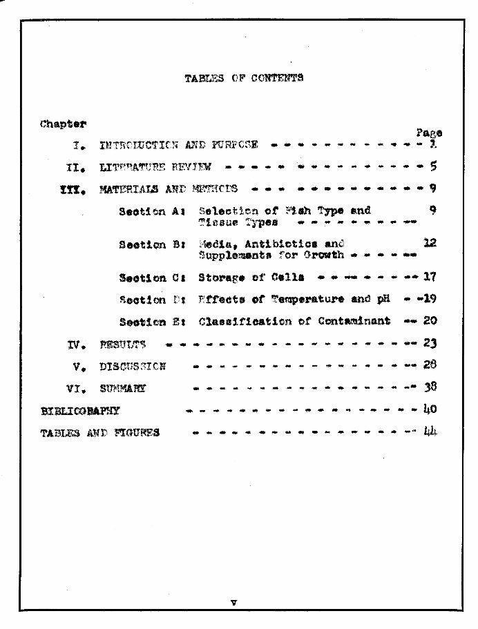
Chapt."
1'. .. .. .. • _ _ _ _ _ • • ~af..
-.. ..... - - - - - - .. 5 Itt. MATERIALS AWt ~1(,l'S ...... _................. .. 9
a.otton Al Select1efl. ot ~1Ih TJpe md 9 Tissue T)~.. .... - - - ... .. - _.
Sutton B. f~edla, Antibiotio. antS 12 ~upple~nt. ro~ Growth .. .. .. ... ..
Seotion 01 Storage of Cella .... - ..... - -- 11
Scot! on r I Rtf.cta ot' 'rem.p ... ture and pH .. -19
a.otten Et Classification of ContMlnant -- 20
• • - - - - - - ~ - - - - - ~ - - - _. 23 v. DISCUS~Ir;N
VI. s'tnnu.m
BlBU:COJtAP}lY
'fASIBS AWL'; fIGURES
• - - • • - - - - - - - ~ - - - -- 26
- - • - • - - - - ~ - - - - ~ - .- )8
.. .. ... .... .. - .. - ... .. .. - -- - - ., .. - 40
- - - - • - ~ - - - ~ - - - - - -- 44
v
LIST OF FIGURES
Figure Page
I. REPORTS OF VARIOUS HEDlA TYPES ON CELL MULTIPLICATICN .. ... - ... ... .. .. .. .. ... .. 59
II. EFFECT OF TE~!PERATURE ON CELL WJLTIPLICATION ...... - ... ... ... .. ... .. 61
III. EFFECT OF VARIAnCE OF pH ON CELL M'JLTIPLICATICN ... ... ... ... .. ... ... ... - 63
IV. PHOTCGRAPJIS .. .. ... ... ... ... ... .. - .. ... .. 64
V. PHOTOGRAPHS ... - ... ... .. .. .. - .. - - ... 64
VI. PHOTOGRAPHS .. - ... - .. ... ... • - - ... - 65
VII. PHOTOGRAPHS - - - - ... - ... - - - .. - 65
vi

LIST OF TABLES
Table Page I. MORPHOLOGICAL AND PHYSIOLOGICAL CLASSIFICATICN
OF CONTAlUNAUT - - - .. - - ... ... - _... 44
II. TYPES OF MEDIA 5CR~l[D ... ... - ... - - - ... 45 III. RESULTS OF Mb1DIA SCREYNING ON CULTliRES OF
WHOLE SMALL GOLDFISH ... - .. ... ... ... - .... - 46
IV. THE RELATION OF 'rEMPERATURE TO PERCENT OF INCREASE IN CELL NUNBERS F0R WHOLE SMALL GOT .. DFISH .. - - - - .. .. .... .. - - ... - 41
v. THE RELATIC'N OF TE'MPERATURE TO PERCRNT OF INCHEASE IN C FLL hTQI-lBERS T<'OR FISH HEART ... - ... 49
VI. THE REIA'1'1 ON OF TE'1PERATtJRE TO PERCENT OF INCRF~SE IN CELL NUMBERS FOR FISH MUSCLE - .. 51
VII. STORAGE RESULTS WITH TISSUES OF SI-1ALL WHOLE GOLDFISH .... - .. - - - - - .... .... .... .. .... 53
VIII. stORAGE RESULTS FOR TISSUE OF SMALL WHOLE GOLD FISH - - .. - .. - - - .. - - .. 54
IX. STORAGE RE8ULTS pon Pi SH HEART AND FISH MUSCLE CELLS.. - ... - .. - - - - - - - $5
x. EFtT'ECT OF pH VARIANCE IN PERCENT OF CELL NUMBER INCREASE.... - - - - - - - .. - - 56
vii
---
CHAPTER I
IN'rRODUCTION AND PURPOSE
The cultivation of tissues !a vitro is a relatively
young tleld ot lolence. One of the first attempts at studying
the functional properties of cells and how they affect or are
aftected by thelr immediate environment was performed by
von Recklnghau.en (1886) who kept amphibian blood cells alive
under various conditions for as long as 35 days.
The first tlssue culture experimentl were oarried
on by Roux (1885) who by :maintaining the neural plate of a
ohick embrro in warm saline, proved the closure of the neural
tube to be a tunction of the constituent cells and not the
direot effect ot meohanical pressure trom the adjacent
structures.
Harrison (1907) made observations on the living
nerve tiber.
1
2
Carrel (1914) sucoeeded in keeping a strain of chiok
connective tissue oel1s alive and aotively multiplying for
34 years.
Thomson (1914) initiated a different approach to tissue
culture along with Strangeways and Fell. This was the start of
"organ oulture", a teohnique whose aim it is to maintain small
fragments of tissues in a state as olose as is possible to their
invl~o situation, rather than trying to make oe11s grow as
rapidly as possible.
Maitland and Maitland (1928) developed a simple method
for virus multiplication.
Up to this time original methods of tissue culture as
fashioned after the metioulous aseptio teChniques of Carr~l. were
extremely tedious. Thus, the labor required to keep oultures
free from oontamination deterred many biologists from entering
the field.
With the stimulus of World War II., the study of
ohemotherapeutio agents lead to the disoovery of antibiotios.
The incorporation of antibiotics into tissue culture media
lessened the problems of tissue culture to a considerable extent.
No longer were the exacting original teohniques required for
cultura.l.a.sepais. There was a resurgenoe of interest in the
field as a practical art.
A great impetus was given to tissue culture as a

3
field when Enders (1948) ~ a1. showed that the polio virus
could be cultivated in vitro in the absence of nervous tissue.
The contributions of these and others have aided in
the development of prooedures that have proved and are proving
to be advantageolls to all fields of experimental biology and
medicine. The importance and potentIal of tissue culture in the
areas ot m.orphogenesis, cancer resea.rch and virology is far
reaohing.
Tissues of mammalian, amphibian, and avian origins
have been used a great deal for tissue culture studies, but
comparatively tew observers have used fish tissues.
Since fish are poikilothermic, the development of fish
cell lines has many possibilities. In their natural environment
they metabolize at relatively low temperatures. If cells of fish
origin could be cultivated and stored at low temperatures, they
would be usetul for cultivating agents which cannot be maintained
at the higher temperatures required by aome of the mammalian cell
lines. They may one day provide an interesting vehicle for viral
research.
The purpose of this work was two-fold. The primary
objective was to establish a fish cell line in continuous
culture trom fish cells ot varied tissue origins. The second
objeotive was to explore the tolerance of suoh cells to
variations in media, antibiotics, temperature, pH, and the
means by which they could be stored.
LI!ERATURE REVIEW
Osowski (1914) observed cellular movement in fragments
of tadpoles and trout (~almo Galrdnerl), wnich were maintained
tor 24 hours in Ringer' a solution. Lewi. (1916) studied
embrJon1c tIssue maintenance using aterile a.a water aa a tisaue
cultuN medium. Dr.derer (1921) in studying the behavior of
embrJon1o flab eotoderm cella in tisaue culture (trom FQnduB
heteroclltus), u •• d II aea water baa. and fish bouillon. The.e
cultures were only viab1e tor 10 days. Chlopin (1922) oultured
tIlluel from pike and eruslan carp in rabbIt plasma, "dlluted
with homOrenouB extraot".
The above works oomprise the rudimentary beginnings
of the culturing of fIsh tissues in vi.tro. Thea. first attempts ............ .. involved priMArIly the 1";lat:n.tenance of' fish abryoa.
4
5
The next trend of investigation is seen in the works of
suoh investigators as Grand (1935, 1938, 1941), Gordon (1938,
1941), and Cameron (1935. 1941), who concerned themselves wIth
the morphologioal and physiological aspects of fish melanomas and
1eiomyomas.
Sohumberger (1949) studying neoplastic goldfish tissue,
added embryonic fish extraots to salt solutions enriohed with
ohicken, human cord or flsh sera.
Boret and Sanders (1954) suoceeded in propagating the
virus of Eastern equine encephalomyelitis in fish embryos.
Wood (1955) was able to isolate and identify agents whioh were
oausative of a myoosis-like granuloma of salmon.
Grutzner (1956), of the Robert Kooh Institute, Berlin,
examined the applioation of tissue oultures of Lebiatee
retioulatus (Peters) and l\facropodus operculari. (linne) in
virology. An attempt waa made to demonstrate the lymphooystis
virus and the virus oausative ot oarp pox in tissue oultures ot
tropioal fish. Speoific diagnostio oell alterations were observed
14-18 days after infection with oarp pox in tissue oultures Of
Labiates retioulatus.
In the attempts to demonstrate the virua of lymphoo~tis
morphologicaloell alterations appeared, however, their speelfloltJ
requires more extensive examination. They were able, in some
instances, to suooessfully transmit lymphocystls disease !!!. vivo
6
fram viral inoouluMS grown in these tissue oultures. Electron
miorographs of the tumors show what may be the elementary bodies
of the virus. More investigative work must be carried out before
a positive identification can be made.
Grutzner (1958) successfully cultivated the liver and
kidney tissues of Tinea vulgaris (a viviparous carp) 1a vitr~.
Monolayers developed w:1thj.n 5 - 6 days and could be maintained
in a healthy condi tion for :3 - 1.t- weeks. The medium employed con
sisted of 8M 199 (synthetio medium 199) supplemented with beef
amniotio fluid plus l5-2~ calf serum, other media used for
culture, but whioh gave poer growth, were (1) isotonic medium 199
plus 20~ calf serum, (2) Lactalbumin-hyd.rolysate (isotonic) plus
~~ calf serum, (3) Lactalbumin-hydrolysate plus isotonic 199,
plus 2~ oali' serum, (4) beef amniotic fluid (isotoniC) plus
15-2~ oalf serum, and (5) beef amniotio fluid plus Lactalbumin
hydrolysate (isotoniC) plus 3~ calf serlun. These oultures were
oarried through three sub-oultures. flle optimal growth was
aohieved at 20°C.
One of the most fruitful endeavors whioh involved .fish
tissue culture was reported by G. Bargen and A. Wessing (1960) ot
the Bonn University Zoologioal Institute. They oultivated
embryos of Lebiates retioularis (viviparous toothed oarp,Bupples)
for use in studying a virus wflieh leads to the formation ot
malignant tumore in various tropioal .fish. Their observations

7
a.re based on m.onola.,..·X' oultures and pla.sma clot cultun8.
The nutrient medium contained 1 part embryo extract (9 .. 10 da.y
old chicken embryo), l~ parts ~.C! (phosphate containing) solution
and 4 parts hutnan umbilical cord serum or an artif'iclal nutrient
medium, (T.C.1\1. 199, Morgan, l1orton 8....1'ld Parker), to which ~ um
bilical cord se~um is added. The cells were cultivated 2 - 3
weeks without transfer and were maintained through sub-culturing
for 6 - A weeks. Observations made during the first few days
after establishing the cultures. revealed epithillal cells which
give way to fibroblastio forr.ls as the cultures increased with a.ge
The fol1owlng observations of cellular structure were noted:
1. ~e nucleus is oval or sometimes dumbbell
shaped.
2. Nuoleoli are sharply defined.
3. :11 toehondria can be seen in freshly grown
fIbroblasts. They were unusually long:
a few strands exceed the dimensions of the
nucleus and extend from the cell oenter to
the smallest oellular branoh.
~-_ Golg! apparatus is evident after etaining
with sulfates.
;;. A cell well structure was found that was
peouliar tiO Apithelial cells only_
8
In order to make the observations oited, the fixation
and staining process developed by K. E. Wohlfarth-Bottermann
(1957) was employed. It involves these basic phases: Fixation
of the eells with osmium tetroxide (isotonic) ~ and potassium
diohromate 1%. This is followed by exposure to a mixture of 1%
phosphotungstic anhydride and uranyl aoetate .5~ in 70~ methanol.
After exposure of the oells to the above procedure, the oell
population is fixed in a manner in which all of the struotural
elements of the living oel1 are preserved and are visible
microsoopically.
Wolt and Dunbar (1957) oultivated adult teleost tissues
!E vitro (trout and goldfish). Trout (Salmo gairdneri,Salvelinus
fontinalis, and Salmo trutta), was successfully cultured for 65 days atlqOO or lower in 20% serum, 3~ synthetio medium 199,
4~ Earl's solution and 5% fish embryo extraot. Good results
were attained with human oord, human homologous sera or bovine
amniotio fluId at 2~. Temperatures below 20°0 were thought to b
essential for growth.
Section A:
CHAfTER III
MATERIALS AND MRl'HODS
Seleotion of Fish Type and Tissue Types
Previous work with fish tissue culture has centered on
trout (Salmo sp.). To a limited extent, goldfish (Carassius
auratus) have been studied, however, there is little information
concerning their response to their physical and chemical environ
ment. They have not been mainta1ned.ln oontinuous oulture for an
extensive time period. Since so little is known about a fish
which is so readily attainable, and one which might have great
worth as a tissue culture tool, goldfish were chosen as the
speoies to be used in this investigation.
Three tissues were chosen for culturing; whole fish,
fish musole (from the pectoral region), and fish heart muscle.
Culturing of whole small goldfish was based upbn the
assumption that a greater initial yield of oells might be obtaine
10
atter e~matic tissue digestion.
Fiah muscle and fiSh heart were chosen respectively
sinoe they oould be obtained, through aseptic dissection, in
a state free from baoterial contmninationJ a problem which posed
difficulty in culturing whole amall fish.
Methods of Tissue Pre~arati0!l
Prior to culturing, the fiSh were maintained for one
week to 10 days in tap water containing the following antibiotics:
Penicillin 100 units/ml
Streptomysin 100 A S/ml
Pungizone S ~ g/ml
Water wal Changed twice daily.
The general prooedure tor tissue preparation tollows
the methods of Parker (1961) and involves the following steps:
1. Tiesueawere obtained under aseptic conditions.
2. Tissues were eut into discrete fragments.
3. The fragments were washed several times in
phosphate buffered saline (prepared according
to Kalter, 1963) at a pH of 7.5. 4. The fragments were suspended in O.25~ trypsin
(Difeo 1:250). This suspension was p1aoed in
a trypsin1zing flask (Bel1oo), on a magnetic
stirrer for speoific, time intervals.
11
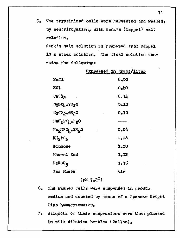
s. The t~sln1 •• d 0.,11a weN harvested and waahed.
byce.nt!"'ltugation, with Rank'a (Cappel) aait
solution.
Jiank'. salt solution i. prepQ"tl from Cappel
10 X atook tlolut1.on. The flnal solution con
tains the follwin.g.
NaCl.
Eel
CaC~
~\l504. 7BaO
'if8C12_6f120
ft"aB21'~.n2o
liaifP04·2n20
m2Pc4 GlfJooa.
fhAtng,l Red
Ndeo, (huJ Pl .. s.
~1mn:.t!a .• 'A. la srw/Al~!~ 8.00
0.40 0.1.h,
0.10
0.10
0.06
0.06
1.00
0.02
0.)$
Air
(pH 7.2:!)
6. ~ waat~d oella were suspenaed 1n r.l~th
mecU.\U'4 and counted by rlCl4ns of a r;pencer Bright
Line hemacytometet'.
7. AlIQ.uot. of these IHusp~n.lon. \If.:)" th0n planted
lnmllk dilution bottle. ('8elloo).
12
Section Bt
Media, Antibiotics and Supplem~~ts for ~rowth
In establishment of a cell line a major faotor is the
selection of an effioient growth medium and an effioient mainte
nance medium. To be erfective; a growth medium must stimulate
maximum yields of metabolizing cella. To be an effective mainte
nance media, it must support oells in an actively metabolizing
state with limits on the degree of multiplioation.
Media U.8.d. for the work was seleoted as 9. result of
three e2perlments:
E!periment I
Small whole goldfish, 2 - 3 inches in length, were
trypsinized in .o25~ trypsin (Difco 1:250), prepared according
to the recommendation of Marcus (1956). Trypsinization was
oarried out for four hours at 100e. The cells were then washed
2 - 3 times with Hankts solution, suspended in growth medium.
oounted and inoculated into milk dilution bottles.
The growth medium used consisted of Melnick mediUM,
prepared according to }1elnick (1955) t with 10% bovine serum and
P.S.F. antibiotic solution.
ins:
P .S.F. antihio.tic s~lution consists of the follow-
Antibiotic
Penicillin Streptomyoin Fungizone
Final Coneentratlo~
100 units/ml lOO -1 sImI
5 gjml
13
The freShly inoculated bottles were incubated at
37°0 and 20°0.
Samples of media, trypsin, basic salt solution, and
serum, were inooulated into Brain Heart Infusion broth (Diroc)
and Sabouraudts Dextrose agar (Diroo) to check for bacterial and
for fungal contamination.
Ff9?erlment g. Whole fish oultures were trypsinlzed, washed,
counted and planted accorcUng to previously stated methods.
This time the growth medium consisted ot Melniok medium supple
mented with 2~ bovine serum and P.S.F. antibiotio solution.
Sterility controls of the media Ware run according
to pl'ooedure previoasly oited.
E;peri!nent .1
As will be seen from the results, exoessive contami
nation was enoountered in Experiment. 1 and 2. 4 soreening ot
various media types and varIous antlblotIc.types and concentra
tions was undertaken. An enumeration of media and antibiotics
screened is found in Table I_ page14.

TABLE I --------------..........:;;:.;:.::.::=0.....;;:: __ .. ___ ,, ____ . __ " _____ _ Code Letter
A
B
C
D
E
F
G
R
I
J
It
L
M
N
Base ~edlum __ .Anti l?J.ott<1 .... !.IE.! __ .
Melnick P.S.F. n n
" »
It oP.S.F.
199(Capell) Aehromycin(Lederle) It ft If
tt If tt
ft tt *'**Aohromycln
Scherer's(Capel1) ft
ft ft
If P.S.F.
1t n
199 ( Capell) HP.S.F.A.
" ft
Concentra.!ion.
4ml/IOO
3ml/IOO
2ml/lOO
W/IOO
lml/IOO
3/4ml/IOO
~/IOO
!m1/IOO
itta/lOO
.;m1/100
lml/10O
2ml/IOO
lml/IOO
2ml/IOO 1------------,- .. ____ .. ____ . __ _ Final antibiotic concen-o 1ml P.S.F./IOO rul of media --+ tration of (Penicillin 100 units/.ml (Streptomycin 100 ~g/.ml (Fungizone $ j{g;m1
** 1ml P.S.F.A./IOO mlmedia ---1 Final antibiotic concentration of (Penicillin 100 units/.ml (Streptomycin 100 A g/ml (FungizClne 5 ~g/ml (Achromycin 12.5 ~g/ml
*** 1/4 ml Achromvcin/IOO ml media ~ Final antibiotic concen-tJ ~ tration of
(12.51" g/ml
**** All of the respective media types were supplemented with 20% Bovine Serum
1$
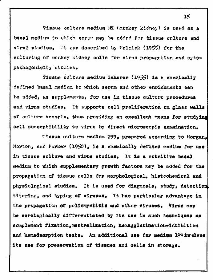
fJ:/1ar.HH.l eultul"'O m.ectlum l~ (.uOnkey k'.dney) 1s use-a ae a
basal .ed1u~tl to \.,hich eerurn may be edcled for tissue culture and
viral .tudios. It \J'4\0 desoribed by ?fclnlck (19!)!5) tor the
culturing ot tlonkey kidney oalla for virua propagation anc1 cyto
pathogetliolty studies.
TIssue oulture med1aM SCherer (19~$) 18 a c.hemicall,.
defined 'baeul.l medium to whioh •• rum and other enrlcWuenta can
be added, 2S supploments. ror use in tissue culture proeeduree
a~.d viru. studIes. It supports eell pro11feration en gla8G wall.
of oulture vessels, thus providing an exoellent mea.na tor stud),luI
cell suseeptIbll1ty to virus by d1rcu'.It {nicro.oople e.ltn.Minatlon.
Tis8U. cuitu1'*\) medlum 199. prepared acoordlng to ?!organ,
~1orton, ane'! Parker (19$0), 11 .. chemloally detlned mediwa for use
in tissue oulture and viru. studt... It 1. a nutritive ba.al
taed1um to whlah suppleuntU7 growth raotor-a _:'9' be added tor the
propag,ation or tiasue cella .tel' morphological. h1etocheidcal and
physiological .tndle.t. It la ueed tot' diagnosis, stud)" deteeliIot,
tltering, and typing or .11'0.... It haa partioular advantage 1n
the propagat1on of pol1011JeUtl. and bthet- yiPU.... Virua mal'
be .erologicall,. 41fterentiated by lta us. in suoh teobn1qu •• a.
oomplement t1¥atIon.ftlut~11a.tlon, bemagglutlnatlon.lnhlbltlon
and hema4aorptlon te.ta. An additional u •• to. med1 .. 199_01 •••
Ita us. tot' pH •• X'Vat1on or tisaue8 and cell. 1n atonge.
16
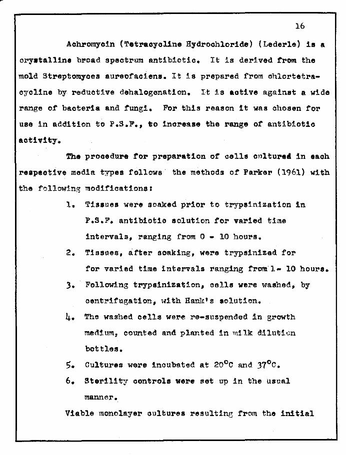
Achromyoin (Tetraoyoline Hydrochloride) (Lederle) is a
orystalline broad spectrum antibiotic. It is derived from. the
meld Stl'eptODl7ces aure-ofacienl. It is prepared f'rom. oh1ortetra
cycline by r-eductlve deha1egenation. It is active a.ga.inst a wide
r-ange of bacteria and fungi. FbI' this reason it was chosen for
use in addition to P.S.P., 'lio Ino:r:-ease the range of' antibiotic
activit,..
-.rh. pl"ocedupe tor p:reparation of cellI Cl)ltuHi in each
respective medi. typel follows the methods of Parker (1961) with
the following modifications:
1. Tissues were soaked prior te trypsinization in
P.S.F. antIbiotic solution tor varied time
intervals, ranging from 0 - 10 hours.
2. Tissues, after soaking, were trypslnized tor-
tor varied time intervals ranging from 1 .. 10 hours.
3. Following trypsinization, cella were washed, by
centrifugation, ,\11th Hank's lolution.
4. The washed cells were re-suspended in growth
medium, eounted and planted in milk d11uticn
bottles.
S. Cultures were inoubated at 20°0 and 37°0.
6. Sterility controls were set up in the usual
manner.
Viable monolayer oultures resulting trom the initial

17
cultures were oarried on in oontinuous culture. The procedure
employed. .for the sub-ou1turing of oe11s 1s enumel"ated below:
1. Bottles containing complete monolayers were
inoubated 15 minutes at 370 with .02'~ trypsin,
in order to free the cells fro~ the glass.
2. Cells were oentrifuged and washed 2 .. :3 t'.mea
in Ha..l'l.k f s solution.
3. The cells were re-suspended in. growth m.edium,
counted, and inooulated into milk dilution
bottles.
Maintenanoe mediUM was su.hsti tuted for growth .. tUum
when healthy monolayerA were developed and the cells were con
tinuously re-fed ~.th this medium until they were ready to be
sub-ou1tured.
Section c:
St,~!\£e __ oL_C.e!:-t-~w
A problem which poses numerous praotical difficulties
in the use of tissue culture methods 1s the storing of cell
line.. These stored eells should retain the ability to aotively
metabol'.ze for use in further sub-oulture. A luccessful long
term prooess for storage of cell and tissue oultures would
eliminate the risks ooncomitant with continuous growth of a
oulture over an extended period or time, namely; the increased

18
possIbilIty of mutation, the possibility of baoterial, fungal
and/or viral contamination, and the possible 10s8 of the oulture.
One method of storing oells is by means of freezing
at extremely low temperatures. The technique used to insure
maximum reoovery of viable cultures depends upon the particular
tissue or cell type.
A method with whioh the best results have been obtained
and one whioh is commonly employed today, involves the following
steps, (Moline 1964):
1.
2.
The cells or tissues to be stored are cooled at a
precisely controlled rate. The rate of cooling
usually falls within 0 the range ot leper minute 0
to 20 C per minute.
Protective additives are added to the !I1eM um in
which the cultures are to be stored. The usual
additives are either glycerol or dimethyl
sulfoxide in concentrations ranging from 5~ to 20~.
Glycerol 1s a hydrophilic and prevents excessive
injury to the cells during freezing and thawing.
Dimethyl sulfoxlde apparently has a vast potential
as an addItive. It rapidly diffuses into and out
of the cell, thus minimizing osmotic shock when
the cells are cllnted for culturing after thawing.
3. The cells are cooled by use of liquid nitrogen and
19
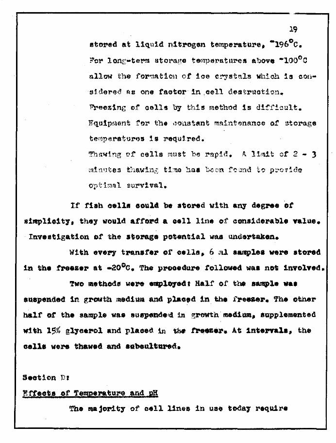
.toNd at liquid. nitI"Ggen temperature, -196°0.
F'or lonG-term storage temperatures above -100°0
allow the torr;1tltion of 10e crystals whioh is con
lddered as one factor in ,cell destruction.
11reezlng of oells by this m.ethod 1s d!fi"icult.
Equlpr.uilnt for the ()o·l,1.a~ant rnn1nt~ma,"lee ot ~tora.ge
tel'l'1p4!ratuMB 1s required.
'!'ha'l41.ng of oel1& roust be rapid. A l1adt of :2 - 3
optimal survival.
It flab oell. oou14 be a'o"d wi tih any deS ... 8 ot
s1mpllo1t7. they would afford a .e11 line of canalderable value.
Inve.'lgatlon of the storase potentIal vas und.rtaken.
With eve.,. tpan8te,.. or cella. 6 ml aample. weN .tON4
1n the freezer at -20°0. The pPOO.4ure followed was not lnvolved.
Two •• thod. were emplote4t Bal.! of tbe ...,1. vaa
au.pended 1n grovth medium and 10'1&0.4 1n the .fre_er. 'fhe otber
halt ot the sample was auspend.tel in growth' mecl1_. IQPpl ... nted
with 1~% g17cerol and plaoed in the .tPees ••• At In,e1"l&1a, the
oelli "eN thavea and •• beultal"ttd.
Seotion Dt
f.,ft!ela et '!5!!D!i"1t! ,&n11 eB 'fhe u jor1 t7 of cell line. in US8 todaJ :reqUire

20
temperatures ot 300 - 37°e for optimal growth. It would be advan
tageous if a cell line could be established that would have a
wider growth temperature range.
Fish are poikilothermio. Fish cell cultures might show
optimal growth at temperatures lower than 37°0. Cultures were
maintained at temperatures of 100e, 200 e, and 37°0.
Most mammalian and avian cells are sensitive to changes
in the pH of the medis.. The pH range to which n. eh cells can be
subjected, without loss of viability, was examined.
The media were adjusted to pH ranging from 6 - 8. pH
was determined with hydrlon paper and verified by pH m.eter.
These media, : used to feed the cells for given periods ot time.
Viability was determined by observation of cell multiplication.
Seotion E, OlassiflcatJon ot Conta~~~nt
A oontaminating m.ioroorganism. resistant to P.S.F.
antibiotic solutiQn vas encountered. The morphologioal and
physiological classirication of the bacterium was established.
The morphological data was determined by perfo~lng
the following:
'1. Gram. stain acoording to McClung (1957).
2. Capsule stain according to McClung (19$7).
3. Flagella stain acoording to Leirson (1951). run
Diagnostic biochemical testa were/In order to establish
21
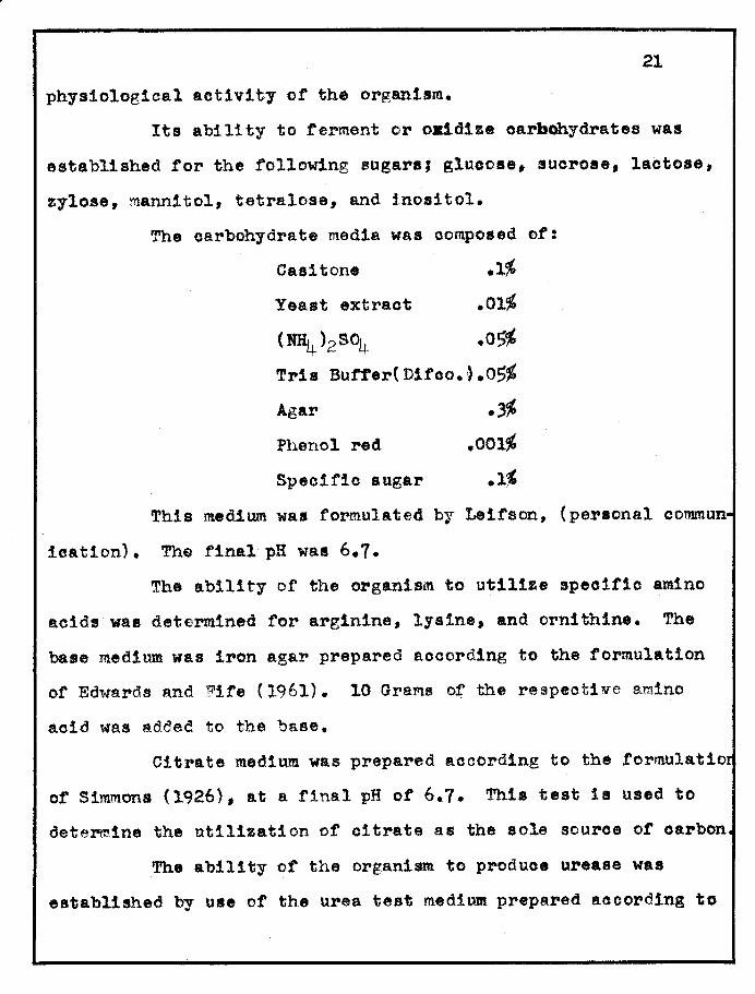
physiological activity ot the organism.
Its ability to ferment or o~dize carbOhydrates was
established tor the following sugars, glucose, sucrose, lactose,
zylose, mannitol, tetralose, and 1.nosi tole
The oarbohydrate media was oomposed of:
Casitone .l~
Yeast extract .Ol~
(NH4 )2S~ .O~
Tria Butrer(Ditco.~.05~
Agar .~
Phenol red
Speoifio sugar
This medium was formulated by Leifson, (personal OOMmun
ioation). The final pH was 6.7.
The ability of the organism to utilize specifio amino
aoids was determined tor arginine, lysine, and ornithine. The
base medium was iron agar prepared aooording to the formulation
ot Edwards and Fite (1961). 10 Grams of the respeotiYe amino
aoid was added to the base.
Citrate medium was prepared according to the tormulatior
of Simmons (1926), at a final pH of 6.7. This test is used to
determine the utilization of citrate as the sole source of carbon
The ability of the organism to produoe urease was
established by use of the urea test medium. prepared aocording to
22
the t'orrnulation oJ." Rust1gan ana :-"to~rt (1941). The tinal pH or
the test medium was 6.8.
The agar medium and the reagents for tne nitrate reduc
tion test were prepared a.eccr{l~.11g to Bal1ey (19f)2). '1'h. test
shows the abl1 ~.t,. or laok of abIlity ot' the organism to recluc.'H'J
nitrates to nItrites or to free nltrogfln. The results ot a
n.ge.t1ve nltrete test werft confirmed by the a(kd.ticn ot a small
amount of zinc dust to the tned1ua after incubation. If 1 t were
a false negative, the presence ot reduced nitrate 1s revealed by
the development ot a red color.
The prot~o11tlc capacity of the organism was determined
by testing its a~111ty to liquefy gelatin. Wutt-ient gelatin pre ...
pared according to Ewlng (1962) was the specifio medium used.
There are some microorganisms which possess the ability
to deaminate oertain Amino acida. To find out it the bacterium
in question possesa.d t}~ls capaclty, the deandruuse tesG tollow
ing the methods of Ew1Dg (1957) was perform... The apeeltl0 test
medium. used was Phen:rlalanlne Agar_ and the test reagent waa a
l~ (w/v) solutIon or ferric chloride. It ph.nflalan10. haa been
deam1nated to phenylpyruvio acid, a green color develops In the
syneresis fluid and 1n the slant.

CHAPTER IV
RESULTS
Experiments 1 and 2,where medium MK (monkey kidney),
supplemented with 10~ bovine serum and P.5.F. antibiotic
solution was utilized as the growth medium, were unsuccessful.
Both of these initial attempts at egtablishment of fish cell
oultures were terminated within 10 - 12 days respectively, due
to contaminar.ion by a bacterium which was resistant to P.S.F.,
at the concentrations used.
In both of theae experiments, m.edia sterility con
trols which had been rlm in Bra.in Hear·t infusion broth and on
Sabouraud's Dextrose agel' remained negative to baoterial or
fungal oontamination. Thus, the bncterium came from. the fish
itself and was not introduced from externel soorC~8.
Sinoe the oontaminant isolated from the cultures
23

24
in Experiments 1 and 2 was definitely resistant to P.S.F. at the
concentrat1.ons of Penicillin 100 units/ml, Streptomycin 100-1 s/ml,
and Fungizone 5 J{ g./ml, not only was there a need for finding a
proper growth medium, but also for finding an antibiotio which
would eliminate the contam.inant cO<n:rJon to the fish being used.
'Phe classifioation of the oontaminant from the aspects
of its morphological and physiological nature was established.
The results of the various determinations will be found in Table I •. "
The organism 1s a Gram neg~tive polar flagellated rod similar to
~~romona·bJdrophll~.
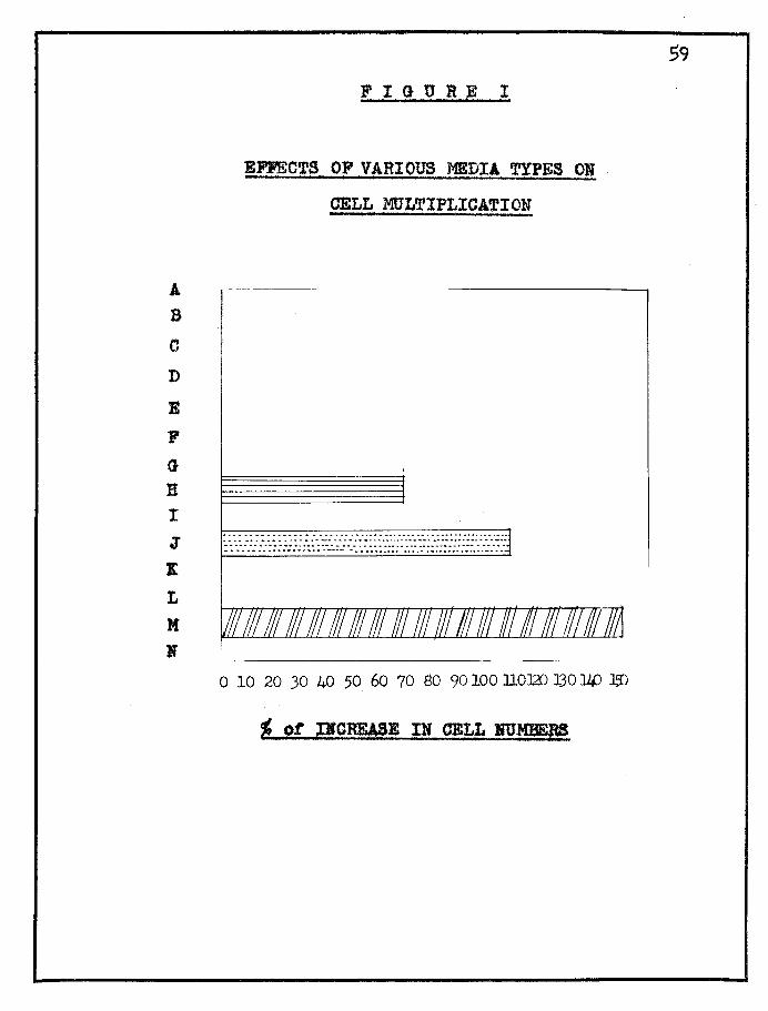
Experiment 3 entailed the sereen1.ng of 14 different
media types, which were supplemented with various antibiotic
mixtures. A listing of media types tested will be noted in
Table II. The results of the media and antibiotio screenlng will
be fonnd in Ta,ble III.
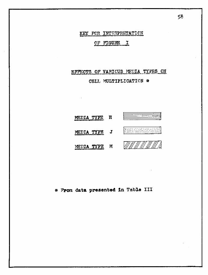
As can be seen from the results cited in Table III,
medias H., J., nnd M were the most effective. Media H, wS.thout
Fungizone, was discont1nued due to jts suseeptibllity to con
tam1.nntlon by H!.old.
A grnphioftl oomparison of the relatlve value of the
three medig typ~s will be found in Figure I. Media M , as is
illustrated, gave the greatest inorease in oell numbers. Media J
ga~e excellent oellular growth, but the inorease in cell numbers
was more limited than with Media 1'1. These results were to be

2$
espected due to the composition of their basal medias, 199 and
Scherer's base respectively.
Cells on these aedia types survived continuous culture.
Since media. d and M were by ~ar more etfl.lent, media M was
chosen as the growth medium and media d was ohosen as the main-
tenanae mediull1 for further experiments. P.S.F.A. was substituted
fo]" the single anti biotio Aohro!)l:ycin in mfl'ldia J. This was done
in order to take additional precautions aga.inst the poss:tble
development of other bacterial or fungal contaminants.
As observed, ooncomitant with the results of the media
screening, it W8S round that P.S.F. was not effective against
contamination unless used at concentrations far in excess of
those normally employed. At these high conoentrations it was
found to be toxic to the fish tissues.
Achromycin was effeotive. However, it was observed that
it was toxic to the fiSh cel18 at concentrations in excess of
12.5/fg/ml. of growth medium.
To date the cultures derived from small whole goldfish 28
have undergone/sucoessful transfers in oontinuous oulture. This
is enumerated in Table IV.
After a satisfaotory growth and maintena,nce media were
selected, oultures of fish peotoral muscle and fish heart musole
were established. To date the fish muscle oells have undergone
17 transfers. These results are in Table VI. The fish heart
26
cells have also undergone 18 transfers. as is enumerated in
Table V. Sub-culturing h~y(;nd .20 passages or over a period of Si3
months is taken as an indioation of the establishment of a stable
cell line. :Prel:1111inary experimentation wi th the length of expo
sure to trypsinization and exposure to antibiotic soaking of the
tissue fragments prior to trypsinization was performed. For
prevention of contamination the most effeotive period of anti
biotio soaking was from 4 to 6 hours of exposure. '):lhe best yields
of cells were obtained, for the initial establishment of a
culture, when the trypstnization process was continued for six
hours or more before harvest.
The ability to multiply after stora.ge of the 1~ish tissues
was determined. Ea.ch tim.e the cells were transf'erred 6 tn.l SB.fltples o werte stored at -20 e. A limited number of these storec cells
havp. been thawed and carried in continuous culture. The results
so far have been encouraging. Fnllowing thawing, cultures of
small Whole goldfish have undergone transfers to date and are
growing satisfactorily. 'rhess resl..11 ts p,re cited in Tables VII
and VIII. Cultures of i'ish peotoral muscle and fish heax't muscle
have been brought out of storage and are at present grOWing satis
factorily. ~~e~e results are in Table IX.
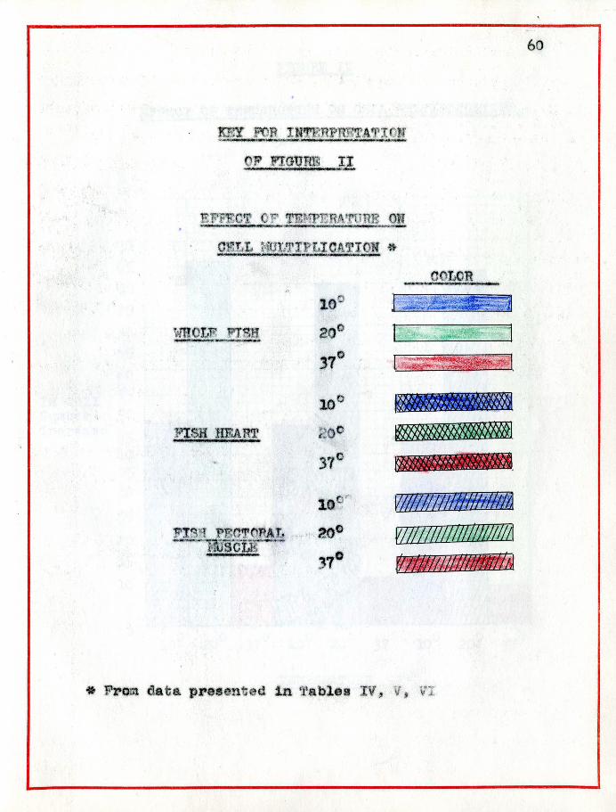
Temperatures of 100e, 20°C, and 37°0 were employe~ in the
cultivation of the various cell cultures. A graphical analysis
of the effect of temperatures on the increase in number of ce118
27
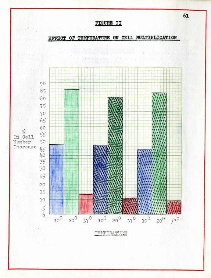
of all three types 1s illustrated in Figure II.
The optimal cell growth or increase was at 20°0; good
growth was obtained at 1000J cells grew at 37°0, but at a very
slow rate, and they could nbt be carried beyond six trans!'ers,
without the final loss or the cultures.
The ability of the cells to withstand varied degrees of
pH, ranging from 6.0 to 8.0 was determined. It was round that
all three types of tissue could withstand this range. ~h.
optimum for all tissue types was from 6.8 to 7.2 , the be8t~wth
being achieved about 7.2. (Table X).
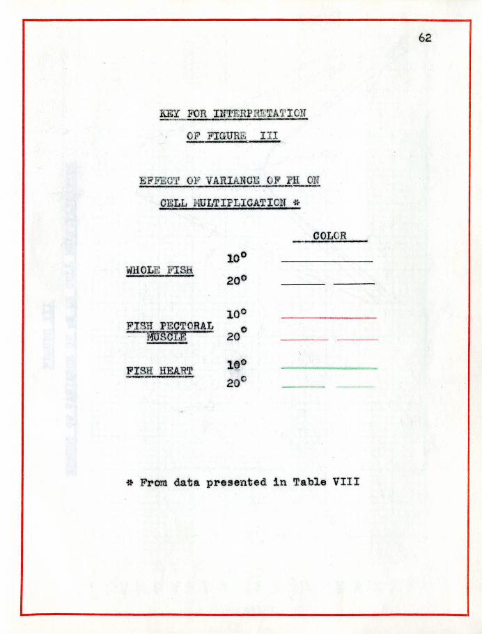
A graph representing the relationship or pH to oell
number increase, for all three types of tissues, wilL be found
in Figure III. As seen in the graph the peak of maximal multi
plioation occurs at pH 7.2.
It was observed that in the initial differentiated
cultures, two cell types predominated. The young cultures dis
played great numbers of epithelial-like cells. As the oultures
underwent sub-oulturing the cells began to de-difrerentiate and
the ribroblaetic rorms began to predominate. (Figures IV through VIL)

CHAPTER V
DISCUSSION
Consideration of all the data reveals certain facts.
One problem to oOl ... side1>, concerns the initial establishing ot
fish oells in culture. In oulturing of whole srnall goldf1sh,
1t is difficult to cbtain initially sterile tissue preparations.
Contamination is introduoed from the external body ooverings of
the fish and from. the ga$trointest1nal traot. This problem was
enoountered during the fi.rst attempts at working with stloh
cultures. One Sl.lch oonta...'l'linant prado'l'linl:lted in the initial
oulture experiments. After examining the results of the
determinatIon of the morphological and physiological charaoter
istics of the baoteriwfL, a correspondenoOj of thf' obtai'.1ed char
aot.eristles with those set down for the genus Aerolnonas was
eVident, therefore, the baoterium was designated to be an
Aeromonad. A more definitive evaluation places the organIsm a8 a
28

29
strain of Aeromonas hydrophila. Aeromonas sp. are comman to fish
both as normal flora and as etiologioal agents of pathological
lesions. The oonolusion that the oontaminant was introduced by
the tissue fragments per sa and not from extl"aneOUB sources 1s
substantiated by media sterility controls remaining negative
and fts complete elimination in subsequent studies by use ot
Aohromycin.
Several aspeots were considered in. arrlving at appropriate
methods for suoceesful establishment of fish cell lines. A study
ot exposure of the tissues to antibiotio containing salt solution
prior to initial trypsinization was perfo~ed and it was observed
that prlor soaklng for 6 hours el1min&.ted contamination.
Trypsinization time was evaluated. Cells harvested after
one hour could not be grown i;o. vitro. An interesting phenomenon
was that if the cells freed after one bNlr exposure to trypsin
were not removed from the trypsinizing flask, none of the cells
(no matter how long the exposure to trypsin) could be gr~m !E . t
vit~~. If the first houra ylelo was dlsoarced, harvested cells
were viable atter two hours or more trypsinization. The best
yields of cells were obtained after L. ... 6 hours trypsinization.
Thus, it is assumed that during the first hour of enzymatio
digestion, the fish cells give off some substance which is
extremely inhibitory to further cell growth and 13 toxic to all
O8lls being exposed to them. Either B. cell substanoe 18 l1ber-

30 ated during the first hour of trypsinization, whioh ia
inhibitory of growth, or the trypsin itself may oombine with
liberated substances to beoome an inhibitory factor.
Experiments were performed to determine if trypsin is inhibitory. The method being to incorporate inactivated trypsin
into the culture media. If inhibition is encountered, the noxt
step is a determination of the degree of reveraibility. If no
inhibition 1s observed, then a cellular liberation produot must
have been responsible. The product can be identified by chroma
tographioal analysis.
Incorporation of inactivated trypsin into the media revealed no deleterious effects to cell growth or m.ultiplication. It
can, therefore, beconcluded that a cellular liberation product was
responsible for inhibition.
Figure I reveals medium. 199 supplemented with 20% Eovm.e
Si)rum and P.S.F.A,. pr-ovided an excellent growth medium, beeause,
as per defi~ition, it allowed the maximum oell multiplioation.
This was media M of the variety of media tested.
Media J. Scherer t s medium, supplemented wi th 2~ Bovine serum and 12.$ ~ simI Achromyoin supported good cellular growth with emphaSis on maintaining cell quality more than quantIty.
It has been noted that Achromycin ia toxic at concen
trations above 12.5~g/ml media. It was observed that this
tetrpoycline 1n solution is very acidio. This is most likely
attributable to ita being commeroially prepared as a hydro-
chloride, which may account for its toxicity.

31
Whole small goldfish were cultured with ease after a
suitable media and antibiotic were determined. Fish pectoral
muscle and fish heart muscle de not present the contamination
difficultues that are inherent in whole flah cultures, since they
are obtained as sterile, when a.septic dissection procedures are
followed:
Definite ccrresponcence between the result of all three
tissue types cultured can be observed. There is agreement in the
several results recardlnr the influence of suah ~lyslca1 factors
as pH and temperature en the amCiunt of multiplication for all
types of goldft sh cells.
It is evident in Figure n that temperature plays an
important re1e in effective culturing techniques. Wolf and
Dunbar (1957), believed temperatures below 20PC to be essential
for growth of fish cells. In this work it was established that
the cells will grow as high as 37°0. The growth rate i. slow in
comparison to lower temperatures and they did not survive more
than seven transfers, but they did grow. At this moment the
usefulness of growth at such a temperature is not evident, but
such cultures might be fruitful for the study of enzymes active
only at such a temperature range, or perhaps may be benefioial for
immunological stUdies and for virological studies.
C id i i 1 i 1 t di lth h th t 37°0 ons er ng v ro og 0& sues, a oug grow a
is not as rood as at lower temperatures" if these ..<"lsh cells
32
support viral growth, they may be used for study of some known o animal viruses Which require temperatures around 37 o. This
would enable study of virus adaptability to change of host and
environment. For example, Eastern Equine encephalomyelitis has
been grown at 19°0 in fish embryo cells by Scret and Sanders
(1954). This virus is also known to l.lrow in ohickens, (normal
body temperature of 42°0) and man (normal body temperature 37°0)
and insects (ncrmal body temperature 20°. 37 cO). Possibly it
oan be grown in these poikilothermic cells line at temperature
ef 10°0 or less. The transoendenoe of temperature and speoies
by viruses is as yet not explained. It is known that the
oommon cold virus is grown at 33°0 for optim.al results. Some
Arboviruses and Picornaviruses have been grown in oold-blooded
animal oells. Fish eells may yield good growth at 300 - 33°.
This adds value to eell lines of oold-blooded species.
These fiBh cell lines May afford a practical v~hicle for viral
studies.
In the comparative graph (Figure II) the best growth
is achieved at 20°0, and gooc growth at 10°0. This ndght be,
for by nature fish are cold-blooded and their normal envtron
mental temperature is approximately 200 e. Being cold-blooded
they have the ability to adapt to the temperature of their
surroundings, thus, they grow well at 100 e. They adapt to growth
at 37°0, but it is more incongruous with nature, therefore, it
33
is poor. Possibly enzyme systems are retarded at 37°C for heat
tends to inhibit or destroy enzym.es, where cold does not neces
sarily do so. Their ability to grow at low temperatures may make
them valt.lable as a cell line fer studies requiring this temper
ature range. Growth at lODe and at 4°. 6°0 enables refrigerator
storare of such cultures, for they survive at these temperatures
without re-feeding for two weeks. A sampling of bottles was
maintained at refrigeratoI· temperature. Their metabolism rate
slowed but they remain aotive and healthy. This provides eell
lines that minimize maintenance tasks.
Figure III 1s a graphical evaluation of the effect
pH has on fish cell multiplication. Sat::'s.f'actory cell number
increase is attained within an initial range or pH 6.8 - 7.2,
the optimal inorf!ase occurring at initial pH of 7.2. A marked
deorease 1n multiplication occurs upon exposure to pH 6.0 or
pH 8.0. It has 'been observed that tlle cells ~dll survive at
these lim.its for two weeks without re .. feed~.ng (with media .s.t
pH 6.8 - 7.2), but multiplication is al~o.t negligible. The
oel1s are sensitive to these pH limits but are not killed by
suoh exposure. At pH5 and pH 8.5 no growth occurs. 'rhus, they
can be oonsidered as hardy-oell lines.
Today a number cf d:i.soiplines util'.ze tissue and cell
cuI ture techniques for resea!·ch. 'I'he preservati on of cells in
an unaltered state is a highly desirable prerequisite. The
34 ability of a cell line to successfully undergo prolonged storage
enhances its value as a tool. As prev:1.o!lsly stated, storage at
very low temperatures 1s an elaborat~ task requlrinr special
apparatus and the use of oultnre additives to be succe'3sful. In
addition the ability of these fish cell lines to survive storage
has been investigated. Tables VII and VIII summarize the results
obtained for small Whole goldfish. Table IX contains the results
for fish heart and fish peotoral muscle. These oells will sur
vive storage at .20°0 without the use of protective additives.
A comparison of the results obtained from oells frozen merely in
growth media and cells frozen in ;:!'owth media conta:i.ning 15?t
glyee:rol show 11 ttle ,;'s.rIat1c'n 1n the~!' abil:l ty to be: brought
back in a healthIly metabolizin.p: state qna to survive sub-culture.
It must be noted here that the data in TableR VII and VIII is
based on values obta.ined at weekly inttJrvals, whereas other data.
x-efer:ring to ( of cell number increasE'! was hasec. on readings
taken at two week intervals. Tn the or5.gi!lal datA at readings
of two weeks, the peroen.te.ges in Tables VII and VIII would be
increased by' approx1.mately 30 - 1.!.~. It c~n b~ concluded' that
no cellular alterations occurred as a result of storage.
Morpholo~lca.lly the cells are small in ccmparison to
some animal cell li-nes. For example, chick oells are 201 in
dIameter, Hela cells 151 and fish oel1s 101- Cultures of small
Whole goldfish initially conta.:\n cells of both the epi.the11al and
35
fl b!'oblastla fom. After sub-ell1 ture they de-differentl ate and
and the fibroblastic form is the type surviving. Cultures of
fish pectoral muscle and fish heart muscle display the fibro
blastic appearance initially and throll~hout sub-etllture.
All three types of fish tissue exhibit a co~on
mOr?hological phenomenon. Young culture~ rive clearly defined
cellula!' shapes. As they are, m1.crcscoploally th.oy Elve the
appearance of bone tissue cr hyaline cart1.1aGe. This char'B.cter
lstie has also been observed by other ~.,.ve9tigator~. Figur-es IV
through VII photograph5.oally depiot this ftbroble.stic nature of
the flsh cells in oulture. \-Inrk ooncern~.ng determination of an
efficient stalnin~ procedure for these fish oells is in progress.
This fixative employed for the staJned cells in the photographs
was Osmium tetroxide l~. The stain is Delafield's hematoxylin
and eosin.
Hea.lthy monolayers develop w:!th1n 24 - h8 hours.
P.fonolayers become exceedtngly thick. Elimination of the serum.
8l'pplement enti.rely slows the growth; (monols:yers not being oom
plete before 96 hours). However, there is a poor definition of
cellular differentiation. Therefore, the serum-tree medium is
not sat1.sfaotory for morphological studies.
Perhaps a serum faotor is ~\"nV'olved. Studies of
incorporation of specific serum fractions, the use of serum ultra
filtrates ~r dialyzed serum may eliminate this completeooalesoing
of the oells. This is an area ot tuture research.
36
~1onolayers adhere strongly to the glass. If the mono
layer beoomes extremely thiok, the edges begin curling baok and
a sheet of new oells is laid down in their place immediately.
They are very hard to remove from the glass. For transfer a
trypsinization time of 20 minutes followed by vigorous agitation
to free them from the glass i8 required. Even after this
stringent prooedure, many cells are not removed and are lost for
transfer. They are very sturdJ. After transfer the oells begin
to adhere to the glass atter 2 • 3 hours.
One side experiment yields an interesting faoet regard
ing oel1 transfer and the hardiness ot the cell lines. There are
always some cells remaining on the glass after trypsinization.
If the bottles containing remaining monolayer fragments are
waShed with Hank's salt solution and re-fed, monolayers are again
developed within 48 hours. This is supported by the faot that \ two of the origlna~bottles started in August, have been re-fed
after 27 exposures to trypsin and are still actively producing
healthy monolayers.
The nex.t phase of this work w1.l1 involve testing of
these oells to determine their usefulness for viral studies.
Four viruses will be tested: Sindbis, an arbovirus; Vaa.inia, a
pox virus, Newoastle's disease virus, a myxo virus, and Polio
myelitus virus, a pioornavirus. The objeotive is to study
oytopathogenio effects and other evidence of viral multiplioation
Multiplication will be determined by hemagglutination and
neutralization tests.
37
This work is in progress at the present time: however,
due to the scope .of such an investigation it will be the subject
of future reports and the results are not incorporated as a part
of this thesis.
If these goldfish cells are able to support viral
growth, they would be valuable cell lines.' Tissue culture is
always in need 0 f new cell lines for virus research. A line
with as flexible a range of survival in terms of physical
properties and one which is so readily available, easily estab
l1aned and maintained, has many distino~ advantages as a tool for
viral and other studies.
CHAPTER VI
SUMMARY
Tissue of Ca~assius auratul (goldflsh) has successfully
undergone continuous cultu~e. Three cell lines have been estab-
11shed, whole small goldfish, fish pectoral muscle, and flsh
heart muscle, ~espectively.
To date whole fish cultures have been taken through
27 sub-cultures; fish pectoral muscle and fish heart mUlcle
have undergone 18 Bub-cultures.
The growth medium consisting of TO medium 199 (Cappel)
supplemented with 20% Bovine serum; the maintenance media con
sisting of TO medium Scherer (Cappel) supplemented with 2O~Bovine
serum proved satisfactory.

39 The fish cell lines have been found to grow at temper
atures ranging from 4°0 - 31°0, the best cell growth and multio plication being observed to occur at 20 C.
Fish cell lines survive a pH range of 6 - 8, the best
growth and mu1tiplioation occurring at initial pH 7.2.
The cell lines have been observed to withstand a
simplified storage process) freezing 'at -20°C. in growth media
without incorporation of proteotive additives, with no apparent
alteration of the oel1s.
The oe11 lines are hardy, easily established and main
tained, and exhibit potential usefulness, as cell lines for
virological studies.
40
BIBLIOGRAPHY
Sargen, G.v. and Wes.ing, A., 1959, Untersuchungen Ober einen Viruabedlngten Tumor bei Fi.chen. Arch. Virustorschung 9, 521-536. -
Cameron, G. and Grand, O.G. and Gordon, M., 1941, Neoplasm Studies. Cancer Reaearch. 1,66. -
Oameron, G. "and Grand, C.G. and Chambers, R., 1935, Neoplasm Studies. Am. J. ot Oancer. 24,36.
. --Carrel, A., 1914, Present Conditions ot a Strain of Connective
Tia~e Twenty-Eight Montha Old. J. Exper. Med. 2011.
Carrel, A., 1931, The Bew Crtology. Science. 731297.
Ohain, E., and othera, 1940, Lancet. !s226.
Chain, E., and others, 1941, Laacet. !.1'77.
Chlopin, N., 1922, tn>er in vitro J,ulturen in dem Embryonalengewebe der Slugetiere. Arch. t. Mikra. Anat., Bonn, !2!!s435-493.
Drederer, P.H., 1921, The Behavior of Cells in Tissue Oultures ot Fundus heteroclitus withSpecial Reterence to the Ectoder.m. Bio1. Bull. 41,221. -
Duggar, B.M., 1948, Ann. N.Y. Acad. Sci. ~s177.
Ehrlich, P., 1897, Klin. Janrb. ~s299.
Enders, J.F., Weller, T.H., and Robbins, F.O., 1949, Cultivation of the Lansing Strain of Poliomyelitis Virus in Oultures ot Various Human Blabryonic Tissues. Science. ~,85.
Finlay, A.O., and others, 1950, Science. !!l,8S. Pleming, A., 1929, Brit. J. Esper. Path. !Q,226.
Flory, N.E. and Flory, H.W., 1943, Lancet. 1,387. Flory, H.W. and others, 1949, Antibiotics, London, Oxford
University Presa.
41 -
Gordon, M., and Smith, G.M., 1938, Progressive Growth stages of Heritable Melanotic Disease in Tissue of Fish from the Da7 of Birth. Am. J. of Cancer. gjJ.'36.
Gordon, M., 191.11, Genetics of Melanomas in Fishes. Cancer Research. 1:656. -
Grand, C.G., Chambers, R., and Cameron, G., 1935, Neoplasm Studies Am. J. of Cancer. ~.36.
Grand, C. G., 1938, )fe~lasm Studies. Am. J. of Ca."'1cer. Jl:394.
Grand, C.G., Gordon, M., and Cameron. G., 1941, Neoplasm Studies. Cancer Research. 1#660.
Gr\\tzner, L., 19S6, 1tberpl"ltung eln1ger Anvendungsmogllcbkelten del" Gewebeku1turen von Lebist •• reticulatus und *cr01>o<1us ?-erCularis in cter VIrusforsohung. Zbl. Bakt., 6r!g~~:8~6.
Grfttsner, L., 19;8, Ders,lIn vitro-z!ehtung de. Leber und Nierengewebes von Tinea vUls;ulirls Cuv., in Trypsinn1erten Einschlohtgewe&kulturen. 21it. Bakt., Orlg. ill.t195-l97.
Harrison, R.G., 1907, Observation on the Livlng Developing Nerve Fiber. Proc. Soc. Exper. Blo1. and Med. ~:140.
Kalter, 5.S., 1963, Procedure tor Rou~ne Laborat0ft Diagnosis of Virus and Ricketts!al Dlseasea. Burgess Pub shIng lompan'1, Minneapons, MInn.
Lewis, M.R., 1916, Sea water as a Media. for Tisaue Cultures. Anatom.Reoord. 10c287. - .
Osowski, R., 1914, Arch. Entwicklungemechen. Organ., J!t :S47 •
Maitland_B.B. and Maitland, M.C., 1928, Cultivation of Vaccinia Virus without Tissue Oulture. Lancet. 212.596.
l~rcua, P.I., Creclura, 8.3. and luck, !.T., 1956, Clonal Growth in vitro or Epithelial Cella from Normal Human Tilauea. J. ~er. Med. 10~:615.
Meleney, F.L. and Johnson, B., 1947. J. A. M. A. 1ll:675.
---42
Melnick, 19$$, N. Y • .lead. Scl. 611755. -McClung, L.S., 19$7, General Baoteriology Laboratory l~ual, w.
B. Saunders Co., 'hl1aaeipbla and London.
Moline, S. W., 1964, The Low Temperature Preservation of Tissue and Oell Cultures. Linde Oryobiology, Linde Company.
Morgan, J.P., Morton, B.J., and Parker, R.O., 1950, Nutrition of Animal Cells in Tissue Culture. I. Initial Studies on Synthetio Medium. Proc. Soc. Exper. Bl01. andM_d. 11:1-8.
Parker, R.C., 1961, Methods of Tissue Culture. Paul B. Hoeber, Inc. Medical DivisIon or Harper ana Bros.
Paul, John, 1960, Oell and Tisaue Oulture. The Williams and 'Wilkins Company, <lmore.
Roux, VI., 188$, Beitrlge !lUI' Entwlck1ullgsmechanik des Embryo. Ztsoher. Biol. 21:411. -
Scherer, 1953, Am. J. P~th. ~ :113.
von Reekl1nghausen, F.D., 1866, Ubel' die erseugung von l'othen Blutk&rpersehen. Arch. M.1kroskop. Anat. g 1137.
SchWMberger, R. G. L 1949, Outaneous Leiomyomas of Goldfish. Am. J. Path. ~:2tl7.
Soret, M.G., and Sandel'S, M., 1954, In vitro Method for Cultivation of Eastern Equine Encephalomyelitis Virus in Teleoat Emb1'1oa. Soc. for Exper. Biol. and Med. ~.526.
Strangewaya, T.S.P.,and Fell, B.B •• 1925-26, Experimental Studiea on the Differentiation of Embryonic Tissue Growing in vivo and 1n vItro. I. The Development of the Undifferentiated LImb-bud (a) When Subcutaneously Grafted into the Poat-Emb17onie Chick and (b) When Oultivated !!! ~tro. Proc. Roy. Soc. London. s. B. 22:340.
Strangeways. T.S.P. and Fell, R.B., 1926_ Experimental Studies 0 the Differentiation of Embryonic Tissues Growing in vivo and !!l vitro. LL. The Development of the Isolatecf"!'ar!,. Embl'7on1c lYe of Fowl when Cultivated !!! vitro. Proc. Roy. Soo. London. s. B. 100.273. -
43
Thompson, D., 1914, Some Futher Researches on the Cultivation or Tisaue in vitro. Proc. Roy. Soo. Med. 1:21 (Marcus Beck Lab. Rep.). -
Wakeman, S.A., 1941t, Prac. Soc. Exper. Biol. and Med • .21:244.
Wolf, K. and Dunbar, C.E., 1951, Cultivat10n ot Adult Teleoat Tisaue !.u vitro. Proo. Soc. Exper. Biol. ane. Med. 2.2.!455-45a
WOhltarth-bottermann, X.E., 1951, Die Kontrast1erung T1er1scher Zellen und Gewebe 1m Rahmen Ib.rer Elektronemikroskoplschen Untersuohung an Ultradftnen SCbnitten. Naturw1saenachatten • .h!t.:281-288 •
Wood, E.Iof., Yasutake, W.T. and Lehman, V .L •• 1955, A Mycosis ... like Granuloma of Pieh. J. of: lni'. Diseases. ll:262.

TABLE 1 44
MORPHOLOGICAL AND PHYSIOLOGICAL CLASSIFICATION OF CONTAMmANT
Morphology
Substance
Glucose
Lactose
~ucro8e
·altose
Zylose
Mannitol
Tetralose
Inositol
Physiology
Citrate
Urea
Nitrate
Gelatin
1y's1ne
Argenine
Ornithine
Deamin&S8
1. Gram Negative
2. No Capsule
:.3. Polar ¥lOnotrichous or
Polar Mulitrichous Flagella
48 HRS
Ferment.r+Acid~as +Acid and Gas
+ +
+ +
Allcaline Top .. Acid Butt
+ +
+ +
+
.. +
+
+
+
TABLE II 45
TYPES OF MEDIA SCREENED
Code Ba.se Serua Antibiotic .coneetltration
A Melnick 20% Bovine P. s. F. 4 ml/100
B " It " " .3 ml/l00
C " II II " 2 ml/1OO
D " " " It 1 rnl/loo
E 199 " tt Achrol'1\Ycin lml/l00
F It " n " 3/4 ml/1OO
G If It " " 1/2 ml/loo
H If " " " 1/4 ml/1OO
I Scherer " If " 1/2 ml/1OO
J " It " n 1/4 ml/100
K .. 1\ " P • s. F. 1 ml/1OO
L n " " It 2 ml/1OO
M 199 " " P. s. F. A. 1 1Il/1OO
N If tt " " 1 ml/1OO
Note: See page for antibiotic concentration conversion factor

'fABLE nI
RBSt1LTS OF MEDIA scruwJIIG ON CULTURE OF WHOLE mWJ.. GOLDFISH . Media Ant.1blot.1c Type A~IniUal Average FiBa1 Tzpe " CO!!!!Drat.1on C.1J emm . Cell Coun1rs No •• f TrtIl!1"m
A P .5.1. 4al/1OO 250.fXXJ/al - 0 B tt 3ml/100 300 ,OCJO/ml - 0 C Jt 2aijloo 4SOPJO/ml Contaminated -D ** " lal/100 '2lXJ.OCJO/JIl. .. -E Achromy-
cirl hil/loo 230,fXXJjmJ. .... - v , u )/4al/.100 2fX),OCJJ/ral - 0 G "i/2ai/lOO 4fX),COO/ml 0 H** :~= 450,rxYJ/al 7SO-;ciXJ/ml 6 I 3OO,ocxi/riJ. 0 J " 1/4al/1OO 200 ,CtJO/ml W,OCC/111 23 K P .5.1 0; lml/lOO 250,OOOjml Cont.Wnated -L n 2Ifil/lIXJ 300 ,CXXJ/ml tt -M** P.S.F.A lal/lOO JOO ,C1Jfi/ml 7.52,OOO/ml 23 N n 2ml/lOO . 450 ,OOO/rtil 0 -
". Cult urea wiped out by mold. (1 ml of P.S.F. {Pc1cU11n 100 un1J.s/ml (in 100 ml of media (Streptom.rc1n 100../ &1m!
** Antibiotic conversicn factors tor concentration per ml media
~ (Fungizone '1 g/ml
( 1/4 ml Achromyein per 100 ml media. ( (Penicillin 100 un1t.s/ml ( {Streptom,-c1n llJfM/gfml ( 1 III P .5.1 .A. in (Fl8'lg1zcme sj{iilml ( 100 mle.ot media (Achroa'G"C1n 12"'1 g/ml.
% C.ll Number imr!!se
0 0
-0 0 0
66-2/,% * 0
llZ -
150.6% 0
(12.SJjg/ml (
.-------1) All media cont.a1ned. 3>'$ Bovine SGrua 2) pH of Media: 6.8 at t1me of feeding and transters ,) All cultures grown at J.OOC, '2IPC, )70(;,: Data bere baaed on cultures grown at UC.
47
TABLE IV (Part 1)
THE RELATION OF TEMPERATURE TO % OF INCREASE IN CELL NUMBERS FOR WHOLE SMALL GOLDFISH
Transter Average Average Days in l'emperature NWllber Original r.oynt Final Count S Increase CUl\ure
10° Initial 250,000 - - -20 200,000 - - -37 Inoculum 170,000 - , - -10° #1 l!JO,OOO 226,000 50.6% II 20 1 220,000 400,000 81.S% II 37 1 100,000 123,000 22.0% 11 100 #2 150,000 223,000 48.6% 18 20 2 210,000 378,000 80.0% 18 37
0 2 100,000 120,000 20.0% 18
10 113 100,000 150,000 50.0% 25 20 3 120,000 227,000 89.2% 25 '7 3 200,000 234,000 17.0% 25 10° #4 110,000 164,000 49.1% 34 20 4 100,000 190,000 90.0% 34 37 4 150,000 177,000 18.0% 34 10° #5 190,000 283,000 49.0% 41 20 5 100,000 189,000 89.0% 41 37 16 II8:888 195,000 14.1% 41 10° 16,,000 50.9% 47 20 6 330,000 240,000 90.'/% 47 37 6 100,000 l.O4,OOO 4.~ It.7 100 #7 120,000 100,000 50.0% 56 20 7 150,000 286,000 90.6% ;6 10° #8 100,000 151,000 51.0% 65 20 8 llO,OOO 208,000 8<).0% 65 10° #9 150,000 222,000 48.0% 74 200 9 100,000 189,000 89.0% 7lt. 10 #10 110,000 166,000 50.9% 85 10 10 100,000 190,000 90.0% 85 10° #11 170,000 253,000 48.8% 93 20 II 100,000 190,000 90.0% 93 10° #12 120,000 182,000 51.6% 99 20 12 150,000 287,000 91.3% 99 10° #13 100,000 150,000 50.0% 111 20 13 220,000 417,000 8<).5% III 100 #14 120,000 l81,OOO 5O.S% 120 20 14 100,000 190,000 90.0% 120 10° #15 100,000 151,000 51.0% 128 20 15 180,000 344,000 91.1% 128 10° Hit 100000 ~6:888 ~:~ it! 20 130:000

46
TABU~ IV (Part 2)
THE R1~lJiTIOri v.l" Tr:.NP1:;t,;,A'l'UHE TO % OF INCHt:ASE 1M C1:'lLL NUMBERS
FoR lfiILE SHAlJ,. g<>LDfISI!
Tl"8J1ster Average Average D&711 in 1!1'w!r:atH:t! .N1.!Bbsr Otlg1!l@J, CP,!l!lt F~ Csaust % lncr~a! Cu1!=ure
100 1111 120,000 179,000 49.1% 149 20 17 200,000 382,000 91.0% 149 100 #l8 100,000 151,000 51.0'~ 161 20 18 190,000 362,000 90.,% 161. lOO 1/19 110,000 162,000 47.2% 161 20 19 260,000 492,000 89.2:' 167 100 6'1!J:) 160,000 ~"ooo 49.3$ 113 20 20 190,000 ,61.000 90.0% 173 10° fa 1OO,Ot'() lSO,OOO so.o% 184 20 U l3O,ooo 248,000 90.8% 184 uP 1122- lOG,ooo 151,000 51.0'; 193 20 22 170,000 _,000 90.6% 19' WO 123 llD,OOO 16',000 so.o% 206 20 23 190,000 363.000 93..1$ 206 J.OO 1'4 110,000 165,000 50.0% 214 20 24 l$S,OOO 291,000 81.7% 214 lIJo 1125 100,000 10.000 53.0% 228 20 2, 100,000 19#,000 92.~ 228 10° I~ 100,000 *,000 41.4% 2'33 20 26 l35.OOO U',OOO W1.~ 2:33 J.OO II'¥! 100,000 :u.s,ooo 48.0';t 241 20 '¥l 170,000 '30.000 9I..U 24l 1JI' 1/28 100,000 15',000 '3.0$ 26; 20 28 125,000 232,000 85.6;( 26;
pH ot Heclia I 6.8

.- ,
49 TABLE V (Part 1)
THE RELATIOIi (;1" fr~1>U'ERATUn.E '1'0 ~ OF INChEASE IN CEIL NUMBERS _____ . _______ ~f( .F:!SH _ H~~ ~SC~< ________ , ______ -___ _
Transter Average Average % Dqa 1n Ta.'Jerlture llJ!!:l:!er __ Or1g1ngJ. CoY&4 .1!n!.l_Q9unt Ingrey! ._~ul.!m'!
10° Initial 250,000 -20 250,000 - .. 37 Inoculum 2;0,000 - ... 10° #1 250,000 378,000 51.2% 14 20 1 250,000 450,000 ao.o~ 14 37 1 200,000 247,000 23.5% 14 10° 12 100,000 149,000 49.~ 20 ao 2 120,000 222,000 85.0% 20 37 2 110,000 l'S,OCO 25.4% ao 10° 113 110,000 169,000 '3.6% 26 20 .3 lOO,ooo 199,000 99.0% 26 '7 , 150,000 174,000 16.0% 26 10° 14 100,000 149,000 49.0$ '3 20 4 120,000 22',000 8'1.~ 33 37 4 1»,000 1)3,000 10 •• " 100 " 1;0,000 228,000 ,2.0% 41 !lt.) 5 110,000 202,000 83.6~ 41 37 ') 100,000 llJ,OOO 13.0% 41 100 116 100,000 150,000 50.0% 47 20 6 170,000 .315,000 85.)% 47 37 6 110,000 U3,OOO 2.7% 47 100 n 12),000 180,000 ')0.0% 56 20 1 150,COO 285,000 90.~ 56 .37 7 llO,OOO Cells died -lOO iJS l2O,ooo 185,000 54.1% 66 20 a l4O,OOO 251,000 79.3% 66 10° tl9 100,000 153,000 ,).3.~ 75 20 9 160,000 299,000 P:7.0% 75
---_.- -.-Growth I>fedia : M
~1aintenance ;-;EI(iia : J
pH of i'.{edia : 6.8
Initial Antibiotic so&k 6 hours
Tr.rpsinization initia~ 6 hours
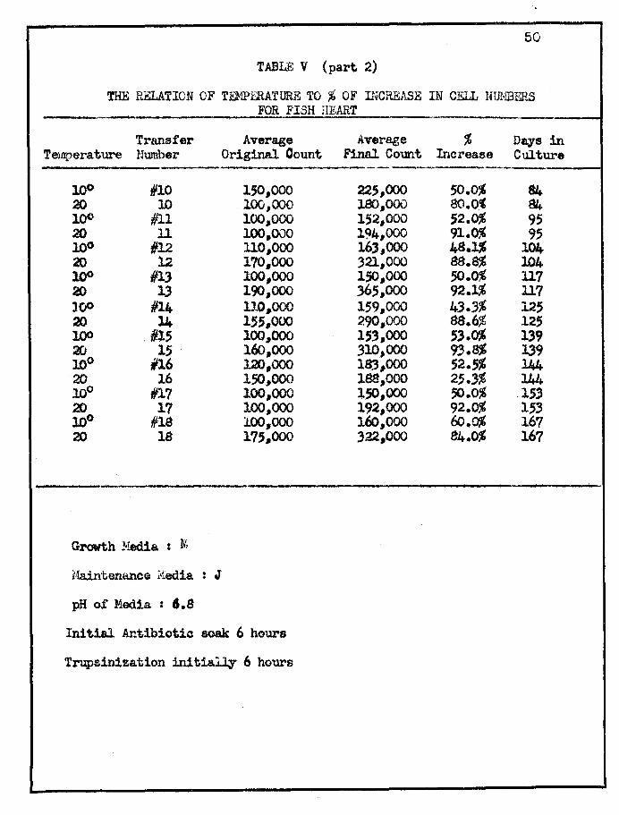
50
TABLE V (part 2)
THE RELATION OF TEMPERATURE TO % OF INCREASE IN CELL Nll{BERS FOR FISH HEART .
Transfer Average Average % Days in Tempera.ture Number Original Count Final Count Increase Culture
--"-- - --100 1/10 150,000 225,000 50.0% 84-20 lO 100,000 180,000 80.0" 84 100 #ll 100,000 152,000 ;2.0% 95 20 11 100,000 194,000 91.0% 95 10° 112 110,000 163,000 48.]$ 104 20 12 170,000 321,000 8$.8% 104 l.()O 113 100,000 150,000 ;0.0% 117 20 13 190,000 36;,000 92.1% ll7 100 1114 1l0,000 159,000 43.3% 12; 20 14 1';,000 290,000 88.6$ 125 100 IllS 100,,000 15',000 53.0% 139 20 1S 160,000 310,000 93.8% 139 10° #16 l2O,ooo 18:3,000 ;2.5% l44 20 16 150,000 188,000 25.3% 144 100 1117 100,000 150,000 50.0% 153 20 17 100,000 192,000 92.0% 1;3 10° #18 100,000 160,000 l:J:J.C'$ 167 20 18 175,000 322,000 84.0% 167
-- ,
Growth Hedia : M.
Haint.enancG I'iedia. : J
pH of Media : 6.8
Initial Antibiotic soak 6 hours
Trupsinization in1tia.~ 6 hours
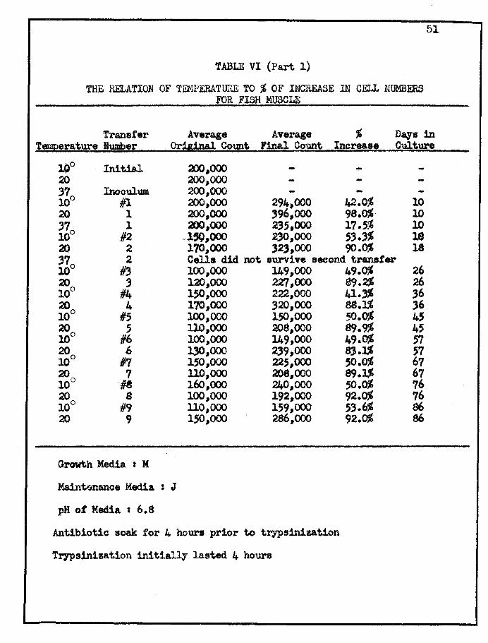
TABLE VI (Part 1)
THE RELATION OF TF1<IPERATUllE TO % OF INCREASE 1N CELL NUMBERS FOR FISH MUSCLE
51
Transfer Average Average ~ Days in Tft!rature Humber Orii&nal Count. Fip!l Count Increase Culture
lJ)0 Initial 3JO J OOO .... 20 2OO~OOO
370 Inoculum 200.000 ... 10 III 200,000 294,000 42.0% 10 20 1 200,000 '9'.000 98.0% 10 370 1 200,000 23;.000 17.;% 10 10 #2 l~,OOO 230,000 ;3.3% 18 20 2 170,000 )2,,000 90.0% 18 370 2 Cells did. not. survive second t.ranster 10 #3 100,000 149,000 20 3 120,000 2Z'/,OOO 10° 114 lSO,ooo 222,000 20 4 170,000 320,000 10° Ii; 100,000 lSO,OOO 20 ; 110,000 208,000 10° 116 100,000 149,000 20 6 130,000 2)9,000 10° In 150,000 22.5,000 200 7 110,000 208,000 10 H 160.000 240.000 20 8 100,000 192,000 10° #9 1l0,OOO 159,000 20 9 lSO,OOO 286.000
Growth Media. : M
Ka1nt!inartce Media : J
pH of Media : 6.8
Antibiotic soak for 4 hours prior to trypsinization
Trypsinization initially lasted 4 hours
49.0% 26 89.2% 26 41.'% 36 88.]$ 36 50.0% 4' 8<).9% 45 49.0% 'Yl 8).3$ '7 ,0.0% 67 8<).3$ 67 .50.0% 76 92.0% 76 .53.6% 86 92.0% 86

5(;;
TABLE VI (Part 2)
THE RELATION OF TEMPERATURE TO % OF DlCREASE IN CELL NUMBERS
FOR FISH HlJSCl..E
Transfer Average Average % Dqs in Temperature Number Or~ Count l!inal Count Increase Culture
10° #10 l4O.000 210,000 50.0% 95 20 10 150,000 284,000 89.'% 95 10° #ll 100,000 150,000 50.0% 106 20 11 155,000 2<)0,000 ef/.l% 106 10° #12 137,000 204,000 48.9% 115 20 12 1SO,OOO 342,000 90.0% li5 100 #13 120,000 182,000 51.7% 128 20 1, 149,000 284,000 90.6% 128 10° #14 130,000 200,000 53.8% 136 20
0 14 100,000 193,000 93.0% 136 10 #15 150,000 222,000 48.0% 141 20 15 110,000 205,000 86.3% l4l 10° #16 100,000 152,000 52.0% 149 20 16 200,000 390,000 95.0% 149 10° #17 130,000 179,000 37.7% 163 20 17 100,000 207,000 107.0% 163
Growth Media : M
Maintenance Media t J
pH of Media : 6.8
Antibiotic soak for 4 hours prior to trypsinization
Trypsinization initially lasted 4 hours
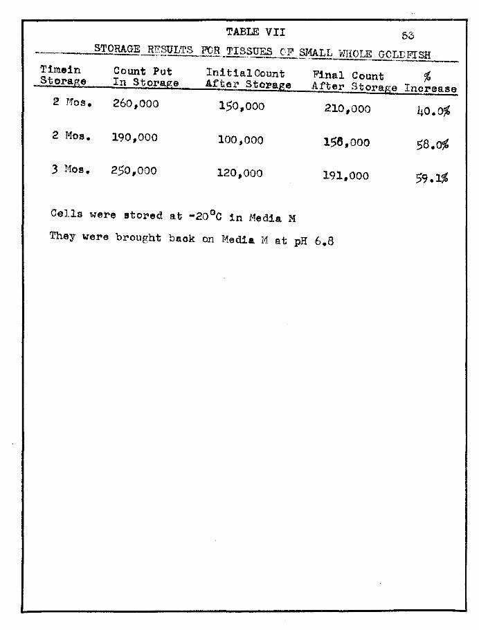
TABLE VII 53 STORAGE-B.~§ULTS FOR TISSUES CF SMALL itIHOLE GOLDFISH -"'---"---_ .. ,,-- ~.-,
Tim.in Count Put In! tial Count Final Count ~ Storage In Storage After Storage After Storage Increase 2 ],'1013. 260,000 150,000 210,000 40.~
2 Mos. 190,000 100,,000 158,000 58.0%
3 r1os. 250,000 120,000 191,000 59.1"
Cells were stored at -20°C in Media M
They were brought baok on Media M at pH 6.8
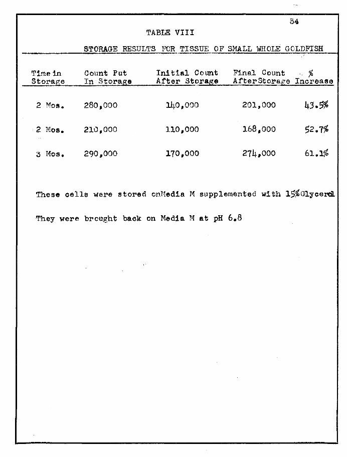
54 TABLE VIII
STORAGE RESULTS FOR TISSUE OF SMALL WHOLE GOLDFISH ; ~~."
Time in Count Put In! tial Count Final Count % Storage In StoraS8 After St~r8.g8 AfterStorage Increase
2 1\10s. 280,000 140,000 201,000 43.5%
2 Mos. 210,000 110,000 168,000 52.7%
3 Mos. 290,,000 170,000 274,000 61.1%
These cells were stored ont·1edia M supplemented wi th 15~G1ycercil.
They were brought back on Media M at pH 6.8

Tissue Type
Fish Heart
Fish. HUBcle
Fish Heart
Fish Muscle
TABLE IX
STORAGE RESULTS FOR FISH HEART AND FISH 1,mSCLE Initial Final
Time fu. Count fut ~£untAfter Count After Storage In Storage orage S~orage
5 !>1os. 149,000 100,000 222,000
!~ Hos. 294,000 220,000 395,000
Cells were stored at -20°C in Media M
Cells were brought baok on Media M
Counts are average of 5 transfers
5 t4os. 149,000 80,000 222,000
4 Mos. 294.,000 220,000 402,000
55
CELLS
% Increase
122%
8~
o Cells were stored at -20 C in Media M supplemented
with 15% Glycerol
Cells were brought baok on Bedia M
Counts are average of 5 transfers
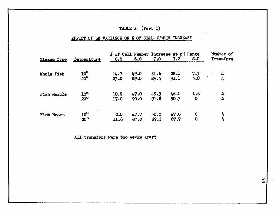
TABLE X (J>art 1)
EFFECT OF pH VARIANCE ON % OF CELL NUNBER INCREASE
% of Cell Number In~rea8e at pH Range Number of T!88ue Tzpe T!!P!l"!ture 6,0 6.8 7.0 7.2 8.0 Transfers
Whole Fish 100 1.4.7 49.0 51.6 28.1 7.3 4 2JJo 23.0 89.0 89.5 91.1 5.0 4-
Fish Muscle l.()O 10.9 47.0 49.3 46.0 4.6 4 20° 17.0 90.0 91.$ 90.3 0 4
'ish H-.rt 100 8.0 47.7 50.0 47.0 0 4 '2[)0 1l.6 87.0 89.3 87.7 0 4
All transfers were two weeks apart

TABLE X (Part 2)
Cell Counts from which the % of increase in No. ot cells was calculated
pH 6.0 % pH 6.8 % pH 7.0 % Whole Fish Initial Final !ncr. Wtial F1nal Iner, In! t1al Final Iner.
10° 150,000 172,000 14.7% 100,000 149,000 49.0% 120,000 l82,ooo 51.6% 200 100,000 123,000 2).0% 100,000 189,000 8<].0% 220,000 417,000 89.5%
Fish I1usc1e 100 llO,ooo 122,000 10.9% 115,000 169,000 47.($ 150,000 224,000 49.'% 200 100,000 117,000 17.0% 140,000 266,000 90.0% 110,000 2ll,ooo 91.8%
Fish Heart 10° 165,000 178,000 8.0% 170,000 251,000 47.7% 100,000 150,000 50% 2fjo 120,000 1.34,000 11.6% 130,000 243,000 87.0% 140,000 265,000 89.3%
ph 7.2 % pH 8.0 % Whole Fish Init.ial F:1nal Iner. Initial Final. Incr.
10° 110,000 141,000 28.1.% 150,000 161,000 7.3~ 20° 180,000 344,000 91.1% 160 ,000 168,000 5.0%
Fish Muscle 10° 115,000 168,000 46.0% 125,000 183,000 4.6% 200 145,000 276,000 90.3% 110,000 0
Fish Heart 10° 100,000 147,000 47.0% 155,000 0 21)0 130,000 244,000 81.7% 140,000 0
Counts are average values ot tour transfers
KEY FOR INTERPRETATION 1 ... • ..
OF FIGURE I
~.FP'EC~g OF .vA~~CUS MER+4 .TYPj;~_ ON
CELL MULTIPLICATION *
MEDIA TYPE H .. . ..
MEDIA TYl'E J • II
1- ----. --. --.. -. -. ---\ J _____ • _____ - • - • _. ---
- ---- _. ----- . - - -. - - - . --: ::-.::: .-.-.-.::: .-.-.-. -.".::: .-:.".
WlIUJm
* From data presented In Table III
A
B
o D
E
F
G H !
J
It
L
M )J
FIGURE ,I
EPPEOTS OPVARIOUS MEDIA TYPES ON
CELL MULTIPLICATION
-. _ .. -- - ... _ .. - -. -........ -- ................... --"'~ _ .... -_ .. --- -." ... -- ----... .... --_ .. -... -_ ..... _ .. _ .. ---_ .............. _ .. --_ ... --- ------------- .. -_ .... . _ .......... -- ................ -_ ... -.... -. --. -.-..... _ ... _ ..... - .. _._"._. -----_ .. -...
o 10 20 30 40 50 60 70 80 90 100 1l0:ro ]30 Jl.f) J;I)
_ Or IBOREASE IN CELL WMBERS
59
o ~T L ~(Jtl:rlP'tlOAlfION "" , - ! ........ .. - ~', .' . '
}I'l5B llE,:A RT . _ . I
10
20° j
31° [i~~~1.
10° '
toO
37°
'V1llfJ!/tlf!illV//J/!!///III111///I/JI ,.~
-From (lata, pr-ee nted ill 'l'ablea Iv, ' V' .
60
90
85 80 75 70 65 60
In ~ell 55 Numbe r 50 Increase 45
40 3'5
61 FIGURE II
EFFECT OF' TEMPERA'l'URE ON CELL MtTLT IPLICAT I ON , t • ~ • - • - T _ _ • •
-""
f to- , It!
t T ~ ~ ,~
-~ .. - , -1 ' -'<-
-3i -,- T I I , , , tiT lIt I T. I
TEJ1PERA TURK
KEY It"UR I~rTERPR ·'TATI ON - - .
o.F FIGURE I II ......-. ' .
EFFEC OF VARIANCE OF p~
CELL lULTIPLI OATION *
WHOLE FI SH
10° FI SH PEOTORAL
. MUSCI£ 200
FISH HEA~T
COLOR .
* Fram data presented in Table VIII
62

FIGURE III
Ef1$CT OF VARIANCE o'p pH , ON CELL MtfL'l'IPUGATlOlJ ' , , .
100 t
95 90 85 80
75 70 65
. 60
55_' So 45
0 35 30 25 20
15 10
5
o 4 6 pH

'n. __ 1'".. Filla IIaut Celb
n..\lwl o.w. 1'et.J!ltoJWle
stall'll ~ .. iba.
'iCa" IV I Let",..,. lOX
F1&U'e "I J ap. Jr!:1 431
fi..p.ft VI. i1p n.., ,.,1 i"~ YII. au ~1!!Jnioa VII

PIGURE IV
• MAY • 64
MAG)7IFlCA,TT01U 10 X
, FILM, Kodacolor X
FIGURE 'V
• MAY • 64
MAGNIFICATICN : 43 x FILMs Koc3acolor X
64
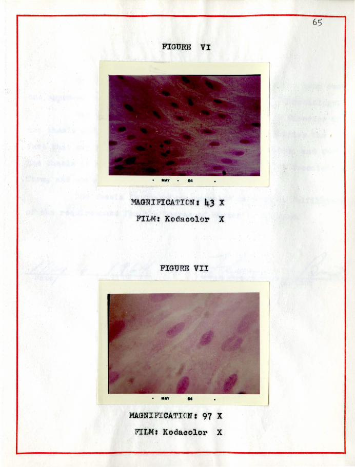
FIGURE VI
• IIIAY • &4
MAGNI FICATI ON t 43 X
FILM: Kodacolor X
FIGURE VII
• IIIAY &4
MAGNI FICAT! Nr 97 X
FILM I Kodaoolor X
6S
APPR0V'AL SHEET ............ _-----:r'hs thesis submitted by Helen Mae Kroeker has been read
and approved by three members of the Department of Microbiology.
The final copies have been examined by the director of
the thesis and the signatlJr'e wh:tch appea~s below verifies the
fact that any necessary changes have been ineorporated" and that
the thesis is now given final approval with referenoe tocontent,
form, and mechanical accuracy.
The thesis is therefore accepted in partial fulfl1l""1ent
of the requirements for the Degree 01' Haster of SeIenee.