Embed Size (px)
Citation preview

Neotropical Ichthyology, 2014Copyright © 2014 Sociedade Brasileira de IctiologiaDOI: 10.1590/1982-0224-20130178
The essential oil from Lippia alba induces biochemical stress in the silvercatfish (Rhamdia quelen) after transportation
Joseânia Salbego1, Alexssandro G. Becker1, Jamile F. Gonçalves1, Charlene C. Menezes2,Clarissa G. Heldwein3, Rosélia M. Spanevello4, Vania L. Loro2, Maria Rosa C.
Schetinger2, Vera M. Morsch2, Berta M. Heinzmann3 and Bernardo Baldisserotto1
This study investigated the effects of the essential oil (EO) from Lippia alba on biochemical parameters related to oxidativestress in the brain and liver of silver catfish (Rhamdia quelen) after six hours of transport. Fish were transported in plastic bagsand divided into three treatments groups: control, 30 µL L-1 EO from L.alba and 40 µL L-1 EO from L.alba. Prior to transport, thefish were treated with the EO from L. alba (200 µL L-1 for three minutes), except for the control group. Fish transported in bagscontaining the EO did not have any alterations in acetylcholinesterase, ecto-nucleoside triphosphate diphosphohydrolaseand 5’nucleotidase activity in the brain or superoxide dismutase activity in the liver. The hepatic catalase (CAT), glutathione-S-transferase (GST), glutathione peroxidase (GPx), nonprotein thiol and ascorbic acid levels were significantly lower comparedto the control group. However, the hepatic thiobarbituric acid-reactive substances, protein oxidation levels and the lipidperoxidation/catalase+glutathione peroxidase (LPO/CAT+GPx) ratio were significantly higher in fish transported with bothconcentrations of the EO, indicating oxidative stress in the liver. In conclusion, considering the hepatic oxidative stressparameters analyzed in the present experiment, the transport of previously sedated silver catfish in water containing 30 or 40µL L-1 of EO from L. alba is less effective than the use of lower concentrations.
Este estudo investigou os efeitos do óleo essencial (OE) de Lippia alba sobre parâmetros bioquímicos relacionados aoestresse oxidativo em cérebro e fígado de jundiá (Rhamdia quelen), após seis horas de transporte. Os peixes foram transportadosem sacos plásticos e divididos em três tratamentos: controle, 30 μL L-1 e 40 μL L-1 de OE de L.alba. Antes do transporte, ospeixes foram tratados com o OE de L. alba (200 μL L-1 por três minutos), exceto para o grupo controle. Os peixes transportadosem sacos contendo o OE não tiveram alterações na atividade da acetilcolinesterase (AChE), ecto-nucleosídeo trifosfatodifosfohidrolase (NTPDase) e 5’nucleotidase, em cérebro ou superóxido dismutase (SOD) no fígado. O tiol não proteico(NPSH), os níveis de ácido ascórbico, catalase (CAT), glutationa-S-transferase (GST) e glutationa-peroxidase (GPx) hepáticos,foram significativamente mais baixos em comparação com o grupo controle. No entanto, as substâncias reativas ao ácidotiobarbitúrico (TBARS), os níveis de oxidação proteica e a taxa de peroxidação lipídica/catalase+glutationa peroxidase (LPO/CAT+GPx) foram significativamente maiores nos peixes transportados com ambas as concentrações de OE, indicando estresseoxidativo no fígado. Em conclusão, considerando os parâmetros de estresse oxidativo do fígado analisados no presenteexperimento, o transporte de jundiás previamente sedados em água contendo 30 ou 40 µL L-1 de OE de L.alba é menos efetivoque utilizando concentrações menores.
Key words: Anesthetics, Antioxidant defenses, Biochemical parameters, Fish transport, Oxidative stress.
Departamentos de Fisiologia e Farmacologia1, de Química2 e de Farmácia Industrial3, Universidade Federal de Santa Maria, 97105-900 SantaMaria, RS, Brazil. [email protected] (JS) [email protected] (AGB) [email protected] (JFG),[email protected] (CCM), [email protected] (VLL), [email protected] (MRSC), [email protected](VMM), [email protected] (CGH), [email protected] (BMH), [email protected] (BB)4Centro de Ciências Químicas, Farmacêuticas e de Alimentos, Setor de Bioquímica, Universidade Federal de Pelotas, Campus UniversitárioCapão do Leão, 96010-900 Pelotas, RS, Brazil. [email protected] (RMS)

The essential oil from Lippia alba induces stress in Rhamdia quelen
Introduction
The transportation of fish in Brazil involves the use ofplastic bags. This system has limitations, such as a finiteoxygen supply and the build-up of ammonia and carbon dioxidelevels (Golombieski et al., 2003; Carneiro et al., 2009; Beckeret al., 2012). Generally, fish farmers add pure oxygen to theplastic bags prior to transport, which can cause variations inthe dissolved oxygen levels, depress the metabolic rate andprovoke blood flow rearrangement and effective methods ofenergy production (Nilsson & Renshaw, 2004).
The acetylcholinesterase (AChE) is an important regulatoryenzyme, which hydrolyses acetylcholine, a neurotransmitterwith important role in the regulation of cognitive functions,mainly found in the brain, muscles, erythrocytes andcholinergic neurons. AChE besides cholinergic transmissionis related to several non-cholinergic actions such as responsesto stress situations. Stress can increase due to free radicalswhich impair enzyme function (Gutierres et al., 2012, 2014)and lead to disorders in locomotion (Blenau et al., 2012) andfish erratic swimming (Salbego et al., 2010).
Adenosine triphosphate (ATP) is the primary intracellularenergy source is which is also one of the most importantneurotransmitters in the purinergic system and is responsiblefor modulating signaling and biosynthetic processes,including vascular homeostasis, cell size maintenance,neuronal signaling, immune function, and protein and lipidmodifications (Marcus et al., 2003; Tort, 2011). The enzymeecto-nucleoside triphosphate diphosphohydrolase(NTPDase) hydrolyzes ATP and adenosine diphosphate(ADP) to adenosine monophosphate (AMP). The resultingAMP is subsequently hydrolyzed to adenosine by ecto-5’-nucleotidase (Stefan et al., 2005; Colgan et al., 2006; Schmatzet al., 2009).
Exposure to hyperoxic, anoxic or hypoxic environmentsmay result in oxidative changes because oxygenconsumption contributes to the levels of reactive oxygenspecies (ROS) generated and the antioxidant status of thecells (Wilhelm-Filho et al., 2001, 2002; Lushchak et al., 2001,2005; Azambuja et al., 2011). The oxidative metabolism ofcells is a continuous source of ROS (resulting from theunivalent reduction of O2) that can damage most cellularcomponents such as carbohydrates, lipids and proteins aswell as promoting cell death (Ahmad et al., 2000; Morales etal., 2004). To protect themselves from these highly reactiveintermediates, living organisms possess a biochemicaldefense system consisting of enzymatic and non-enzymaticantioxidants that scavenge any reactive species. However,in several situations, the rate of ROS generation exceedsthat of their removal, resulting in oxidative stress (Halliwell& Gutteridge, 2000; Livingstone, 2001). The most importantantioxidant enzymes are superoxide dismutase (SOD), whichdetoxifies O2; catalase (CAT), which reduces H2O2;glutathione peroxidase (GPx), which reduces both H2O2 andorganic peroxides by a glutathione-dependent reaction; and
glutathione reductase (GR),which catalyzes the NADPH-dependent regeneration of glutathione (a nonproteinthiol[NPSH]) from the oxidized form (GSSG) generated byGPx (Halliwell & Gutteridge, 2000).
Variations in the water parameters, such as ammonia andcarbon dioxide levels, can provoke stress that could beminimized, at least in some species, through the addition ofeither sedatives or anesthetics in the transport water(Azambuja et al., 2011; Cunha et al., 2011; Becker et al., 2012).The essential oil (EO) from Lippia alba (Mill.) N.E. Brown(Verbenaceae) is a novel anesthetic whose action has beenestablished for the silver catfish Rhamdia quelen (Cunha etal., 2010; Heldwein et al., 2012), and the slender seahorseHippocampus reidi (Cunha et al., 2011).When this oil wasadded to the transport water, the redox state (Azambuja etal., 2011) and ionoregulation (Becker et al., 2012) wereimproved, and lipid oxidation was delayed in the fillets ofsilver catfish (Veeck et al., 2013). However, an increasedventilation rate was observed during the 30 min of transport(Becker et al., 2012).
Increases in the plasma cortisol and glucose levels areclassics indicators of stress responses (Iwama et al., 2004;Urbinati & Carneiro, 2004; Tort, 2011). However, otherbiochemical parameters, such as enzymatic activities, are alsoimportant to understand stress at the cellular level (Lushchaket al., 2001, 2005). In this context, the purpose of this studywas to investigate if a rapid pre-transport sedation andtransport with the EO from L. alba could change enzymesrelated to purinergic neurotransmitters in the whole brain(AChE, NTPDase and 5’-nucleotidase activities) and improveenzymatic parameters (SOD, CAT, GST, and GPx) that act byinactivating free radicals, in the liver. Furthermore, this studyalso measured non-enzymatic parameters that can repairdamage caused by oxidative stress (NPSH and ascorbic acid),changes in pro-oxidant parameters, such as thiobarbituricacid-reactive substance (TBARS) levels and protein oxidationresulting from damage to lipids and proteins respectively, inthe liver of Rhamdia quelen after transport.
Material and Methods
Essential oil extraction. Lippia alba was cultivated in SãoLuiz Gonzaga, Rio Grande do Sul State, Brazil. The aerial partsof the plant were collected in January 2009. The plant materialwas identified by botanist Dr. Gilberto Dolejal Zanetti,Department of Industrial Pharmacy, Universidade Federal deSanta Maria (UFSM), and a voucher specimen (SMDB No.10050) was deposited in the herbarium of the Department ofBiology, UFSM.
Essential oil (EO) was obtained from the fresh leaves ofthe plant by steam distillation for 2 h using a Clevenger-typeapparatus. In this method, the distillate is collected and theaqueous phase is automatically reused by returning it to thedistillation flask (European Pharmacopoeia, 2007). The EOsamples were stored at -4°C in amber glass bottles.
J. Salbego et al.
Experimental procedure. Silver catfish (mean weight ± SEM420.1 ± 8.8 g; 21.2 ± 2.3 cm mean length ± SEM) were capturedfrom a cage net at a fish farm. Fish did not go through adepuration period because this procedure, despite itsrecommendation (Amend et al., 1982), is not implemented bymost fish producers in southern Brazil (Golombieski et al.,2003). Fish were transported at a loading density of 275.1 g L-
1 for 6 h in 9 plastic bags with 7 L of water and 8 L of pureoxygen, and they were divided into three treatment groups(three replicates each, n = 12 per group) that were treated withdifferent concentrations of EO from L. alba: control, 30 µLL-
1and 40 µLL-1 of EO from L. alba diluted 1:10 in ethanol. Fishthat were transported with either of the EO treatments wererapidly sedated with a higher concentration of the same EO(200 µL L-1) for three minutes before placing them into theplastic bags. This concentration induces sedation within thetime proposed (Cunha et al., 2010). Control fish were placeddirectly into the plastic bags. The transport time andconcentrations of EO from L. alba were chosen according toBecker et al. (2012) and were within a sedative safe range forsilver catfish (Cunha et al., 2010). The loading density waschosen according to Carneiro et al. (2009).
Water parameters were monitored before and aftertransport, with the values (mean ± SEM) at the end of transportas follows: dissolved oxygen (8.29 ± 0.98 mg L-1), carbondioxide (58.13 ± 2.51 mg L-1), alkalinity (30.89 ± 2.09 mg CaCO3L-1), water hardness (22.78 ± 1.64 mg CaCO3 L
-1), pH (6.07±0.07), temperature (26.33 ± 0.81 ºC), total ammonia nitrogen(3.21 ± 0.16 mg L-1) and un-ionized ammonia (0.0023 ± 0.0002mg L-1). Dissolved oxygen and temperature were measuredwith an YSI oxygen meter. The pH was verified with a DMPH-2 pH meter. Nesslerization was used to verify the total ammonianitrogen levels using the method of Eaton et al. (2005). Un-ionized ammonia levels were calculated according to Colt(2002). Water hardness was analyzed by the EDTA titrimetricmethod. Alkalinity was determined according to Boyd &Tucker (1992). Carbon dioxide was calculated by the methodof Wurts & Durborow (1992).
After transport, all of the fish were euthanized by spinalcord section, and whole brain and liver tissues were carefullyremoved and frozen to posterior analysis of biochemicalparameters.
Enzymatic assays in the brainAcetylcholinesterase (AChE; E.C. 3.1.1.7). Brain was
homogenized with 150 mM NaCl. The homogenates werecentrifuged for 15 min at 3000 g at 5 ºC, and the supernatantwas used as the enzyme source. AChE activity was measuredas described by Ellman et al. (1961).
NTPDase (ecto-apyrase, ecto/CD39; E.C. 3.6.1.5).Enzymatic assay of the whole brain was carried out in areaction medium containing 5 mM KCl, 1.5 mM CaCl2, 0.1 mMEDTA, 10 mM glucose, 225 mM sucrose and 45 mM Tris–HClbuffer (pH 8.0) in a final volume of 200 µL, as described bySchetinger et al. (2000).
5’-nucleotidase (CD73; E.C. 3.1.3.5). Activity wasdetermined by the method of Heymann et al. (1984) in a reactionmedium containing 10 mM MgSO4 and 100 mM Tris-HCl buffer(pH 7.5) in a final volume of 200 μL. The tubes were thenchilled on ice for 10 min, and the released inorganic phosphates(Pi) were assayed by the method of Chan et al. (1986).
Enzymatic antioxidant activity in the liverSuperoxide dismutase (SOD; E.C. 1.15.1.1). Activity was
determined as the inhibition rate of autocatalytic adenochromegeneration at 480 nm in a reaction medium containing 1 mMepinephrine (0.017 mL) and 50 mM glycine-NaOH (pH 10.5) (1mL). A unit of SOD is defined as the amount of enzyme thatinhibits the speed of detector (epinephrine) reduction by 50%.Enzyme activity was expressed in units mg protein-1 usingthe method described by Misra & Fridovich (1972).
Catalase (CAT; E.C. 1.11.1.6). Activity was assayed byultraviolet spectrophotometry. Change of H2O2 absorbanceafter 60 s was measured at 240 nm. Catalase activity wascalculated and expressed in μmol min-1 mg protein-1 using themethod described by Nelson & Kiesow (1972).
Glutathione -S-transferase (GST). Activity was measuredbased on the method described by Habig et al. (1974) using1-chloro-2,4-dinitrobenzene (CDNB) (0.15 mL) as a substrate.The extinction coefficient used for CDNB was 9.6 mM cm1,and the activity was expressed as μmol GS-DNB min-1 mgprotein-1.
Glutathione peroxidase (GPx; EC 1.11.1.9). The enzymeactivity was measured according to Paglia & Valentine (1967).The assay solution contained 100 mM potassium phosphatebuffer (pH 7.0) 1 mM GSH, 0.15 mM NADPH, 0.1 U mL-1
glutathione reductase, 100 mM azide and a suitable sample ofenzyme solution. Enzyme activity was determined at 37°C bymeasuring the depletion of NADPH at 340 nm and expressedas µmol NADPH min-1 mg protein-1.
Nonenzymatic antioxidants in the liverNon-protein thiol groups (NPSH). NPSH levels were
determined by the method of Ellman (1959) with 0.05 mL of 10mM 5, 5’-dithio-bis (2-nitrobenzoic acid) (DTNB) and 0.7 mLof 0.5 mM phosphate buffer (pH 6.8) added to 0.25 mL ofsupernatant. The results were expressed as μmol non-proteinthiols g tissue-1.
Ascorbic acid content (AsA). It was determined by themethod of Roe (1954). To measure the ascorbic acid levels, analiquot of the supernatant was mixed with 2,4-dinitrophenylhydrazine (4.5 mg mL-1), 0.6 mg mL-1thiourea,CuSO4 (0.075 mg mL-1), and 13.3% trichloroacetic acid followedby incubation for 3 h at 37 ºC. Afterwards, H2SO4 65% (v/v)was added to the medium. The results were expressed as µmolAsA g tissue-1.
Prooxidants in the liverLipid peroxidation estimation. The lipid peroxidation was
estimated by a thiobarbituric acid-reactive substances
The essential oil from Lippia alba induces stress in Rhamdia quelen
(TBARS) assay and performed by a malondialdehyde (MDA)reaction with 2-thiobarbituric acid (TBA), which was opticallymeasured according to Buege & Aust (1978). TBARS levelswere expressed as nmol MDA mg protein-1.
Protein carbonyl assay. The protein carbonyl content wasassayed by the method described by Yan et al. (1995). Theassay was performed in duplicate, and two blank tubes treatedwith 2N HCl (0.2 mL) without DNPH were included for eachsample. The total carbonylation was calculated using a molarextinction coefficient of 22000 M cm-1. The protein carbonylcontent was expressed as nmol carbonyl mg protein-1.
Protein total determination. The protein concentration wasdetermined by the Coomassie Blue method following theBradford method (1976) using bovine serum albumin as astandard, and the absorbance of the samples was measuredat 595 nm.
Statistical analyses. All data are expressed as the mean ±SEM. The homogeneity of variances between treatments wastested with Levene’s test. The data presented homogeneousvariances, so comparisons between the different treatmentswere made using a one way ANOVA and Tukey’s test.Analysis was performed using the Statistica ver. 7.0 software(Stat Soft, Tulsa, OK), and the minimum significance levelwas set at P < 0.05.
Results
No mortality was recorded in any treatment followingtransport. The AChE, NTPDase and 5’-nucleotidase activitiesin the whole brain of silver catfish showed no significantdifferences between the treatments (Table 1).
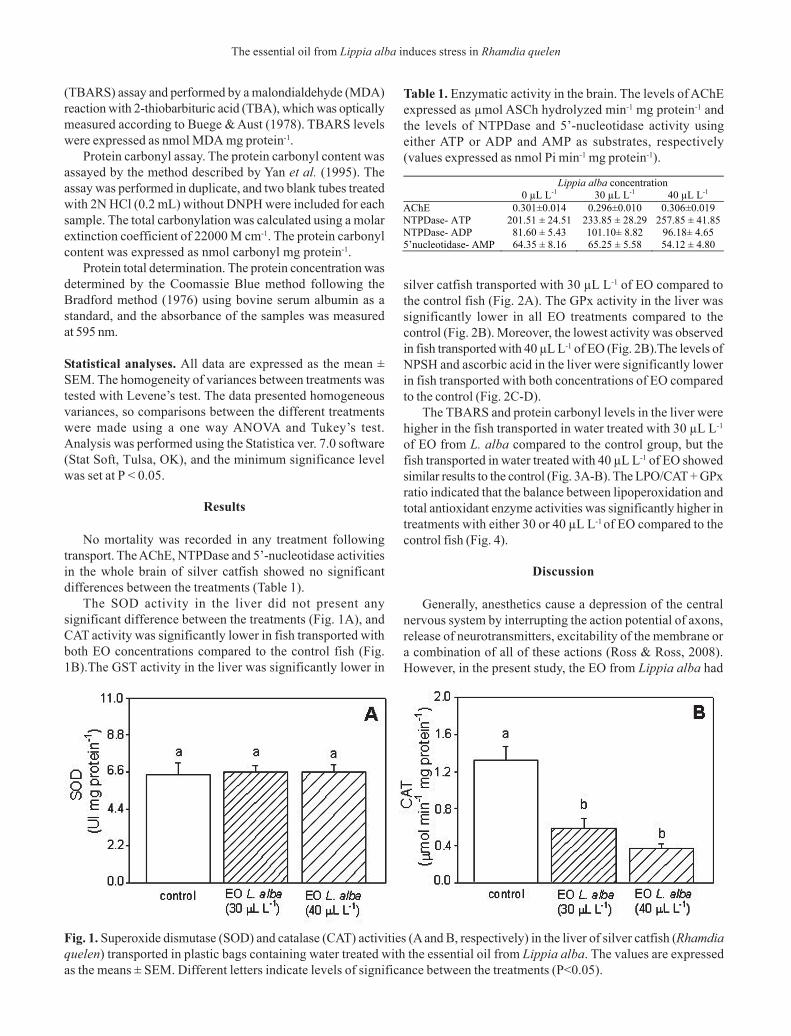
The SOD activity in the liver did not present anysignificant difference between the treatments (Fig. 1A), andCAT activity was significantly lower in fish transported withboth EO concentrations compared to the control fish (Fig.1B).The GST activity in the liver was significantly lower in
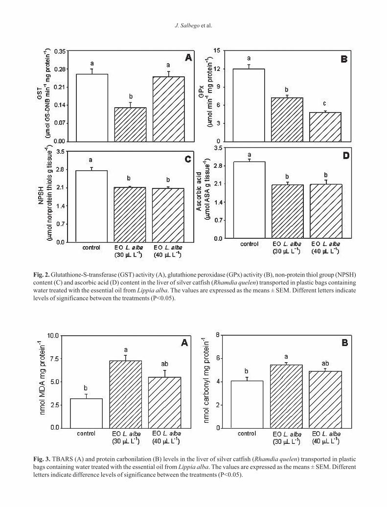
silver catfish transported with 30 µL L-1 of EO compared tothe control fish (Fig. 2A). The GPx activity in the liver wassignificantly lower in all EO treatments compared to thecontrol (Fig. 2B). Moreover, the lowest activity was observedin fish transported with 40 µL L-1 of EO (Fig. 2B).The levels ofNPSH and ascorbic acid in the liver were significantly lowerin fish transported with both concentrations of EO comparedto the control (Fig. 2C-D).
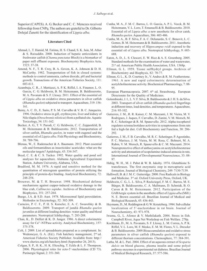
The TBARS and protein carbonyl levels in the liver werehigher in the fish transported in water treated with 30 µL L-1
of EO from L. alba compared to the control group, but thefish transported in water treated with 40 µL L-1 of EO showedsimilar results to the control (Fig. 3A-B). The LPO/CAT + GPxratio indicated that the balance between lipoperoxidation andtotal antioxidant enzyme activities was significantly higher intreatments with either 30 or 40 µL L-1 of EO compared to thecontrol fish (Fig. 4).
Discussion
Generally, anesthetics cause a depression of the centralnervous system by interrupting the action potential of axons,release of neurotransmitters, excitability of the membrane ora combination of all of these actions (Ross & Ross, 2008).However, in the present study, the EO from Lippia alba had
Table 1. Enzymatic activity in the brain. The levels of AChEexpressed as µmol ASCh hydrolyzed min-1 mg protein-1 andthe levels of NTPDase and 5’-nucleotidase activity usingeither ATP or ADP and AMP as substrates, respectively(values expressed as nmol Pi min-1 mg protein-1).
Fig. 1. Superoxide dismutase (SOD) and catalase (CAT) activities (A and B, respectively) in the liver of silver catfish (Rhamdiaquelen) transported in plastic bags containing water treated with the essential oil from Lippia alba. The values are expressedas the means ± SEM. Different letters indicate levels of significance between the treatments (P<0.05).
Lippia alba concentration 0 µL L-1 30 µL L-1 40 µL L-1 AChE 0.301±0.014 0.296±0.010 0.306±0.019 NTPDase- ATP 201.51 ± 24.51 233.85 ± 28.29 257.85 ± 41.85 NTPDase- ADP 81.60 ± 5.43 101.10± 8.82 96.18± 4.65 5’nucleotidase- AMP 64.35 ± 8.16 65.25 ± 5.58 54.12 ± 4.80
J. Salbego et al.
Fig. 2. Glutathione-S-transferase (GST) activity (A), glutathione peroxidase (GPx) activity (B), non-protein thiol group (NPSH)content (C) and ascorbic acid (D) content in the liver of silver catfish (Rhamdia quelen) transported in plastic bags containingwater treated with the essential oil from Lippia alba. The values are expressed as the means ± SEM. Different letters indicatelevels of significance between the treatments (P<0.05).
Fig. 3. TBARS (A) and protein carbonilation (B) levels in the liver of silver catfish (Rhamdia quelen) transported in plasticbags containing water treated with the essential oil from Lippia alba. The values are expressed as the means ± SEM. Differentletters indicate difference levels of significance between the treatments (P<0.05).

The essential oil from Lippia alba induces stress in Rhamdia quelen
no effect on the activities of the enzymes NTPDase and 5’-nucleotidase, which constitute an enzymatic complex able toregulate the extracellular concentrations of adeninenucleotides and nucleosides (Stefan et al., 2005; Colgan etal., 2006; Schmatz et al., 2009; Gutierres et al. 2012, 2014).
Studies by Heldwein et al. (2012) suggest the involvementof the gamma-aminobutyric acid -A (GABAA) benzodiazepinereceptor in the anesthetic effect of the EO from L. alba. Silvercatfish transported in water containing the EO from L. albawere less agitated (Becker et al., 2012). The results of theAChE activity assay corroborate with the aforementionedfindings because the enzyme activity was unaltered and thechange in the locomotor behavior was not due to alterationsin its function. Furthermore, consolidates that EO from L.alba is able to reduce or maintain the basal levels of thecholinergic neurotransmitters.
This study did not detect any significant differences inthe SOD activity between the treatments after transport ofthe catfish. The lower CAT and GST activities in the liver ofsilver catfish transported with 30 µL L-1 of EO in the watercould be attributed to the increase of the oxidant levels, asobserved by the highest levels of TBARS that were generatedby this treatment. It is possible that in silver catfish transportedwith 40 µL L-1 of EO in the water the decrease of CAT wasenough to avoid the increase of TBARS. Azambuja et al.(2011) reported that silver catfish transported (fish density140 – 200 g L-1) for 6 h in normoxic conditions with 10 µL L-1 ofEO of L. alba exhibited no significant alterations in hepaticGST, SOD and CAT activities, but TBARS levels also did notchange.
Alterations in GPx activity are generally accompanied bychanges in NPSH levels because the NPSH is a co-substratefor H2O2 breakdown by GPx (Sies, 1999). The major cellular
thiol that participates in cellular redox reactions – NPSH –displayed an important role in the detoxification ofelectrophilic metabolites catalyzed by GST (Sies, 1999; Latha& Pari, 2004).
High NPSH levels may protect cellular proteins againstoxidation either via the NPSH redox cycle or by directlydetoxifying the ROS generated by exposure to stressor agents(Ruas et al., 2008), but the low NPSH content could modulatethe activity of GPx and GST enzymes (Brouwer & Brouwer,1998) as suggested in the present study in silver catfishtransported in water containing the EO from L. alba. Theformation of peroxides and TBARS was lower in the frozenfillets of silver catfish transported with either 30 or 40 µL L-1
of EO (Veeck et al., 2013). In the present study, lower levels ofascorbic acid were also observed in the liver of silver catfishtransported in water containing the EO from L. alba.Antioxidants such as ascorbic acid are active ROS scavengersinvolved in the lipid peroxidation process (Halliwell &Gutteridge, 2000; Trenzado et al., 2006; Kochhann et al., 2009).This study showed that the TBARS and protein carbonyllevels in the liver of silver catfish were higher whentransported in water containing 30 µL L-1 of EO, indicatinghigher lipid peroxidation and protein oxidation activity anddemonstrating that the antioxidant defenses were notcompletely able to effectively scavenge the ROS produced.The increased LPO/CAT+GPx ratio also suggests thathydrogen peroxide were produced, overcoming the capacityof CAT and GPx in neutralizing ROS production (Ruas et al.,2008) and resulting in a LPO in the liver.
The use of 10-20 µL L-1 of EO from L. alba increasedventilation rate of silver catfish during the first 30 min oftransport, indicating a higher agitation of the fish (Becker etal., 2012). Therefore, it was hypothesized that the use of apre-transport sedation with 200 µL L-1 of EO from L. albawould reduce this initial agitation and the transport with 30 or40 µL L-1 of EO from L. alba in the water would be moreeffective. However, this hypothesis was not confirmedconsidering the hepatic oxidative stress parameters analyzedin the present experiment and the lower concentrations (10-20 µL L-1 of EO from L. alba) used by Azambuja et al. (2011)and Becker et al. (2012) are more effective for transportingsilver catfish.
Acknowledgments
This study was supported by research funds from theFundação de Amparo à Pesquisa do Estado do Rio Grande doSul (FAPERGS/PRONEX, process 10/0016-8), the ConselhoNacional de Desenvolvimento Científico e Tecnológico(CNPq, process 470964/2009-0) and the Ministério da Pesca eAquicultura/FINEP (process 01.12.0130.00). B. Baldisserotto,V. Loro, M. R. C. Schetinger, V. M. Morsch, B. M. Heinzmann,and R. M. Spanevello received research fellowship. J. Salbego,J. F. Gonçalves, and C. G. Heldwein received fellowship fromCoordenação de Aperfeiçoamento de Pessoal de Nível
Fig. 4. LPO/CAT+GPx ratio in the liver of silver catfish(Rhamdia quelen) transported in plastic bags containingwater treated with the essential oil from Lippia alba. Thevalues are expressed as the means ± SEM. Different lettersindicate levels of significance between the treatments (P<0.05).
J. Salbego et al.
Superior (CAPES). A. G. Becker and C. C. Menezes receivedfellowship from CNPq. The authors are grateful to Dr. GilbertoDolejal Zanetti for the identification of Lippia alba.
Literature Cited
Ahmad, I., T. Hamid, M. Fatima, H. S. Chand, S. K. Jain, M. Athar& S. Raisuddin. 2000. Induction of hepatic antioxidants infreshwater catfish (Channa punctatus Bloch) is a biomarker ofpaper mill effluent exposure. Biochemystry Biophysics Acta,1523: 37-38.
Amend, N. F., T. R. Croy, B. A. Goven, K. A. Johnson & D. H.McCarthy. 1982. Transportation of fish in closed systems:methods to control ammonia, carbon dioxide, pH and bacterialgrowth. Transactions of the American Fisheries Society, 111:603-611.
Azambuja, C. R., J. Mattiazzi, A. P. K. Riffel, I. A. Finamor, L. O.Garcia, C. G. Heldwein, B. M. Heinzmann, B. Baldisserotto,M. A. Pavanato & S. F. Llesuy. 2011. Effect of the essential oilof Lippia alba on oxidative stress parameters in silver catfish(Rhamdia quelen) subjected to transport. Aquaculture, 319: 156-161.
Bainy, A. C. D., E. Saito, P. S. M. Carvalho & V. B. C. Junqueira.1996. Oxidative stress in gill, erythrocytes, liver and kidney ofNile tilapia (Oreochromis niloticus) from a polluted site. AquaticToxicology, 34: 151-162.
Becker, A. G., T. V. Parodi, C. G. Heldwein, C. C. Zeppenfeld, B.M. Heinzmann & B. Baldisserotto. 2012. Transportation ofsilver catfish, Rhamdia quelen, in water with eugenol and theessential oil of Lippia alba. Fish Physiology and Biochemistry,38: 789-796.
Blenau, W., E. Rademacher & A. Baumann. 2012. Plant essentialoils and formamidines as insecticides/ acaricides: what are themolecular targets? Apidologie, 43: 334-347.
Boyd, C. E. & C. S. Tucker. 1992. Water quality and pond soilanalyses for aquaculture. Alabama Agricultural ExperimentStation, Auburn University, Alabama, USA.
Bradford, M. M. 1976. A rapid and sensitive method for thequantitation of microgram quantities of protein utilizing theprinciple of protein-dye binding. Analytical Biochemistry, 72:248-254.
Brouwer, M. & T. H. Brouwer. 1998. Biochemical defensemechanisms against copper-induced oxidative damage in theblue crab, Callinectes sapidus. Archives of Biochemistry andBiophysics, 351: 257-264.
Buege, J.A. & S.D. Aust. 1978. Microssomal lipid peroxidation.Methodos in Enzymology, 52: 302-309.
Carneiro, P. C. F., P. H. S. Kaiseler, E. A. C. Swarofsky & B.Baldisserotto. 2009. Transport of jundiá Rhamdia quelenjuveniles at different loading densities: water quality and bloodparameters. Neotropical Ichthyology, 7: 283-288.
Chan, K., D. Delfert & K. D. Junger. 1986. A direct colorimetricassay for Ca2+-NTPase activity. Analytical Biochemistry, 157:375-378.
Colt, J. 2009. List of spreadsheets prepared as a complement. In:Wedemeyer, G. A. (Ed.). Fish hatchery management, 2nd ed.American Fisheries Society Publisher. Available from: http://www.sheries.org/afs/hatchery.html (September 26, 2013).
Colgan, S. P., H. K., H. K. Eltzschig, T. Eckle & L. F. Thompson.2006. Physiological roles for ecto-5’-nucleotidase (CD 73).Purinergic Signal, 2: 351-360.
Cunha, M. A., F. M. C. Barros, L. O. Garcia, A. P. L. Veeck, B. M.Heinzmann, V. L. Loro, T. Emanuelli & B. Baldisserotto. 2010.Essential oil of Lippia alba: a new anesthetic for silver catsh,Rhamdia quelen. Aquaculture, 306: 403-406.
Cunha, M. A., B. F. Silva, F. A. C. Delunardo, S. C. Benovit, L. C.Gomes, B. M. Heinzmann & B. Baldisserotto. 2011. Anestheticinduction and recovery of Hippocampus reidi exposed to theessential oil of Lippia alba. Neotropical Ichthyology, 9: 683-688.
Eaton, A. D., L. S. Clesceri, E. W. Rice & A. E. Greenberg. 2005.Standard methods for the examination of water and wastewater,21st ed. American Public Health Association, USA. 1368p.
Ellman, G. L. 1959. Tissue sulfhydryl groups. Archives ofBiochemistry and Biophysis, 82: 70-77.
Ellman, G. L., K. D. Courtney Jr., V. Andres & R. M. Featherstone.1961. A new and rapid colorimetric determination ofacetylcholinesterase activity. Biochemical Pharmacology, 7: 88-95.
European Pharmacopoeia. 2007. 6th ed. Strassbourg, EuropeanDirectorate for the Quality of Medicines.
Golombieski, J. I., L. V. F. Silva, B. Baldisserotto & J. H. S. da Silva.2003. Transport of silver catfish (Rhamdia quelen) fingerlingsat different times, load densities, and temperatures. Aquaculture,216: 95-102.
Gutierres, J. M., R. R. Kaiser, C. M. Mazzanti, J. M. Vieira, M. V.Rodrigues, J. Jaques, F. Carvalho, D. Zanini, V. M. Morsch, M.R. C. Schetinger & R. M. Spanevello. 2012. Alpha-tocopherolregulates ectonucleotidase activities in synaptosomes from ratsfed a high-fat diet. Cell Biochemistry and Function, 30: 286-292.
Gutierres, J. M., F. B. Carvalho, M. R. C. Schetinger, P. Agostinho,P. C. Marisco, J. M. Vieira, M. M. Rosa, C. Bohnert, M. A.Rubin, V. M. Morsch, R. Spanevello & C. M. Mazzanti. 2014.Neuroprotective effect of anthocyanins on acetylcholinesteraseactivity and attenuation of scopolamine-induced amnesia in rats.International Journal of Developmental Neuroscience, 33: 88-97.
Habig, W. H., M. J. Pabst & W. B. Jakoby. 1974. Glutathione S-transferases. The first enzymatic step in mercapturic acidformation. Journal of Biological Chemistry, 249: 7130-7139.
Halliwell, B. & J. M. C. Gutteridge. 2000. Free Radicals in Biologyand Medicine. 3rd ed. Oxford University Press, Oxford, UK.
Heldwein, C. G., L. L. Silva, P. Reckziegel, F. M. C. Barros, M. E.Bürger, B. Baldisserotto, C. A. Mallmann, D. Schmidt, B. O.Caron & B. M. Heinzmann. 2012. Participation of theGABAergic system in the anesthetic effect of Lippia alba (Mill.)N. E. Brown essential oil. Brazilian Journal of Medical andBiological Research, 45: 436-443.
Heymann, D., M. Reddington & G. W. Kreutzberg. 1984. Sub cellularlocalization of 5’-nucleotidase in rat brain. Journal ofNeurochemistry, 43: 971-978.
Iwama, G., L. Afonso & V. Mathilakath. 2004. Stress in fish.Campbell River, Aqua Net Workshop on Fish Welfare. 278p.
Kochhann, D., M. A. Pavanato, S. F. Llesuy, L. M. Correa, A. P. K.Riffel, V. L. Loro, M. F. Mesko, E. M. M. Flores, V. L. Dressler& B. Baldisserotto. 2009.Bioaccumulation and oxidative stressparameters in silver catfish (Rhamdia quelen) exposed todifferent thorium concentrations. Chemosphere, 77: 384-391.
Latha, M. & L. Pari. 2004. Effect of an aqueous extract of Scopariadulcis on blood glucose, plasma insulin and some polyolpathway enzymes in experimental rat diabetes. Brazilian Journalof Medical Biological Research, 37: 577-586.

The essential oil from Lippia alba induces stress in Rhamdia quelen
Livingstone, D. R. 2001. Contaminant-stimulated reactive oxygenspecies production and oxidative damage in aquatic organisms.Marine Pollution Bulletin, 42: 656-666.
Lushchak, V. I., L. P. Lushchak, A. Mota & M. Hermes-Lima. 2001.Oxidative stress and antioxidant defenses in goldfish Carassiusauratus during anoxia and reoxygenation. American Journal ofPhysiology Regulatory Integrative and Comparative Physiology,280: 100-107.
Lushchak, V. I., T. V. Bagnyukova, O. V. Lushchak, J. M. Storey &K. B. Storey. 2005. Hypoxia and recovery perturb free radicalprocess and antioxidant potential in common carp (Cyprinuscarpio) tissues. International Journal of Biochemistry & CellBiology, 37: 1319-1330.
Marcus, A. J., M. J. Broekman, J. H. F. Drosopoulos, N. Islam, D.J. Pinsky, C. Sesti & R. Levi. 2003. Heterologous cell-cellinteractions: thromboregulation, cerebroprotection andcardioprotection by CD39 (NTPDase-1). Journal of Thrombosisand Haemostasis, 1: 2497-2509.
Misra, H. P. & I. Fridovich. 1972. The role of superoxide anion inthe auto-oxidation of epinephrine and a simple assay forsuperoxide dismutase. Journal of Biological Chemistry, 247:3170-3175.
Morales, A. E., A. Pérez-Jiménez, M. C. Hidalgo, E. Abellán & G.Cardenete. 2004. Oxidative stress and antioxidant defenses afterprolonged starvation in Dentex dentex liver. ComparativeBiochemistry and Physiology, 139: 153-161.
Nelson, D. P. & Kiesow, L.A. 1972. Enthalpy of decomposition ofhydrogen peroxide by catalase at 25ºC (with molar extinctioncoefficients of H2O2 solution in the UV). AnalyticalBiochemistry, 49: 474-478.
Nilsson, G. E. & G. M. C. Renshaw. 2004. Hypoxic survivalstrategies in two fishes: extreme anoxia tolerance in the NorthEuropean crucian carp and natural hypoxic preconditioning in acoral-reef shark. Journal of Experimental Biology, 207: 3131-3139.
Paglia, D. E. & W. N. Valentine. 1967. Studies on the quantitativeand qualitative characterization of erythrocytes glutathioneperoxidase. Journal of Laboratory and Clinical Medicine, 70:158-168.
Roe, J. H. 1954. Chemical determination of ascorbic, dehydroascorbicand diketogulonic acids. Pp. 115-139. In: Glick, D. (Ed.).Methods of Biochemical Analysis. Interscience Publishers Inc.,New York.
Ross, L. G. & B. Ross. 2008. Anaesthetic and sedative techniquesfor aquatic animals, 3rd ed., Blackwell Publishing Ltd., Oxford.
Ruas, C. B. G., C. S. Carvalho, H. S. S. de Araújo, E. L. G. Espíndola& M. N. Fernandes. 2008. Oxidative stress biomarkers ofexposure in the blood of cichlid species from a metal-contaminated river. Ecotoxicology and Environment Safety, 71:86-93.
Salbego, J., A. Pretto, C. R. Gioda, C. C. Menezes, R. Lazzari, J.Radünz Neto, B. Baldisserotto & V. L. Loro. 2010. Herbicideformulation with glyphosate affects growth,acetylcholinesterase activity, and metabolic and hematologicalparameters in piava (Leporinus obtusidens). Archives ofEnvironmental Contamination and Toxicology, 58: 740-745.
Schetinger, M. R. C., N. M. Porto, M. B. Moretto, V. M. Morsch,J. B. T. da Rocha, V. Vieira, F. Moro, R. T. Neis, S. Bittencourt,H.G. Bonacorso & N. Zanatta. 2000.New benzodiazepines alteracetylcholinesterase and ATPDase activities. NeurochemicalResearch, 25: 949-955.
Schmatz, R., M. R. C. Schetinger, R. M. Spanevello, C. M.Mazzanti, N. Stefanello, P. A. Maldonado, J. Gutierres, M. C.de Carvalho, E. Girotto, M. B. Moretto & V. M. Morsch. 2009.Effects of resveratrol on nucleotide degrading enzymes instreptozotocin-induced diabetic rats. Life Sciences, 84: 345-350.
Sies, H. 1999. Glutathione and its role in cellular functions. FreeRadical Biology and Medicine, 27: 916-921.
Stefan, C., S. Jansen & M. Bollen. 2005. NPP-typeectophosphodiesterases: unity in diversity. Trends inBiochemical Sciences, 30: 542-550.
Summerfelt, R. C. & L. S. Smith. 1990. Anesthesia, surgery andrelated techniques. Pp. 213-272. In: C.B. Schreck, P.B. Moyle(Eds.). Methods for Fish Biology. American Fisheries Society,Bethesda, Maryland.
Trenzado, C., M. C. Hidalgo, M. Garcia-Gallego, A. E. Morales,M. Furné, J. Domezain & A. Sanz. 2006. Antioxidant enzymesand lipid peroxidation in sturgeon Acipenser naccarii and troutOncorhynchus mykiss. A comparative study. Aquaculture, 254:758-767.
Tort, L. Stress and immune modulation in fish. 2011. Developmental& Comparative Immunology, 35: 1366-1375.
Urbinati, E. C. & P. C. F. Carneiro. 2004. Práticas de manejo eestresse dos peixes em piscicultura intensiva. Pp. 171-194. In:Cyrino, J. E. P., E. C. Urbinati, D. M. Fracalossi & N. Castagnolli(Eds.). Tópicos especiais em piscicultura de água doce tropicalintensiva. São Paulo: Tec Art.
Veeck, A. P. L., B. Klein, L. F. Ferreira, A. G. Becker, C. G. Heldwein,B. M. Heinzmann, B. Baldisserotto & T. Emanuelli. 2013. Lipidstability during the frozen storage of fillets from silver catfishexposed in vivo to the essential oil of Lippia alba (Mill.) NEBrown. Journal of the Science of Food and Agriculture, 93: 955-960.
Wilhelm Filho, D., T. Tribess, C. Gáspari, F. D. Claudio, M. A.Torres & A. R. M. Magalhães. 2001. Seasonal changes inantioxidant defenses of the digestive gland of the brown mussel(Perna perna). Aquaculture, 203: 149-158.
Wilhelm Filho, D., F. Sell, L. Ribeiro, M. Ghislandi, F. Carrasquedo,C. G. Fraga, J. P. Wallauer, P. C. Simões-Lopes & M. M. Uhart.2002. Comparison between the antioxidant status of terrestrialand diving mammals. Comparative Biochemistry andPhysiology, 133: 885-892.
Winkaler, E. U., T. R. M. Santos, J. G. Machado-Neto & C. B. R.Martinez. 2007. Acute lethal and sublethal effects of neem leafextract on the neotropical freshwater fish Prochilodus lineatus.Comparative Biochemistry and Physiology, 145: 236-244.
Wurts, W. A. & R. M. Durborow. 1992. Interactions of pH, carbondioxide, alkalinity and hardness in fish ponds. Southern RegionalAquaculture Center Publication, 464: 1-4.
Yan, L. J., M. G. Traber & L. Packer. 1995. Spectrophotometricmethod for determination of carbonyls in oxidatively modifiedapolipoprotein B of human low-density lipoproteins. AnalyticalBiochemistry, 228: 349-351.
Submitted October 4, 2013Accepted April 20, 2014 by Adalberto L. Val


![[Lippia alba (Mill) NE Brown] pode variar](https://img.dokumen.tips/doc/110x75/58720c351a28ab02618c08c0/lippia-alba-mill-ne-brown-pode-variar.jpg)


