Embed Size (px)
Citation preview
RoskildeUniversity
The escherichia coli chromosome replication initiator protein, DnaAMutational analysis of the DNA binding domain and analysis of amino acid substitutions inDnaA5 and DnaA46Nyborg, Malene
Publication date:2000
Citation for published version (APA):Nyborg, M. (2000). The escherichia coli chromosome replication initiator protein, DnaA: Mutational analysis ofthe DNA binding domain and analysis of amino acid substitutions in DnaA5 and DnaA46. Roskilde: RoskildeUniversitet.
General rightsCopyright and moral rights for the publications made accessible in the public portal are retained by the authors and/or other copyright ownersand it is a condition of accessing publications that users recognise and abide by the legal requirements associated with these rights.
• Users may download and print one copy of any publication from the public portal for the purpose of private study or research. • You may not further distribute the material or use it for any profit-making activity or commercial gain. • You may freely distribute the URL identifying the publication in the public portal.
Take down policyIf you believe that this document breaches copyright please contact [email protected] providing details, and we will remove access to thework immediately and investigate your claim.
Download date: 29. Apr. 2019
HI^ Escherichia coli Chromosome Replication Initiator
Protein, DnaA .
Mutational Analysis of the DNA Binding Domain
and
Analysis of Amino Acid Substitutions in DnaA5 and
DnaA46
Marlene Nyborg
Department of Life Sciences and Chemistry
Roskilde University

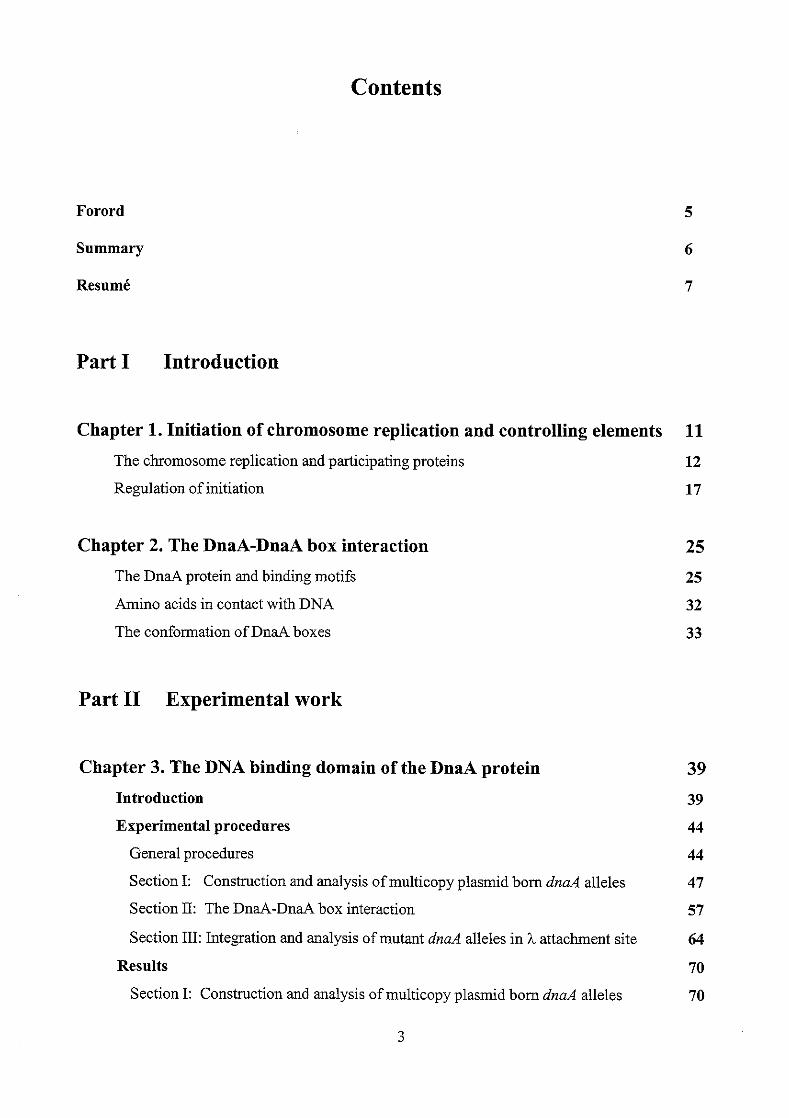
Contents
Forord
Summary
Resume
Part I Introduction
Chapter 1. Initiation of chromosome replication and controlling elements The chromosome replication and participating proteins
Regulation of initiation
Chapter 2. The DnaA-DnaA box interaction The DnaA protein and binding motifs
Amino acids in contact with DNA
The conformation of DnaA boxes
Part I1 Experimental work
Chapter 3. The DNA binding domain of the DnaA protein
Introduction
Experimental procedures
General procedures
Section I: Construction and analysis of multicopy plasmid born dnaA alleles
Section 11: The DnaA-DnaA box interaction
Section III: Integration and analysis of mutant dnaA alleles in h attachment site
Results
Section I: Construction and analysis of multicopy plasmid born dnaA alleles
11
12
17
25 25
32
33
39 39
44
44
47
57
64
70
70
3
Section 11: DnaA-DnaA box interaction 100
Section III: Integration and analysis of mutant dnaA alleles in h attachment site 115
Discussion 133
Chapter 4. Analysis of the A184V, H252Y and G426S substitutions in the DnaA
protein 146 Introduction 146
Experimental procedures 147
Results 149
Discussion 155
Recapitulation 157
References 165
Publication I: Two types of cold sensitivity associated with the A184-+V change
in the DnaA protein
4
Forord
Denne Ph.D &andling er udfmt ved Roskilde Universitetscenter i perioden 1995-1999.
Jeg vi1 fnrst og fremmest takke min vejleder Tove Atlung for saerdeles god vejledning
og diskussioner og for at vaere positiv og inspirerende nir resultateme lader vente p i
sig. Endvidere takkes Ole Skovgaard og Ole Michelsen for hjaelp med diverse computer
programmer/problemer, Benny Pedersen for diskussioner af in vitro assays, Flemming
G. Hansen for stor hjaelp med flow cytometret og alle fra 'Mandagsmde-sjakket' for
gode diskussioner og rid. Til sidst vi1 jeg gerne takke Kirsten Olesen for med b j t
humerr at have holdt mig med selskab i laboratoriet, hjulpet mig med diverse tekniske
opstillinger og ikke mindst for at have hjulpet mig med sekvenseringen af de mange
ha4 mutanter.
Denne afhandling er delt op i Part I og Part 11. Part I er introducerende og inkluderer
Chapter 1 (The initiation of chromosome replication and controlling elements) og
Chapter 2 (The DnaA-DnaA box interaction), mens Part II inkluderer det
eksperimentelle arbejde, som er delt op i to kapitler. Chapter 3 indeholder underwgelser
det DNA bindende domaene af DnaA proteinet (The DNA binding domain of the DnaA
protein) og Chapter 4 indeholder underwgelser af DnaA(A184V) proteinet og andre
DnaA proteiner (Analysis of the A184V, H252Y and G426S substitutions in the DnaA
protein). Afhandlingen indeholder endvidere publikationen : Two types of cold
sensitivity associated with the A1 84+V change in the DnaA protein.
Roskilde Universitetscenter, April 2000.
5
Summary
The experimental work presented in this thesis involve mutational analysis of the DNA
binding domain of the DnaA protein and analysis of the A184V substitution in the ATP
area of domain III and other amino acid substitutions found in the DnaA5 and DnaA4G
proteins.
To analyse the DNA binding domain, more than 100 functional DnaA proteins with
amino acid substitutions in the DNA binding domain were constructed and studies in
vivo by complementation analysis o f the high temperature sensitive &a446 phenotype
by induction of plasmid born mutant dnaA(IV) alleles. The results indicate that the
whole domain is involved in DNA binding. However, there seems to exist more
residues in the first half and the veIy C-terminal region that can be substituted with non-
closely related amino acids with no apparent effect on DnaA activity. Based on
alignment of the DNA binding domain of the DnaA protein with proteins with known
binding motifs, it is suggested that helix 3 and 4 in the DNA binding domain o f the
DnaA protein carries a modified helix-turn-helix motif The results of the in vivo
complementation analysis carried out in this study support this proposal. A more
extensive analysis was carried out with 20 mutant DnaA proteins. The study included in
vivo and in vitro binding analysis. The results of these analyses indicate that many of
the mutant DnaA proteins retained relatively high aflinity for oriC, but reduced
specificity. DnaA proteins with the highest afhity, but lowest specificity had amino
acid substitutions in a-helix 1 and in the following basic loop. A DnaA protein with a
K397E substitution had higher aflinity and specificity for oriC in vitro than the DnaA+
protein had, and a strain harbouring the mzaA(K397E) allele on the chromosome
overinitiated chromosome replication at 32OC and 42°C with a DnaA(K397E)
concentration o f only one fourth of the DnaA concentration in a wild type strain. Higher
DnaA(K397E) concentrations seemed to inhibit initiation of chromosome replication
probably due to too tight binding of the oriC area by DnaA(K397E) proteins.
The A184V substitution in the ATP area of domain III of the DnaA protein and other
amino acid substitutions found in the DnaA5 and Dm446 proteins were studied in vivo
and in vitro. Multicopy dnnA(A184V) and dnaA5 strains, which are cold sensitive,
initiated chromosome replication extensively at non-permissive temperature, but the
initiations were not elongated and DNA synthesis stopped. On the contrary, multicopy
dnaA46 strains (and the dnaAcos mutant), which are also cold sensitive, overinitiated
G
chromosome replication at non-permissive temperature, but (some of) these initiation
were elongated and resulted in an increased DNA concentration per mass especially in
the multicopy M 4 6 cells. Multicopy dnaAW52Y) and dnd(G426S) strains are
temperature resistant like multicopy M' strains. However, in vitro binding analysis
indicates that both the DnaA(H252Y) and DnaA(G426S) proteins have reduced affinity
and specificity for oriC as the DnaA46 and DMM proteins, while the DnaA(A184V)
protein retained as high afsnity and specificity for oriC in vitro as the DnaA' protein. A
strain harbouring the dnaA(A184V) allele on the chromosome overinitiated
chromosome replication at 42°C under conditions where the DnaA concentration was
only one fourth of the DnaA concentration in a wild type strain. At 32T, the initiation
of chromosome replication decreased severely with increasing DnaA(A184V)
concentrations.
Resume
Det eksperimentelle arbejde, som praesenteres i denne a&mllimg omfatter en
mutationsanalyse af det DNA binding domzne af DnaA proteinet og en analyse af
A184V substitutionen i det ATP bmdende domaene og andre aminosyre substitutioner
fhndet i DnaA5 og DnaA46 proteinerne.
For at undersnge det DNA bindende domEne blev mere end 100 W i o n e l l e DnaA
proteiner med aminosyre substitutioner i det DNA bindende d o m n e konstrueret og
studeret in vivo. Analysen bestod i at underssge hvor godt inducerbare plasmidbime
dnaA(IV) alleler komplementerede den bj-temperatur sensitive M 4 6 mutant.
Resultateme indikerer, at hele domaenet er involveret i DNA binding. Der er imidlertid
flere aminosyrer i den fsrste halvdel og i den helt C-terminale del, som kan erstattes
uden nogen tilsyneladende effekt p i DnaA aktiviteten. Ved at sanmenligne aminosyre
sekvensen af det DNA bindende domaene af DnaA proteinet med aminosyre sekvenser
fia andre proteiner med kendte bindingsmotiver er det foresliet, at helii 3 og 4 i det
DNA bindende d o m n e af DMA proteinet kan have et modificeret helix-turn-helix
bmdingsmotiv. Resultaterne af in vivo kompiementationsanalysen stdter dette forslag.
Der blev foretaget en mere indgiende undersngelse af omkring 20 mutante DnaA
proteiner. Underssgelsen inkluderede in vivo og in vitro bindingsanalyser. Resultaterne
af disse undersngelse indikerer, at mange af DnaA proteinerne stadig har relativ hnj
7
&nitet til oriC, men til gengseld er bmdingen mindre speciiik. De DnaA proteiner med
den bjeste affinitet men lavest specificitet baerer aminosyre substitutioner i a-helix 1
og i det eftdlgende basiske loop. DnaA proteinet med K397E substitution har hsjere
finitet og specificitet til orzC sammenlignet med DnaA+ proteinet. N i r M ( K 3 9 7 E )
allelen m e s ind p i kromosomet overinitieres kromosom replikationen ved 32°C og
42"C, selvom DnaA(K397E) koncentrationen kun er en fjerdedel af hvad DnaA
koncentrationen er i en vild type stamme. Det Ser ud til, at h~ijere DnaA(K397E)
koncentrationer hammer initieringen af kromosom replikationen. Dette skyldes miiske
at DnaA(K397E) proteinerne binder oriC omridet med for hsj affinitet.
A184V substitutionen i det ATP bmdende omride i domaene III og andre aminosyre
substitutioner fundet i DnaAS og DnaA46 proteinerne blev underssgt in vivo og in vitro. Multi-kopi &d(A184V) og &d5 stammer er kulde sensitive. De initierer kromosom
repliiionen voldsomt ved ikke-permissive temperaturer, men initieringerne bliver
imidlertid ikke elongeret, hvorved DNA syntesen stopper. Multi-kopi ha446 stammer
(og h d c o s mutanten) er ogsi kulde sensitive. Her overinitieres kromosom
replikationen og&, men nogle af initieringer elongeres, hvilket resulterer i et hqere
DNA indhold pr. masse. Multi-kopi hd(H252Y) og dnaA(G426S) stammerne er
temperatur resistente ligesom multi-kopi M' stammer. in vitro bindingsanalyser tyder
dog pi, at bide DnaA(HZ52Y) og DnaA(G426S) proteinerne samt DnaA46 og DnaA5
proteinerne har reduceret &nitet til O K , mens DnaA(A184V) proteinet har samme
hsje afkitet og specificitet til oriC som DnaA' proteinet. Nir dd(A184V) allelen
szttes in p i kromosomet resulterer det i overinitiering af kromosom repliiationen ved
42T, selvom DnaA(Al84V) koncentrationene kun er en fjerdedel af hvad DnaA
koncentrationen er i en vild type stamme. Ved 32°C Ser det ud til, at initieringen af kromosom replikationen ophmer med stigende DnaA(A184V) koncentrationer.
8
Part I
Introduction

Chapter 1
Initiation of chromosome replication and controlling elements
Life is dependent on cell division and therefore dependent on replication of the
chromosome, which is regulated at the level of initiation (Cooper and Helmstetter,
1968; for review see von Meyenburg and Hansen, 1987; Weigel and Messer, 1996;
Skarstad and Boye, 1994). Initiation of chromosome replication is precisely coupled to
the growth rate so that the bacterial genome is initiated once every cell cycle. Cooper
and Helmstetter (1968) defined two constants based on experiments with E.colz B/r: the
C period, which is the time between initiation and termination of the DNA replication
(app. 40 min.), and the D period, which is the time between termination of DNA
replication and cell division (app. 20 min.). If the C+D period is shorter than the
doubling time, there will be a gap between cell division and initiation, and the cell has
one origin and one terminus, prior to an initiation. Yet, if the C+D period is longer than the doubling time, replication reinitiates before termination of the previous initiation,
and the cell has more origins than termini depending on the doubling time. However,
since the cell mass increase with increasing growth rate (Schaechter et al., 1958), the
cell mass per replication origin, at the time of initiation, is constant and independent of
the growth rate of the cells (Donachie, 1968). Experimental results with fast growing
E.coZi B/r have shown agreement about the length of the C and D period being app. 40
and 20 min., respectively (Helmstetter, 1996; Skarstad et al., 1983). However, slowly
growing E.colz B/r strains have a longer C period (Skarstad et al., 1983; Churchward
and Bremer, 1977). The same tendency is observed with E.colz K12 strains (Allman et
al., 1991; for review see Helmstetter, 1996). More recent results with E.colz B/r and
E.colz K-l2 strains grown with different growth rates have shown that the C-period
decrease from about 70 min. at a growth rate of 0.6 doubliigs per hour to 33 min, at a
growth rate of 3.0 doubliigs per hour (Bipatnath et al., 1998). The reason why the
length of the C period increases with decreasing growth rate, and what controls the
DNA synthesis velocity, is not clear. The controlling elements may be directly or
indirectly growth rate regulated, and it could be a protein that atTects the DNA
polymerase and/or the DNA conformation.
11
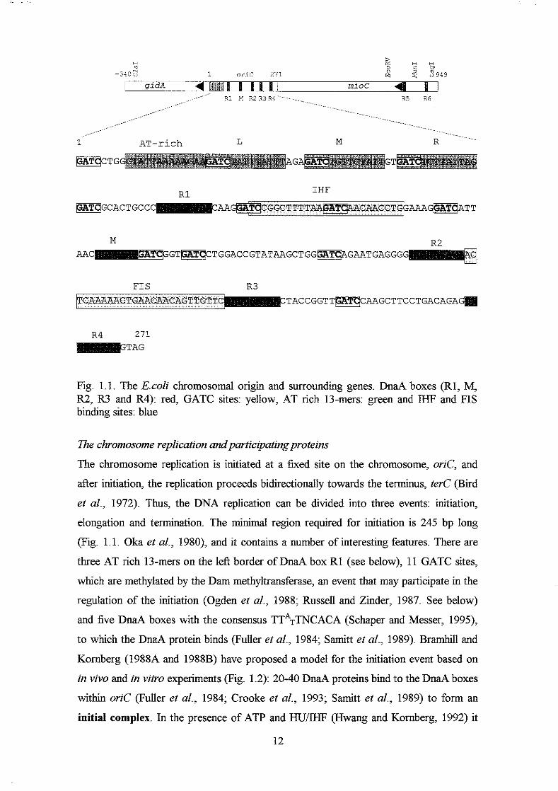
FIS R 3
Fig. 1.1. The E.coli chromosomal origin and surrounding genes. DnaA boxes: red, GATC sites: yellow, AT rich 13-mers: geen and IHF and FIS binding sites: grey
The chromosome replication andpnrtrcipting proteins
The chromosome replication is initiated at a k e d site on the chromosome, oriC, and
after initiation, the replication proceeds bidirectionally towards the terminus, terC (Bird
et al., 1972). Thus, the DNA replication can be divided into three events: initiation,
eiongation and termination. The minimal region required for initiation is 245 bp long
(Fig. 1.1. Oka et al., 1980), and it contains a number of interesting features. There are
three AT rich 13-mers on the lei3 border of DnaA box R1 (see below), 1 1 GATC sites,
which are methylated by the Dam methyltransferase, an event that may participate in the
regulation of the initiation (Ogden et al., 1988; Russell and Zider, 1987. See below)
and five DnaA boxes with the consensus TT*=TNCACA (Schaper and Messer, 1995),
to which the DnaA protein binds (Fuller et al., 1984; Samitt et al., 1989). Bramhill and
Komberg (1988A and 1988B) have proposed a model for the initiation event based on
in vivo and in vitro experiments (Fig. 1.2): 20-40 DnaA proteins bmd to the DnaA boxes
within oriC (Fuller et al., 1984; Crooke et al., 1993; Samitt et aZ., 1989) to form an
initial complex. In the presence of ATP and HUEEE (Hwang and Komberg, 1992) it
12

1 A T - r i c h L M R " " " '
~ C T G AG GT
R1 IHF
~ G C A C T G C C C AA~GGCTTTTAA~CAACCTG+AAA-ATT
M AAC
R2 GTJCTAT~CTGGACCGTATAAGCTGG~JLTC~AGMTGAGGG
F1 S R3 TACCGGTT[GRITTqCMGCTTCCTGACAGA
R4 271 TAG
Fig. 1.1. The E.coZi chromosomal origin and surrounding genes. DnaA boxes (Rl, M, R2, R3 and R4): red, GATC sites: yellow, AT rich 13-mers: green and IHF and FIS binding sites: blue
The chromosome replication andparticipatingproteins
The chromosome replication is initiated at a fixed site on the chromosome, oriC, and
after initiation, the replication proceeds bidirectionally towards the terminus, terC (Bird
et al., 1972). Thus, the DNA replication can be divided into three events: initiation,
elongation and termination. The minimal region required for initiation is 245 bp long
(Fig. 1.1. Oka et aZ., 1980), and it contains a number of interesting features. There are
three AT rich 13-mers on the left border of DnaA box RI (see below), 11 GATC sites,
which are methylated by the Dam methyltransferase, an event that may participate in the
regulation of the initiation (Ogden et al., 1988; Russell and Zinder, 1987. See below)
and five DnaA boxes with the consensus TTA~TNCACA (Schaper and Messer, 1995),
to which the DnaA protein binds (Fuller et al., 1984; Samitt et al., 1989). Bramhill and
Kornberg (1988A and 1988B) have proposed a model for the initiation event based on
in vivo and in vitro experiments (Fig. 1.2): 20-40 DnaA proteins bind to the DnaA boxes
within oriC (Fuller et aZ., 1984; Crooke et al., 1993; Samit t et al., 1989) to form an
initial complex. In the presence of ATP and HU/IHF (Hwang and Kornberg, 1992) it
12
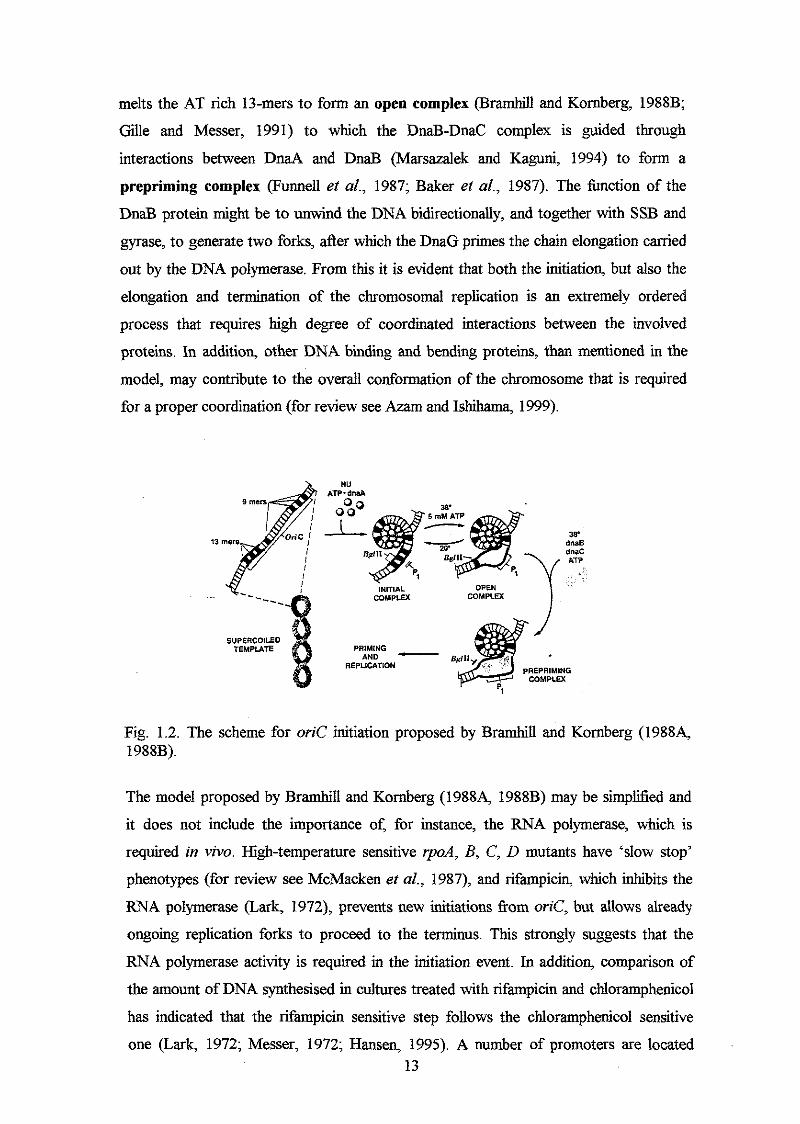
melts the AT rich 13-mers to form an open complex (BramhiU and Kornberg, 1988B;
Gille and Messer, 1991) to which the DnaB-DnaC complex is guided through
interactions between DnaA and DnaB (Marsazalek and Kaguni, 1994) to form a
prepriming complex (Funnel1 et al., 1987; Baker et al., 1987). The function of the
DnaB protein might be to unwind the DNA bidirectionally, and together with SSB and
gyrase, to generate two forks, &er which the DMG primes the chain elongation carried
out by the DNA polymerase. From this it is evident that both the initiation, but also the
elongation and termination of the chromosomal replication is an extremely ordered
process that requires high degree of coordinated interactions between the involved
proteins. In addition, other DNA binding and bending proteins, than mentioned in the
model, may contribute to the overall conformation of the chromosome that is required
for a proper coordination (for review see Azam and Ishihama, 1999).
. COMPLEX
INITIAL COMPLEX OPEN
PRlMlNG - REPLICATION
AND U
Fig. 1.2. The scheme for oriC initiation proposed by Bramhill and Kornberg (19884 1988B).
The model proposed by Bramhill and Kornberg (1988A, 1988B) may be simpliiied and
it does not include the importance of, for instance, the RNA polymerase, which is
required in vivo. High-temperature sensitive rp0.4, B, C, D mutants have ‘slow stop’
phenotypes (for review see McMacken et al., 1987), and rifampicin, which inhibits the
RNA polymerase (Lark, 1972), prevents new initiations &om oriC, but allows already
ongoing replication forks to proceed to the terminus. This strongly suggests that the
RNA polymerase activity is required in the initiation event. In addition, comparison of
the amount of DNA synthesised in cultures treated with rifampicin and chloramphenicol
has indicated that the rifampicin sensitive step follows the chloramphenicol sensitive
one (Lark, 1972; Messer, 1972; Hansen, 1995). A number of promoters are located 13
around and within orzC. Some are negatively regulated (P,;& and Poti.~) whereas others
are positively regulated (Pz;& and Por;.~) by DnaA (Asai et al., 1992; Lsbner-Olesen et
al., 1987; Szalewska-Palashz et al., 1998). The h c t i o n of the RNA polymerase i s still
unknown. However, analysis of transcriptions from surrounding promoters on the
initiation from plasmid born oriC, it has been suggested that transcription of gidA
activates replication by induction of negative superhelicity behind the polymerase, and
that this superhelicity facilitates unwinding of the DNA duplex required in the
formation of the open complex (Ogawa and Okazaki, 1991; Asai et al., 1992). On the
contrary, transcription fiom the mioC promoter had a negative effect on the initiation
process at least when the transcription frequency was too high (Ogawa and Okazaki,
1991). Studies of the effect of transcription of gz& and mzoC on the initiation from the
chromosomal orzC have indicated that the transcriptions have only minor (or no) effect
on the initiation frequency (Lsbner-Olesen and Boye, 1992; Bogan and Helmstetter,
1996), and may only be needed under sub-optimal conditions (Bates et al., 1997). This
is in spite of the observation that the mzoC and gzdA transcription fluctuate during the
cell cycle; the mzoC transcription is shut off while gidA transcription peaks prior to an
initiation (Ogawa and Okazaki, 1994; Bogan and Helmstetter, 1996; Theisen et al.,
1993). The term transcriptional activation may therefore not involve transcriptions of
and mzoC, but instead transcriptions from promoter within orzC. Alternatively, as
suggested by Bates et al. (1997), transcriptional activation could include transcriptions
of genes in general. When rifampicin is added, all transcriptions are shut down, and this
may severely alter the structure of oriC. Thus, the rifampicin effect may be non-specific
and not due to the inhibition of the mzoC and @& promoters. von Freiesleben and
Rasmussen (1992) have found that when the level of supercoiling is altered in gyrB and
top4 mutants, the replication is initiated asynchronously, and thus, the DNA topology
influences the initiation event. Specific qwB mutations suppress the high-temperature
sensitive phenotype of certain &d mutants (Atlung et al., 1984; Bagdasarian et al.,
1977; Szalewska-Palashz et al., 1998), and t h i s may indicate that the RNA polymerase
and the DnaA protein interact. Alternatively, rpoB mutations may result in an altered
transcription frequency from different promoters including the dnd promoter. It has
been shown that a mutation in the rpoC gene, which encode the 0’ subunit of the RNA
polymerase, resulted in a significant increase in the DnaA concentration (Petersen and
Hmsen, 1991). Thus, although the D subunit remained intact, its afsnity to various
promoters seemed affected by amino acid substitutions in other subunits. The
hypothesis that the RNA polymerase and the DnaA protein interact is further supported 14
by the observation that rifampicin resistant initiations can take place in certain d n d
mutants, maybe because the interaction with mutant DnaA proteins prevent rifampicin
from bindmg to the 8-subunit of the polymerase (Hansen, 1995; Tippe-Schindler et al.,
1979; Hanna and Carl, 1975). Thus, the results could indicate that the RNA polymerase
participates in the initiation of chromosome replication in a more complex manner, and
it may both interact with the DnaA protein and transcriptionally activate oriC. In the model proposed by Bramhill and Komberg (1988) (Fig. 1.2), it is obvious that
the DnaA protein is a key protein in the initiation event. In addition, several experiment
indicate that the DnaA concentration determines when the initiation takes place, and
thus, the DnaA protein may be the regulator of the chromosomal replication (Hirota,
1970; Lsbner-Olesen et al., 1989; Hansen and Rasmussen, 1977; Atlung et al., 1987,
for reviews see Skarstad and Boye, 1994; von Meyenburg and Hansen, 1987; Messer
and Weigel, 1996). Most of the experiments consist of analysis of 1) mutant ha4
strains 2) strains in which the DnaA protein or DnaA box concentration has been
changed, and 3) measurements of DnaA concentrations in various strains under different
growth conditions.
Early experiments with the high-temperature sensitive M 4 6 and M 5 mutants
showed that the mutants stop initiation of chromosome replication at non-permissive
temperature. This, in itself, does not indicate that the DnaA protein regulates the
initiation, but can be explained by a lost m t y to the DnaA boxes in oriC. The more
interesting observation was, however, that the strains accumulated initiation capacity at
non-permissive temperature, which resulted in a burst of initiations upon return to
permissive temperature (Hirota, 1970; Hansen and Rasmussen, 1977; Hanna and Carl,
1975; Tippe-Schindler et al., 1979; Hansen and Atlung, 1995q Hansen, 1995). This
strongly suggests that the mutant DnaA proteins accumulated at non-permissive
temperature, and reactivation, maybe by conformational changes of the proteins (see
below), at permissive temperature resulted in overinitiations. Thus, other initiation - and
replication participating proteins did not seem to be limiting. The observed initiation
burst upon retum to permissive temperature did not require protein synthesis. This may
be explained by the finding that the ha4 promoter, which carries a DnaA box (Hansen
et al., 1982), is autoregulated (Atlung et al., 1985; Braun et al., 1985), and this lead to
an increased mutant DnaA concentration at non-permissive temperature (Braun et al.,
1985) probably due to a decreased DnaA box affinity. Alternatively, the concentration
of a negatively acting protein on the initiation decreases when chloramphenicol is added
(Tippe-Schindler et al., 1979). The mutant DnaA proteins may have an altered and 15
inactive conformation at high temperatures. This is suggested in the light of the
observation that oversupply of GroELS proteins suppresses the temperature sensitive
ha446 mutant. GroELS proteins interact with misfolded proteins, and may stabilise the
unfolded state of proteins. Thus, the suppression of the temperature sensitive M 4 6
mutant by an oversupply of GroELS protein could indicate that the DnaA46 protein is
misfolded and therefore 'rescued' - or activated - by GroELS proteins (Katayama and
Nagata, 1991).
Overproduction of wild type DnaA protein also result in overinitiation of chromosome
replication with a resulting decrease in initiation mass (Athmg et al., 1987; Atlung and
Hansen, 1993; Lebner-Olesen et al., 1989; Skarstad et aZ., 1989). The overinitiations
are, however, stalled or aborted and therefore, do not lead to an increased DNA content
of the cells (Churchward et al., 1983; Atlung et al., 1987). This observation strongly
suggests that the DnaA protein is the limiting factor under normal conditions. The
reason why the overinitiations are aborted or stalled, and therefore do not lead to
replicated chromosomes, is still not understood. Atlung et al. (1987) suggest that a
certain factor is missing, for instance a DNA unwinding protein. In the opposite
situation where the DnaA concentration has been lowered, the initiation mass increases
(L~bner-Olesen et al., 1989). The presence of additional DnaA boxes, either from oriC
(Christensen et al., 1999) or from the &tA locus (Kitagawa et al., 1996; 1998) lead to
an increased initiation mass and increased transcription from the hd promoter
(Hansen et al., 1987). Thus, presence of additional DnaA boxes resembles the situation
where the DnaA concentration was lowered, and the results are in full agreement with
the generally accepted idea that DnaA regulates when the initiation takes place. As will
be discussed below, the results also indicate that the DnaA boxes on the chromosome
participate in the regulation of initiation by titrating DnaA proteins away from oriC
most of the cell cycle (Hansen et al., 1991B).
In contrast to other ha4 mutants which have a increased initiation mass, a cold
sensitive ha4 mutant, haAcos, seems to overinitiate chromosome replication in vivo
and in vitro at non-permissive temperature, and thus, have a decreased initiation mass
(Kellenberger-Gujer et al., 1978; Katayama and Kornberg, 1994; Katayama et al., 1997;
Frey et al., 1984). The observed overinitiation is probably not a result of increased
DnaAcos concentration, since it is only 65% of the DnaA concentration in an isogenic
ha4' strain (Katayama and Kornberg, 1994). Thus, the DnaAcos protein may be in an
initiation-competent form for a longer period of time as compared with the DnaA'
16
protein or, alternatively, the DnaAcos protein is inert to negative regulation (Katayama,
1994. See below).
The experimental results discussed above, and the observation that the initiation mass
‘follows’ the DnaA concentration (Hansen et al., 1991; for review see Hemck et al.,
1996) strongly suggests that the DnaA protein is a key molecule in the initiation
process, although there are still some disagreements about the regulatory bnction (see
below).
The DnaA protein also seems to innuence the initiation synchrony. Flow cytometric
analysis of rifampicin treated dnaA+ strains display discrete peaks corresponding to 2”
initiated origins per cell (i.e. 1, 2, 4, 8, 16 etc.) depending on the growth rate (Skarstad
et al., 1986). Thus, all origins are initiated simultaneously within one cell cycle. On the
contrary, drzd mutants initiate the replication very asynchronously especially d n d
mutants that carry a mutation that gives rise to an A184V substitution in the DnaA
protein (i.e. DnaA46, DnaA5, DnaA601 and DnaA604, Hansen et al., 1992) (Skarstad
et al., 1988). Asynchronous initiation is also observed in dam mutants (Boye and
L~bner-Olesen, 1990) and other mutants encoding proteins involved in the structural
arrangement ofthe chromosome (see below).
Regulation of initiation
The initiation of chromosome replication is not a single step event, and therefore its
regulation may be a combination of different mechanisms and may not solely depend on
the DnaA protein. Any proposal of a control mechanism must satisfy several
requirements. First, the signal must be cyclical and have the same periodicity as the cell
cycle, and secondly, the signal must ensure that each origin is initiated once and only
once per cell cycle. The initiation can be considered to happen when i) a certain initiator
has accumulated (the Autorepressor Model by Sompayrac and Maalere, 1973) or ii) the
concentration of an inhibitor is reduced sufficiently due to the increase in cell volume
(the Inhibitor Dilution Model by Prichard et d., 1969) or when iii) the concentration of
the initiator has increased while the concentration of the inhibitor has decreased (the
Initiator Titration Model by Hansen et al., 1991B).
The DnaA protein seems to fit the requirement as an initiator well for several reasons.
As discussed above, there are no doubts that the DnaA protein influence the initiation
mass. When the DnaA concentration (or activity) increases, the initiation mass
decreases, and vice versa (L~bner-Olesen et al., 1989; Atlung et al., 1987; Atlung and
Hansen, 1993; Kellenberger-Gujer et aZ., 1978; Hirota, 1970). In addition, the dnaA
17
gene transcripts, and thus, the DnaA concentration, may very likely fluctuate during the
cell cycle. First, the dnaA gene is autoregulated, and the transcription can be repressed
down to 25% of the normal expression and derepressed up to 400% (Atlung et al., 1985;
Braun et al., 1985). Secondly, Dam methylation of the GATC sites in the promoter
stimulates dnaA transcription (Braun and Wright, 1986), and thus, its hemimethylated
state following replication (Campbell and Kleckner, 1990) may have a repressing effect
in itself In a darn- strain, the DnaA concentration was found to be 3-fold lower than in
the isogenic dam' strain (Landoulsi et al., 1989). Third, the hemimethylated dm4
promoter may be sequested in the membrane like oriC (Ogden et al., 1988), and thus,
the M gene will not be transcribed following its replication. This is consistent with
experimental results that indicate that the mRNhnd concentration is highest at the time
of initiation and lowest following a replication (Theisen et al., 1993; Campbell and
Kleckner, 1990; Ogawa and Okazaki, 1994). Fourth, results have indicated that the
M transcription is growth rate regulated (Chiaramello and Zyskind, 1990).
Whether or not the initiation mass is independent of growth rate has been discussed
for some time, and a constant initiation mass (Donachie, 1968) seemed to be important
for proper understanding of the cell cycle and control of the initiation. However, there
does not really exist any problem with an initiation mass that depends on the growth
rate, as long as the 'initiator', the DnaA protein (or another protein for that matter), also
depend on the growth rate, and thus, settles the initiation mass. In fact, at different
growth rates, the initiation mass will only be constant if the initiator is synthesised as a
constant fraction of total proteiq and there does not seem to be a reason why that should
be the case. What seems more important is that the initiation mass within a population is
constant, and that has not been disproved. The initiation mass of a whole cell population
seems to depend on the strain in question. The initiation mass of E.coli K-l2 was found
to increase with decreasing growth rate (Wold et al., 1994), whereas in E.coli B/r the
opposite was found (Churchward el al., 1981). More recent experiments with E.colz K-
12 and B/r showed that the initiation mass was constant at different growth rates, but
25% higher in E.coli K-l2 (Bipatnath et al., 1998). The DnaA concentration has,
however, not been measured together with the initiation mass in any of the published
results, and thus, it is not known if (or how) the DnaA concentration depends on the
growth rate in the strains discussed above. Instead, The DnaA concentration has been
measured in other strains and whether or not its concentration depends on the growth
rate also seem to depend on the strain. The DnaA concentration in E.coZi K-l2 strains
was found to be independent of the growth rate (Hansen et al., 1991A) or increasing
18
with increasing growth rate (Chiaramello and Zyskind, 1989; Polaczek and Wright,
1990). In E.colz B/r it was found to increase with decreasing growth rate (Hansen et al.,
1991A). By combining the data of the initiation mass at different growth rates in E.colz
K-l2 (Wold et al., 1994) and B/r (Churchward et al., 1981) and the DnaA
concentrations in E.coZi K-l2 and B/r strains at different growth rates (Hansen et al.,
1991A), Henick et al. (1996) observed a good correlation between the initiation mass
and the reciprocal DnaA concentration. The Initiator Titration Model (Hansen et al.,
1991B) includes the Autorepressor- and Inhibitor Dilution Model (Sompayrac and
W e , 1973 and Pritchard et al., 1969, respectively). In the model, the 'initiator' is the
DnaA protein and the 'inhibitor' is the DnaA boxes on the chromosome. Right after
replication, the free DnaA concentration is low, because the DnaA box concentration
exceeds the DnaA concentration. At a certain point, all the DnaA boxes are saturated
with DnaA proteins. This will result in an increase in the free DnaA concentration and
subsequent interaction with the DnaA boxes @U) within oriC. Introduction of
additional DnaA boxes, either from orzC or from the &A locus, clearly titrate DnaA
protein as discussed previously (Christensen et al. 1999; Kitagawa et al., 1996, 1998)
indicating that the DnaA boxes on the chromosome will do the same. The Initiator
Titration Model requires a signiticant numbers of DnaA boxes or, alternatively, areas to
which the DnaA protein has high affinity. The DnaA box consensus TT*TTNCACA
would statistically result in about 140 DnaA boxes present on the chromosome. This
may not be enough when considering that the average number of DnaA proteins per cell
is larger; according to &sen et al. (1991B) in the order of 1000-1500 DnaA proteins
per cell, according to Schaefer and Messer (1991) about 1600 and according to
Sekimizu et al. (1988) between 800 and 2100 DnaA proteins per cell. Schaefer and
Messer (1991) quantified the relative DnaA binding affinity to different DnaA boxes in
vivo, and defined a new DnaA box consensus sequence to which the relative affinity
was greater than 0.2: (TK) (TIC) (A/T/C) T (MC) C (NG) (A/C/T) (MC). Data base
research revealed that these DnaA boxes are distributed uniformly over the genome with
a frequency of 3.3 DnaA boxes per 10 kb, which gives 1600 DnaA boxes per genome
equivalent. Whether all DnaA boxes actually titrate DnaA proteins may seem unlikely.
Instead, there seems to exist high affinity loci on the chromosome that may be
responsible for the DnaA titration. These areas are the &tA locus (IGtagawa et al.,
1996, 1998; Roth and Messer, 1998), the ompT-appY region, the mutH gene, and the
narU gene (Roth and Messer, 1998). These loci are distributed uniformly on the
chromosome. Thus, a likely explanation why de novo protein synthesis is required 19
before another round of initiation can take place is that the DnaA boxes on the
chromosome titrate the free DnaA proteins. Alternatively, or in addition, the
requirement for de novo protein synthesis may be that once the DnaA protein has
participated in an initiation event, it cannot be re-used (Zyshd and Smith, 1992).
Maha@ and Zyskind (1989) have proposed a model partly based on the observation
that the DnaA concentration increases with increasing growth rate (Chiaramello and
Zyskind, 1989) and therefore is growth rate regulated (Chiaramello and Zyskind, 1990).
The authors suggested that the DnaA protein can exist in an active ATP form and an
inactive DnaA-ADP form, and that the concentration of the active DnaA proteins is
independent of the growth rate. This proposal is partly supported by the observation that
when DnaA proteins are purified from a DnaA overproducing strain, about half of the
proteins exist in an aggregated form, which contains phospholipids. This aggregated
form is unable to initiate replication in vzho unless it is activated by either DnaK or
phospholipase AZ in the presence of ATP (Hwang et al., 1990). Recently it has been
shown that the p subunit of DNA polymerase III accelerates hydrolysis of ATP bound
to DnaA in vitro (Katayama et al., 1998) and in vivo (Kurokawa et al., 1999) resulting
in a DnaA-ADP form that is unable to initiate replication. It is suggested that this
negatively regulation, called RIDA (regulatory inactivation of DnaA), of the DnaA
activity is the key control mechanism of the replication cycle (Katayama et al., 1998).
The authors agree with the model proposed by Maha@ and Zyskind (1989) and thus,
de novo protein synthesis is suggested to be required due to an (ir)reversible inactivation
of the DnaA protein. A DnaA protein with an E204Q substitution has been shown to
have decreased intrinsic ATPase activity although the &nity for ATP and ADP was
intact in vitro. This mutant DnaA protein may overinitiate replication in vivo when it is
overproduced in a wild type strain and this has led to the proposal that the intrinsic
ATPase activity negatively regulates chromosomal replication (Ivbushima et al., 1997),
and therefore is part of the FUDA system (Katayama et al., 1998). However,
overproduction of wild type DmA" proteins also result in overinitiation of replication
(L~bner-Olesen et al., 1989; Atlung and Hansen, 1993; Skarstad et al., 1989), which
may suggest that intrinsic ATPase activity is not a negative regulator of the initiation
process, but, if any, then rather of the elongation process. This seems in agreement with
the observation that overproduction of the j3-subunit of polymerase III suppresses the
lethal phenotype of multicopy hd(E204Q) strain @&se et al., 1999). Recent results
have shown that exponentially growing wild type cells have abundant DnaA-ADP and
that only 30% of the DnaA protein exists in an ATP form. In addition, in a synchronised 20

culture, it was found that the DnaA-ATP form oscillates and that the concentration
reaches maximum (80% DnaA-ATP) upon initiation in presence of de novo protein
synthesis (Kurokawa et al., 1999). Many of the authors cited above believe in the RIDA
hypothesis partly because of experimental results that indicate that the DnaAcos protein
is inert towards negative regulation in vitro (Katayama, 1994) and therefore,
overinitiates replication. However, recent in vivo analysis of the initiation frequency in
the cold sensitive dndcos mutant at non-permissive temperature indicated that the
mutant does not overinitiate replication extensively, although the DNA content
increased slightly (Nyborg et al., 2000). Thus, the DnaAcos protein may not be inert
towards negative regulation in vivo. h addition, although the results may indicate that
the DnaA protein is re-activated by ATP binding which is essential for open complex
formation in vitro (Bramhill and Kornberg, 1988B), it has been shown that not all DnaA
proteins have to exist in a DnaA-ATP form in order to participate in the initiation in
vitro (Yung et al., 1990; Crooke et al., 1992). In addition, several other mutant DnaA
proteins have been shown not to bind, or have severely reduced affinity for ATP and
ADP in vitro @Wang and Kaguni, 1988; Hupp and Kaguni, 1993; Carr and Kaguni,
1996). Yet, they are able to initiate chromosome replication in vivo, and no
overinitiations are observed (at least not when the &d allele exists as a single copy on
the chromosome). Thus, the function of ATP and ADP binding to DnaA in vivo may be
important in the (fine)tuning of the replication control, and probably not the ultimate
controlling element. Interestingly, a new six base pair sequence has been identified as a
target for the DnaA protein in its ATP, but not ADP form (Speck ef al., 1999). These
sites are, among others, located in the three 13-mers (Fig. l.l), and they are suggested to
be important in the formation of the open complex. Although this remains to be further
investigated, the results indicates that so-called ATP/ADP switch may contribute to an
even more sophisticated control mechanism.
As mentioned previously, all origins in the cell are initiated once and only once per
cell cycle. All high-temperature sensitive d d mutants initiate the replication
asynchronously, especially those dnaA mutants with a mutation that give rise to a
A184V change in the ATP binding domain of the DnaA protein (Skarstad et al., 1988).
However, the DnaA protein is probably not the only protein responsible for
synchronous initiations, since dam, him m), $S, g y B and topA mutants also initiate
the chromosome replication asynchronously (Boye and Lsbner-Olesen, 1990; von
Freiesleben and Rasmussen, 1992). The way the cell ensures that all origins are initiated
at the same time in the cell cycle bas been proposed by Lsbner-Olesen et al. (1994). In 21
the so-called Initiation Cascade model, which partly relies on the Initiator Titration
model (Hansen et al., 1991), one (or more) origin@) are initiated when the DnaA
concentration reaches a certain level. The subsequent release of DnaA proteins from
that particular origin results in a local increase in the DnaA concentration leading to the
initiation of another origin in the cell and so fourth. This initiation cascade will ensure
that all origins are initiated (almost) simultaneously within the cell cycle. The existence
of a mechanism that blocks secondary initiations was proposed by Russell and Zinder
(1987), who found that transformation of a dam strain with methylated
minichromosomes resulted in an accumulation of hemimethylated minichromosomes.
To explain this, the cell membrane has been drawn into the picture. Actually, it was
proposed long time ago by Jacob et al. (1963), in the Replicon Model, that the
replication origin is an integrated part of the membrane. Upon initiation, which was
thought to be stimulated positively by an initiator, the membrane would grow in
between the replicating origins resulting in chromosome separation. oriC, which
contains a high number of GATC sites (Fig. 1 . l), remains hemirnethylated in vivo from
eight to ten minutes after initiation (Ogden et al., 1988; Campbell and Kleckner, 1990),
and this relatively long period of time required for methylation is suggested to be due to
sequestration of oriC on the cell membrane. Thus, the binding on the membrane may
prevent initiations of already initiated origins shortly after initiation. At the time oriC is re-methylated; the number of 'newborn' DnaA boxes on the chromosome has increased
sufliciently to titrate the free DnaA proteins preventing initiation at any origin.
Accordingly, dam mutants do not initiate replication synchronously since oriC remains
unmethylated and therefore, does not bind on the membrane. The cell seems to initiate
replication from random origins until the increase in DnaA box concentration has pulled
DnaA proteins away from oriC. A protein named SeqA has been suggested to be
involved in sequestration, because a seqA mutant allows methylated minichromosomes
to replicate in dam strains, and furthermore, the observed re-methylation delay of orzC
(Ogden et al., 1998; Campbell and Kleckner, 1990) is reduced in the seqA mutant (Lu et
al., 1994). However, the function of SeqA may be more complex. Based on the
observation that SeqA may act directly on DnaA, it has been speculated that the SeqA
protein regulates the exchange rate of ADP with ATP, and that when SeqA is absent,
this exchange is much faster (von Freiesleben et al., 1994). More recent in vifro
experiments indicate that SeqA affects the DNA conformation and thereby inhibits open
complex formation (Torheim and Skarstad, 1999). Thus, although there seems to be
22
agreement about SeqA affecting the initiation of replication negatively, the function of
SeqA and the sequestration mechanism remains to be resolved.
The asynchronous initiations observed in him @n'),fzs, gyrB and topA mutants might
be a result of changed DNA conformation. The IHF and FIS proteins bind oriC (Fig. 1.1
and below), and this probably affects the DNA topology. Gyrase and topoisomerase
affect the local - and global - superhelicity. Thus, although the DnaA protein may settle
the initiation mass, factors involved in the structural arrangement of oriC can affect the
DnaA-DnaA box affinity, and thereby the precise timing and synchrony of the initiation.
The DnaA boxes R1, R2 and R4 have bound DnaA proteins throughout most of the
cell cycle in vivo (Samitt et al., 1989; Cassler et al., 1995). According to these results,
the binding of DnaA proteins to R3 could be the final event for initiation to happen. The
FIS protein binds to oriC between R2 and R3 (Fig. 1. l), and the DNA bends upon FIS
binding (GiUe et al., 1991). in vivo and in vitro footprinting experiments have indicated
that the FIS site is protected throughout most of the cell cycle. However, at the time of
initiation, the FIS site was no longer protected, whereas DMA box R3 was. It has been
suggested that binding of FIS occluded the binding of DnaA to R3 and thereby prevents
initiation of replication (Cassler et al., 1995; Gille et al., 1991). However, recent in vitro
experiments indicate that the FIS protein does not occlude the DnaA protein. Instead,
the inhibitory effect of FIS seemed to be due to absorption of negative superhelicity
required in the initiation process (Marguhes and Kaguni, 1998). The IHF protein, on the
contrary, seems to bind oriC at the time of initiation coincident with the filling of DnaA
at R3 (Cassler et al., 1995). Based on recent in vitro footprinting experiments, it has
been shown that IHF directs the DnaA protein from stronger to weaker binding sites in
oriC (Grimwade et al., 2000). The authors also find new sequences that do not resemble
the DnaA box consensus to which DnaA binds in vitro in presence of IHF. Based on
mutational analysis of DnaA boxes in oriC, it has been suggested that DnaA box R3 is
not required for replication from oriC, since scrambled or inverted R3 resulted in oric"
behaviour with respect to replication of minichromosomes Ganger et al., 1996).
Instead, the DnaA box R3 is suggested to play a regulatory role in the fine-tuning of the
initiation in the cell cycle. However, the results indicate that R3 is dispensable for
minichromosomes replication, and therefore its regulatory role seems of less
importance. It would be interesting to see if R3 is important for synchronous initiations
fiom oriC.
In spite of the contribution of many proteins in the initiation and replication process,
experimental data, some of which have been discussed above, strongly points at the
DnaA protein as the key protein in the control of the initiation. The maybe strongest
evidence is that the DnaA concentration is proportional to the number of origins in the
cell and inversely proportional to the initiation mass. Binding of ATP to (some of) the
DnaA proteins may be crucial, but whether the hydrolysis of ATP is a controlling
component needs more evidence.
24
Chapter 2
The DnaA-DnaA box interaction
DNA-protein interactions are very important in the genetic life of the cell. They are
required in many events including transcription, repair, recombination and, not least,
chromosome replication. As discussed in the previous chapter, the DnaA protein seems
to be the protein that, upon binding to O K , trigger the initiation event leading to
chromosome replication. Therefore, it is of special interest to understand the interaction
between the DnaA protein and its target, the DnaA boxes. Unfortunately, the three-
dimentional structure of the DnaA-DnaA box complex is not known yet (neither is the
structure of the DnaA protein). It has not been possible to crystalise the protein for X-
ray analysis, and although the NMR technique has improved with respect to protein
sizes, the DnaA protein is still too large for a proper analysis.
The aim of this chapter is to discuss the DNA-protein interaction, in general, and to
describe the different interaction types in structure-known DNA binding proteins. Focus
will be put on those interaction types - or binding motifs - the DnaA protein may be
classiied under. The discussion is mainly based on review articles and books, and is
therefore not a detailed discussion of all DNA binding proteins and their specific
structures.
The DnaA protein and binding motifs
DNA binding proteins can be classified into several groups according to their DNA
binding motif. One of the well characteTised motifs is the heiii-turn-helii motif, which
was the first motif to be characterised (for reviews see Hamson, 1991; Brennan and
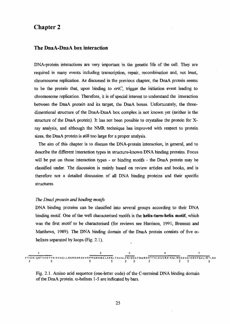
Matthews, 1989). The DNA binding domain of the DnaA protein consists of five a-
helixes separated by loops (Fig. 2.1).
1 2 3 4 5 P
V T l D N I C I K T V A E Y Y K l K V A \ D L L S K R R S R S V A R P R ~ M A M A L A K E L T N H S ~ P E I G D A F G G R D H ~ T V L H A C R U ~ E Q L R E E S ~ D I K E D F S ~ ~ ~ R T L S S I
D - e a x F 1 - ,x 8 - $ 5 - ~~ ~
Fig. 2.1. Amino acid sequence (one-letter code) of the C-terminal DNA binding domain of the DnaA protein a-helixes 1-5 are indicated by bars.
25
A guess would be that the DnaA protein carries a helix-turn-helix motif and that this
motif is responsible for the DNA contact. The Lac repressor, Tpr repressor, catabolite
activator protein (CAP) and the FIS protein (inversion stimulation factor) bear, among
other procaryotic proteins, the helix-turn-helix motif (see Fig. 2.2). The proteins that
cany this motif do not generally share structural similarities, and this contributes to the
unique recognition of different DNA sequences by the various helix-turn-helix proteins.
The length of the turn (loop) in the procaryotic group is four recidues, and a glycine
occurs at the second position (Brennan and Matthews, 1989). A core of hydrophobic
recidues allows the two helixes to pack together and form a compact tertiary structural
domain. The DNA contact takes place through base contact by residues in tbe second
helix that penetrates the major groove of the DNA. In the major groove, but also in the
minor groove, the edges of base pairs expose hydrogen bond donor and acceptor groups.
This hydrogen bond pattern is unique for each base pair in the major goove, but not in
the minor, and this is probably the reason why amino acid residues often recognise and
interact with the bases in the major groove. As mentioned, this interaction may be in
shape of hydrogen bonding but it can also have a hydrophobic character and involve
van der Waals interactions with the non-polar surface of C5 of pyrimidines. Although
the second helix in the helix-turn-helix is referred to as the recognition helix, it should
be stressed that if the helix, or the whole helix-turn-helix motif, is separated from the
rest of a larger DNA binding domain, DNA binding capacity is lost (Pabo and Sauer,
1992). In addition, experiments called 'helix swap' experiments where amino acid
residues in one 'recognition' helix is substituted with residues that match another
'recognition' helix only works in special cases. Thus, specific recognition includes more
than interactions between residues in the second helix of the helix-turn-helix motif and
bases in the major groove (reviewed by Harrison and Aggerwal, 1990). Accordingly, in
the h repressor, for instance, otha amino acids than the ones that are part of the helix-
turn-helix motif contact the DNA backbone.
According to secondaq structure predictions, the DnaA protein may have a short
strech of coiled structure, probably four or five amino acids long, between helix 4 and 5
of the DNA binding domain (Fig. 2.1 and 3.3), but no glycine is seen among these
residues. Instead, the amino acids present in this small coiled area are a relatively
unconserved glutamic acid, a serine and a histidine, and therefore the presence of a well
conserved turn among the different DnaA proteins seems unlikely (Fig.2.1 and Fig. 3.4).
26
434 Rep.
434 Cro
h Rep.
h Cro
CAP
Trp Rep.
Lac Rep.
FIS
Gal Rep
Tet (Tnl 0)
Antp
LexA Rep.
DnaA ex. 1
turn
- -
-
1 2 3 4 5 6 7 8 9 10 11 12 13 1 4 15 1 6 1 7 18 1 9 2 0
-
DnaA
DnaA
ex. 2
ex. 3
G l n Ala
G l n Thr
G l n G l u
G l n Thr
Arg G l n
G l n Arg
Leu T y s
G l n T h r
Ile LYS
T h r A r g
Arg Ile
L y s Ala
Pro
T h r T h r Val Leu
Ile L y s G l u ' A s p
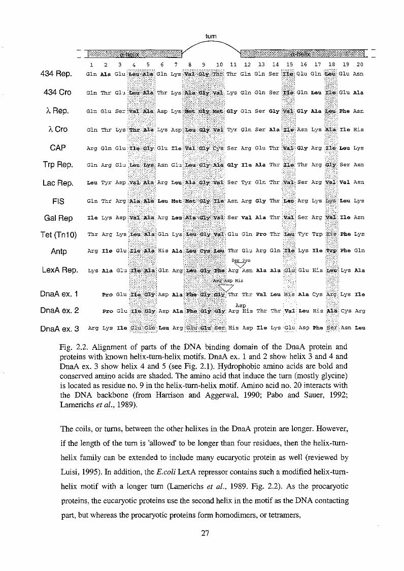
Fig. 2.2. Alignment of parts of the DNA binding domain of the DnaA protein and proteins with known helix-turn-helix motifs. DnaA ex. 1 and 2 show helix 3 and 4 and DnaA ex. 3 show helix 4 and 5 (see Fig. 2.1). Hydrophobic amino acids are bold and conserved amino acids are shaded. The amino acid that induce the turn (mostly glycine) is located as residue no. 9 in the helix-tun-helix motif. Amino acid no. 20 interacts with the DNA backbone (from Harrison and Aggerwal, 1990; Pabo and Sauer, 1992; Lamerichs et al., 1989).
The coils, or turns, between the other helixes in the DnaA protein are longer. However,
if the length of the turn is 'allowed' to be longer than four residues, then the helix-tum-
helix family can be extended to include many eucaryotic protein as well (reviewed by
Luisi, 1995). In addition, the E.coEi LexA repressor contains such a modified helix-tum-
helix motif with a longer turn (Lamerichs et al., 1989. Fig. 2.2). As the procaryotic
proteins, the eucaryotic proteins use the second helix in the motif as the DNA contacting
part, but whereas the procaryotic proteins form homodimers, or tetramers,
27
and bind to palindromic DNA sequences, the eucaryotic proteins normally bind as
monomers to non-palidromic sites. If the DnaA protein binds in a helix-turn-helix
manner, it could seem that the interaction has a more eucaryotic character, since the
DnaA box is non-palindromic, and thus, the turn between the two helixes may be longer
than four residues. The eucaryotic heli-turn-helix proteins also have glycine(s) in the
turn separating the helixes (reviewed by Travers, 1993). Therefore, the alignment
indicates that helix 3, the following turn, and helix 4 may constitute a (modified) helix-
turn-helix motif in the DnaA protein (Fig. 2.2). This also seem consistent with the
observation that this area is highly conserved among DnaA proteins from different
organisms (Fig. 3 .l and 3.2), and the loop (turn) between helix 3 and 4 contains two
highly conserved glycines. In addition, it is possible for the helixes to make a
hydrophobic core due to the presence of a number of hydrophobic amino acids..
Comparison of the tertiary structure of proteins with helix-turn-helii motifs has shown
that residue 1-7 and 12-20 forms the helixes and the residues at positions 4, 8, 10, 15
and 18 are normally hydrophobic and participate in formation of the tertiary structure
through van der Waals interactions. Residue number 5 is usually glycine or alanine. A
small amino acid is required at this position, since larger side chains interfere with the
peptide backbone in the N-terminal part of the following helix. Residue 9 in the turn is
almost always a glycine as mentioned previously, but serine, cystein and glutamic acid
have been found at this position (reviewed by Harrison and Aggerwal, 1990).
According to the secondary structure prediction of the DNA binding domain of the
DnaA protein (Fig. 3.3), the alanine at position 7 (A428) is helical, and so is the
histidine (H434) and threonine (T435) in the end of the loop shown in 'DnaA ex. 1' (Fig.
2.1). If they are included in the helix as indicated in "A ex. 2', the hydrophobic
amino acids follow the pattern with hydrophobic amino acids at positions 15 and 18.
Amino acid no. 20, typically a hydrogen bond doner or acceptor, in the helix-turn.-helix
motif contacts the DNA backbone (Harrison and Aggerwal, 1990; Pabo and Sauer,
1992). This observation supports the proposal that 'DnaA ex. 2' is the correct alignment
of the DnaA protein, because amino acid no. 20 in this case is a well conserved arginine
(R442, Fig. 2.1), which will be able to interact with the DNA backbone through
hydrogen bonding. 'DnaA ex. 3' (Fig. 2.2) shows the amino acid sequence of a-helix 4
and 5 of the DNA binding domain of the DnaA protein with Glu Glu Ser His as the loop
(turn) residues (discussed previously). This area of the DnaA protein does not fit the
helix-turn-helix properties discussed above well. In addition, the residues in the loop
(turn) area and helix 5 (supposed to be the 'recognition' helix) is not well conserved 28
among DnaA proteins from different organisms (see Fig. 3.1 and 3.2). Therefore, this
area probably does not constitute a helix-turn-helix motif. Neither does helix 2 and 3 .
The helixes do not seem to contain hydrophobic amino acids at the right positions, and
residue no. 5, which is supposed to be a small hydrophobic amino acid (reviewed by
Harrison and Aggerwal, 1990), is a large charged amino acid (E416, Fig. 2.1). In
addition, the loop between the helixes does not contain any glycine, and the serine
located there is not placed at the right position. Helix 1 and helix 2 with the basic loop
in between do not fit the requirements for a helix-turn-helix motif either. The loop i s
very long (19 residues) and it does not contain any glycines (Fig. 2.1). However, there
are two well conserved serines, but they are located far away (10 and 14 residues,
respectively) fiom the first helix. The presence of the high number of basic amino acids
in this loop is, however, conspicuous and they may be important in the interaction with
the DNA backbone. Instead of a helix-turn-helix motif, this area of the DnaA protein
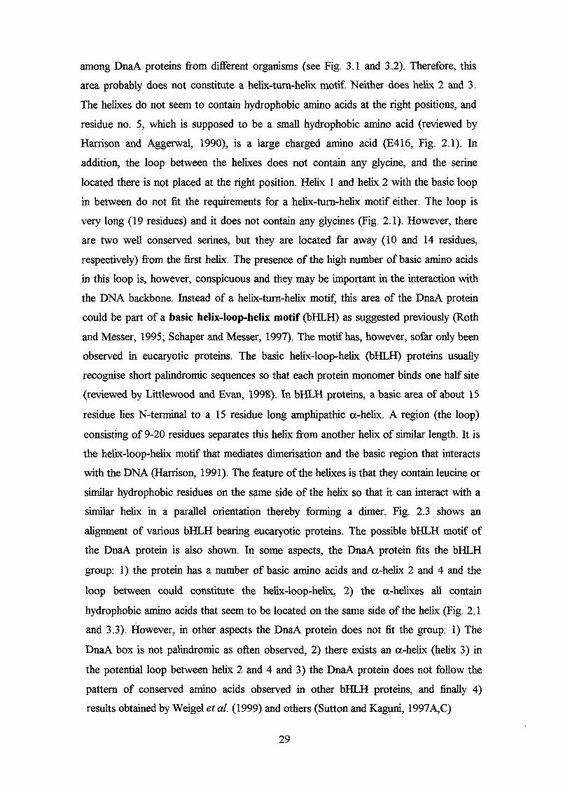
could be part of a basic helix-loop-helix motif @HLH) as suggested previously (Roth
and Messer, 1995; Schaper and Messer, 1997). The motif has, however, sofar only been
observed in eucaryotic proteins. The basic helix-loop-helix (bHLH) proteins usually
recognise short palindromic sequences so that each protein monomer binds one half site
(reviewed by Littlewood and Evan, 1998). In bHLH proteins, a basic area of about 15
residue lies N-terminal to a 15 residue long amphipathic a-helix. A region (the loop)
consisting of 9-20 residues separates this helix from another helix of similar length. It is
the helix-loop-helix motif that mediates dimerisation and the basic region that interacts
with the DNA (Harrison, 1991). The feature of the helixes is that they contain leucine or
similar hydrophobic residues on the same side of the helix so that it can interact with a
S i a r helix in a parallel orientation thereby forming a dimer. Fig. 2.3 shows an
alignment of various bHLH bearing eucaryotic proteins. The possible bHLH motif of
the DnaA protein is also shown. In some aspects, the DnaA protein fits the bHLH
group: 1) the protein has a number of basic amino acids and a-helix 2 and 4 and the
loop between could constitute the helix-loop-helix, 2) the a-helixes all contain
hydrophobic amino acids that seem to be located on the same side of the helix (Fig. 2.1
and 3.3) . However, in other aspects the DnaA protein does not fit the group: 1) The
DnaA box is not palindromic as often observed, 2j there exists an a-helix (helix 3 ) in
the potential loop between helix 2 and 4 and 3 ) the DnaA protein does not follow the
pattern of conserved amino acids observed in other bHLH proteins, and finally 4)
results obtained by Weigel et al. (1999) and others (Sutton and Kaguni, 1997A,C)
29
1 0 W W W W W W W
I x H W W e a a z
W W
H
Y a v 4 x rl
> E.
E. 2
a 0
W
3
a: 9
W
H
W
a
m cl
2 z h rl
W
Y 4 d
4 x a: x W
a a a 4 > m a m n: a u rr)
cl cl 0 a:
%
30
indicate that it is the N-terminal part of the DnaA protein that is responsible for
dimerisation, and that area lies 400 amino acids away from the basic region in the DNA
binding domain. Thus, if the basic area in the DNA binding part of the DnaA protein is
responsible for the DNA contact, the binding mode may have a new, and yet undefined,
character. For instance, helix 1 and 2 and/or helix 4 may interact with each other
through van der Waals interactions (in a leucine zipper fashion), allowing the basic loop
to penetrate and interact with the DNA (Schaper and Messer, 1997). The basic loop
could, however, also have a rather different function. If helix 3 and 4 constitute a helix-
turn-helix mot$ the basic region could particicipate in bending the DNA through
electrostatic attractiodinteractions between the basic residues and the DNA backbone.
In this scenario, the basic loop would be important for DnaA function, but it would not
be responsible for the specific DnaA box recognition.
A second very large class of specific DNA binding proteins is the zinc-bearing DNA
binding domains, which is stabilised structually by tetrahedrally co-ordinated Zn”
ion(s). In all of the so far characterised groups of zinc containing proteins, the zinc ion
always complex with cysteines and sometimes also histidines. Considering that the
DNA biding domain of the DnaA protein only carries one not very conserved cysteine,
and that zinc is not required in in vitro replication systems, it does not seem liiely that
the DnaA protein should possess any zinc dependent conformations. The zipper group
is, like the zinc-containing proteins, so far composed of eucaryotic proteins. The DNA
binding domain of the DnaA protein may contain primary and secondary structures that
could allow it to be classified under the zipper group (see below). The zipper consists of
leucines or other hydrophobic residues that are part of a-helixes, either from two
different proteins or from two helixes within one protein. These hydrophobic amino acids are separated by three or four residues, which corresponds to one helical turn, and
the hydrophobic residues are therby located on one side of the helix. The amino acids
can make hydrophobic interactions (the zipper) with a parallel a-helix that carries the
same pattern of hydrophobic amino acids. The leucine-zipper is often used in the
dimerisation of DNA binding proteins. In cases where protein dimers bind the DNA, the
DNA sequence is palindromic. That is not the case with the DnaA box. The leucine
zipper may, however, exist between two a-helixes within the same DnaA protein, for
instance between helix 4 and 5 or maybe helix 1 is involved in a leucine zipper.
Other DNA binding structures clearly exists, although the helix-turn-helix and the
zinc finger motifs are the best characterised. A number of DNA binding proteins use
31
residues in P-sheets (either parallel or anti-parallel sheets (MetJ)) in the interaction with
bases in the major groove of the DNA helix. The DNA binding domain of the DnaA protein probably does not contain any 0-sheets why such an interaction type seems
rather unlikely. Some DNA binding proteins seem to have unique binding motifs. The
EcoRI restriction enzyme, for instance, recognise its site through amino acids located in
the ends of parallel a-helixes. As with the other binding motifs, it is bases in the major
groove that is in contact with the protein. The DnaA protein could in principle also
possess such a bmding motif The EcoRV restriction enzyme and Dnase I use loops as
the recognition element that penetrates into the major groove of the recognition site.
Again, this could be a possible binding motif of the DnaA protein. The RNase H bound
to a DNA-RNA hybrid shows that, unliie any of the other proteins or motifs discussed
so far, contact is mediated through a loop that penetrates the minor groove. Yet other
proteins use both a-helixes and 0-sheets to penetrate and contact bases in the minor and
major groove (JSlenow fragment of DNA polymerase I, for instance).
From this discussion it appears that many different binding motifs exist, and that the
DnaA protein may possess either one of them, or a combination of already characterised
motifs. Based on secondary structure analysis, it seem unavoidable that contact is
mediated through either a-helixes andior loops, and that the DnaA protein possess a
potential helix-turn-helix motif including helix 3 and 4. In the experimental part of this
thesis, the effect of different amino acid substitutions in the DNA binding domain will
be discussed, and this may lead to a better understanding of the DnaA-DnaA box
interaction.
Amino acids in contact with DNA
All amino acid residues seem to be able to interact with the DNA either through
hydrogen bonding or van der Waals interactions. Thus, it is not possible to conclude
that a certain area of a DNA binding protein will or will not interact with the DNA on
basis of the primary structure of the protein. Amino acid residues can interact with the
suger phosphate backbone of the DNA or with the base pairs. The interaction with the
DNA backbone is often through hydrogen bonds to nonestedied phosphate oxygens
from peptide -NH groups, neutral -NI32 groups of glutamine and asparagine and from - OH groups of serine and threonine. Hydrogen bonds from lysine and arginine to the
phosphates appear with only modest frequency (reviewed by Hanison and Aggerwal,
1990). This may seem difficult to understand, because such an interaction could be
32

expected to be stronger than ordinary hydrogen bonds due to the electrostatic character
of the interaction. Base pairs in major groove, and minor groove, can be contacted by
various sidechains that donate or accept hydrogen bonds. In addition, non-polar
hydrophobic interactions are often present and they participate in the specific distinction
between cytidine and thymine.
Except for tryptophan, the DNA binding domain of the DnaA protein consists of all
twenty amino acids, and the area that may constitute the helix-turn-helix motif(Fig. 2.2)
includes both hydrophobic residues and hydrogen bond donor and acceptor amino acids.
It is not possible on the basis of analysis of the interaction between other DNA binding
proteins and their DNA targets to determine which amino acids in the DnaA protein that
may be responsible for the interaction with the DnaA boxes. As mentioned previously,
part of the experimental work in this thesis includes analysis of various amino acid
substitutions in the DNA bmding domain of the DnaA protein, and this analysis may
shed light on which amino acids that are liiely to be involved in the DnaA box
recognition and binding
l;he conformation of the DnaA boxes
When analysing protein-DNA interactions, it may not be correct to consider the protein
as the active part and the DNA as the passive part in the interaction and recognition.
Actually, it may be just as correct to say that a certain DNA sequence recognises a
certain protein with the correct structure. The DNA molecule is not a uniform unit, but
the structure of it changes throughout the chromosome. The conformation depends on
the actual DNA sequence and the base pair interactions leading to several types of
comformational flexibilities (Fig. 2.4). Accordingly, the DNA sequence may have a B-
form shape or a A-type shape, or the DNA sequence may bend or curve (reviewed by
Luisi, 1995; Travers, 1993). Following parameters are important in the final
conformation of DNA fragment (Fig. 2.3): 1) The propeller twist: Favorable stacking of
bases affect the propeller twist moving the base pairs away from a planar configuration.
A consequence of this is that the GC base pair has lower propeller twist than the AT
basepair due to the higher number of hydrogen bonds. 2) The Helical twist: The twist
angle vary corresponding to a winding or unwinding of the DNA sequence. Changes in
the axial flexibility leads to changes in the helical axis from B-type DNA to A-type
DNA or vice versa. B-DNA has a twist of 36' per base pair equivalent to 10 base pairs
per turn, whereas A-DNA contains 11 base pairs per turn. As the twist decreases, the
minor groove becomes wider and shallower, while the major groove becomes narrower 33
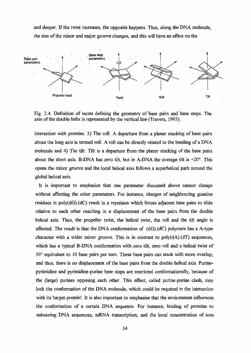
and deeper. If the twist increases, the opposite happens. Thus, along the DNA molecule,
the size of the minor and major groove changes, and this will have an effect on the
Bawpair parameters * .;;"$
Propeller twist Twist Roll Tin
Fig. 2.4. Definition of terms defining the geometry of base pairs and base steps. The axis of the double helix is represented by the vertical line (Travers, 1993).
interaction with proteins. 3) The roll: A departure from a planar stacking of base pairs
about the long axis is termed roll. A roll can be directly related to the bending of a DNA
molecule and 4) The tilt: Tilt is a departure from the planar stacking of the base pairs
about the short axis. B-DNA has zero tilt, but in A-DNA the average tilt is +20". This
opens the minor groove and the local helical axis follows a superhelical path around the
global helical axis.
It is important to emphasize that one parameter discussed above cannot change
without affecting the other parameters. For instance, charges of neighbouring guanine
residues in poly(dG).(dC) result in a repulsion which forces adjacent base pairs to slide
relative to each other resulting in a displacement of the base pairs from the double
helical axis. Thus, the propeller twist, the helical twist, the roll and the tilt angle is
affected. The result is that the DNA conformation of (dG).(dC) polymers has a A-type
character with a wider minor groove. This is in contrast to poly(dA).(dT) sequences,
which has a typical B-DNA conformation with zero tilt, zero roll and a helical twist of
36" equivalent to 10 base pairs per turn. These base pairs can stack with more overlap,
and thus, there is no displacement of the base pairs from the double helical axis. Purine-
pyrimidine and pyrimidine-purine base steps are restricted conformationally, because of
the (large) purines opposing each other. This effect, called purine-purine clash, may
lock the conformation of the DNA molecule, which could be required in the interaction
with its 'target-protein'. It is also important to emphasise that the environment influences
the conformation of a certain DNA sequence. For instance, binding of proteins to
nabouring DNA sequences, mRNA transcription, and the local concentration of ions
34
will undoubtly affect the DNA conformation. Thus, due to all these parameters, of
which some have been mentioned in this chapter, it seems difficult to determine the
exact structure of a certain DNA sequence, including the DnaA box sequence, on basis
of the sequence.
The DnaA box sequence d8er among the various DnaA boxes, but in general they
contain a high number of A and T (Table 2.1), indicating that the overall conformation
of the DnaA box has a B-DNA character.
Table 2.1. The sequence of a number of DnaA boxes found in the oriC region and in the dnaA promoter DnaA box Sequence Affinity l’
R1 TTGTTATCCACAGGG High
matsui CGATCATTCACAGTT Low
R2
R3
R4
R5
GGGTTATACAUGCT Medium
TAGTTATCCAAAGAA Low
GAGTTATCCACAGTA High
TACTTTTCCACAGGT Medium
d n a A promoter GATTTATCCACAGGA High 1) The &nitiy of the ~ n a ~ + protein to the boxes has been determind ty s & e d authors using in vivo and in vitro measurements. The affinities are therefore only expressed qualttatively as high, medium
of > 200 nM @&sing, 1999; Schaefer andMesser, 1991; Schaper andMesser, 1995). and low, where high is in the order of &= 1-2 nh.s medium in the order of 30-50 nM and low in the order
The most stringent DnaA box consensus sequence is TTA/~TNCACA (Schaper and
Messer, 1995). It seems that the DnaA protein has the highest affinity to the sequence
TTATCCACA, and deviation from this sequence results in decreased ani@ of DnaA
to the box. If CS is substituted with A (as in R3) the affinity is severely reduced, which
indicates that the maybe relatively locked structure of CACA (due to purine-purine
clashes, see above) is important in the interaction with DnaA. Substitution of T2 with C
(as the M box) also seem to have severe effects on the atlinity, whereas substitutions of
A3 ( R 5 ) or CS (R2 box and M box) seem to be less deleterious. Schaefer and Messer
(1991) have quantified the relative DnaA binding a&ities to different DnaA boxes by
meauring the ability of the DnaA-DnaA box complexes to block in vivo trancription. On
basis of their analysis, they were able to suggest a more relaxed DnaA box consensus
sequence to which the relative affinity is greater than 0.2: (TK) (TK) (A/T/C) T (MC)
C (NG) (NUT) (NC). Further analysis has been canied out by Speck et al. (1997),
who determined which thymines in the DnaA box that are important for the interaction 35
with DnaA. The authors found that Tz, T+ T7 and T9 are required for DnaA binding,
which indicates that hydrophobic interactions with the methyl groups of the DnaA box
are imporatant. Interestingly, these thymines are located opposite each other in pairs
allowing interaction from two sides of the DnaA protein. However, it is probably not
only the major grooves to which the DnaA protein mediates contacts, but also the minor
groove (Schaper and Messer, 1995). Thus, it seems that except for the nucleotide in the
middle of the DnaA box, the rest are important for specific binding. The binding of
DnaA to the DnaA box induces a bend in vitro of about 40° (Schaper and Messer,
1995), and this may be important for a correct structure of the DNA that allows the
formation of a higher order nucleoprotein complex.
According to the discussions above the DnaA box has been analysed using different
experimental approches. It seems that all nine nucleotides in the DnaA box (and maybe
even additional 5' and/or 3' nucleotides. Marguhes and Kaguni, 1996; Weigel et al.,
1997) are important for proper DnaA binding, the only exception being the middle
nucleotide. The interaction between DnaA and its target may include contacts in major
and minor groove through hydrogen bonding and hydrophobic interactions.
36
Part I1
Experimental work

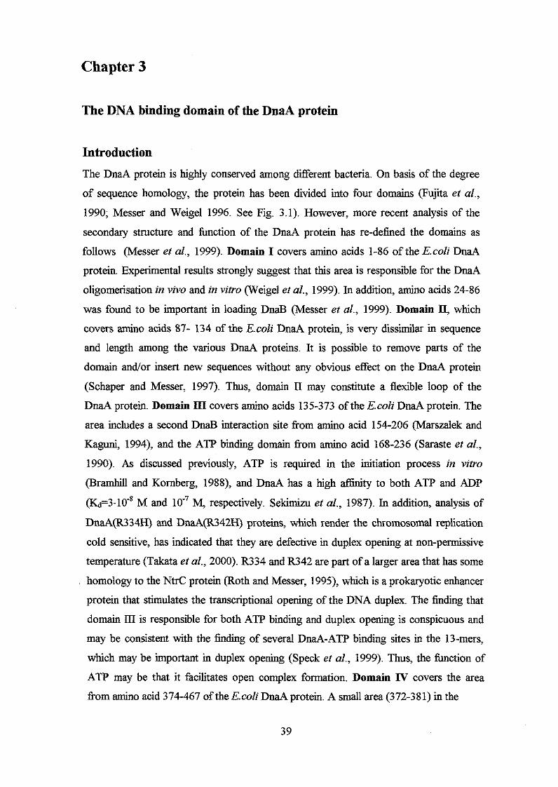
Chapter 3
The DNA binding domain of the DnaA protein
Introduction The DnaA protein is highly conserved among different bacteria. On basis of the degree
of sequence homology, the protein has been divided into four domains (Fujita et al.,
1990; Messer and Weigel 1996. See Fig. 3.1). However, more recent analysis of the
secondary structure and function of the DnaA protein has re-defined the domains as
follows (Messer et al., 1999). Domain I covers amino acids 1-86 of the E.coli DnaA
protein. Experimental results strongly suggest that this area is responsible for the DnaA
oligomerisation in vivo and in vitro (Weigel et al., 1999). In addition, amino acids 24-86
was found to be important in loading DnaB (Messer et al., 1999). Domain II, which
covers amino acids 87- 134 of the E.coZz DnaA protein, is very dissimilar in sequence
and length among the various DnaA proteins. It is possible to remove parts of the
domain andor insert new sequences without any obvious effect on the DnaA protein
(Schaper and Messer, 1997). Thus, domain I1 may constitute a flexible loop of the
DnaA protein. Domain III covers amino acids 135-373 of the E.coli DnaA protein. The
area includes a second DnaB interaction site from amino acid 154-206 (Marszalek and
Kaguni, 1994), and the ATP binding domain &om amino acid 168-236 (Saraste et al.,
1990). As discussed previously, ATP is required in the initiation process in vitro
(Bramhill and Kornberg, 1988), and DnaA has a high aEmity to both ATP and ADP
(&=3.10-* M and M, respectively. Sekimizu et al., 1987). In addition, analysis of
DnaA(R334H) and DnaA(R342H) proteins, which render the chromosomal replication
cold sensitive, has indicated that they are defective in duplex opening at non-permissive
temperature (Takata et al., 2000). R334 and R342 are part of a larger area that has some
homology to the NM: protein (Roth and Messer, 1995), which is a prokaryotic enhancer
protein that stimulates the transcriptional opening of the DNA duplex. The finding that
domain LI1 is responsible for both ATP binding and duplex opening is conspicuous and
may be consistent with the finding of several DnaA-ATP binding sites in the 13-mers,
which may be important in duplex opening (Speck et al., 1999). Thus, the fimction of
ATP may be that it facilitates open complex formation. Domain N covers the area
f?om amino acid 374-467 of the E.coli DnaA protein. A small area (372-381) in the
39
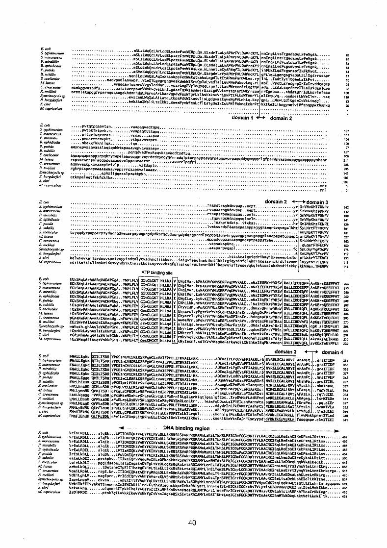
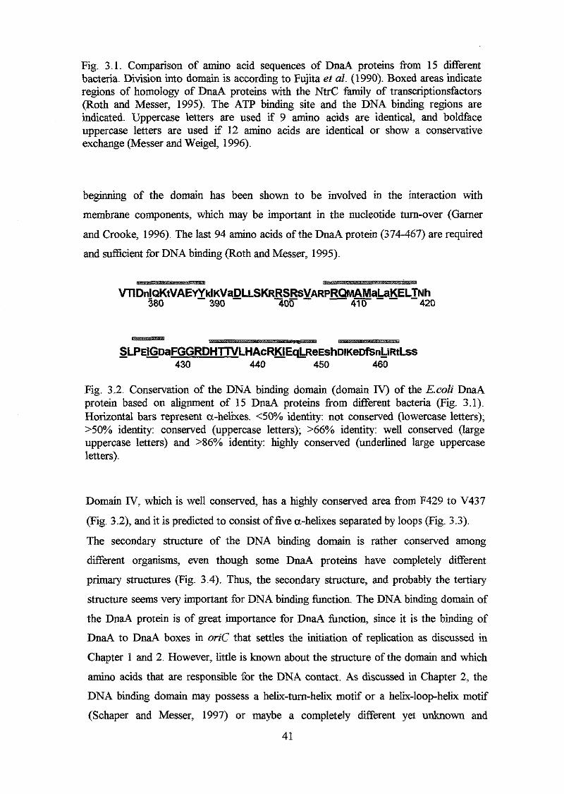
Fig. 3.1. Comparison of amino acid sequences of DnaA proteins from 15 different bacteria. Division into domain is according to Fujita et al. (1990). Boxed areas indicate regions of homology of DnaA proteins with the NtrC family of transcriptionsfactors (Roth and Messer, 1995). The ATP binding site and the DNA binding regions are indicated. Uppercase letters are used if 9 amino acids are identical, and boldface uppercase letters are used if 12 amino acids are identical or show a conservative exchange (Messer and Weigel, 1996).
beginning of the domain has been shown to be involved in the interaction with
membrane components, which may be important in the nucleotide turn-over (Gamer
and Crooke, 1996). The last 94 amino acids of the DnaA protein (374-467) are required
and sufficient for DNA binding (Roth and Messer, 1995).
WlDnlQKtVAEflklKVa~LSKRRSRS~ARPRQMAMaLa~ELTNh 380 390 7Ob 41 0 - 420
SLPE~LmFGGRDHrrVLHAcR~Eql$eEshDIKeDfsn~RtLss -
- 430 440 450 460
Fig. 3.2. Conservation of the DNA binding domain (domain IV) of the E.colz DnaA protein based on alignment of 15 DnaA proteins from different bacteria (Fig. 3.1). Horizontal bars represent a-helixes. <50% identity: not conserved (lowercase letters); >50% identity: conserved (uppercase letters); >66% identity: well conserved (large uppercase letters) and >86% identity: highly conserved (underlined large uppercase letters).
Domain IV, which is well conserved, has a highly conserved area from F429 to V437
(Fig. 3.2), and it is predicted to consist of five a-helixes separated by loops (Fig. 3.3).
The secondary structure of the DNA binding domain is rather conserved among
different organisms, even though some DnaA proteins have completely different
primary structures (Fig. 3.4). Thus, the secondary structure, and probably the tertiary
structure seems very important for DNA binding function. The DNA binding domain of
the DnaA protein is of great importance for DnaA function, since it is the binding of
DnaA to DnaA boxes in orzC that settles the initiation of replication as discussed in
Chapter 1 and 2. However, little is known about the structure of the domain and which
amino acids that are responsible for the DNA contact. As discussed in Chapter 2, the
DNA binding domain may possess a helix-tum-helix motif or a helix-loop-helix motif
(Schaper and Messer, 1997) or maybe a completely different yet unknown and
41
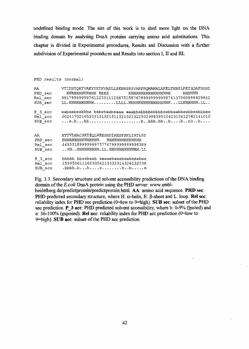
undefined binding mode. The aim of this work is to shed more light on the DNA
binding domain by analysing DnaA proteins carrying amino acid substitutions. This
chapter is divided in Experimental procedures, Results and Discussion with a firther
subdivision of Experimental procedures and Results into section I, 11 and III.
PHD results (normal)
AA PHD sec
VTIDNIQKTVAEYYKIKVADLLSKRRSRSVARPRQ~ELTNHSLPEIGDAFGGRD
RelIsec 99179999999?612233111266?515~7678999999933374137563~8~62~842 HHHHHHHHHHHH EEEE HHHHHHHHHHHHHHHHH HHHHHH
SUB-sec LL.HHHHHHHHHH... ...... LLLL.HHHHHHHHHHHHHHHHH...LLHHHHHH.LL.. P-3acc eebeebeebbbe bebebeebeeee eeebbebbbbbbbbbeebbeebbeebbeebbbee Rel-acc SUB-acc
002417021652031313213113210213225023383~51241313412?62141010 ... e.b. .. bb ..................... b..bbb.bb .. b....b .. bb..b ....
AA PHD-sec HHHHHHHHHHHHHHH HHHHHHHHHHHHHH
HTTVLHACRKIEQLREESHDIKEDFSNLIRTLSS
Rel-sec 4455318393333373774733333333336'383 SUB-sec ..HH..HHHHHHHHH.LL.HHHHHHHHHHHH.LL
P 3-acc bbbbb bbeebeeb eeeeebeeebeebeeebee Relacc 1558506111623004213332314326132236 SUBacc .bbbb.b ... b....e... ..... b..b ..... e
Fig. 3.3. Secondary structure and solvent accessibility predictions ofthe DNAbinding domain of the E.coZi DnaA protein using the PHD server: www.ernb1- heidelberg.de/predictprotein/predictprotein.html. A A : amino acid sequence. PHD sec: PHD predicted secondary structure, where H: a-helix, E p-sheet and L: loop. Re1 sec: reliability index for PHD sec prediction (+low to 9=high). SUB sec: subset of the PHD sec prediction. P-3 ace: PHD predicted solvent accessibility, where b 0-9% (buried) and e: 36-100%0 (sxposed). Ret ace: reliability index for PHD acc prediction (O=low to 9=high). SUB acc: subset of the PHD acc prediction.
42

Fig. 3.4. Secondary structure predictions of the (proposed) DNA binding domain of DnaA proteins from various organisms. The PHD server with following address was used in the prediction: www.embl-heidelberg.de/predictprotein/predictprotein.html. Horizontal bars represents a-helixes and arrows p-sheets.
43
Experimental procedures
General procedures
PI transduction andpreparation
Preparation of P1 lysate was carried out essentially as described by Miller (1992):
Overnight cultures grown in LB medium were diluted 20 times in LB medium
supplemented with 10 mM CaClz and grown for 1-2 hours. 100 pl cell suspension was
incubated 20 min. at 37°C with different amounts of P1 lysate (typically 1, 10 and 100
p1 lo5 P1 phageshl). 5 ml top-agar supplemented with 5 mM CaC4 was added, and the
suspension was plated on LB plates. The following day a few drops of chloroform was
added to the supernatant (P1 phages) which was kept at 5°C. P1 transductions were also
carried our essentially as described by Miller (1992). 100 pl of an Overnight culture
grown in LB medium supplemented with 5 mM CaClz were incubated 20 min. at 37OC
with different amounts of P1 lysate (typically 1, 10 and 100 p1 lo5 P1 phages/ml). 100
p1 Na-citrate was added to a final amount of 1% and the suspension was plated on
selective plates and incubated overnight. When kanamycin resistance was selected, the
bacteria + phage suspensions were incubated with 1 ml LB supplemented with 1% NaCi
for 3 hours, centrifbged and plated on selective plates.
Competent cells and transformation
Cells were made competent and transformed with plasmids essentially as described by
Sambrook et al. (1989): An over night culture of the strain to be transformed was
inoculated 1:25 in LB and grown until OD450 1.0. 10 ml was harvested and 4 ml 50
mM CaCh was added. After 20 min., the cells were centrifuged again and dissolved in 2
ml 50 mM CaClz. After 1 hour, the cells were competent for about a week. 200 p1
competent cells was incubated with an appropriate amount of plasmid (depending on the
type of transformation, ranging &om 10 ng to 0.5 pg) and incubated on ice for 30 min.
The transformation mixture was heat shocked at 42OC for 5 min. 1 ml LB was added
and the transformation mixture was incubated at room temperature or 3OoC for 30-120
min. depending on the selection.
44
Growth medium
The growth medium was either LB medium (10 g NaCI, 10 g Difco Bacto tryptone, 5 g
Difco yeast extract and 20 g Difco agar per litre) or AB medium (Clark and Maalse,
1967) supplemented with 0.2% glucose, 2 &ml thiamine and 1% casamino acids
(Difco). When required, the growth medium was supplemented with 50 &ml
kanamycin (Calbiochem), 10 pg/d tetracycline (Sigma), 100 &ml ampicillin (Sigma),
different concentration of IPTG (Sigma) and/or X-gal (5-bromo-4-chloro-3-indolyl-B-
D-galactopyranoside. Apollo).
Mini-scale plasmid preparation
Plasmid was prepared as described by Sambrook et al. (1989) combined with a further
improvement by using a PCR purification kit from Boehringer Mannheim: 2 ml over
night culture was harvested and dissolved in 100 p1 50 mM glucose, 10 mM EDTA, 25
mM Tris-HC1 pH 8.0. After app. 5 min. 200 p1 0.2 M NaOy 1% SDS was added. After
5 min. incubation, the samples were incubated on ice with 150 p1 3 M NaAc pH 4.8 for
15 min. The samples were centfiged, and the supernatant was transferred to 0.8 ml
96% EtOH. The DNA was precipitated and dissolved in 100 p1 0.5 mM EDTA 2 mM
Tris-HC1 pH 8. 0.2 p1 25 mg/d RNase was added and the samples were incubated at
room temperature for 15-30 min followed by a column purification using the PCR
purification kit from Boeringer Mannheim
Immunologicalprocedures
Samples were prepared for immunoblot analysis essentially as described previously
(Hansen et al., 1991A). Cells were harvested and dissolved in standard loading buffer
for sodium dodecyl sulfate polyacrylamide gel electrophoresis (SDS-PAGE) and boiled
for 5 min. 12% resolving, 4% stacking Tris-HC1 Ready Gels &om BioRad was used in
the protein separations. M e r electrophoresis, proteins were transferred to Immun-Blot
PVDF Membranes (BioRad) by using a semi-dry blotting procedure (Kyhse-Andersen,
1984). Protein transfer was for 2 hours. The membrane was blocked over night in Tl3S
buffer (0.05 M Tris (pH 10.4), 0.15 M NaCI) containing 2% Tween. The following day,
the membrane was washed 3 times with TBS buffer containing 0.05% Tween and
incubated with primary DnaA antibody (Landoulsi et al., 1989) in TBS, 0.05% Tween
and Blocking reagent (Boeringer Mannheim) or streptavidin conjugated alkaline
phosphatase (BioRad) in TBS, 0.05% Tween for 1-2 hours. M e r three times washes in
45
TBS, 0.05% Tween, the filters were incubated with alkaline phosphatase conjugated pig
immunoglobulin to rabbit immunoglobulins (DAKO) for 2 hours. After three additional
washes, the filters were stained with NBT (4-nitro blue tetrazolium chloride) and XP (5-
bromo-4-chloro-3-indolyl-phosphate) (Boehringer Mannheim) as described previously
(Blake et al., 1984). For all immunoblots DnaA protein was used as standard
concentration marker by using experimental results indicating that 1 ml culture from a
&d+ strain at OD450=1 corresponds to 25 ng DnaA/ml (Hansen et al., 1991A). The
western immunoblots were quantified by using a BioRad model GS-700 imaging
Densitometer.
Enzyme reactions
All restriction enzyme reactions were carried out in KAc buffer (0.1 M KAc, 25 mM
Tris-acetat (pH 7 3 , 10 mM MgAc, 0.1 mg/ml bovine serum albumin, 1 mM p- mercaptoethanol) and incubated at the temperatures recommended by the supplier (New
England Biolab., Amersham and Promega). Ligase (MBI Fermentas) was used in
ligations reactions that were carried out in ligase buffer (SO mM Tris-HC1 (pH 7.5), 10
mh4 MgClz, 0.025% Np40), 20 mM dithiothreitol (DTT), 1 mM ATP. The ligation mix was incubated at room temperature for 2 hours or more.
Standard PCR reaction
1 ~.rl linearized DNA (app. 0.05 ng) or cells corresponding to one colony was incubated
with 2.5 mM MgC12, 0.8 pM of each primer, 0.2 mM of each dNTP, thermo prime
DNA polymerase buffer and 1 unit Thenno prime DNA polymerase O(EB0) to a total
volume of 25 pl. The cycle program was 94°C for 1 min., 50-52OC for 1 min., and 72'C
for 2 min.
46
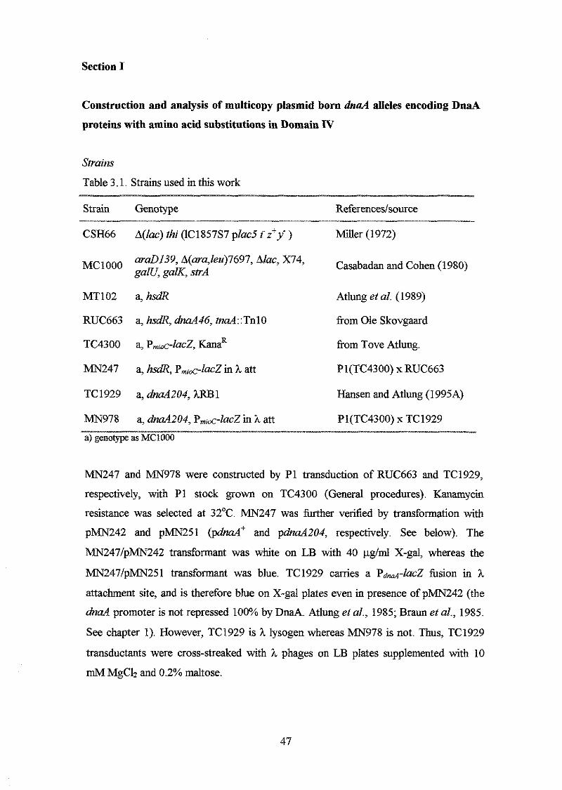
Section I
Construction and analysis of multicopy plasmid born dnd alleles encoding DnaA
proteins with amino acid substitutions in Domain IV
Spains
Table 3.1. Strains used in this work
Strain Genotype Refermces/source
CSH66 A(Zac) thi (lC1857S7 plac5 i-z'y-) Miller (1972)
Oo0 gaZU, galK, SPA araD139, A(ara,Zeu)7697, Alae, X74, Casabadan and Cohen (1980)
MT102 a, hsdR Atlung et al. (1989)
RUC663 a, h&, haA46, tnd::TnlO fiom Ole Skovgaard
TC4300 a, PmiK-lacZ, KanaR from Tove Atlung.
MN247 a, h&, P,,~-ZacZ in h att Pl(TC4300) x RUC663
TC 1929 a, dmA204, ?LIU3 1 Hansen and Atlung (1 995A)
MN978 a, dnaA204, Pmi&-ZacZ in ?L att Pl(TC4300) x TC1929 a) genotype as MC1000
____
MN247 and "978 were constructed by P1 transduction of RUC663 and TC1929,
respectively, with P1 stock grown on TC4300 (General procedures). Kanamycin
resistance was selected at 32OC. MN247 was further verified by transformation with
pMN242 and pMN251 (pdnad and pdnaA204, respectively. See below). The
MN247/pMN242 transformant was white on LB with 40 &ml X-gal, whereas the
MN247/pMN251 transformant was blue. TC1929 carries a Pdn&acZ fusion in h
attachment site, and is therefore blue on X-gal plates even in presence of pMN242 (the
dnd promoter is not repressed 100% by DnaA. Atlung et al., 1985; Braun et al., 1985.
See chapter 1). However, TC1929 is h lysogen whereas MN978 is not. Thus, TC1929
transductants were cross-streaked with h phages on LB plates supplemented with 10
mM MgCl2 and 0.2% maltose.
47
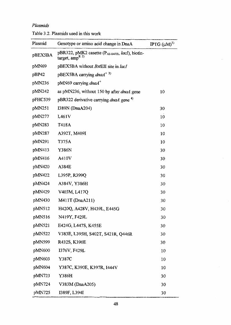
Plasmids
Table 3.2. Plasmids used in this work
Plasmid Genotype or amino acid change in DnaA
pBEX5BA
pMN69
pBP42
pMN23 6
pMN242
pFHC539
pMN25 1
pMN277
pMN283
pMN287
pMN291
pMN4 1 3
pMN4 1 6
pMN420
pMN422
pMN424
pMN429
pMN430
pMN5 12
pMN516
pMN52 1
pMN522
p m 5 9 9
pMN600
pMN603
pMN604
pMN723
p m 7 2 4
pMN725
~
pBR322, pMK2 casette (P~1-04103, lucI), biotin- target, ampR ’)
pBEX5BA without BstEII site in lacl
pBEX5BA carrying &d’ ’) pMN69 carrying dnaA+
as pMN236, without 150 bp &er dnd gene
pBR322 derivative carrying gene 4,
I389N (DnaA204)
L46 1 V
T41 SA
A392T, M4091
T375A
Y386N
A410V
A3 84E
L395P, R399Q
A384V, Y386H
V403M, L4 17Q
M411T@naA211)
H420Q, A428V, H439L, E445G
N419Y, F429L
E424G, L447S, K455E
V383E, L395y S402T, S421R, Q446R
R432S, K390E
I376V, F429L
Y387C
Y387C, K390E, K397q I444V
Y386H
V383M @naA205)
I389F, L3941
~~~~~ ~ ~~~
IPTG (@Q’’
10
30
10
10
10
10
30
30
30
30
30
30
30
30
30
30
30
30
10
10
10
30
30
30
48
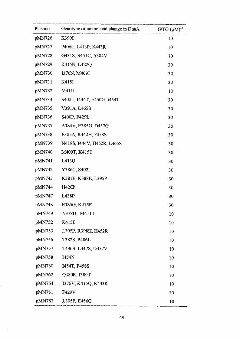
Plasmid Genotype or amino acid change in DnaA IPTG (m1) pMN726 K3901 10
pMN727 P406L, L413P, K443R 10
pMN728 G431S, S451C, A384V 10
p m 7 2 9 K415N, L4224 30
pMN730 I376N, M4091 30
pMN73 1 K4 151 30
p m 7 3 2 M41 11 10
pMN734 S402L, I444T, E450G, I454T 30
pMN735 V391A, L465S 30
pMN736 S400P, F429L 30
pMN737 A384V, E385G, D457G 30
pMN738 E385A, R442H, F458S 30
pMN739 N419S, I444V, H452R, L465S 30
pMN740 M409T, K415T 30
pMN741 L413Q 30
pMN742 Y386C, S402L 30
pMN743 K381E, K388E, L395P 30
pMN744 H420P 30
pMN747 L438P 30
pMN748 E385G, K415E 30
pMN749 N378D, M411T 10
pMN752 K415E 10
pMN753 L395P, R398H, H452R 10
pMN756 T382S, P406L 10
pMN757 T436S, L447S, D457V 10
pMN758 I454N 10
pMN760 I454T, F458S 10
pMN762 Q380R, I389T 10
pMN764 I376V, K415Q, K443R 10
pMN781 F429V 10
pMN783 L395P, E456G 10
- ...~~ ~.~~~~~~~ ~ . ~ ~ . ~ . ~ ~ ....... ~ ................ ~ ........ ~~.~...~~~~..~ ~~ . ~ . ~ ~ ~ ........ ~ ................
- 49

pMN786
pMN787
pMN788
pMN789
pMN790
pMN79 1
p m 7 9 3
pEvIN796
p m 7 9 9
p m 8 0 1
pMN802
pMN803
p m 8 0 4
p m 8 0 5
p m 8 0 6
p m 8 10
pMN819
pMN822
pMN823
pMN833
pMN834
pMN835
pMN836
pMN845
pMN846
pMN847
pMN848
pMN849
pMNS5 1
pMN852
V383A 10
D393Y, N460D, I462F, L465M 10
I462F, I379T 10
Y387N, K397R 10
L394H, D427Y, E445G 10
A4 14V, L4 1 7 4 10
E416D, L465S 10
F429S 10
D453A 10
A410T 10
Q380R, K415R, I389V, L422Q, Q446R 10
M411V, D453N 10
E416G, C441R 10
K390E, M411V 10
A392V, I425S, E456G 10
I389T 10
N378Y, F429L 10
K381R, R448H 30
K415E, K455E 30
K397q I425T 30
F429Y, T436S, E445G, K455Q 30
E416G, I389TS 30
S400P, C441G 30
K381E 10
Y386C, K443R 10
K397E 10
K390E, L417M, L465S 10
E445G, L461F 10
I376L, M409T 10
M409V, T418N 10 ~~ --p- -~
50
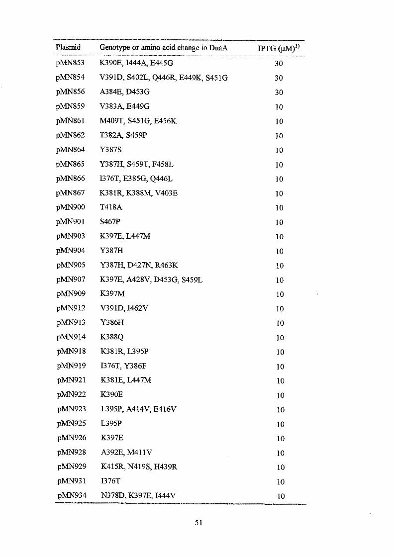
Plasmid Genotype or amino acid change in DnaA IPTG (p“)’) pMN853 K390E, 14444 E445G 30
p m 8 5 4
pMN856
pMN859
pMN861
pMN862
pMN864
pMN865
pMN866
pMN867
pMN900
pMN901
p m 9 0 3
p m 9 0 4
pMN905
pMN907
pMN909
pMN912
pMN9 13
pMN914
pMN9 18
pMN919
pMN92 1
pMN922
pMN923
pMN925
pMN926
pMN928
pMN929
p m 9 3 1
p m 9 3 4
V391D, S402L, Q446R, E449K, S451G 30
A384E, D453G 30
V3834 E449G 10
M409T, S451G, E456K 10
T3824 S459P 10
Y387S 10
Y 3 8 m S459T, F458L 10
I376T, E385G, Q446L 10
K381R, K388M, V403E 10
T418A 10
S467P 10
K397E, L447M 10
Y387H 10
Y387J3, D427N, R463K 10
K397E, A428V, D453G, S459L 10
K397M 10
V391D, I462V 10
Y386H 10
K388Q 10
K381R, L395P 10
I376T, Y386F 10
K381E, L447M 10
K390E 10
L395P, A414V, E416V 10
L395P 10
K397E 10
A392E, M41 1V 10
K415R, N419S, H439R 10
I376T 10
N378D, K397E, I444V 10 ~
51
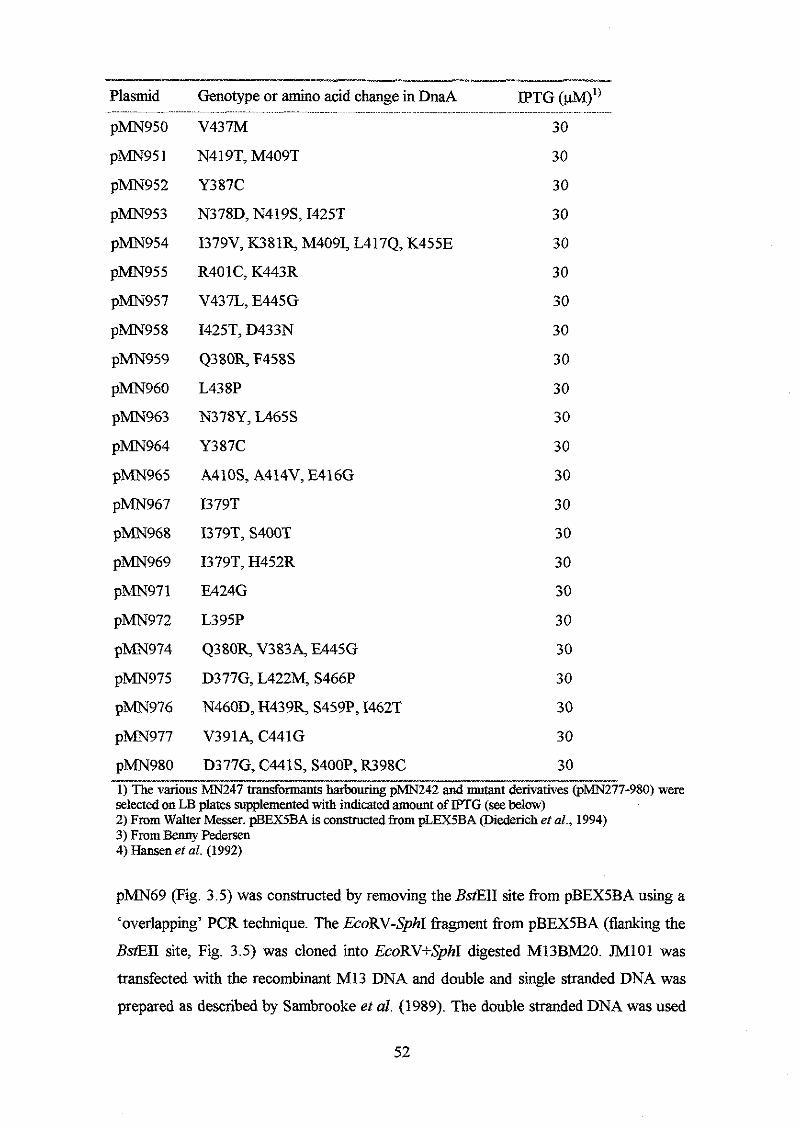
Plasmid Genotype or amino acid change in DnaA IPTG (W’’ - -
pMN950 V437M 30
pMN95 1 N4 19T, M409T 30
pMN952 Y387C 30
pMN953 N378D, N419S, I425T 30
pMN954 I379V, K381R, M4091, L417Q, K455E 30
pMN955 R401C, K443R 30
pMN957 V437L, E445G 30
pMN958 I425T, D433N 30
pMN959 Q380R, F458S 30
pMN960 L438P 30
pMN963 N378Y, L465S 30
pMN964 Y387C 30
pMN965 A410S, A414V, E416G 30
pMN967 I379T
p m 9 6 8 I379T, S400T
30
30
pMN969 I379T, H452R 30
pMN971 E424G 30
pMN972 L395P 30
pMN974 Q380R, V3834 E445G 30
pMN975 D377G, L422M, S466P 30
pMN976 N460D, H439R, S459P, I462T 30
pMN977 V3914 C441G 30
pMN980 D377G, C441S, S400P, R398C 30
selected on LB plates supplemented with indicated amount of IPTG (see below) 1) The various MN247 transformants harbouring p m 2 4 2 and mutant derivatives (pMN277-980) were
2) From Walter Messer. pBEX5BA is constructed from pLEX5BA (Diederich ei al., 1994) 3) From Benny Pedersen 4) &sen et nl. (1992)
p m 6 9 (Fig. 3.5) was constructed by removing the BstEII site from pBEX5BA using a
‘overlapping’ PCR technique. The EcoRV-SphI fragment from pBEX5BA (flanking the
BstEII site, Fig. 3.5) was cloned into EcoRV+S’hI digested M13BM20. J M l O l was
transfected with the recombinant M13 DNA and double and single stranded DNA was
prepaxed as described by Sambrooke et al. (1989). The double stranded DNA was used
52
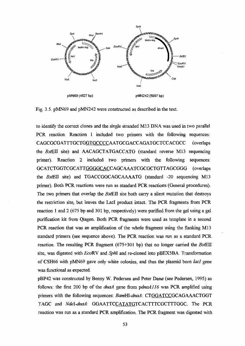
Sphi \
Non Non l NO8
pMN69 (4627 bp) pMN242 (5997 bp)
Fig. 3.5. p m 6 9 and p m 2 4 2 were constructed as described in the text.
to identify the correct clones and the single stranded M13 DNA was used in two parallel
PCR reaction. Reaction 1 included two primers with the following sequences:
CAGCGCGATTTGCTGGTGCCCCAATGCGACCAGATGCTCCACGCC (overlaps
the BstEII site) and AACAGCTATGACCATG (standard reverse M13 sequencing
primer). Reaction 2 included two primers with the following sequences:
GCATCTGGTCGCATTGGGGCACCAGCAAATCGCGCTGTTAGCGGG (overlaps
the BstEII site) and TGACCGGCAGCAAMTG (standard -20 sequencing M13
primer). Both PCR reactions were run as standard PCR reactions (General procedures).
The two primers that overlap the BstEII site both cany a silent mutation that destroys
the restriction site, but leaves the Lac1 product intact. The PCR fragments from PCR
reaction 1 and 2 (675 bp and 301 bp, respectively) were purified from the gel using a gel
purification kit from Qiagen. Both PCR fragments were used as template in a second
PCR reaction that was an amplification of the whole fragment using the flanking M13
standard primers (see sequence above). The PCR reaction was run as a standard PCR
reaction. The resulting PCR fragment (675+301 bp) that no longer carried the BstELI
site, was digested with EcoRV and SphI and re-cloned into pBEX5BA. Transformation
of CSH66 with pMN69 gave only white colonies, and thus the plasmid born ZacZ gene
was functional as expected.
pBP42 was constructed by Benny W. Pedersen and Peter Dam (see Pedersen, 1995) as
follows: the first 200 bp of the dnaA gene from pdnculIl6 was PCR amplified using
primers with the following sequences: BamHI-dnaA: CTGGATCCGCAGAAACTGGT
TAGC and NdeI-dnaA: GGAATTCCATATGTCACTTTCGCTTTGGC. The PCR
reaction was run as a standard PCR amplification. The PCR fragment was digested with
53
NdeI and filled with Klenow fragment of DNA polymerase I. The polymerase was
inactivated and the PCR fragment was digested with BamHI. pBEX5BA was digested
with Nm1 and BamHI and ligated with the PCR product. The correct clone was verified
by sequencing using a Thermo Sequenase cycle sequencing kit from Amersham with
the NdeI&d primer as sequencing primer. Finally, the EcoRI-PstI fragment from
p d n d i i 6 was cloned into the pBEX5BA derivative canying the first part of the dnaA
gene.
pMN236 is a 3 187 bp MuI-CZaI fragment from pBP42 (canying the dnaA gene) ligated
to the 2973 bp MZuI-CZaI fragment from pMN69.
pMN242 (Fig. 3.5) was constructed as follows: Two primers flanking the sequence
encoding the DNA binding domain was used to PCR ampm this area - both in order to
construct mutations in the area (see below) and in order to construct a wild type
derivative. The primers have the following sequences: ‘BstEII’: GCGCGACTTGCTGG
CATTGC ; ‘HindIII’: CGTTCTACGGTAAATTTCATTTACG. The ‘BstEII’
primer m e a l s just upstream the BstEII site in the dnaA gene, and thus, close to the
DNA binding domain encoding sequence. The ‘HindIW primer anneals in the very end
of the dnaA structural gene. This primer has a HzndIII site build in so that the PCR
fragment can be cut and ligated with the BstEII-HindIII fragment of pMN236. Thus,
pMN242 and pMN236 are identical except for the 150 bp between the end of the dnaA structural gene and the HindIII site in pMN236, which are missing in pMN242. The
PCR was run as described below, but without MnCl2.
pMN242 derivatives canying mutations in the sequence that encodes the DNA binding
domain (pMN277-980) were constructed by using a mutagenic PCR technique as follows: The PCR amplification was run under conditions that reduces the fidelity of the
Taq polymerase (Leung ef al., 1989): 0.06 ng linear pMN236 was mixed with Taq
polymerase buffer, 6 mM MgC12, 0.5 mM MnC12, 0.2 mM of each d m , 0.8 @l of the
‘BstEII’ and ‘HindIW primers and 4 units taq polymerase (Perkin Elmer). The cycle
program was 1 min 94”C, 1 min 49‘C, and 1 min 72OC. The PCR kagments were
digested with BstEII and HindIU and ligated to the gel purified BstEII + HzndIII
digested pMN242 fragment. The point mutation frequency was 7%.
Transfomtion, selection and sequencing mutant dnaA aZleles
The ligation mix was used to transform MN247 (dnaA46, P,,d-ZacZ, see table 3.1).
Transfonnants were selected at 42°C on LB plates with ampicillin and 10 or 30 @l
IPTG. These IPTG concentrations were chosen because MN247 harbouring p m 2 4 2 54
(pdna43 formed colonies on 10, but not 30 pM IPTG plates, whereas the presence of
pMN251 (pdnaA204) required 30 pM IPTG in order to complement the heat sensitive
M 4 6 phenotype and form colonies. After transformation, the colonies were re-
streaked and plasmids were purified (General procedures). The area of the dna4 gene
that encodes the DNA binding domain was sequenced using a Thermo Sequenase
Sequencing kit ffom Amersham with the 'BstEII' primer as sequencing primer. Further
analysis was carried out with those dnd mutants that had one and two mutations (see
below).
Complementation analysis
MN247 carrying different multicopy mutant &ad alleles were grown over night in LB
medium at 32°C. The over night cultures were diluted 10, lo2, lo3, lo4 times in a
microtiter tray and app. 5 p1 was plated on LB plates with 100 pg/d ampicillin and 40
&d X-gal and 0,6,10,30 and 100 IPTG. The plates were incubated at 30,39 and
42OC. Complementation analysis was carried out with two independently isolated
MN247 transformants from each transformation. Complementation and growth was
evaluated visually the following day by counting and measuring the number and sizes of
colonies. Complementation of the heat sensitive dnaA46 (MN247) and &d204
phenotype (MN978) was also determined by re-transformations and plating on LB
plates with ampicillin and different amounts of IPTG.
Ver$cation of the dnaA allele on the chromosome
The presence of dnaA46 dele on the chromosome of the MN247 transformants was
verified to rule out the possibility that the M genes on the multicopy plasmids had
recombined with the one on the chromosome. This was done by PCR amplification of
the chromosomal d n d gene with primers that flank the allele specific dm446
mutations. The forward primer anneals upstream the dnaA structural gene and can
therefore anneal to the chromosomal M but to the plasmid W . The primers have
the following sequences: FLF 1 : GCCAATTTTTGTCTATGGTG FLR2:
TTGAGAAGGACTCTAG CAAG. The chromosomal &d allele ffom two colonies of
each m 2 4 7 transformant were PCR amplified as described in General procedures. One
of the M 4 6 mutations disrupts a SphI recognition site in the a'naA gene. The PCR
ffagments were therefore digested with SphI to venfy the presence of the dnaA46 allele
on the chromosome.
55
Stability measurements of D d &ion proteins
Cells were grown in LB medium at 32OC for 5-8 generations. At OD450 = 0.5, 1 mM
IPTG was added to the culture. After 1 hour incubatioq 200 &ml chloramphenicol
was added and the culture was divided in two portions; one portion was incubated at
42°C and the other remained at 32OC. Samples were taken at times corresponding to 1,
2, 3 and 4 generations after chloramphenicol addition. Aliquots corresponding to 100 p1
cuiture OD450 = 1.0 was run on SDS-PAGE gels and immunoblotted as described in
General procedures.
Secondary structure prediction
Secondary structure prediction of the wild type DnaA protein was obtained from the
PHD server: www.emb1-heideIberg.de/predictprotein/ predictprotein.htm4 which
includes algorithms developed by Rost and Sander (1994). Alternatively, the secondary
structure was obtained from a secondary structure prediction program developed by
Claus A. Andersen (1998). Both programs are a two level system based on neural
network on each level. The secondary structures of all DnaA proteins with amino acid
substitution constructed in this work and listed in Table 3.2, were obtained using the
CBS program. When the helix prediction reliability was 0.7 or higher, it was accepted
as the likely secondary structure. Otherwise, it was interpreted to be a coil. According to
the prediction servers used in this work, the DNA binding domain of the E.coZi DnaA
protein probably does not contain P-sheets.
56
Section II
The DnaA-DnaA box interaction
Strains andplasmids
Table 3.3. E.coli strains and plasmids used in this work
Strain Genotype / plasmid (amino acid change in -II_
DnaA protein)
MN978
MN98 1
MN987
MN982
m 9 9 4
MN983
MN984
MN985
MN986
MN988
MN989
MN990
MN991
m 9 9 5
MN996
m 9 9 7
MN998
MN999
MNlOOO
MNlOOl
MN 1002
MN1003
dnaA204, Pmi&acZ
MN97WpBEX5BA
MN978/pMN242 @naA+)
MN978/pMN724 (V383M, DnaA205)
MN97UpMN251 (I389N, DnaA204)
MN978/pMN430 (M411V, DnaA211)
MN978/pMN864 (Y387S)
m978/pMN950 (V437M)
MN97WpMN960 (L438P)
MN97WpMN972 (L395P)
MN978/pMN971 (E424G)
MN978/pMN955 (R401C, K443R)
MN97WpMN859 (V3834 E449G)
MN978/pMN424 (A384V, Y386H)
MN978/pMN735 (V3914 L465S)
MN97WpMN758 (1454N)
MN97WpMN796 (F429S)
MN97WpMN847 (K397E)
MN97WpMN900 (T418A)
MN978/pMN913 (Y386H)
MN97WpMN967 (1379T)
MN97WpMN977 (V3914 C441G)
pTAC1257 pBR322 derivative carrying oriC and mioC
The constructions of strains and plasmids are described in Experimental procedures
section I (Table 3.1 and 3.2). Transformation of MN987 was carried out as described in 57
General procedures. pTAC1257 was obtained from Tove Atlung: The Taql (upstream
mioC) -Cia1 (in g.&) fragment was cloned into pTAC909, which is a pBR322
derivative with a deleted HindIII site. Thus, pTAC1257 carries the whole oriC area with
DnaA box RI, M, R2, R3, R4, R5 and R6 (see Fig. 1.1 in Chapter 1).
Buffers
Binding buffer I: 50 mM HEPES-KOH pH 7.6, 100 mM K-Glutamate, 1 mM DTT and
1mM ATP. Binding buffer TT: as Binding buffer I supplemented with 15% glycerol,
0.4% Triton X-100,2 mM EDTA and 5 mg/ml BSA. TBS: 50 mM Tris-HC1 pH 10.0,
150 mM NaCI. PBS: 140 mM NaCI, 3 mM KCI, 100 mM Na2Hp04,20 mM KH2P04,
pH 7.4.
Overproduction of DnaA fusion proteins
MN981-MN1003 were inoculated directly from a fresh over night LB plate with 100
&ml ampicillin in liquid LB medium. 500 &ml ampicillin was added to the growth
meduim continuously (app. every 1 hour) to avoid growth of plasmid free cells. The
cultures were grown exponentially at 32°C for approximately eight generations and at
OD450 = 0.5, 1 mM IPTG (Sigma) was added. After 1-2 hours induction, the cells were
harvested and washed twice in binding b e e r I. The cell pellets were dissolved in
binding buffer I to a 10’ up-concentration (i.e. 100 ml bacterial culture was dissolved in
1 ml binding b e e r I). The cell suspensions were incubated 30 min. on ice with 1 mg/ml
lysozyme before sonication (5 x 10 sec.). The sonicated cells were centrikged 20 min.
at 20.000 G and the supernatant was kept in the freezer until it was used in binding
experiments.
Production of ‘hot’ and ‘cold’ oriCfiagment
pTAC1257 was PCR amplified with the following pBR322 primers: pBR-1:
CGAGGCCCTTTCGTCTTC, pBR-2: CTGCGTTAGCAATTTAACTGTG. The PCR
amplification was run as a standard PCR reaction (General procedures). PCR fragments
used as ‘cold’ orzC was purified using a Boeringer Mannheim PCR purification kit,
whereas when it was used in preparation of ‘hot’ orzC, it was gel purified using a
Qiagen gel purification kit. Thermo sequenase sequencing kit %om Amersham was used
to prepare ‘hot’ oriC. This procedure consists of a labelling step and an elongation step,
and the procedure was carried out essentially as described by the supplier. Labelliig
step: 6 ng/@ gel purified PCR fragment (see above) was mixed with reaction buffer, 29 58
nM of primer pBR-1 and pBR-2, 0.17 pM dGTP and dTTP, 0.29 pCi/pl 35S-dATP and
2 units Thermo Sequenase DNA polymerase per 17.5 p1 reaction mix. The cycle
program was 95°C 15 sec. and 57°C 30 sec. for 50 cycles. Elongation step: the labelled
PCR fragment was mixed with 80 dNTP in reaction buffer (dNTP was used and not
deoxy-NTP's purchased from supplier). l unit Thermo Sequenase DNA polymerase was
added per 30 pl reaction mix. The cycle program was: 95°C 30 sec., 57OC 30 sec. and
72°C 2 min. for 10 cycles. The labelled PCR fragment was purified using a PCR
purification kit from Boeringer Mannheim. To make sure that the labelling procedure
was successful, the 'hot' oriC DNA was run on a 0.7% agarose gel, which was dried and
exposed on a Kodak film. The 'hot' oriC concentration was in the order of 1-2 ng/p1
and counted between 10.000 and 20.000 cpdng oriC in a Liquid Scintillation Counter.
The DNA concentration was determined by using a h BstEII marker with known
concentration. In addition, the DNA concentration was determined by measuring OD260
(ODzm=l corresponds to 50 pg DNNml, Sambrooke et al., 1989).
BindingJission DnaA proteins to m a l Streptavidin coated magnetic beads
20 p1 streptavidm coated magnetic beads (Dynal) and 5 p1 sonicated bacterial lysate was
used in each binding assay. The total amount of magnetic beads to be used in one
experiment (typically 2-300 pl per mutant DnaA protein) was washed once with PBS
buffer and once with the binding buffer 1. The total amount of bacterial lysate was
added and the mixture was incubated for one hour at room temperature with vigorous
shaking. The supernatant (i.e. proteins that do not bmd to streptavidin) was removed and
kept for quantification of bound DnaA protein on SDS-PAGE gels and immunoblots
(see below). The magnetic beads with bound fkion DnaA protein was washed twice
with binding buffer I and divided into 20 p1 portions. The supernatant was removed and
the magnetic beads were ready to be used in oriC-DnaA saturation experiments,
competition experiments and protein concentration determinations (see below, and Fig.
3.6).
o r i C - D d saturation
20 p1 binding buffer with seven different oriC concentrations was added to the magnetic
beads - DnaA suspensions. The oriC concentrations, and the percentage of 'cold' oriC,
are shown in table 3.4.
59
Table 3.4. orzC concentrations used in saturation experiments, I__-
[orzc] (nM) ‘cold’ orzC (%) -_ 0.126 76
0.252 88
0.378 92
0.63 95.2
1.26 97.6
2.52 98.8
5.04 99.4
I
oriC was allowed to bind to the DnaA protein for one hour at room temperature. The
supernatant was removed and the magnetic beads - DnaA suspensions were washed
several times with binding buffer I. Each orzC concentration was assayed in duplicate,
and a sample was always removed from each concentration (corresponds to [orzqtotal.
Fig. 3.6). The wild type DnaA protein was included in each experiment.
Competition assay
20 p1 binding buffer I with either 0.63 nM or 1.26 nM oriC and increasing amount of
salmon sperm DNA was added to the magnetic beads - DnaA suspensions in order to
measure the specificity of the mutant DnaA proteins towards oriC. The salmon sperm
DNA was sonicated to a fiagment size of 500 bp in average, and 10, 50, 125,250,500
and 1000 fold excess was added simultaneously with the orzC fragment. Each salmon
sperm DNA concentration was assayed in duplicate, and the wild type DnaA protein
was included in each experiment.
Quant@atrion of bound oriC
The concentration of orzC-DnaA complex was quantified by subtracting the number of
counts in the supematant after incubation with the magnetic bead-DnaA suspensions
([0riCJ6~) from the number of counts in the oriC solutions that had not been incubated
with magnetic bead-DnaA ( [ o r i h ) . A liquid scintillation counter was used. It was
tried several times to elute the orzC by adding 1.5 M NaCl (Roth and Messer, 1995), but
it was never possible to recover all the counts (i.e. [ o r i a - l - [ o r i q k >> [orzc],,d).
Counting the bound orzC together with the magnetic beads was not successful either,
because the magnetic beads quenched too much to be counted properly.
60
c Magnetic beads +sonicated bacterial lysate
b Incubation 1 hour
v eh qr
distribution into tubes with 20 p1 in each, remove supernatant
Addition of different concentrations of radioactive labelled O K fragment - or increasing amount of salmon sperm DNA to a constant onC concentration Incubation 1 hour
Transfer supernatant (free oriC) to vials
1 Count total oriC 0 and free oriC (F) in liquid scintillation counter T - F = Bound O K
Fig. 3.6. Flow diagram of in vitro assay used to determine binding constants and specificity towards oviC. F: free orzC and T: total oriC. See text for explanations.
Besides, the [oriqt*l - [ o r z q b = [oriC]bound approach seemed valid, since the
magnetic beads do not bind oriC themselves, and the pBEX5BA lysate do not bind
oriC either.
Determination of DnaA concenPations
The amount of DnaA protein that had bound to the magnetic beads was quantified using
three approaches: 1) the bacterial lysate (before and after incubation with magnetic
beads) was run on SDS-PAGE gels and Western blotted as described in General
procedures. 2) A sample was taken from each mutant DnaA protein and processed in a
61

Westem-like manner (Benny W. Pedersen, unpublished results): 150 p1 TBS, 2%
Tween was added to the magnetic bead-DnaA pellet and the samples were incubated
over night with vigorous shaking. The samples were washed with TBS and DnaA
antibody in TBS, 0.05% Tween was added. After one hour incubation, the samples were
washed several times with TBS, 0.05% Tween. 100 p1 lo5 dilution Pork-anti-Rabbit
Immunoglobulins @ M O ) were added and the samples were incubated one hour at
room temperature. The samples were washed several times with TBS, 0.05% Tween
and once with 150 mM Tris-HCI pH 8.8. 200 p1 Tris-HCI pH 8.8 was added and the
samples were pre-incubated at 37OC. 200 p1 18.6 mg/ml PNPP (p-nitrophenyl-phosphate
disodium, Sigma) was added at t = 0, and the samples were incubated at 37°C for 30-60
min. The Supernatant was transferred to 800 p1 1 M NaOH and was measured with
PNPP as reference. A pBEX5BA sample was always included in the protein
measurements. The amount of protein is expressed as units as follows: units DnaA
protein = (&dJhA)- hlo(pBEX5BA) x 1000) / t (min). Samples were taken in
duplicate. Unfortunately, it was not easy to reproduce the results. The amount of DnaA
proteins, expressed as units, differed more than 2-fold when comparing othenvice
similar samples. Therefore, the DnaA concentrations were estimated by 3) plotting
toric-DnaA] as a function of [or iqh (see below).
Binding constants
in vitro dissociation constants (G) and total DnaA protein concentrations were
determined in the saturation experiments (see above) by plotting bound oriC as a
fUnction of free oriC. GraFit (Erithacus Software) was used to make the curve fits and
estimate the binding constants using the equation for the binding of one ligand (in this
case one oriC fragment per DnaA protein): [DnaA-oriq = [oriCJh . DnaAltd / o(d +
[or zqh) . The total DnaA concentration can be read as the horizontal asymptote at
saturation and KD when half of the total amount of DnaA protein has bound oriC.
in vivo affinig: pgalactosidme assay
Over night cultures were diluted and grown exponentially for app. 6 generations at 32°C
in AB medium (Clark and Maalee, 1967) supplemented with 0.2% glucose, 2 pg/ml
thiamine and 1% casamino acids. At OD450 = 0.1 the cultures were divided in five
portions. Four portions were incubated at 42" with 0, 10, 30 and 100 pM IPTG and the
fdlh portion was incubated at 32°C with no IPTG. 3 x 1 ml samples were taken different
62
times after the shift, added to 200 pg/d chloramphenicol and kept at 0°C. At the end of
the experiment, the samples were shaken with toluene. The following day the p- galactosidase assay was carried out essentially as described by Miller (1972). Enzyme
units was calculated as follows: units 0-galactosidase = (1000 . oD42o)/t . v . O D ~ ~ O ) ,
where t = min. incubation with ONPG and v = volume of the culture.
South- Western analysis
On a low Bis SDS-PAGE gel, the fusion protein always appear as a double band after,
but not before, sonication (Fig. 3.17 in Results, section 11). This indicates that the fusion
protein is degraded. To measure if both the intact and degraded protein bind oriC,
South-Westem analysis was carried out essentially as described by Sutton and Kaguni
(1997B). Sonicated lysate corresponding to 0.58 pmol DnaA protein per lane was run
on a SDS-PAGE gel, and the proteins were blotted onto a Immun-Blot PVDF
Membrane PioRad). The filter was then incubated in 30 mi binding buffer I1 at 4°C for
one hour. 35S-oriC fiagment was added to the filter to a final concentration of 50 n g h l
and incubated over night at 4°C. The filter was washed twice in binding buffer II and
exposed on a Kodak film. Bound DNA was stripped fiom the filter in 154 mM NaCI, 10
mM Tris-HC1 (pH 7.4), 0.25% SDS. The filter was then processed as in Western
blotting (General procedures).
FX, cleavage
The fusion DnaA protein was cleaved with FX, (Promega) in 100 mM NaCI, 20 mM
Tris-HC1 pH 8.0 according to recommendations fiom supplier. Cleavage of fusion
DnaA protein was verified on SDS-PAGE gels and Western blotting as described in
General procedures.
63
Section III
Integration and analysis of mutant d n d alleles in h attachment site of the
chromosome
Strains andplasmiJs
Table 3.5. Strains and plasmids used in this work ~- I__L_I(--
Source or relevant genotype Reference/source Strain
W 1 7 1 8
MC 1000
MT 102
RI3210
TC3422
TC3482
~~ ............ ~~~~ ..,......
TC1929
NK5525
NK6034
MN1127
MN1128
MN1184
m 1 1 9 1
MN1193
MN1195
MN1197
MN 1240
m 1 2 1 0
m 1 2 1 1
W 1 2 1 3
dnaA46, recBC from Christoph Weigel
araDI39, A(ara,leu)7697, Alac, X74, galU, gaZK, shA (1980) Casabadan and Cohen
a, hsdR Atlung et al. (1 989)
a, hRB 1 (pdnaA-lac9 Braun et al. (1 985)
a, hsdRlpTAC3422 (int plasmid), camR Atlung et al. (1991)
a, hRB1, dnaA::cat, rnh373 Hansenetal. (1991A)
a, hRI3 1, dnaA204
F,proA81::TnlO, rh', IN(mD-rrnE)
€E, car-96::Tn10, del(gpt-lac)5, relAl, spoTI, thi-I
Hansen and Atlung (1 995A)
from E.coli Genetic Stock Ceter (#7089)
from E. coli Genetic Stock Ceter (#6 18 1)
a, hRB1, dnaA::cat, mh373, car-96::TnlO TC3482 x Pl(NK6034)
a, hRB1, dnaA::cat, rnh373, car-96::TnlO/pUF747 this work
a, dnaA::cat, rnh373, car-96::TnlO/pUF747, Pm- biotindnaA' in h att
this work
a, dnaA204, P~-biotin&mA(L395P) in h att this work
a, dnaA204, Prn-biotin&aA@C397E) in h att this work
a, dna4204, Pm-biotin- dnaA(A384V,Y3.86H) in h att this work
a, dnaA204, P~-biotin&aA(C441G,V391A) in h att this work
a, dnaA204, Pm-biotin&aA+ in h att this work
a, &&::cat, Pm-biotin-dnuA(L395P) in h att, mh+ this work
a, dnaA::cat, P~~-biotin&mA(C441G,V391A) in h this work att, rnh'
a, dnaA::cat, Pmbiotin-dnaA(K397E) in h att, rnh' this work
64
MN1215 a, dnaA::cat, P~~-biotin-dnaA(A384V,Y386H) in h this work att, mh+
"242 a, dnaA::cat, Pm-biotin&aA' in h att, rnh' this work
pTAC3463 AttP-apb-4 DNA integration casette Atlung et al. (1 99 1)
pMNl122 as pTAC3463, NotI adaptor in EcoFU site this work
pUF747 PBm-mh' ~ ~~ ..... . .~. .... ~ . . .
a) genotype as MC1000
From Ulrik von Freiesleben
.~ . .
Construction of pMN1122 was carried out as follows: pTAC3463 was digested with
EcoRI and ligated with 10 times molar excess of NotI adaptors with the following
sequences : AATTCAGGCCTGCGGCCGCC and AATTGGCGGCCGCAGGCC.
After transformation, plasmids were prepared and the correct clone, carrying a Not1 site,
was identified. pm747 was constructed by PCR amplification of the mh' gene with
following primers: CAGGAAGTCTACCAGCCATGGTTAAACAGGTAG and
CCCAAGCTTCAAGCCACGAATTCGCCAGG. The PCR fragment was digested with
NcoI and EcoRI and cloned into the pBADhis.4 vector (Invitrogens).
"127 was constructed by P1 transduction of TC3482 with a P1 stock grown on
NK6034 (General procedures). Tetracycline resistance was selected, and the
transductants were screened for arginine and uracil requirements. MN1127 was
transformed with pUF747 to construct MN1128. The transformation was carried out as
described in General procedures.
Construction of "128, TC1929 and TC3482 derivatives with dnaA alleles in h
attachment site were carried out as follows (Fig. 3.7): pMN242 and pMN242 derivatives
(Table 3.2) were digested with NotI and CZuI. The fragment canying the dnd gene p i g
3.5) was gel purified using a gel purification kit from Qiagen, and ligated to the gel
purified NotI-AccI fragment of pMNl122. This ligation results in Iigated circles that
lack origin of replication and they are therefore not replicated in the cell. Kanamycin
resistant colonies only appear if the circular DNA molecules have integrated on the
chromosome. TC3422, carrying the znr plasmid, was transformed with the ligation mix
and kanamycin resistant colonies were selected. Integration of the DNA molecules in h
attachment site of the chromosome in kanamycin resistant cells was verified by PCR
amplification with primers that anneal to the qhA and attR sequence, respectively
(Kana: CCGAGTACGTGCTCGCTCG, AttR: GGCGCAATGCCATCTGG).
65
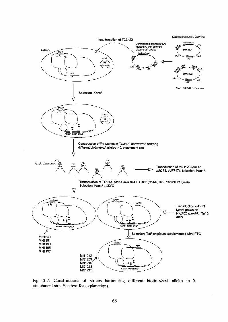
4 Selection: KanaR
Commction of P1 lysates of TC3422 derivatives carrying different biotin-dnaA alleles in h atbchrnent site
Kanan, b i o t i n 4 n a A A Transdudon of MN1128 (dnaA0.
4 mh373. pUF747). Selection: K a n a R
Transduction of TC1929 (dnaA204) and TC3482 (dnaA0. mh373) with P1 lysate Selection: KanaR at 32°C
MN1240 MN1191 MN1193 MN1195 MN1197
L Selection: TetR on plates supplemented with IPTG
Fig. 3.7. Constructions of strains harbouring different biotin-dnd alleles in h attachment site. See text for explanations.
66
The PCR reaction was run as a standard PCR reaction (General procedures). P1 lysates
were prepared from TC3422 derivatives canying the different dnaA genes in h
attachment site and used to transduce MN1128, TC3482 and TC1929 (General
procedures). Kanamycin resistance was selected on LB plates at 32'C. TC3482
transductants were screened for rnh depended high-temperature sensitivity. In addition,
to avoid selection of cells that had recombined the whole plasmid into h attachment site,
the TC3482 transductants were screened for cam'. TC1929 transductants were screened
for cam' for the same reason as above, and for growth at high temperature (42OC) in
presence of different amounts of IPTG due to induction of the dnaA gene in h
attachment site. The rnh37.3 mutation in the TC3482 transductants was removed by P1
transduction with P1 lysate grown on NK5525 (proA::TnlO, rnh'). Tetracycline
resistant transductants were selected on LB plates supplemented with 0, 30 and 75 &l
IPTG. The distance betweenproA and rnh is 0.6 min. (Bachmanq 1990) and thus, co- transduction is approximately 33% (Wu, 1966). Tetracycline resistant transductants
were screened for mh+ phenotype (no high-temperature sensitivity) on LB plates
supplemented with IPTG. Growth of all transductants were IPTG dependent - or
sensitive (see Table 3.13).
Using W 1 7 1 8 to tran$er dnaA mutants to their normal chromosomal location
pMN242 and a selection of pMN242 derivatives were transformed into W 1 7 1 8 and
plated on LB plates supplemented with 10 or 30 pIvf IPTG and 100 pg/d ampicillin,
and incubated at 42'C. The following day, four colonies from each transformation were
streaked at 42'C on LB plates without ampicillin and without IPTG. This treatment
selects recombinants that have recombined the dnaA46 allele out of the chromosome.
The streaking procedure was repeated five times to allow growth of plasmid free cells.
Chromosomal DNA from ampicillin sensitive and temperature resistant cells were PCR
amplified (General procedures) using primers that A) flank the allele specific dnaA46
mutations and B) flank the sequence that encodes the DNA binding domain (for primer
sequences see Experimental procedures, Section I: FLFl, FLR2 and BstEII , HzndIII).
Absence of dnaA46 allele on the chromosome was verified by SphI digestion of PCR
fragment A. A number of dnaA alleles that were selected for recombination into their
normal chromosomal location cany mutations that introduce or remove recognition
sequences for various restriction enzymes (Table 3.6). The correct isolate could
61
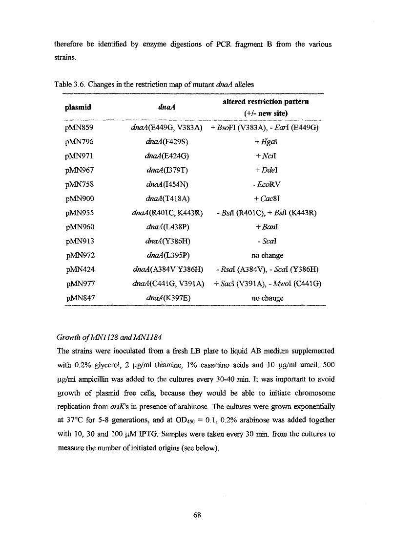
therefore be identified by enzyme digestions of PCR fragment B from the various
strains.
Table 3.6. Changes in the restriction map of mutant dnaA alleles
plasmid altered restriction pattern
(+b new site) dnaA
pMN859 &d(E449G, V383A) +&OFT (V383A), -Ear1 (E449G)
pMN796 dnaA(F429S) + HgaI
pMN97 1 dnaA(E424G) + NciI
pMN967 dmA(I379T) + DdeI
pMN758 dnaA(I454N) - EcoRV
p m 9 0 0 dnaA(T418A) + Cuc8I
p m 9 5 5 dnaA(R401C, K443R) -BSA (R401C), +BSA (K443R)
pMN960 dnaA(L438P) + Ban1
pMN913 dnaA(Y386H) - ScaI
pMN972 dnaA(L395P) no change
pMN424 dnaA(A384V Y386H) - RSUI (A384V), - SCUI (Y386H)
p m 9 7 7 dnaA(C441G, V391A) + Sac1 (V391A), -MW01 (C441G)
pMN847 haA(K397E) no change - x
Growth ofAANI128 andAANll84
The strains were inoculated from a fresh LB plate to liquid AB medium supplemented
with 0.2% glycerol, 2 pg/d thiamine, 1% casamino acids and 10 uracil. 500
pg/d ampicillin was added to the cultures every 30-40 min. It was important to avoid
growth of plasmid free cells, because they would be able to initiate chromosome
replication from oriXs in presence of arabinose. The cultures were grown exponentially
at 37°C for 5-8 generations, and at OD450 = 0.1, 0.2% arabinose was added together
with 10, 30 and 100 ph4 IPTG. Samples were taken every 30 min. from the cultures to
measure the number of initiated origins (see below).
68
Growth of TC1929 derivatives carrying dzfferent IPTG inducible dnu4 alleles in A attachment site
Cells were grown exponentially at 32°C in AB medium supplemented with 0.2%
glucose, 2 pg/d thiamine and 1% casamino acids. At OD450 = 0.1, the culture was
shifted to 42OC for 1 hour. The culture was diluted 20 times and divided in ten portions;
five portions were krther incubated at 4 2 C and the rest were shifted back to 32'C.
IPTG was added to final concentrations of 0, 10,30, 50 and 100 @l to the cultures at
42 and 32'C. Samples were taken from the cultures before and 1, 2, 3 and 4 hours &er
IPTG addition to measure the number of initiated origins and DnaA concentrations (see
below).
Growth of rnh' derivatives of TC3482 (WO) carrying dfferent IPTG inducible dnd
alleles in ,L attachment site
Strains were grown exponentially at 32°C and 42OC in AB medium supplemented with
0.2% glucose, 2 pg/ml thiamine, 1% casamino acids and 0 or 30 pM IPTG
dependending on the strain. At OD4504.5, the cultures were diluted 10 times in the
same media supplemented with 0, 30 and 100 pM IPTG and samples were taken from
the cultures after 2, 3 and 4 hours to measure the number of initiated origins and DnaA
concentrations (see below).
Number of initiated origins usingjlow cytometry
1 ml samples were incubated minimum three hours with 300 &ml rifampicin (kindly
denoted by Ciba Geigy NS) and 36 pg/d cephalexin (Sigma). The cells were
harvested and dissolved in 100 p1 10 mM Tris-HC1 pH 7.4. 1 ml cold 77% ethanol was
added to fixate the cells. The cells were kept at 5°C over night or longer. Between 0.1
(if OD450=0.5) and 1 ml k e d cells were harvested and dissolved in 250 p1 10 mM Tris-
HCI @H 7.9, 10 mM MgC12, 90 pg/ml mitramycin and 20 &ml ethidium bromide.
The cells were kept dark and on ice until they were analysed on a BioRad flow
cytometer.
DnaA protein concentration
The DnaA concentration was determined as described in General procedures
69
Section I
Construction and analysis of multicopy plasmid born d n d alleles encoding DnaA
proteins with amino acid substitutions in domain TV
Modified PCR conditions were used to construct p m 2 4 2 derivatives with mutations in
the DNA binding domain of the DnaA protein. The method is described by Leung et al.
(1989), who has shown that the frequency of point mutations increased fiom 0.4% to
2% by adding 0.5 mM MnClz and five times reduced amount of dATP relative to the
other d m ' s . If the amount of dATP was not reduced, the mutation frequency
decreased to 1.4%. Under both conditions, the number of transitions and transversions
was fifty-fifiy. In this study, it was not possible to run a mutagenic PCR reaction with
decreased amount of dATP relative to the other dNTP's, whereas the DNA yield in the
presence of M n 2 + was unailkcted. However, in spite of the non-optimal mutation
conditions, a higher mutation frequency - about 7% - was obtained. This resulted in
PCR fragments that carried many mutations and therefore, were not further analysed. In
addition, most of the mutations (75%) were transitions (Table 3.7 and 3.9). This is not
in agreement with results obtained by Leung et al. (1989). The different results may be
due to differences in the Taq polymerase activity. The mutagenic PCR fkagments were
digested with BstEII and Hind111 and cloned into pMN242 pig. 3.9, and used to
transform MN247, which canies a &a446 allele on the chromosome and a P,~o~-ZacZ
fusion in h attachment site. The transformants were selected at 42°C on LB plates
supplemented with ampicillin and 10 or 30 PM IPTG. Selection at 42°C ensured that
only hctional DnaA protein encoding &a4 alleles were selected, due to the
temperature sensitive phenotype of the dna446 host. 10 and 30 @l IPTG were chosen,
because the pdnaA' transformant formed colonies in the presence of 10, but not 30
IPTG, whereas the pdmA204 transformant required 30 W IPTG in order to
complement the &a446 phenotype. Thus, this selection resulted in selection of both not
so harmful mutations @naA activities close to that of the wild type DnaA protein) and
more harmful mutations that require more IPTG to complement the &a446 allele.
Plasmids from 74 colonies selected on 10 @l IPTG and 59 colonies selected on 30
IPTG were prepared and the &a4 sequence encoding domain IV was sequenced. 70
Table 3.7. The number of times a particular amino acid change was observed in . . . .
Amino acid change Number Mutation Amino acid change Number -_I _I_
______--_ T375A I376V I376N I376L I376T D377G N378D N378Y I379V I379T Q380R K38lR K38lE T382A T382S V383A V383E V383M A3 84V A384E E385G E385A Y386N Y386C Y386H Y386F Y387C Y387N Y387S Y387H K388E K388M K388Q I389F I3 89T I389V
1 2 1 1 3 2 3 2 1 4 4 4 3
1 1 3 1 1 3 2 3 1 1 2 3 1
4 1 1 3 1 1 1 1 3
1
A+G A+G T+A A+C T+C A+G A+G A+T A+G T+C A+G A+G A+G A+G A+T T+C T+A G+A C+T C+A A+G A+C T+A A 4 T+C A+T A+G T+A A+C T+C A+G A+T A+G A+T T+C A+G
K390E K3901 V391A V391D A392T A392V A392E D393Y L3941 L394H L395P L395H K397R K397E K397M R398H R398C R399Q S400P S400T R401C S402T S402L
V403M V403E P406L M4091 M409T M409V A410V A4 10T A4 1 OS M41 1T M4111 M41 1V
5 1 2 2 1 1 1 1
1 1 S 1 3
5 1
1 1 1
3 1 1 1
3
1 1 2 3 4 1 1 1 1 2 1 3
1
__I
Mutation
A+G A+T T+C T+A G+A C+T C+A G+T C+A T+A T+C T+A A+G A+G A+T G+A C+T G+A T+C T+A C+T T+C C-tT G+A T+A C+T G+A T+C A+G C+T G+A G+T T+C G+A A+G
i L4 13P T+C __g. _I__
71
Amino acid change Number .~ ..,...... ...~.~
Mutation Amino acid change Number Mutation .~ ~~ ................ ~ ~ . ~ ~ ~ ~ ~ . ~ . ~
A414V K4 15N K4 151 K415T K4 15E K415Q K4 15R E416D E4 16G E416V L417Q L417M T418A T4 1 8N N419Y N419S N4 19T H420Q H420P S421R L422Q L422M E424G I425V 1425s I425T D42W D427N A428V F429L F429L F429V F429S F429Y G43 1 S R432S D433N
~~.~ ............
3 1 1 1 3 1 2 1 3
1 3 1 2 1 1 3
1 1 1 1 2 1 3
1 1 3
1
1 2 3 1 1
1 1 1 1
C+T A+T A+T A+C A+G A+C A+G A+T A+G A+T T+A C+A A+G C+A A+T A+G A+C C+A A+C T+A T+A C-+A A+G A+G A+G T+C G+T G+A C+T T+C T+A T+G T* T+A G+A
C+&T+C
T436S v43 7M V437L U38P H43 9L H439R C441R C44 1 G C441S R442H K443R I444V I444T I444A E445G Q446R Q446L L447s IA47M R448H E449K E449G E450G S451C S451G H452R D453A D453N D453G I454T 1454N K455E K455Q E456G E456K D457G
1 G+A D457V
2
1 1 2 1 2 1 2 1 1 4 3 2 1 7 3 1 2 2 1 1 1 1 1 2 3 1 1 2 2 1 3 1
2 1 1 1
A+T G+A G+C T+C A+T A+G T+C T+G T+A G+A A+G A+G T+C
A+G,T+C A+G A+G A+T T+C T+A G+A G+A A+G A+G A+T A-+G A+G A+C G-+A A+G T+C T+A A+G A+C A+G
G+A A+G A+T
72
F458L 1 T+C I462V 1 A+G S459P 2 T+C I462T 1 T+C S459T 1 T-+A R463K 1 G+A S459L 1 C+T L465S 5 T+C
N460D 2 A+G L465M 1 T+A L461V 1 T+G S466P 1 T+C L461F 1 A+T S467P 1 T+C
i__l__ i_. __I_
The obtained amino acid changes in the DnaA protein are shown in table 3.2
(Experimentd procedures, section I), and Table 3.7 shows the number of times the same
amino acid substitution was observed in different isolates. The table also shows the type
of mutation that caused the amino acid substitutions. The silent mutations are not
included. An amino acid change at a given position (i.e. E456G) normally appeared 1-3
times, with few exceptions as the L395P change (and K397E and L465S). These areas
may be mutational hot spots.
If the mutations are separated according to the amino acid change, with no regard to
the position, it is clear that the amino acid changes Ile+Thr, Gln-+Arg, Lys+Glu,
Giu-+Giy, Tyr+His, Phe-+Leu, Phe+Ser and all the Met substitutions are the most
common amino acid changes (Table 3.8).
73
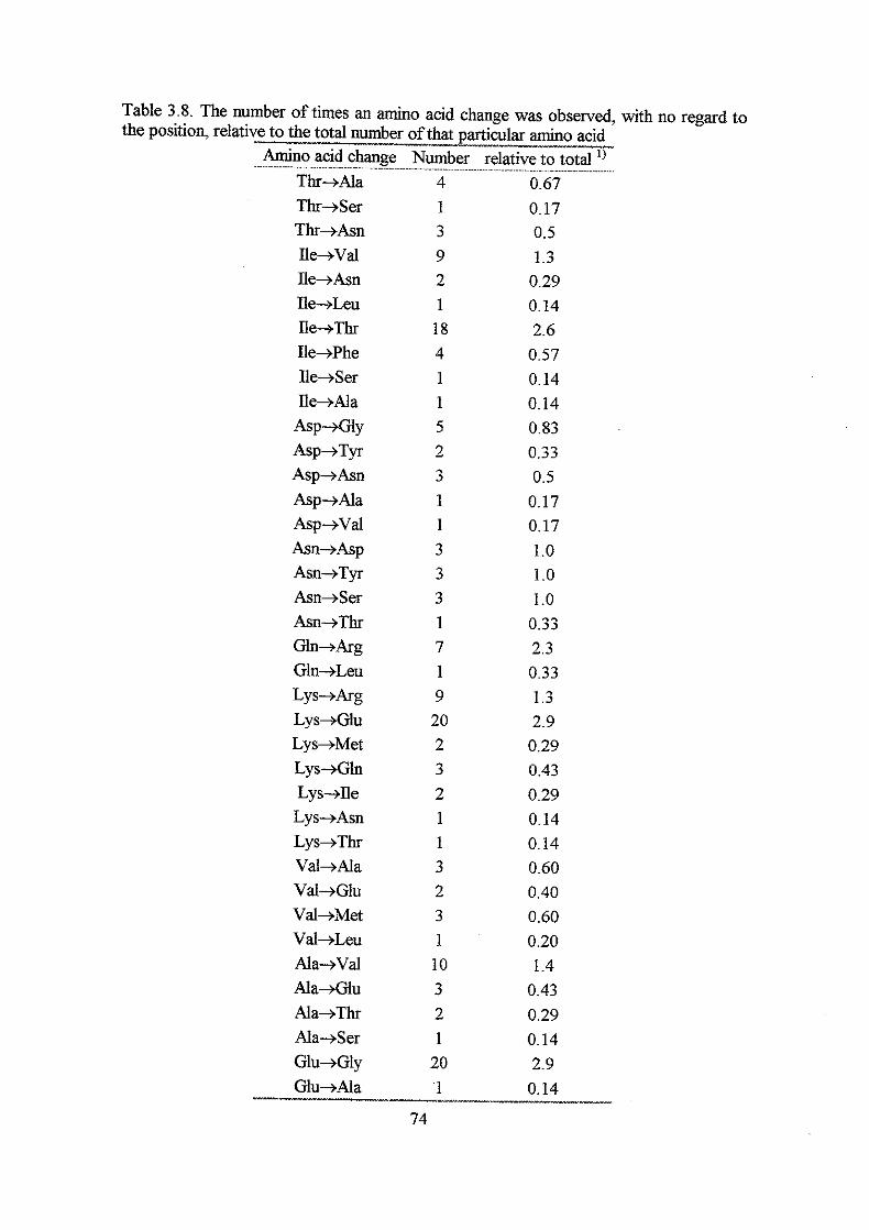
Thr+Ala Thr+Ser Thr+Asn Ile+Vd ne+Asn Ile+Leu Ile-+Thr Ile+Phe Ile+Ser Ile+Ala
Asp+Gly Asp+Tyr Asp+Asn Asp+Ala Asp+Vd Asn-+Asp Asn+Tyr Asn+Ser Asn+Thr Gln+Arg Gln+Leu Lys+Arg Lys+Glu Lys+Met Lys+Gln Lys-+Ile Lys+Asn Lys+Thr Val+Ala Val+Glu Val+Met Val+Leu Ala+Vd Ala+Glu Ala+Thr Ala+Ser G l u 4 l y
4 1 3 9 2 1 18 4 1 1 5 2 3 1 1 3 3 3 1 7 1 9 20 2 3 2 1 1 3 2 3 1 10 3 2 1
20 '1
0.17 0.5 1.3
0.29 0.14 2.6
0.57 0.14 0.14 0.83 0.33 0.5 0.17 0.17 1.0 1.0 1 .o
0.33 2.3 0.33 1.3 2.9 0.29 0.43 0.29 0.14 0.14 0.60 0.40 0.60 0.20 1.4
0.43 0.29 0.14 2.9
0.14 Glu+Ala I__
74
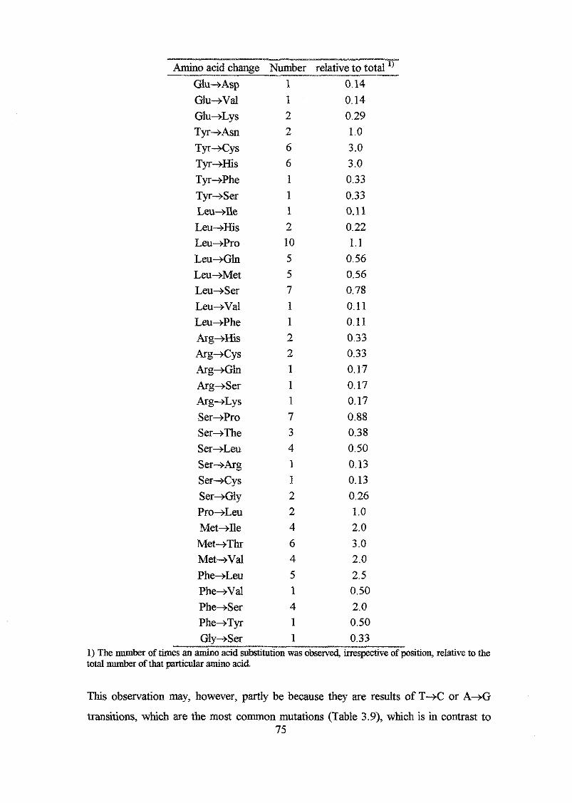
Amino acid change Number relative to total __ Glu+Asp Glu+Val Glu+Lys Tyr+Asn Tyr+Cys Tyr+His Tyr+Phe Tyr+Ser Leu+Ile Leu+His Leu+Pro Leu+Gln Leu+Met Leu+Ser Leu+Val Leu+Phe Arg+His Arg+cys Arg+Gln Arg+Ser Arg+Lys Ser+Pro Ser+The %+Leu Ser-+Arg Ser+Cys Ser+Gly Pro+Leu Met+Ile Met+Thr Met+Val Phe+Leu Phe-+Val Phe+Ser Phe+Tyr Glv+Ser
1 0.14 1 2 2 6 6 1 1 1 2 10 5 5 7 1 1 2 2 1 1 1 7 3 4 1 1 2 2 4 6 4 5 1 4 1 1
0.14 0.29 1.0 3.0 3.0 0.33 0.33 0.11 0.22 1.1
0.56 0.56 0.78 0.11 0.11 0.33 0.33 0.17 0.17 0.17 0.88 0.38 0.50 0.13 0.13 0.26 1.0 2.0 3 .O 2.0 2.5 0.50 2.0 0.50 0.33
1) The number of times an amino acid substitution was obsew@ irreqshve of position, relative to the total number of that particular amino acid
This observation may, however, partly be because they are results of T+C or A+G
transitions, which are the most common mutations (Table 3.9), which is in contrast to 75

observations done by Leung et al. (1989), who found a transition:transversion ratio of
1: l .
Table 3.9. The number of different mutations Mutation Number / % of total number of mutations
T+C
T+A
T+G
C+T
C+A
C+G
72 l 25
23 18.1
4 11.4
18 16.3
9 l 3.2
010
A+G 106 l 37
A+T 22 17.7
A+C 8 12.8
G+A 19 16.7
G+T 3 11.1
G+C 1 10.4
DnaA proteins with one and two amino acid substitutions were analysed firther (see
below), whereas those with 3, 4 or 5 amino acid substitutions were not due to
difficulties in the later interpretations of the effects of individual amino acid
substitutions.
Secondary structure predictions of DnaA proteins carrying one and two amino acid
substitutions in the DNA binding domain
Secondary structure predictions of the various DnaA proteins constructed in this work,
and the DnaA' protein, were carried out using a program developed by Claw A.
Andersen (1998), referred to as the CBS program. The program is a two level system
with neural network at each level. The more commonly used PHD server that includes
algorithm developed by Rost and Sander (1994), referred to as the PHD program, was
used to predict the secondary structure of the wild type DnaA protein for comparison
(Fig. 3.8). This program is also based on neural networks. When both programs are used
to predict the secondary structure of the DNA binding domain of the DnaA+ protein two
differences in the secondary structure predictions appear: A) the probability for a longer 76
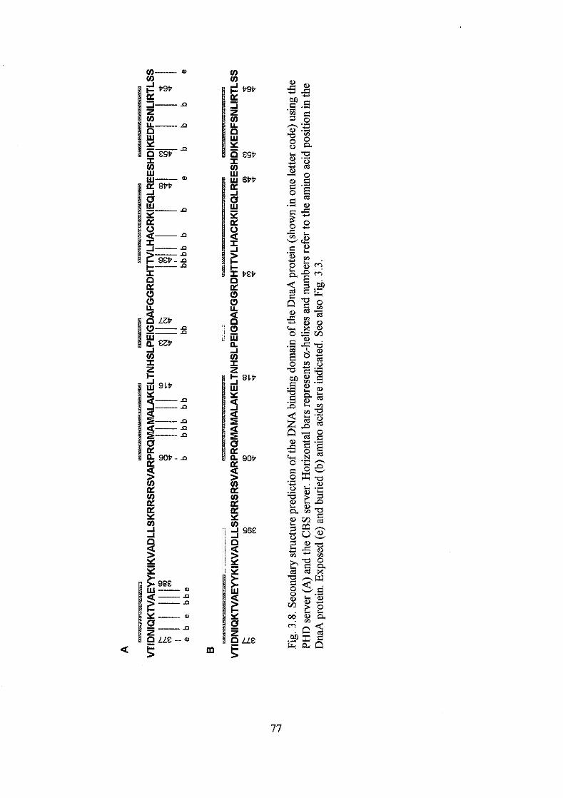
P9P
ESP
6PP
E P
BLP
90P
S6E
?LE
helix 1 is higher when the CBS program is used (0.61-0.64) compared to the PHD
program (probability only 0.1-0.4) and B) the existence of helix 3 from P423-D427 has
lower probability (0.65) using the CBS program than when the PHD program is used
(0.8-0.9). The different amino acid changes constructed in this work will be used in the
discussion of these contradictions (see below).
Complementufion of the heat-sensitive dnaA46 allele by induction of multicopy mutant
dnaA alleles
aha4 alleles with one and two mutations were hrther analysed in the &a446
background (MN247). Dilutions of over night cultures were plated on LB plates
supplemented with different concentrations of IPTG (0, 6, 10, 30, 100 @l) and
incubated at 30,39 and 42'C. The following day, growth and ability to complement the
high temperature sensitive M 4 6 phenotype were estimated by measuring the number
and sues of the colonies. The results of the complementation of the heat-sensitive
h a 4 4 6 phenotype by induction of different mutant dnaA alleles are shown in Fig. 3.9.
Growth data at 30°C are not shown, because they were quite similar among the different
strains with few exceptions that will be mentioned in the text (see below). Generally,
there was good growth at 3 0 T , which is expected due to the presence of DnaA46
proteins synthesised from the chromosomal dnaA gene. However, there was generally
no growth on LB plates supplemented with 100 pM IPTG. The high IPTG
concentration was not toxic itself, because the pBEX5BA (no hu4) transformant grew
on these plates (see below), which indicates that high DnaA concentrations are lethal.
pBEX5BA
MN247 ( h d 4 6 ) harbouring pBEX5BA (no dnaA allele) formed colonies at 30°C
independently of the IPTG concentration (data not shown), whereas no colonies
appeared at 39 or 42OC due to the heat sensitive phenotype of the host (Fig. 3.9).
*d+ m 2 4 7 (dnaA46) harbouring pdnaA' formed colonies at 42°C in the presence of 6 and
10 pM IPTG. Higher IPTG concentrations seemed to inhibit growth at 42°C (Fig. 3.9).
The growth at 30 and 39O was less affected by high IPTG concentrations. The DnaA'
protein is not temperature sensitive, and thus, growth of the transformant was
expected to be independent of the temperature. The fact that the p & d + transformant
formed colonies on 30 pM P T G plates at 30 and 39OC, but not at 42"C, may therefore 78
be due to the presence of some DnaA46 activity at the lower temperatures. This also
seems consistent the observation that the PM' transformant formed colonies without
IPTG induction at 39OC, but not at 42OC (Fig. 3.9). The amount of DnaA+ protein that
was synthesised fiom the Zac promoter under non-induced conditions was enough,
together with the (in)active DnaA46 protein, to initiate chromosome replication and
allow growth at 39°C. This was also observed in a dnaA204 background (data not
shown). Leakiness of the lac promoter was also observed in the I)-galactosidase assays,
where the specific activity in the pdnaA+ transformant was lower under non-induced
conditions than in most of the other transformants (Results, Section II).
What is the efJect of the biotin-target on the DnaA protein?
Although the biotin target is fused to the N-terminal part of the DnaA protein, and
therefore has maybe only minor effect on the DNA binding, its presence could have
some effect on the protein activity in vivo. As discussed previously, the N-terminal
domain of the DnaA protein seems to be responsible for protein-protein interactions,
which are important in the assembly of the initiation complex (Weigel et al., 1999).
Blasing (1999) have shown that M 4 6 cells harbouring pm?aAII6, which cany a
&d'' allele (without the biotin tag) under the same lac type promoter control as used in
this work, formed colonies at 4 2 T with 5 and 10 pM IPTG, which is in agreement with
the observations done in this work. However, Blasing (1999) also observed (reduced)
complementation at 42°C without IPTG induction, and that was not observed in this
work at 42OC, only at 39OC. This could indicate that the biotin tag somehow reduces the
DnaA activity slightly, but it could also be due differences in the strain backgrounds
resulting in differences in the transcription fYequency fiom the lac promoter under non-
induced conditions and/or differences in the DnaA46 concentrations. In general, the
results indicate that the DnaA protein fused to the biotin tag is functional, and has an
activity comparable to the wild type DnaA protein.
79
39oc 4 2 C 39°C 42°C Aminoacidchange o 6 IO 30100 o 6 T O 30100 Amino acidchange o 6 10 30100 o 6 l o 30100 LLM IPTG
DnaA' no DnaA 00000 00000 T375A
~ .. .. .
1376T, y386f 1376T
1376N, m409i 1376V, F429L 1376L, M409T
N378D, M411T
N378Y, L465S N378Y, F429L
Iq70T
1379T, 1462F 1379T, S400T 1379T, H452R Q380R, 1389T Q380R, F458S
K381 E, L447M K381 E
K381 R, R448H K381 R, L395P
T382A, S459P T382S, P406L
V383A
V383A, E449G V383M
A384E, D453G A384E
A384V, Y386H E385G, K415E
Y386H y386h, A384V
Y386N Y386C, K443R Y386C, S402L Y386F, 1376T
Y387H
..,,",
Y387C
Y387N. K397R Y387S
1389N K3880
~~~
1389T 1389T, Q380R 1389F, L3941
K3901
K390E, R432S K390E
V391A, L465S K390E, M411V
V391A, C441G V391D, 1462V A392T, M4091 A392E, M411V
L3941, 1389F
L395P, R3990 L395P
L395P, E456G L395P, K381 R
00..0 000.0
000.0 000.0 000.0 000.0
000.0 000.0 000.0 0.. 00 000.0
000.0
00..0 .... c .... 0
00000 00.00
000.0 000.0
000.0 000.0
000.0
00000 00.00
00.00
K397M K397E
K397E. L447M K397R,Y387N R399Q, L395P S400P, F429L
S400P, C441G S400T, 1379T
R401C, K443R S402L. Y386C ~ 4 0 3 ~ 1 L4176
P406L. T382S M4091, A392T M4091, 1376N
M409T, K415T M409T, 1376L M409T. N419T M409V,'T418N
A41 OV A41 OT M41 1 I
M41 1T (dnaA211) M41 lT, N378D
00.00 000.0
000.. 00000 000.0
000.0 000.0
000.0
M411V. K390E O m 0 0 0 00 M411V, D453N 06.06 00
A414V, L417Q L41 3Q
K41 5E K415E. E385G
K4151
K415E; K455E K415N, L4220 K415T, M409T E416D, L465S E416G. C441 R L417Q,'A414V~ L4170, V403M
T418N, M409V T41 8A
N419T, M409T N419Y, F429L
L422Q, K41 5N H420P
E424G E424G, 1425V 1425T, D433N
F429S F429V
F429L,kk5V F429L, S4OOP F429L, N378Y F429L, N419Y R432S, K390E D433N, 1425T
V437L. E445G V437M
L438P C441G, V391A C441G, S400P
@@.a0 00..0
000.0 000
000 000 0.. 000 000.. 000.0 000.0 000.0
000.0 000.0 000.0 000. 000. 000. 000.0
00080 C0000 000.0 000.0 00000
00000 0 C C C 0 C C C 000.0 000.0 00000
000.0 000.0 00000
80
39oc 4 2 C
Amino acid chanae 0 6 10 30100 0 6 10 30100 uM IPTG C441 R, E416& 0 0 0 K443R. Y386C 000 K443R,’R401C 000 E445G, V437L 000
L447M, K397E E445G, L461F 000
L447M. K381 E 0 0
E449G. V383A R448H, K381R 000
0 H452d, 1379T 666
D453A 000 D453N,-M411 V 0 0 0 D453G, A384E 000
1454N 00@
_ _ _
1454T, F458S 000 K455E. K41 5E 0 0 0
_ _ _
E456G, L395P 0 0 0 F458S:I454T 000
S459P, T382A 6 6 6 L461 F, E445G 0 0 0
L461 V 0.0
1462F, 1379T 00
L465S. V391A 000 1462V, V391D 000
L465S: E416D 00 L465S, N378Y 0 0 0
- - _
S467P
10 000.0
10 00000 10 00000 10 000.0 10 00000 10 000.0 10 000.0 10 000.0
IO 00.00
Fig. 3.9. Complementation of high-temperature sensitive dnaA46 phenotype by IPTG induction of plasmid born mutant dnaA alleles. The strains were diluted and plated on LB plates supplemented with hfferent amounts of IPTG and incubated at 3OoC (data not shown), 39°C and 42’C. The following day growth was determined visually by counting the number and measuring the sizes of the colonies. Following symbols are used to describe the growth of the various strains:
0 : good growth
: slightly reduced growth : reduced growth
0 : severely reduced growth
0 : no growth
81
First and last part of the DnaA protein may not be directly involved in DNA binding
The result of the complementation analysis may seem rather complex (Fig. 3.9) and
generally, there does not seem to exist areas of the DNA binding domain that are not, if
not directly then indirectly, involved in the DNA interaction. There are, however, amino
acids scattered over the domain that probably are not in direct contact with the DNA.
This is based on the observation that certain amino acids can be substituted with non-
closely related amino acid(s) apparently without affecting the DnaA activity. These
amino acids are: 1376, K381, Y386, K388, K390, V391, L395, K397, M411, T418,
E456, F458, L461, L465, S467 (Fig. 3.9 and 3.10).
* * * * * * * ** * * * V T I D N I Q K T V A E Y Y K I K V A D L L S U R R S R S V A R P R Q M A M A L A U E L T N H S L P E I G D A F G G R D H T T V L H A C R K I E Q L R E E S H D ~ K E D ~ S ~ L I R ~ L S S
* * * - E - m - " 8. - 5 5 ? 3 1 - - a
Fig. 3.10. Amino acids that can be substituted with non-closely related amino acid(s) may not be in direct contact with the DnaA box (*). See text for explanations. The horizontal bars represent a-helixes. Only the amino acid sequence of domain IV is shown.
The first half of domain IV and the very C-terminal region seems to contain more amino
acids that may not be in direct contact with the DNA, compared to he!& 3 and 4 and the
loop in between. The relatively high number of amino acids in helix 5 that may not be in
direct contact with the DNA is consistent with the observation that this area mainly
consists of not conserved amino acids (Fig. 3.2).
Neutral mutations
The PAM250 matrix was used to define neutral amino acid substitutions. The matrix is
calculated from a protein evolution model that involves amino acid frequencies and pair
wise substitution frequencies observed in existing alignments of proteins. A match
involving a rarely occuning amino acid counts more than a match involving a common
amino acid does, and if two different amino acid sequences are aligned, a mismatch
between two easily exchangeable amino acids gives a higher score than a mismatch
between two unrelated amino acid. A neutral substitution was defined to be a change
that counts 50% of that particular amino acid, and that the score goes 'both ways'. In
addition, firther restrictions concerning charge and size were included. The following
amino acid changes has been regarded as neutral: Arg ts Lys, Asp t) Glu, Gln ts His,
Ile # Val, Leu ts Met, Phe t) Tyr, Ser -+ Thr. Unfortunately, this PAM250 matrix
82
was not consulted in the beginning of this work, and therefore, the A+V substitution
has been considered neutral as well (and, as it turned out, in many cases it seems
acceptable. See below).
Eke effect of mino acid changes in the DNA binding domain on DnaA activity
The effects of the various amino acid substitutions constructed in this work on the
activity of the DnaA protein is discussed in the following sections. Activity is based on
complementation analysis of the high-temperature sensitive dna446 strain shown in Fig.
3.9. In some cases, the result of the complementation analysis is rather difficult to
interpret due to the presence of double mutations, and it is not easy to draw much
information. Therefore, focus is put on those amino acid changes where the effect on the
DnaA protein is more clear. The secondary structure was predicted for all DnaA
proteins (data not shown).
The classical dna4 mutants
The well conserved V383 is substituted with a hydrophobic methionine in the DnaA205
protein. This change seems to affect the DnaA knction severely, and the high-
temperature sensitive dm446 phenotype was only complemented weakly. The
DnaA205 protein is unstable at higher temperatures (see Table 3.10), and this may
partly explain the weak complementation. On the other hand, Hansen et al. (1992)
showed that the presence of multicopy dm4205 alleles (under dnaA promoter control)
resulted in temperature resistant growth. Thus, it would be expected that at a certain
IPTG concentration, good complementation was observed. The reason why that is not
observed may be that the correct IPTG concentration was not used and therefore, the
DnaA205 concentration in the cell never equalled that in the multicopy dnaA205 strain
used by Hansen et d . (1992). The V383 can be substituted with the also very
hydrophobic, but smaller alanine without affecting the DnaA protein much. Since they
are all hydrophobic, and none of the amino acid substitution seems to change the
secondary structure (data not shown), the results indicate that at this position it is
important, for steric reasons, that the amino acid is not larger than valine. I389 is
substituted with asparagine in the DnaA204 protein, and that amino acid change seem
very deleterious, whereas when it is substituted with threonine, the function of the
DnaA protein is unaffected. The I389N substitution may result in a longer helix l (data
not shown), which of course can have an effect on the DnaA protein. The isoleucine is
very hydrophobic, whereas both the threonine and asparagine are neutral, but can act as 83
a donor and/or acceptor in hydrogen bonding. Thus, the negative effect of asparagine
compared to threonine may lie in difFerences in the functional groups of the two amino
acid and/or differences in the molecular structures. As with the multicopy ha.4205
(above) and M 2 1 1 strains (below), a certain IPTG concentration would be expected
to result in good complementation of the dnaA46 phenotype by the DnaA204 protein,
since a multicopy dm4204 strain with the dnaA allele under Pdnd control is temperature
resistant (Hansen et al., 1992). A M411T substitution is found in the DnaA211 protein.
As with the two other classical dnaA mutants, the DnaA211 protein complements the
h a 4 4 6 phenotype rather weakly, although better than DnaA204 and DnaA205 proteins
do. When M411 is substituted with isoleucine, the DnaA protein is not affected
severely, which is consistent with the nature of the amino acids; methionine and
isoleucine are hydrophobic whereas threonine has a more hydrophilic character. The
results indicate that at this position a strictly hydrophobic amino acid is required and
even the presence of a hydroxyl group disrupts the structure. A large area of helix 2, in
which the M411 lies, is predicted to be protected &om the solvent (Fig. 3.3 and 3.8),
which is in agreement with the results in this work.
Helix I (W377 + Y386/L395)
The well conserved T375 can apparently not be substituted with alanine without
affecting the DnaA protein severely. The protein only complements the d~a.446
phenotype weakly. Threonine can, in contrast to alanine, act as donor and acceptor in
hydrogen bonding, and this ability may be important at this position. The nabour, I376,
can, however, be substituted with the non-closely related threonine without affecting the
DnaA protein. Thus, T375 cannot be substituted with a hydrophobic amino acid,
whereas its hydrophobic neighbour I376 can be substituted with a threonine. This does
seem a little obscure, but may indicate that the structure of the DNA binding domain is
very sensitive towards certain amino acid changes. None of the amino acid substitutions
discussed above seem to change the secondary structure of the binding domain (data not
shown). N378 is substituted with aspartic acid and histidine, but both substitutions co-
exist with other substitutions, which make the N378 substitutions difficult to interpret.
The I379T substitution probably changes the secondary structure of the DnaA protein
by shortening helii 1 in the N-terminal (the he i i is predicted to start at T382 in the
DnaA(I379T) protein, data not shown), and this may be responsible for the effect the
substitution has on the DnaA protein. However, the substitution of the highly conserved
isoleucine could be deleterious in itself. The accessibility predictions indicate that I379 84
is protected fiom the solvent (Fig. 3.3 and 3 .S), and thus, a change into threonine would
be expected' to have an effect on the existing hydrophobic van der Waals interactions
between I379 and other hydrophobic amino acids, and thereby change'the structure of
the protein. The Q380R substitution co-exists with I389T (and F458S), a substitution
that apparently has no effect on the DnaA protein, although it may extend helii 1 (see
classical mufatiom). Thus, the Q380R change, which probably has no effect on the
secondary structure, is responsible for the reduced DnaA function observed with the
DnaA(Q380K I389T) protein. Glutamine is a polar, but neutral residue, whereas
arginine is strongly basic and thus, always positively charged. This positive charge
together with the multiple hydrogen bonds donating capacity makes the arginine a
surface amino acid. However, Q380 is not predicted to be located on the surface of the
DnaA protein (the probability for it being exposed or buried is zero, Fig. 3.3), and ifthe
amino acid is located inside the protein, a substitution with arginine would be expected
to have dramatic effect on the structure of the protein. T382 is probably not located on
the surface of the protein either, because it can be substituted with alanine without any
apparent major changes of the DnaA function, or the secondary structure (the T382A
substitution co-exists with S459P). This is consistent with the observation that T382 is
not conserved and often substituted with hydrophobic amino acids in other organisms
pig. 3.1 ). On the contrary, K381 may very likely be interacting with water, and
thereby located on the surface, since it can be substituted with glutamic acid without
any apparent effect on either secondary structure or DnaA function. The K381 is located
n+4 amino acids away fiom the predicted exposed D377 and E385, which places the
hydrophilic amino acids on the same side of the a-helix. The A384V substitution co-
exists with the harmless Y386H substitution (see below). Substitution of the well-
conserved alanine has an effect on the protein. Depending on the prediction server (Fig.
3.4), the secondary structure may be changed as follows: helix 1 is divided into two
smaller a-helixes, the first cover the area T382-+A385 and the second the area
K3884L395. It does, however, not seem to be the changes in the secondary structure
that is responsible for the effect of the amino acid substitution, since the A384E
substitution probably does not change the secondary structure, but has a even more
deleterious effect on the DnaA hnction. It seems more likely that at this position, a
small hydrophobic amino acid is required. This is consistent with the accessibility
predictions, which predicts that A384 is buried (Fig. 3.3 and 3.8). As mentioned above,
the Y386H substitution has no effect on the DnaA fimction, although it may interrupt
helix 1 with a small stretch of coil consisting of 2-3 amino acids around the substitution, 85
like the A384V substitution (and the Y386N substitution). Y386 is conserved, but other
residues are seen at this position (mainly histidine, but also glutamine, serine, lysine and
glutamic acid, Fig. 3.1). A substitution with arginine has an effect on the DnaA protein.
Both histidine and arginine can act as donor and acceptor in hydrogen bonding, but the
orientation is of course not identical. This may indicate that Y386 participates in
hydrogen bonding and that histidine, but not arginine, has the correct orientations of the
atoms. However, Y386 can also be substituted with phenylalanine without any severe
effects, and although phenylalanine may have weakly polar properties, it cannot be
considered as having hydrogen bonding capacity. Y387 is a highly conserved amino
acid. It can, however, as Y386 be substituted with histidine without any apparent effect
on the DnaA function (although helix 1 may be interrupted from K388 to V391),
whereas substitutions with cysteine, serine or arginine reduce the activity (substitutions
with arginine may interrupt the helix from Y386 to A392. This amino acid substitution
co-exists with K397E, a substitution that apparently does not reduce the DnaA activity,
see below). They can all act as donor and acceptor in hydrogen bonding, but as
discussed above, tyrosine and histidine are somewhat similar in shape which may be
required for a correct orientation and angle of the hydrogen bonds between tyrosine (or
histidine) and another residue. The tyrosine is predicted to be buried, although with low
reliability, which indicate that the hydrogen bonding is between two residues and not
between tyrosine and water. This is also consistent with the apparent sensitivity towards
substitutions with other amino acids with hydrogen bonding capacity (Cys, Ser and
Arg), because in contrast to other amino acid residues, water molecules may adapt new
orientations more easily. V391A (well conserved) and A392E (not conserved) may also
interrupt helix 1, but as the other helix interrupters, these DnaA proteins complement
the M 4 6 phenotype almost as well as the DnaA'protein does (the A392E substitution
co-exists with M411V. The latter substitution may not have any effect on the DnaA
protein, since the M4111 substitution, which exists as single mutant, does not). The
V391D and A392T substitutions very likely reduce the size of helix 1 by several amino
acids (the helix stops at Y386). This does not seem to have any severe effect on the
DnaA protein either (the A392T change co-exists with M4091, an amino acid change
with a relatively high score according to the PAM250 matrix, and thus, is likely not to
have severe effect on the DnaA protein). The A392 is not conserved among different
organisms, and often substituted with hydrophilic amino acids, which is consistent with
the results of the complementation analysis.
86
S u m m a n r of a-helix 1: According to the alignment and secondary structure predictions
of the DNA binding domain of DnaA proteins from various organisms (Fig. 3.4), it
seems quite conspicuous that the length of helix 1 is rather well conserved in spite of the
fact that this area does have dissimilarities in amino acid sequence. Ifthe helix goes all
the way to L395, as predicted by the CBS server, there are a number of amino acid
changes, constructed in this work, that interrupt the helix without any apparent effect on
the DnaA activity. This seems contradicting with fact that the length of helix 1 is well
conserved, and indicates that the first helix actually does stop around Y386 as predicted
by the PHD server, and thus, the many amino acid substitutions that appeared to
interrupt helii 1, actually do not. Instead, they are located in the following loop area.
The amino acid changes that may shorten the helix N-terminaUy (i379T and A384V) or
extend the helix @389N, DnaA204) do indeed seem to affect the DnaA activity.
Helix 1 may be located on the surface of the DnaA protein, with D377, K381 and
E385, which are located on the same side of the a-helix, exposed to the solvent, and the
rest, especially 1379, V383 and A384 pointing away from the solvent and probably
participating in hydrophobic interactions with other residues of the DnaA protein,
maybe in a leucine-zipper fashion (Chapter 2). Except for 1376, K381 and Y386 it does
not seem possible on basis of this complementation analysis to point at amino acids in
helix 1 that are more likely to be interacting with the DNA target than others. On the
other hand, this area of the DnaA protein does seem sensitive towards amino acid
changes, in general, although the secondary structure is only rarely changed. This
indicates that the amino acids in the helix, and not only the helix itself, help stabilise the
DnaA structure andlor are responsible for DNA contact.
Loop I, the basic loop (Y387 + R405) Generally, amino acid changes in especially the first part of this area of the DnaA
protein seem, at least at first glance, to have minor effect on the function of the protein,
since many of the DnaA proteins complement the dnaA46 phenotype as well as the
DnaA+ protein does. The DnaA(K388Q) protein complements the & d 4 6 phenotype as
well as the DnaA’ protein does. Thus, it seems that the positive charge of lysine is of
less importance, whereas the ability to participate in hydrogen bonding may be more
important. K388 is not very conserved; some DnaA proteins have asparagine at this
position, which is consistent with the observation that glutamine is a non-harmful
substitution. Yet, other DnaA proteins have a glycine at this position. Glycine and
asparagine are often located in turns, and may therefore be considered as helix breakers. 87

Maybe the most important fhction of the first amino acids following helix 1 is to
ensure that the helix does not continue. The conserved K390 is probably located on the
surface of the protein, since a substitution with glutamic acid does not affect the protein
whereas a substitution with isoleucine reduces the DnaA function. This 'surface
hypothesis' is consistent with the observation that the K388E (see above), K397E and
R398H substitutions do not seem to affect the DnaA protein (the R398H co-exists with
two other substitutions. See Table 3.2 (pMN753): the triple mutant was isolated on 10
p M IPTG). None of the amino acid substitutions seem to change the secondary structure
(data not shown). They are not highly conserved, and several organisms have other
hydrophilic amino acids at these positions (Fig. 3.1). The hydrophobic amino acids
V391 and A392 can be substituted with other amino acids, either with hydrophobic or
hydrophilic character without any major effects on the DnaA protein (and without any
apparent changes in the secondary structure). The alanine is not very conserved and
DnaA proteins from other organisms have serine, glutamic acid, glutamine, lysine or
asparagine at this position. This is consistent with the observation that both the A392T
and A392E substitutions have no effect on the DnaA protein. The V391 is not well
conserved per se, but in other organisms substituted with isoleucine (closely related) or
leucine (related), and only very few organisms have glutamine, arginine or histidine at
this position. Therefore, the V391A substitution (co-exists with the non-hamfid L465S substitution, see below) would be expected to have maybe only minor effect on the
DnaA protein, whereas the V391D substitution (co-exist with I462V, a neutral
substitution) would be expected to have some effect. That is also observed. Thus, V391
may be protected f?om the solvent in agreement with the accessibility prediction (Fig.
3.3). The L395P substitution may introduce a very short stretch of @-sheet like structure
around K390 (data not shaown). However, this does not seem to have any effect on the
DnaA protein, since the DnaA(L395P) protein complements the & d 4 6 phenotype as
well as the DnaA+ protein does. This is consistent with the observation that secondary
structure predictions of DnaA protein f?om different organisms indicate that some of
them may have a small area of P-sheet like structure between helix 1 and 2 (Fig. 3.4).
The highly conserved R399 cannot be substituted with glutamine without affecting the
DnaA protein (the R399Q exists as double mutant with L395P, a substitution without
any effect on the protein, see above), indicating that the the arginine has other functions
than to be interacting with water. The substitution does not seem to change the
secondary structure of the area (data not shown). Thus, at this position, a positively
charged arginine seems to be required for 111 DnaA activity. This also seems true for 88
the highly conserved R401. Although the effect seems smaller, this amino acid cannot
be substituted with cysteine without affecting the DnaA activity. The amino acid
substitution does not seem to change the secondary structure of the DnaA protein (data
not shown). A substitution with cysteine may, however, not be informative when
considering the function of the arginine in the basic loop due to the non-closely related
natures of arginine and cysteine. Unfortunately, no other mutants with R399 nor R401
substitutions were isolated. Blasing (1999) substituted these arginines with alanines, and
found that especially the R399A substitution had severe effect on the DnaA protein,
although neither substitutions destroyed the DnaA activity completely. The S400T
substitution affects the DnaA protein (the substitution co-exists with I379T, a substitution that does not affect the h c t i o n of the DnaA protein, see helzxl, above).
This seems obscure, since other organisms have the closely related threonine at this
position. The substitution does not seem to change the secondary structure (neither does
the S400P substitution, data not shown). The DnaA(M02L) protein complements the
&d46 phenotype with 100 IPTG as one of the few. The substitution probably does
not alter the secondary structure of the protein (data not shown). In addition, the serine
is not very conserved and some organisms have alanine or valine at this position. Thus,
the apparant deleterious effect of the S402L substitution may not be due to reduced oriC
affinity per se, but more likely due to an unstable DnaA(S402L) protein. This was,
however, not further investigated.
Summarv of loop I (basic loop): Except for the L395P substitution, none of the amino
acid changes seem to change the secondary structure of the DnaA protein. The basic
arnino acids K388, K390 and K397 can all be substituted with the oppositely charged
glutamic acid, indicating that the function of these amino acids are to interact with the
solvent and thereby keep the loop on the surface of the protein. In this work, R398 was
substituted with histidine without any effect on the DnaA protein. Other organisms have
serine at this position, indicating that the R398, like the lysines discussed above, is
required for solvent interactions. The highly conserved R399 and R401 may have other
functions than to interact with the solvent. The R399Q substitution has severe effect on
the protein. Unfortunately, it is not possible to determine whether these basic amino
acids participate in the DNA interaction due to the few amino acid substitutions at these
positions. Bliising (1999) constructed and analysed DnaA proteins with R399A and
R401A substitutions. These amino acid substitutions clearly have an effect on the
protein, although they are, especially the DnaA@401A), able to complement the
&d46 phenotype. In general, the solvent accessibilities are predicted with low 89
reliability, but there seems to be more exposed than buried amino acids, indicating that
this loop is located on the surface of the protein.
Helix 2 (P406 4 E416)
Helix 2 probably starts with the not so well conserved P406. A substitution of this
amino acid with leucine has some effect on the DnaA protein, although it was possible
to obtain good growth on 30 pM IPTG plates. The P-+L substitution may extend helix 2
resulting in helii start at S402. It does not, however, seem to be the likely reason for the
effect on the DnaA protein since helix 2 in DnaA proteins from other organisms may
start at that position (Fig. 3.4). In this work, the highly conserved amino acids R407 and
Q408 were not substituted in any of the isolated dnaA mutants. This could indicate that
substitutions of these amino acids have a severe effect on the DnaA protein. However,
Blasing (1999) isolated several R407 substitutions (R407y R4074 R407C) and a
single Q408 substitution (Q408R). Although they all (except R407A) seem to have
severe effect on the DnaA activity, the proteins are stiU functional to some extend
(except the DnaA(R407H) protek which did not complement the dnaA46 phenotype at
all. This is unexpected considering the related characteristics of Arg and His),. M409
can be substituted with isoleucine and valine without any effects on the DnaA protein.
This is consistent with the observation that other organisms have these amino acids at
this position. The highly conserved A410 cannot be substituted with neither valine nor
threonine without affecting the DnaA function severely. These amino acid substitutions
do not seem to change the secondary structure (data not shown), which indicates that at
this position a small hydrophobic amino acid is required. The highly conserved M411
can be substituted with the related amino acid isoleucine without any remarkable effect,
whereas a change into threonine (DnaA211) has a much more severe effect (see
classical M mutants). Like the A410 substitutions, the M411 substitutions probably
do not change the secondary structure of the DnaA protein (data not shown). Thus, at
this position a hydrophobic amino acid also seems to be required. Surprisingly, the
DnaA(L413Q) protein complements the dnaA46 phenotype relatively well (although 30
ph4 IPTG is required). The highly conserved leucine in helix 2 is predicted to be
protected from the solvent (Fig. 3.3 and 3.8), and therefore, substitution with glutamine
could be expected to have a more severe effect on the DnaA fimction than observed.
The double mutants DnaA(A414V, L417Q) and DnaA(E416G, C441R) may have a
shortened he l i that stops at K415 (and not E416), which may account for the reduced
DnaA activity. However, it may seem wrong to attach much weight into a single amino 90
acid difference, but when considering the secondary structure &om other organisms (see
Fig. 3.4) it is obvious that the helix start position may differ among the different
organisms, but it stop at a position corresponding to the E.colz E416. The
DnaA(K415E) protein complements the dnaA46 phenotype rather well with 30 pM
IPTG, whereas the DnaA(K415I) does not. None of the amino acid substitutions seem
to change the secondary structure of the protein (data not shown), which indicates that a
charged amino acid is preferred at this position. Whether it is differences in charges or
Siply the substitution of the highly conserved lysine that is responsible for the after all
reduced activity of DnaA(K415E) cannot be answered, but the results indicate that the
lysine has functions other than to be interacting with water.
Summarv of a-helix 2: According to the accessibility prediction, many amino acids in
helix 2 are protected from the solvent, indicating that a part of this helix is located
inside the protein with other hydrophobic amino acids in the surroundings, and very
little space around A410. This hypothesis is consistent with the observation that the
A410V and M41 11 substitutions have a less severe effect on the DnaA protein than the
A410T and M411T substitutions. DnaA(L413Q) protein complements the dnaA46
phenotype with 30 pM IPTG indicating that the neutral, but polar glutamine somehow
fits the presumed hydrophobic core. Blasing (1999) also isolated the L413Q substitution
and found that the protein complemented the dnaA46 phenotype poorly and only in the
presence of 100 pM IPTG.
Generally, the amino acid substitutions in this area of the DnaA protein do not
change the secondary structure to large extends indicating that the helix structure can
not be changed without damaging the DnaA protein. The length of helix 2 corresponds
to between 2.5 and 3.5 a-helical turns among the different organisms (see Fig. 3.4).
None of the DnaA proteins seem to have an equal number of helical turns, which may
be an important factor in the direction of the following loop. Since helix 2 is liely to be
located inside the protein, this helix may not interact directly with the DNA target.
Helix 3 and small loops surrounding it (L41 7-T435)
The L417Q substitution seems to have severe effect on the DnaA protein (the
substitution co-exists with V403M, which is expected to have minor effect due to the
related character of valine and methionine). The neighbours of the well conserved U 1 7
are hydrophilic, but never the less a hydrophobic amino acid seems to be required at
position 417. T418 is highly conserved among DnaA proteins from different organisms
91
(Fig. 3.1 and 3.2). Therefore, the observation that a substitution with alanine has no
apparant effect on the DnaA protein seem contradicting. The reason could be that helix
2 is predicted to be extended to N419 in the DnaA(T418A) protein (data not shown). On
the contrary, the non-conserved H420 cannot be substituted with proline without
afTecting the DnaA protein severely. The substitution may not change the secondary
structure (data not shown) although the probability that helix 3 exists is reduced (see
below), and other organisms have either negatively changed or hydrophobic amino
acids at this position, which makes it difficult to understand why the H420P substitution
is hamfull. The dnaA46/pcina4(H420P) strain forms colonies on 100 phi IPTG plates.
This could indicate that the DnaA(H420P) protein is unstable, and explain why it
apparently complements the heat sensitive phenotype weakly. The highly conserved
S421 is not substituted in any of the isolated mutants constructed in this work.
However, Blasing (1999) isolated both a DnaA(S421G) and DnaA(S421N) version of
the DnaA protein and they both complemented the M 4 6 phenotype well.
The probability that helix 3 exists is reduced in the DnaA(U22Q, K415E) protein with
no apparent consequences. The DnaA(E424G) protein may also lack helix 3 (with high
reliability) and this may account for the apparent very severe effect the amino acid
substitution has on the DnaA protein. The I425 and G426 amino acids are both highly
conserved among different organisms (see Fig. 3.1 and 3.2). Never the less, I425 can be
substituted with threonine and still complement the dnaA46 phenotype well with 30 PM
IPTG (the I425T co-exists with D433N, which is expected not to have negative effects
on the DnaA protein. This assumption is based on observations done by Blasing (1999),
who found that the DnaA(D433G) and DnaA(D433A) proteins complemented the
M 4 6 phenotype even better than the DnaA' protein did). The highly conserved G426
is not substituted in any of the mutants isolated in this work or by Franca Blasing. This
could indicate that a glycine is required at this position, maybe as a cc-helix breaker.
However, the G426 is substituted with serine in the DnaA5 protein (the G426S
substitution co-exists with the Al84V substitution in the DnaA5 protein). This has some
effect in vzho (see Chapter 4), but multicopy Pdnd&d(G426s) strains are temperature
resistant like multicopy Pd,,&-dnaA+ strains, and not cold sensitive like the multicopy
Pd"d-dnaA5 and Pdn&dd(A184V) strains (Nyborg et al., 2000). The secondary
structure is not affected by the G426S substitution, and thus, ifthe hnction of G426 is
to break the helii a serine seems to have the same effect. The F429 is the first amino
acid in a highly conserved area of the DnaA protein that covers amino acids from F429
to V437. In this work, many different F429 substitutions were isolated, both as single- 92
and double mutants. They all have an effect on the DnaA protein. Phenylalanine may
contribute to the tertiary structure by stacking with other amino acids (tyrosine or
tryptophane). The highly conserved G430 and G431 are not substituted neither in this
work nor in wotk done by Blasing (1999). This indicates that these glycines are
important for DnaA function, maybe because they induce a turn of the coiled structure.
R432 is substituted with leucine in the cold sensitive dnax suppressor DnaA721 protein
(Walker mutant). In this work, it is substituted with serine, which also seems to affect
the activity of the DnaA protein. Whether the protein is cold sensitive was not hrther
investigated. The highly conserved D433 can be substituted with non-closely related
amino acid with no effect (or if any, then positive) on the DnaA protein (Blasing, 1999).
It was not possible in this work to isolate any substitutions of the highly conserved
H434 or T435. However, Biasing (1999) isolated H434Y, H434A and T435P
substitutions, and although the complementation ability is reduced, it is far from
abolished. T435 is substituted with lysine in the second Walker mutant dnd71. This
mutant is cold sensitive like dnaA721. In addition, Sutton and Kaguni have analysed the
DnaA(T425M) protein in vitro, and they found that T435 may participate in the specific
recognition of DnaA boxes (see Section 4 and Discussion)
Summarv of a-helix 3 and loops surrounding it: It was not possible in this work to
substitute many of the highly conserved amino acids, indicating that they cannot be
substituted without deleterious effects on the DnaA protein. The function and
importance of helix 3 is difficult to interpret. Some amino acid substitutions (E424G
and maybe H420P) indicate that the helix cannot be removed without severely affecting
the DnaA protein, yet another substitution (R422Q) does not seem to have any effect on
the DnaA protein, although the protein may lack helix 3 (however with lower reliability
than the E424G substitution, data not shown). The presence of highly conserved G430
and G431 that were not substituted neither in this work nor in Blasing's (1999) may
indicate that DnaA activity depends on helix 3, that on the other hand must be broken
and followed by a turn before the next helix starts. As discussed in Chapter 2, this area could be part of a modified helix-turn-helix motif. The results of the complementation
analysis do not argue against this proposal.
u-Helix 4 mad 5 (T436 + T467)
The highly conserved V437 is substituted with the related methionine, and that has
some effect on the DnaA protein although good complementation was obtained with 30
pM IPTG. V437 is predicted to be buried, and a substitution with methionine may 93
disrupt the hydrophobic interactions with other residues. The DnaA(L438P) protein
does not complement the m?aA46 phenotype at 42T, which may be due to the predicted
reduction of the length of the helix that starts at A440 and not T436. However, the
length helix 4 differs among DnaA proteins f?om different organisms (Fig. 3.4). U 3 8 is
predicted to be protected &om the solvent, but proline is also hydrophobic so this is
probably not the reason for the deleterious effect of the substitution. The well conserved
H439 and A440 are not substituted in any of the isolated mutants. The A440 is
predicted to be protected form the solvent. The C441G substitution require 30 p.M
IPTG in order to complement the heat sensitive M 4 6 phenotype (the substitution co-
exists with V3914 which has no apparent effect on the DnaA protein). The secondary
structure may be influenced rather dramatically (data not shown): a stretch of @-sheet
like structure may be substituting the first part of the a-helix, which is followed by a
loop, allowing the a-helix to start at K443. C441 is not very conserved. However, none
of the other organisms has glycine at this position. The LA38P substitution (see above)
has a more severe effect on the DnaA protein than the C441G substitution has,
indicating that the s d l changes in the secondary structure caused by the U38P
substitution are not alone responsible for the deleterious effect on the DnaA protein. The
well conserved R442 and the highly conserved K443 and 1444 are not substituted with
any non-related amino acids, which makes the function of the amino acids difficult to
interpret. It does however indicate that amino acids with special characteristics are
required at these positions. I444 is predicted to be buried (Fig. 3.3 and 3.8). The E445G
substitution co-existing with L461F (see below) complements the M 4 6 phenotype
we& and that is in spite of the observation that the E445G substitution may extend the
small loop between helix 4 and 5 with seven amino acids from I444 -+ D453 (data not
shown). Q446 is not substituted in any of the analysed DnaA proteins, which is obscure
because this glutamine is not conserved among different organisms (Fig. 3.1). The
highly conserved L447 is substituted with the closely related methionine, and this
substitution has no effect on the DnaA protein. The R448H substitution probably does
not change the secondary structure (data not shown), but it has an effect on the DnaA
protein. Since the R+H substitution is an exchange of one polar amino acid to another,
the results indicate that the size of the amino acid and the orientation of the positive
charge may be important at this position. The E449G substitution seems to extend the
loop between helix 4 and 5, with five amino acids (Q446 + D453). This could be the
reason why this substitution has severe effect on the DnaA protein. The D453G
94
substitution may also extend the loop between helix 4 and 5 by three amino acids (E450
+ K455), but in contrast to the E449G substitution, the D453G substitution has severe
effect on the DnaA activity (the D453G substitution co-exists with A384E. The latter
also exists as single mutant, and that amino acid change has some effect on the DnaA
protein (reduced growth 30 m. However, when the D453G change is added to
A384E, the effect on the DnaA protein is much more deleterious, Fig. 3.9). When D453
is substituted with alanine the loop between helix 4 and 5 may disappear, and this seems
to have just as deleterious effect on the DnaA protein as when the loop is extended (see
above). However, the reason for the apparent damaging effect of D453A could also be
that a charged amino acid is required at this position, since this amino acid is predicted
to be exposed to the solvent. When D453 is substituted with asparagine, the activity of
the DnaA protein is close to that of the D&+ protein. This substitution probably does
not change the secondary structure of the DnaA protein. The intact secondary structure
in addition to an exchange with a somewhat related amino acid is very likely the reason
why the DnaA(D453N) protein is a l l l y functional protein. The I454T substitution also
affect the DnaA activity, and the ha446 transfonnant requires 30 pM IPTG in order to
form colonies at 42°C (the substitution co-exists with F458S that probably does not
affect the protein). The DnaA(I454T) protein may have an extended loop (E450 + K455) between helix 4 and 5 (data not shown), which could be the reason for the
reduced activity of DnaA(I454T). Synechocystis sp. has threonine at this position (the
secondary structure is not changed, probably due to the general different primary
structure of this DnaA protein, Fig. 3.4), indicating that it is not the presence of
threonine at this position that affects the DnaA protein per se, but the changes in the
secondary structure. When I454 is substituted with asparagine, a substitution that
apparently has no effect on the secondary structure (data not shown), no
complementation at 42°C was observed. This strongly suggests that a hydrophobic
amino acid is required at this position. This is consistent with the observation that other
organisms have hydrophobic amino acids at this position (Fig. 3.1), and the residue is
therefore predicted to be protected from the solvent (Fig. 3.3 and 3.8). The K455E substitution co-exists with K415E. The latter substitution, which also exists as a single
mutant, has an effect on the DnaA protein, but good complementation was obtained
with 30 fl IPTG. As discussed in 'helix 2' (see above) this could indicate that a
positively charged amino acid is preferred at this position. When the K445E substitution
is added to the K415E substitution, almost no complementation was observed. This
95
strongly suggests that K455, like K415, is required at this position for other reasons
than to be involved in solvent interactions. However, the fimction of the positively
charge of K445 becomes unclear when considering its conservation among different
organisms; tyrosine, glutamine, serine and asparagine at this position seem to result in
hnctional DnaA proteins (Fig. 3.1). These amino acids are all neutral, but polar to some
extent, and able to participate in hydrogen bonding. So far, several amino acid
substitutions indicate that the small loop between helix 4 and 5 is of great importance - not only is it important that it exists, but the length of it also seems to be critical.
Therefore, it is rather contradicting when the E445G (see above) and E456G
substitutions are predicted to have an extended loop between helix 4 and 5 (data not
shown) without any apparent effect on the activity of the DnaA protein. Generally,
amino acids from S459 to S467 of the DnaA protein can be substituted with non-closely
related amino acids without any severe effects on the DnaA protein. This seems
consistent with the observation that this area is less conserved among different
organisms (except for L461, Fig. 3.1 and 3.2). The U65S and S467P substitutions may
shorten helix 5 slightly without any apparent effect on DnaA activity. In this work, the
highly conserved L461 is substituted with the hydrophobic amino acids valine and
phenylalanine, which are all somehow related. Blasing (1999) isolated a DnaAF461S)
version, and found that this protein did not complement the M 4 6 phenotype at all,
indicating that a strictly hydrophobic amino acid is required at this position.
Accordingly, L461 is predicted to be buried with high probability.
Summan of a-helix 4 and 5: In general, this area of the DNA bmding domain of the
DnaA protein includes few single substitution and many double substitution, and this
makes it relatively difficult to interpret the results. However, there seems to be a
tendency that the presence of a coiled structure between helix 4 and 5 is important, and
that the length of it is critical. Helix 4 is more sensitive towards amino acid substitutions
than helii 5, which is in agreement with how conserved the regions are. Those amino
acids that are predicted to be protected form the solvent with high reliability cannot be
substituted with polar amino acids without affecting the DnaA protein. These amino
acids include V437, A440, 1444, 1454, F458 and L461. The hydrophobic amino acids
are located on the same side of the a-helix (or close to). The two hydrophobic amino
acids I454 and F458 are substituted with threonine and serine, respectively, in one of the
double mutants. This DnaA(I454T, F458S) protein complements the dnaA46 phenotype
better than DnaA(1454N) and DnaA(F458S, Q380R), which indicates that there could
be (weak) interactions between I454 and F458 that is maintained to some extend in the 96
double mutant where both amino acids are substituted with polar, hydrogen bonding
residues. Helix 4 and 5 are probably located on the surface of the DnaA protein with a hydrophobic and hydrophilic side and a hydrophilic coil in between. Helix 4 could be
responsible for DNA contact and as mentioned previously be part of a modified helii-
turn-helix motif.
The complementation analysis discussed above was carried out in a dnaA46 strain. The
DnaA46 protein binds oriC at 42°C @Wang and Kaguni, 1988; C m and Kaguni, 1996),
and therefore, some of the DnaA proteins constructed in this work might be able to
complement the heat sensitive phenotype due to 'help' from the DnaA46 proteins in the
cell (maybe especially at 39°C). A number of pMN242 derivatives carrying mutant
d n d alleles were therefore transformed into an isogenic M 2 0 4 strain (MN987). A
detailed complementation analysis was not carried out. Instead, the transformation mix was plated on 10 and 30 pM IPTG plates and incubated at 30 and 42°C. The results of
this analysis were identical in the ha446 and dnaA204 strain backgrounds (data not
shown).
Verification of the dnd allele on the chromosome
Presence of dnaA46 on the chromosome of the m 2 4 7 transformants was verified to
rule out the possibility that the dnaA genes on the multicopy plasmids had recombined
with the one on the chromosome. This was done by PCR amplification of the
chromosomal dnd gene with primers that flank the dnaA46 mutations. The forward
primer anneals upstream the dnaA structural gene and can therefore anneal to the
chromosomal &d but not to the plasmid M. AmpWcation and subsequent SphI
digestion of PCR fragments from the various strains showed that they all carried a & d 4 6 allele on the chromosome (data not shown). Later, experiments indicated that it
was difficult (impossible) to recombine the dnaA46 allele out of the MN247
chromosome even when the strain was under recombination 'pressure' (see
Experimental procedures, section III and results, section III): the MN247/pMN242
(pdna43 was plated and incubated at 42°C without IPTG. This treatment allowed
selection of recombinants since the host is high-temperature sensitive. The potential
recombinants were further streaked 4-5 times on LB plates without ampicillin to allow
growth of plasmid &ee cells. When the chromosomal d n d gene from ampicillin
sensitive and high-temperature resistant colonies was PCR ampltfied and SphI digested,
it turned out that they apparently still had a ha446 allele on the chromosome and 97
therefore had not recombined with the dm4 gene on the plasmid. Thus, the MN247
transformants formed colonies at high temperature due to formation of suppressor
mutations that were not krther analysed. In the complementation analysis (Fig. 3.9),
most of the observed colonies at 39 and 42OC did not carry suppressor mutations, since
the number of colonies at permissive and non-permissive temperature was identical. In
some cases, a few colonies appeared in the IO' dilution spot. The cells in these colonies
were probably carrying suppressor mutations and were of course not interpreted as a
dm446 complementation by the plasmid born dm4 allele.
DnaA amino acid substitutions have eflect on stability
So far, the DnaA amino acid substitutions have been analysed with no regard to protein
stability. Thus, an apparently weak complementation of the high temperature sensitive
ha446 phenotype by a mutant DnaA protein (i.e. low afsnity to orI'C) and low in vivo
asfinity to the DnaA box R5 and R6 in the mzoC promoter (Section 11) could be due to
degration of the protein and not reduced mty per se. Therefore, the stability of
various proteins was estimated at 32 and 42°C. m 9 8 7 (dnaA204) strains harbouring
plasmid with Werent dm4 alleles were grown at 32", IPTG was added to induce the
lac promoter and after app. one hour, chloramphenicol was added to stop protein
synthesis. Samples were taken different times after drug addition, and the relative
amounts of protein was estimated by Western blotting. Fig. 3.1 1 shows the degration of
the DnaA+, DnaA(K397E) and DnaA@401C, K443R) protein, and Table 3.10
summarises the results with all tested proteins.
Stability of DnaA+ is independent on temperature as expected. M e r 4 generations with
chloramphenicol, there is app. 60% protein left (Fig. 3.1 1). The stability of the
100 V I Q
1 2 3 4
time afler chloramphenicol addition (generations) 98
Fig. 3.1 1. Degration of DnaA' (white symbols), DnaA(K397E) (grey squares) and DnaA(R401C, K443R) (grey circles). Cells were grown exponentially and IPTG was added. After 1 hour, chloramphenicol was added and the amount of protein measured different times after drug addition. The degration is expressed as percentage protein left compared to t=O (black circle) as a knction of time after drug addition (expressed as generation times)
DnaA(K397E) protein is also independent on the temperature. In addition, this protein
seems even more stable than the wild type protein. On the contrary, the DnaA(R401C,
K443R) protein is more unstable than the D d * protein, especially at 42OC, where the
half-life is 2.8 generations (Fig. 3.11 and Table 3.10).
Table 3.10. Half-lives ofwild type and mutant DnaA proteins.
DnaA protein T% (generations)
DnaA'
I379T
DnaA205
A384V,Y386H
Y386H
Y387S
DnaA204
L395P
K397E
R401C, K443R
DnaA211
T41 8A
E424G
F429S
V437M
L438P
C441G, V391A
E449G, V383A
I454N
L465S, V391A
32°C 42'C
1 4 > 4
> 4 > 4
2.2 1.4
> 4 > 4
> 4 > 4
1.5 1.5
1.9 1.5
4.0 2.1
> 4 > 4
> 4 2.8
> 4 2.1
> 4 > 4
3.25 1.75
> 4 4.0
2.4 1.6
2.2 1.8
> 4 3.1
2.2 1.8
> 4 2.0
> 4 2.8 1) The half-life (Tliz) was determines by adding chloramphenicol to IPTG induced m 9 8 7 strains harbouring plasmids carrying various M alleles. Samples were taken several times after drug addition (see experimental procedures). T112 is expressed as generation times.
The DnaA(I379T), DnaA(Y386H), DnaA(T418A), DnaA(K397E), DnaA(A384V,
Y 3 8 6 3 and DnaA(F429S) proteins have stabilities comparable to that of the DnaA' 99
protein (table 3.10). Thus, the reduced complementation of the dnaA46 phenotype by
the DnaA(I379T), DnaA(A384V, Y386H) and DnaA(F429Sj proteins seems to be due
to actual lowered affinity (or specificity) to oriC. The DnaA(Y386H), DnaA(T418A)
and DnaAQC397E) protein are also as stable as the DnaA' protei& which is expected
since they complement the M 4 6 phenotype as well as the wild type protein does (Fig.
3.9). The DnaA(L395P) protein is degraded twice as fast at 42°C than the wild type
protein, but this does not seem to affect the overall DnaA concentration in the
exponentially growing cell, since the protein complements the d n d 4 6 phenotype well.
Thus, the stability of the DnaA(L395P) protein may represent the lower limit for protein
stability without it causing a significant effect on the DnaA concentration and growth of
the cell. Alternatively, the DnaA(L395P) protein has a higher affinity to oriC at 42°C
than the DnaA+ protein has. The phenotype of the multicopy dnaA46IpdnaA2II strain
seems to be due to reduced affinity (or speciticity) to orzC, and not due to instability of
the protein (Table 3.10). On the contrary, the phenotypes of the multicopy dnaAZ04,
M 2 0 5 , dnaAQ3424G), W ( E 4 4 9 G , V383A), M(V437M), dnaA(Y387Sj and
dnaA(R401C, K443R) strains may partly be because the proteins are more unstable,
especially at 42OC.
Section I1
The DnaA box-DnaA interaction
in vivo affinity to D n d box R5 and R6 in mioCprornoter
The dna446 and dnaA204 strains used in this work and discussed in section I have a
P,;&acZ fhion in h attachment site. The mioC promoter carries the DnaA boxes R5
and R6 (see Fig. l.l), which makes it possible to measure the relative affinities of the
various DnaA mutant proteins constructed in this work for box R5 and R6 by measuring
the p-galactosidase activities under different growth conditions. p-galactosidase assays
were carried out several times in the M 4 6 strain background, but it was not possible
to reproduce the results. Therefore, the p-galactosidase assays were carried out in the
h d 2 0 4 strain, in which the results were reproducible. Fig. 3.12 shows the p- galactosidase activities in various m 9 8 7 strains harbouring p m 2 4 2 (pclnaA+) and
pMN242 derivatives under different growth conditions, and Table 3.11 summarises the
100
results. When no IPTG was added, the MN987/pMN242 (pdnaA') strain had lower p- galactosidase activity than any of the other strains carrying plasmid born mutant M
alleles. This indicates that the lac promoter is leaky and the (small) amount of DnaA+
protein that is synthesised is sufficient to repress the mioC promoter 1.5 fold at 42"C,
but not sufficient to complement the high temperature sensitive phenotypes of &a446
and d?za4204 (Section I, Fig. 3.9). Generally, the DnaA proteins analysed in this in vivo
afiinity experiment, retained 50-100% DnaA box affinity at 32'C and 42'C (Table
3.11). Even the DnaA(I454N), DnaA(E424G), DnaA(L438P), DnaA(E449G, V383A),
DnaA204 and DnaA205 proteins that appeared to have either severely reduced or
absolutely no affinity to oriC pig. 3.9). Presence of DnaA(Y386H), DnaA(K397J3,
DnaA(T418A) and DnaA(L395P) proteins resulted in p-galactosidase activities
comparable to those of the wild type DnaA protein (Fig. 3.12). This is consistent with
the observation that these DMA proteins complemented the high temperature sensitive
ha446 and M 2 0 4 phenotypes as well as the DnaA' protein did (Fig. 3.9). However,
they all had a slightly higher activity at 32°C when no IPTG was added, which indicates
that the affinity to the DnaA boxes in the mzoC promoter is somewhat lower than that of
the wild type DnaA protein (Table 3.11). The p-galactosidase activities of the rest of the
m 9 8 7 strains shown in Fig. 3.12 and Table 3.1 1 had higher activities than the
strain at 32OC and 42T . Cells in which DnaA(I379T), DnaA(Y387S), DnaA(V437M),
DnaA(F429S), DnaA(A384V, Y386H) and DnaA(V39lq C441G) proteins were
expressed had slightly reduced repression of the mzoC promoter, which is consistent
with the observation that 30 @l IPTG was required for complementation of the heat-
sensitive ha446 and M 2 0 4 phenotypes. The DnaA(R401C, K443R) and
DnaA(V39lq L465S) proteins, on the contrary, had reduced mzoC promoter
repression, and that is in spite of the observation that especially the DnaA(L465S,
V391A) protein complemented the h a 4 4 6 and ha4204 phenotypes as well as the
DnaA' protein did (Fig. 3.9). The classical M mutant proteins (DnaA204, 205 and
21 1) also had reduced mioC promoter affinity. However, the DnaA204 and DnaA205
proteins seem to bind the R5 and R6 box in the mzoC promoter with higher aftinty than
expected from the complementation data, where the high temperature sensitive ha446
and ha4204 phenotypes were only weakly complemented.
101
35
30 25
DnaA‘
0 0 Y
- 25 V 0
15 ,. .. .’ . 0 10
5;‘ - 5
. - Y
. - . - . - - - - - Y
0
- - l <.
15 10
5
DnaA(Y387S)
10 20 30 40 50 60 70 80 90
l - DnaA(L438P)
DnaA(V391A, L465S)
D
7 DnaA(M411T)
DnaA(V437M)
10 20 30 40 50 60 70 80 90
DnaA(1454N)
haA(K397E)
InaA(L395P)
10 20 30 40 50 60 70 80 90
Time (min. incubation at 42°C with different IPTG)
102
35 I DnaA('379T)
v 0
0 DnaA(E424G) : 35
10 20 30 40 50 60 70 80 90
.. ,. .. . .. .. . - . - .-_ -k -
*, Y
.. - - _ .--e
10 20 30 40 50 60 70 80 90
Time (min. incubation at 42°C with different IPTG)
Fig. 3.12. P-galactosidase assays were carried out to estimate the relative ani t ies of various DnaA proteins for DnaA box R5 and R6 in vivo. The strains (MN987 background) were grown exponentially at 32OC. At OD450=0.1 the cultures were divided in five portions. Four portions were incubated at 42T with indicated amounts of IPTG and the fifth portion was incubated at 32°C with no IPTG. Samples were taken 30 and 90 min. after IPTG induction and P-galactosidase activities determined as described in experimental procedures. The dashed line represents the DnaA' P-galactosidase activities.
103
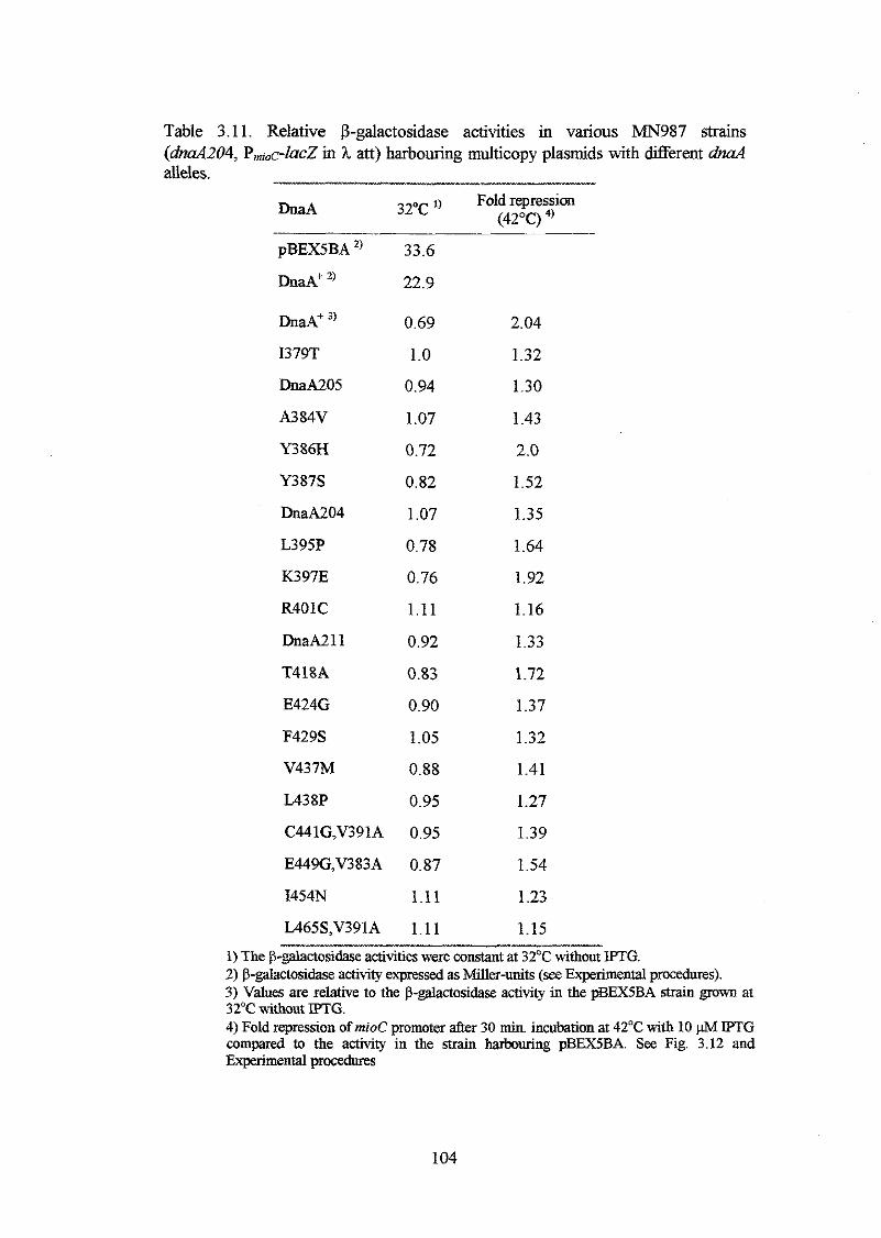
Table 3.1 1. Relative P-galactosidase activities in various MN987 strains (dnaA204, PmiOc-ZacZ in h att) harbouring multicopy plasmids with different dnuA alleles. -
DnaA 32°C ') Fold repression (42°C) 4,
pBEX5BA
DnaA' ')
D n a A + 3,
I379T
DnaA205
A384V
Y386H
Y387S
DnaA204
L395P
K397E
R401C
DnaA211
T418A
E424G
F429S
V437M
L438P
C441G,V391A
E449G,V383A
I454N
L465S,V391A
33.6
22.9
0.69
1.0
0.94
1.07
0.72
0.82
1.07
0.78
0.76
1.11
0.92
0.83
0.90
1.05
0.88
0.95
0.95
0.87
1.11
1.11
2.04
1.32
1.30
1.43
2.0
1.52
1.35
1.64
1.92
1.16
1.33
1.72
1.37
1.32
1.41
1.27
1.39
1.54
1.23
1.15 1) The p-galactosidase activities were cons& at 32'C without IPTG. 2) &&ctosidase activity exoressed as Miller-units (see Exuerimental urocedures). . . - 3) Values are relative to the p-galactosidase activi6 in the pBEX5BA strain &m at 32OC without IPTG.
- -
4) Fold repression of mzoC promoter after 30 min. incubation af 42°C with 10 IPTG
Experimental procedures compared to the activity in the strain harbouring pBEX5BA. See Fig. 3.12 and
104
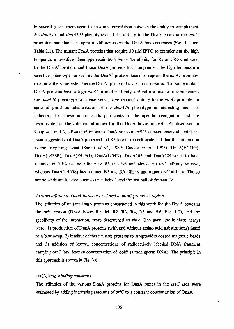
In several cases, there seem to be a nice correlation between the ability to complement
the dna446 and dnu4204 phenotypes and the affinity to the DnaA boxes in the mioC
promoter, and that is in spite of differences in the DnaA box sequences (Fig. 1.1 and
Table 2.1). The mutant DnaA proteins that require 30 pM IPTG to complement the high
temperature sensitive phenotype retain 60-70% of the affinity for R5 and R6 compared
to the DnaA' protein, and those DnaA proteins that complement the high temperature
sensitive phenotypes as well as the DnaA+ protein does also repress the mioC promoter
to almost the same extend as the DnaA' protein does. The observation that some mutant
DnaA proteins have a high mzoC promoter &nity and yet are unable to complement
the dnaA46 phenotype, and vice versa, have reduced affinity to the mioC promoter in
spite of good complementation of the & d 4 6 phenotype is interesting and may
indicates that these amino acids participate in the specific recognition and are
responsible for the different W t i e s for the DnaA boxes in oriC. As discussed in
Chapter 1 and 2, different affinities to DnaA boxes in oriC has been observed, and it has
been suggested that DnaA proteins bind R3 late in the cell cycle and that this interaction
is the triggering event (Samitt et al., 1989; Cassler el d., 1995). DnaA(E424G),
DnaA(L438P), DnaA(EA49G), DnaA(I454N), DnaA205 and DnaA204 seem to have
retained 60-70% of the affinity to R5 and R6 and almost no oriC affinity in vivo,
whereas DnaA(L465S) has reduced R5 and R6 affinity and intact oriC afiinity. The se
amino acids are located close to or in helix 1 and the last half of domain IV.
in vitro @nity to DnaA boxes in oriC and in mioCpromoter region
The affinities of mutant DnaA proteins constructed in this work for the DnaA boxes in
the oriC region @naA boxes RI, M, R2, R3, R4, R5 and R6. Fig. l . l) , and the
specificity of the interaction, were determined in vitro. The main line in these assays
were: 1) production of DnaA proteins (with and without amino acid substitutions) hsed
to a biotin-tag, 2) binding of these fusion proteins to streptavidin coated magnetic beads
and 3) addition of known concentrations of radioactively labelled DNA fragment
canying oriC (and known concentration of 'cold' salmon sperm DNA). The principle in
this approach is shown in Fig. 3.6.
oriC-DnaA binding constants
The afiinities of the various DnaA proteins for DnaA boxes in the oriC area were
estimated by adding increasing amounts of oriC to a constant concentration of DnaA
105
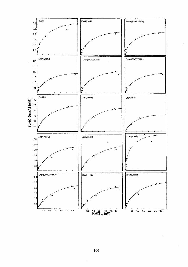
30
25
aa4(E424G)
3.0 W 4 3 7 M )
25
30
25
W C U l G . V B 1 A )
::I_ Q5 1.0 0.5 1.0 1.5 20 25 3.0 ~ Q5 1.0 1.5 20 25 3.0
106
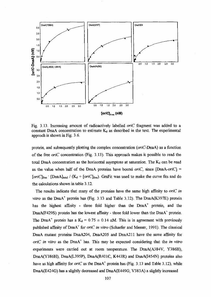
Fig. 3.13. Increasing amount of radioactively labelled orzC fragment was added to a constant DnaA concentration to estimate & as described in the text. The experimental approch is shown in Fig. 3.6.
protein, and subsequently plotting the complex concentration (oriC-DnaA) as a function
of the free oriC concentration (Fig. 3.13). This approach makes it possible to read the
total DnaA concentration as the horizontal asymptote at saturation. The E;d can be read
as the value when half of the DnaA proteins have bound oriC, since [DnaA-oriq =
[or zqk ' [DnaA]u / (h + [oric]s,,>. GraFit was used to make the curve fits and do
the calculations shown in table 3.12.
The results indicate that many of the proteins have the same high affinity to oriC in
vztro as the DnaA' protein has (Fig. 3.13 and Table 3.12). The DnaA(K397E) protein
has the highest affinity - three fold higher than the DnaA+ protein, and the
DnaA(F429S) protein has the lowest affinity - three fold lower than the DnaA' protein.
The DnaA+ protein has a & = 0.75 * 0.14 nM. This is in agreement with previously
published afhity of DnaA' for oriC in vitro (Schaefer and Messer, 1991). The classical
DnaA mutant proteins DnaAZ04, DnaA205 and DnaA211 have the same affinity for
oriC in vitro as the DnaA' has. This may be expected considering that the in vitro
experiments were carried out at room temperature. The DnaA(A384V, Y386H),
DnaA(Y386H), DnaA(L395P), DnaA(R401C, K443R) and DnaA(I454N) proteins also
have as high affinity for oriC as the DnaA+ protein has (Fig. 3.13 and Table 3.12), while
DnaA(E424G) has a slightly decreased and DnaA(E449G, V383A) a slightly increased
107
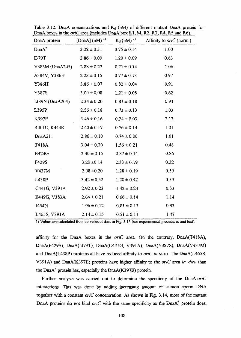
Table 3.12. DnaA concentrations and & (nM) of different mutant DnaA protein for DnaA boxes in the oriC area (ichdes DnaA box R1, M, R2, R3, R4, R5 and R6). DnaA protein [ ~ n a ~ ] (M) (d) *) to oriC (norm.)
DMA+
I379T
V383M (DnaA205)
A384V, Y386H
Y386H
Y387S
I389N (DnaA204)
L395P
K397E
R401 C, K443R
D M 1 1
T418A
E424G
F429S
v43 7h4
L438P
C441G, V391A
E449G, V383A
I454N
L4655, V391A
3.22 f 0.31
2.86 i 0.09
2.88 f 0.22
2.28 f 0.15
3 . 8 6 i 0.07
3.00 f 0.08
2.34 f 0.20
2.56 f 0.18
3.46 1 0.16
2.40 f 0.17
2.86 f 0.10
3.04 f 0.20
2.30 * 0.15
3.20 10.14
2.98 10.20
3.42 f 0.52
2.92 i 0.23
2.64 f 0.21
1.96 f 0.12
2.14f0.15
0.75 1 0.14
1.20 f 0.09
0.71 1 0.14
0.77 f 0.13
0.82 f 0.04
1.21 f 0.08
0.81 f0.18
0.73 f 0.13
0.24 f 0.03
0.76 i 0.14
0.74 f 0.06
1.56 f 0.21
0.87 f 0.14
2.33 f 0.19
1.28 f 0.19
1.28 i 0.42
1.42 f 0.24
0.66 f 0.14
0.81 f 0.13
0.51 fO.11
1 .oo 0.63
1.06
0.97
0.91
0.62
0.93
1.03
3.13
1.01
1.01
0.48
0.86
0.32
0.59
0.59
0.53
1.14
0.93
1.47 1) Values are calculated from m e f i t s of data in Fig 3.13 (see experimental procedures and text).
atfinity for the DnaA boxes in the oriC area. On the contrary, DnaA(T418A),
DnaA(F429S), DnaA(I379T), DnaA(C441G, V391A), DnaA(Y387S), DnaA(V437h4)
and DnaA(TA38P) proteins all have reduced afsnity to oriC in vitro. The DnaA(L465S,
V391A) and DnaA(K397E) proteins have higher affinity to the oriC area in vitro than
the DnaA' protein has, especially the DnaA(K397E) protein.
Further analysis was canied out to determine the specificity of the DnaA-orzC
interactions. This was done by adding increasing amount of salmon sperm DNA
together with a constant oriC concentration. As shown in Fig. 3.14, most of the mutant
DnaA proteins do not bind oriC with the same specificity as the DmA' protein does.
108
The DnaA(K397E) and DnaA(Y386H) proteins have, however, somewhat increased
specificity towards oriC, whereas the DnaA(T418A), DnaA(C441G, V391A),
DnaA(E449G, V383), DnaA(F429S) and DnaA(I379T) proteins still have high
specificity towards oriC, although lower than what is observed with the DnaA' protein.
The rest of the DnaA proteins have reduced specificity toward oriC, the DnaA(A384V,
Y386H), DnaA(E424G), DnaA204 and especially DnaA(L395P) have severely reduced
specificity towards oriC. The results are rather interesting, and indicates that even
though the affinity for oriC is reduced, the specificity towards oriC can still be intact
(DnaA(F429S) for instance), and vice versa, a high affinity to oriC does not automaticly
mean that the interaction is specific towards oriC (DnaA(L395P)). Fig. 3.15 summarises
the results of the in vivo and in vitro binding experiments. The DnaA proteins with the
highest in vitro affinity but low specificity are the classical DnaA mutant proteins
DnaA205 and DnaA204 and DnaA(A384V, Y386H), DnaAF395P) and the
DnaAQWOlC, K443R) protein pig. 3.15). These amino acid substitutions are located in
a-helix 1 and in the basic loop, and this may suggest that this part of the DNA binding
domain participate in the specific recognition of the DnaA box. Generally, the in vivo
affinities of mutant DnaA proteins for the DnaA box R5 and R6 boxes are lower relative
to that of the DnaA' protein compared to the in vitro affinities for the oriC area (which
includes DnaA box Rl-R6. See Fig. 1.1). It is also conspicuous that the in vivo affinities
in many cases 'follow' the specificity, that is, when the in vivo affinity is low so is the in
vitro specificity. This indicates that the observed reduced aflinity to R5 and R6 in vivo
may be due to a titration of DnaA proteins by the rest of the chromosome. Thus, there
seems to be correlation between the in vivo and in vitro binding experiments.
109
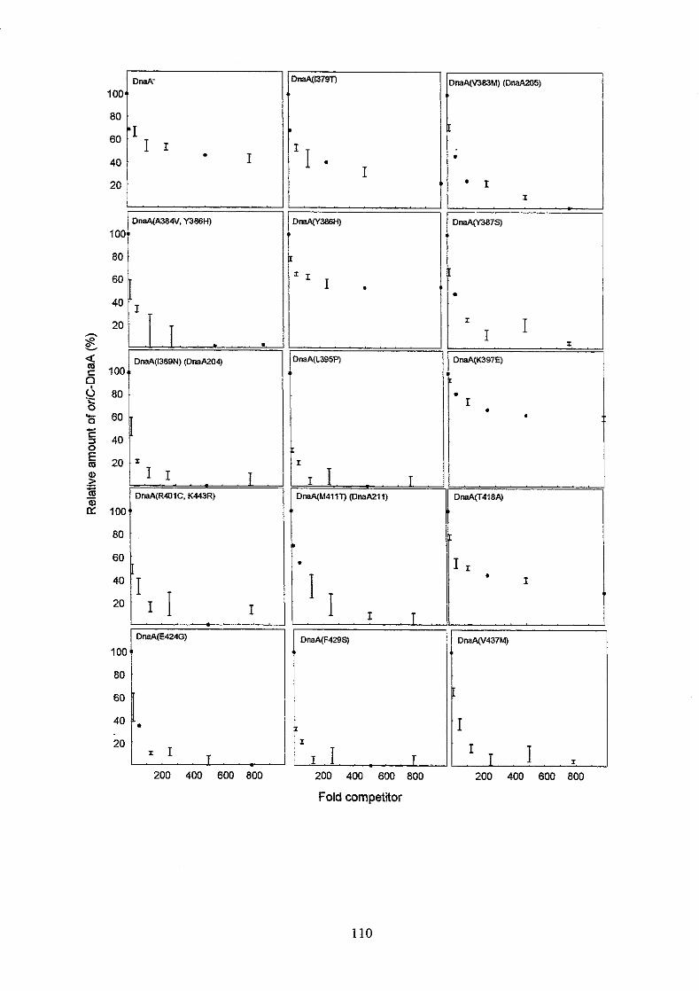
100-
80
60
40 I 20
DreA'
'I I 1 .
loo,,
80
60
OmA(A384V. Y386H)
g - 2 0 / .I T - .
! I I I , , , 20 , ~ I,
80
:: p. 2 0 1 T I r
I I . . _ 200 400 600 800
' I ' I
= I I
DnaA(L3954
t
I
I , I . T DnaA(M411T) (OnaA211)
. / I ,
.I I I . 200 400
f
- 1 I
Dm%A(vJ87S)
*
T T I I
I.
DnaA(K397E)
. I
DnaA(T418A)
T rr-l- I
DnaA(F429S) DnaAy437M)
200 400 600 800 200 400 600 800
Fold competitor
110
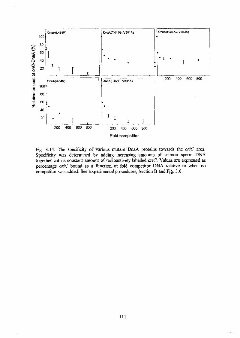
100
C . m .
I
200 400 600 800 200 400 600 800
Fold competitor
200 400 600 800
Fig. 3.14. The specificity of various mutant DnaA proteins towards the orzC area. Specificity was determined by adding increasing amounts of salmon sperm DNA together with a constant amount of radioactively labelled oriC. Values are expressed as percentage orzC bound as a hnction of fold competitor DNA relative to when no competitor was added. See Experimental procedures, Section I1 and Fig. 3.6.
111
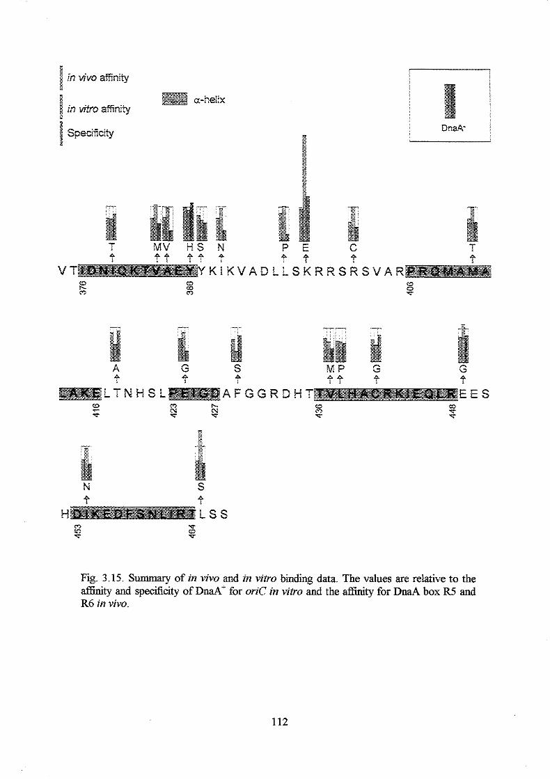
* a-heiix
in vitp asnity
specmity
T MV H S M P 4 ? P P
l
VT KIKVADLLSKRRSRSVAR 2 0
22 m a 0 -r
4 ? 8 44 P P P
A F G G R D H T E E S n h. .ic f
F- h. f
$2 'c
c? f T
4 P H ISS 0 LD f
'p
T c?
Fig. 3.15. Summary of in vivo and in vitro binding data. The values are relative to the affinity and specificity of DnaA' for oriC in vitro and the affinity for DnaA box R5 and R6 in vivo.
112

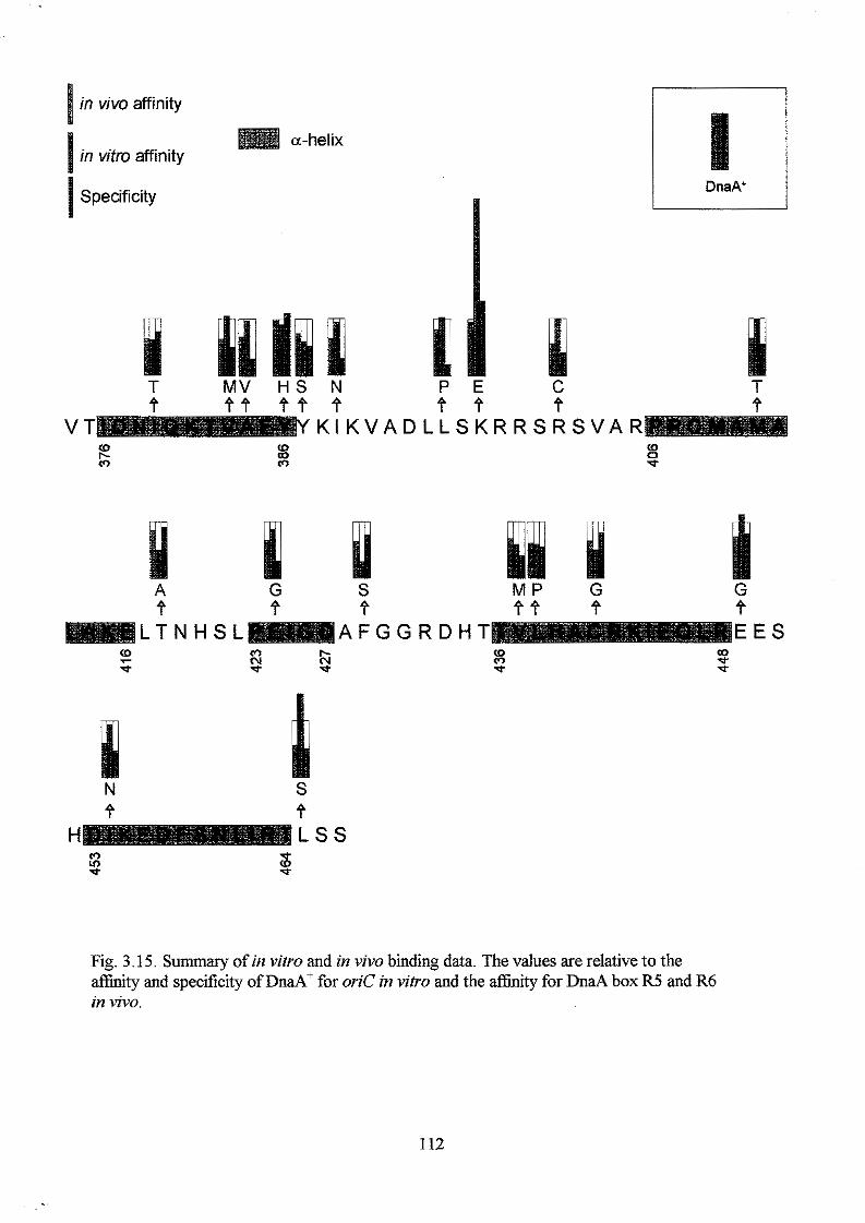
in vivo affinity
a-helix in vitro affinity
1 DnaA’ l , I
.
Specificity
T MV H S N P E f ? f
c f
T
V T K I K V A D L L S K R R S R S V A R E m m g
‘p
A G S f f f
M P G e + e e L T N H S L A F G G R D H T E E S
. .
: m N ‘p
IC N ‘p
8 d-
m .;r d-
N S f f
H L S S m v) ‘p
‘p (D ‘p
Fig. 3.15. Summary of in vitro and in vivo binding data. The values are relative to the affinity and specificity of DnaA’ for oriC in vitro and the affinity for DnaA box R5 and R6 in vivo.
112
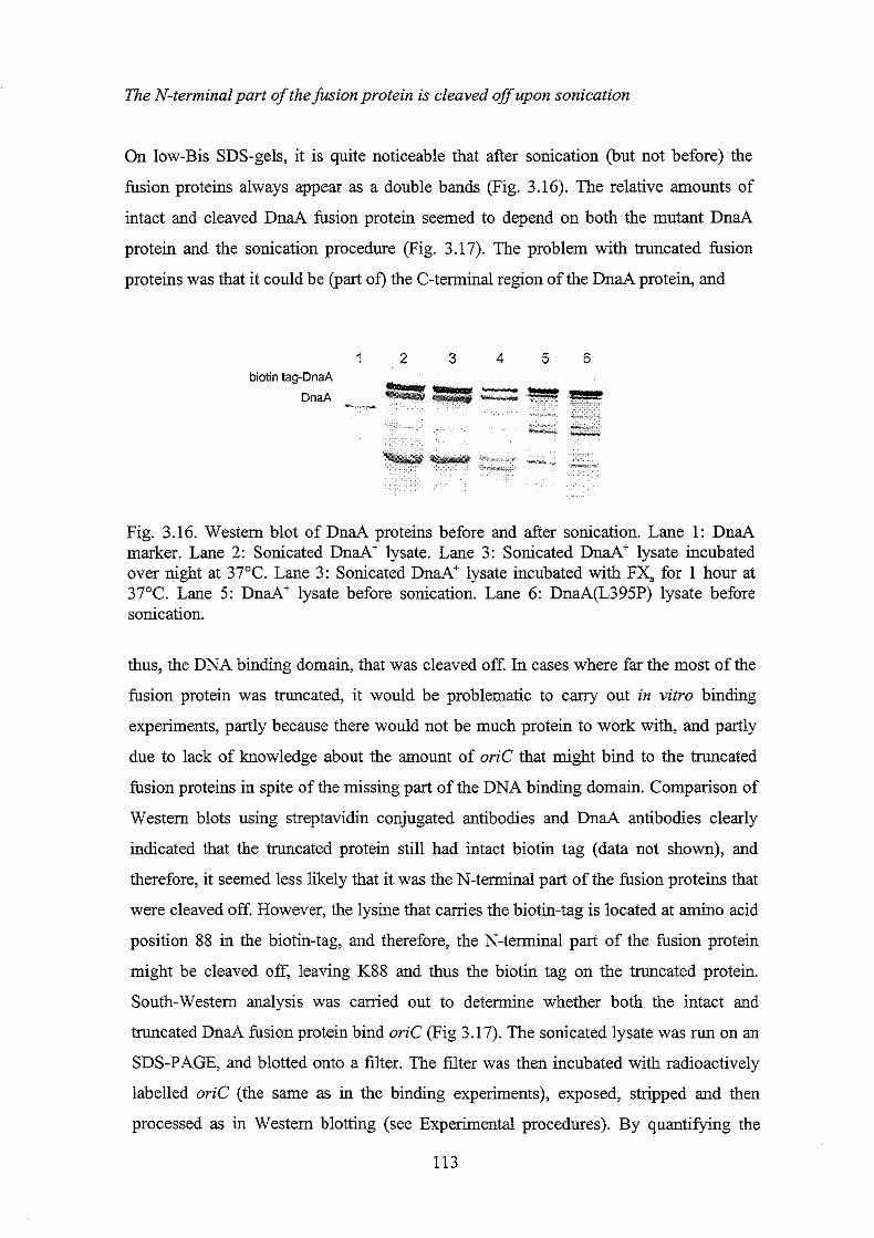
The N-terminalpart of the fusion protein is cleaved off upon sonication
On low-Bis SDS-gels, it is quite noticeable that after sonication (but not before) the
fusion proteins always appear as a double bands (Fig. 3.16). The relative amounts of
intact and cleaved DnaA fusion protein seemed to depend on both the mutant DnaA
protein and the sonication procedure (Fig. 3.17). The problem with truncated fusion
proteins was that it could be (part of) the C-terminal region of the DnaA protein, and
1 2 3 4 5 6
Fig. 3.16. Westem blot of DnaA proteins before and after sonication. Lane 1: DnaA marker. Lane 2: Sonicated DnaA' lysate. Lane 3: Sonicated D&+ lysate incubated over night at 37°C. Lane 3: Sonicated DnaA+ lysate incubated with FX, for 1 hour at 37°C. Lane 5 : DnaA' lysate before sonication. Lane 6: DnaA(L395P) lysate before sonication.
thus, the DNA binding domain, that was cleaved off. In cases where far the most of the
fusion protein was truncated, it would be problematic to cany out in vitro binding
experiments, partly because there would not be much protein to work with, and partly
due to lack of knowledge about the amount of oriC that might bind to the truncated
fusion proteins in spite of the missing part of the DNA binding domain. Comparison of
Western blots using streptavidin conjugated antibodies and DnaA antibodies clearly
indicated that the truncated protein still had intact biotin tag (data not shown), and
therefore, it seemed less likely that it was the N-terminal part of the fusion proteins that
were cleaved off. However, the lysine that carries the biotin-tag is located at amino acid
position SS in the biotin-tag, and therefore, the N-terminal part of the fusion protein
might be cleaved off, leaving K88 and thus the biotin tag on the truncated protein.
South-Westem analysis was carried out to determine whether both the intact and
truncated DnaA fusion protein bind oriC (Fig 3.17). The sonicated lysate was run on an
SDS-PAGE, and blotted onto a filter. The filter was then incubated with radioactively
labelled oriC (the same as in the binding experiments), exposed, stripped and then
processed as in Westem blotting (see Experimental procedures). By quantifylng the
113
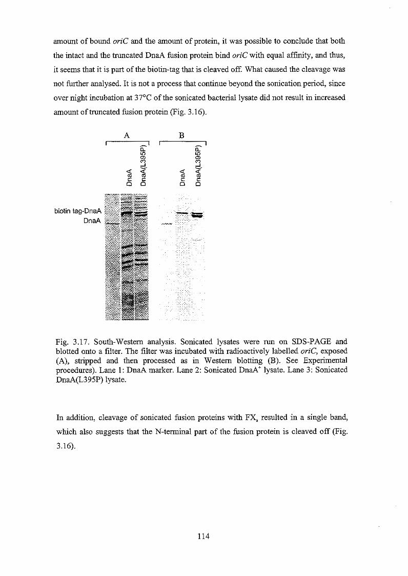
amount of bound oriC and the amount of protein, it was possible to conclude that both
the intact and the truncated DnaA fusion protein bind oriC with equal affinity, and thus,
it seems that it is part of the biotin-tag that is cleaved off. What caused the cleavage was
not further analysed. It is not a process that continue beyond the sonication period, since
over night incubation at 37OC of the sonicated bacterial lysate did not result in increased
amount oftruncated fusion protein (Fig. 3.16).
biotin
Fig. 3.17. South-Westem analysis. Sonicated lysates were run on SDS-PAGE and blotted onto a filter. The filter was incubated with radioactively labelled oriC, exposed (A), stripped and then processed as in Westem blotting (B). See Experimental procedures). Lane 1: DnaA marker. Lane 2: Sonicated DnaA+ lysate. Lane 3: Sonicated DnaA(L395P) lysate.
In addition, cleavage of sonicated fusion proteins with FX, resulted in a single band,
which also suggests that the N-terminal part of the fusion protein is cleaved off (Fig.
3.16).
114
Section III
Integration and analysis of mutant dnaA alleles in h attachment site of
the chromosome
To h t h e r analyse the effect of amino acid substitutions on the function and activity of
the DnaA protein, it was tried to insert several d d mutant genes into their normal
chromosomal location, and later into the h attachment site of the chromosome. Several
different approaches were used, and they will be described in the following. The last
attempt was successful, and the effect of the A384V, L395P, K397E and C441G
substitutions will be discussed (and the A184V substitution in Chapter 4).
Recombination into chromosomal location using dnaA46, recBD strain
A dnuA46, recBD strain (kindly fiom Christoph Weigel) was used to insert mutant
dnaA genes into their normal chromosomal location. The strategy was rather
straightforward, and it consisted of re-streaking of high temperature resistant dnaA46,
recBD recombinants (or, as it turned out, revertants). First, several pMN242 derivatives
canying various dnaA mutant alleles were transformed into the dm446, recBD strain
and plated at high temperature (42°C) on rich media supplemented with ampicillin and
IPTG. The following day several colonies from each transformation were streaked on
rich media supplemented with ampicillin but without IPTG and incubated at 42T.
Lack of IPTG in the growth media generally resulted in reduced and 'sick' growth of the
transformants, but (some of) the colonies that appeared as 'healthy' should consist of
cells in which the high temperature sensitive W 4 6 allele had recombined out of the
chromosome. These high temperature resistant colonies were re-streaked at 42OC on
rich media without ampicillin and IPTG several times to allow growth of plasmid free
cells. Verification of the chromosomal dnuA alleles of high temperature resistant and
ampicillin sensitive cells were carried out by PCR amplification of both the last part of
the dnd gene encoding the DNA binding domain, and the area that includes the
&a446 mutations followed by restriction enzyme digestions (see Experimental
procedures, section III). The principle in this technique works well, and it was very
straightforward to substitute the M 4 6 allele with a allele (data not shown).
However, it was not possible to insert new mutant dnuA alleles into the chromosome. It
115
was unproblematic to recombine the M 4 6 mutations out of the chromosome, but
never possible to recombine the C-terminal mutations into the chromosome. The M
gene of 30 potentially high-temperature resistant and ampicillin sensitive dnaA(I379T)
and hd (Y386H) strains were PCR amplied and digested with DdeI and ScaI,
respectively (see Experimental procedures, section III). All were &d+. It was expected
to be possible to insert the haA(Y386H) allele on the chromosome because the
DnaA(Y386H) protein seems very wild type like with respect to complementation of
the M 4 6 allele, affinity to the mioC promoter, in vitro affinity to oriC and specificity
towards oriC (see sections I and II). Thus, it seems that there was not enough DNA for
the recombination event. Alternatively, (some oQ the DnaA proteins encoded by the
mutant dm4 alleles are high temperature sensitive as the DnaA46 protein, and
therefore, selection at 42°C would not select correct recombinants. However, lowering
the temperature from 42OC to 39OC did not result in recombinants with the desired dnaA
allele either.
Analysts of M O strains carrying mutant dnaA genes in 1 attachment site ana'
harbouring aplasmid with P~rnrnh'fi&sion.
If the mutant dnaA genes cannot be introduced in their right chromosomal position, the
second best seemed to be to insert them into the h attachment site in a strain that does
not carry a dnaA gene (t3m.A'). Such a strain has been constructed. It is called TC3482
and it carries a chloramphenicol cassette in the h a 4 gene (Hansen et al., 1991. See
Experimental procedures). The cell survives due to a mutation in the rnhA gene, a gene
that encodes RNase H. RNase H normally removes RNA-DNA hybrids, and thus, when
it is absent those hybrids are allowed to exist, and the chromosomal replication can
initiate from DnaA independent oriK's (for review see Messer and Weigel, 1996). The
experimental strategy was to insert various mutant IPTG inducible M alleles into the
h attachment site of a &do strains harbouring a plasmid with the rhnA gene under
arabinose promoter control and analyse the initiation frequency from oriC by adding
arabinose to the growth medium. The construction of the various strains went well, but
unfortunately, it turned out that the PBm-rnh+ plasmid probably did not synthesise
enough RNase H to prevent oriK initiations. This was first under suspicion when
colonies, although very small, appeared on LB plates supplemented with arabinose, and
later concluded from the observation that when strains canying the PBm-rnh' plasmid
were grown in media supplemented with arabinose and treated with rifampicin (which
also inhibits initiations from oriK), the DNA distributions were similar to the ones in a 116
cell population that did not cany the P~m-mh' plasmid. The oriK initiations in
presence of arabinose was not a result of growth of plasmid free cells, because the
growth medium was supplied with 500 pg/d ampicillin every 40 min.
Analysis of mutant d n d alleles in a & d 2 0 4 and &do background A selection of mutant dnaA alleles and the &df allele were inserted into a strain that
carried a & d 2 0 4 allele on the chromosome (TC1929), or alternatively, into the dnaAo,
mh373 strain (TC3482) mentioned above. Both strains can grow at 32OC independently
on the dna4 allele in h attachment site. However, at 42T, the M 2 0 4 strains were
IPTG dependent (- or sensitive) as expected (Table 3.13). The mh373 mutation was
transduced out of the d n a A o rhn373 strains carrying different dnaA alleles in h
attachment site (Fig. 3.7) making these strains IPTG dependent (- or sensitive) (Table
3.13). The various strains were grown in the presence of different amounts of IPTG as
described in Experimental procedures, section III, samples were take% treated with
rifampicin (+cephalexin) and the chromosome configurations were analysed using flow
cytometry. Samples were also taken to measure the DnaA concentration using Western
blotting as described below and in Experimental procedures (section III and General
procedures).
117
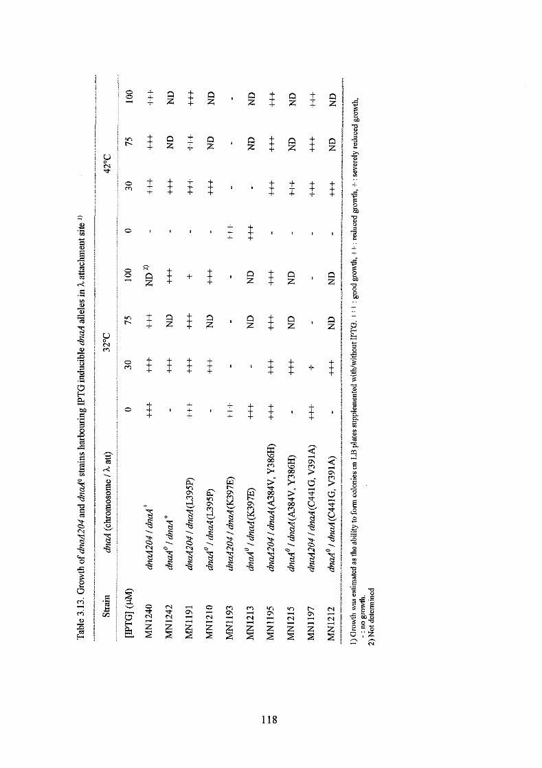
0 0 3
v, t.
0 m
0
0 0
v, r-
0 m
0
g E v
k?
+ + +
+ + +
+ + +
- N i2
+ + +
+ + +
+ + +
+=S B
-3 B
. p. 0 "1
0 d G
n z
i2
+ + +
+ + +
!2
+ + +
+ -3 B
B % .
N d
+ + +
+ + +
+ + +
+
+ + +
+ + +
+ + +
E %
B 8 2
. "1 P
-Q
i2
+ + +
+ + +
n z
+ + +
E %
B
B
. '%
2
+ + +
+ + +
+ + +
+ + +
+ + +
+
+ + +
g W
F 2" 3 W
P
h S
S
B
. 2
v,
e m
S
+ + +
+ + +
+ + +
+
+ + +
z S
5
3 m
d
B S -3 B
. "1
b
2
3
118
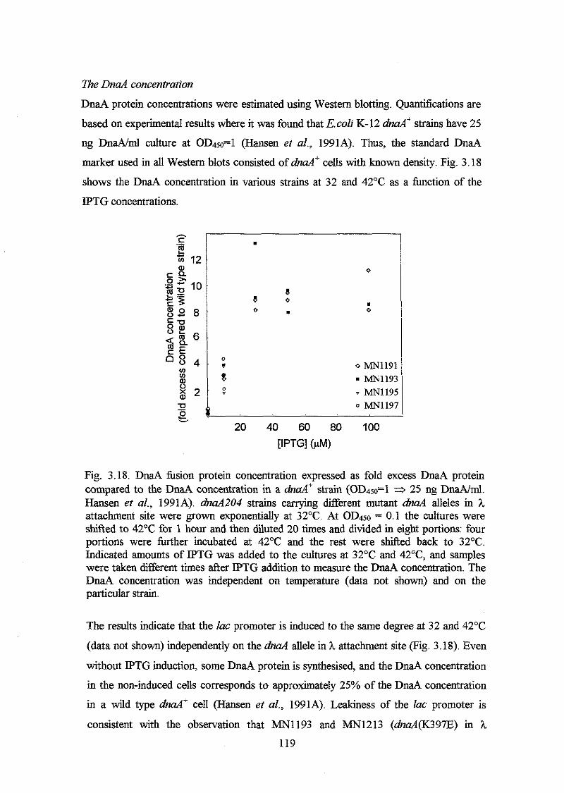
n e DnaA concentration
DnaA protein concentrations were estimated using Western blotting. Quantifications are
based on experimental results where it was found that E.coZi K-l2 M" strains have 25
ng DnaA/ml culture at oD450=1 (Hansen et al., 1991A). Thus, the standard DnaA
marker used in all Western blots consisted of &ad cells with known density. Fig. 3.18
shows the DnaA concentration in various strains at 32 and 42°C as a hnction of the
IPTG concentrations.
. 0
8 a 0 .
0 . 0
D
. v 0 m 1 1 9 1 8 1 m 1 1 9 3 v v m 1 1 9 5
0 MN119i
0
b 20 40 60 80 100
[IPTG] ($4
Fig. 3.18. DnaA fusion protein concentration expressed as fold excess DnaA protein compared to the DnaA concentration in a M+ strain (OD450=1 3 25 ng DnaA/ml. Hansen et al., 1991A). dnaA204 strains canying different mutant M alleles in h attachment site were grown exponentially at 32OC. At OD450 = 0.1 the cultures were shified to 42°C for 1 hour and then diluted 20 times and divided in eight portions: four portions were further incubated at 42°C and the rest were shifted back to 32OC. Indicated amounts of IPTG was added to the cultures at 32°C and 42T, and samples were taken different times &er IPTG addition to measure the DnaA concentration. The DnaA concentration was independent on temperature (data not shown) and on the particular strain.
The results indicate that the lac promoter is induced to the same degree at 32 and 42OC
(data not shown) independently on the M allele in h attachment site (Fig. 3.18). Even
without IPTG induction, some DnaA protein is synthesised, and the DnaA concentration
in the non-induced cells corresponds to approximately 25% of the DnaA concentration
in a wild type &d+ cell (Hansen et al., 1991A). Leakiness of the lac promoter is
consistent with the observation that MN1193 and MN1213 (dnaA(K397E) in h
119
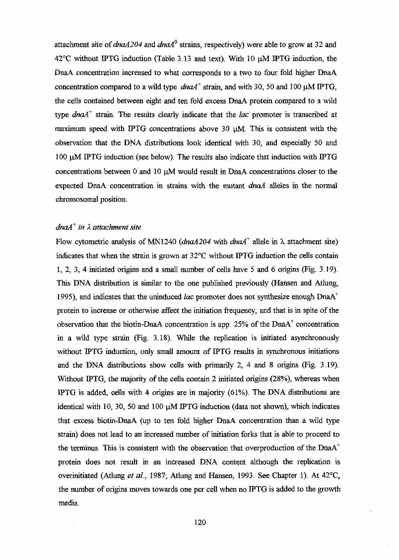
attachment site of M 2 0 4 and &do strains, respectively) were able to grow at 32 and
42°C without IPTG induction (Table 3.13 and text). With 10 pM IPTG induction, the
DnaA concentration increased to what corresponds to a two to four fold higher DnaA
concentration compared to a wild type M' strain, and with 30,50 and 100 pM IPTG,
the cells contained between eight and ten fold excess DnaA protein compared to a wild
type &d+ strain. The results clearly indicate that the lac promoter is transcribed at
maximum speed with IPTG concentrations above 30 pM. This is consistent with the
observation that the DNA distributions look identical with 30, and especially 50 and
100 pM IPTG induction (see below). The results also indicate that induction with IPTG
concentrations between 0 and 10 @l would result in DnaA concentrations closer to the
expected DnaA concentration in strains with the mutant &d alleles in the normal
chromosomal position.
dnd' in ;1 attachment site
Flow cytometric analysis of MN1240 (dm4204 with &d+ allele in h attachment site)
indicates that when the strain is grown at 32OC without IPTG induction the cells contain
l , 2, 3, 4 initiated origins and a small number of cells have 5 and 6 origins (Fig. 3.19).
This DNA distribution is similar to the one published previously (Hansen and Atlung,
1995), and indicates that the uninduced lac promoter does not synthesize enough DnaA+
protein to increase or otherwise affect the initiation frequency, and that is in spite of the
observation that the biotin-DnaA concentration is app. 25% of the DmA' concentration
in a wild type strain (Fig. 3.18). While the replication is initiated asynchronously
without IPTG induction, only small amount of IPTG results in synchronous initiations
and the DNA distributions show cells with primarily 2, 4 and 8 origins (Fig. 3.19).
Without IPTG, the majority of the cells contain 2 initiated origins @S%), whereas when
IPTG is added, cells with 4 origins are in majority (61%). The DNA distributions are
identical with 10,30, 50 and 100 PM IPTG induction (data not shown), which indicates
that excess biotin-DnaA (up to ten fold higher DnaA concentration than a wild type
strain) does not lead to an increased number of initiation forks that is able to proceed to
the terminus. This is consistent with the observation that overproduction of the DnaA+
protein does not result in an increased DNA content although the replication is
overinitiated (Atlung et al., 1987; Atlung and Hansen, 1993. See Chapter 1). At 42"C,
the number of origins moves towards one per cell when no IPTG is added to the growth
media.
120
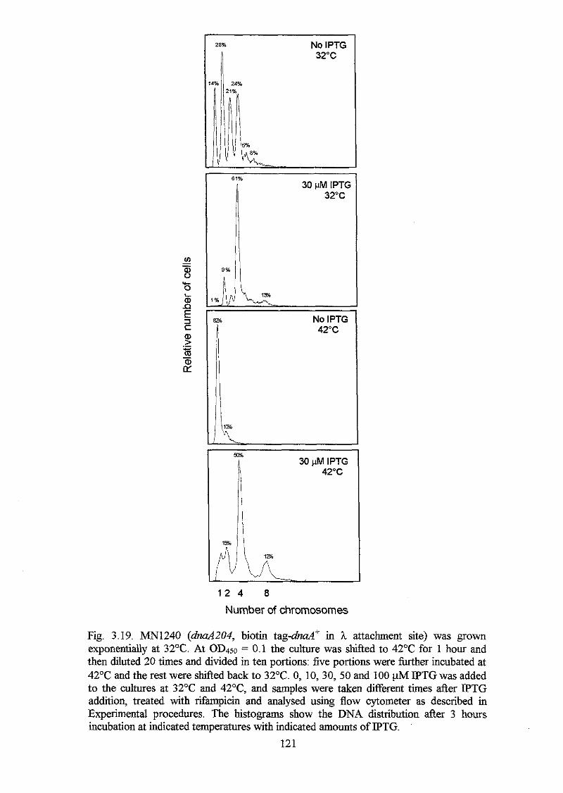
28% No IPTG 32°C
61% 30 PM IPTG
32°C
No IPTG 42°C
1 2 4 8
Number of chromosomes
Fig. 3.19. M 1 2 4 0 (dna4204, biotin tag&d* in h attachment site) was grown exponentially at 32T. At OD450 = 0.1 the culture was shifted to 4 2 T for 1 hour and then diluted 20 times and divided in ten portions: five portions were hrther incubated at 42°C and the rest were shifked back to 32°C. 0, 10,30,50 and 100 pM P T G was added to the cultures at 32°C and 42OC, and samples were taken different times &er IPTG addition, treated with rifampicin and analysed using flow cytometer as described in Experimental procedures. The histograms show the DNA distribution after 3 hours incubation at indicated temperatures with indicated amounts of IPTG.
121
Thus, without IPTG the biotin-DnaA concentration is not sufficiently high to allow
initiation of chromosome replication, which is consistent with the data at 32OC and with
the observation that m 1 2 4 0 only forms colonies at 4 2 T in the presence of IPTG
(table 3.13). When 10, 30, 50 and 100 pM IPTG is added, chromosome replication is
initiated, and the cells contain 1, 2, 4, and 8 origins. At 42OC there is a large number of
very big cells that contain 1 (and 2) chromosomes. The size of these cells seem to
increase with increasing incubation time (Fig. 3.20). The existence of a fraction of big
cells has been observed previously in the same &a4204 strain background (Hansen and
Atlung, 1995A).
A B
l * 2 4 8 2 4 8
Number of chromosomes
Fig. 3.20. Contour blots of "240 cells (&d+ in dnuA204 background) grown at 42°C with 30 pIvf IPTG. A: after 2 hours growth and B: after 3 hours growth.
The cells do not seem to be able to initiate chromosome replication although the growth
medium is supplemented with IPTG, and it seems as if this population of cells is stalled
in a locked situation. The population of cells grew exponentially, but more slowly than
at 32°C. This is unexpected and may be explained by the presence of these large cells
not responding to IPTG.
MN1242 (M+ in strain background) was able to initiate chromosome
replication at both 32 and 42°C with only 3 pM IPTG induction, and the number of
origins per cell depended on the temperature and only slightly on the amount of IPTG
(Fig. 3.21). Thus, it seems that enough DnaA' protein is synthesised from the 3 pM
122
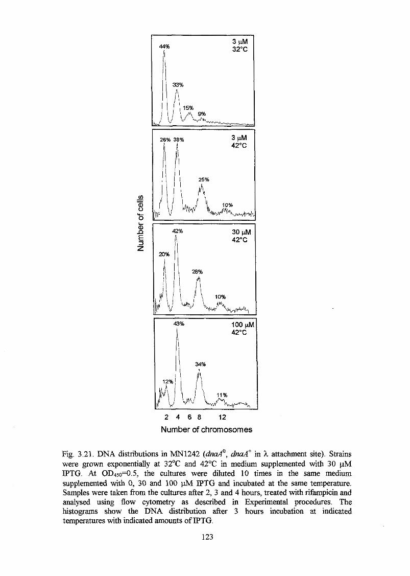
44%
i 3 PM 32°C
42% n 42°C 30 pM
2 4 6 8 12
Number of chromosomes
Fig. 3.21. DNA distributions in "242 (&d", dnd' in L attachment site). Strains were grown exponentially at 32OC and 42°C in medium supplemented with 30 pM IPTG. At OD450=0.5, the cultures were diluted 10 times in the same medium supplemented with 0, 30 and 100 m IPTG and incubated at the same temperature. Samples were taken from the cultures after 2, 3 and 4 hours, treated with rifampicin and analysed using flow cytometry as described in Experimental procedures. The histograms show the DNA distribution after 3 hours incubation at indicated temperatures with indicated amounts of IPTG.
123
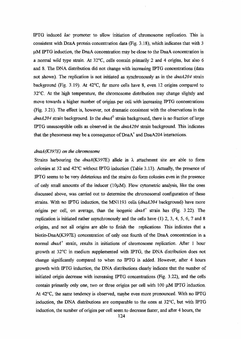
IPTG induced lac promoter to allow initiation of chromosome replication. This is
consistent with DnaA protein concentration data (Fig. 3.18), which indicates that with 3
@l IPTG induction, the DnaA concentration may be close to the DnaA concentration in
a normal wild type strain. At 3 2 T , cells contain primarily 2 and 4 origins, but also 6
and 8. The DNA distribution did not change with increasing IPTG concentrations (data
not shown). The replication is not initiated as synchronously as in the dnaA204 strain
background (Fig. 3.19). At 42OC, far more cells have 8, even 12 origins compared to
32°C. At the high temperature, the chromosome distribution may change slightly and
move towards a higher number of origins per cell with increasing IPTG concentrations
(Fig. 3.21). The effect is, however, not dramatic consistent with the observations in the
& d 2 0 4 strain background. In the &do strain background, there is no fiaction oflarge
IPTG unsusceptible cells as observed in the dnaA204 strain background. This indicates
that the phenomena may be a consequence of DnaA+ and DnaA204 interactions.
dnaA(K397E) on the chromosome
Strains harbouring the dnaA(K397E) allele in h attachment site are able to form
colonies at 32 and 42OC without IPTG induction (Table 3.13). Actually, the presence of
IPTG seems to be very deleterious and the strains do form colonies even in the presence
of only small amounts of the inducer ( l o w ) . Flow cytometric analysis, like the ones
discussed above, was carried out to determine the chromosomal configuration of these
strains. With no IPTG induction, the MN1193 cells ( M 2 0 4 background) have more
origins per cell, on average, than the isogenic &d+ strain has (Fig. 3.22). The
replication is initiated rather asynchronously and the cells have (1) 2, 3, 4, 5, 6, 7 and 8
origins, and not all origins are able to finish the replications. This indicates that a
biotin-DnaA(K397E) concentration of only one fourth of the DnaA concentration in a
normal d n d ' strain, results in initiations of chromosome replication. After 1 hour
growth at 32OC in medium supplemented with IPTG, the DNA distribution does not
change si,y%cantly compared to when no IPTG is added. However, after 4 hours
growth with IPTG induction, the DNA distributions clearly indicate that the number of
initiated origin decrease with increasing IPTG concentrations (Fig. 3.22), and the cells
contain primarily only one, two or three origins per cell with 100 p M IPTG induction.
At 42"C, the same tendency is observed, maybe even more pronounced. With no IPTG
induction, the DNA distributions are comparable to the ones at 32"C, but with IPTG
induction, the number of origins per cell seem to decrease faster, and &er 4 hours, the 124
After 1 hour 32°C After 4 hours 32°C
25%
41% -I
After 1 hour 42°C After 4 hours 42°C
42% -1
2 4 6 2 4 6 2 4 6
3%
I
L
Number of chromosomes Fig. 3.22. m1193 (dnaA204, dnaA(K397E) in h attachment site) was grown exponentially at 32OC. At OD450 = 0.1 the culture was shifled to 42°C for 1 hour and then diluted 20 times and divided in ten portions: five portions were hrther incubated at 42°C and the rest were shifted back to 32°C. 0, 10,30,50 and 100 @l IPTG was added to the cultures at 32OC and 42T, and samples were taken different times after IPTG addition, treated with rifampicin and analysed using flow cytometer as described in Experimental procedures. The histograms show the DNA distribution after 1 and 4 hours incubation at indicated temperatures with indicated amounts of IPTG.
125
cells have only one or two origins with 30,50 and 100 IPTG induction (Fig. 3.22).
The result of this analysis could indicate that the DnaA(K397E) protein has an increased
affinity to orzC and/or decreased afFnity to (some of) the DnaA boxes on the rest of the
chromosome (see Discussion). The results also indicates that MN1193 (and MN1213,
see below) does not form colonies at neither 32 nor 42OC in presence of IPTG (Table
3.13) due to an apparent cease of initiation of chromosome replication. Alternatively,
the initiation of replication actually continues, but the forks do not pass the orzC area as
observed with other dnaA mutants (Nyborg et al., 2000) and in strains in which the
DnaA' protein is overproduced (Atlung et al., 1987; Atlung and Hansen, 1993). Flow
cytometric analysis of MN1213 (dnd(K397E) in dndo background) did not display
discrete peaks (data not shown). However, there seems to be a tendency that the number
of origins per cell at 32' moves towards one, which is consistent with the observations
in the M 2 0 4 strain background (data not shown).
dnd(L395P) on the chromosome
The high temperature sensitive dnaA204 phenotype is complemented when the IPTG
inducible M ( L 3 9 5 P ) allele is inserted into h attachment site and IPTG is added to the
growth medium (Table 3.13). Consistent with this observation, MN1210 ( M ( L 3 9 5 P )
in d n a A o background) forms colonies only in the presence of IPTG (Table 3.13). Flow
cytometric analysis of MN1191 ( M 2 0 4 background) indicates that at 32OC, the
number of origins per cell is rather constant and independent of the IPTG concentration,
although there may be a tendency that the number of origins decrease after 4 hours
incubation with the highest IPTG concentrations (Fig. 3.23). Without IPTG induction,
the DNA distributions at 32°C are comparable to the ones in the isogenic &d+ strain
(MN1240, Fig. 3.19), but unlike MN1240, "191 does not begin to initiate
replication synchronously with IPTG induction. At 42T , MN1191 does, eventually,
seem to stop initiation of replication without IPTG induction, but with only small
amounts of IPTG the replication is overinitiated rather dramatically, especially &er 4
hours. With the highest IPTG concentrations there seem to be too many initiated origins
per cell to make a completion of the many replication forks possible (Fig. 3.23). M e r
one hour growth with IPTG induction at 42OC, a portion of the cells has only one origin,
but unlike the isogenic &df strain (Fig. 3.20), these cells have a normal size (data not
shown). In spite of the apparent dramatic increase in the number of origins at 42OC in
presence of IPTG, MN1191 forms normal colonies on LB plates supplemented with
126
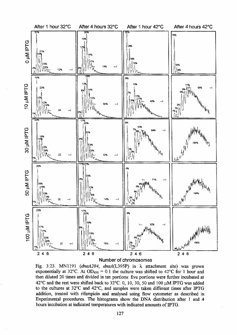
After 1 hour 32°C 31%
l- 1% -I
19%
After 4 hours 32°C After 1 hour 42°C After 4 hours 42°C
17%
17%
$ 1 !-
2 4 6
Number of chromosomes 2 4 6
Fig. 3.23. MN1191 (dnaA204, dnaA(L395P) in X attachment site) was grown exponentially at 32OC. At OD450 = 0.1 the culture was shifted to 42°C for 1 hour and then diluted 20 times and divided in ten portions: five portions were further incubated at 42OC and the rest were shifted back to 3 2 T . 0, 10,30, 50 and 100 pM IPTG was added to the cultures at 32'C and 42OC, and samples were taken different times after IPTG addition, treated with rifampicin and analysed using flow cytometer as described in Experimental procedures. The histograms show the DNA distribution after 1 and 4 hours incubation at indicated temperatures with indicated amounts of IPTG.
127
IPTG. Unfortunately, it was dficult to grow MN1210 (Mo background) in liquid
medium, and therefore, there are no flow cytometric data of this strain.
dnaA(A384K Y386H) on the chromosome
Results of growth analysis and DNA distributions in rifmpicin treated strains
harbouring the M ( A 3 8 4 V , Y386H) allele in h attachment site are similar to strains
harbouring the M ( L 3 9 5 P ) allele (see Table 3.13 and Fig. 3.24). The DnaA(A384V, Y386H) protein also seems to overinitiate replication at 42OC, whereas at 32OC, the
number of origins per cell decreases slightly with increasing IPTG concentrations (Fig.
3.24). m 1 1 9 5 (dna4(A384V, Y386H) in dnaAZ04 background) forms colonies at
32OC with and without IPTG, whereas at 42°C P T G is required (Table 3.13). Thus, like
the isogenic dnaA(L395P) strain, the apparent high number of origins at 42°C in
presence of IPTG does not seem to have any influence on colony formation. Unlike any
of the other &d mutants studied in this work, M 1 2 1 5 (dna4(A384V, Y386H) in
d d 0 background) shows discrete peaks after rifampicin (+ cephalexin) treatment (Fig.
3.25). However, the DNA distributions in "215 do not change with increasing IPTG
concentrations at neither 32 nor 42°C. This is not consistent with observed
overinitiation of replication observed at 42°C in the dm4204 strain background.
128
Aftw 1 hour 32°C
J3sb l /l
l1
After4 hwrs 32°C After 1 hour 42°C After 4 hours 42°C I, c 18% -4
I1 i
Fig. 3.24. m 1 1 9 5 (dnaA204, dnaA(A384V, Y386H) in h attachment site) was grown exponentially at 32T . At OD450 = 0.1 the culture was shifted to 42'C for 1 hour and then diluted 20 times and divided in ten portions: five portions were further incubated at 42OC and the rest were shifted back to 32OC. 0, 10,30,50 and 100 IPTG was added to the cultures at 32OC and 42OC, and samples were taken different times &er IPTG addition, treated with rifampicin and analysed using flow cytometer as described in Experimental procedures. The histograms show the DNA distribution after 1 and 4 hours incubation at indicated temperatures with indicated amounts of IPTG.
129
L 0 pM IPTG I i i 0 UM IPTG
2 4 6 8 1 0
Number of chromosomes Fig. 3.25. DNA distributions in MN1215 (Mo, dnaA(A384V, Y386H) in h attachment site). Strains were grown exponentially at 32°C and 42°C in medium supplemented with 30 pM IPTG. At OD450=0.5, the cultures were diluted 10 times in the same medium supplemented with 0,30 and 100 IPTG and incubated at the same temperature. Samples were taken from the cultures after 2, 3 and 4 hours, heated with rifampicin and analysed using flow cytometry as described in Experimental procedures. The histograms show the DNA distribution after 3 hours incubation at indicated temperatures with indicated amounts of IPTG.
130
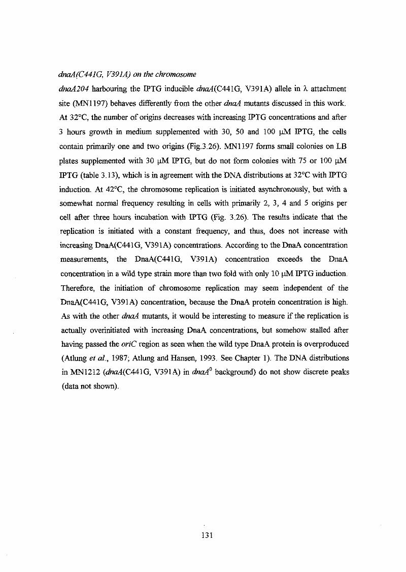
dnaA(C441G, V391A) on the chromosome
W 2 0 4 harbouring the IPTG inducible dmA(C441G, V391A) allele in h attachment
site (IvfN1197) behaves differently from the other d n d mutants discussed in this work.
At 3 2 T , the number of origins decreases with increasing IPTG concentrations and after
3 hours growth in medium supplemented with 30, 50 and 100 @l IPTG, the cells
contain primarily one and two origins (Fig.3.26). W 1 1 9 7 forms small colonies on LB
plates supplemented with 30 @l IPTG, but do not form colonies with 75 or 100
IPTG (table 3.13), which is in agreement with the DNA distributions at 32°C with IPTG
induction. At 42T, the chromosome replication is initiated asynchronously, but with a
somewhat normal frequency resulting in cells with primarily 2, 3, 4 and 5 origins per
cell after three hours incubation with IPTG (Fig. 3.26). The results indicate that the
replication is initiated with a constant frequency, and thus, does not increase with
increasing DnaA(C441G, V391A) concentrations. According to the DnaA concentration
measurements, the DnaA(C441G, V391A) concentration exceeds the DnaA
concentration in a wild type strain more than two fold with only 10 p M IPTG induction.
Therefore, the initiation of chromosome replication may seem independent of the
DnaA(C441G, V391A) concentration, because the DnaA protein concentration is high.
As with the other &d mutants, it would be interesting to measure if the replication is
actually overinitiated with increasing DnaA concentrations, but somehow stalled after
having passed the orzC region as seen when the wild type DnaA protein is overproduced
(Atlung et al., 1987; Atlung and Hansen, 1993. See Chapter 1). The DNA distributions
in "212 (dnaA(C441G, V391A) in W' background) do not show discrete peaks
(data not shown).
131
After 1 hour 32°C After 3 hours 32°C After 1 hour 42°C r
48% -l
29% -l
2 4 6 2 4 6 2 4 6
After 3 hours 42°C 28%
28% -I
22%
22%
15% I
2 4 6 Number of chromosomes
Fig. 3.26. m 1 1 9 7 (dnaA204, h d ( C 4 4 1 G , V391A) in h attachment site) was grown exponentially at 32OC. At OD450 = 0.1 the culture was shifled to 42OC for 1 hour and then diluted 20 times and divided in ten portions: five portions were further incubated at 42°C and the rest were shifted back to 32OC. 0, 10, 30, 50 and 100 @l IPTG was added to the cultures at 32OC and 42OC, and samples were taken different times after IPTG addition, treated with rifampicin and analysed using flow cytometer as described in Experimental procedures. The histograms show the DNA distribution after 1 and 4 hours incubation at indicated temperatures with indicated amounts of IPTG.
132
Discussion
In this study, more than 100 different DnaA proteins with amino acid substitutions
scattered over Domain N were constructed and studied. The study consisted of a
complementation analysis of the high temperature sensitive dnuA46 and dnaA204
alleles by induction of multicopy mutant dnaA(Iv) alleles. Growth at different
temperatures on plates supplemented with various amounts of IPTG was determined
visually after over night incubations. The result of the complementation analysis, which
is shown in Fig. 3.9, indicates that the whole area participates in DNA binding, either
directly or indirectly. However, there seems to be a relatively higher number of amino
acids in the first half of domain IV and in helix 5 that can be substituted with non-
closely related amino acids without any apparent effect on the DnaA protein (Fig. 3.10).
The following summarises the results of the complementation analysis carried out in
this work. The results are compared to those obtained by Bl%ing (1999) and Sutton and
Kaguni (1997).
The length of helix 1 seems important for DnaA function. This is based on the
observation that amino acid changes that either shorten the helix N-terminally (I379T
and A384V) or extend the helix (I389N, DnaA204) have (severe) effects on the DnaA
activity. Helix 1 may be located on the surface of the DnaA protein, with D377, K381
and E385, which are located on the same side of the a-helix, exposed to the solvent, and
the rest, especially 1379, V383 and A384 protected from the solvent and probably
participating in hydrophobic interactions with other residues of the DnaA protein. It
does not seem possible on basis of this complementation analysis to point at amino
acids in helix 1 that are more likely to be interacting with the DNA target than others.
On the other hand, this area of the DnaA protein seems sensitive towards amino acid
changes, in general, although the secondary structure is only rarely changed. This
indicates that some of the amino acids in helix 1, and not only the helix itself, help
stabilise the DnaA structure andior are responsible for DNA contact.
All lysines (K388, K390 and K397) in the basic loop can be substituted with the
oppositely charged glutamic acid without any apparent effect on the DnaA protein. This
could indicate that these amino acids are not in direct contact with the DNA, but instead
interacting with the solvent and thereby keep the loop on the surface of the protein. This
is in agreement with the observation that these lysines are not highly conserved and
substituted with other polar amino acids in DnaA proteins from other organisms (Fig.
133
3.1 and 3.4). The results also indicate that R398, as the lysines discussed above, is
required for solvent interaction. On the contrary, the highly conserved arginines R399
and R401 cannot be substituted with glutamine or cysteine, respectively, without
affecting the DnaA protein. Especially the R399Q substitution has severe effect on the
DnaA protein. These observations are in agreement with results obtained by Blasing
(1999), who constructed DnaA proteins with R399A and R401A substitutions, and
found that the R399A substitution, in particular, had deleterious effect on the DnaA
protein in vitro. In fact, it was found that DnaA(R399A) did not bind DnaA boxes R2,
R4 and R5+R6 in vitro, which led to the suggestion that R399 may be one of the
residues interacting with the DNA (Blasing, 1999). This proposal is not in conflict with
results obtained in this study, but it should, however, be noted that the presence of
multicopy dnuA(R399A) alleles complemented the high temperature sensitive dnaA46
phenotype in vivo, and thus, the DnaA(R399A) protein probably has some affinity to the
DnaA boxes in oriC in vivo (BEsing, 1999). The basic loop has been proposed to be
part of a basic helix-loop-helix motif (Schaper and Messer, 1997). Alignment of the
DNA binding domain of the DnaA protein with other proteins that carry a basic helix-
loop-helix motif (Fig. 2.3) indicates that if the DnaA protein carries such a motif, it may
be rather modified due to the poor similarity to known helix-loop-helix proteins.
According to the accessibility prediction, many amino acids in helix 2 are protected
from the solvent, indicating that a part of this helix is located inside the protein with
other hydrophobic amino acids in the surroundings. This hypothesis is consistent with
the observation that for instance the A410V and M4111 substitutions have less severe
effects on the DnaA protein than the A410T and M41 1T substitutions have. The results
also indicate that there may be very little space around A410. It was not possible to
construct DnaA proteins with R407 and Q408 substitutions, and this may indicate that
substitutions of these residues have severe effects on the DnaA protein. Blasing (1999)
isolated several R407 and a single Q408 substitution, and found that the DnaA proteins
were severely affected especially in vitro. in vivo, however, there seemed to exist some
affinity to the DnaA boxes, and the DnaA(Q408R) protein actually repressed the m i d
promoter (DnaA box R5 and R6) almost as well as the DnaA' protein did (Blasing,
1999). However, Blasing suggests that both R407 and Q408 interact with the DNA. The
proposal that R407 participate in DNA binding is in agreement with results obtained by
Sutton and Kaguni (1997), who found that the DnaA(R407H) protein retained only 20%
of the affinity for oriC in vitro compared to the affinity of the DnaA' protein. Sutton and
134
Kaguni (1997) also found that substitution of the not conserved A412 in helix 2 with
proline resulted in lost affinity to oriC. This does not seem to be due to changes in the
secondary structure (data not shown).
It was not possible in this work to obtain substitutions of many of the highly
conserved amino acids in helix 3 and the loops surrounding it. This may indicate that
they cannot be substituted without deleterious effects on the DnaA protein. The
presence of highly conserved G430 and G431 that were not substituted neither in this
work nor in previously published work may indicate that DnaA activity depends on a
turn between helix 3 and 4. Based on alignment of domain IV of the DnaA protein with proteins that carry a helix-tun-helix motif, it was suggested in Chapter 2 (Fig. 2.2) that
this area could be part of a modified helix-tun-helix motif, and the results of the
complementation analysis presented in this study do not argue against this proposal.
Sutton and Kaguni (1997) constructed a DnaA(G426D) protein, and found that it
retained only 12% of the affinity to oriC. This seems contradicting with results
presented in this study, where it was found that the DnaA(G426S) protein (the
secondary amino acid substitution in the DnaA5 protein) retained 70% of the affinity for
oriC in vitro. None of the amino acid substitutions seems to change the secondary
structure (data not shown).
In general, helix 4 and 5 have few single substitutions and many double substitutions,
and this makes it relatively difficult to interpret the results. However, there seems to be
a tendency that the presence of a coiled structure between the two parts of the helix is
important, and that the length of it is critical. Helix 4 is more sensitive towards amino
acid substitutions than helix 5, which is in agreement with how conserved the regions
are (Fig. 3.2). Amino acids in the beginning of helix 4 that are highly conserved are not
substituted in any of the DnaA proteins constructed in this work. This strongly suggests
that they are required for DnaA function, and they may be interacting with the DNA.
Those amino acids that are predicted to be protected form the solvent with high
reliability cannot be substituted with polar amino acids without affecting the DnaA
protein. These amino acids include V437, A440, 1444, 1454, F458 and L461. The
hydrophobic amino acids are located on the same side of the a-helix (or close to). The
two hydrophobic amino acids I454 and F458 are substituted with threonine and serine,
respectively, in one of the double mutants. This DnaA(I454T, F458S) protein
complements the dnuA46 phenotype better than DnaA(I454N) and DnaA(F458S,
Q380R), which indicates that there could be (weak) interactions between I454 and F458
135
that is maintained to some extend in the double mutant where both amino acids are
substituted with polar, hydrogen bonding residues. Helix 4 and 5 are probably located
on the surface of the DnaA protein with a hydrophobic and hydrophilic side and a
hydrophilic coil in between. Helix 4 could be responsible for DNA contact and as
mentioned previously be part of a modified helix-tu-helix motif (Fig. 2.2). Blbing
(1999) also suggests that relatively many amino acids in helix 4 interact with the DNA.
These include H434, T435, T436 and A440. The proposal is mainly based on in vitro
binding analysis where it was found that substitutions of these residues severely reduced
the affinity for DnaA box R2, R4 and R5+R6. However, in vivo complementation
analysis of the dnaA46 phenotype by induction of multicopy mutant dnaA alleles
showed that substitutions of these residues in helix 4 resulted in good complementation
of the dnaA46 phenotype (Blasing, 1999). Thus, it seems that the mutant DnaA proteins
are somehow activated in vivo, or alternatively, the DnaA46 proteins present in the cells
in the in vivo experiments bind the DnaA boxes, and thereby participate in the initiation
of replication. Sutton and Kaguni (1997B) isolated DnaA proteins with T435M, V437M
and A44OV/T substitutions in helix 4. They find that they all, except for the
DnaA(T435M) protein, have retained only app. 10% of the affinity for oriC in vitro.
However, the DnaA(A440T) protein was nearly wild type like in vivo (Sutton and
Kaguni, 1997C), which suggests that A440 does not interact with the DNA. The
DnaA(T435M) retained 50% of the affinity for oriC in vitro, but the binding was found
to be non-specific, and thus, it is suggested that T435 participate in the specific
recognition of oriC (Sutton and Kaguni, 1997B). T435 is substituted with lysine in the
cold sensitive dnaX suppressor DnaA(71) (Gines-Candelaria et al., 1995). Binding
analysis indicates that the affinity of DnaA(T435K) for various DnaA boxes are reduced
in vitro, but more than 50% of the affinity is retained in vivo (Blasing, 1999).
Dissociation constant of the DnuA'-oriC interaction
In this study, the in vitro dissociation constant (Kd) of DnaA' for DnaA boxes in the
oriC area (including R5 and R6) was estimated to be 0.75 i 0.14 nM. This is in good
agreement with results obtained by Schaper and Messer (1995), who measured the
dissociation constant of DnaA bound to the whole oriC in vitro by using gel retardation
assay and found that I(d (DnaA-oriC) = 0.4 * 0.2 nM. The dissociation constants of
DnaA binding to individual DnaA boxes were lower, ranging from 0.9 to >200 nM, which is in agreement with results obtained by Bl%sing (1999). However, the
136
dissociation constant determined in this study is 10 fold lower than obtained by
Kitagawa et al. (1996). They measured the dissociation constant in vitro by using
nitrocellulose filter assays, and found that I(d @naA-oriC) was 8.6 nM. Majka et al.
(1999) studied the interaction of DnaA from Streptomyces lividans to its target and
found that the dissociation constant was in the order of 1.25 to 12 nM depending on the
number of DnaA boxes. Thus, the in vitro dissociation constant of the DnaA-oriC
complex obtained in this study is qualitatively similar to previously published in vitro
dissociation constants that were determined using other methods.
in vivo and in vitro binding data of DnaA proteins with amino acid substitutions in
domain IV
In this study, further work was carried out with a selection of mutant DnaA proteins.
The analysis included in vivo and in vitro binding assays. In addition, the stability of the
various proteins was determined in vivo. Table 3.14 summarises the results. The
following discussion is an attempt to compare the different characteristics of the various
DnaA proteins. However, it does seem problematic primarily because the experiments
were not carried out under the same conditions. One of the important differences
between the in vivo and in vitro binding data is that the in vivo binding data are based on
experiments carried out at 42°C (to avoid too much influence of the chromosomally
encoded DnaA204 or DnaA46 proteins), while the in vitro binding data have been
calculated from experiments camed out at room temperature. Therefore, an apparent
disagreement between the oriC affinities determined in vivo and in vitro might be
because the activity of the DnaA protein in question is temperature dependent. In addition, the oriC fragment used in vitro include DnaA box R1, M, R2, R3, R4, R5 and
R6, while the mioC promoter used in estimation of in vivo affinities only carries DnaA
box R5 and R6. Since the initiation of chromosome replication probably does not
require binding to R5 or R6, the in vivo affinity for oriC (= the ability to complement
the heat sensititive dnaA46 and dnaA204 phenotypes) involve binding to R1, M, R2, R3
and R4.
The DnaA(I379T) protein is as stable as the DnaA' protein, and thus, the apparent
reduced in vivo activity seems to be due to reduced oriC and mioC promoter affinity.
This is also consistent with the in vitro data. The substitution may change the secondary
structure by shortening helix 1 N-terminally. Blasing (1999) isolated an 1379s version
of the DnaA protein, and this substitution seems more severe than the substitution with
137

8 DnaA dnaA46ldnaAZO4 in vivo affinity in vitro in vitro Stability 32°C Stability 42°C
promoter oriC (norm.) ‘) T% (T%) (norm.)” (norm.) 3, (generations)” generation^)^'
complementation” to mioc affmity to Specificity
DnaA’
I379T
DnaA208 (V383M)
A384V, Y386H
Y386H
Y387S
DnaA204 (I389N)
L395P
K397E
R401C, K443R
DnaA211 (M41 1T)
T41 8A
E424G
F429S
V437M
L438P
C441G, V391A
E449G, V383A
I484N
L465S, V391A
+++++ ++ +
+++ +++++
++ +
+++++ W+++
+++ +t
++++ + +t
+x
0
+++ ++ 0
U++
1 .oo 0.65
0.64
0.70
0.98
0.75
0.66
0.80
0.94
0.57
0.65
0.84
0.67
0.65
0.69
0.62
0.68
0.76
0.60
0.56
1.00
0.63
1.06
0.97
0.91
0.62
0.93
1.03
3.13
1.01
1.01
0.48
0.86
0.32
0.59
0.59
0.53
1.14
0.93
1.47
1 .oo 0.77
0.52
0.33
1.11
0.53
0.34
0.21
1.33
0.39
0.56
0.88
0.33
0.78
0.41
0.87
0.80
0.79
0.49
0.50
> 4
> 4
2.2
> 4
> 4
1.5
1.9
4.0
> 4
2 4
> 4
> 4
3.3
2 4
2.4
2.2
> 4
2.2
2 4
> 4
2 4
2 4
1.4
> 4
2 4
1.5
1.5
2.1
2 4
2.8
2.1
2 4
1.8
4.0
1.6
1.8
3.1
1.8
2.0
2.8
1) Complementation of high-temperature sensitive dnaA46 and dnaA204 strains harbouring different multicopy IPTG inducible mutant dnaA alleles. 5 +: good complementation like DnaA” (complementation with 6 and 10 pM IPTG), 4 +: slightly reduced complementation, 3 +: reduced complementation (requires 30 p M IPTG), 1-2 f: severely reduced complementation, Q no complementation. See Results, section I. 2) The P-gal activity was measured in a dnuA204, PmhClucZ strain harbouring plasmids with different IPTG inducible d n d alleles. Fold repression (= af f i ty to DnaA box R5 and R6) is normalised to that of the DnaA’ protein. See Results, section II. 3) was calculated by GraFit using the formula for the binding of one ligand. The values are expressed as affmities to DnaA boxes in oriC normalised to the affiity of the DnaA’ protein. See Results, section II. 4) Specificity was determined by adding increasing amounts of salmon sperm DNA together with a fixed amount of oriC DNA to the various DnaA proteins. The specificity is normalised to that of the DnaA+ protein. S) Stability was determined by measuring the amount of DnaA protein different times after chloramphenicol addition. The proteins half-lives are expressed as generation times. See Results, section I.
138

threonine both in vivo and in vitro. The DnaA205, DnaA204 and DnaA211 proteins are
similar. They are all unstable in vivo compared to the DnaA' protein, especially at 4 2 T ,
and this may partly explain why the proteins apparently complement the dnuA46
phenotype and bind the mioC promoter weakly. The affinity for oriC in vitro is
unaffected, whereas the specificity is reduced. The intact affinity in vitro may be
expected considering that the experiments were carried out at room temperature.
However, if the specificity is reduced in vivo as observed in vitro then it is expected to
observe an apparent reduced affinity for oriC in vivo, because the chromosome titrates
the DnaA proteins. Blasing (1999) did not analyse these proteins in vitro. However, the
affinity of DnaA205 for the mioC promoter at 37OC was similar to that of the DnaA'
protein, whereas the DnaA211 protein was found not to repress the mioC promoter at
all. The DnaA204 protein had reduced affinity to the mioCpromoter as obtained in this
study. The DnaA(R401C, K443R) protein has (slightly) reduced affinity for oriC and
the mioC promoter in vivo, while the affinity in vitro is unaffected. The protein has
retained 30-40% of the specificity towards oTiC, and this may be the reason for the
reduced complementation of the dnaA46 allele and reduced mioC promoter affinity,
because as discussed above, the DnaA proteins bind elsewhere on the chromosome.
R401 is one of the highly conserved basic residues in the basic loop, and the in vivo and
in vitro binding data indicates that R401 is required for the specific recognition of oriC
and not for obtaining a high affinityper se. Bliising (1999) found that the DnaA(R401A)
protein bound as well to the DnaA box R5 and R6 in the mioC promoter as the DnaA+
protein did. However, in vitro results indicated that the protein had between 25 and 100
fold reduced affinity depending on the DnaA box. The DnaA(Y386H) protein behaves
like the DnaA' protein in all aspects, and thus, Y386 probably does not interact with the
DNA. DnaA(Y387S) and DnaA(V437M) proteins have reduced in vivo and in vitro
affinities, reduced specificity and reduced stability. The substitutions probably do not
change the secondary structure of the DnaA protein, and thus, the reduced function of
DnaA(Y387S) and DnaA(V437M) may be a consequence of substituting the highly
conserved tyrosine and valine. According to the complementation analysis, substitution
of Y387 with histidine has no effect on the DnaA protein, which indicates that the
shape of the amino acid is important at this position. Sutton and Kaguni (1997)
analysed DnaA(V437M) in vitro and found that the protein only retained 9% of the
affinity to oriC, which is an even more severe effect than observed in this study.
139
DnaA(T418A) has retained most of the in vivo affinity while the in vitro affinity is
reduced. This protein has high specificity towards oriC, which may explain why the in
vivo affinities are unaffected. Helix 2 is predicted to be slightly extended in the
DnaA(T418A) protein. The DnaA(E424G) protein complemented the dnuA46 and
dnaA204 phenotypes poorly, whereas the protein retained 67% of the affinity to DnaA
box R5 and R6 in vivo. This may be due to different affinities to different DnaA boxes
in vivo (see below). The affinity for 0% in vitro is only slightly reduced, whereas the
specificity is more severely affected. This seems consistent with the in vivo results and
indicates that the DnaA(E424G) protein has reduced specificity towards DnaA boxes in
oriC and not towards the mioC promoter. According to the secondluy structure
predictions, the DnaA(E424G) protein may not have a helix 3. Therefore, it is expected
that the substitution has effect on the DnaA protein. In fact, an even more severe effect
could be expected if this helix were part of a helix-tum-helix motif as suggested in
Chapter 2 (see Fig. 2.2). However, the affinity for oriC is dramatically reduced in vivo
in agreement with the proposal. The DnaA(F429S) protein has the lowest affinity for
oriC in vitro than any of the other proteins studied in this work. Accordingly, the in vivo
affinity to oriC is reduced, while the in vivo affinity for the mioC promoter and the in
vitro specificity towards oriC is less affected. DnaA(L438P) does not complement the
dnaA46 phenotype at 42"C, and thus, the protein apparently has no affinity for oriC in
vivo at high temperatures. The in vivo and in vitro affinities and the specificity, is
approximately 60% of the values of the DnaA' protein. The L438P substitution may
reduce the length of helix 4 N-terminally, and this may be the reason for the observed
severe effects on DnaA activity in vivo, especially if helix 4 is part of a helix-tun-helix
motif. The secondary structure is affected in the DnaA(E449G, V383A) protein where
the small loop between helix 4 and 5 may be extended with a few amino acids. This
may be the reason for the reduced affinity of DnaA(E449G, V383A) for oriC in vivo.
The in vivo affinity to DnaA box R5 and R6 is less affected, and the in vitro affinity not
affected at all. In fact, the results indicate that DnaA(E449G, V383A) has slightly higher
affinity to oriC in vitro than the DnaA' protein has and the protein has retained 80% of
the in vitro specificity towards oriC. DnaA(I454N) does not complement the dnuA46
and dnuA204 phenotypes at 42°C indicating that the protein is high temperature
sensitive. However, the protein binds DnaA box R5 and R6 in vivo, and the affinity for
oriC in vitro seems unaffected. The DnaA(L465S, V391A) protein has 50% higher
affinity for oriC in vitro than the DnaA' protein has. However, a higher affinity is not
140
reflected in vivo where the mioC promoter is two fold derepressed and the dnuA46
phenotype not complemented as well as when DnaA' proteins are present. This may
partly be explained by the observation that the protein has lost 50% of the specificity
towards oriC, and therefore these DnaA proteins may have a tendency to bind elsewhere
on the chromosome.
The DnuA(384K Y386H), DnuA(L395P), DnuA(K397E) and DnuA(C44IG, V39IA)
proteins
In addition to the in vivo and in vitro binding experiments discussed above, the initiation
frequency was analysed in a number of strains carrying different dnuA alleles in h
attachment site. The strains that were analysed carry a dnuA204 allele in the normal
chromosomal position and a dnuA', dnuA(384V, Y386H), dnaA(L395P), dnuAW97E)
and dnuA(C441G, V391A) allele in h attachment site. The results of the in vivo and in
vitro binding data and the initiation frequencies at different temperatures under different
growth conditions are summarised below (see Table 3.14 and 3.15).
Those in vivo binding results that are based on complementation of the dnuA46
phenotype and binding of the mioC promoter by IPTG induction of multicopy
dnaA(A384V, Y386H) alleles indicate that the protein has reduced in vivo affinity for
the oriC and the mioC promoter at 42"C, while the in vitro affinity is unaffected (Table
3.14). The proteins have retained 30-40% of the specificity towards oriC, and this may
be the reason for the reduced complementation of the dnuA46 allele and reduced mioC
promoter affinity in vivo, because the DnaA proteins bind elsewhere on the
chromosome. However, when the IPTG inducible dnuA(A384V, Y386H) allele is
inserted into h attachment site on the chromosome of a dnaA204 strain (MN1195), the
strain seems to overinitiate replication at 42"C, whereas at 32"C, the number of origins
per cell decreases slightly with increasing IPTG concentrations (Fig. 3.24 and Table
3.15). Thus, a reduced affinity for oriC in vivo is not reflected in MN1195 where the
dnuA(A384V, Y386H) allele is present in single copy. On the contrary, the affinity for
oviC in vivo seems increased at 42'C and maybe also at 32°C. "195 forms colonies
at 32OC with and without IPTG, whereas at 42OC IPTG is required (Table 3.13 and
3.15). The apparent high number of origins at 42OC in presence of IPTG does not seem
to have any influence on colony formation. The results indicate that if the dnaA(A384V,
Y386H) allele were inserted into its normal chromosomal position, the strain would be
temperature resistant.
141

142
The DnaA(L395P) protein has retained (almost) all in vivo and in vitro affinity for
oriC, but seems to have lost the specificity towards oriC in vitro (Table 3.14). The
amino acid substitution probably introduces a b-sheet like structure in the loop, and this
may be one of the reasons for the lost specificity observed in vitro, which on the other
hand is not reflected in the in vivo experiments. dnuAZ04 carrying the IPTG inducible
dnuA(L395P) allele in jl attachment (MNl191) site resembles the strain with the
dnaA(A384V, Y386H) allele. With only small amounts of IPTG, the replication is
overinitiated extensively at 42OC, while it is relatively unaffected at 32OC, although the
replication is initiated asynchronously (Fig. 3.23 and Table 3.15). Thus, although the
DnaA(L395P) protein seems very DnaA' like in vivo when the dnaA(L395P) allele is
present in multicopy, the single copy version reveals large differences between the
DnaA(L395P) and DnaA' proteins. The DnaA(L395P) protein might have increased
affinity for oriC at 42OC, which is not detected in vitro due to the in vitro experiments
were carried out at room temperature. MN1191 forms colonies in presence of IPTG at
32°C and 42OC, which indicates that if the dnaA(L395P) allele was inserted into its
normal chromosomal position, the strain may be temperature resistant.
When present in multicopy, the results indicate that the DnaA(K397E) protein has in
vivo affinities comparable to those of the DnaA' protein, while in vitro, the affinity is
three fold higher and the specificity 1.3 fold higher compared to DnaA'. The strain
harbouring the IPTG inducible dnaA(K397E) allele (MN1193) cannot form colonies in
the presence of IPTG (Table 3.13 and 3.15). Flow cytometric analysis of rifampicin
treated cells indicates that the strain stops initiation of replication in presence of IPTG,
while it initiates chromosome replication asynchronously without IPTG induction (Fig.
3.22 and Table 3.15) where the DnaA concentration is only one fourth of the DnaA
concentration in a dnuA' strain (Fig. 3.18). The results strongly suggest that the
DnaA(K397E) protein has increased oriC affinity in vivo as well as in vitro. The reason
why this strain cannot initiate replication in presence of IPTG could be that too high
DnaA(K397E) concentrations result in accumulation of a large number of
DnaA(K397E) proteins at oriC that will not 'fall off, which makes it impossible for the
replication machinery to elongate the initiation. If the dnuA(K397E) allele was inserted
into its normal chromosomal position, the strain may not survive due to the high oriC
affinity.
143
The DnaA(C441G, V391A) protein has reduced affinity for oriC and the mioC
promoter in vitro and in vivo (Table 3.14). The protein is slightly unstable at 42OC, but
that cannot account for the observed effect the substitution has on the DnaA protein. A
strain that carries the IPTG inducible dnaA(C441G, V391A) allele in h attachment site
("197) has a different chromosome configuration compared to the other strains. At
32"C, colony formation is IPTG sensitive (table 3.13), while at 42"C, gowth is
independent on the IPTG concentration. Flow cytometric analysis of rifampicin treated
cells indicates that at 3 2 T , the initiation of replication stops in presence of high
DnaA(C441G, V391A) concentrations (Fig.3.26 and Table 3.15), while at 4 2 T , the
chromosome replication is initiated asynchronously, but with a somewhat normal
frequency. If the dnaA(C441G, V391A) allele were inserted into the normal
chromosomal position, the results indicate that the strain may be cold sensitive. An
explanation for the observed effect on the DnaA protein may be that helix 4 is predicted
to start at K443 (data not shown), and thus, a big part of helix 4 may actually be missing
in the DnaA(C441G, V391A) protein.
Summary of binding data
The DnaA proteins with the highest in vitro affinity but low specificity are the classical
DnaA mutant proteins DnaA205 and DnaA204 and DnaA(A384V, Y386H),
DnaA(L395P) and the DnaA(R401C, K443R) protein. These amino acid substitutions
are located in a-helix 1 and in the basic loop, and this may suggest that this part of the
DNA binding domain participates in the specific recognition of the DnaA box. In
Chapter 2 it is proposed that helix 3 and 4 constitute a helix-turn-helix motif based on
amino acid alignments of proteins with known helix-tum-helix motifs (Fig. 2.2) and that
the basic loop between helix 1 and 2 may participate in DNA bending through
electrostatic attraction between the basic residues and DNA backbone. These residues
are therefore suggested not to be important for the specific recognition of the DnaA box.
However, the in vitro affinity and specificity constants indicate that it may in fact be
residues in helix 1 and in the basic loop that are responsible for the specificity towards
DnaA box sequences. Thus, this may point towards the already published proposal that
the DnaA protein possess a basic helix-loop-helix motif (Roth and Messer, 1995;
Schaper and Messer, 1997). However, substitutions of the basic residues in the loop
seem less deleterious than expected, except for R399 substitutions. On the contrary,
substitution of K397 with glutamic acid actually seems to increase the DnaA activity in
144
vivo and in vitro. In this work, it was not possible to substitute the highly conserved
amino acids in helix 4 (proposed 'recognition helix', Chapter 2), and these residues may
therefore interact with the DNA. This seems in agreement with results obtained by
Blasing (1999) and Sutton and Kaguni (1997), who suggest that H434, T435, T436,
V437 and A440 may interact with the DNA. In addition, no DnaA mutants with
substitutions of the highly conserved glycines G431 and G432 in the loop between helix
3 and 4 has been isolated and analysed.
In several cases, there seem to be a nice correlation between the ability to complement
the dnaA46 and dnaAZO4 phenotypes and the affinity for the DnaA boxes in the mioC
promoter, and that is in spite of differences in the DnaA box sequences (Fig. 1.1 and
Table 2.2). The mutant DnaA proteins that required 30 pM IPTG to complement the
high temperature sensitive d n d 4 6 phenotype retained 60-70% of the affinity for R5
and R6 compared to the DnaA* protein, and those DnaA proteins that complemented the
high temperature sensitive dnaA46 phenotypes as well as the DnaA' protein did also
repressed the mioC promoter to almost the same extend as the DnaA' protein did. The
observation that some mutant DnaA proteins have a high mioC promoter affinity and yet
are unable to complement the dnaA46 phenotype, and vice versa, have reduced affinity
to the mioC promoter in spite of good complementation of the dnaA46 phenotype is
interesting. It may indicate that these amino acids participate in the specific recognition
and are responsible for the different affinities for the DnaA boxes in oriC. As discussed
in Chapter 1 and 2, different affinities to DnaA boxes in oriC has been observed, and it
has been suggested that DnaA proteins bind R3 late in the cell cycle and that this
interaction is the triggering event (Samitt et al., 1989; Cassler et al., 1995). DnaA
proteins with the following amino acid substitutions E424G, L438P, E449G, I454N,
V383A (DnaA205) and I389N (DnaA204) seem to have retained 60-70% of the affinity
to R5 and R6 and almost no oriC affinity in vivo, while the DnaA(L465S) protein has
reduced R5 and R6 afinity and intact o& affinity. The amino acids are located close to
or in helix 1 and the last half of domain IV.
145
Chapter 4
Analysis of the A184V, H252Y and G426S substitutions in the DnaA
protein
Introduction The dnd(A184V) mutation is found in several &d mutants. However, the mutation
never exists alone, but always in the presence of secondary intragenic mutations (Hansen
et al., 1992). It has been shown that multicopy dnaA(Al84V), &d5 and dnaA46
strains are cold sensitive (Nyborg et al., 2000; Hansen et al., 1992), whereas multicopy
dnaA(H252Y) and dnd(G426S) (secondary mutations from &ml46 and dnaA5,
respectively) strains are temperature resistant like multicopy d&' strains. The
multicopy dnaA(Al84V), M 5 and dnaA46 strains (and the dndcos mutant) were
further analysed, and it was found that the mutants could be divided into two groups.
The first group, which included the multicopy dnaA(Al84V) and d n d 5 strains, initiated
chromosome replication extensively at non-permissive temperature, but the initiations
were not elongated and DNA synthesis stopped. Group two, which included the
multicopy dnaA46 strain and the d d c o s mutant, also overinitiated chromosome
replication at non-permissive temperature, but (some of) these initiation were elongated
and resulted in an increased DNA content in the cells, especially in the multicopy M 4 6
cells (Nyborg et al., 2000).
In this study, the &nity and specificity of DnaA(A184V), DnaA5, DnaA46,
DnaA(H252Y) and DnaA(G426S) for oriC was analysed in vitro. In addition, strains
carrying the dnaA(A184V) allele in h attachment site were constructed and the initiation
frequencies determined using flow cytometry.
146
Experimental procedures
E.coli strains andplasmids used in this work
Strain or plasmid Genotype Referendsource
MC 1000
TC 1 929
MN1229
TC3482
MN1209
pdnaA(H252Y)
pdnaA(G426S)
pdnaA(A184V)
pdnaA46
pdnaA5
pMN242
pMN1007
pMN1150
pMN1151
pMNl152
pMNl153
araD139, A(ara,leu)7697, Alac, X74, galU, galK, strA
a, dnaA204, hRBl
a, dnaA204, P~-biotin-dnaA(A184V) in h att
a, &do, rnh373
a, dnaAo, Pm-biotin-dnaA(A184V) in h att
pFHC539, dnaA(H252Y)
pFHC539, dnaA(G426S)
pFHC539, dnaA(Al84V)
pFHC539, d d 4 6 (A184V, H252Y)
pFHC539, dnaA5 (A184V, G426S)
pBEXSBA, dnd'
as pMN242, dnaA(A184V)
as pMN242, dnaA(H252Y)
as pMN242, & d 4 6
as pMN242, dnaA(G426S)
as pMN242, dmA5
Casabadan and Cohen (1980)
Hansen and Atlung (1995A)
This work
Hansen ef aL(1991A)
This work
Nyborg et al. (2000)
Nyborg et al. (2000)
Nyborg et al. (2000)
Hansen et al. (1 992)
Hansen et al. (1 992)
b) this work
this work
this work
this work
this work a) genotype as MC1000 b) For construction of pMN242 see Chapter 3, Experimental procedures, section I and Fig. 3.5
MN1229 and MN1209 were constructed as described in Chapter 3, Experimental
procedures, section III and shown in Fig. 3.7. pMN1007 was constructed by ligation of
the 753 bp BszWI-Age1 fragment from pdmA(A184V) with the BszW + Age1 digested
fragment from pMN242. Both fragments were gel purified before ligation. The correct
clone no longer carried the SphI recognition site in the dnaA gene due to the
dnaA(A184V) mutation. pMNl150 and pMN1151 were constructed as follows:
pahaA(H252Y), pdnaA46 and pMN242 were digested with EcoRI and BsfEII. The 1058
147
bp fragment canying the &a446 mutation(s) &om pdnaA(H252Y) and pdnaA46 was
cloned into pMN242 to construct pMN1150 and pMN1151, respectively. pMNl152 and
pMNl153 were constructed by PCR amplication of pdm.7445 using primers that flank
the DNA binding domain (for primer sequence see Chapter 3, Experimental procedures,
section I. Primers are called 'BstEII' and 'HindIII'), The PCR reaction was run as a
standard PCR reaction (Chapter 3, General procedures) with an annealing temperature of
49OC. One of the primers anneal just upstream the BstEII site prior the DNA binding
encoding sequence, and the other anneal just after the dnaA gene and has a HindIII site
build into it. The PCR fragment was digested with BstEII and HzndIII and cloned into
p m 2 4 2 to construct pMN1152 or into pMN1007 to construct pMN1153. The part of
the dnad genes that encode domain IV in pMNl152 and pMNl153 were sequenced
using a Thermo Sequenase Sequencing kit f?om Amersham with the 'BstEW primer as
sequencing primer (see Chapter 3, Experimental procedures, section I).
oriC-DnaA afsinity and speczjicity
See Chapter 3, Experimental procedures, section I1 and Fig. 3.6.
Seconday structure predictions
The PHD server with the following address was used to predict the secondary structure
of the DnaA proteins in this study: www.emb1-heidelberg.de/predictprotein/
predictprotein.html.
Stability of biotin-tag DnaA proteins
See Chapter 3, Experimental procedures, section I
Growth ojdnaA204 and W', mh' strains with dnaA(AI84v allele in il attachment
site
S e e Chapter 3, Experimental procedures, section III.
148
Results
in vitro affinity and specificity
Strains harbouring pMN242 (pdnd') and pMN242 derivatives carrying dnaA(Al84V),
&d5, dnuA46, DnaA(H252Y) and &d(G426S) alleles were induced with IPTG and
the supernatant of the sonicated and centrifuged bacterial lysates were mixed with
streptavidin coated magnetic beads as described in Experimental procedures.
Radioactively labelled oriC was added with or without increasing amounts of salmon
sperm DNA, and the concentration of the DnaA-oriC complex was determined as
described in Experimental procedures and shown i Fig. 3.6.
Fig. 4.1 (A and B) shows the concentration of the DnaA-oriC complex as a function of
the concentration of free oriC (used in estimation of I Q and as a function of fold excess
salmon s p a DNA, respectively. Table 4.2 summarises the results. The table also
includes in vivo stability data. Generally, the experiments clearly indicate that the DnaA proteins have high aflinity and specificity for oriC in vitro. The DnaA5 and DnaA46
proteins have the lowest afsnity for oriC compared to the other proteins, whereas the
DnaA(Al84V) protein has the same high affinity for orzC as the DnaA' protein has.
Thus, it seems that the combination of the A184V substitution and the H252Y or
G426S substitution has the most dramatic effect on the afsnity for oriC in vitro. The
specificity towards oriC is reduced in the DnaA5 and DnaA(G426) proteins, whereas it
is only slightly affected in the DnaA(A184V), DnaA(H252Y) and DnaA46 protein
proteins. In addition to the A184V substitution, the DnaA5 protein has an amino acid
substitution in the DNA binding domain of the DnaA protein (G426S), and therefore,
both the DnaA5 and DnaA(G426S) proteins could be expected to have altered afiinity
and specificity for oriC compared to the DnaA' protein. This is in agreement with the
results of this in vitro analysis and with results obtained by C m and Kaguni (1996).
G426 is highly conserved among DnaA protein from different organisms (Fig. 3.2). In
addition, it is located in helix 3 which is suggested to be part of a helix-turn-helix motif
(Chapter 2 and 3). Therefore, a substitution with serine could be expected to have a
greater effect than observed in this study. The G426S substitution does not change the
secondary structure of the D m 4 protein according to the secondary structure prediction,
and this may account for the only minor effect the substitution has on the DnaA protein.
149
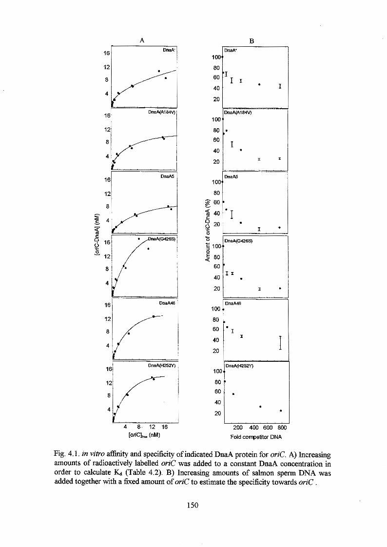
16 m A
12
4
121 I
16-
12/ l
1 01
80
60
40
20
1 oc 80
60
40
20
1 oc 80
g 60
40 6 g 20 0
0 L
c 5 1oc
80
60
40
20
0
100
80 60
40
20
1 O[
80
60
40
20
B )MA'
saA(A184V)
I
I . I .
'naA(G426S)
I
I
N " a A 4 6
l I T
. 200 400 600 801
Fold competitor DNA
Fig. 4.1. in vitro afftnty and specificity of indicated DnaA protein for oriC. A) Increasing amounts of radioactively labelled oriC was added to a constant DnaA concentration in order to calculate & (Table 4.2). B) Increasing amounts of salmon sperm DNA was added together with a fixed amount of orzC to estimate the specificity towards oriC .
150
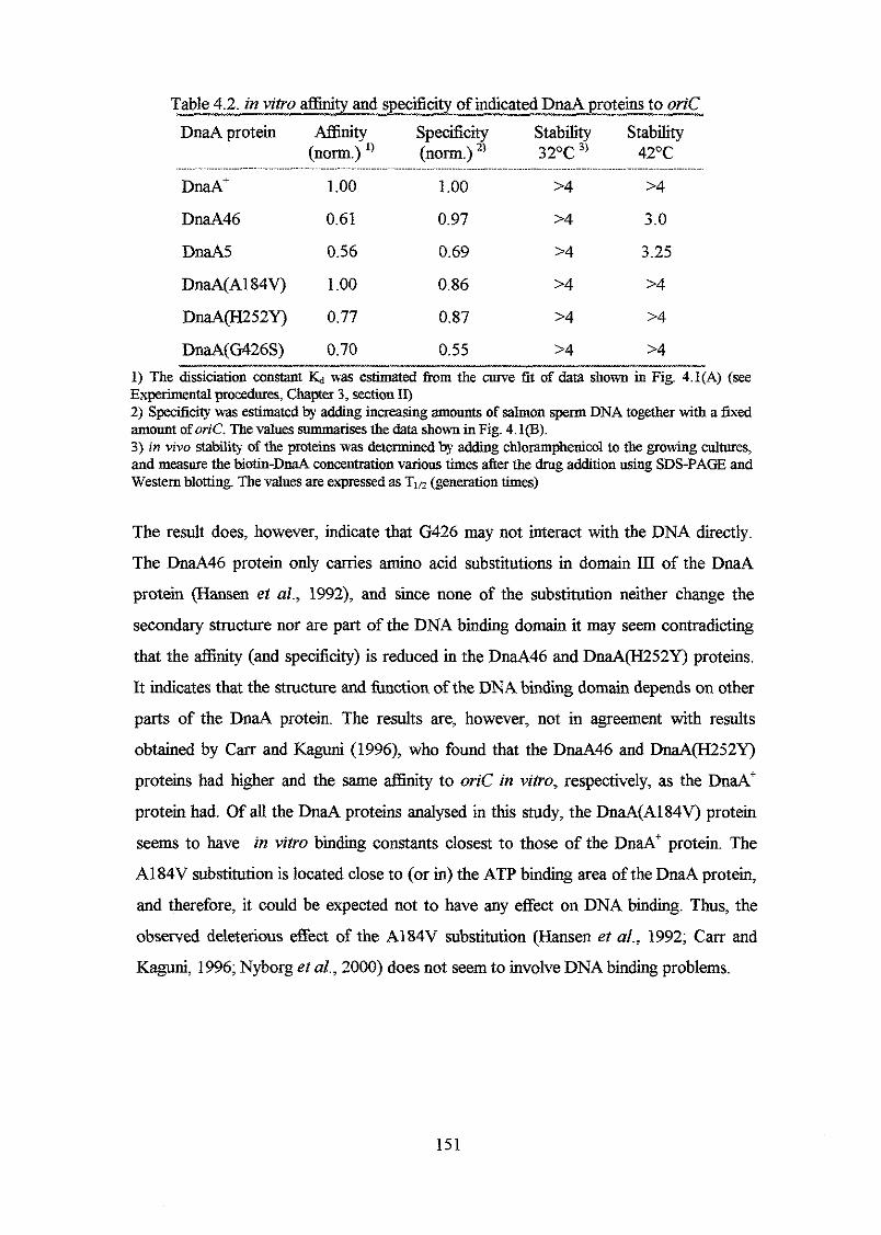
DnaA protein Affinity Specificity Stability Stability (norm.) ') (norm.) 32°C 3' 42OC
DnaA' 1 .oo 1 .oo 24 >4
DnaA46 0.61 0.97 >4 3.0
DnaA5 0.56 0.69 >4 3.25
DnaA(A184V) 1.00 0.86 >4 >4
DnaA(H252Y) 0.77 0.87 >4 >4
DnaA(G426S) 0.70 0.55 >4 >4
Experimental procedures, Chapter 3, section IQ 1) The dissiciation constant was estimated from the curve fit of data shown in Fig. 4.1(A) (see
2) Specifcity was estimated by adding increasing amounts of salmon sperm DNA together with a fixed amount of oriC. The values summarises the data shown in Fig. 4.1@). 3) in vivo stability of the proteins was determined by adding chloramphenicol to the growing cultures, and meawe the biotin-DnaA concentration various times after the drug addition using SDS-PAGE and Western blotting. The values are expressed as T,o (generation times)
The result does, however, indicate that G426 may not interact with the DNA directly.
The DnaA46 protein only cames amino acid substitutions in domain III of the DnaA
protein (Hansen el al., 1992), and since none of the substitution neither change the
secondary structure nor are part of the DNA binding domain it may seem contradicting
that the affinity (and specificity) is reduced in the DnaA46 and DnaA(H252Y) proteins.
It indicates that the structure and function of the DNA binding domain depends on other
parts of the DnaA protein. The results are, however, not in agreement with results
obtained by Can and Kaguni (1996), who found that the DnaA46 and DnaA(H252Y)
proteins had higher and the same affinity to oriC in vitro, respectively, as the DnaA'
protein had. Of all the DnaA proteins analysed in this study, the DnaA(A184V) protein
seems to have in vitro binding constants closest to those of the DnaA' protein. The
A184V substitution is located close to (or in) the ATP binding area of the DnaA protein,
and therefore, it could be expected not to have any effect on DNA binding. Thus, the
observed deleterious effect of the A184V substitution (Hansen et al., 1992; Cam and
Kaguni, 1996; Nyborg et al., 2000) does not seem to involve DNA binding problems.
151
Stability of theproteins
The DnaA(Al84V), DnaA46, DnaA5, DnaA(H252Y) and DnaA(G426S) proteins are all
stable both at 32 and 42OC, although the DnaA46 and DnaAS proteins have a slightly
reduced Tm at 42OC (table 4.2). This high stability of the heat sensitive DnaA46 and
DnaA5 proteins at non-permissive temperature is consistant with the observation that
when & d 4 6 and &a45 strains are grown at permissive temperature, shifted to non-
permissive temperature (42') for 1.5 hours, and then shifted back to permissive
temperature, the chromosome replication is initiated immediately (Hanna and Carl, 1975;
Tippe-Schindler et al., 1979; Hansen, 1995). The DnaA(A184V) protein is also stable at
both 32 and 42OC, and thus, sofar there does not seem to be any reason why the
haA(A184V) mtation would force the cell to pick up other intragenic mutations in
order to survive.
dnaA(A184) allele on the chromosome
h a 4 2 0 4 and M', mh' strains harbouring the dnaA(A184V) allele in h attachment site
were constructed and grown as described in Chapter 3, Experimental procedures, section
III and shown i Fig. 3.7. Samples were taken from the exponentially growing cultures,
treated with rifampicin (+cephalexin) and analysed using flow cytometry (Fig. 4.2).
Generally, the DNA distributions do not look as nice as the distributions in the isogenic
M' strain pig. 3.19 and 3.21), and the dnaAo strain carrying the dnaA(A184V) allele
did not display discrete peaks (data nor shown). The replication is initiated in MN1229
(dnaA(A184V) in M 2 0 4 background) at 32 and 4 2 C without IPTG induction (Fig.
4.2). This is consistent with the observation that the strain forms colonies at 42OC
without IPTG induction (Table 4.3). The replication is initiated somewhat synchronous
at both temperatures and most of the cells contain four and eight origins. At 32"C, the
initiation frequency clearly decrease with increasing IPTG concentrations, and ailer 4
hours growth in medium supplemented with 100 ph4 IPTG, half of the cells contain
either one or two chromosomes. This is consistent with the results of experiments with
multicopy Pdn&na4(A1S4V) strains. These strains are cold sensitive and it was shown
that DNA synthesis stopped when the cultures were shifted to non-permissive
temperatures in spite of an overinitiation of chromosome replication (Nyborg et al.,
2000). However, "229 forms colonies at 32°C on LB plates supplemented with 100
p.M IPTG, although the colonies are small (Table 4.3). This apparent contradiction may
152
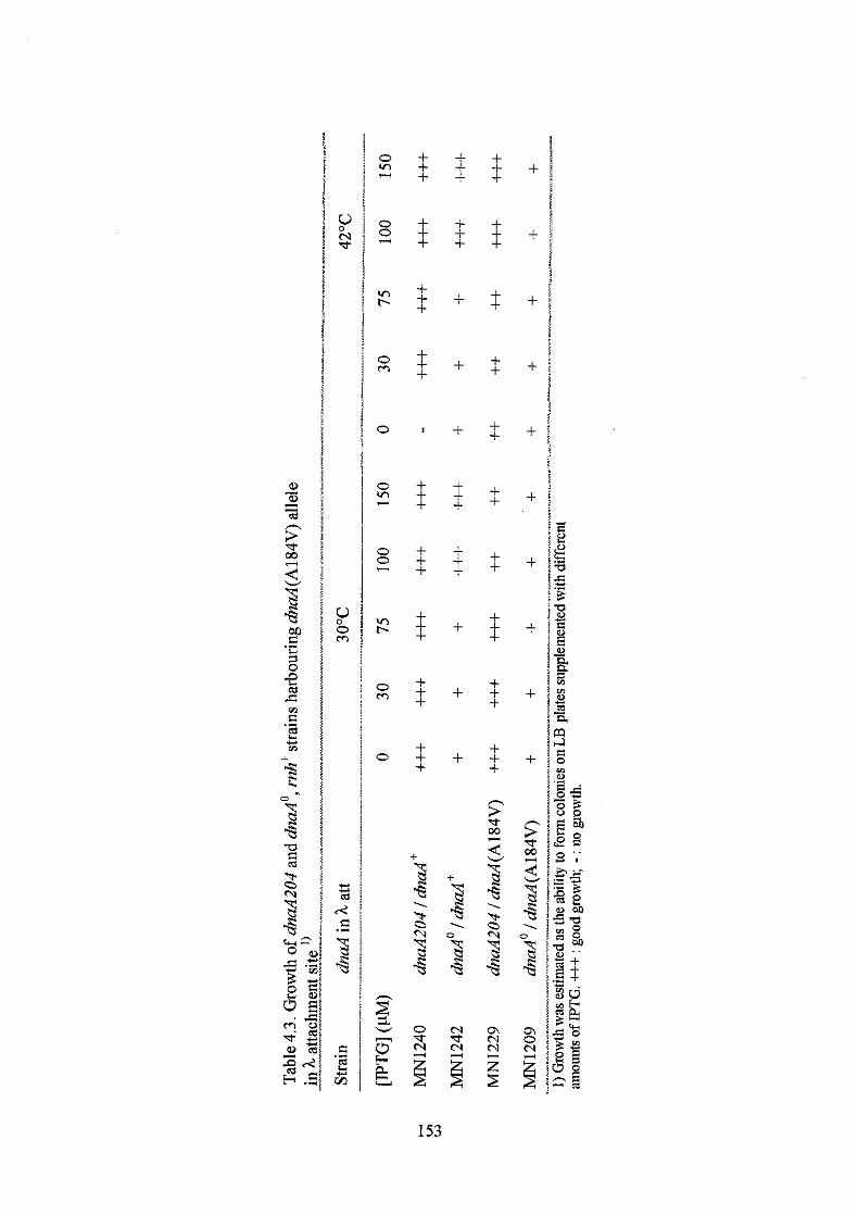
t +
+ + +
+ :: $ +
+ :: + + +
I: +
+ t
t +
5
2 -8 2
-8
.
0 Tr
153
:: +
+ + +
+
+
+
+ r: + + +
+
+
+
+3
% 4
-8
.
N Yt
+ I:
:: +
+ +
+ +
+ +
+ +
+ +
+ t + t
t +
h
'cr 00
+
5
2
2
-8 . -3. 0
-8
m N
+
+
+
+
+
+
+
+
+
+
h > 3 2
-8
-&
5 3 .
m 0
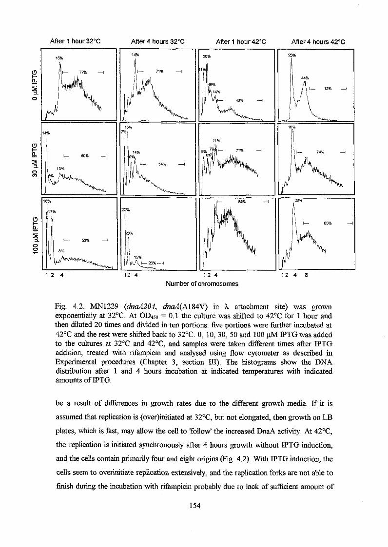
After 1 hour 32°C
1 4%
a 1 I- 60% -1
1 2 4
After 4 hours 32'C After 1 hour 42°C After 4 hours 42'C 14% n i- 71% -l
il
Pb
12 4
1k 40% ,-l
71% -I
! 18% n / l
Number of chromosomes
Fig. 4.2. MN1229 (dnaA204, dnaA(Al84V) in h attachment site) was grown exponentially at 32°C. At OD450 = 0.1 the culture was shifted to 4 2 T for 1 hour and then diluted 20 times and divided in ten portions: five portions were hrther incubated at 42OC and the rest were shifted back to 32OC. 0, 10,30, 50 and 100 pM IPTG was added to the cultures at 32'C and 42OC, and samples were taken different times after IPTG addition, treated with rifampicin and analysed using flow cytometer as described in Experimental procedures (Chapter 3, section III). The histograms show the DNA distribution after 1 and 4 hours incubation at indicated temperatures with indicated amounts of IPTG.
be a result of differences in growth rates due to the different growth media. If it is
assumed that replication is (0ver)initiated at 32T, but not elongated, then growth on LB
plates, which is fast, may allow the cell to 'follow' the increased DnaA activity. At 42OC,
the replication is initiated synchronously after 4 hours growth without IPTG induction,
and the cells contain primarily four and eight origins (Fig. 4.2). With IPTG induction, the
cells seem to overinitiate replication extensively, and the replication forks are not able to
finish during the incubation with rifampicin probably due to lack of sufficient amount of
154
nucleotides etc. Without IPTG induction, the DnaA concentration is only one fourth of
the DnaA concentration in a normal &d' strain (Fig. 3.18), whereas 30 pM IPTG
induction results in DnaA concentrations from two to four fold higher than the DnaA
concentration in a dnd' strain. This indicates that only small amounts ofDnaA(A184V)
protein is enough to initiate chromosome replication, and that a DnaA(A184V)
concentration corresponding to what it would be if the (61aA(A184V) allele was inserted
into its normal chromosomal position, might result in overinitiation of chromosome
replication at 42°C and probably also at 32OC. The overinitiations at 32°C may not pass
the oriC area as observed with multicopy M ( A l 8 4 V ) strains (Nyborg et al., ZOOO),
and thus, the strain is likely to be cold sensitive. MN1209 (dnaA(A184V) allele in ha.4'
strain background) grows on LB plates at 32 and 42°C both with and without IPTG, but
the colonies are small. This independency of IPTG (or sensitivity), may indicate that the
strain carries unknown supressor mutations, but several independently isolated strains
were isolated and tested for growth on plates supplemented with different amount of
PPTG. They all grew with and without IPTG.
Discussion We have previously shown that multicopy dnar146, dnu.45 and d d ( A l 8 4 V ) strains are
cold sensitive, and that the chromosome replication is overinitiated at non-permissive
temperatures. These overinitiations are, however, only elongated in the multicopy
d d 4 6 strain and not in the multicopy &d5 and dnaA(A184V) strains. We have also
shown that multicopy d d ( H 2 5 2 Y ) and dnaA(G426S) strains are temperature resistant
and thus, have the same phenotype as multicopy dd' strains.
In this study, in vitro binding assays indicate that the DnaA5 and DnaA46 protein have
retained 50-60% of the affinity to DnaA boxes in orzC compared to the DnaA' protein,
DnaA(H252Y) and DnaA(G426S) have retained 70-80% and the DnaA(A184V) protein
has the same athity for DnaA boxes in oriC as the DnaA' protein has. DnaA(A184V)
has also retained high specificity towards oriC, whereas especially DnaA5 and
DnaA(G426S) have reduced specificity, which may be due to the G426S substitution
located in the DNA binding domain of the DnaA protein. Generally, the protein have a
relatively high affinity for oriC, and this is in agreement with results obtained by Can and
Kaguni (1996). However, they observed a slightly increased aflinity of both
DnaA(A184V) and DnaA46 for oriC at 30 and 42"C, and that only the DnaA(G426S)
155

had a decreased af€inity at these temperatures. The dicrepancy between the results
obtained by C m and Kaguni (1996) and the results in this study may be due to different
incubation temperatures. In addition, the proteins used in this work all have a biotin-tag
fused to the N-terminus of the DnaA protein. Sutton and Kaguni (1997) analysed the
DnaA(G426D) protein and found that it retained only 12% of the in vitro aflinity to
oriC. The amino acid substitution does not change the secondary structure, and thus the
results indicate that substitution of G426 with serine is tolerable while substitution with
aspartic acid has deleterious effect on the DnaA hc t ion . The results of this work
indicate that the DnaA(A184V) protein does not differ significantly from the DnaA'
protein with respect to oriC interaction in vitro. The in vivo stability of DnaA(A184v) is
also very similar to that of the DnaA' protein both at 32 and 42°C. Thus, the dominance
of the multicopy dnaA(AlS4V) over the singie &d' allele on the chromosome
published previously (Nyborg et al., 2000) may not be due to altered aflinity or
specificity of the DnaA(A184V) protein for oriC. The result of the experiment also
indicate that the so-called secondary DnaA46 and DnaA5 substitutions have an effect on
the DnaA protein in vitro. The proteins have reduced oriC aflinity, and especially the
DnaA(G426S) has reduced specificity towards oriC. The observation that the
combination of the A184V substitution and the H252Y or G426S substitutions has the
most severe effect on the affinity for oriC may seem dfficult to explain. However, if the
A184V substitution is the most deleterious one, then its presence may lead to other
DnaA substitutions that reduce the aflinity of the DnaA protein for oriC, and thereby
reduce the initiation frequency from oriC dowing (better) growth. As shown in Fig. 4.2,
the chromosome replication is probably overinitiated at 42°C and most likely also at
32OC (although these initiations are not elongated). It would be interesting to measure
the number of origins per mass in isogenic &aA46, M S , dnaA(H252Y) and
ahnaA(G426) strains.
156
Recapitulation
The DnaA protein is the key protein in the initiation of the chromosomal replication of
Eschen'chiu coli. The DnaA protein binds to DnaA boxes in origin of replication, oriC,
to form an initial complex. In the presence of ATP it melts part of the oriC to which the
rest of the replication machinery can bind and elongate and eventually terminate the
chromosomal replication. Based on analysis of secondary structure and function, the
DnaA protein has been divided in four domains. The N-terminal domain I is responsible
for protein-protein interactions, domain I1 constitute the flexible loop, domain III carries
the ATP binding area and domain IV, which include the last 94 amino acids of the
DnaA protein, constitute the DNA binding domain. Domain IV is predicted to consist of
five cc-helixes separated by loops, and this structure is highly conserved among DnaA
proteins from a large number of organisms.
The experimental work presented in this thesis involve 1) mutational analysis of the
DNA binding domain of the DnaA protein and 2) analysis of the Al84V substitution in
the ATP area of domain III and other amino acid substitutions found in the DnaA5 and
DnaA46 proteins.
To analyse the DNA binding domain, more than 100 functional DnaA proteins with
amino acid substitutions in the domain IV (fused to a biotin tag) were constructed and
studies in vivo by complementation analysis of high temperature sensitive dnuA46 and
dnaA.204 phenotypes by induction of plasmid born mutant dnuA(1V) alleles. The result
of the complementation analysis indicates that the whole area participates in DNA
binding, either directly or indirectly. However, the complementation analysis indicates
that there are a relatively higher number of amino acids in the first half of domain IV
and in helix 5 that can be substituted with non-closely related amino acids without any
apparent effect on the DnaA protein. These include 1376, K381, Y386, K388, K390,
V391, L395, K397, M411, T418, E456, F458, L461, L465 and S467. None of the
mutant DnaA proteins complement the high temperature sensitive phenotype better than
the DnaA' protein does. The secondary structures of the mutant DnaA proteins were
determined, and when compared with the results of the complementation analysis, the
results suggest that the secondary structure of domain IV cannot be changed without
affecting the function of the DnaA protein severely. This seems consistent with the
highly conserved secondary structure observed in a large number of DnaA protein form
other organisms.
157
In agreement with solvent accessibility predictions, the complementation analysis
indicates that helix 1 may be located on the surface of the DnaA protein, with D377,
K381 and E385, which are located on the same side of the a-helix, exposed to the
solvent, and the rest, especially 1379, V383 and A384 protected from the solvent and
probably participating in hydrophobic interactions with other residues of the DnaA
protein. Generally, helix 1 is sensitive towards amino acid substitutions even though
they do not change the secondary structure of the protein. This indicates that amino
acids in the helix, and not only the helix itself, help stabilise the DnaA structure and/or
are responsible for DNA contact. All lysines (K388, K390 and K397) in the basic loop
between helix 1 and 2 can be substituted with the oppositely charged glutamic acid
without any apparent effect on the DnaA protein with respect to complementation. This
could indicate that these amino acids are interacting with the solvent. On the contrary,
the highly conserved arginines R399 and R401 cannot be substituted without affecting
the DnaA protein, which indicates that these residues may interact with the DNA.
According to the accessibility prediction, many amino acids in helix 2 are protected
from the solvent, indicating that a part of this helix is located inside the protein with
other hydrophobic amino acids in the surroundings. This hypothesis is consistent with
the complementation analysis and may indicate that residues in helix 2 does not interact
with the DNA, because of the buried nature of the helix. However, except for M41 1,
other residues are sensitive towards substitutions, and in addition, it was not possible to
construct DnaA proteins with R407 and Q408 substitutions, which suggests that
residues in helix 2 may in fact be interacting with the DNA. It was not possible in this
work to substitute many of the highly conserved amino acids in helix 3 and the loops
surrounding it. This may indicate that they cannot be substituted without deleterious
effects on the DnaA protein. The presence of the highly conserved G430 and G431 that
were not substituted either may indicate that DnaA fimctionality depends on a turn
between helix 3 and 4. Alignment of DnaA with proteins harbouring a helix-turn-helix
motif suggests that helix 3 and 4 of the DNA binding domain of the DnaA protein could
constitute a modified helix-turn-helix motif. The results of the complementation
analysis are in agreement with this proposal. Generally, helix 4 and 5 carry few single
substitutions and many double substitutions, which could indicate that amino acid
substitutions in this area require secondary substitutions elsewhere in order to maintain
DnaA activity. There seems to be a tendency that the presence of a coiled structure
between the two parts of the helix is important, and that the length of it is critical. Helix
158
4 is more sensitive towards amino acid substitutions than helix 5, which is in agreement
with how conserved the regions are. Thus, many amino acids in helix 5 can be
substituted with non-closely related amino acids without affecting the function of the
DnaA protein. On the contrary, amino acids in the beginning of helix 4 that are highly
conserved are not substituted in any of the DnaA proteins constructed in this work. This
strongly suggests that they are required for DnaA activity, and they may be interacting
with the DNA. In addition, those amino acids that are predicted to be protected form the
solvent with high reliability cannot be substituted with polar amino acids without
affecting the DnaA protein severely. The hydrophobic amino acids are located on the
same side of the a-helix (or close to). The two hydrophobic amino acids I454 and F458
are substituted with threonine and serine, respectively, in one of the double mutants.
This DnaA(I454T, F458S) protein complements the high temperature sensitive
phenotype of the host better than when the substitutions exist alone, which indicates that
there could be (weak) interactions between I454 and F458 that is maintained to some
extend in the double mutant where both amino acids are substituted with polar,
hydrogen bonding residues. Helix 4 and 5 are probably located on the surface of the
DnaA protein with a hydrophobic and hydrophilic side and a hydrophilic coil in
between. Helix 4 could be responsible for DNA contact and as mentioned previously be
part of a modified helix-turn-helix motif.
Further work was carried out with a selection of twenty DnaA proteins with amino
acid substitutions in domain IV. The analysis included in vivo and in vitro binding
studies and in vivo stability measurements. In addition, the IPTG inducible
dnuA(A384V), dnd(L395P), dnuA(K397E) and dnuA(C441G) alleles were inserted
into the h attachment of a dnuA204 and dnd(nul1) strain and the initiation frequencies
determined at different temperatures under different growth conditions using flow
cytometry. The relative in vivo affinities of the various DnaA proteins for DnaA box R5
and R6 was determined by measuring the P-galactosidase activities in multicopy
pdnd(IV) strains harbouring a P,,,-ZucZ hsion in h attachment site. The in vitro
affinities and the specificity towards oriC in vitro was determined by using streptavidin
coated magnetic beads to which the biotin-tagged DnaA proteins were bound. To
determine Kd different amounts of oriC was added and to determine the specificity
towards oriC, increasing amounts of competitor DNA was added together with the oriC
fragment. The oriC fragment included DnaA box R1, R2, R3, R4, R5 and R6. The
results of the in vivo and in vitro affinity assays indicate that DnaA proteins with amino
159
acid substitutions in Domain IV have affinities comparable to those of the wild type
DnaA protein (I& = 0.75 f 0.14 nM), and the variation of the affinities only differed
three-fold. There seems to be a tendency that amino acid substitutions in the area from
T418 to C441 result in lower in vitro affinities than amino acid substitutions elsewhere
in the domain. Generally, the in vivo affinities were lower than the in vitro affinities,
and the in vivo affinities seem to 'follow' the specificity. Thus, a protein with high in
vitro specificity towards oriC (which also included the R5 and R6 box) also had high in
vivo affinity to the mioC promoter. The DnaA proteins that had high affinity, but low
specificity towards oriC are located in helix 1 and in the basic loop and include
DnaA205, DnaA(A384V, Y386H), DnaA204, DnaA(Y387S), DnaA(L395P) and
DnaA(R401C, K433R). This indicates that this part of the DNA binding domain is
responsible for the specificity towards oriC. The DnaA(L395) protein, .in particular, had
low specificity, whereas the in vivo and in vitro affinities were comparable with those of
the DnaA+ protein. Strains carrying dnuA(A384V, Y384H) and dnaA(L395P) alleles in
h attachment site of a dnaA204 strain overinitiated chromosome replication extensively
at 42OC, but maintained normal initiation frequency at 32°C as compared with the
isogenic dnuA' strain. The DnaA concentration in these strains was from two to four
fold higher than the DnaA concentration in a normal wild type strain. This indicates that
especially the DnaA(L395P) protein has increased affinity for oriC at 42°C in the light
of the observation that the protein is more unstable than the DnaA' protein at 42°C. The
in vitro experiments were carried out at room temperature, and therefore an increased
affinity at 42OC could not be observed. Unlike the isogenic dnaA' strain, the replication
in dnuA(A384V, Y386H) and dnuA(L395P) were initiated asynchronously at both 32
and 42°C. Interestingly, the DnaA(K397E) protein, which has substituted one of the
basic residues in the basic loop between helix 1 and 2 with an oppositely charged amino
acid, has a three fold higher affinity for oriC in vitro than the DnaA+ proteins has, and
the binding is more specific. The higher affinity is, however, not reflected in the in vivo
affinity experiments with strains harbouring multicopy dnuA(K397E) alleles. However,
when the IPTG inducible dnaA(K397E) allele is inserted into the chromosome in h
attachment site of a dnuA204 and dnuA(nul1) strain, the strains initiate replication
(asynchronously) without IPTG induction even though the DnaA concentration is only
one fourth of the DnaA concentration in a wild type d n d ' strain. When the
dnaA(K397E) allele is induced to a DnaA concentration corresponding to two fold
higher than in a wild type strain, initiation of chromosome replication seems to stop, and
160
cells with only one chromosome accumulate. The replication may, however, be
overinitiated with increasing DnaA(K397E) concentration, but subsequently aborted or
stalled and therefore not detected in the experiments carried out in this work. The results
are in agreement with the in vitro data and indicate that the DnaA(K397E) protein has
increased oriC affinity. The results also indicate that a strain with the dnaA(K397E)
allele at its normal chromosomal position may not survive due to the high oriC affinity.
The DnaA(C441G, V391A) protein has retained app. 60% of the in vivo and in vitro
affinity and 80% in vitro specificity for oriC. When the dnaA(C441G, V391A) allele is
inserted into the h attachment site of a dnaA204 strain, the replication is initiated
asynchronously, but with a somewhat normal frequency at 42"C, whereas initiation of
replication seems to stop at 32OC when the DnaA concentration is high (more than two
fold higher than in a normal wild type dnaA' strain). The results indicate that a strain
with the dnaA(C441G, V391A) alleles at its normal chromosomal position will be cold
sensitive.
The amino acid substitutions found in the DnaA5 and DnaA46 proteins were
separated into DnaA(A184V), DnaA(H252Y) and DnaA(G426S) proteins and analysed
in vivo and in vitvo. Multicopy dnaA(H252H) and dnaA(G426S) strains (secondary
amino acid substitutions from DnaA46 and DnaA5, respectively) were temperature
resistant like multicopy dnaA' strains, while multicopy dnaA(Al84V), dnaA46 and
dnaA5 strain were high temperature resistant, but cold sensitive. The multicopy
dnaA(Al84V), dnaA5 and dnaA46 strains (and the dnaAcos mutant) were further
analysed, and it was found that the mutants could be divided into two groups. The first
group, which included the multicopy dnaA(A184V) and dnaA5 strains, initiated
chromosome replication extensively at non-permissive temperature, but the initiations
were not elongated and DNA synthesis stopped. Group two, which included the
multicopy dnaA46 strain and the dnaAcos mutant, also overinitiated chromosome
replication at non-permissive temperature, but (some 04 these initiation were elongated
and resulted in a increased DNA content in the cells, especially in the multicopy dnaA46
cells (Nyborg et al., 2000).
In this study in vitro binding assays as the ones discussed above indicate that the
DnaA5 and DnaA46 proteins have retained 50-60% of the affinity to DnaA boxes in
oriC area compared to the DnaA' protein, DnaA(H252Y) and DnaA(G426S) have
retained 7040% and the DnaA(Al84V) protein has the same high affinity to DnaA
161
boxes in oriC as the DnaA' protein has. DnaA(Al84V) has also retained high
specificity towards oriC, whereas especially DnaA5 and DnaA(G426S) have reduced
specificity, which may be due to the G426S substitution located in the DNA binding
domain of the DnaA protein.
The results of this work indicate that the DnaA(A184V) protein does not differ
significantly from the DnaA+ protein with respect to oviC interaction in vitro. The in
vivo stability of DnaA(A184V) is also very similar to that of the DnaA' protein both at
32 and 42°C. Thus, the dominance of the multicopy dnaA(Al84V) over the single
d n d ' allele on the chromosome may not be due to altered oriC affinity or specificity of
the DnaA(A184V) protein. The result of the experiment also indicates that the so-called
secondary DnaA46 and DnaA5 substitutions have an effect on the DnaA protein in
vitro. The proteins have reduced oriC affinity, and especially the DnaA(G426S) has
reduced specificity for oriC. The observation that the combination of the A184V
substitution and the H252Y or G426S substitutions has the most severe effect on the
affinity for oriC may seem obscure. However, if the A184V substitution is the most
deleterious substitution, then its presence may lead to other DnaA substitutions that
reduce the affinity of the DnaA protein for oriC, and thereby reduce the initiation
&equency from oriC allowing better growth. When the IPTG inducible dnaA(A184V)
allele is inserted into h attachment site of a dnuAZO4 strain, the chromosome replication
is overinitiated at 42OC even without IPTG induction where the DnaA concentration is
only one fourth of the DnaA concentration in a normal d n d ' strain. At 32°C the
initiation frequency seems to decrease severely with increasing IPTG induction and
cells with one and two chromosomes accumulate. The replication may, however, be
overinitiated at 32°C as observed with multicopy dnuA(A184V) strains, but the
initiation are stalled or aborted. The results indicate that a dnaA(Al84V) strain with the
dnuA allele at its normal chromosomal location is cold sensitive.
162
References

References
Allman, R.; Schjerven, T.; Boye, E. (1991) Cell cycle parameters of Escherichia coli K-
12. J. Bacteriol. 173(24):7970-7974.
Andersen, C.A. (1998) Neural Network Assignment of Protein Secondary Structure
with Increased Predictability. Master thesis, CBS, Technical University, DK.
Asai, T.; Chen, C-P.; Nagata, T.; Takanami, M.; Imai, M. (1992) Transcription in vivo
within the replication origin of the Escherichia coli chromosome: a mechanism for
activating initiation of replication. Mol. Gen. Genet. 23 1: 169-178.
Atlung, T. (1984) Allele-specific supression of dnaA(Ts) mutations by rpoB mutations
in Escherichia coli. Mol. Gen. Genet. 197: 125-128.
Atlung, T.; Clausen, E.S.; Hansen, F.G. (1985) Autoregulation of the dnaA gene of
Escherichia coli K12. Mol. Gen. Genet. 200: 442-450.
Atlung, T.; L@bner-Olesen, A.; Hansen, F.G. (1987) Overproduction of DnaA protein
stimulates initiation of chromosome and minichromosome replication in
Escherichia coli. Mol. Gen. Genet. 206:51-59.
Atlung, T.; Nielsen, A.; Hansen, F.G. (1989) Isolation, characterization, and nucleotide
sequence of appY, a regulatory gene for growth-phase-dependent gene expression in
Escherichia coli. J. Bacteriol. 171(13): 1683-1691.
Atlung, T.; Nielsen, A.; Rasmussen, L.J.; Nellemann, L.J.; Holm, F. (1991) A versatile
method for integration of genes and gene fusions into the h attachment site of
Escherichia coli. Gene 107: 11-17.
Atlung, T.; Hansen, F.G. (1993) Three distinct chromosome replication states are
induced by increasing concentrations of DnaA protein in Escherichia coli. J.
Bacteriol. 175(20):6537-6545.
165
&am, T.A.; Ishihama, A. (1999) Twelve species of the nucleoid-associated protein
from Escherichia coli. J. Biol. Chem. 274(46): 33105-33113.
Bachmann, B.J. (1990) E.coli K-l2 linkage map. Microbiol. Rev. 54: 130-197.
Bagdasarian, M.M.; Izakowska, M.; Bagdasarian, M. (1977) Suppression of the DnaA
phenotype by mutations in the rpoB cistron of ribonucleic acid polymerase in
Salmonella typhimurium and Escherichia coli. J. Bacteriol. 130(2): 577-582.
Baker, T.A.; Funnell, B.; Komberg, A. (1987) Helicase action of DnaB protein during
replication from the Escherichia coli chromosomal origin in vitro. J. Biol. Chem.
262(14): 6877-6885.
Baker, T.A.; Kornberg, A. (1988) Transcriptional activation of initiation of replication
from E.co2i chromosomal origin: an RNA-DNA hybrid near oriC. Cell 55: 113-123.
Bates, D.B.; Boye, E.; Asai, T.; Kogoma, T. (1997) The absence of effect ofgid or
mioC transcription on the initiation of chromosome replication in Escherichia coli.
Proc. Natl. Acad. Sci. USA 94: 12497-12502.
Bipatnath, M.; Dennis, P.P.; Bremer, H. (1998) Initiation and velocity of chromosome
replication in Escherichia coli B/r and K-12. J. Bacteriol. 180 (2): 265-273.
Bird, R.E.; Louarn, J.; Martuscelli, J.; Caro, L. (1972) Origin and sequence of
chromosome replication in Escherichia coli. J. Mol. Biol. 70: 549-566.
Blake, M.S.; Johnston, K.H.; Russell-Jones, G.J.; Gotschlich, E.C. (1984) A rapid
sensitive method for detection of alkaline phosphatase-conjugated anti-antibody on
Westem blots. Anal. Biochem. 136: 175-179.
Bogan, J.A.; Helmstetter, C.E. (1996) mioC transcription, initiation of replication, and
the esclipse in Escherichia coli. J. Bacteriol. 178(11): 3201-3206.
166
Boye, E.; Lerbner-Olesen, A. (1990) The role of dum methyltransferase in the control of
DNA replication in E.coZi. Cell 62: 981-989.
Brasing, F. (1999) Analyse der DNA-Bindungsdomhe des DnaA Proteins von E.coli.
Dissertation (Ph.D thesis). Der Freien UniversiCat, Berlin, Germany
Bramhill, D.; Komberg, A. (1988A) A model for initiation at origins of DNA
replication. Cell 54:915-918.
Bramhill, D.; Komberg, A. (1988B) Duplex opening by DnaA protein at novel
sequences in initiation of replication at the origin of the E.coZi chromosome. Cell
52:743-755.
Braun, R.E.; O’Day, K.; Wright, A. (1985) Autoregulation of the DNA replication gene
dnuA in E.coZi K-12. Cell 40:159-169.
Braun, R.E.; Wright, A. (1986) DNA methylation differentially enhances the expression
of one of the two E.coZz dnuA promoters in vivo and in vitro. Mol. Gen. Genet.
202~246-250.
Braun, R.E.; O’day, K.; Wright, A. (1987) Cloning and characterization of dnuA(Cs), a
mutation which leads to overinitiation of DNA replication in Escherichia coli K-12.
J. Bacteriol. 169(9):3898-3909.
Brennan, R.G.; Matthews, B.W. (1989) The helix-turn-helix binding motif. J. Biol.
Chem. 264(4): 1903-1906.
Campbell, J.L.; Kleckner, N. (1990) E.coli 0% and the dnuA gene promoter are
sequestered &om dum methyltransferase following the passage of the chromosomal
replication fork. Cell 62: 967-979.
Cam, K.M.; Kaguni, J.M. (1996) The A184V missense mutation of the dnuA5 and
dnuA46 alleles confers a defect in ATP binding and thermolability in initiation of
Escherichia coli DNA replication. Mol. Microbiol. 20: 1307-1318.
167

Casabadan, M.J.; Cohen, N.S. (1980) Analysis of gene control signals by DNA fusion
and cloning in Escherichia coli. J. Mol. Biol. 138:179-207.
Cassler, M.R.; Grimwade, J.E.; Leonard, A.C. (1995) Cell cycle-specific changes in
nucleoprotein complexes at a chromosomal replication origin. EMBO J. 14(23):
5833-5841.
Chiaramello, A.E.; Zyskind, J.W. (1989) Expression of Escherichia coli dnaA and mioC
genes as a function of growth rate. J. Bacteriol. 171(8):4272-4280.
Chiaramello, A.E.; Zyskind, J.W. (1990) Coupling of DNA replication to gowth rate in
Escherichia coli: a possible role for guanosine tetraphosphate. J. Bacteriol.
172(4):2013-2019.
Christensen, B.B.; Atlung, T.; Hansen, F.G. (1999) DnaA boxes are important elements
in setting the initiation mass of Escherichia coli. J. Bacteriol. 181(9): 2683-2688.
Churchward, G.; Bremer, H. (1977) Determination of deoxyribonucleic acid replication
time in exponentially growing Escherichia coli B/r. J. Bacteriol. 130(3):1206-1213.
Churchward, G.; Astiva, E.; Bremer, H. (1981) Growth rate dependent control of
chromosome replication initiation in Escherichia coli. J. Bacteriol. 145(3): 1232-
1238.
Churchward, G.; Holmans, P.; Bremer, H. (1983) Increased expression of the dnaA gene
has no effect on DNA replication in a dnaA' strain of Escherichia coli. Mol. Gen.
Genet. 192506-508.
Clark, D.J.; Maalere, 0. (1967) DNA replication and the division cycle in Escherichia
coli. JMol Biol 23: 99-1 12.
Cooper, S . ; Helmstetter, C. (1968) Chromosome replication and the division cycle of
Escherichia coli. J. Mol. Biol. 31519-540.
168
Crooke, E.; Castuma, C.E.; Komberg, A. (1992) The chromosome origin ofEscherichia
coli stabilizes DnaA protein during rejuvenation by phospholipids. J. Biol. Chem.
267(24):16779-16782.
Crooke, E.; Thresher, R.; Hwang, D.S. ; Griffith, J. ; Komberg, A. (1993) Replicatively
active complexes o f DnaA protein and the Escherichia coli chromosomal origin
observed in the electron microscope. J. Mol. Biol. 233:16-24.
Diederich, L.; Roth, A.; Messer, W. (1994) A versatile plasmid vector system for the
regulated expression o f genes in Escherichia coli. BioTechniques 16(5): 916-923.
Donachie, W.D. (1968) Relationship between cell size and time o f initiation of DNA
replication. Nature 219: 1077-1079.
Frey, J.; Chandler, M.; Caro, L. (1984) Overinitiation o f chromosome and plasmid
replication in a dnaAcos mutant ofEscherichia coli K12. J. Mol. Biol. 176:171-183.
Fuller, R.S.; Funnell,, B.E.; Komberg, A. (1984) The DnaA protein complex with the
E.coli chromosomal replication origin (oric) and other DNA sites. Cell 38389-900.
Funnell, B.E.; Baker, T.A.; Komberg, A. (1987) In vitro assembly o f a prepriming
complex at the origin o f the Escherichia coli chromosome. J. Biol. Chem.
262:10327-10334.
Fujita, M.Q.; Yoshikawa, H.; Ogasawara, N. (1990) Structure o f the dnaA region of
Micrococcus luteus: conservation and variations among eubacteria. Gene 93: 73-78.
Gamer, J.; Crooke, E. (1996) Membrane regulation of the chromosomal replication
activity o f E.coli DnaA requires a discrete site on the protein. EMBO J. 15(9): 2313-2321.
169
Gille, H. ; Messer, W. (1991) Localized DNA melting and structure1 pertubations in the
origin o f replication, oriC, o f Escherichia coli in vitro and in vivo. EMBO
10(6):1579-1584.
Gille, H.; Egan, B.J.; Roth, A.; Messer, W. (1991) The FIS protein binds and bends the
origin o f chromosomal DNA replication, O K , of Escherichia coli. Nucleic Acid
Res. 19(15): 4167-4172.
Gines-Candelaria, E.; Blinkova, A.; Walker, J.R. (1995) Mutations in Escherichia coli
dnaA which suppress a dnaX(Ts) polymerization mutation and are dominant when
located in the chromosomal allele and recessive on plasmids. J. Bacteriol. 177(3):
705-715.
Grimwade, J.E.; Ryan, V.T.; Leonard, A.C. (2000) IHF redistributes bound initiator
protein, DnaA, on supercoiled oriC ofEscherichia coli. Mol. Microbiol. 35(4): 835-
844.
Hanna, M.H.; Carl, P.L. (1975) Reinitiation of deoxyribonucleic acid synthesis by
deoxyribonucleic acid initiation mutants of Escherichia coli: role o f ribonucleic acid
synthesis, protein synthesis, and cell division. J. Bacteriol. 121(1):219-226.
Hansen, F.G.; Rasmussen, K.V. (1977) Regulation o f the dnaA product in Escherichia
coli. Mol. Gen. Genet. 155:219-225.
Hansen , F.G.; Hansen, E.B.; Atlung, T. (1982) The nucleotide sequence of the dnaA
gene promoter and of the adjacent rpmH gene, coding for the ribosomal protein L34,
ofEscherichia coli. EMBO J. 1:1043-1048.
Hansen, F.G.; Koefoed, S.; Smensen, L.; Atlung, T. (1987) Titration of DnaA protein
by oriC DnaA-boxes increases dnaA gene expression in Escherichia coli. EMBO J.
6(1): 255-258.
170
Hansen, F.G.; Atlung, T.; Braun, R.E.; Wright, A.; Hughes, P,; Kohiyama, M. (1991A)
Initiator @naA) protein concentration as a function of growht rate in Escherichia
coli and Salmonella Typhimuriurn. J. Bacteriol. 173(16):5194-5199.
Hansen, F.G.; Christensen, B.B.; Atlung, T. (1991B) The initiator titration model:
computer simulation of chromosome and minichromosome control. Res. Microbiol.
1421161-167.
Hansen, F.G.; Koefoed, S.; Atlung, T. (1992) Cloning and nucleotide sequence
determination of twelve mutant dnaA genes of Escherichia coli. Mol. Gen. Genet.
234114-21.
Hansen, F.G. (1995) Reinitiation kinetics in eight dnaA(Ts) mutants ofEscherichia coli:
rifampicin resistant initiation of chromosome replication. Mol. Microbiol.
15(1):133-140.
Hansen, F.G.; Atlung, T. (1995A) Reversibility of the DnaA protein activity in the
'irreversible' dnaAZO4 mutant ofEscherichia coli. Mol. Microbiol. 15(1): 141-148.
Hansen, F.G.; Atlung, T. (1995B) Initiation of chromosome replication after induction
of DnaA protein synthesis in a dnaA(nul1) rnh mutant ofEscherichia coli. Mol.
Microbiol. 15(1): 149-154.
Harrison, S.C. (1991) A structural taxonomy of DNA-binding domains. Nature 353:
715719.
Harrison, S.C.; Aggerwal, A.K. (1990) DNA recognition by proteins with the helix-
turn-helix motif. Annu. Rev. Biochem. 59: 933-969.
Helmstetter, C.E. (1996) Timing of synthetic activities in the cell cycle. F.C. Niedhardt,
J.L. Ingraham, K.B. Low, B. Magasanik, M. Schaechter, H.E. Umbarger:
Escherichia coli and Salmonella typhirnuriurn, cellular and molecular biology.
American Society for Microbiology, Washington, D.C.
171
Hemck, J.; Kohiyama, M.; Athmg, T.; Hansen, F.G. (1996) The initiation mess? Mol.
Micro. 19(4): 659-666.
Hirota, Y.; Mordoh, J.; Jacob, F. (1970) On the process of cellular division in
Escherichia coli. J. Mol. Biol. 53:369-387.
Hupp, T.R.; Kaguni, J.M. (1993) DnaA5 protein is thermolabile in initiation of
replication &om the chromosomal origin of Escherichia coli. J. Biol. Chem.
268(18):13128-13136.
Hwang, D.S.; Kaguni, J.M. (1988) Interaction of DnaA46 protein with a stimulatory
protein in replication from the Escherichia coli chromosomal origin. J. Biol. Chem.
263(22):10633-10640.
Hwang, D.S.; Crooke, E. ; Komberg, A. (1990) Aggregated DnaA protein is dissociated
and activated for DNA replication by phospholipase or DnaK protein. J. Biol.
Chem. 265(31):19244-19248.
Hwang, D.S.; Komberg, A. (1992) Opening of the replication origin of Escherichia coli
by DnaA protein with protein €€U and IHF. J. Biol. Chem. 267(32): 23083-23086.
Jacob, F.; Brenner, S.; Cuzin, F. (1963) On the regulation of DNA replication in
bacteria. Cold Spring Harbor Symp. Quant. Biol. 28:329-348.
Katayama, T. ; Nagata, T. (1991) Initiation of chromosomal DNA replication which is
stimulated without oversupply of DnaA protein in Escherichia coli. Mol. Gen.
Genet. 226:491-502
Katayama, T. (1994) The mutant DnaAcos protein which overinitiates replication of the
Escherichia coli chromosome is inert to negative regulation for initiation. J. Biol.
Chem. 269:22075-22079
172
Katayama, T.; Komberg, A. (1994) Hyperactive initiation of chromosomal replication in
vivo and in vitro by a mutant initiator protein, DnaAcos, of Escherichia coli. J. Biol.
Chem. 269:12698-12703.
Katayama, T.; Takata, M.; Sekimizu, K. (1997) CedA is a novel Escherichia coli
protein that activates the cell division inhibited by chromosomal DNA over-
replication. Mol. Microbiol. 26(4): 687-697.
Katayama T.; Kubota T.; Kurokawa K.; Crooke E.: Sekimizu K. (1998) The initiator
function of DnaA protein is negatively regulated by the sliding clamp of the E. coli
chromosomal replicase. Cell 94: 61-71
Kellenberger-Gujer, G.; Podhajska, A.J.; Caro, L. (1978) A cold sensitive dnuA mutant
of E.coli which overinitiates chromosome replication at low temperature. Mol. Gen.
Genet. 162:9-16.
Kitagawa, R.; Mitsuki, H.; Okazaka, T.; Ogawa, T. (1996) A novel DnaA protein-
binding site at 94.7 min on the Escherichia coli chromosome. Mol. Microbial.
19(5): 1137-1147.
Kitagawa, R.; Ozaki, T.; Moriya, S.; Ogawa, T. (1998) Negative control of replication
initiation by a novel chromosomal locus exhibiting exceptional affinity for
Escherichia coli DnaA protein. Gen. Devel. 12: 3032-3043.
Kurokawa, K.; Mizushima, T.; Kubota, T.; Tsuchiya, T.; Katayama, T.; Sekimizu, K.
(1998) A stimulation factor for hydrolysis of ATP bound to DnaA protein, the
initiator of chromosomal DNA replication in Escherichia coli. Biochem. Biophys.
Res. Comm. 243: 90-95.
Kurokawa, K.; Nishida, S.; Emoto, A.; Sekimizu, K.; Katayama, T. (1999) Replication
cycle-coordinated change of the adenine nucleotide-bound forms of DnaA protein in
Escherichia coli. EMBO J. 18(23): 6642-6652.
173

Kyhse-Andersen, J. (1984) Electroblotting of multiple gels: a simple apparatus without
buffer tank for rapid transfer of proteins fiom polyacrylamide to nitrocellulose. J.
Biochem. Biophys. Methods lO(3-4): 203-209.
Lamerichs, R.M.J.N.; Padilla, A.; Boelens, R.; Kaptein, R.; Ottleben, G.; Ruterjans, H.;
Granger-Schnarr, M.; Oertel, P,; Schnarr, M. (1989) The amino-terminal domain of
LexA repressor is a-helical but differs from canonical helix-tam-helix proteins: a
two-dimentional 'H NMR study. Proc. Natl. Acad. Sci. USA 86: 6863-6867.
Landoulsi, A.; Hughes, P.; Kern, R.; Kohiyama, M. (1989) dum methylation and the
initiation of DNA replication on oriC plasmids. Mol. Gen. Genet. 216:217-223.
Langer, U.; Richter, S.; Roth, A.; Weigel, C.; Messer, W. (1996) A comprehensive set
of DnaA-box mutations in the replication origin, oriC, of Escherichia coli. Mol.
Microbiol. 21(2): 301-311.
Lark, K.G. (1972) Evidence for the direct involvement of RNA in the initiation of DNA
replication in Escherichia coli 15T'. J. Mol. Biol. 64:47-60.
Leung, D.W.; Chen, E.; Goeddel, D.V. (1989) A method for random mutagenesis of a
defined DNA segment using a modified polymerase chain reaction. Technique 1: 11-
15.
Littlewood, I.; Evan, G.I. (1998) Helix-loop-helix transcription factors. Oxford
University Press, New York, USA,
Lother, H.; Messer, W. (1981) Promoters in the E.coZi replication origin. Nature 294:
376-378.
Lu, M.; Campbell, J.L.; Boye, E.; Kleckner, N. (1994) SeqA: A negative modulator of
replication initiation in E.coli. Cell 77: 413-426.
Luisi, B. (1995) DNA-protein interaction at high resolution. In DNA-protein: structural
interactions. Edited by D.M.J. Lilley. Oxford University Press, New York, USA.
174
Lerbner-Olesen, A.; Atlung, T.; Rasmussen, K.V. (1987) Stability and replication control
ofEscherichia coli minichromosome. J. Bacteriol. 169(6):2835-2842.
L0bner-Olesen, A.; Skarstad, K.; Hansen, F.G.; von Meyenburg, K.; Boye, E. (1989)
The DnaA protein determines the initiation mass of Escherichia coli K-12. Cell
57:881-889.
Lerbner-Olesen, A.; Boye, E. (1992) Different effect of mioCtranscription on initiation
of chromosomal and minichromosomal replication in Escherichia coli. Nucleic Acid
Res. 20(12): 3029-3036.
Lerbner-Olesen, A.; Hansen, F.G.; Rasmussen, K.V.; Martin, B.; Kuempel, P.L. (1994)
The initiation cascade for chromosome replication in wild-type and Dam methyltransferase deficient Escherichia coli cells. EMBO J. 13(8): 1856-1862.
Mahaffy, J.M.; Zyskind, J.W. (1989) A model for the initiation of replication in
Escherichia coli. J. Theor. Biol. 140:453-477.
Majka, J.; Jakimowics, D.; Messer, W.; Schrempf, H.; Lisowski, M.; Zakrewska-
Czenvinska, J. (1999) Interactions of the Streptomyces lividans initiator protein
DnaA with its target. Eur. J. Biochem. 260: 325-335.
Makise, M.; Sakamoto, K.; Tsuchiya, T.; Mizushima, T. (1999) Identification of a high-
copy-number plasmid supressor of a lethal phenotype caused by mutant DnaA
protein which has decreased intrinsic ATPase activity. Biol. Pharm. Bull. 22(9):
904-909,
Margulies, C.; Kaguni, J.M. (1996) Ordered and sequential binding of DnaA protein to
oriC, the chromosomal origin ofEscherichia coli. J. Biol. Chem. 271(29): 17035-
17040.
175

Margulies, C.; Kaguni, J.M. (1998) The FIS protein fails to block the binding of DnaA
protein to oriC, the Escherichia coli chromosomal origin. Nucleic Acid Res. 26(22):
5170-5175.
Marszalek, J.; Kaguni, J.M. (1994) DnaA protein directs the binding of DnaB protein in
initiation of DNA replication in Escherichia coli. J. Biol. Chem. 269(7): 4883-4890.
Matsui, M.; Oka, A.; Takanami, M.; Yasude, S.; Hirota, Y. (1985) Sites of DnaA
protein-binding in the replication origin of the Escherichia coli K-l2 chromosome.
J. Mol. Biol. 184(3):529-533.
McMacken, R.; Silver, L.; Georgopoulos, C. (1987) DNA replication. F.C. Niedhardt,
J.L. Ingraham, K.B. Low, B. Magasanik, M. Schaechter, H.E. Umbarger:
Escherichia coli and Salmonella typhimurium, cellular and molecular biology.
American Society for Microbiology, Washington, D.C.
Messer, W. (1972) Initiation of deoxyribonucleic acid replication in Escherichia coli
B/r: chronology of events and transcriptional control of initiation. J. Bacteriol.
112(1): 7-12.
Messer, W.; Blasing, F.; Majka, J.; Nardmann, J.; Schaper, S.; Schmidt, A.; Seitz, H.;
Speck, C.; Tiingler, D.; Wegrzyn, G.; Weigel, C.; Welzeck, M.; Zakrzewska-
Czerwinska, J. (1999) Functional domains of DnaA proteins. Biochimie 81: 819-
825.
Messer, W.; Weigel, C. (1996) Initiation of chromosome replication. In Escherichia coli
and Salmonella typhimurium. Cellular and Molecular Biology, 2nd edn. Neidhardt,
F.C., Ingraham, J.L, Low, K.B., Magasanit, B., Schaechter, M., and Umbarger, H.E.
(eds), Washington, DC: ASM, pp. 1579-1601.
Miller, J.H. (1972) Experiments in molecular genetics. Cold Spring Harbor Laboratory
Press, Cold Spring Harbor, New York, USA.
176
Miller, J.H. (1992) A short course in bacterial genetics. A laboratory manual and
handbook for Escherichia coli and related bacteria. Cold Spring Harbor Laboratory
Press, Cold Spring Harbor, New York, USA.
Mizushima, T.; Nishida, S.; Kurokawa, K.; Katayama, T.; Miki, T.; Sekimizu, K. (1997)
Negative control of DNA replication by hydrolysis of ATP bound to DnaA protein,
the initiator of chromosomal DNA replication in Escherichia coli. EMBO J. 16:
3724-3730.
Ogawa, T.; Okazaki, T. (1991) Concurrent transcription from the gid and mioC
promoters activate replication of an Escherichia coli minichromosome. Mol. Gen.
Genet. 230:193-200.
Ogawa, T.; Okazaki, T. (1994) Cell cycle-dependent transcription from the gid and
mioCpromoters ofEscherichia coli. J. Bacteriol. 176(6):1609-1615.
Ogden, G.B.; Pratt, M.J.; Schaechter, M. (1988) The replicative origin of the E.coZi
chromosome binds to cell membrane only when hemimethylated. Cell 54:127-135.
Oka, A.; Sugimoto, K.; Takanami, M.; Hirota, Y . (1980) Replication origin of the
Escherichia coli K-l2 chromosome: the size and structure of the minimum DNA
segment carrying the information for autonomous replication. Mol. Gen. Genet.
178: 9-20.
Pabo, C.O.; Sauer, R.T.(1992) Transcription factors: Structural families and principles
ofDNA recognition. Annu. Rev. Biochem. 61: 1053-1095.
Pedersen, B.W. (1995) Plasmidkonstruktion til udtryk afbiotinkoblet Escherichia coli
DnaA. 2. modul molekylmbiologi, Roskilde University, DK.
Petersen, S.K.; Hansen, F.G. (1991) A missense mutation in the rpoC gene affects
chromosomal replication control in Escherichia coli. J. Bacteriol. 173(16):5200-
5206.
177
Pierucci, 0.; Helmstetter, C.E.; Rickert, M.; Weinberger, M,; Leonard, A.C. (1987)
Overexpression of the dnuA gene in Escherichia coli B/r: Chromosome and
minichromosome replication in the presence of rifampin. J. Bacteriol. 169(5):1871-
1877.
Polaczek, P.; Wright, A. (1990) Regulation of expression of the dnaA gene in
Escherichia coli: role of two promoters and the DnaA box. New Biol. 2: 564-582.
Prichard, R.H.; Barth, P.T.; Collins, J. (1969) Control of DNA synthesis in bacteria.
Symp. Soc. Gen. Microbiol. 19:263-297.
Rost, B; and Sander, C. (1994) Combining evolutionary information and neural
networks to predict protein secondary structure. Proteins 19: 55-74.
Roth, A.; Messer, W. (1995) The DNA binding domain of the initiator protein DnaA.
EMBO J. 14(9): 2106-2111.
Roth, A.; Messer, W. (1998) High-affinity sites for the initiator protein DnaA on the
chromosome of Escherichia coli. Mol. Microbiol. 28(2): 395-401.
Russell, D.W.; Zinder, N.D. (1987) Hemimethylation prevents DNA replication in
Ecoli. Cell 50:1071-1079.
Sambrook, J.; Fritsch, E.F.; Maniatis, T. (1989) Molecular cloning. A laboratory
manual. Second edition. Cold Spring Harbor Laboratory Press.
Samitt, C.E.; Hansen, F.G.; Miller, J.F.; Schaechter, M. (1989) In vivo studies of DnaA
binding to the origin of replication of Escherichia coli. EMBO 8(3):989-993.
Saraste, M.; Sibbald, P.R.; Wittinghofer, A. (1990) The P-loop - a common motif in
ATP- and GTP-binding proteins. Trends Biochem. Sci. 15: 430-434.
178
Schaechter, M.; Maal0e, 0.; Kjeldgaard, N.O. (1958) Dependency on medium and
temperature of cell size chemical composition during balanced growth of
Salmonella typhimurium. J. Gen. Microbiol. 19: 592-606.
Schaefer, C.; Messer, W. (1991) DnaA proteinDNA interaction. Modulation of the
recognition sequence. Mol. Gen. Genet. 226:34-40.
Schaper, S.; Messer, W. (1995) Interaction of the target initiator protein DnaA of
Escherichia coli with its DNA target. J. Biol. Chem. 270(29): 17622-17626.
Schaper, S . ; Messer, W. (1997) Prediction of the structure ofthe replication initiator
protein DnaA. Proteins 28: 1-9.
Sekimizu, K.; Bramhill, D.; Komberg, A. (1987) ATP activates DnaA protein in
initiating replication of plasmids bearing the origin ofthe E.C& chromosome. Cell
50~259-265.
Sekimizu, K.; Yat-ming, B.; Komberg, A. (1988) The DnaA protein of Escherichia coli.
J. Biol. Chem. 263(15): 7136-7140.
Skarstad, K.; Steen, H.B.; Boye, E. (1983) Cell cycle parametres of slowly growing
Escherichia coli Blr studied by flow cytometry. J. Bacteriol. 154(2):656-662.
Skarstad, K.; Boye, E.; Steen, H.B. (1986) Timing of initiation of chromosome
replicationin individual Escherichia coli cells. EMBO 5(7):1711-1717.
Skarstad, K.; von Meyenburg, K.; Hansen, F.G.; Boye, E. (1988) Coordination of
chromosome replication initiation in Escherichia coli: effects of different d n d
alleles. J. Bacteriol. 170(2):852-858.
Skarstad, K.; L~bner-Olesen, A.; Atlung, T.; von Meyenburg, K.; Boye, E. (1989)
Initiation of DNA replication in Escherichia coli after overproduction of the DnaA
protein. Mol. Gen. Genet. 218:50-56.
179
Skarstad, K.; Boye, E. (1994) The initiator protein DnaA: evolution, proporties and
function. Biochim. Biophys. Acta. 1217:lll-130.
Sompayrac, L.; Maalae, 0. (1973) Autorepressor model for control of DNA replication.
NatureNew Biology 241:133-135.
Speck, C.; Weigel, C.; Messer, W. (1997) From foorprint to toeprint: a close-up of the
DnaA box, the binding site for the bacterial initiator protein DnaA. Nucleic Acid
Res. 25(16): 3242-3247.
Speck, C.; Weigel, C.; Messer, W. (1999) ATP- and ADP-DnaA protein, a molecular
switch in gene regulation. EMBO J. 18(21): 6169-6176.
Sutton, M.D.; Kaguni, J.M. (1997A) The Escherichia coli dnaA gene: four functional
domains. J. Mol. Biol. 274 546-561.
Sutton, M.D.; Kaguni, J.M. (1997B) Threonine 435 of Escherichia coli DnaA protein
confers sequence-specific DNA binding activity. J. Biol. Chem. 272(37): 23017-
23024.
Sutton, M.D.; Kaguni, J.M. (1997C) Novel alleles of the Escherichia coli dnuA gene. J.
Mol. Biol. 271: 693-703.
Szalewska-Palasz, A.; Wegrzyn, A.; Blaszczak, A.; Taylor, K.; Wegrzyn, G. (1998)
DnaA-stimulated transcriptional activation of orih: Escherichia coli RNA
polymerase p subunit as a transcriptional activator contact site. Proc. Natl. Acad.
Sci. USA 95: 4241-4246.
Takata, M.; Guo, L.; Katayama, T.; Hase, M.; Seyama, Y.; Miki, T.; Sekimizu, K.
(2000) Mutant DnaA proteins defective in duplex opening of oriC, the origin of
chromosomal DNA replication in Escherichia coli. Mol. Microbiol. 35(2) : 454-462.
180
Theisen, P.W.; Grimwade, J.E.; Leonard, A.C.; Bogan, J.A.; Helmstetter, C.E. (1993)
Correlation of gene transcription with the time of initiation of chromosome
replication in Escherichia coli. Mol. Microbiol. lO(3): 575-584.
Tippe-Schindler, R.; Zahn, G.; Messer, W. (1979) Control of the initiation of DNA
replication in Escherichia coli. Mol. Gen. Genet. 168:185-195.
Torheim, N.K.; Skarstad, K. (1999) Escherichia coli SeqA protein affects DNA
topology and inhibits open complex formation at oriC. EMBO J. 18: 4882-4888.
Travers, A. (1993) DNA-protein interactions. Chapmann and Hall, London, UK.
von Freiesleben, U.; Rasmussen, K.V.; Schaechter, M. (1994) SeqA limits DnaA
activity in replication from oriC in Escherichai coli. Mol. Microbiol. 14(4): 763-
772..
von Freiesleben, U,; Rasmussen, K. (1992) The level of supercoiling affects the
regulation of DNA replication in Escherichia coli. Res. Microbiol. 143: 655-663.
von Meyenburg, K.; Hansen, F.G. (1980) The origin of replication, oriC, of the Escherichis coli chromosome: genes near oriC and construction of oriC deletion
mutations. UCLA Symp. Mol. Cell Biol. 19:137-159.
von Meyenburg, K.; Hansen, F.G. (1987) Regulation of chromosome replication. F.C.
Niedhardt, J.L. Ingraham, K.B. Low, B. Magasanik, M. Schaechter, H.E. Umbarger:
Escherichia coli and Salmonella typhirnurium, cellular and molecular biology.
American Society for Microbiology, Washington, D.C.
Weigel, C; Schmidt, A.; Ruckert, B.; LW, R.; Messer, W. (1997) DnaA protein binding
to individual DnaA boxes in the Escherichia coli replication origin, oriC. EMBO J.
16(21): 6574-6583.
181
Weigel, C.; Schmidt, A.; Seitz, H.; Tiingler, D.; Welzeck, M.; Messer, W. (1999) The
N-terminus promotes oligomerization of the Escherichia coli initiator protein DnaA.
Mol. Microbiol. 34(1): 53-66.
Wold, S.; Skarstad, K.; Steen, H.B.; Stokke, T.; Boye, E. (1994) The initiation mass for
the DNA replication in Escherichia coli K-l2 is dependent on growth rate. EMBO J.
13(9): 2097-2102.
Wu, T.T (1966) A model for three-point analysis of random general transduction.
Genetics 54(1):405-410.
Yung, B.Y.; Crooke, E.; Komberg, A. (1990) Fate of the DnaA initiator protein in
replication at the origin of the Escherichia coli chromosome in vitro. J. Biol. Chem.
265(3): 1282-1285.
182

Publication I

Statement of Scientific Contribution to Paper included in PhD Thesis
Title of publicatiodmanuscript:
Two types of cold sensitivity associated with the A184-->V change in the DnaA protein
Mol. Microbiol. 35: 1202-1210. 2000
Authors:
Nyborg M, T. Atlung, 0. Skovgaard, and F.G. Hansen.
The PhD student: Marlene Nyborg
has contributed to the project as described below:
0 =No contribution
1 = Minor
2 = Substantially
lg to th a) Basic formulation of the project leadin
b) Strategy for the project.
c) Implementation of the project.
d) Analysis and conclusion.
e published n rork. 0
1
2 ?. L
Author:
Tove Atlung
Signature:
Ole Skovgaard Flernming G. Hansen
Universitetsvej 1, Postboks 260, DK- 4000 Roskilde. Telefon nr. 45 46 74 2470 (direkte nr.) Fax nr. 46 74 301 1

Molecular Micmbiology (m) 35(5), IZOZ-1210
Two types of cold sensitivity associated with the A184+V change in the DnaA protein
Marlene Nyborg,’ Tove Atlung,” Ole Skovgaard’ and Flemming G. Hansen‘ ‘Depamnt of Life Sciems and Chemistry (17.2), Roskilde University, PO Box 260, DK-4OOO Roskilde. Denmark?Department of MiMobidogy, Technical University of Denmark, 2800 Lyngby, Denmak.
Summary
Multicopy d n w s ) strains carrying the dnaA5 or dnaA46 allele are high-temperature res.mant but are cold sensitive for colony formation. The DnaA5 and DnaA46 proteins both have an A184-V change in the ATP binding motif of the protein, but they also have one additional mutation. The mutations were sepa- rated, and it was found that a plasmid carrying exclusively the A184-y mutation conferred a phenc- type virtually identical to that of the dnaA5 plasmid. Strains carrying plasmids with either of the addiional mutations behaved like a strain carrying the dnaA+ plasmid. In temperature downshi from 42°C to W C , chromosome replication was stimulated in the multicopy dnaA46 strain. The DNA per mass ratio increased threefold, and exponential growth was maintained for more than four mass doublings
the dna45 gene behaved differently. The temperature Strains carrying plasmids with the dnaA(Al84-V) or
downshift resulted in run out of DNA synthesis and the ~nseventualiyceasedgrowth.lheamstofDNA synthesis was not due to the inability to initiate chromosome replication because marker frequency analysis showed hiih initiation act iv i i after tempera- ture downshii. However, the marker frequencies indicated that most, if not all, of the newly initiated repkation forks were stalled SOMI alter the onset of chromosome replication. Thus, it appears that the multicopy dnaA(Al84-V) strains are cold sensitive
temperature. The multicopy dnaA46 strains, on the because of an inability to doongate replication at low
contrary, exhibit productive initiation and normal fork movement. In this case, the cold-sensitive phenotype may be due to DNA overpmduction.
Received 28 September. 1999; revised 25 November, 1999; accepted 1 December, 1999. ’For cnnespondence. E-mail atlung@ m.dk Tel. (+e) 4674 24M; Fax (+G) 4674 3011.
0 2oM1 Blackwell Science Ltd
Introduction
The DnaA protein is a key prdein in the initiation of chromosome replication from oriC in Escherichia cob (von Meyenburg and Hansen, 1987; Skarstad and b y e , 1994; Messer and Weigel, 1996). The protein binds to the DnaA boxes within oriC (Fuller etal., 1984; Sam& et at., 1989) and mens the AT-rich 19mers in the presence of ATP,
mutants carry a G-T mutation within the Sphl recognition HU and IHF (Bramhill and Komberg, 1988). Several dnaA
site of the dnaA gene, giving rise to an Ala-Val change a1 amino acid 184 (Hansen etal.; 1992). This is close to the ‘P-loop’ motii (GX4GKT from amino acid 172 to amino acid 179; Saraste eta/., 1990; Meser and Weigel, 1996) likely to be responsible for the binding of ATP to the DnaA protein that is necessary for DNA synthesis in vitro
temperature sensitive in DNA synthesis in W, and has (SMmizu et al., 1987). The DnaA(Al84-V) protein is
an activity similar to the wild-type DnaA protein at permissive temperature (Carr and Kaguni, 1996). So tar, all dnaA mutants harbouring the dnaA(Al84-V) mutation carry secondary mutatian(s) within the dnaA gene (dnm, dna.446, dnaA601, dnaA6@4 and dfmAcus) (&am et al., 1987; Hamen st al., 1992), indicating that the A184-V change is not tolerated by the cell when present &ne. dnaA(Ts) mutants transformed Wah plasmids carrying the same d d allele become high-temperature resistant (Hansen et al., 1992). However, introduction of dnaA alleles harbouring the dnaA(A184-V) mutation resulted in cold-sensitive phenotypes (Hansen eta/., 1992). Cold- sensitive phenotypes are also observed with the multi- copy dnaA(G177dD) strain (Sutton and Kaguni, 1997) and with the dnaAcas mutant, which was isohted as an intragenic dnaA46 suppressor (Kellenberger-Gujer et ai.,
two dnaA46 mutations (Braun et ai., 1987). It has been 1978) and which carries two mutations in addtion to the
suggested that the dnaAcos mutant is cold sensitive because of overinitiation and subsequent overproduction of DNA at non-permissive temperature (Kellenberger- Gujer et al., 1978; Katayama and Komberg, 1994). The cold sensitivity associated with amino acid charges in the pmposed ATP binding region of the DnaA protein (Kellenberger-Gujer et al., 1978; Katayama and Nagata, 1991; Hansen et al., 1992; Sutton and Kaguni, 1997) may be a resuk of reduced ATP and/or ADP affinity (Katayama, 1994; Carr and Kaguni, 1996). The wild-type DnaA protein binds ATP and ADP wth high affinity
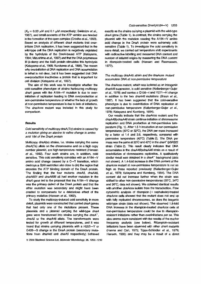
(& = 0.03 pM and 0.1 pM respectively; Sekimizu et ai.,
in the formation of the open complex (Crmke et af., 1993). 1987). and small amounts of the ATP version are needed
Based on amlysis of diierent dnaA mutants that over-
wild-type cell the DNA replication is negatively regulated initiate DNA replication, it has been suggested that in the
by the hydrolysis of the DnaA-bound ATP (Katayama, 1994; Miishima etal., 1997) and mat the DNA polymerase III @damp and the IdaB protein stimulates this hydrolysis (Katayama etal., 1% Kurobwa etal., 1998).The reason why overinitiation of DNA replication and DNA acwmulatii
overproduction inactivates a protein that is important for islemalisndck?ar,butithasbeensuggestedthatDNA
cell division (Katayama et ai., 1997).
cold-sensitive phenotype oi strains hahring muHicopy The aim of this work was to investigate whether the
initiation of replication leading to DNA overproduction at dnaA genes with the A 1 8 4 4 mutati is due to over-
non-permissive temperature or whether the la& of gmwth at non-permissive temperature is due to la& of initiations. The dnaAm mutant was included in this study for comparison.
Results
Cofdsenskivityof multicopydnaA(TsJ strains is causedby a mutation giving an alanine to valine change in amino acid 184 of the DnaA protein
Multicopy dnaA(ts) strains, i.e. strams carrying the same dnaA(Ts) allele on the chromosme and on a high copy number plasmid, are high-temperature resistant (Hansen et ai., 1992). Four such strains are, in addition, cold sensitive. This c& sensitivity coincides with an AlM-V amino acid change caused by a C-T transition, which destroys a Sphi restriction site close to (in) the region that encodes the ATP binding domain of the DnaA protein. The finding that the four mutants dnaA5, dnaA46, dnaA601 and dm4606 all had another mutation in the dnaA gene led to the pwposal that the A184-V change was the primary defect of the DnaA protein and that the other mutation was secondary and might have been created to compensate for a deleterious effect of the primary mutation (Hansen et af., 1992).
To study the multimpy-induced cdd sensitivity in more
that had only one of the mutations present. These detail, plasmids were constructed that carried dnaA genes
plasmids and a plasmid canying the wild-type dnaA gene were tramformed into strains carrying the d&+, dnaA5 or the dm446 allele. The transfwmants were tested for gmWh at dierent temperatures, and it was found that strains canying plasmids with a H252-Y or G4X-S charge in the DnaA protein (secondary muta- tions from dm446 and dm45 respectively) behaved
0 m Blackwell science Ltd. Mdecular Mioobio(qly. 35,1a2-1210
Cdd-sensitive DnaA(AAl%VJ 1203
exactly as the strains carrying a plasmid with the wild-type dnaA gene (TaMe 1). In contrast, the strains carrying the
acid change in the DnaA protein were extremely cdd plasmid with the mutat i i causing the A184-V amino
sensitive (Table 1). To investigate the cold sensitivity in more detail, we carried out temperature shift experiments with radioactive labelling and measured DNA content and number of initiated origins by measuring the DNA content in rifampicin-treated cells (Hansen and Rasmussen, 1977).
accumulate DNA at non-permissive temperature The multicopy d W 4 6 strain and the dnaAm mutant
The dnaAm mutant, which was isolated as an intragenic dnaA46 suppressor, is wld sensitive (Kellenberger-Guier
in addiion to the two dna.446 mutations (Braun et a/., etai., 1978) and carries a Q156-I and Y271-H change
phenotype is due to overinitiation of DNA replication at 1987). It has been suggested that the cold-sensitive
non-permissive temperature (Kellenberger-Gujer et al., 1978; Katayama and Komberg, 1994).
Our results indi i te that the dnaAcas mutant and the dnaA4i7pdnaA46strain continue initiation of chromosome
perature (Fig. 1). After 1.5 h incubation at non-permissive replication and DNA production at non-permissive tem-
temperatures (30°C (x 3Z"C), the DNA per mass increased by a factor of 1.4 and 3.6, respe&ely, wmpared with permissive temperature (42°C) (T-2). The DNA per mass was the same at 30% and 42°C in the dnaA+/pBR322 strain (Table 2). This result dearly imes that DNA accumulates in the dnaANpdnaA46 strain as a resuit of overinitiation of chromosome replimtion. A quaIiitively similar result was o b t a i d in a d&+ backgmund (data not shown). A 1.4-fokl increase in the DNA content of the dnaAcas mutant at non-permissive temperature is not as high as those reported previously (Kellenberger-Gujer et ai., 1978; Katayama and Komberg, 1994). The DNA content dd not increase further when the strain was shined to other non-perm'wive temperatures (W%, 34°C and 36°C; data not shown). We obtained identical results with another dnaAcas isolate from the transduction. Flow cytometric analysis of rifampicin (+ cephah?xin)-treated dnaAcos cells showed that the mutant does not end up with fully replicated chromosomes, as does the isogerjc wild-type strain (data not shown). The observed 1 .S-fold DNA increase in the rifampicin-treated dnaAcas cells at non-permissive temperature could be due to rifampicin- resistant initiations rather than overinitiations perse. This
frequency analysis (see below). Warnpicin-resistant also seems more consistent with the resuits of the marker
initiations have been observed with other dnaA mutants (Hanna and Carl, 1975; Tppe-Schindler et al., 1979; Hansen, 1995) and they may be a resuit of a direct
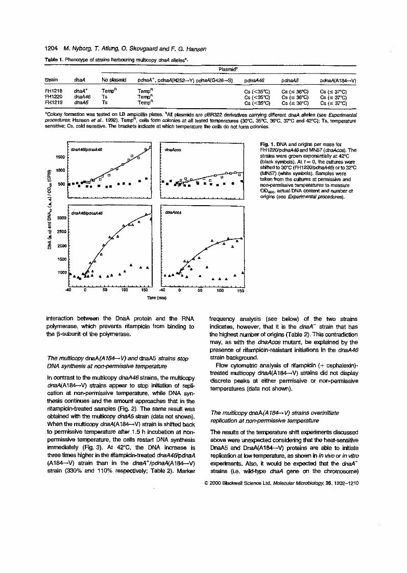
1794 M. Nyborg, T. Atlung, 0. Skovgaard and F. G. Hansen Table 7. Phenotype oi slraim harbxring m&ticopy dnaA allelesa.
P!asmi&
Saain dnaA No plasmid pdnaA+. pdnaA(H2~2+~) pdnaA(G4m-S) pdnaA46 pdnaA5 PdnaA(A184-V)
FHlpO M 4 6 Ts M1218 dnaA+ TempR TempR
Temp" M1279 dnaA5 Ts TempR CS (<%QC) CS (C 36%) CS (S 37°C)
'Colony formation was tested on LB ampicillin plates. 'All plasmids are pBR322 delivaiives canying different dnaA alleles (see Experimental procedureq Hamen et al.. 7592). TempR. cells form mlonies at all tested temperawes (WC, 35°C. 36°C. JPC and 42°C); Ts, tempemre sensitive: CS. cold sensiiiie. The brackets indicate at which temperature the cells do not farm mlonies.
CS (<%*C) CS (C 36%) CS (C 3PC) CS (C35SC) CS (C 36°C) CS (S V C )
Fig. 7. DNA and origins per m a s s for
Strairs were gmwn exponentially at 42°C FHlZWpdna446and MN57 (dmAm). The
(bl& symbols). At t = 0, the cultures were shnedx,30"C(FHl~/PM46)orx,32320C
takenfrom the wltures at permissive and (MN57) (white symbols). Samples were
ODlso, a w l DNA content and number of origins (see Expermental pmedum).
... ~ ~ p e r m i s s i ~ e temperatures to measure
4 0 0 50 100 1%
interaction between the DnaA protein and the RNA pdymerase, which prevents rifampicin from binding to the psubunil of the polymerase.
The muiticopy dnaA(A18h.v) anddnaA5 strains stop DNA synthesis at nonpermissive temperatute
In contrast to the multicopy dnaAMstrains, the mukicopy dnaA(A184-V) strains appear to stop initiation of repli- cation at non-petmissive temperature, while DNA syn- thesis continues and the amount approaches that in the rifampicin-treated samples (fig. 2). The same result was obtained with the m u l t i i dnaA5strain (data not shown). When the mmicopy dnaA(Al84-V) strain is shfted back to permissive temperature after 1.5 h incubation at non- petmissive temperature, the cells restart DNA synthesis immediately (Fg. 3). At 42°C. the DNA increase is three times higher in the rifampicin-treated dnaAWpdnaA (A184-V) strain than in the dnaA+/pdnaA(Al84-V) strain (330% and 110% respectively; Table 2). Marker
frequency analysis ( s e e below) of the two strains
the highest number d origins Fable 2). This contradiction indicates, however, that it is the dnaA+ strain that has
may, as with the dnaAws mutant, be explained by the presence d rifampicin-resistant initiations in the dnaA46 strain background.
treated multicopy dnaA(Al84-V) strains did not display Flow cytometric analysis of rifampicin (+ cephalexin).
discrete peaks at either perrrissive or non-petmisskve temperatures (data not shown).
The muhicopy dnaA(A 184-v) strains overinitiate replication at nonpermissive temperature
The results of the temperature shift experiments discussed above were unexpected considering that the heat-sensitiie DnaA5 and DnaA(A184-V) proteins are able to initiate reNiticm at low temperature, as shown in in vivo or in vitro experiments. Also, il would be expected mat the dnaA+ strains (i.e. wikl-type dnaA gene on the chromosome)
B 2ooo BhckweN Science Ltd, Mdecuiarh4i~ob.dc.q~ 35,1m2-1210
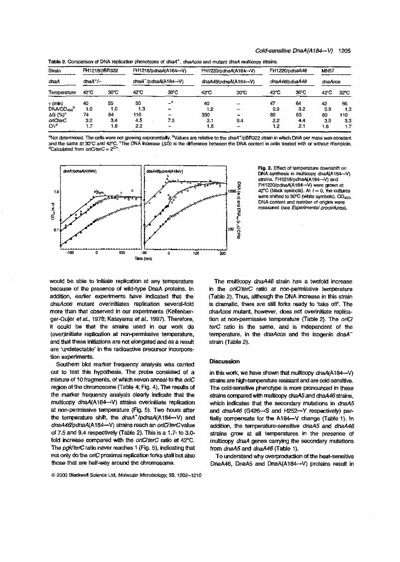
Cold-sensitive DnaA(Al84-V) 1205
Table 2. Cornparisan of DNA rephtion phenotvpes of M*, dnaAmand mutant dnaA multicopy strains.
Strain FHl2781pBR322 Ftil276/pdn~Al84-V) FHIm/pdnaA(A184-V) FH12201pdnaA46 MN57
dnaA &aA% dnaAi/pdna4(A184-V) dnaAWpdnaA(Al84-V) dnaA46ipdnaA46 dnaAms
Temperature 42% 30C QC 30°C 42% 30°C 42°C 30°C 42% 32%
T (min) 40 55 50 - 47 ONNOD,," 1.0 1 .Q 1.3
40 1 2
€4 4 2 5 6
4G (%)' 74 84 110 - 330 Orif2er-C 3.2 3.4
- 69 63 4.5 . 7.5
60 110
C/# 3.1 9.4
- - - 0.9 3.2 0.9 1.3
1.7 1.8 2.2 - 1.6 - 2.1 1.6 1.7 2 2 4.4 1.2
3.0 3.3
BNd determined. The cells were not gmwing exponentially. 'Values are relative to the &aAilpBR3ZZ m i n in which D N A per m a s was mmm and the Same at 30°C and 4?C. %e DNA increase (4G) is the diference between the DNA mntent in cells treated with OT without riiampicin. ~calcubted frm WiCJferc = 2U'.
would be able to initiate replication at any temperature because of the presence of wild-type DnaA proteins. In addition, earlier experiments have indicated that the
more than that obSeNed in our experiments (Kelknber- dnaAcrxs mutant overmitiates replication several-fold
ger-Gujer eta/., 1978; Katayama eta/., 1997). Therefore, it could be that the strains used in our work do
and that these initiations are not elongated and as a resuii (0ver)initiate replication at non-permissive temperature,
tion experiments. are 'unddectable' in the radimctive precursor incorpora-
Southem blot marker frequency analysis was carried
mixture of 10 fragments, of which seven anneal to the on% out to test this hypothesis. The probe consisted of a
the marker frequency analysis clearly indicate that the region of the chromosome (Table 4; Fig. 4). The results of
muiticopy dnaA(Al84-V) strains overinitiate replication at non-permissive temperature (Fig. 5). Two hours after the temperature shA, the dnaA'lpdnaA(Al84-V) and dnaA4@'pdnaA(Al84-V) strains reach an oriQterCvalue
fold increase compared wah the oriUferC ratio at 42%. of 7.5 and 9.4 respectivety (Table 2). This is a 1.7- to 3.0-
The pgMterCratii never reaches 1 (Fig. 5). indicatingthat not d y dothe oriC proximal reflkatii forks stall but also those that are halfway around the chromosome.
Fig. 2. Effect of ternperamre dowmhi m DNA symhesis In mAtkow dnaA(Ai84-V) strain?.. FHl2lWp&aA(Al84-v) and
42°C (black symbols). At r = 0, the cuftures FHlpglpdnaA(A184-.V) were gmwn at
were shifted to 30°C (Mi symbols). ODao. DNA mntent and number of wigim were measured (see Expenmenrd pm&res).
in the oriUterC ratio at non-permissive temperature The multicopy dnaA46 strain has a twofold increase
(Table 2). Thus, although the DNA increase in this strain is dramatic, there are still f o k ready to 'take off'. The dnaAoos mutant, however, does not overinitiite replica- tion at non-permissive temperature (Table 2). The oriU terC ratio is the s a m e , and is independent of the temperature, in the dnaAm and the isogenic dnaA+ strain (Table 2).
Discussion
In this work, we have shown that multiwpy dnaA(A184-V) strains are high-temperature resistanf and are d sensitive. The cold-sensitive phenotype is more pronowad in these strains mpared with multicopy dnaA5and dna446strains, which indicates that the secondary mutations in dnaA5 and dnaA46 (G42GS and W52-Y r e w i v e l y ) par- tiaUy oompensate for the AI-V change (Table 1). In addition, the temperature-sensitive dnaA5 and dnaA46 strains grow at all temperatures in the presence of
from dnaA5 and dnaA46 (Table l). multicopy dnaA genes carrying the secondary mutations
To understand why overproductiin of the heat-sensitive DnaA46, DnaA5 and DnaA(A184-V) proteins result in
e 2om Blackwell Science Ltd. MdecumrMinobiology. 35,1202-1210
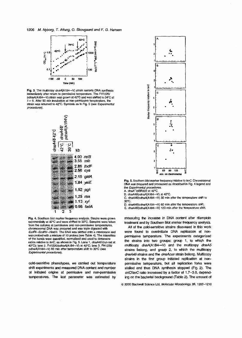
1206 M. Nybog, T. Atfung, 0. Skovgaard and F. G. Hamen
5 i“
-1w 5 0 0 50 1M m e (m)
Fig. 3. The multicopy dnaA(A184-V) strain restam DNA synthesis immediately after return to permissive temperature. The FHtZ20I pdnaA(Al84-V) strain was gmwn at 42% and was shined to 34°C at
strain was r e m & to 42°C. symbols as in Fig. 2 (see Expenmema! t = 0. Affer 90 min incubation at non-permissive temperature. the
procedures).
Fig. 4. Swthem blot marker frequency analysis. Strains were grown exponentially at 42°C and were shfied to 30°C. Samples were laken imm the cUnures at pem$?swe and nonpermissive temperatures, chmmosamal DNA was prepared and was triple digested with EccRI-ECORV-Hidlll. The DNA was blctted MO a membrane and was probed wah a m.mre of 10 probes (see Table 4). The intemitiffi d the bands were quanIjiied. normalized and used m determine ratios relarive m ten?. as sbwn in F@. 5. Lane 1, &d46 (run om at 42’C): lane 2. FHlpOlpdnd(A184-V) at 42%; lane 3, F H l W pdnaA(Al84-V) €0 min after temperalure shift to W C ( s e e Expermental pmcedures).
cold-sensitiie phenotypes, we carried out temperature shift experiments and measured DNA content and number of intied origiis ai pemilssive and nm-permissive temperatures. The last parameter was estimated by
B
l 1
Fig. 5. S a m m blot marker frequency relative to &C. Chromosomal DNA was prepared and pmce~Sed as desaiiwd in Fig. 4 legend and me Expenrnentai procedures. A. dnaA‘ipBR322 at 42°C. 8. dnaA46/pdnd(A184-V) at 42°C. C. dnaAWpdnaA(Al84-V) 30 min after the temperame stiff to 30°C. D. dnd4@pdnaA(Ata4-V) 60 min after the temperame shi. E. dnaAWpdnaA(Al84-v) 120 min after the temperature shift
treatment and by Southem blot ma&r frequency analysis. measuring the ircrease in DNA content after rifampicin
All of the coldsensitive strains discussed in this work were found to overinitiate DNA repliition at non- permissive temperature. The experiments categorized me strains into two groups: group 1, to which the multicopy dnaA(A184-V) and the multicopy dnaA5 strains belong, and group 2, to which the multicopy
strains in the first group iniliated replication at non- dnaA46 strains and the dnaAcas strain belong. Muiticopy
permissive temperature, but all replication forks were stailed and thus DNA synthesis stopped (Fig. 2). The oriUterC ratio increased by a factor of 1.7-3.0, depend- ing on the bacterial background (Table 2). The amount of
0 2oM) Blackwell Science Ltd. MolecularMiaobiolagy. 35.1202-1210

Cold-sensitive DnaA(A186V) 1207
Table 3. Svains used in this W&.
Strain Genome Reiere"CW
FH1218
FHlPO FH1219
MN60 MN57
M441
mu-l, am-9, bx-3. mn-l. msL.8, sopE44 .VC*) hp-3. his-4, pyrB.:TnS, thkl. g a l K 2 , lacy?. Hansen et al. (1991)
As FH1218. Ma446 As FH1218, &a45 Hansen et d. (1991)
As FH1218. haA::Tn?ff. ha4-s This work Hansen er d. (1991)
As FH1218. maA::Tnlff &&m, hsA::Tnlff
This work Katayama and Kcinberg (1994)
that already ongoing replications forks finished, but the DNA synthesized after the temperature shtt suggested
marker frequency analysis showed that existing forks must also stall because the pgwterc ratio (marker located halfway around the chromosome) also increased slightly (Fig. 5). Strains in group 2 also initiated replication at non- permissive temperature but (some of) these initiations did
especially in the multicopy dnaA46 strains, which have a continue and resulted in a higher actual DNA content,
more M threefold hisher DNA cnntent at m-pemksiive temperature. The DNA content in the dnaAcos mutant only increased 1.4-fold when shtted to non-permissive tem- perature (Table 2: Fig. 1). The DNA increase in dnaAcos at non-permissive temperature has previously been reported to be more dramatic (Kellenberger-Gujer et al.,
due to diierent growth canditions a d o r the diierences in 1978; Katayama etal., 1997). Thedifferent results may be
the strain background. dnaA mutants have previously been reported to
become cold sensitive when the dnaA(A184-V) allele is involved; either as a single wpy on the chromosome as
the dnaA46 mutant with GroE chaperone overproduction the dnaAcas mutant (Kellenberger-Gujer et al., 1978) or
(KEdayamaandNa~,199l),orasmultimpydnaAgenes (Hansen et al., 1992). Also, a multicopy dMA(Gl77-D) strain is wld sensitive (Won and Kaguni, 1997). and because both amino add changes are lccated close to, or
the mutations may alter the ATP binding affinity and/or in, the presumed ATP binding region of the DnaA protein
indicated that the DnaA5, DnaA46, DnaA(A184-V) and ATP hydrolysis. ln Vmo nitrocellulose binding assays have
the DnaAms proteins do not bind ATP (Hwang and Kaguni, 1988; Hupp and Kaguni, 1993; Katayama, 1994; Carr and Kaguni, 1996). However, the four mutant proteins bind oric with an affinity similar to that of the
non-permissive temperatures (Katayama, 1994; Carr wild-type DnaA protein in vitro? at both permissive and
and Kaguni. 1996). AWough recent in vitro experiments showed that the DnaA(A184-N) protein wuld be activated for lower affinity ATP binding by monomerization (Carr and Kaguni, 1996), the functiw of ATP binding and hydrolysis is still unclear. The importance of ATP binding may be more indirect, in the sense that ATP hydmiysis is
c3 MW Blackwell Science Ltd. M d w ! a r Mimbidcgy, 35,1202-1210
GCTGCCGATTCCTCGACTGA AAGAATGGCTGGGATCGTGG ATCGAGGTTACTGCGGATCA GATCCAGnGACTGGTGTTG CAlTGAGCGAAAGCGATCCG CAATTAACCAATCAGTCGCA CATGGTGAATCCTCTCGlTG AGCACTGGATGAGGTGATCA GATATCGGCGTCATCCAGGC AGCAGCTCGATTACCGTACG TTCCACAGACCTACCATCAG GACGATAAAGGCTGCmGT CGCTCATGCCAGAACGGAAC
~
(Mizushima et al., 1997: Kurakawa et al., 1998). As required for a negative regulation of the initiation
suggested previously for the OnaAccs protein (Katayama, 1994), the lower affinity of the four mutant proteins for ATP may render them inert to negative regulation through ATP hydrolysis and thus give rise to overinitiation of
ture, where the mutant proteins have a higher activity that replicatiin when the mutants are shifted to low tempera-
may be due to a more normal conformation. There are, however, other dnaA mutatiins !mated in the ATP binding
GI75-D and P43-+L-Gl77-S, Sulton and Kaguni, region (G172-A and V185-A, Skovgaard et al., 1998;
although the mutant proteins are active for chromosome 1997) that do not confer multicopy cold sensitivity,
bus for ATP binding or hydrolysis than the AIM-V initiatiin. These mutations might therefore be less deleter-
mutation.
ATP bound to DnaA is strongly stimulated by replicating It has recently been shown in viho that the hydrolysis of
DNA polymerase III holoenzyme (Katayama eta/., 1998). We propose that in vivo the ATP hydrolysis may be needed to help release DnaA protein bound to DnaA
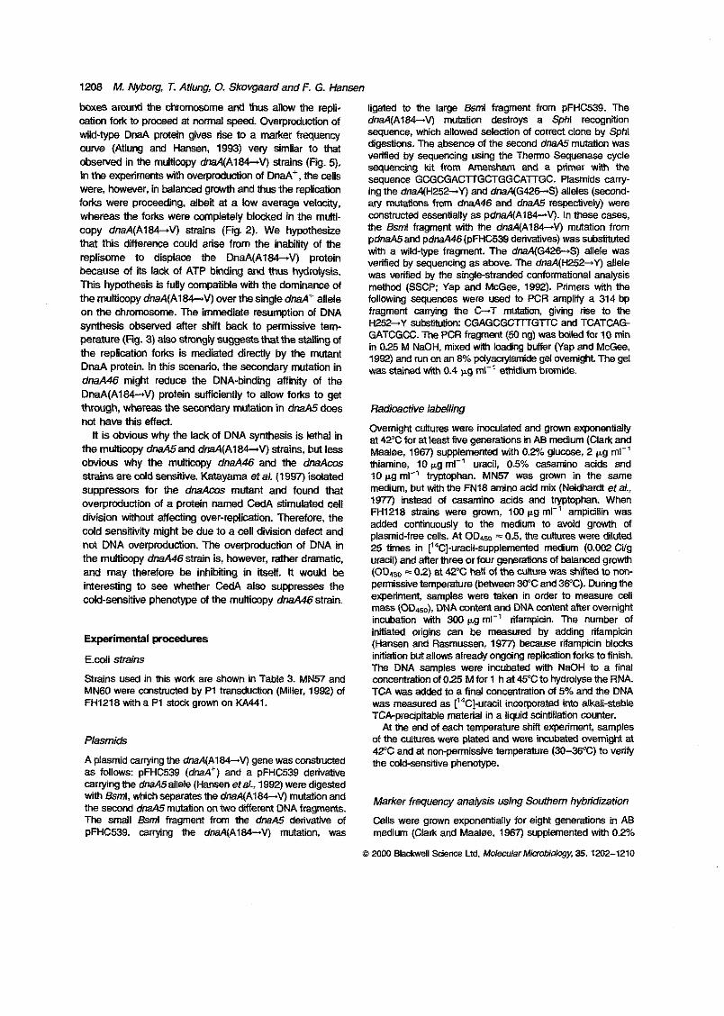
1208 M. Nyborg, T. Atlung, 0. SKovgaard and F. G. Hansen
Catim fork to proceed at normal speed. Overproduction of boxes amund the chromosome and thus allow the repli-
wild-type DnaA protein gives rise to a marker frequency curve (Atlung and Hansen, 1993) vety similar to that &sewed in the multicopy dnaA(A184-V) strains (Fu. 5). In the experiments with overprcdwtii of D m + , the cells
forks were proceeding, albei at a low average velocity, were, however, in balanced grcrwth and thus the replication
copy dnaA(A184-V) strains (Fig. 2). We hypothesize whereas the forks were mpletely bl&ed in the multi-
that this difference cwld arise fmm the inability of the replisome to displace the DnaA(A184-V) protein because of its lack of ATP binding and thus hydrolysis. This hypdhesis is fully compatible with the dominance of the multicopy dnaA(Al84-9 over the single d&+ allele on the chromosome. The immediate resumption of DNA synthesis observed after shft back to psrmissiie tem- perature (Fig. 3) also s tmgv suggests that the stalling of the replication forks is mediated directly by the mutant DnaA protein. In this scenario, the secondary mutation in dnaA46 might reduce the DNA-binding affinity of the
through, whemas the SeCMlctary mutation in dnaA5 daes DnaA(A184-V) protein sufficiently to allow forks to get
not have this effect. It is obvious why the lack of DNA synthesis is lethal in
the multicopy dnaA5and dnaA(A184-V) strains, but less obvious why the multicopy dm446 and the dnaAcas strains are mld sensitive. Katayama et al. (1997) isdated suppressors for the dnaAcas mutant and found that overpmduction of a protein named CedA stimulated cell
cold sensitivty migM be due to a cell division defect and division without affecting over-replication. Therefore, the
not DNA overproduction. The overpmduction of DNA in the multicopy dmA46strain is, however, rather dramatic, and may therefore be inhibiting in itself. It would be interesting to see whether CedA a b suppresses the cold-sensitive phenotype of the multicopy dnaA46strain.
Experimental procedures
E.coli strains
Strains used in this work are shown in Table 3. MN57 and MN60 were constructed by P1 transdudion (Miller, 1992) of FH1218 with a P1 st& grown on KA441.
Plasmids
A plasmid carrying the dnaA(A184-V) gene was wnstructed as follows: pFHC539 (dnaA‘) and a pFHC539 derivative carrying the dmA5allele (Hansen eta., 1992) weredigested
the second dm45 mutation on two different DNA fragments. with &ml, whichseparates me dnaA(A164-V) mutation and
The Small Bsml fragment frwn the dmA5 derivative of pFHC539, carrying the dnaA(A184-V) mutation, was
ligated to the large &ml fragment from pFHC539. The
sequence, which allowed selection of correct clone by Sphl dnaA(Als4-V) mutation destroys a Sphl reccgnition
digestions. The absence of the sewnd dnaA.5 mutation was verified by sequencing using the Therm0 Sequenase cycle sequencing kit from Amersham and a primer with the sequence GCGCGACTTGCTGGCATGC. Plasmids carry-
ary mutations from dnaA46 and dm45 respectively) were ing the dnaA(H252+Y) and dnaA(G426-S) alleles (second-
wnstructed essentially as pdnaA(A184-V). In these cases, the &mi fragment with the dnaA(Al84-V) mutation from pdnaA5and pdnaA46(pFHC539 derivatives) was substituted with a wild-type fragment. The dnaA(G42€-S) allele was verified by sequencing as above. The dmA(H252-Y) allele was verified by the single-stranded conformational analysis method (SSCP Yap and McGe, 1992). Primers with the following sequences were used to PCR amplify a 314 bp fragment carrying the G-T mutation, giving rise to the H252-Y submution: CGAGCGGTI’TGTTC and TCATCAG- GATCGCC. The PCR fragment (50 ng) was boiled for 10 min in 0.25 M NaOH, mixed wah loading buffer (Yap and MC-,
was stained with 0.4 pg ml-’ ethidim brwnide. 1992) and run on an 8% pdyacMarnide gel overnight. The gel
Radioactive labdling
OvemigM cuitures were inoculated and grown exponentially at 42°C for at least five generamns in AB medium (Clark and
thiamine, 10 pg ml-’ uracil, 0.5% m i n o adds and Maaloe, 1967) supplemented with O.% g~ucase, 2 pg ml-’
10 pg ml-’ tryptcphan. MN57 was grown in the same medium, but WiM the FN18 amino add mix (Neidhardt et al., 1977) instead of casamino acids and twtophan. When
added continuously to the medium to avoid growth of FH1218 strains were grown, 100 +g ml-’ ampidliin was
plasmid-free cells. A t OD4- = 0.5, the cultures were diluted 25 times in [’4C]-uracil-suppkrnented medium (0.002 Ciig uracil) and after three or fwr generations of balanced growth (ODas0 = 02) at 42°C hail of the culture was Shitted to non-
experiment, samples were taken in order to measure cell permissive temperature ( W e e n W C and 3VC). During the
mass (ODaso), DNA mntent and DNA content after overnight incubation with 300 pg ml-’ rifampicin. The number of initiated origins can be measured by adding rifampicin (Hansen and Rasmussen, 1977) because rimpicin blccks initiation but allows already ongoing replication forks to finish. The DNA samples were incubated with NaOH to a final
TCA was added to a final concentration of 5% and the DNA concentration of 025 M for 1 h at 45°C to hydroiyse the RNA.
was measured as (‘4C1-uracil incorporated into a!ka!i-stabie TCA-precipitable material in a liquid scintillation counter.
of the cultures were plated and were incubated wemight at At the end of each temperature shft experiment, samples
42°C and at non-permissive temperature (30-36‘C) to veri* the cold-sensiiive phenotype.
Marker frequency anaiysis using Soothem hybridization
C e l l s were grown exponentially for eight generations in AB medium (Clark and MaaJoe, 1967) supplemented with 0.2%
c3 20M) Blackwell Science Ltd, MolecoarMimbidcgy, 35,1X12-1210
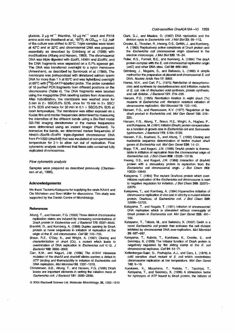
ghose, 2 +g m!-’ thiamine, 10 pg mi-’ uracil and FN18 amino acid mix (Neidhardl et al., 1977). At ODaSo = 0.2 ,half of the culture was s h i i d to 32°C. Triple samples were taken at 42°C and at 32°C and chromosomal DNA was prepared essentially as described by Grimberg et al. (1989) with modifications (Atlung and Hansen, 1993). The chromosomal
the DNA fragments were separated on a 0.7% agarose gel. DNA was triple digested with EcoRi, HinClll and ECORV, and
The DNA was transferred ovemigM to a nylon membrane (Amersham), as described by Sambrd et al. (1989). The membrane was prehybridized with denatured Salmon sperm
at W C with r5S]dATP-labelled probe. The probe mnsisted DNA for more than 1 h ai 65°C and was hybridized overnight
of 10 purified PCR fragments from different positions on the chrcinosome (Table 4). The DNA fragments were labelled
After hybridization, the membrane was washed once for using the megaprime DNA labelling system from Amersham.
5 min in 2x SSC/0.5% SDS, once for 15 min in 2x SSC/ 0.1% SDS and twice for 30 min in 0.1 x SSC/O.5% SDS at room temperature. The membrane was either exposed MI a
the intens’mes of the different bands using a EIwRad model Kodak film and marker frequencies determined by measuring
GS-700 imaging densitometer or the marker frequencies were determined using an instant Imager (Packard). To normalize the bands, we determined ma&er frequendes d HinClll-EmRI-EroRV tripledigested chromoscinal DNA from FHIZO (dnaA46) that was incubated at non-permissive temperature for 2 h to allow Nn out of replication. Flow cytometric anaiysis confirmed that these cells contained fully replicated chromosomes.
Flow cytometric analysis
sen et al., 1999). Samples were prepared as described previously (Christen-
Acknowledgements
We thank Tsutomu Katayama for supplying the Strain W441 and Ole Micheisen and Sven MOIler for discussions. This Study was supponed by the Danish Centre of Microbiology.
References
Atlung, T., and Hansen. F.G. (1993) Three disiinci c h r m m e replication States m induced by increasing concernrations d DnaA protein in Escherichia coli J Bacterid 175: 653-6545.
Bramhill, D., and Kombeq, A. (1988) Duplex opening by DnaA
origin of the E. wlichmosome. Cell52: 743-755. protein at novel sequences in initiation d replication at the
Braun. R.E.. ODay. K., and Wright, A. (1987) Cloning and characterization of dnaA (CS). a mutant which leads to overinitiation of DNA repiication in Escherichia coli K.12. J
Carr, K.M.. and Kaguni, J.M. (1996) The AtMV missense Bacteriol169 3898-303.
mutalion d the dnaA5 and dnaA46 alleles m i e n a defeci in ATP binding and thermolability in initiation of Eschews mli
Christensen. BB.. Atlung. T.. and Hansen. F.G. (1999) DnaA DNA replication. MolMicrobioiZO: 1307-1318.
boxes are impatant elements in setting the initiation mass d Escbrichla coli J Bactenol 181: 2683-2688.
0 W Blackwell Swnw Lid, MdwukrMimbioiwy. 35.1202-1210
Cold-sensitive DnaA(A184- VJ tz09
Clark, D.J., and Maabe, 0. (1967) DNA replication and the
C&e, E., Thresher. R., Hwang. D.S.. Grffiih, J.. and Komberg. division cycle in Escherichia col J Mol Bid 2 3 99-1 12.
A. (1993) Replicatively active CmpleXeS d DMA protein and the Escherichia coli chrmosomal origin ohsewed in the
Fuller, R.S.. Funnell. B.E.. and Komberg. A. (1984) The dnaA electron microscope. J Mol Sid233: 16-24.
protein complex with the E. colichrom~~~mal replication origin (0riC) and other DNA sites. cell 38: 889-900.
Grimberg. J., Maguire, S., and Belium’o, L (1969) A simple method forthe preparation d plasmid and chromosomal E. coli
H ~ M . M.H.. and Cad, P.L (1975) Reinidation d deoxyribonu- DNA. Nucleic Acids Res 17: 8893.
cleic acid synthesis by deoxyribonucleic acid initiation mutants of E. wli role d ribonucleic acid synthesis. protein synthesis,
Hansen, F.G. (1995) Reiniliation kjnetics in eight dmA (Ts) and cell division. JBacteridl21: 219-226.
chrwnmme replication. Mol McmbioIIS: 133-140. mufanis d Escherichia coli rifampicin resistam initiation d
Hansen. F.G., and Rasmussen, K.V. (1977) Regulation of the
225. dna.4 produd in Escherichia coli. Mol Gen Genet 155: 219-
Hansen, F.G., Atlung. T., Braun. RE.. Wright. A., Hughes, P,. and Kohima. M. (1991) Inititor(DnaA) proteinconcenhation as a function of growlh rate in Escherichia coli and SalmOneXa
Hansen. F.G., Koetoed, S., and Atlung, T. (1992) Cloning and fyphimurium. JBacferdl73: 5194-5199,
nwleotide sequence determination d twelve muIan1 dmA genes of Escherichia mli. Mol Gen Genet 234.14-21.
Hupp, T.R.. and Kaguni. J.M. (1993) DnaA5 protein is thermo- labile in initiation of replication from the chrcinmal origin of
Hwang. D.S., and Kaguni. J.M. (1988) interactian of dnaA46 Eschekhia coli. J Biol Chem 268: 131 28-1 31 36.
protein with a Stimulatory protein in replication from the Escherichia coli chromosmd wigin. J Biol Chem 2W:
Katayama, T. (1994) The mutant DnaAcos pmtein which over- t0633-to640
initiates replication of the EschercMa cob chromosome is inen to negative regulation for initiation. J Bid Chem 269 22075-
Katayama, T., and Kcinberg. A. (1994) Hypera~Ne initiation d 22079.
chromosome replication in vivo and in vitro by a mutant iwiator protein. DnaAzos. of Escherichia coli. J Biol Chem 269:
Katayama, T., and Nagata. T. (1991) Initiation d chromosomal 12698-12703.
DNA replication which is Stimulated wilhwt oversupply of
5 M . DnaA protein in Escherichia coli. Mol Gen Genet 226 491-
Katayama. T., Taka& M., and Sekjmizu. K. (1997) CedA is a novel Escherichia coli protein that activaies the cell division
26: 687-697. inhimed by chromosomal DNA over-replication. Mol Microbbl
Katayama. T.. Kubota, T., Kurokawa. K.. Crmke, E., and Sekjmizu. K. (1998) The iniiiitw function d DnaA protein is negatively regulated by the sliding clamp of the E. CON
Kellenberger-Gujer, G.. Podhajska, A.J., and Caro, L. (1978) A chromosoW replicase. Cell94 61-71.
cold sensmYe d& mutant of E. coli which overiniliates
162: 9-16. chrmmome repiication at low temperature. Mol Gen Genet
Kurokawa. K., Mimshima. T., Kubota. T., Tsuchiya. T., Katayama, T., and Sekimizu. K. (1998) A stimulation factor for hydrolysis d ATP bwnd to DnaA protein, the initiator of
1210 M. Nybog, T. Atlung, 0. Skovgaard and F. G. Hansen
chrommomal DNA replication in Escherichia coli. Bi&em
Messer, W., and Weigel, C. (1996) Initiation of chromosome Biophys Res Comun 243: 93-95,
repbation. In Escherchia cofi and Salmonelia tvphimurim
Ingraham, J.L., LOW, KB., Magasanit, B.. Schaechter, M., and Callular and Molecular Biolw, 2nd edn. Neidhardt. F.C.,
Umbarger, H.E. (eds). Washington, DC: American society for Micmbiolqy Press. pp. 1579-1601.
vm Meyenburg. U. and Hansen. F.G. (1987) Regulation of chromosome replication. In Eschsrichia coli and Salmmlia
Ingraham, J.L., Low, KB., Magasanit. B.. Schaechter. M.. and fyphimurium Cellular and Mdewiar Bology. Neidhardt, F.C..
Umbarger, H.E. (eds). Washington, DC: American Society for
Miller, J.H. (1992) A shorl course in bactwial genetics. A Mircbiolqy Press. pp. 1555-1537.
LaborafoIy Manual and HanaRwok for EsCf~€ficflia coli and Relatedsactera. Cold Spring Harkr. W: Cold Spring Harbor Latmatoiy Press.
Mizushima. T., Nishida. S.. Kumkawa. K., Kaiayama,T., Mild, T., and Sekimizu. K. (1997) Negative control of DNA replication by hydrolysis of ATP bound to DnaA protein. the initiator of chromosomal DNA replication in E&e!ichia m/i EMBO J16: 3724-3730.
Neidham. F.C., Bloch. P.L.. Pedersen, %.and Reeh, S.V. (1m Chemical measurernerds d steady-sate levels of ten aminoa- cykransfer ribonucleic acid symhelaser in Escherichia coli. J Baderiol 129: 37Gm.
Sambrwk. J., Fritsch, E.F.. and Maniatis, T. (1989) MoI-iar
Cloning. A LaboratoIy Manual, 2nd edn. Cold Spring Harbor,
Samm, C.E.. Hansen, F.G.. Miller. J.F., and Schaechtw, M. Ny: Cold Spring Harbor Laboratory Press.
(1989) In vivo studies of DnaA binding to the origin d
Same. M., Sibbald. PR., and winingfrofer, A. (1990) The P- replication d Escherichia coli EMBO J 8: 989-993.
Imp - a common mota in ATP- and GTP-binding proleins. Trends Biochem Sci 15: 430-434.
Seldmizu, K., Bramhill. D.. and Kornbwg. A. (1987) ATP activates DnaA pmtein in initiating replication of plasmids bearing the
Skarsiad, K., and Boye. E. (1994) The initiator protein DnaA: origin of the E. colichrwnosome. Ce1150: 259-265.
evolm-on propelties and fundon. Biochim Biophys Acta 1217 111-130.
Skwgaard. 0.. Olesen. K., and Wright, A. (1998) The cemral lysine in the P-imp motif of the Escherichia cofi DnaA
chromosomal wigin. O G and the F faclw origin. oris, but is protein is essenlial lw initiating DNA replication from t n e
dispensable for the initiation from the P1 plasmid origin. O M
Plaund40: 91-99. Suaon,M.D..andKaguni.J.M.(1997)The Escherichiacolidna4
gene: four functional domains. J Mol Bioi 274: 546-561. Tippe-Schindier, R.. Zahn. G., and Messer, W. (199) Control d
the iniliaFan of DNA replication in Em%Iichia d i . l. Negative
Yap, E.P.. and Mffiee. J.O. (1992) Nonmisdopic SSCP detection control d initiation. Mol Gen Genetl68: 185-195.
in PCR produC15 by ethidium bromide staining. Trends Genet 8: 49.