Embed Size (px)
Citation preview
The Enteropathogenic E. coli Effector EspF Targets andDisrupts the Nucleolus by a Process Regulated byMitochondrial DysfunctionPaul Dean*, Jon A. Scott, Andrew A. Knox, Sabine Quitard, Nicholas J. Watkins, Brendan Kenny*
Institute for Cell and Molecular Biosciences, Medical School, University of Newcastle, Newcastle upon Tyne, United Kingdom
Abstract
The nucleolus is a multifunctional structure within the nucleus of eukaryotic cells and is the primary site of ribosomebiogenesis. Almost all viruses target and disrupt the nucleolus—a feature exclusive to this pathogen group. Here, using acombination of bio-imaging, genetic and biochemical analyses, we demonstrate that the enteropathogenic E. coli (EPEC)effector protein EspF specifically targets the nucleolus and disrupts a subset of nucleolar factors. Driven by a defined N-terminal nucleolar targeting domain, EspF causes the complete loss from the nucleolus of nucleolin, the most abundantnucleolar protein. We also show that other bacterial species disrupt the nucleolus, dependent on their ability to delivereffector proteins into the host cell. Moreover, we uncover a novel regulatory mechanism whereby nucleolar targeting byEspF is strictly controlled by EPEC’s manipulation of host mitochondria. Collectively, this work reveals that the nucleolus maybe a common feature of bacterial pathogenesis and demonstrates that a bacterial pathogen has evolved a highlysophisticated mechanism to enable spatio-temporal control over its virulence proteins.
Citation: Dean P, Scott JA, Knox AA, Quitard S, Watkins NJ, et al. (2010) The Enteropathogenic E. coli Effector EspF Targets and Disrupts the Nucleolus by aProcess Regulated by Mitochondrial Dysfunction. PLoS Pathog 6(6): e1000961. doi:10.1371/journal.ppat.1000961
Editor: Guy Tran Van Nhieu, Institut Pasteur, France
Received December 4, 2009; Accepted May 24, 2010; Published June 24, 2010
Copyright: � 2010 Dean et al. This is an open-access article distributed under the terms of the Creative Commons Attribution License, which permitsunrestricted use, distribution, and reproduction in any medium, provided the original author and source are credited.
Funding: The work was funded by a Wellcome Trust senior fellowship awarded to Brendan Kenny - Grant number WT064409MA, www.wellcome.ac.uk. Thefunders had no role in study design, data collection and analysis, decision to publish, or preparation of the manuscript.
Competing Interests: The authors have declared that no competing interests exist.
* E-mail: [email protected] (BK); [email protected] (PD)
Introduction
Central to the pathogenesis of many viral pathogens is the
requirement to target the nucleolus [1], a sub-nuclear structure
found in all eukaryotic cells that is the primary site of ribosome
biogenesis. Although the main function of the nucleolus is the
synthesis of ribosomes, it is a highly dynamic and multifunctional
organelle with a proteome of over 4,500 proteins [2] and has many
cell biological functions (reviewed in [3]). The dense concentration
of interacting proteins and nucleic acids is crucial to nucleolar
function, which if disrupted, can have serious consequences to the
cell, leading to disease [3]. One of the best-studied and most
abundant nucleolar proteins is nucleolin, an RNA-binding
phosphoprotein that represents up to 10% of total nucleolar
protein [4] and is crucial for rRNA processing. Although nucleolin
is primarily confined to the nucleolus, it is a multifunctional
protein, able to shuttle between the nucleus and the cytoplasm and
plays important roles in the pathogenesis of many viruses including
HIV, poliovirus and hepatitis C [1].
The specific targeting of proteins to the nucleolus is a well-
established viral infection strategy exhibited by almost all viral
pathogens [1]. Indeed, for several decades viruses have been
reported to subvert or hijack specific nucleolar proteins by causing
their relocalisation from the nucleolus to another subcellular site
such as the cytoplasm where they are presumably unable to
perform their nucleolar functions [1]. Unlike their viral counter-
parts, no other pathogen group including fungi, protozoa or
bacteria are known to target or disrupt the nucleolus, presumably
reflecting the viral dependence on the host transcription or
translation machinery. Many notorious animal and plant patho-
genic bacteria that cause some of our most devastating diseases,
possess type three- or type four secretion systems to deliver
multiple effector proteins directly into eukaryotic cells - a process
that is essential to cause disease [5]. These effectors exhibit diverse
biochemical activities, subverting many important aspects of host
cell physiology and are often highly multifunctional [6,7]. An
emerging theme is functional redundancy between co-delivered
effector proteins and therefore it is often difficult to determine the
role of individual effectors in disease. A successful approach in
understanding the roles of effectors has been to identify effector
families or common host cell targets that may be important across
a wide range of bacterial pathogens.
Enteropathogenic E. coli (EPEC) is a bacterial pathogen that
delivers multiple effector proteins into host cells and targets the
human small intestine causing severe watery diarrhea with high
infant mortality [8]. Unlike related bacterial species such as
Salmonella, EPEC is non-invasive and from an extracellular position
delivers its effectors [7], of which three - Tir, Map and EspF are
the best studied [7]. Tir inserts into the host plasma membrane to
act as a receptor for the outer membrane protein Intimin [9],
mediating intimate bacterial attachment to the host cell. Tir-
Intimin interaction also initiates actin-polymerisation to form an
actin-rich ‘pedestal’ beneath the bacterium. Map and EspF are
highly multifunctional with many overlapping functions as both
target mitochondria [10,11], disrupt tight junctions [12,13], efface
microvilli and inhibit the water transporter SGLT-1 [14] with
PLoS Pathogens | www.plospathogens.org 1 June 2010 | Volume 6 | Issue 6 | e1000961

EspF, but not Map, inhibiting phagocytosis [15]. Although many
functions of EPEC effectors have been identified, we know little
about their subcellular behaviour or how they are regulated within
host cells.
Here, we present the first example of a bacterial protein that
specifically targets and disrupts the nucleolus. The EPEC effector
EspF is shown to target the nucleolus late in infection where it
disrupts a subset of nucleolar factors that are essential for
ribosomal biogenesis. We further uncover a novel regulatory
mechanism whereby nucleolar targeting by EspF is temporally
controlled by EPEC’s exploitation of mitochondrial function
which is the first example of a host organelle regulating the
activities of a bacterial effector. Finally, we demonstrate that other
important bacterial species disrupt the nucleolus dependent on
effector protein delivery, suggesting that the nucleolus is a
common bacterial target.
Results
The EPEC effector protein EspF targets the nucleolusPrevious studies have revealed that EPEC EspF targets
mitochondria in infected host cells [10,16]. Given its many
reported functions, we predicted that EspF would target multiple
sites in host cells and examined the subcellular location of this
effector during early, mid- and late-stage infection. Consistent with
previous reports, microscopy of infected HeLa cells revealed an
early accumulation (within 30 min) of EspF within punctate
cytoplasmic structures (Figure 1Aa-c) that were verified to be
mitochondria (Figure S1A). No other cytoplasmic organelle was
visibly targeted by EspF during infection (Figure S1A). Mitochon-
drial targeting by EspF increased up to 60 min post-infection, after
which no visible increase in mitochondrial EspF was evident
(Figure 1 Aa-c). However, to our surprise, z-axis confocal
sectioning (see Materials and Methods) through late-stage
(.60 min) infected host cells revealed an unexpected punctate
localisation of EspF in the nucleus, within compartments 2–6 mm
in size that stained poorly with the DNA dye DAPI (Figure 1Ae-g)
– both characteristics of the nucleolus. Parallel studies using cells
infected with an EspF-deficient mutant (espF) confirmed the
specificity of EspF staining (Figure 1Ad). EspF accumulation in
the nucleolus was verified by co-staining for nucleolar markers
such as nucleolin (Figure 1B; also see Figure 2B), fibrillarin and
BMS1 (data not shown). Other EPEC effectors such as Map
(Figure S1B) (which has a similar molecular size and similar
functions as EspF [7]) and Tir (data not shown) did not accumulate
in the nucleus or nucleolus at any time during infection. Thus,
EspF specifically targets the nucleolus and is the first example of a
bacterially-encoded protein to localise to this sub-nuclear struc-
ture.
Quantification of the EspF signal revealed that nucleolar
targeting was strictly a late event in infection. Thus, EspF rapidly
associated with the mitochondria within 5–15 min of bacterial
attachment (see Figure 1Ca) and by 30 min, cells exhibited a
marked asymmetry of EspF staining in favour of mitochondria
near to the bacterial attachment site (Figure 1Cb), with little, if
any, EspF in the nucleus (Figure 1Ca&b; p,0.001). However, by
60 min, the mitochondrial EspF signal began to plateau with no
significant increase thereafter (p = 0.21). Following the plateau,
EspF became more prominent within the nucleolus - with a ,40
fold increase between 30 and 120 min (Figure 1Ca). The in vivo
relevance of this finding was supported by EPEC infection of TC-7
polarised intestinal cells (which represent the natural site of EPEC
infection) with EspF targeting the mitochondria of intestinal cells
at early time points (Figure 1Da) and nucleolar accumulation
consistently a later event (Figure 1Db-c).
Nucleolar targeting by EspF is temporally regulated bymitochondria
During infection, EPEC progressively causes the dissipation of
mitochondrial membrane potential - upon which the mitochon-
drial import of EspF relies [11,16]. We therefore hypothesised that
EPEC was exploiting mitochondrial import activity to regulate the
location of EspF to ensure nucleolar targeting is a late event. To
test this hypothesis, we took advantage of an EspF(L16E) variant
that cannot target mitochondria [16] and examined its subcellular
behaviour. HeLa cells were infected with an EspF-deficient mutant
expressing either plasmid-encoded EspF or EspF(L16E) and the
cells were stained to examine EspF’s location. EspF(L16E) was
mainly cytoplasmic (Figure 2Aa) but also targeted the nucleolus
much stronger than expected (Figure 2Aa-d), filling the entire
nucleolar (non-DAPI stained) region (Figure 2Ac&d). By contrast,
the nucleolar staining pattern seen with chromosomal- (see
Figure 1) or plasmid-encoded native EspF (Figure 2Ca) was much
weaker and more punctate. The nucleolar marker nucleolin
confirmed that EspF(L16E) did indeed target the nucleolus
(Figure 2B).
Quantification of the EspF signal in infected HeLa cells revealed
that the L16E mutation resulted in a significantly more rapid
nucleolar signal which was much stronger (at least ,25 fold;
p,0.0001) than native EspF (Figure 2Cb). Indeed, by as early as
15 min. post-infection, the levels of EspF(L16E) in the nucleolus
were similar (p = 0.11) to that of native EspF at 120 min,
demonstrating that in the absence of mitochondrial targeting,
nucleolar uptake occurs almost 8 times faster. This suggested that
the late nucleolar targeting of native EspF during infection was
directly regulated by mitochondria activity. To further test this
prediction, we chemically inhibited mitochondrial membrane
potential (MMP) with valinomycin prior to EPEC infection to
prevent the mitochondrial import of native EspF. As expected,
EspF in these cells was mainly cytoplasmic (Figure 2Da) similar to
the L16E variant but also targeted the nucleolus significantly more
rapidly and stronger (,75 fold increase; p,0.0001) compared to
untreated EPEC-infected cells (Figure 2Db). These data support
the hypothesis that the dissipation of mitochondrial membrane
Author Summary
Many of the world’s most important diseases are causedby bacterial pathogens that deliver effector proteins intothe cells of their host. Effector proteins are collectivelyresponsible for causing disease and an important area ofresearch is to define the functions of these proteins andidentify how they are regulated once inside the host cell.Here, we show that EspF, a well-studied effector ofenteropathogenic E. coli, targets the host’s nucleolus inboth infected and transfected cells and causes extensivenucleolar changes. Previously, only viruses were known totarget and disrupt the nucleolus but we show that bacteriaother than E. coli also disrupt this organelle. Our work alsouncovered a novel regulatory mechanism whereby E. coliutilises the host mitochondrion to control the extent andtiming of EspF nucleolar targeting, ensuring it is strictly alate event in infection. This is the first example of a hostorganelle controlling the functions of a bacterial effectorprotein. Taken together, this work reveals a new cellulartarget for bacterial pathogens and a novel mechanism toregulate the spatio-temporal activity of bacterial effectorproteins.
Bacterial Disruption of the Nucleolus
PLoS Pathogens | www.plospathogens.org 2 June 2010 | Volume 6 | Issue 6 | e1000961
potential induced by EPEC during infection dictates when EspF is
available to target the nucleolus and represents a novel mechanism
for regulating the cellular location and subsequent function of
effector proteins. It is also the first example of a host organelle
controlling the function and location of a bacterial effector protein
within infected host cells.
EspF induces extensive redistribution of nucleolin intothe cytoplasm
To investigate a specific function for nucleolar targeting by
EspF, we assessed changes in important nucleolar components. As
nucleolin is the most abundant nucleolar protein, essential for
ribosomal biogenesis, it was the primary focus. Microscopy of
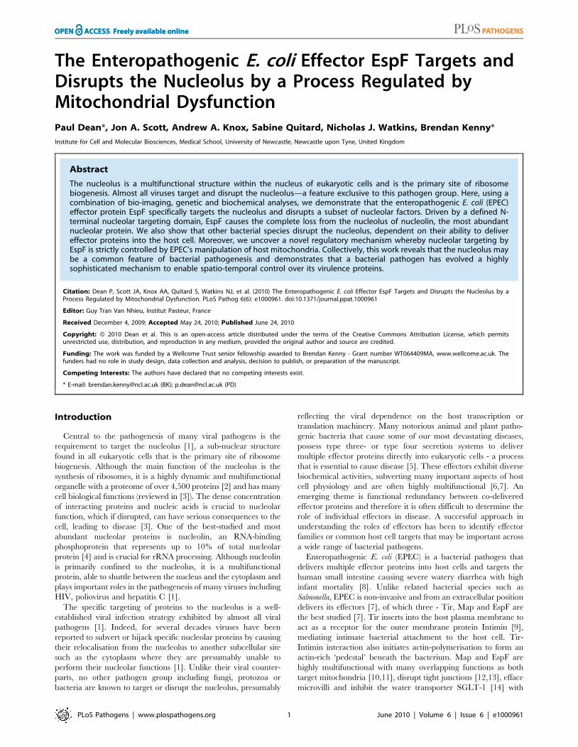
Figure 1. The EPEC effector protein EspF targets the nucleolus late in infection. (A) Immunofluroescence of HeLa cells infected with EPECor EspF-deficient (espF) strain at indicated time points. Images (a–d) show combined confocal z-sections while image (e) shows a single confocal z-section through an infected cell. (f) Punctate EspF within non-DAPI stained nucleolar region (red outline). (g) Confocal z-series through the nucleus ofan infected cell - red line encloses the non-DAPI stained nucleolar region. (B) Single confocal z-section through an EPEC-infected cell (2 h) stained fornucleolin and EspF. (C)(a) Quantification of EspF signal in cellular compartments. Units are based on fluorescence intensity; results show mean 6 SE(n = 3) with approx. 60 cells per experiment. (b) EPEC-infected cell showing EspF staining concentrated beneath attached bacteria. (D) Single confocalcross-sections of intestinal epithelia infected for 45 min (a) or 4 h (b) with EPEC. The image given in (c) is a magnification of the intestinal TC7 cellnucleus (arrow) showing EspF staining.doi:10.1371/journal.ppat.1000961.g001
Bacterial Disruption of the Nucleolus
PLoS Pathogens | www.plospathogens.org 3 June 2010 | Volume 6 | Issue 6 | e1000961

uninfected HeLa cells stained with nucleolin antibodies revealed
nucleolin was exclusively found within the nucleus/nucleolus (not
shown), which remained unchanged following a 30 min EPEC
infection (Figure 3A). However, at later infection times
(.120 min), EPEC caused a dramatic relocalisation of nucleolin
from the nucleolus into the cytoplasm (Figure 3A), which by
180 min was almost exclusively cytoplasmic. Quantification of the
nucleolin signal in the cytoplasm and nucleus revealed an inverse
relationship during EPEC infection (Figure 3Ba), suggesting the
cytoplasmic nucleolin arose directly from the nuclear pool.
Importantly, the espA EPEC mutant (which cannot deliver
effectors into host cells) did not induce any visible changes of
nucleolin (Figure 3A) suggesting that nucleolin relocalisation was
indeed mediated by effector proteins.
Similar results were obtained using polarised intestinal TC-7
cells (Figure S2Aa) and supported by Western blot analysis which
showed that at late infection, nucleolin levels decreased in the
‘insoluble’ (nuclei-containing) and membrane fractions with a
corresponding increase in the cytoplasmic fraction (Figure 3Bb).
Both events coincided with increased EspF in the nuclear fraction
between 2–4 h post-infection (Figure 3Bb) while the EspF signal in
the ‘membrane’ fraction (containing mitochondria) increased
Figure 2. EspF nucleolar targeting is regulated by mitochondrial activity. (A) HeLa cells infected with the EPEC EspF-deficient strain (espF)carrying a plasmid expressing the EspF(L16E) variant with a (a) Confocal z-stack showing EspF(L16E) in the cytoplasm and indicated nuclei (redoutline) (b) Single confocal z-section through an infected cell showing cytoplasmic and nucleolar staining. (c) Localisation of EspF in the non-DAPIstained nuclear region. (d) Composite deconvolved confocal image of an infected nucleus showing EspF(L16E) throughout the nucleolar region. (B)Confocal section of a HeLa cell infected as in (A) revealing EspF(L16E) colocalises with nucleolin in the nucleolus. (C)(a) Single confocal section of HeLacells infected with the espF strain expressing native EspF from a plasmid. (b) Comparison of nucleolar levels of EspF and EspF(L16E) during infection.Units refer to arbitrary fluorescence signal; results show mean 6 SE (n = 3); p,0.01 for all time points compared to t = 0. (D)(a) Confocal section ofHeLa cells treated with valinomycin (1 mM) for 2 h prior to EPEC infection showing an increase of EspF in the cytoplasm and nucleolar regions (redoutline). (b) Quantification of nucleolar EspF signal from 50 random cells during infection (results show mean 6SE, n = 3; p,0.01 for all time pointscompared to t = 0).doi:10.1371/journal.ppat.1000961.g002
Bacterial Disruption of the Nucleolus
PLoS Pathogens | www.plospathogens.org 4 June 2010 | Volume 6 | Issue 6 | e1000961

Figure 3. EPEC causes extensive redistribution of nucleolin dependent on EspF. (A) Immunofluorescence of HeLa cells infected with EPECor the effector delivery-defective strain espA (B)(a) Quantification of nucleolin signal in the cytoplasm and nucleus of HeLa cells infected with EPEC.Results show mean 6SE (n = 3). (b) Western blots of EPEC-infected intestinal TC-7 cell fractions probed for nucleolin, EspF or actin. Insoluble fractioncontains nucleus plus bacteria (C)(a) Immunofluorescence of HeLa cells infected with EPEC strains: WT (wild type EPEC), espA (effector deliverydefective), espF, tir, eae (Intimin), goc (deficient for delivery of at least 11 effectors, including EspF). (b) Cytoplasmic nucleolin signal in host cells thatwere infected with effector-deficient EPEC strains was quantified over 10 fields of view per experiment (bars show mean 6 SE, n = 3). (D) HeLa cellstreated with an inhibitor of nuclear export (leptomycin B; 5 ng/mL) for 2 h prior to 3 h EPEC infection and stained for nucleolin. Arrow indicates actinpedestals mediated by EPEC effectors.doi:10.1371/journal.ppat.1000961.g003
Bacterial Disruption of the Nucleolus
PLoS Pathogens | www.plospathogens.org 5 June 2010 | Volume 6 | Issue 6 | e1000961

gradually from 30 min up to 3 h (Figure 3Bb) – consistent with the
previous data.
To determine which EPEC effector was causing the relocalisa-
tion of nucleolin, we infected HeLa cells for 3 h with various
EPEC strains lacking effector genes and stained for nucleolin
(Figure 3C). This revealed a central role for EspF in nucleolin
relocalisation with no role for the effectors EspG, Orf3, Map, Tir
or the outer membrane protein Intimin/eae. The finding that the
espF mutant carrying the EspF L16E variant on a plasmid induced
greater cytoplasmic nucleolin than native plasmid-encoded EspF
supported the idea that nucleolar targeting may be involved
(Figure S2Ab) Quantification of nucleolin levels in infected cells
did reveal a minor but significant (p,0.001) increase in
cytoplasmic nucleolin in DespF-infected cells (Figure 3Cb) com-
pared to cells infected with the espA mutant, suggesting a lesser role
for other effector(s) in the process. These effectors are evidently
missing from the multiple knockout mutant espGorf3Dcore (goc;
Figure 3Cb) that is deficient for delivery of at least 11 EPEC
effectors, including EspF [17]. Importantly, the espF mutant
displayed no significant defects in adherence or effector-mediated
actin-pedestal formation compared with wildtype EPEC (Figure
S2B).
Nucleolin can shuttle between the nucleus and cytoplasm [4]
and EspF may alter its equilibrium in favour of cytoplasmic
accumulation. To test this hypothesis, cells were pre-treated with
leptomycin B (LMB) to inhibit nuclear export of proteins prior to
EPEC infection. Although this treatment had no effect on
nucleolin location in uninfected cells and did not interfere with
EPEC effector-driven actin rearrangements (Figure 3D), it
abolished any detectable EPEC-mediated mobilisation of nucleo-
lin into the cytoplasm (Figure 3D). However, LMB treatment
failed to prevent EPEC-mediated nucleolin mobilisation from the
nucleolus into the nucleus (Figure 3D and Figure S2C), suggesting
EspF specifically induces the loss of nucleolin from the nucleolus
which is then mobilised into the cytoplasm via classical (LMB-
sensitive) nuclear export.
EPEC disrupts a subset of nucleolar factors essential forribosomal biogenesis
To determine whether nucleolin mobilisation into the cytoplasm
was a specific EspF-mediated event, several nucleolar proteins
were assessed by immuno-detection or tagging with EGFP. The
location of EGFP-tagged B23 (a nucleo-cytoplasmic shuttling
protein), upstream binding factor (UBF, a nucleolar transcription
factor) or fibrillarin (found in the dense fibrillar component (DFC)
of the nucleolus) remained unchanged after a 3 h EPEC infection
(Figure 4A) - despite extensive nucleolin redistribution in the same
cells (Figure S3A). This was supported by immunostaining for the
nucleolar proteins fibrillarin and BMS-1, which remained
unaltered following infection (Figure S3B). Of the nucleolar
proteins tested, only EGFP-nucleolin entered the cytoplasm
following a 3 h EPEC infection (Figure 4A and Figure S3A),
revealing that the redistribution of nucleolin into the cytoplasm is a
specific event. Surprisingly, we did not detect a significant loss of
EGFP-nucleolin from the nucleolus during infection (Figure 4A)
unlike that seen with native nucleolin (Figure 3B) suggesting that
the N-terminal EGFP tag or the high level of expression of EGFP-
nucleolin may affect the mobilisation of this protein.
Given nucleolin’s essential role in ribosome biogenesis, we
examined whether other ribosome-associated factors were altered
by EPEC infection. Small nucleolar RNAs (snoRNAs) U8 (located
in the nucleolar DFC) and U3 (located in DFC and granular
component of the nucleolus) are essential for ribosomal biogenesis
[18,19]. In situ hybridisation for U8 and U3 snoRNA in uninfected
cells revealed particulate and diffuse nucleolar staining patterns
respectively (Figure 4B), consistent with previous reports [20].
Infection with EPEC for 1 h had no detectable effect on U3 or U8
staining pattern (Figure S3D) but after a 3 h infection the
particulate U8 signal strongly coalesced while U3 remained
unaltered (Figure 4B). This effect on U8 was not induced by the
espA- (effector-delivery defective strain) or the EspF-deficient
mutant (espF) (Figure 4B), revealing that EspF was responsible
for the change in U8 snoRNA. By contrast, EPEC did not alter the
distribution of native fibrillarin (Figures S3A&B), which is
associated with U8 in the DFC [19], implying that the EspF-
induced alteration of U8 distribution is a highly specific event.
The importance of U8 snoRNA and nucleolin in ribosomal
biogenesis further led us to examine the levels and distribution of
ribosomal proteins RPL9 and RPS5. In agreement with previous
work [21], EGFP-RPL9 was detected in the nucleus along with a
weak cytoplasmic localisation (Figure 4C). EPEC infection
significantly reduced the total amount of EGFP-RPL9 in both
compartments (Figure 4Ca&b; p = 0.002 in both cases). This was
supported by Western blot of native RPL9 which was reduced by
wild type EPEC infection, dependent on EspF (Figure S3C) By
contrast, EGFP-RPS5 was mainly cytoplasmic and remained
unaffected by EPEC infection (Figure 4Ca&b; p = 0.7) suggesting
that EPEC alters the levels of specific ribosomal proteins.
Unexpectedly, although EPEC did not affect RPS5 levels,
expression of EGFP-RPS5 completely prevented the EPEC-
mediated mobilisation of nucleolin, an event that was evident in
neighbouring non-transfected (EGFP-RPS5 negative) cells
(Figure 4Ca; arrow). This inhibitory effect of EGFP-RPS5 in
preventing nucleolin mobilisation was supported by quantification
(Figure S3E), suggesting that either directly or indirectly, this
specific ribosomal protein is able to interfere with EspF-mediated
nucleolin redistribution. Overall, these results show that EPEC
alters the distribution and/or levels of a specific subset of nucleolar
proteins (nucleolin and not fibrillarin, B23, UBF and BMS1), small
nucleolar RNAs (U8 and not U3) and ribosomal proteins (RPL9
and not RPS5). As these factors are all essential for ribosomal
biogenesis, this supports the notion that ribosome biosynthesis may
be specifically disrupted by EPEC. Preliminary Northern blot data
also indicates that transcription of pre-rRNA and downstream
rRNA cleavage events (data not shown) are also disrupted by EspF
when expressed in host cells, dependent on a defined nucleolar
targeting domain (as described below).
EspF alone mediates nucleolar targeting via an N-terminal domain to cause nucleolin relocalisation
To determine whether EspF alone is sufficient to target the
nucleolus and mediate the redistribution of nucleolin, we expressed
EspF (and its L16E variant) as an EGFP fusion protein within host
cells. Microscopy revealed EGFP alone (data not shown) or
EspF(L16E)-EGFP were predominantly cytoplasmic, while native
EspF-EGFP targeted the mitochondria (Figure 5Aa-c). In addition,
both EspF fusion variants strongly targeted the nucleolus of
polarised (TC-7) and non-polarised (HeLa) cell types (Figure 5Ad
and Figure S4A) revealing EspF alone is sufficient to target this
organelle. Quantification of the EGFP cellular signal in HeLa
cells revealed that EspF(L16E)-EGFP was present within the
nucleolus 2–3 days before EspF-EGFP (Figure 5Ba), despite no
significant differences in total expression levels (Figure 5Bb;
p.0.8). The delay for native EspF to accumulate in the
nucleolus, compared with the L16E variant, further supports the
idea that mitochondrial import regulates EspF-nucleolar target-
ing, as shown with the infection data.
Bacterial Disruption of the Nucleolus
PLoS Pathogens | www.plospathogens.org 6 June 2010 | Volume 6 | Issue 6 | e1000961

Figure 4. EPEC disrupts a subset of nucleolar factors. (A) Quantification of EGFP-tagged nucleolar proteins in the cytoplasm or nucleolus ofuninfected or EPEC-infected (3 h) HeLa cells; bars shows mean 6 SE, n = 3. There was no significant differences for any construct before or afterinfection (p.0.3) except for increased cytoplasmic nucleolin (p,0.0001) and decreased fibrillarin (p = 0.002). (B) Epifluorescence of U3 and U8snoRNA antisense probes in HeLa cells infected for 3 h with wildtype (WT) EPEC or the EspF-deficient (espF) strain. Images show infected cell nuclei.
Bacterial Disruption of the Nucleolus
PLoS Pathogens | www.plospathogens.org 7 June 2010 | Volume 6 | Issue 6 | e1000961

Host cells that were transfected with EspF unexpectedly
displayed a complete loss of nucleolin in all cellular compartments
in both non-polarised and polarised cells (Figure 5Ca&b).
EspF(L16E)-EGFP induced a more rapid loss of nucleolin than
EspF-EGFP (data not shown), presumably due to its more rapid
accumulation in the nucleolus. Quantification of nucleolin within
EspF(L16E)-EGFP-transfected cells revealed that nucleolin grad-
ually diminished ,25-fold to near background levels by day 4
post-transfection (Figure 5 Cc&d) correlating with increasing
EspF(L16E)-EGFP expression (Figure S4B). Indeed, after 4 days,
most cells expressing EspF(L16E)-EGFP (,88%) exhibited no
detectable nucleolin above background levels (Figure 5Cd) while
cells transfected with control or empty EGFP vectors displayed
normal nucleolin levels (Figure S4C). The complete absence of
nucleolin in cells transfected with EspF, in contrast to EPEC-
infected cells, possibly reflects incubation time differences (i.e.
hours vs. days respectively), levels of EspF or a role for additional
EPEC factors.
Despite the differences in nucleolin fate, the transfection system
provided a convenient means to screen for features of EspF that
are required for nucleolar targeting and/or nucleolin loss.
Bioinformatic analysis of the 206 residue sequence of EspF failed
to identify a putative nuclear localisation signal (NLS; see
Materials and Methods) while no consensus nucleolar localisation
signal (NoLS) is known at present [1]. We therefore investigated
the ability of EspF variants carrying internal deletions to
accumulate within the nucleolus. Deletion of EspF residues 50–
194, which removes three polyproline repeats (PRR) that make up
the majority of this protein (Figure 5Da), only partially impaired
nucleolar accumulation (Figure 5Db) while deletion of residues
101–184 displayed no visible defect in nucleolar targeting, ruling
out a role for the polyproline repeats (Figure 5Db). The residual
ability of EspF D50–194 to accumulate in the nucleolus suggested
that the remaining N- or C-terminal regions were important. N-
terminal EGFP fusions were also defective in nucleolar targeting
(Figure 5Db) implicating the N-terminal EspF region. Indeed,
deletion of region 21–74 completely abolished EspF accumulation
in the nucleolus (Figure 5Db). However, like EGFP alone, the
EspF(D21–74) variant was able to enter the nucleus (Figure S4D) –
thus revealing a specific role for residues 21–74 in targeting EspF
to the nucleolus. Interestingly, the closely related EPEC pathogen,
enterohemorrhagic E. coli (EHEC 0157:H7) encodes two EspF
homologues – EspF and EspFU/Tccp (herein Tccp) which differ
greatly in the N-terminal (21–74) region (98% vs. 32% identity
respectively; see Figure 6B). Indeed, whereas EHEC EspF-EGFP
targeted the nucleolus, Tccp-EGFP did not (Figure 5Db), further
supporting a role for region 21–74 in nucleolar targeting by EspF.
The identification of a putative nucleolar targeting region
enabled us to determine whether EspF disruption of nucleolin was
specifically linked to EspF nucleolar targeting. Thus,
EspF(L16E)D21–74 fused to EGFP was expressed in mammalian
cells and nucleolin levels were quantified revealing that unlike full
length EspF (Figure 5E), the D21–74 variant had no effect on
nucleolin levels relative to untransfected cells (Figure 5E; p = 0.83).
Furthermore, the EHEC EspF homologue also caused extensive
nucleolin loss (Figure 5E) while the Tccp homologue was similar to
untransfected cells (Figure 5E; p = 0.55). Taken together, these
data demonstrate that the ability of EspF to target the nucleolus is
directly linked with the loss of nucleolin.
The nucleolus – a common target of bacterialpathogens?
To investigate whether the nucleolus is targeted by other
bacterial species that deliver effectors into host cells, we initially
examined the ability of other EspF-encoding pathogens to induce
nucleolin redistribution. Interestingly, while the human-specific
pathogen enterohemorrhagic E. coli (EHEC) induced nucleolin
relocation into the cytoplasm of HeLa cells, two other closely-
related strains RDEC-1 (rabbit-specific EPEC) and Citrobacter
rodentium (mouse-specific; Cr) did not (Figure 6Aa&b) even after
very long infection times. This inability was not linked to effector
delivery defects as both Cr and RDEC-1 triggered extensive
effector-mediated actin-pedestal formation (Figure 6Aa and Figure
S5A) and importantly both delivered high levels of EspF into host
cells (Figure 6Ac and Figure S5B-C). Interestingly, comparison of
the EspF sequences linked to nucleolar targeting (i.e. residues 21–
74) revealed 1, 12, 17 and 36 substitutions for EspF of EHEC,
RDEC, Cr and EHEC EspFU/Tccp, respectively, compared with
EPEC EspF (Figure 6B). Thus, the presence of multiple
substitutions in the nucleolar targeting region of EspF likely
explains the inability of RDEC and Cr to induce nucleolin
redistribution.
Importantly, studies with Salmonella species that target humans
(S. typhimurium) and cattle (S. dublin) but do not encode EspF
homologues revealed that both species induced extensive mobili-
sation of nucleolin into the cytoplasm (Figure 6Ca-b). Salmonella
encode two effector delivery systems, SPI-1 and SPI-2, with the
former essential for host cell invasion, while both systems
contribute to the formation of Salmonella-containing vacuoles
(SCV) [22]. Interestingly, a SPI-1 mutant failed to induce
nucleolin redistribution (Figure 6Ca-b) in HeLa cells suggesting
that effectors delivered by the SPI-1 system are required for this
process. By contrast, a SPI-2 mutant that invades host cells and
delivers SPI-1 effectors [23] induced significantly greater levels of
cytoplasmic nucleolin (p = 0.008) compared with the wild type
strain (Figure 6Cc and Figure S5D), suggesting that SPI-2
effector(s) act to attenuate redistribution. Western blot analysis
supported the microscopy data as wild type Salmonella and the SPI-
2 mutant caused a progressive decrease in nuclear nucleolin that
was not evident with the SPI-1 mutant (Figure 6Cd). In-depth
confocal examination of host cells infected with wildtype Salmonella
revealed diffuse cytoplasmic nucleolin by 3 h post-infection
(Figure 6Da) which strongly sequestered around intracellular
SCV by 5–8 h post-infection (Figure 6Db-d and Figure S5E) as
supported by the absence of nucleolin in bacterial-free cytoplasmic
regions (Figure S5F). Parallel studies with the SPI-2 mutant
revealed a major defect in nucleolin sequestration (Figure 6De),
suggesting a role for SPI-2 delivered effector(s) in this process.
Overall, these findings support the contention that the nucleolus
and its major component nucleolin are commonly targeted, not
only by viruses, but also by bacterial pathogens.
Discussion
In this study, we describe the first example of a non-viral
pathogen that specifically targets a protein to the nucleolus and
also define a novel mechanism for the spatial/temporal control of
a bacterial effector protein within host cells. We further
demonstrate that bacterial pathogenic species with invasive or
Arrowheads indicate the condensation of U8 snoRNA in wildtype EPEC infected cells. (C) (a) Confocal image of EGFP-RPS5 and EGFP-RPL9 expressedin HeLa cells before and after a 3 h EPEC infection. Arrow shows a non-transfected cell stained with nucleolin. (b) Quantification of EGFP levels in thenucleolus and cytoplasm of transfected cells from (C) (a); bars show mean 6 SE, n = 3.doi:10.1371/journal.ppat.1000961.g004
Bacterial Disruption of the Nucleolus
PLoS Pathogens | www.plospathogens.org 8 June 2010 | Volume 6 | Issue 6 | e1000961

Figure 5. The N-terminal domain of EspF mediates nucleolar targeting and loss of nucleolin. (A) Confocal images of HeLa (a–c) andintestinal TC-7 cells (d) expressing EspF-EGFP or EspF(L16E)-EGFP. Inset in d) shows a contrast-enhanced magnified nucleus. (B) Quantification of the
Bacterial Disruption of the Nucleolus
PLoS Pathogens | www.plospathogens.org 9 June 2010 | Volume 6 | Issue 6 | e1000961

non-invasive lifestyles employ their effector delivery systems to
disrupt the nucleolus. This work not only reveals a novel effector
function and a new eukaryotic target for bacterial effectors, it also
shows that bacteria have evolved a highly sophisticated mecha-
nism to control the activities of their virulence proteins by utilising
host organelles.
Multiple lines of evidence support the contention that
enteropathogenic E. coli specifically targets EspF to the nucleolus.
Firstly, in EPEC-infected cells, EspF specifically colocalised with
nucleolar markers within a distinct nuclear sub-compartment (2–
6 um sized DAPI-refractive organelle). Secondly, EspF-EGFP
fusions targeted the nucleolar region alone, irrespective of EPEC
infection. Thirdly, two other EPEC effectors, Tir and Map, were
never detected in the nucleus/nucleolus despite Map sharing
many features with EspF [7]. Fourthly, nucleolar targeting by
EspF induced specific redistribution of nucleolin (but not B23,
fibrillarin, UBF1 or BMS1) into the nucleoplasm from where it
entered the cytoplasm via the host’s canonical nuclear export
pathway. And finally, EspF residues 21–74 were identified as the
nucleolar targeting domain required for nucleolar accumulation
and mobilisation of nucleolin. The identity of the putative
nucleolar targeting domain was supported by the finding that
EspF homologues carrying multiple substitutions within this region
failed to target the nucleolus and/or trigger nucleolin redistribu-
tion, unlike a homologue with a single substitution.
At present, there is little understanding about how bacterial
effectors with multiple functions, such as EspF, are regulated
during infection. Exceptions include Salmonella SopE and SptP
whose functions are temporally controlled through host-mediated
proteosomal degradation and ubiquitination [24,25] while Yersinia
YpkA activation is dependent on host factors [26]. Here, we report
a new mechanism of effector regulation involving the activity of a
host organelle - the mitochondrion. Thus, during infection, EspF
rapidly accumulates in mitochondria – dependent on a functional
mitochondrial membrane potential (MMP) [10,11,16]. We
postulated that the progressive loss in MMP caused by EPEC
during infection [11,16] would regulate when EspF became
available for nucleolar targeting. This hypothesis was supported by
(i) chemically inhibiting MMP and (ii) abolishing EspF’s
mitochondrial signal sequence – both of which dramatically
increased the speed and intensity of EspF within the nucleolus.
Thus, the data suggest that EPEC induces mitochondrial
dysfunction to control when EspF is available to target the
nucleolus. This manipulation of host mitochondria represents a
novel regulatory mechanism to control effector proteins that could
potentially be employed by other pathogens that target proteins to
this organelle [27].
One obvious question is why does EPEC target EspF to the
nucleolus? The late nucleolar targeting of EspF within polarised
intestinal epithelia suggests that EspF’s nucleolar function is
unlikely to be involved in the rapid disease-associated events such
as intimate adherence, actin nucleation, microvilli effacement or
inhibition of water transporter - all events linked with EspF
function [7]. Although EspF nucleolar accumulation correlates
temporally with EPEC’s disruption of epithelial barrier function,
we have found no evidence for a link between the two processes as
EspF in an eae mutant - which cannot disrupt barrier function [13],
targets the nucleolus and causes nucleolin mobilisation, suggesting
that EspF nucleolar targeting alone is not linked to tight junction
disruption. Intriguingly, we did find that nucleolin is recruited to
the EPEC infection site, similar to reports with EHEC [28], but no
role for EspF nucleolar targeting could be found in the process (not
shown). EspF’s role in mediating apoptosis was also not considered
to be involved in nucleolar targeting as the L16E EspF variant,
which readily targets the nucleolus and causes nucleolin
mobilisation, has been documented to not cause apoptosis in host
cells [16,29]. In addition, we find very low levels of apoptosis in
HeLa cells infected with the Intimin-deficient EPEC mutant (not
shown), despite EspF targeting the nucleolus in this strain.
A likely clue about why EspF targets the nucleolus relates to the
extensive EspF-mediated relocation of nucleolin into the cyto-
plasm and the altered distribution of the U8 small nucleolar RNA
(snoRNA) – both essential for ribosome biogenesis. These
nucleolar changes were highly specific as other nucleolar proteins
(B23, fibrillarin, UBF and BMS1) and U3 snoRNA remained
unaltered by EPEC infection. Ribosome biogenesis relies upon the
precise co-localisation of specific nucleolar factors within the
nucleolus and therefore the complete removal of nucleolin from
the nucleolus, along with the marked alteration in U8 snoRNA
would undoubtedly have a negative impact on ribosome
biogenesis. In line with this, the levels of the ribosomal protein
RPL9 (native and the EGFP-tagged variant) were reduced
following EPEC infection that was dependent on EspF, while
previous proteomic studies on intestinal cells show that the levels of
many ribosomal proteins are reduced following EPEC infection
[30]. Preliminary data also reveals a blockage during pre-rRNA
processing in host cells expressing EspF, which is dependent on
EspF’s nucleolar targeting domain (data not shown). Future studies
will attempt to decipher the mechanism of ribosomal synthesis
inhibition and its role in EPEC infection. The reason for targeting
ribosomal factors is unclear but shutting down ribosome biogenesis
would potentially free up resources for the bacterium as it
represents a large proportion of the total energy consumption by
host cells [31]. Mammalian ribosomes are very stable (60–120 hr
half life) [32], suggesting that inhibition of de novo ribosomal
biogenesis would not have an immediate impact on protein
synthesis but would undoubtedly have greater significance during
in vivo infections which can last days to weeks [8]. Interestingly,
ribosomal proteins also have extra-ribosomal functions in
modulating transcriptional factor activity and/or translation of
specific mRNAs [33] providing another putative rationale for
targeting specific ribosomal proteins.
Like many bacterial effectors, EspF does not play an essential
role in disease as espF-deficient mutants have only a partial or
negligible defects in virulence, at least in the mouse-Citrobacter
model [34]. This is likely due to effector redundancy as Citrobacter,
(a) nuclear and (b) total cellular EGFP signal from HeLa cells transfected with EGFP-tagged EspF or EspF(L16E). Units represent arbitrary fluorescentsignal (mean 6 SE, n = 3, 10 random cells per experiment). Asterisks indicate significant differences between the EspF variants (p,0.01) atcorresponding time points. (C) Expression of EspF(L16E)-EGFP in (a) HeLa or (b) polarised intestinal TC-7 cells and stained for nucleolin. (c)Quantification of total levels of nucleolin in HeLa cells expressing EspF(L16E)-EGFP. (mean 6 SE, 10 random transfected cells, n = 3) (d) Percentage ofHeLa cells exhibiting nucleolin signal at or above background levels following transfection with EspF(L16E)-EGFP (mean 6 SE, 10 random transfectedcells, n = 3). (D) (a) EspF protein sequence indicating the N-terminal secretion (SEC) domain, three C-terminal polyproline repeats (PPR), amitochondrial targeting sequence (MTS) and residue L16E critical for mitochondrial targeting. (b) Nucleolar accumulation of EspF(L16E) constructsand EHEC EspF homologues (EspF and Tccp) fused to EGFP after 1–5 day expression in host cells. Nucleolar score was based on EGFP intensity in thenucleolar region with native EspF given (+++++) and EGFP alone given (2) (n = 4 with 5 fields assessed per experiment). (E) Nucleolin signal in cellstransfected with EspF variants quantified as described in (C).doi:10.1371/journal.ppat.1000961.g005
Bacterial Disruption of the Nucleolus
PLoS Pathogens | www.plospathogens.org 10 June 2010 | Volume 6 | Issue 6 | e1000961

Figure 6. Nucleolin is mobilised by other bacterial pathogens dependent on effector delivery. (A) Nucleolin location within host cellsfollowing infection with EPEC-related strains that infect humans (EHEC), rabbits (RDEC-1) or mice (Citrobacter rodentium; C.r.). (a) Immunofluorescence
Bacterial Disruption of the Nucleolus
PLoS Pathogens | www.plospathogens.org 11 June 2010 | Volume 6 | Issue 6 | e1000961

like EPEC, delivers over 20 effector proteins into the host cell that
individually only have small effects in vivo. Unfortunately, there is
no amenable animal model for EPEC and therefore a role for
EspF nucleolar targeting in disease has not been possible to
ascertain. The finding that RDEC (rabbit-specific) and Citrobacter
(mouse-specific) do not disrupt the nucleolus/nucleolin during
infection suggests that these bacterial species would not be suitable
to determine EspF’s nucleolar role in disease. Thus, while the role
of nucleolar targeting is an intriguing aspect of EspF’s function, its
role in EPEC disease remains unclear but likely contributes to the
overall fitness of the pathogen in the host environment.
In this study, four out of six tested bacterial strains/species that
have either invasive (S. typhimurium or S. dublin) or non-invasive
(EPEC and EHEC) life-styles induced an almost complete
redistribution of nucleolin from the nucleolus to the cytoplasm,
often resulting in no detectable nucleolin within the nucleolus.
Nucleolin provides a good indicator of nucleolar subversion because
it is the most abundant nucleolar protein and plays an essential role
in ribosome biogenesis [4,35]. The consequences of a complete loss
of nucleolin from the nucleolar region are undoubtedly deleterious
to the host cell as nucleolin, by inference, could no longer perform
its vital nucleolar functions. Further investigations with Salmonella
showed that two separate effector delivery systems (SPI-1 and SPI-
2), which deliver different sets of effectors into the host cell,
differentially modulate nucleolin relocation. Thus, the ability of S.
typhimurium to mobilise nucleolin into the cytoplasm was dependent
on the SPI-1 system while SPI-2 was required to sequester
cytoplasmic nucleolin around intracellular bacteria. This co-
cooperativity of two distinct effector-delivery systems in altering
the cellular location of nucleolin supports the contention that
subversion of nucleolin is a specific virulence-associated event.
Collectively, this work suggests that various bacterial pathogens
which deliver proteins into the host may also target and manipulate
the nucleolus and/or nucleolar proteins.
In conclusion, the involvement of the nucleolus and disruption
of nucleolar factors is a new concept in bacterial pathogenesis and
the nucleolar field. For decades, the importance and relationship
between viruses and the nucleolus has been well established and in
light of the work presented here, this relationship should now be
extended to bacterial pathogens. Indeed, this work should
encourage efforts to determine whether many other important
bacterial pathogens target and utilise this sub-nuclear structure. As
over 350 bacterial effector proteins have been identified [6] that
are delivered into human, animal or plant hosts, it is highly likely
that a subset of these proteins behaves like EspF and target the
nucleolus. Moreover, with recent proteomic advances in the
nucleolar field and the acceptance that the nucleolus is highly
dynamic and multi-functional, bacteria will undoubtedly provide
an important resource to further our understanding of the
nucleolus and its role in health and disease.
Finally, bacterial effectors are intriguing molecules – often
highly modular by design and displaying multiple functions. How
these proteins are regulated once inside the host cell remains an
important question and the work presented here demonstrates the
high level of sophistication employed by bacterial pathogens to
tightly control their effector proteins. By evolving such regulatory
mechanisms, bacterial pathogens ensure the functional repertoire
of their virulence proteins are maximised – thereby increasing the
bacterium’s capacity to subvert cellular processes.
Materials and Methods
Cell culture, bacterial strains, plasmids and generalprocedures
Infection assays, immunofluorescence, Western blot and cell
culture methods used in this study have been described elsewhere
[13,14,36] although a detailed description of these methods are
given in Protocol S1. Strains, plasmids, oligonucleotides and
reagents are given in Table S1. EspF deletion constructs were
made by inverse PCR as described in Protocol S1. In situ
hybridisation for U8 and U3 was performed as previously described
[20]. To assess levels of ribosomal protein L9 in infected TC-7 cells,
the cells were infected for 5 h with various EPEC strains and the
bacteria were killed by exposure to 100 mg/mL gentamycin for 1 h.
Cells were left for an additional 36 h, after which they were lysed
with triton X-100 and processed for Western blot as described in
Protocol S1. Where cell synchronisation was sought, particularly for
quantification analysis, a standard double thymidine block was used
by incubating cells in DMEM containing 2 mM thymidine for 19 h,
removing the thymidine for 10 h and replenishing 2 mM thymidine
for a further 17 h. After this time the thymidine was removed and
the synchronised cells were used the following day(s). For
transfections, Lipofectamine 2000 was used for all cells types
according to the manufacturer’s instructions.
Confocal microscopy and image analysisConfocal analysis was performed on a Leica TCS SP2UV
confocal microscope. Cells were fixed and stained cells on
coverslips or membrane filters as previously described [13]. Cells
were visualised with a 663 objective lens by making a series of
optical slices through the cell along this z-axis (i.e. parallel to the
coverslip). Images were routinely deconvolved using Huygens
Professional Deconvolution software with default parameters but
with at least 50 iterations. Maximal confocal projections (the entire
reconstructed ‘z-stack’) or single z-slices are indicated in Figure
legends. Fluorescence intensity was determined using Leica
quantification software or Image J (NIH) and presented as
arbitrary fluorescence values based on the mean numbers of
pixels for each channel. Total fluorescence from individual cells
was determined by capturing the cell as a region of interest (ROI)
of infected cells after a 3 h (EHEC), 15 h (C.r.) or 8 h (RDEC-1) infection. Red line indicates nuclear perimeter. Yellow boxes are enlarged to showeffector-driven pedestal formation (yellow arrow heads) by RDEC-1 and Citrobacter respectively – demonstrating that effector delivery is notcompromised in these strains (b) Quantification of cytoplasmic nucleolin levels in cells infected with indicated bacterial pathogens (mean 6 SE, n = 3independent experiments). (c) Western blot showing changes in nucleolin levels in cytoplasmic and nuclear (insoluble) fractions after infection withCitrobacter and RDEC-1 for the indicated time points. Host cell delivery of EspF (16 kDa and 31 kDa respectively) by these strains is also shown and isquantified in Figure S5C. (B) EPEC EspF sequence 21–74 (putative nucleolar targeting region) and corresponding regions of EspF homologues fromrelated pathogens; conserved residues in red. (C) (a) Immunofluorescence of infected cells after 8 h infection with WT (wildtype) Salmonellatyphimurium or a Salmonella strain (SPI-1) unable to deliver effectors through the SPI-1 system. Red line indicates nuclear perimeter. (b) Quantificationof cytoplasmic nucleolin after 3 h and 5 h EPEC infection or 3 h and 8 h infections with S. typhimurium (S. typ WT or S. typ SPI-1) or Salmonella dublin(mean 6SE for 3 separate experiments). (c) Quantification of cytoplasmic nucleolin in cells infected with S. typhimurium strains (mean 6 SE for 3separate experiments). (d) Representative Western blot of nucleolin in the insoluble (nuclear-containing) fraction of host cells infected for theindicated times with the Salmonella strains given. (D) Representative confocal z-sections of cells infected with wild type S. typhimurium (a–d) or theSPI-2 mutant (e) with the latter having a clear defect in recruiting nucleolin. The red box in (b) is magnified in Figure S5E.doi:10.1371/journal.ppat.1000961.g006
Bacterial Disruption of the Nucleolus
PLoS Pathogens | www.plospathogens.org 12 June 2010 | Volume 6 | Issue 6 | e1000961

using confocal software. The nuclear/nucleolar signal was
measured by making an ROI around these cellular structures
while the cytoplasmic signal was determined by subtracting the
nuclear signal from the total cell fluorescence. Routinely, negative
control slides were used to set base parameters for each series of
slides, which was maintained during visualisation, ensuring the
detected signal was specific to the fluorophore being examined.
Statistical analysis and bioinformaticsIn all cases, unless otherwise stated, experiments were repeated
independently 3 times. Presented graphs represent the mean 6
SEM and where confocal microscopy was used for quantification,
results represent at least 50 cells for each experiment over 3–5
randomly chosen fields of view unless otherwise stated. Where
necessary, comparison of means was performed using the non-
parametric Mann-Whitney U test with p values less than 0.01
taken as a significant. Bioinformatic analysis of the EspF protein
was performed using BLASTP, BLAST PSI, PredictNLS (http://
cubic.bioc.columbia.edu/cgi/var/nair/resonline.pl) and MITO-
PROT (www.expasy.org).
Supporting Information
Table S1 Cell lines, Bacterial Strains, Plasmids, and Oligonu-
cleotides
Found at: doi:10.1371/journal.ppat.1000961.s001 (0.04 MB
DOC)
Figure S1 EspF and Map staining in infected HeLa cells. (A)
Colocalisation of DsRED-MITO (a mitochondrial marker, red)
with EspF (green) in HeLa cells following a 60 min infection with
EPEC. (B) Immunofluorescence using HA antibodies to detect
MapHA (green) in HeLa cells after a 3 h infection with Dmap/
pmapHA. Actin staining (red) shows pedestals on the cell surface;
DNA (blue).
Found at: doi:10.1371/journal.ppat.1000961.s002 (0.03 MB PDF)
Figure S2 Nucleolin localisation and bacterial binding in host cells
infected with EPEC. (A) (a) Quantification of the cytoplasmic
nucleolin signal in EPEC infected TC-7 intestinal cells. Cytoplasmic
nucleolin levels were counted over 6 fields of view (results show
mean 6 SE). (b) Quantification of the cytoplasmic nucleolin signal in
HeLa cells infected with the indicated EPEC strains. (B) Left Graph:
Number of pedestals per infected cell was not significantly different
between WT and espF mutant after 40 min infection period (10 fields
of view counted, results show mean 6 SE). Right Graph: The
number of bacteria attached to infected host cells after 40 min
infection with indicated EPEC strains (results show mean 6 SE, 10
fields of view). (C) A confocal z-series through the nuclei of HeLa
cells infected for 3 h with EPEC after treatment with leptomycin B
and stained for nucleolin (green), actin (red) and DAPI (blue).
Found at: doi:10.1371/journal.ppat.1000961.s003 (0.17 MB PDF)
Figure S3 Effects of EPEC infection on nucleolar proteins and
snoRNA. (A) Cellular location of prominent nucleolar proteins
using N-terminal EGFP fusions and expressed in HeLa cells before
and after a 3 h EPEC infection. Cells were co-stained with
nucleolin antibodies (red). Arrow indicates nucleolin is the only
EGFP fusion to enter the cytoplasm. (B) Immunofluorescence for
the nucleolar markers fibrillarin (green) and BMS1 (red) before
and after a 3 h EPEC infection in HeLa cells. (C) In situ
hybridisation for U8 and U3 snoRNA after a 1 and 3 h infection
with EPEC strains. (D) Quantification of cytoplasmic nucleolin in
cells before and after transfection with EGFP-RPS5 (mean 6 SE,
10 cells counted for each treatment).
Found at: doi:10.1371/journal.ppat.1000961.s004 (0.25 MB PDF)
Figure S4 Transfection of EspF and EspF variants into HeLa
cells. (A) Magnification of Figure 5A and colocalisation (inset) of
EspF-EGFP with nucleolin in transfected cells (B) Relative levels of
expression of L16E-EspF-EGFP in transfected cells at different days
post transfection measured by quantification of fluorescent signal
(10 cells were randomly chosen each day from one of three separate
experiments, mean 6 SE shown) (C) Percentage of nucleolin
negative cells after transfection with the indicated control plasmids
(x-axis). Results represent the mean percentage 6 SE of 15
randomly chosen transfected cells.(D) Representative image of a
HeLa cell transfected with pL16E-espFdelta21-74EGFP showing no
nucleolar accumulation but with nuclear and cytoplasmic localisa-
tion. The right image shows the DAPI-stained nucleus from the cell
with the nucleoli clearly evident (pseudo-coloured red).
Found at: doi:10.1371/journal.ppat.1000961.s005 (0.12 MB PDF)
Figure S5 Nucleolin or EspF levels in HeLa cells infected with
non-EPEC strains. (A) Enlarged confocal image from Figure 6Aa
showing actin pedestal formation on HeLa cells by Citrobacter
rodentium (B) Representative confocal image showing EspF staining
pattern in HeLa cells following Citrobacter infection (C) Quantifi-
cation of EspF in from three Western blots of lysates from HeLa
cells infected with the indicated bacterial species for the indicated
time points. Data points represent relative densitometrical values
(mean 6 SEM). (D) Non-polarised TC-7 cells infected with
Salmonella typhimurium SPI-2 mutant for 3 h induced extensive
nucleolin redistribution into the cytoplasm. Image shows confocal
section of cells stained for nucleolin (green), DAPI (blue) and actin
(red) and revealed little nucleolin in the nucleus. (E) Magnification
of red box in Figure 6Db showing nucleolin (green) recruitment
around intracellular bacteria(blue). (F) HeLa cells infected for 8 h
with S. typhimurium showing cytoplasmic nucleolin (green) seques-
tered around the intracellular bacteria (blue) with regions of the
cytoplasm (arrow) displaying no or little nucleolin.
Found at: doi:10.1371/journal.ppat.1000961.s006 (0.17 MB PDF)
Protocol S1 Extended Protocols
Found at: doi:10.1371/journal.ppat.1000961.s007 (0.05 MB
DOC)
Acknowledgments
We are indebted to Scott Willox for help in generating plasmids, Kate
Sloan for help with Northern blots, Drs. Trevor Booth for confocal
bioimaging assistance (Newcastle Bioimaging), Dave Matthews (University
of Bristol, UK) for the nucleolar EGFP constructs and Sui Huang (North
Western University, USA) who generated these plasmids, Monique
Rousset for the TC-7 cell line, Neal Alto (South Western Medical Centre,
USA) for EGFP-EspF and Profs. Chihiro Sasakawa (University of Tokyo,
Japan) for the L16E plasmids and David Holden (Imperial College
London) for the Salmonella SPI-2 mutant. We also thank Dr. Sabrina
Muhlen, Profs. Martin Embley and Jeff Errington for critical reading of this
manuscript.
Author Contributions
Conceived and designed the experiments: PD NJW. Performed the
experiments: PD JAS AAK SQ. Analyzed the data: PD NJW BK. Wrote
the paper: PD BK.
References
1. Hiscox JA (2007) RNA viruses: hijacking the dynamic nucleolus. Nat Rev
Microbiol 5: 119–127.
2. Ahmad Y, Boisvert FM, Gregor P, Cobley A, Lamond AI (2009) NOPdb:
Nucleolar Proteome Database–2008 update. Nucleic Acids Res 37: D181–184.
Bacterial Disruption of the Nucleolus
PLoS Pathogens | www.plospathogens.org 13 June 2010 | Volume 6 | Issue 6 | e1000961

3. Boisvert FM, van Koningsbruggen S, Navascues J, Lamond AI (2007) The
multifunctional nucleolus. Nat Rev Mol Cell Biol 8: 574–585.4. Ginisty H, Sicard H, Roger B, Bouvet P (1999) Structure and functions of
nucleolin. J Cell Sci 112 (Pt 6): 761–772.
5. Galan JE, Wolf-Watz H (2006) Protein delivery into eukaryotic cells by type IIIsecretion machines. Nature 444: 567–573.
6. Kenny B, Valdivia R (2009) Host-microbe interactions: bacteria. Curr OpinMicrobiol 12: 1–3.
7. Dean P, Kenny B (2009) The effector repertoire of enteropathogenic E. coli:
ganging up on the host cell. Curr Opin Microbiol 12: 101–109.8. Chen HD, Frankel G (2005) Enteropathogenic Escherichia coli: unravelling
pathogenesis. FEMS Microbiol Rev 29: 83–98.9. Kenny B, DeVinney R, Stein M, Reinscheid DJ, Frey EA, et al. (1997)
Enteropathogenic E. coli (EPEC) transfers its receptor for intimate adherenceinto mammalian cells. Cell 91: 511–520.
10. Nougayrede JP, Donnenberg MS (2004) Enteropathogenic Escherichia coli EspF is
targeted to mitochondria and is required to initiate the mitochondrial deathpathway. Cell Microbiol 6: 1097–1111.
11. Kenny B, Jepson M (2000) Targeting of an enteropathogenic Escherichia coli
(EPEC) effector protein to host mitochondria. Cell Microbiol 2: 579–590.
12. McNamara BP, Koutsouris A, O’Connell CB, Nougayrede JP, Donnenberg MS,
et al. (2001) Translocated EspF protein from enteropathogenic Escherichia coli
disrupts host intestinal barrier function. J Clin Invest 107: 621–629.
13. Dean P, Kenny B (2004) Intestinal barrier dysfunction by enteropathogenicEscherichia coli is mediated by two effector molecules and a bacterial surface
protein. Mol Microbiol 54: 665–675.14. Dean P, Maresca M, Schuller S, Phillips AD, Kenny B (2006) Potent
diarrheagenic mechanism mediated by the cooperative action of three
enteropathogenic Escherichia coli-injected effector proteins. Proc Natl AcadSci U S A 103: 1876–1881.
15. Quitard S, Dean P, Maresca M, Kenny B (2006) The enteropathogenicEscherichia coli EspF effector molecule inhibits PI-3 kinase-mediated uptake
independently of mitochondrial targeting. Cell Microbiol 8: 972–981.
16. Nagai T, Abe A, Sasakawa C (2005) Targeting of enteropathogenic Escherichia
coli EspF to host mitochondria is essential for bacterial pathogenesis: critical role
of the 16th leucine residue in EspF. J Biol Chem 280: 2998–3011.17. Ruchaud-Sparagano MH, Maresca M, Kenny B (2007) Enteropathogenic
Escherichia coli (EPEC) inactivate innate immune responses prior to compromisingepithelial barrier function. Cell Microbiol 9: 1909–1921.
18. Dragon F, Gallagher JE, Compagnone-Post PA, Mitchell BM, Porwancher KA,
et al. (2002) A large nucleolar U3 ribonucleoprotein required for 18S ribosomalRNA biogenesis. Nature 417: 967–970.
19. Peculis BA, Steitz JA (1993) Disruption of U8 nucleolar snRNA inhibits 5.8S and28S rRNA processing in the Xenopus oocyte. Cell 73: 1233–1245.
20. Granneman S, Vogelzangs J, Luhrmann R, van Venrooij WJ, Pruijn GJ, et al.
(2004) Role of pre-rRNA base pairing and 80S complex formation insubnucleolar localization of the U3 snoRNP. Mol Cell Biol 24: 8600–8610.
21. Chen D, Huang S (2001) Nucleolar components involved in ribosome biogenesiscycle between the nucleolus and nucleoplasm in interphase cells. J Cell Biol 153:
169–176.22. Steele-Mortimer O (2008) The Salmonella-containing vacuole: moving with the
times. Curr Opin Microbiol 11: 38–45.
23. Hensel M, Shea JE, Waterman SR, Mundy R, Nikolaus T, et al. (1998) Genesencoding putative effector proteins of the type III secretion system of Salmonella
pathogenicity island 2 are required for bacterial virulence and proliferation inmacrophages. Mol Microbiol 30: 163–174.
24. Kubori T, Galan JE (2003) Temporal regulation of salmonella virulence effector
function by proteasome-dependent protein degradation. Cell 115: 333–342.25. Patel JC, Hueffer K, Lam TT, Galan JE (2009) Diversification of a Salmonella
virulence protein function by ubiquitin-dependent differential localization. Cell137: 283–294.
26. Juris SJ, Rudolph AE, Huddler D, Orth K, Dixon JE (2000) A distinctive role for
the Yersinia protein kinase: actin binding, kinase activation, and cytoskeleton
disruption. Proc Natl Acad Sci U S A 97: 9431–9436.
27. Kozjak-Pavlovic V, Ross K, Rudel T (2008) Import of bacterial pathogenicity
factors into mitochondria. Curr Opin Microbiol 11: 9–14.
28. Sinclair JF, O’Brien AD (2002) Cell surface-localized nucleolin is a eukaryotic
receptor for the adhesin intimin-gamma of enterohemorrhagic Escherichia coli
O157:H7. J Biol Chem 277: 2876–2885.
29. Viswanathan VK, Weflen A, Koutsouris A, Roxas JL, Hecht G (2008)
Enteropathogenic E. coli-induced barrier function alteration is not a consequence
of host cell apoptosis. Am J Physiol Gastrointest Liver Physiol 294: G1165–1170.
30. Hardwidge PR, Rodriguez-Escudero I, Goode D, Donohoe S, Eng J, et al.
(2004) Proteomic analysis of the intestinal epithelial cell response to
enteropathogenic Escherichia coli. J Biol Chem 279: 20127–20136.
31. Mayer C, Grummt I (2006) Ribosome biogenesis and cell growth: mTOR
coordinates transcription by all three classes of nuclear RNA polymerases.
Oncogene 25: 6384–6391.
32. Tsurugi K, Morita T, Ogata K (1974) Mode of degradation of ribosomes in
regenerating rat liver in vivo. Eur J Biochem 45: 119–126.
33. Lindstrom MS (2009) Emerging functions of ribosomal proteins in gene-specific
transcription and translation. Biochem Biophys Res Commun 379: 167–170.
34. Mundy R, Petrovska L, Smollett K, Simpson N, Wilson RK, et al. (2004)
Identification of a novel Citrobacter rodentium type III secreted protein, EspI, and
roles of this and other secreted proteins in infection. Infect Immun 72:
2288–2302.
35. Ugrinova I, Monier K, Ivaldi C, Thiry M, Storck S, et al. (2007) Inactivation of
nucleolin leads to nucleolar disruption, cell cycle arrest and defects in
centrosome duplication. BMC Mol Biol 8: 66.
36. Maresca M, Miller D, Quitard S, Dean P, Kenny B (2005) Enteropathogenic
Escherichia coli (EPEC) effector-mediated suppression of antimicrobial nitric oxide
production in a small intestinal epithelial model system. Cell Microbiol 7:
1749–1762.
37. Varadi A, Ainscow EK, Allan VJ, Rutter GA (2002) Involvement of
conventional kinesin in glucose-stimulated secretory granule movements and
exocytosis in clonal pancreatic beta-cells. J Cell Sci 115: 4177–4189.
38. Chantret I, Rodolosse A, Barbat A, Dussaulx E, Brot-Laroche E, Zweibaum A,
Rousset M (1994) Differential expression of sucrase-isomaltase in clones isolated
from early and late passages of the cell line Caco-2: evidence for glucose-
dependent negative regulation. J Cell Sci 107 (Pt 1): 213–225.
39. Levine MM, Bergquist EJ, Nalin DR, Waterman DH, Hornick RB, Young CR,
Sotman S (1978) Escherichia coli strains that cause diarrhoea but do not produce
heat-labile or heat-stable enterotoxins and are non-invasive. Lancet 1:
1119–1122.
40. Donnenberg MS, Kaper JB (1991) Construction of an eae deletion mutant of
enteropathogenic Escherichia coli by using a positive-selection suicide vector.
Infect Immun 59: 4310–4317.
41. Warawa J, Finlay BB, Kenny B (1999) Type III secretion-dependent hemolytic
activity of enteropathogenic Escherichia coli. Infect Immun 67: 5538–5540.
42. Kenny B, Lai LC, Finlay BB, Donnenberg MS (1996) EspA, a protein secreted
by enteropathogenic Escherichia coli, is required to induce signals in epithelial
cells. Mol Microbiol 20: 313–323.
43. Kenny B (2001) The enterohaemorrhagic Escherichia coli (serotype O157:H7)
Tir molecule is not functionally interchangeable for its enteropathogenic E. coli
(serotype O127:H6) homologue. Cell Microbiol 3: 499–510.
44. Alto NM, Weflen AW, Rardin MJ, Yarar D, Lazar CS, Tonikian R, Koller A,
Taylor SS, Boone C, Sidhu SS, Schmid SL, Hecht GA, Dixon JE (2007) The
type III effector EspF coordinates membrane trafficking by the spatiotemporal
activation of two eukaryotic signaling pathways. J Cell Biol 178: 1265–1278.
Bacterial Disruption of the Nucleolus
PLoS Pathogens | www.plospathogens.org 14 June 2010 | Volume 6 | Issue 6 | e1000961