Embed Size (px)
Citation preview
The effects of ankle protectors on lower limb kinematics in male football players :
a comparison to braced and unbraced ankles
Graydon, R, Fewtrell, D, Atkins, SJ and Sinclair, J
http://dx.doi.org/10.3920/CEP160031
Title The effects of ankle protectors on lower limb kinematics in male football players : a comparison to braced and unbraced ankles
Authors Graydon, R, Fewtrell, D, Atkins, SJ and Sinclair, J
Type Article
URL This version is available at: http://usir.salford.ac.uk/42316/
Published Date 2017
USIR is a digital collection of the research output of the University of Salford. Where copyright permits, full text material held in the repository is made freely available online and can be read, downloaded and copied for noncommercial private study or research purposes. Please check the manuscript for any further copyright restrictions.
For more information, including our policy and submission procedure, pleasecontact the Repository Team at: [email protected].

The effects of Ankle Protectors on lower limb kinematics in male football players. A 1
comparison to Braced and Unbraced Ankles. 2
Graydon, R1, Fewtrell, D1, Atkins, S2, and Sinclair, J 1 3
1. Centre for Applied Sport & Exercise Sciences, School of Sport & Wellbeing, 4
University of Central Lancashire, UK 5
2. School of Health Sciences, University of Salford, UK 6
7
Corresponding author contact details: 8
Robert Graydon 9
Centre for Applied Sport & Exercise Sciences 10
University of Central Lancashire 11
Preston 12
Lancashire 13
PR1 2HE 14
E-mail: [email protected] 15
Funding Declaration 16
No external funding was provided. 17
Conflict of Interest Declaration 18
No conflict of interest. 19
20

Keywords: Biomechanics, motion analysis, Ankle Braces, Ankle Protectors, Football, 21
Soccer. 22
Abstract 23
Football (Soccer) players have a high risk of injuring the lower extremities. To reduce the risk 24
of ankle inversion injuries ankle braces can be worn. To reduce the risk of ankle contusion 25
injuries ankle protectors can be utilized. However, athletes can only wear one of these devices 26
at a time. The effects of ankle braces on stance limb kinematics has been extensively 27
researched, however ankle protectors have had little attention. Therefore, the current study 28
aimed to investigate the effects of ankle protectors on lower extremity kinematics during the 29
stance phase of jogging and compare them with braced and uncovered ankles. Twelve male 30
participants ran at 3.4 m.s-1 in three test conditions; ankle braces (BRACE), ankle protectors 31
(PROTECTOR) and with uncovered ankles (WITHOUT). Stance phase kinematics were 32
collected using an eight-camera motion capture system. Kinematic data between conditions 33
were analysed using one-way repeated measures ANOVA. The results showed that BRACE 34
(absolute range of motion (ROM) =10.72° & relative ROM =10.26°) significantly (P<0.05) 35
restricted the ankle in the coronal plane when compared to PROTECTOR (absolute ROM 36
=13.44° & relative ROM =12.82°) and WITHOUT (absolute ROM =13.64° & relative ROM 37
=13.10°). It was also found that both BRACE (peak dorsiflexion =17.02° & absolute ROM 38
=38.34°) and PROTECTOR (peak dorsiflexion =18.46° & absolute ROM =40.15°) 39
significantly (P<0.05) reduced sagittal plane motion when compared to WITHOUT (peak 40
dorsiflexion =19.20° & absolute ROM =42.66°). Ankle protectors’ effects on lower limb 41
kinematics closely resemble that of an unbraced ankle. Therefore, ankle protectors should only 42
be used as a means to reduce risk of ankle contusion injuries and not implemented as a method 43
to reduce the risk of ankle inversion injuries. Furthermore, the reductions found in sagittal plane 44

motion of the ankle could possibly increase the bodies energy demand needed for locomotion 45
when ankle protectors are utilised. 46
47
Introduction 48
Football (Soccer) is an immensely popular sport with an estimated 265 million participants 49
worldwide (FIFA Communications Division, 2007). Unfortunately, as with any sport, there is 50
an inherent risk of injury to participants and football is no exception. Figures for injury 51
incidences vary among studies due to differing methodologies, time frames observed, ability 52
of participants and competitions observed but conclude there are approximately 25 to 43.53 53
injuries per 1000 hours of competitive match play (Andersen, et al., 2004; Hägglund, et al., 54
2013; Hawkins & Fuller, 1999; Salces, et al., 2014). Losing an integral team member can lead 55
to a reduced chance of winning competitive matches and further more lead to loss of major 56
trophies (Hägglund, et al., 2013). Therefore, an understanding of the common types of injury 57
sustained by players and also methods to reduce the occurrence of injury is a high priority for 58
football clubs. 59
60
Footballing injuries mainly occur to the lower extremities (Ekstrand, et al., 2011) with the ankle 61
being one of the most commonly injured sites amongst players (Junge & Dvorak, 2013). Ankle 62
inversion injuries and contusion injuries account for a large proportion of the total amount of 63
ankle injuries (Waldén, et al., 2013). Once a player has suffered an ankle inversion injury they 64
have an increased risk of reinjuring the ankle (Thacker, et al., 1999). To reduce the risk of ankle 65
inversion injuries ankle braces can be worn (Kaplan, 2011), the ankles can be taped (Verhagen, 66
et al., 2000), or a neuromuscular training program can be utilised (McGuine & Keene, 2006). 67
Using tape to support the ankle has been found to be ineffective after approximately fifteen 68
minutes of use (Lohkamp, et al., 2009) and expensive (Olmsted, et al., 2004), whereas 69
neuromuscular training programs have been found to be effective but take long periods of time 70
to implement (Emery & Meeuwisse, 2010). This makes ankle braces an attractive alternative 71
because they are easy to put on, do not need to be regularly replaced, and have been found to 72
reduce the risk of ankle inversion injury by restricting the range of motion of the ankle (Farwell, 73
et al., 2013; Janssen, et al., 2014; Pedowitz, et al., 2008). To reduce the risk of contusion 74
injuries ankle protectors can be worn which utilise foam constructs to reduce forces being 75
transferred to the ankle (Ankrah & Mills, 2002; Ankrah & Mills, 2004). Unfortunately, due to 76
ankle braces and ankle protectors aiming to reduce differing injuries at the same location only 77
one of these devices can be used at any one time. This selection is dependent on whether the 78
wearer wants to reduce the risk of acute or chronic injuries. 79
Ankle braces effects on ankle kinematics have been well established and have been found to 80
reduce the amount of movement of the ankle (Tang, et al., 2010; DiStefano, et al., 2008) whilst 81
having little effect on running performance (Locke, et al., 1997; Gross, et al., 1997; 82
Bocchinfuso, et al., 1994). The effects of ankle braces on knee and hip kinematics has also 83
been previously studied and found to, in some sporting tasks, increase knee axial rotation which 84
could indicate a higher risk of knee injury (Santos, et al., 2004). However, the effects of ankle 85
protectors’ on ankle kinematics during running has, to the author’s best knowledge, had no 86
attention. As the location of ankle protectors are the same as ankle braces there is a possibility 87
that they inadvertently act like ankle braces by reducing the amount of movement of the ankle 88
whilst running. If ankle protectors are found to produce similar ankle kinematics to braced 89
ankles, health care professionals could potentially recommend ankle protectors to reduce the 90
risk of both ankle inversion injuries and ankle contusion injuries. Therefore, the current study 91
aims to investigate; firstly, the effects of ankle protectors on ankle kinematics during the stance 92
phase of a wearers running gait, secondly, compare the effects of ankle protectors on ankle 93
kinematics with braced and unbraced ankles to establish which it more closely resembles, and 94
thirdly, investigate the effects of ankle protectors on knee and hip kinematics. 95
96
Method 97
Participants 98
Twelve male participants took part in this study. Participants were recruited from local and 99
university football teams using poster adverts. The inclusion criteria for the study was that the 100
participant were aged between 18 and 35, currently playing for a football team, and were injury 101
free at the time of testing. All participants provided written consent in line with the University 102
of Central Lancashire’s ethical panel (STEMH 309). 103
104
Ankle Braces and Ankle Protectors 105
The ankle protectors used for the current investigation were a pair of Nike ankle shield 10 (Nike 106
Inc, Washington County, Oregon, USA) and the ankle braces used were a pair of Aircast A60 107
(DJO, Vista, CA, USA). 108
109
***Figure 1 here*** 110
111
Procedure 112
Participants performed running trials across a 22m biomechanics laboratory in three test 113
conditions; wearing ankle braces (BRACE), wearing ankle protectors (PROTECTOR) and 114
with uncovered ankles (WITHOUT). Five successful trials were recorded for each test 115
condition. A successful trial was determined as one in which the participant landed with the 116
whole of their right foot on an embedded force platform (Kistler Instruments Ltd., Alton, 117
Hampshire) located in the centre of the laboratory, did not focus on the force plate as to alter 118
their natural gait pattern (Sinclair, et al., 2014), and kept within a speed tolerance of 3.4 m.s-1 119
± 5%. The force plate sampled at 1000 Hz and was used to determine the start and end of the 120
stance phase during the running trials. These points were determined as the point where the 121
force plate first recorded a vertical ground reaction force (VGRF) that exceeded 20N and ended 122
when the VGRF dropped back down below 20N (Sinclair, et al., 2011). 123
124
Kinematic data were recorded using an eight camera motion capture system (Qualisys Medical 125
AB, Goteburg, Sweden) tracking retro-reflective markers at a sampling rate of 250 Hz. Using 126
the calibrated anatomical system technique (CAST) (Cappozzo, et al., 1995) the retro-reflective 127
markers were attached to the 1st and 5th metatarsal heads, calcaneus, medial and lateral 128
malleoli, the medial and lateral femoral epicondyles, the greater trochanter, Left and right 129
anterior superior iliac spine, and left and right posterior superior iliac spine. These markers 130
were used to model the right foot, shank, thigh, and pelvis segments in six degrees of freedom. 131
Rigid plastic mounts with four markers on each were also attached to the shank and thigh and 132
were secured using elasticated bandage. These were used as tracking markers for the shank and 133
thigh segments. To track the foot the 1st and 5th metatarsal heads and the calcaneus were used 134
and to track the pelvis the left and right anterior superior iliac spine and left and right posterior 135
superior iliac spine were used. In the BRACE condition the medial and lateral malleoli 136
locations were found by placing the index finger under the rigid construct of the brace to locate 137
the anatomical landmark then matching the location to the exterior of the Brace where the 138
marker was then fixed to. In the PROTECTOR condition the medial and lateral malleoli 139
locations were located by palpating the soft foam construct to find the underlying anatomical 140

landmarks. To assess the speed of the participant a single marker was attached to the xiphoid 141
process and was checked for velocity using the QTM software after each trial was recorded. 142
Before dynamic trials were captured a static trial of the participant stood in the anatomical 143
position was captured which was used to identify the location of the tracking makers with 144
reference to the anatomical markers. To define each plane of motion firstly the Z (transverse) 145
axis follows the segment from distal to proximal and denotes internal/external rotation, 146
secondly the Y (coronal) axis is orientated from anterior to posterior of the segment and denotes 147
adduction/abduction, and thirdly the X (sagittal) axis is orientated from medial to lateral of the 148
segment and denotes flexion/extension. 149
150
Data Processing 151
Anatomical and tracking markers were identified within the Qualisys Track Manager software 152
and then exported as C3D files to be analysed using Visual 3-D software (C-Motion, 153
Germantown, MD, USA). To define the centre points of the ankle and knee segments the two 154
marker methods were utilised for both. These methods calculate the centre of the joint using 155
the positioning of the malleoli markers for the ankle centre and the femoral epicondyle markers 156
for the knee centre (Graydon, et al., 2015; Sinclair, et al., 2015). To calculate the hip joint 157
centre a regression equation which uses the position of the ASIS markers was utilised (Sinclair, 158
et al., 2014). The running trials were filtered at 12Hz using a low pass 4th order zero-lag filter 159
Butterworth filter. Data were normalized to 100% of the stance phase then processed trials 160
were used to produce means of the five trials for each test condition for each participant. 3D 161
kinematics of the ankle, knee and hip joints of the right leg were calculated using an XYZ 162
cardan sequence of rotations. The 3D joint kinematic measures which were extracted for further 163
analysis were 1) angle at footstrike, 2) angle at toe-off, 3) peak angle during the stance phase, 164
4) Absolute range of motion (Absolute ROM) calculated by taking the maximum angle from 165
the minimum angle during stance, 5), Relative range of motion (Relative ROM) calculated 166
using the angle at footstrike and the first peak value after footstrike. 167
168
Statistical analyses 169
Data analysis was conducted using SPSS v22.0 (SPSS Inc., Chicago, IL, USA). The means of 170
the five trials for each of the three test conditions were compared using one-way repeated 171
measures ANOVA with significant findings, accepted at P<0.05 level, being further explored 172
using post-hoc pairwise comparisons. Effect sizes were determined using partial Eta2 (η2). 173
174
Results 175
The demographic of the participants of the current study were; age 24.8±4.8 years, height 176
174.8±5.8 cm, body mass 73.4±10.5 kg and BMI 24.0±2.7. 177
Tables 1, 2, and 3 present the key parameters of interest for each condition and Figures 1, 2, 178
and 3 display the 3D kinematic waveforms recorded for each condition in each plane of motion. 179
180
***Tables 1-3 close to here*** 181
182
For the ankle joint, in the Sagittal plane, significant main effects were found for the Angle at 183
footstrike F (2, 22) = 5.04, P<0.05, η2=0.31, Angle at toe-off F (2, 22) = 11.95, P<0.05, η2=0.52, 184
Peak dorsiflexion angle F (2, 22) = 23.27, P<0.05, η2=0.68, and Absolute ROM F (2, 22) = 31.12, 185
P<0.05, η2=0.74. Post-hoc analysis revealed that the BRACE condition exhibited significantly 186
(P<0.05) lower angle at footstrike than the PROTECTOR condition. It also revealed the 187
BRACE and PROTECTOR conditions had a significant (P<0.05) reduction in angle at toe off 188
than the WITHOUT condition. The BRACE condition significantly (P<0.05) reduced peak 189
dorsiflexion when compared to the other groups and all three conditions were significantly 190
(P<0.05) different from each other for Absolute range of motion with the WITHOUT condition 191
having the most ROM and BRACE condition having the least ROM. 192
For the ankle joint, in the coronal plane, significant main effects were found for the Angle at 193
footstrike F (2, 22) =7.34, P<0.05, η2=0.40, Angle at toe-off F (2, 22) = 6.02, P<0.05, η2=0.35, Peak 194
Inversion angle F (2, 22) = 10.22, P<0.05, η2=0.48, Peak Eversion angle F (1.19, 13.14) = 6.80, 195
P<0.05, η2=0.38, Relative ROM F (2, 22) = 18.40, P<0.05, η2=0.63, and Absolute ROM F (2, 22) 196
=25.19, P<0.05, η2=0.70. Post-hoc analysis revealed that the BRACE condition significantly 197
(P<0.05) reduced angle at footstrike, angle at toe off, and peak inversion angle when compared 198
with the WITHOUT condition. The BRACE condition also exhibited significantly (P<0.05) 199
lower peak eversion angle when compared to the PROTECTOR condition. It was also revealed 200
that the BRACE condition had significantly (P<0.05) lower Absolute and Relative ROM’s 201
when compared to both the WITHOUT and PROTECTOR conditions. 202
203
No significant differences (P>0.05) were found in the transverse plane for the ankle or in any 204
of the planes of motion for both the knee joint and the hip joint. 205
206
***Figures 2, 3, and 4 close to here*** 207
208
209

Discussion 210
The aim of the current study was to investigate the effects of ankle protectors on ankle 211
kinematics during the stance phase of a wearers running gait, compare the effects of ankle 212
protectors with braced and unbraced ankles to establish which it more closely resembles, and 213
investigate the effects of ankle protectors on knee and hip kinematics. 214
215
Previous research reviewing the effectiveness of ankle braces has found them to reduce the risk 216
of inversion injury (Farwell, et al., 2013) and it is a reduction in coronal plane kinematics which 217
is likely the main contributor to the reduction in risk of inversion injuries (Tang, et al., 2010). 218
Ankle protectors aim to reduce contusion injuries and have previously been found to be 219
effective at this (Ankrah & Mills, 2004). However, it was previously unknown whether an 220
ankle protector inadvertently restricts the ankle, due to its location, which may cause 221
restrictions similar to ankle braces. It is evident from the results from the current study that 222
ankle protectors do not significantly restrict the ankle in the coronal plane and replicate similar 223
movement to that of an ankle free of orthotic support. The lack of restriction is due to the soft 224
foam construct of the ankle protector which is far less rigid than the plastic polymer contained 225
within the brace. It is this rigidness that is the main contributor to the ankle braces efficiency 226
at restricting the ankle. Therefore, ankle protectors do not offer the benefits of protecting 227
against ankle inversion injuries like ankle braces. 228
229
The sagittal plane results produced some interesting observations. The angle at toe off was 230
significantly reduced in the BRACED & PROTECTOR conditions when compared to the 231
WITHOUT condition. Also Absolute ROM was reduced in these conditions too, these results 232
suggest that there is an impedance on the ankle when wearing an ankle protector. The reduction 233

in movement in this plane might be due to the way both the ankle braces and ankle protectors 234
sit on the ankle. The ankle braces have a support strap that runs around the front and rear of the 235
ankle which allows the brace to be tightened. The tightening of this strap is likely to reduce the 236
movement of the ankle by restricting the ankle in the sagittal plane. As for the ankle protector, 237
although the soft foam is designed not to come all the way over the front of the foot, on many 238
of the participants the foam did encroach on the front of the foot due to its “one size fits all” 239
design. The location of the foam at the front of the ankle joint could possibly explain the 240
reduction of sagittal plane movement when wearing the ankle protector. Reductions in ankle 241
motion in the sagittal plane have been shown to increase energy expenditure (Huang, et al., 242
2015). The reductions in ankle ROM seen in the current study could suggest that ankle 243
protectors could cause earlier onset of fatigue for a wearer during prolong use such as during 244
competitive match play. This is beyond the scope of the current study but should be 245
investigated further. 246
247
Although no restrictions of the ankle in the coronal plane were observed for the ankle protectors 248
there is a possibility they might provide proprioceptive cues to the wearer, which may be 249
beneficial to reduce the overall risk of inversion injury. This has been seen with ankle taping 250
where the effectiveness of the tape does not exceed more than approximately fifteen minutes 251
of use (Lohkamp, et al., 2009) but has been found to significantly reduce the risk of ankle injury 252
when compared to not wearing any tape (Verhagen, et al., 2000). Again this is beyond the 253
scope of the current investigation but one that should be researched in the future to compare 254
inversion injury rates of players wearing ankle protectors’ verses players who do not wear ankle 255
protectors. 256
257
Previous research has shown some ankle devices alter knee and hip kinematics which could 258
increase the likelihood of sustaining an injury higher up the kinematic chain (Santos, et al., 259
2004). Looking at the results of the current study it can be seen that the knee and hip kinematics 260
were found to not be significantly different between the test conditions. The implementation of 261
the ankle braces and ankle protectors used in the current study do not increase the risk of 262
injuring the knee or hip by altering the kinematics of these locations. 263
264
The current study has limited applicability due to the relatively comfortable jogging pace the 265
participants ran at and further research is required to investigate the effects of ankle protectors 266
during nonlinear motion, during jumping, during kicking a football, and also how they affect 267
female footballers. Furthermore, some of the kinematic data show large standard deviations. 268
These large deviations may be due to differing running styles exhibited by the participants, and 269
in some cases such as the hip, due to the movement of the tightly fitted sports shorts worn by 270
participants. Also although markers affixed to the malleoli were not used to track the dynamic 271
movement there is still a possibility that error in their application may cause errors within the 272
data collected as they were used for defining segments in the static model. 273
The current study has established that ankle protectors provide very little restriction to the ankle 274
when jogging and do not restrict the ankle like ankle braces. Therefore, ankle protectors should 275
only be used as a means to reduce risk of ankle contusion injuries and not implemented as a 276
method to reduce the risk of ankle inversion injuries. It must be noted that although no 277
restrictions were seen in the coronal plane there were reductions in sagittal plane motion for 278
the ankle which could possibly increase energy demand needed for locomotion. 279
280
References 281
Ambegaonkar, J. P. et al., 2011. Ankle stabilizers affect agility but not vertical jump or dynamic 282
balance performance.. Foot & Ankle Specialist, 4(6), pp. 354-360. 283
Andersen, T. E., Floerenes, T. W., Arnason, A. & Bahr, R., 2004. Video Analysis of the 284
Mechanisms for Ankle Injuries in Football. The American Journal of Sports Medicine, 32(1), 285
pp. 69-79. 286
Ankrah, S. & Mills, N., 2002. Ankle Protection in Football Shin Guards. The Engineering of 287
Sport, pp. 128-135. 288
Ankrah, S. & Mills, N., 2004. Analysis of ankle protection in Association football.. Sports 289
Engineering, 7(1), pp. 41-52. 290
Bocchinfuso, C., Sitler, M. R. & Kimur, I. F., 1994. Effects of two semi-rigid prophylactic 291
ankle stabilizers on speed, agility, and vertical jump. Journal of Sport Rehabilitation, 3(2), pp. 292
125-134. 293
Burks, R. T., Bean, B. G., Marcus, R. & Barker, H. B., 1991. Analysis of athletic performance 294
with prophylactic ankle devices. The American Journal of Sports Medicine, 19(2), pp. 104-295
106. 296
Cappozzo, A., Catani, F., Croce, U. D. & Leardini, A., 1995. Position and orientation in space 297
of bones during movement: anatomical frame definition and determination.. Clin Biomech, 298
10(4), pp. 171-178. 299
DiStefano, L. J., Padua, D. A., Brown, C. N. & Guskiewicz, K. M., 2008. Lower Extremity 300
Kinematics and Ground Reaction Forces After Prophylactic Lace-Up Ankle Bracing. Journal 301
of Athletic Training, 43(3), pp. 234-241. 302
Ekstrand, J., Hägglund, M. & Waldén, M., 2011. Injury incidence and injury patterns in 303
professional football: the UEFA injury study. British Journal of Sports Medicine, Volume 45, 304
pp. 553-558. 305
Emery, C. A. & Meeuwisse, W. H., 2010. The effectiveness of a neuromuscular prevention 306
strategy to reduce injuries in youth soccer: a cluster-randomised controlled trial. British Journal 307
of Sports Medicine, 44(8), pp. 555-562. 308
Farwell, K. E. et al., 2013. The Effectiveness of Prophylactic Ankle Braces in Reducing the 309
Incidence of Acute Ankle Injuries in Adolescent Athletes: A Critically Appraised Topic. 310
Journal of Sport Rehabilitation, Volume 22, pp. 137-142. 311
FIFA Communications Division, 2007. [Online] 312
Available at: 313
http://www.fifa.com/mm/document/fifafacts/bcoffsurv/bigcount.statspackage_7024.pdf 314
[Accessed 20 01 2017]. 315
Graydon, R., Fewtrell, D., Atkins, S. & Sinclair, J., 2015. The test-retest reliability of different 316
ankle joint center location techniques. The Foot and Ankle Online Journal, 8(1), p. 11. 317
Gross, M. T. et al., 1997. Effect of ankle orthoses on functional performance for individuals 318
with recurrent lateral ankle sprains.. J Orthop Sports Phys Ther., 25(4), pp. 245-252. 319
Hägglund, M. et al., 2013. Injuries affect team performance negatively in professional football: 320
an 11-year follow-up of the UEFA Champions League injury study. British Journal of Sports 321
Medicine, Volume 47, pp. 738-742. 322
Hawkins, R. D. & Fuller, C. W., 1999. A prospective epidemiological study of injuries in four 323
English professional football clubs. British Journal Of Sports Medicine, 33(3), pp. 196-203. 324
Huang, T. P., Shorter, K. A., Adamczyk, P. G. & Kuo, A. D., 2015. Mechanical and energetic 325
consequences of reduced ankle plantar-flexion in human walking. Journal of Experimental 326
Biology, Volume 218, pp. 3541-3550. 327
Janssen, K. W., Mechelen, W. V. & Verhagen, E. A. L. M., 2014. Bracing superior to 328
neuromuscular training for the prevention of self-reported recurrent ankle sprains: a three-arm 329
randomised controlled trial. British Journal of Sports Medicine, Volume 48, pp. 1235-1239. 330
Jones, D., Rawnsley, P. & Switzer, A., 2014. Deloitte. [Online] 331
Available at: http://www2.deloitte.com/content/dam/Deloitte/uk/Documents/sports-business-332
group/deloitte-uk-annual-review-football-finance.pdf 333
[Accessed 28 03 2015]. 334
Junge, A. & Dvorak, J., 2013. Injury surveillance in the World Football Tournaments 1998 -335
2012. British Journal of Sports Medicine, Volume 47, pp. 782-788. 336
Kaplan, Y., 2011. Prevention of ankle sprains in sport: a systematic literature review. British 337
Journal Of Sports Medicine, 45(4), p. 355. 338
Locke, A., Sitler, M., Aland, C. & Kimura, I., 1997. Long-Term use of a Softshell Prophylactic 339
Ankle Stabilizer on speed, agility, and vertical jump performance. Journal of Sport 340
Rehabilitation, Volume 6, pp. 235-245. 341
Lohkamp, M., Craven, S., Walker-Johnson, C. & Greig, M., 2009. The Influence of Ankle 342
Taping on Changes in Postural Stability During Soccer-Specific Activity. Journal of Sport 343
Rehabilitation, Volume 18, pp. 482-492. 344
MacKean, L. C., Bell, G. & Burnham, R. S., 1995. Prophylactic ankle bracing vs. taping: 345
effects on functional performance in female basketball players. J Orthop Sports Phys Ther, 346
22(2), pp. 77-81. 347
McGuine, T. A. & Keene, J. S., 2006. The Effect of a Balance Training Program on the Risk 348
of Ankle Sprains in High School Athletes. The American Journal of Sports Medicine, 34(7), 349
pp. 1103-1111. 350
Olmsted, L. C., Vela, L. I., Denegar, C. R. & Hertel, J., 2004. Prophylactic Ankle Taping and 351
Bracing: A Numbers-Needed-to-Treat and Cost-Benefit Analysis. Journal of Athletic Training, 352
39(1), pp. 95-100. 353
Pedowitz, D. I. et al., 2008. Prophylactic Bracing Decreases Ankle Injuries in Collegiate 354
Female Volleyball Players. The American Journal of Sports Medicine, 36(2), pp. 324-327. 355
Salces, J. N. et al., 2014. Epidemiology of injuries in First Division Spanish Football. Journal 356
of Sports Sciences, 32(13), pp. 1263-1270. 357
Santos, M. J., McIntire, K., Foecking, J. & Liu, W., 2004. The effects of ankle bracing on 358
motion of the knee and hip joint during trunk roatation tasks.. Clinical Biomechanics, 19(9), 359
pp. 964-971. 360
Sinclair, J., Edmundson, C. J., Brooks, D. & Hobbs, S. J., 2011. Evaluation of kinematic 361
methods of identifying gait events during running. Int J Sp Sci Eng, Volume 5, pp. 188-192. 362
Sinclair, J., Hebron, J. & Taylor, P. J., 2015. The test-retest reliability of knee joint center 363
location techniques.. Journal of Applied Biomechanics, Volume 31, pp. 117-121. 364
Sinclair, J. et al., 2014. The Influence of Different Force and Pressure Measuring Transducers 365
on Lower Extremity Kinematics Measured During Running. Journal of applied biomechanics, 366
30(1), pp. 166-172. 367
Sinclair, J., Taylor, P. J., Currigan, G. & Hobbs, S. J., 2014. The test-retest reliability of three 368
different hip joint centre location techniques.. Movement & Sport Sciences, Volume 83, pp. 31-369
39. 370

Tang, Y. M., Wu, Z. H., Liao, W. H. & Chan, K. M., 2010. A study of semi-rigid support on 371
ankle supination sprain kinematics. Scandinavian Journal of Medicine & Science in Sports, 372
Volume 20, pp. 822-826. 373
Thacker, S. B. et al., 1999. The Prevention of Ankle Sprains in Sports. The American Journal 374
of Sports Medicine, 27(6), pp. 753-760. 375
Verhagen, E. A. L. M., van Mechelen, W. & de Vente, W., 2000. The Effect of Preventive 376
Measures on the Incidence of Ankle Sprains. Clinical Journal of Sport Medicine, 10(4), pp. 377
291-296. 378
Waldén, M., Hägglund, M. & Ekstrand, J., 2013. Time-trends and circumstances surrounding 379
ankle injuries in men's professional football: an 11-year follow-up of the UEFA Champions 380
League injury study. British Journal of Sports Medicine, Volume 47, pp. 748-753. 381
Yaggie, J. A. & Kinzey, S. J., 2001. A comparative analysis of selected ankle orthoses during 382
functional tasks. Journal of Sport Rehabilitation, 10(3), pp. 174-183. 383
384
List of figures 385
Figure 1. On the left a pair of Nike ankle shield 10 ankle protectors and on the right an Aircast 386
A60 ankle brace. 387
Figure 2. Ankle joint kinematics during the stance phase of locomotion a. sagittal, b. coronal 388
and c. transverse planes (PROTECTOR = black, BRACE = grey, WITHOUT = dash) (DF = 389
dorsiflexion, IN = inversion, EXT = external rotation). 390
Figure 3. Knee joint kinematics during the stance phase of locomotion a. sagittal, b. coronal 391
and c. transverse planes (PROTECTOR = black, BRACE = grey, WITHOUT = dash) (FL = 392
flexion, AD = adduction, INT = internal rotation). 393

Figure 4. Hip joint kinematics during the stance phase of locomotion a. sagittal, b. coronal and 394
c. transverse planes (PROTECTOR = black, BRACE = grey, WITHOUT = dash) (FL = flexion, 395
AD = adduction, INT = internal rotation). 396
Tables 397
Table 1. Kinematic data (means and stand deviations) for the ankle obtained during stance 398
phase of the running gait. 399
Note. A = significant difference from WITHOUT condition, B = Significant difference from PROTECTOR 400
condition. 401
402
403
404
WITHOUT PROTECTOR BRACE
Sagittal plane (+ = dorsiflexion/ - =
plantarflexion)
Angle at footstrike (°) 6.20 ± 7.42 6.05 ± 6.82 4.15 ± 5.64 B
Angle at toe-off (°) -23.65 ± 4.13 -21.69 ± 3.85 A -21.32 ± 3.22 A
Peak dorsiflexion (°) 19.20 ± 3.21 18.46 ± 2.41 17.02 ± 2.09 AB
Absolute ROM (°) 42.66 ± 3.29 40.15 ± 3.73 A 38.34 ± 2.99 AB
Relative ROM (°) 13.00 ± 6.45 12.41 ± 5.96 12.87 ± 5.41
Coronal plane (+ = inversion/ - =eversion)
Angle at footstrike (°) 3.32 ± 2.86 2.54 ± 3.07 1.46 ± 2.55 A
Angle at toe-off (°) 0.02 ± 3.41 -1.06 ± 3.59 -1.24 ± 3.05 A
Peak Inversion (°) 3.87 ± 2.79 3.16 ± 3.07 1.92 ± 2.74 A
Peak Eversion (°) -9.78 ± 3.70 -10.28 ± 3.78 -8.80 ± 3.74 B
Absolute ROM (°) 13.64 ± 3.23 13.44 ± 3.20 10.72 ± 2.30 AB
Relative ROM (°) 13.10 ± 3.94 12.82 ± 3.69 10.26 ± 2.87 AB
Transverse plane (+ = external/ - =internal)
Angle at footstrike (°) -1.15 ± 2.10 -0.56 ± 2.66 -0.43 ± 2.91
Angle at toe-off (°) 5.06 ± 3.87 5.61 ± 3.95 4.87 ± 4.42
Peak Internal rotation (°) -8.82 ± 4.44 -8.33 ± 4.53 -8.06 ± 4.38
Absolute ROM (°) 13.94 ± 4.18 14.02 ± 4.02 13.12 ± 3.43
Relative ROM (°) 7.67 ± 3.13 7.78 ± 2.83 7.63 2.47
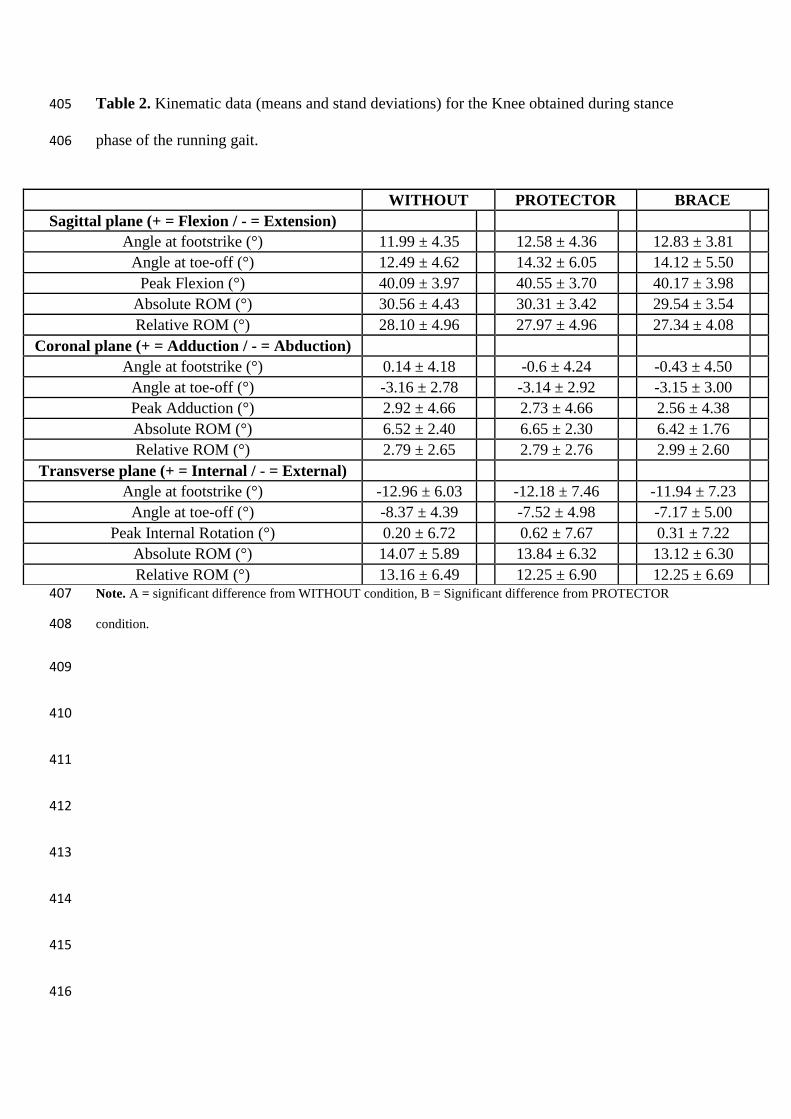
Table 2. Kinematic data (means and stand deviations) for the Knee obtained during stance 405
phase of the running gait. 406
Note. A = significant difference from WITHOUT condition, B = Significant difference from PROTECTOR 407
condition. 408
409
410
411
412
413
414
415
416
WITHOUT PROTECTOR BRACE
Sagittal plane (+ = Flexion / - = Extension)
Angle at footstrike (°) 11.99 ± 4.35 12.58 ± 4.36 12.83 ± 3.81
Angle at toe-off (°) 12.49 ± 4.62 14.32 ± 6.05 14.12 ± 5.50
Peak Flexion (°) 40.09 ± 3.97 40.55 ± 3.70 40.17 ± 3.98
Absolute ROM (°) 30.56 ± 4.43 30.31 ± 3.42 29.54 ± 3.54
Relative ROM (°) 28.10 ± 4.96 27.97 ± 4.96 27.34 ± 4.08
Coronal plane (+ = Adduction / - = Abduction)
Angle at footstrike (°) 0.14 ± 4.18 -0.6 ± 4.24 -0.43 ± 4.50
Angle at toe-off (°) -3.16 ± 2.78 -3.14 ± 2.92 -3.15 ± 3.00
Peak Adduction (°) 2.92 ± 4.66 2.73 ± 4.66 2.56 ± 4.38
Absolute ROM (°) 6.52 ± 2.40 6.65 ± 2.30 6.42 ± 1.76
Relative ROM (°) 2.79 ± 2.65 2.79 ± 2.76 2.99 ± 2.60
Transverse plane (+ = Internal / - = External)
Angle at footstrike (°) -12.96 ± 6.03 -12.18 ± 7.46 -11.94 ± 7.23
Angle at toe-off (°) -8.37 ± 4.39 -7.52 ± 4.98 -7.17 ± 5.00
Peak Internal Rotation (°) 0.20 ± 6.72 0.62 ± 7.67 0.31 ± 7.22
Absolute ROM (°) 14.07 ± 5.89 13.84 ± 6.32 13.12 ± 6.30
Relative ROM (°) 13.16 ± 6.49 12.25 ± 6.90 12.25 ± 6.69
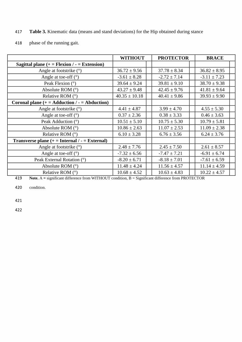
Table 3. Kinematic data (means and stand deviations) for the Hip obtained during stance 417
phase of the running gait. 418
WITHOUT PROTECTOR BRACE
Sagittal plane (+ = Flexion / - = Extension)
Angle at footstrike (°) 36.72 ± 9.56 37.78 ± 8.34 36.82 ± 8.95
Angle at toe-off (°) -3.61 ± 8.28 -2.72 ± 7.14 -3.11 ± 7.23
Peak Flexion (°) 39.64 ± 9.24 39.81 ± 9.10 38.70 ± 9.38
Absolute ROM (°) 43.27 ± 9.48 42.45 ± 9.76 41.81 ± 9.64
Relative ROM (°) 40.35 ± 10.18 40.41 ± 9.86 39.93 ± 9.90
Coronal plane (+ = Adduction / - = Abduction)
Angle at footstrike (°) 4.41 ± 4.87 3.99 ± 4.70 4.55 ± 5.30
Angle at toe-off (°) 0.37 ± 2.36 0.38 ± 3.33 0.46 ± 3.63
Peak Adduction (°) 10.51 ± 5.10 10.75 ± 5.30 10.79 ± 5.81
Absolute ROM (°) 10.86 ± 2.63 11.07 ± 2.53 11.09 ± 2.38
Relative ROM (°) 6.10 ± 3.28 6.76 ± 3.56 6.24 ± 3.76
Transverse plane (+ = Internal / - = External)
Angle at footstrike (°) 2.48 ± 7.76 2.45 ± 7.50 2.61 ± 8.57
Angle at toe-off (°) -7.32 ± 6.56 -7.47 ± 7.21 -6.91 ± 6.74
Peak External Rotation (°) -8.20 ± 6.71 -8.18 ± 7.01 -7.61 ± 6.59
Absolute ROM (°) 11.48 ± 4.24 11.56 ± 4.57 11.14 ± 4.59
Relative ROM (°) 10.68 ± 4.52 10.63 ± 4.83 10.22 ± 4.57 Note. A = significant difference from WITHOUT condition, B = Significant difference from PROTECTOR 419
condition. 420
421
422
![An Ankle-Foot Prosthesis Emulator with Control of ...biomechatronics.cit.cmu.edu/publications/Collins_2015_ICRA.pdf · ankle and knee kinematics [1], reduce metabolic rate [2], and](https://img.dokumen.tips/doc/110x75/5f6b17783154671dfb1d5945/an-ankle-foot-prosthesis-emulator-with-control-of-ankle-and-knee-kinematics.jpg)