Embed Size (px)
Citation preview

Ye et al. SpringerPlus (2016) 5:990 DOI 10.1186/s40064-016-2390-3
REVIEW
The effect of the immune system on ovarian function and features of ovarian germline stem cellsHaifeng Ye2,3, Xiaoyan Li2,3, Tuochen Zheng5, Xia Liang2,3, Jia Li1,2*, Jian Huang2,3, Zezheng Pan2,4 and Yuehui Zheng1,2*
Abstract
In addition to its role in maintaining organism homeostasis, the immune system also plays a crucial role in the modu-lation of ovarian function, as it regulates ovarian development, follicular maturation, ovulation and the formation of the corpus luteum. Ovarian germline stem cells are pluripotent stem cells derived from the ovarian cortex that can differentiate into ovarian germ cells and primary granulosa cells. Recent work has demonstrated that the proliferation and differentiation of ovarian germline stem cells is regulated in part by immune cells and their secreted factors. This paper reviews the role of the immune system in the regulation of ovarian function, the relationship between immune components and ovarian germline stem cells and current research efforts in this field.
Keywords: Immune system, Ovarian function, Ovarian germline stem cells
© 2016 The Author(s). This article is distributed under the terms of the Creative Commons Attribution 4.0 International License (http://creativecommons.org/licenses/by/4.0/), which permits unrestricted use, distribution, and reproduction in any medium, provided you give appropriate credit to the original author(s) and the source, provide a link to the Creative Commons license, and indicate if changes were made.
The immune system maintains tissue homeostasisIntroduction to the immune systemThe immune system, which plays an important role in immune responses and immunologic function, consists of immune organs (including bone marrow, thymus and lymph nodes), immune cells (including lymphocytes, monocytes and neutrophils) and secreted factors such as serum complement proteins, immunoglobulins, inter-ferons and tumour necrosis factor. The immune system is subdivided into several groups. The first division is between innate (non-specific) and adaptive (specific) immunity, and adaptive immunity can be further divided into humoral and cell-mediated immunity depending on the target antigen. The immune system is critical for resisting pathogen invasion and has three vital func-tions: (1) immune defence, or combatting the invasion of pathogenic microorganisms such as viruses, bacteria and other contaminants; (2) immunological surveillance, or recognizing, killing and eliminating mutated cells to
prevent cancers; and (3) immunological homeostasis, or removing aged and dying cells to maintain bodily homeo-stasis. The intersection of these three functions is the key to the maintenance of homeostasis, whereas immune dysfunction can promote disease.
The contribution of the immune system to tissue homeostasis and the maintenance of tissue‑self‑stateMost cells in the body do not directly contact the exter-nal environment but rather exist and survive in an inter-nal environment consisting of extracellular fluid such as plasma, interstitial fluid, lymph or cerebrospinal fluid (CSF). Maintenance of the internal environment is cru-cial for bodily homeostasis. In addition to their role in defence, recent work has shown that the cells and mol-ecules of the immune system can regulate the prolif-eration, differentiation and apoptosis of cells in several tissues, thereby helping to maintain internal environment homeostasis. Carrel (1922) found that leukocyte infu-sion (a liquid analogous to tissue extracts) could stimu-late the proliferation of fibroblasts in vitro, after which he postulated that leukocytes were a source of growth factors. Later work demonstrated that lymphocytes also could promote tissue development and regeneration. For
Open Access
*Correspondence: [email protected]; [email protected] 1 School of Life Science, Nanchang University, Nanchang, China2 Medical Teaching Laboratory Center, Jiangxi Medical College, Nanchang University, Nanchang, ChinaFull list of author information is available at the end of the article
Page 2 of 6Ye et al. SpringerPlus (2016) 5:990
example, Fidler (1980) suggested that lymphocytes could regulate tissue development by acting as trophoblasts. In recent years, monocyte-derive cells (MDCs) have been shown to play a crucial role in tissue maintenance, and cytokines and growth factors derived from this cell type can also influence cellular differentiation and function (Bukovsky et al. 2000). Furthermore, scientists have also found that the immune system not only regulates the bal-ance of the internal environment but also plays an impor-tant role in the maintenance of tissue steady state.
Available data indicate that the functions of all tis-sues are regulated via ‘morphostasis’, a process whereby tissues retain a specific differentiation status after the organism completes morphogenesis. This process is essential to maintain the homeostasis of the internal environment. ‘Morphostasis’ is a complex process that is completed in three steps during embryonic development. The first step involves the renewal of tissue stem cells, while in the second step, specific cells in tissue can be conserved in proper differentiation status. The final step regulates tissue amount and is carried out by the tissue control system (TCS) in which the immune system plays an important role (Bukovsky 2011).
The TCS is mainly composed of cells from the immune and autonomic nervous system, as well as vascular endothelial cells (Bukovsky et al. 1983, 1991, 1995). The functions of these cell types are established early during embryonic development. In addition, MDCs, T lympho-cytes and B lymphocytes also possess crucial functions with respect to the TCS (Bukovsky 2011). According to the prevailing hypothesis in this field, MDCs in the TCS can send out ‘stop effects’ to prevent cells from further differentiation after their respective fates have been spec-ified. Next, autonomic innervation controls the number of volume of cells and tissues, respectively, thus main-taining “morphostasis”. Importantly, this step relies upon ‘stop effects’ from MDCs (Bukovsky et al. 2000). Impor-tantly, the TCS can regulate specific organs and tissues of the body and plays a crucial role in the regeneration of ovarian follicles and age-related anovulation (Bukovsky 2007).
With increasing age, degeneration of the immune sys-tem and reduced ‘stop effects’ from MDCs can alter the “morphostasis” of many organs, including ovaries, and result in functional declines (Bukovsky 2007).
The role of the immune system in ovaryThe relationship between the immune system and ovarySince the 1970s, scientists have made great strides in understanding the relationship between the immune system and the ovaries. For example, Sakakura and Nishizuka (1972) found that the ovaries of athymic mice failed to develop within 2–4 days after birth. In addition,
Russell et al. (1973) used thymocytes from wild type female mice to suppress cyclophosphamide- and X-ray-induced super ovulation. Furthermore, Bukovsky and Presl (1979) postulated that the immune system is a cru-cial mediator of ovarian function, a hypothesis supported by recent research. For example, studies have shown that thymosin injections in neonatal nude mice can help maintain the levels of follicle stimulating hormone (FSH) and luteinizing hormone (LH) during post-natal develop-ment. Therefore, this method can rescue aberrant ovarian development in nude mice (Goya et al. 2007). In addition, studies have also shown that the immune system can degenerate with age, followed by reductions in ovarian function (Bukovsky and Caudle 2012). This correlation indicates the close relationship between the immune sys-tem and ovary.
The interaction between the immune system and ovarian endocrine functionIt is well established that the hypothalamic-pituitary axis is crucial for proper ovarian function. Under the influ-ence of hormones from the hypothalamus, the hypophy-sis secretes FSH and LH, which underlie the physiological changes associated with the female menstrual cycle. In addition, various immune cells exist within the tissues of the hypothalamus–pituitary–ovarian axis, indicating that the immune system may regulate ovarian function and degeneration. Indeed, in the early 1980s and 1990s, stud-ies of the relationship between immunity and the ovary showed that immune cell-derived cytokines can modu-late the production of hormones along the hypothala-mus–pituitary–ovary axis, thereby indicating that the immune system can influence reproductive physiology.
During ovarian follicle maturation, increasing pres-sure in the follicular cavity causes the follicle to rupture, leading to the release of a secondary oocyte surrounded by the zona pelludica and corona radiata. This process is called ovulation, and cytokines play an important role in both early and late stages follicular development (Cre-spo et al. 2010). For example, granulosa cells in the fol-licles produce tumour necrosis factor α (TNF-α), which promotes the secretion of FSH. These cytokines can also inhibit prostaglandin F2a (PGF2a), which in turn, facili-tates ovulation (Nakao et al. 2015). Studies in mice have further shown that ovarian TNF-α can prevent FSH from stimulating aromatase activity and progestin secretion, thereby promoting ovulation (Nakao et al. 2015). In addi-tion, interleukin-1 (IL-1) can reduce the expression of LH receptors in granulosa cells, as well as suppress pro-gestin secretion. Moreover, IL-1 also facilitates the pro-gression of ovulation and oocyte development (Gerard et al. 2004). Furthermore, FSH can synergize with trans-forming growth factor-β (TGF-β) to inhibit follicular

Page 3 of 6Ye et al. SpringerPlus (2016) 5:990
development, thereby regulating the quantity of primor-dial follicles (Wang et al. 2014).
The corpus luteum produced during the menstrual cycle in mammals has a remarkably short life cycle. If the egg is not fertilized, the corpus luteum will regress into the corpus albicans within 2 weeks. Studies of many ani-mal models have postulated that macrophage-derived secretions (e.g., TNF) participate in both the develop-ment and degeneration of corpus luteum (Galvao et al. 2013). Specifically, TNF can stimulate the secretion of vascular endothelial growth factor (VEGF) in the imma-ture and developing corpus luteum, thereby stimulat-ing vasculogenesis and the subsequent development of the mature corpus luteum (Galvao et al. 2013). TNF is also found in the degenerated corpus luteum. Specifi-cally, TNF in the immature corpus luteum promotes its growth, while high levels in an unfertilized corpus luteum assist the progression of its apoptosis and degeneration. This dual role of TNF still remains a mystery (Galvao et al. 2013).
Immunity and the proliferation and differentiation of germline stem cellsOvarian germline stem cells (OSCs)The origins of the primordial follicles and oocytes in the ovaries of mature mammals have been debated for sev-eral hundreds of years. According to the views of tradi-tional medical, most mammals are born with oocytes, which explains the limited supply of oocytes with respect to age. However, researchers in the field of phylogenet-ics hold a different view (Bukovsky et al. 2005). Specifi-cally, Johnson et al. (2004) proposed that gametes possess mitotic activity and can self-renew, even in the mature ovary of adult mammals. Since that time, research on ovarian germline stem cells (OSC) has attracted more attention.
OSCs are epithelial cells that reside on the ovarian sur-face. Several molecular markers, including c-kit, Oct-4, vasa and telomerase have been detected on the ovarian surface epithelium (OSE) in many mammalian species (Bukovsky et al. 2008; Virant-Klun 2015; Bhartiya et al. 2013; Gheorghisan-Galateanu et al. 2014). Therefore, OSCs are also referred to as the ‘germline epithelium’.
In recent years, scientists have made considerable pro-gress in understanding OSCs. For example, Zou et al. (2009) isolated germline stem cells both from new-born and adult mice, introduced a GFP expression construct and implanted the cells into sterile mice. These cells dif-ferentiated into functional follicles, and the sterile mice ultimately gave birth to GFP-expressing progeny. In addi-tion, White et al. (2012) isolated germline stem cells from healthy individuals and injected them into both human ovaries and immunocompromised mice. Surprisingly,
both procedures resulted in the formation of primordial follicles. Based on these results, the consensus in the field indicates that germline stem cells do exist in the ovary from birth. The stem cells can replenish the original pri-mordial follicle pool via self-renewal and differentiation. Work in our laboratory seeks to determine the mecha-nism of OSC function.
Immunity is a part of OSC nicheOSCs can differentiate into oocytes and granulosa cells, and they originate from the bipotential stem cells of the ovarian cortex (Li et al. 2015). This process is facilitated by features of the OSC niche. Specifically, Bukovsky (2011) postulated that factors within the OSC niche con-trol OSC functions. Moreover, the OSC niche contains immune cells and their secreted molecules. The OSC niche is established during early stages of foetal develop-ment and consists of committed ovarian MDCs, T cells and vascular endothelial cells. By contrast, the adult OSC niche contains primary MDCs (CD14+ MDC), acti-vated MDCs ([HLA-DR]+ MDC) and T cells. Therefore, immunity plays an important role in proliferation and differentiation of germline stem cells.
Bukovsky (2007) demonstrated that immune cells and their secreted factors in the OSC niche modulate the asymmetric cell division of OSCs. Ultimately, this pro-cess leads to the production of oocytes, and regulates the symmetric division, migration and generation of new granulosa cells. Immune cells also play an impor-tant role in the development of primordial follicles in both the foetus and adult, and support ovarian homeo-stasis. These conclusions are based on the following: ① According to studies of human ovarian development, fol-licles develop from the inner layer of the ovarian cortex adjacent to the rete ovarii. Importantly, this structure is required for follicular development (Byskov et al. 1977; Hummitzsch et al. 2015) and contains several immune cell types, such as CD14+ MDCs. These cells can differ-entiate into activated MDCs ([HLA -DR]+), which then migrate in reticular passageways and interact with innate MDCs. In addition, inhibitory T cells (CD3+ T cells) and cytotoxic T cells(CD8+ T) also reside in the reticu-lar tubes. ② When OSCs differentiate into reproductive cells, they must then accept ovary-committed bone mar-row cells (OCMT), which in turn, stimulate MDCs and T cells (Bukovsky 2015). OSCs can produce one differ-entiated germ cell and one progeny OSC. The germ cell can then differentiate into an oocyte and migrate to the epithelial layer adjacent to blood vessels in the ovarian cortex. As a consequence of normal circulatory func-tion, germ cells will develop and interact with granulosa cells to form primitive follicles (Kossowska-Tomaszczuk and De Geyter 2013). Ultimately, the development and

Page 4 of 6Ye et al. SpringerPlus (2016) 5:990
migration of germ cells occur in the context of immune cells such as CD14+ MDCs and require asymmetric OSC division. Moreover, symmetric germ cell division is accompanied by CD8+ T cells and is assisted by pri-mary MDCs. Ultimately, primary MDCs facilitate differ-entiation of germ cells to epithelial cells, while activated MDCs (DR+ MDC) are involved in germ cell migration.
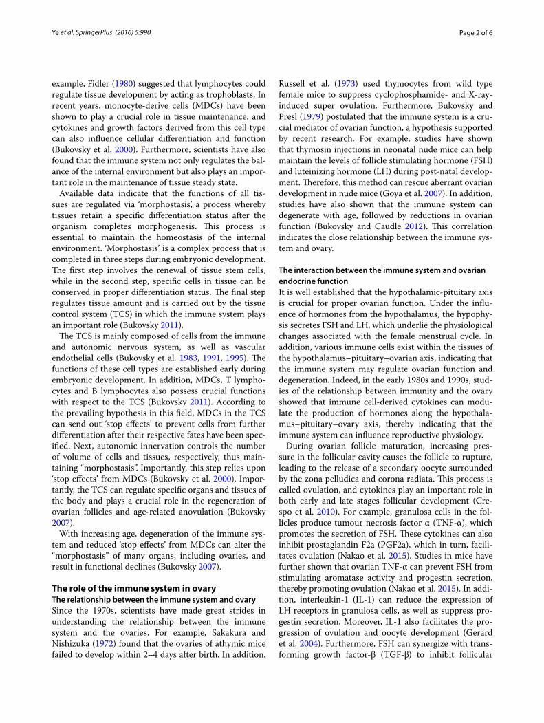
Immunity establishes an ‘ovarian memory’Bukovsky (2011) has hypothesized that uncommitted MDC and T cells (UMT) can recognize and “memorize” OSC in the OSC niche. Bukovsky (2006) further hypoth-esized concerning ‘ovarian memory’: When primi-tive germ-line cells are implanted into undifferentiated gonads, the rete ovarii will stimulate the differentiation of secondary gonocytes to oocytes. During the develop-ment of adaptive immunity, many uncommitted MDCs and T cells (UMT) are present. Some of these UMT can differentiate into “veiled cells” in the channels of the rete ovarii and can transmit chemical messages from oocytes in the rete ovarii to developing lymph tissue. From this point on, these UMT are known as ovarian memory cells (OMC) (Fig. 1), which promote the differentiation of UMT in the channels of the rete ovarii to ovary-commit-ted bone marrow cells (OCMT). These OCMT can then migrate to the ovarian epithelium, where they produce molecular signals to trigger both asymmetric and sym-metric OSC division (Fig. 2). Thus, secondary germ-line cells are then produced. The development of secondary germ-line cells also relies on a suitable hormonal envi-ronment consisting of hCG and estradiol. According to this hypothesis, the developing adaptive immune sys-tem can establish an ‘ovarian memory’ during the foetal period to support the replenishment of OCMT in adult-hood. This immune function will decline by the age of 35–40, concomitant with the replacement of follicles (Bukovsky et al. 2004, 2009).
As the development of the adaptive immune system nears completion, the foetal rete ovarii will undergo degeneration, thereby preventing further oogenesis. This process is caused by reductions in hormonal signals (the foetal hCG barrier) (Bukovsky et al. 2005). Further devel-opment of OMCs in the lymph nodes will then cease, and these cells will ultimately constitute the ovarian memory cell pool. During menarche and the reproductive years, hormonal signals and OMCT can facilitate periodic oogenesis by stimulating OSCs. Continuous renewal of follicles requires cyclic OCMT supplementation, as the memory cell pool will become exhausted if the OCMT proliferation rate in lymph tissue is too high. Therefore, even in the presence of hormone signals, depletions in the frequencies of OMCs will still terminate oogenesis.
Fig. 1 Generation of ovarian memory cells during developmental immune adaptation. UMT uncommitted MDC and T cells, OCMT ovary-committed bone marrow cells, OMC ovarian memory cells, OSC ovarian germline stem cells, LT lymphoid tissue, DIA developmental immune adaptation
Fig. 2 The role of the immune system in promoting the symmetric and asymmetric division and differentiation of ovarian germ stem cells. a Uncommitted OGSCs(u-OGSCs) were produced in the first six weeks of pregnancy; b Primordial germ cells (PGC) were invaded OGSCs layerin the first seven weeks of pregnancy; c–e OGSCs only the joint action of cell signaling (cellular signaling, CS; as CD14 secreted by MDC and CD8 secreted by T lymphocytes) and hormonal signals (hormonal signaling, HS; chorionic gonadotropin, hCG and estradiol, E2) can occur secondary asymmetric division to produce germ cells, germ cells into secondary ovarian cortex and eventually differentiate into oocytes (definitive oocytes, DO). Modified from Bukovsky and Caudle (2012)

Page 5 of 6Ye et al. SpringerPlus (2016) 5:990
According to this view, as age increases, immunological anaplasia is the main point at which oogenesis and ovar-ian replenishment cease. This proposal is supported by extensive experimental data that reveal the close corre-lation between mammalian immunity and reproduction. This work has raised new questions about the influence of immunologically relevant molecules on the prolifera-tion and differentiation of ovarian germline stem cells.
Clinical application of immune system in regulating ovarian germline stem cellsThe research of OSCs will eventually apply to clinical therapy, and the research has shown that there is a great value of clinical application in immune regulation OSCs. In vitro, autologous OSCs in vitro are expected to achieve the in vitro maturation and in vitro fertilization of infer-tile women (Bukovsky 2015). However, due to the lack of similar immune condition, OSCs cannot complete the meiotic differentiation of eggs in vitro. The research indi-cated that OSCs successfully differentiated into eggs by the co-culture of OSCs and mononuclear cells in vitro (Bukovsky et al. 2006), further experiments showed that cell differentiation and the new egg production were obvious by the culture of ovarian cortical cells that stemmed from the premenopausal and postmenopausal women in vitro. Undoubtedly, the above researches pro-vided a theoretical and experimental support for the new in vitro fertilization technology.
In vivo, the our laboratory is trying to promote OSCs proliferation, differentiation and further remodel ovarian function during pathological and physiological ovarian aging by enhance the body immune function. Fortunately, these unpublished results show that the immune func-tion and reproductive function can be improved syn-chronously through the treatment of phase-immune enhancement agents during premature ovarian failure in mice. Similarly, the results are expected to implement enhanced reproductive function in the body.
Conclusion and prospectsThis view of OSC function is distinct from the views of traditional reproductive medicine and reproductive biology. As germline cells self-renew in the ovaries of foetuses and adults, OSCs can maintain ovarian home-ostasis. Thus, the study of cultured OSCs in vitro and in vivo may have clinical applications in the treatment of POF and ovarian infertility, and may improve preg-nancy rates and postpone female ageing. In recent years, ovarian germline stem cells have been shown to reverse infertility in mouse models, but the exact mechanism of this phenomenon regulation is still not well understood. OSCs are influenced by many hormonal signals cell sig-nalling factors. This review focused on the influence of
immune cells and molecules to ovarian function, and considered the possibility that immunity controls the proliferation and differentiation of OSCs.
The immune system plays important roles both in evading foreign pathogens and maintaining tissue home-ostasis. In adulthood, the functions of different tissues (including ovaries) all require mechanisms to regulate (1) stem cell renewal, (2) differentiation status and (3) tissue amount. The TSC is particularly important for the establishment of tissue steady states and also plays an important role in the immune system. Morphostasis takes place in the immune adaptive period, which is a key period in embryonic development. With increasing age, the immune system will undergo degenerative change, also leading to ovarian degeneration. In addition, recent studies indicate that the asymmetric division of OSC into new germline cells requires the stimulation MDCs and T cells. Moreover, immunologically relevant cells and their secretion also modulate the symmetric division of ger-mline cells, as well as their symmetric division, migration and production of new granulosa cells and primitive fol-licles to maintain homeostasis in the ovary.
Scientists have made great strides in understanding immunity, reproduction and mechanisms of in vivo ger-mline stem cell function. However, there is still consid-erable debate regarding the existence of germ-line stem cells. The isolation and purification of ovarian germline stem cells have only been accomplished in in vitro stud-ies. However, further research on the proliferation and differentiation of ovarian germline stem cells may have important clinical applications, as it may help treat ovar-ian insufficiency, ovarian infertility and POF. It may also be a mechanism by which women can delay ageing.
Authors’ contributionsAll authors contributed equally to this work. All authors read and approved the final manuscript.
Author details1 School of Life Science, Nanchang University, Nanchang, China. 2 Medical Teaching Laboratory Center, Jiangxi Medical College, Nanchang Univer-sity, Nanchang, China. 3 The Key Laboratory of Reproductive Physiology and Pathology of Jiangxi Province, Nanchang, China. 4 Faculty of Basic Medical Science, Jiangxi Medical College, Nanchang University, Nanchang, China. 5 School of the 1st Clinical Medical Sciences, Jiangxi Medical College, Nan-chang University, Nanchang, China.
AcknowledgementsThis work was supported by the National Natural Science Foundation of China (Nos. 81160081, 81360100), the Excellence 555 Engineering of Jiangxi Province and the Natural Science Foundation of Jiangxi Province (Nos. 20151ACB20003, 201442BAB205069, 20142BAB205002).
Competing interestsThe authors declare that they have no competing interests.
Received: 14 April 2016 Accepted: 23 May 2016

Page 6 of 6Ye et al. SpringerPlus (2016) 5:990
ReferencesBhartiya D, Sriraman K, Parte S et al (2013) Ovarian stem cells: absence of
evidence is not evidence of absence. J Ovarian Res 6(1):65Bukovsky A (2006) Oogenesis from human somatic stem cells and a role of
immune adaptation in premature ovarian failure. Curr Stem Cell Res Ther 1(3):289–303
Bukovsky A (2007) Cell commitment by asymmetric division and immune system involvement. Prog Mol Subcell Biol 45:179–204
Bukovsky A (2011a) Immune maintenance of self in morphostasis of distinct tissues, tumour growth and regenerative medicine. Scand J Immunol 73(3):159–189
Bukovsky A (2011b) Ovarian stem cell niche and follicular renewal in mam-mals. Anat Rec 294(8):1284–1306
Bukovsky A (2015) Novel methods of treating ovarian infertility in older and POF women, testicular infertility, and other human functional diseases. Reprod Biol Endocrinol 13:10
Bukovsky A, Caudle MR (2012) Immunoregulation of follicular renewal, selec-tion, POF, and menopause in vivo, vs. neo-oogenesis in vitro, POF and ovarian infertility treatment, and a clinical trial. Reprod Biol Endocrinol 10:97
Bukovsky A, Presl J (1979) Ovarian function and the immune system. Med Hypotheses 5(4):415–436
Bukovsky A, Presl J, Zidovsky J et al (1983) The localization of Thy-1.1, MRC OX 2 and Ia antigens in the rat ovary and fallopian tube. Immunology 48(3):587–596
Bukovsky A, Michael SD, Presl J (1991) Cell-mediated and neural control of morphostasis. Med Hypotheses 36(3):261–268
Bukovsky A, Caudle MR, Keenan JA et al (1995) Quantitative evaluation of the cell cycle-related retinoblastoma protein and localization of Thy-1 dif-ferentiation protein and macrophages during follicular development and atresia, and in human corpora lutea. Biol Reprod 52(4):776–792
Bukovsky A, Caudle MR, Keenan JA (2000) Dominant role of monocytes in control of tissue function and aging. Med Hypotheses 55(4):337–347
Bukovsky A, Caudle MR, Svetlikova M et al (2004) Origin of germ cells and formation of new primary follicles in adult human ovaries. Reprod Biol Endocrinol 2:20
Bukovsky A, Svetlikova M, Caudle MR (2005a) Oogenesis in cultures derived from adult human ovaries. Reprod Biol Endocrinol 3:1–13
Bukovsky A, Caudle MR, Svetlikova M et al (2005b) Oogenesis in adult mam-mals, including humans: a review. Endocrine 26(3):301–316
Bukovsky A, Copas P, Virant-Klun I (2006) Potential new strategies for the treat-ment of ovarian infertility and degenerative diseases with autologous ovarian stem cells. Expert Opin Biol Ther 6(4):341–365
Bukovsky A, Caudle MR, Gupta SK et al (2008) Mammalian neo-oogenesis and expression of meiosis-specific protein SCP3 in adult human and monkey ovaries. Cell cycle (Georgetown, Tex) 7(5):683–686
Bukovsky A, Caudle MR, Carson RJ et al (2009) Immune physiology in tissue regeneration and aging, tumor growth, and regenerative medicine. Aging 1(2):157–181
Byskov AG, Skakkebaek NE, Stafanger G et al (1977) Influence of ovarian surface epithelium and rete ovarii on follicle formation. J Anat 123(Pt 1):77–86
Carrel A (1922) Growth-promoting function of leucocytes. J Exp Med 36(4):385–389
Crespo D, Bonnet E, Roher N et al (2010) Cellular and molecular evidence for a role of tumor necrosis factor alpha in the ovulatory mechanism of trout. Reprod Biol Endocrinol 8:34
Fidler IJ (1980) Lymphocytes are not only immunocytes. Biomedicine/[publiee pour l’AAICIG] 32(1):1–3
Galvao AM, Ferreira-Dias G, Skarzynski DJ (2013a) Cytokines and angiogenesis in the corpus luteum. Mediators Inflamm 2013:420186
Galvao AM, Szostek AZ, Skarzynski DJ et al (2013b) Role of tumor necrosis factor-alpha, interferon-gamma and Fas-ligand on in vitro nitric oxide activity in the corpus luteum. Cytokine 64(1):18–21
Gerard N, Caillaud M, Martoriati A et al (2004) The interleukin-1 system and female reproduction. J Endocrinol 180(2):203–212
Gheorghisan-Galateanu AA, Hinescu ME, Enciu AM (2014) Ovarian adult stem cells: hope or pitfall? J Ovarian Res 7:71
Goya RG, Reggiani PC, Vesenbeckh SM et al (2007) Thymulin gene therapy prevents the reduction in circulating gonadotropins induced by thymulin deficiency in mice. Am J Physiol Endocrinol Metab 293(1):E182–E187
Hummitzsch K, Anderson RA, Wilhelm D et al (2015) Stem cells, progenitor cells, and lineage decisions in the ovary. Endocr Rev 36(1):65–91
Johnson J, Canning J, Kaneko T et al (2004) Germline stem cells and follicular renewal in the postnatal mammalian ovary. Nature 428(6979):145–150
Kossowska-Tomaszczuk K, De Geyter C (2013) Cells with stem cell characteris-tics in somatic compartments of the ovary. BioMed Res Int 2013:310859
Li J, Zhou F, Zheng T et al (2015) Ovarian germline stem cells (OGSCs) and the hippo signaling pathway association with physiological and pathological ovarian aging in mice. Cell Physiol Biochem 36(5):1712–1724
Nakao K, Kishi H, Imai F et al (2015) TNF-alpha suppressed FSH-induced LH receptor expression through transcriptional regulation in rat granulosa cells. Endocrinology 156(9):3192–3202
Russell WR, Walpole AL, Labhsetwar AP (1973) Cyclophosphamide: induction of superovulation in rats. Nature 241(5385):129–130
Sakakura T, Nishizuka Y (1972) Thymic control mechanism in ovarian develop-ment: reconstitution of ovarian dysgenesis in thymectomized mice by replacement with thymic and other lymphoid tissues. Endocrinology 90(2):431–437
Virant-Klun I (2015) Postnatal oogenesis in humans: a review of recent find-ings. Stem Cells Cloning Adv Appl 8:49–60
Wang ZP, Mu XY, Guo M et al (2014) Transforming growth factor-beta signaling participates in the maintenance of the primordial follicle pool in the mouse ovary. J Biol Chem 289(12):8299–8311
White YA, Woods DC, Takai Y et al (2012) Oocyte formation by mitotically active germ cells purified from ovaries of reproductive-age women. Nat Med 18(3):413–421
Zou K, Yuan Z, Yang Z et al (2009) Production of offspring from a germline stem cell line derived from neonatal ovaries. Nat Cell Biol 11(5):631–636