Embed Size (px)
Citation preview
The Effect of Superior Colliculus Lesions upon the Visual Fields of Cats with Cortical Ablations
S. MURRAY SHERMAN Department of Physiology, University of Virginia School of Medicine, Charlottesuille, Virginia 22901
ABSTRACT The visual fields of 18 cats were measured before and after various lesions. Preoperatively, all cats had identical fields. With both eyes open, they saw from 90" left to 90" right; with one eye, from 90" ipsilateral to 45" contralateral. Thus the field for nasal retina extends from 90" ipsilateral through to the midline; for temporal retina, from the midline through to 45" contralateral.
In summary, postoperative testing led to two major conclusions. (1) Large occip- ito-temporal cortical lesions produce a stable field blindness, but the blindness is alleviated by a transection of the commissure of the superior colliculus (or a unilateral collicular ablation). This transection yields the same result whether it occurs in an operation before, during, or after the cortical lesion. These data confirm and extend the Sprague effect. (2) Cats made dependent upon retinotectal pathways due to cortical ablations responded much better to stimulation of nasal retina than to stimulation of temporal retina. This presumably is related to the pre- ponderance of nasal retina as a source of the retinotectal pathway. Since even smaller cortical lesions limited to areas 17, 18, and 19 produce this nasal/temporal retinal difference, it is concluded that integrity of the geniculocortical pathways is necessary for good temporal retinal vision as determined by these methods.
In a remarkable series of studies, Sprague and Meikle ('65) and Sprague ('66a) demonstrated that much of the cat's visually guided behavior is subserved by interactions involving midbrain and corti- cal pathways. Sprague ('66b) then reported that the hemianopia that is produced con- tralateral to a large, posterior cortical lesion could be at least partially alleviated either by an ablation of the superior col- liculus contralateral to the cortical lesion or by a transection of the commissure of the superior colliculus.
The neural basis of this dramatic phe- nomenon remains unresolved, but Sprague's (66b) suggestion follows. Each colliculus normally may receive a balanced set of inputs, including q form of functional facilitation from the iGilateral cortex and suppression from the other colliculus via the commissure. Destruction of cortex leads to an imbalance due to surplus sup- pression playing on the ipsilateral collicu-
I. COMP. NEUR., 172. 211-230.
lus, and this interferes with its potential functioning, This functioning can be at least partially restored by destroying either the source of excess sup ression (i.e., the contralateral colliculus P or its pathway (i. e., the collicular commissure).
Despite the profound significance of this study by Sprague ('66b), it has, to my knowledge, neither been confirmed nor re- ported in detail. The purpose of the pres- ent study was to confirm the Sprague effect and extend our knowledge of it. This was found to be a robust and repeatable phe- nomenon in cats, and details are added in this paper. A brief preliminary report of these results was recently published (Sher- man, '74b).
MATERIALS AND METHODS
Subjects and surgical procedures Eighteen cats, purchased as normal
adults, were studied. They were housed in individual cages with a 12-hour light/dark
21 1

212 S. MURRAY SHERMAN
cycle for the duration of this experiment. Most of these cats were also studied for visual discriminations of patterned stimuli, but these data will be presented separately (Loop and Sherman, '77, submitted).
Each cat underwent one or a series of brain lesions consisting of cortical ablation, midsagittal transection of the commissure of the superior colliculus, collicular abla- tion, andlor midsagittal transection of the optic chiasm. The cats were anesthetized with barbiturate, and routine aseptic pre- cautions were taken. All surgery was per- formed under visual control aided by an operating microscope with co-axial illu- mination.
Cortical lesions were created by gentle subpial suction. The smaller ablations were bilateral and were designed to remove the lateral gyrus dorsally and all cortex medial- ly above the splenial sulcus; thus all of areas 17 and 18 plus most of area 19 was ablated. The larger ablations were also bilateral (with the exception of the left cortical lesion in cat C5) and were de- signed to remove nearly all of the occipito- temporal cortex. Thus all known cortical projection areas of the "visual" thalmus (in- cluding the lateral geniculate nucleus plus the inferior, medial and lateral divisions of the pulvinar nucleus *) were ablated (see Sprague et al., '73, for a review of these pathways; also Kawamura, '74; Niimi et al., '74).
The collicular commissure was tran- sected by inserting a fine knife between the colliculi to a depth of approximately 2 mm, and the collicular ablation in cat C5 was achieved by gentle subpial suction. The tectum was commonly approached by first removing the bony tentorium and re- tracting the dura mater between the cor- tex and cerebellum. For some of the com- missure sections, the tectum was ap- proached through a small gap placed in the back of the corpus callosum (only after ex- tensive, bilateral occipito-temporal cortex removal), the knife being guided under the tentorium. Except for the first few tectal operations, intravenous mannitol (50 ml; 30%) was routinely administered preoper-
atively to achieve shrinkage of the hemi- spheres. This greatly facilitated exposure of the tectum.
Cat C4 had its optic chiasm midsagittally sectioned by gentle suction, the approach being through the roof of the mouth.
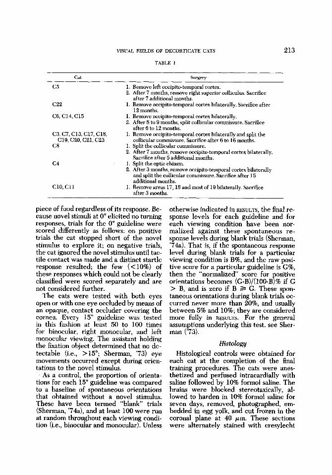
Every cat was tested before and after each surgical procedure. A two to four week postoperative recovery period was allowed before testing, and testing con- tinued for at least three to four months postoperatively in order to assess final, sta- ble performance. Table 1 summarizes the surgical procedures for each cat.
Behavioral testing These cats were tested with techniques
which have been previously described in detail (Sherman, '73, '74a), and these methods will be briefly outlined. Each cat was food-deprived and tested on a table marked off into sixteen 15" sectors by in- tersecting guidelines designated 120"L, 105"L, . . . , etc.. . . , 0", 15"R,. . . etc. . . . , 120"R (cf. fig. 1). The cat was re- strained so that its lateral canthi were aligned with the 90" guidelines and its nose pointed along 0". It was pretrained to fixate to visual and/or auditory cues provided by a piece of dry cat food held in forceps and tapped on the table at 0" and approximate- ly 50 cm in front of the cat's nose; this was the jixation object. While the cat was thus fixating, a novel stimulus (food in forceps or a 1 cm diameter red cardboard circle at the end of a long, stiff wire) was presented ver- tically 20-40 cm from the cat's nose along one of the guidelines. The cat was then im- mediately released from restraint and its behavior noted. A clear and immediate orienting response to the novel stimulus was scored as a positive trial, and any other behavior, as negative. Negative trials near- ly always (>95%) consisted of ignoring the novel stimulus and moving towards the fixation object. The cat was fed a small
To facilitate comparisons with primates, I have chosen the terminology of Niimi et al. ('74) for these thalamic nuclei in cats. The correspondence to older nomenclature is: inferior pulvinar represents the nucleus posterior of Rioch, the medial pulvinar represents the lateral posterior nucleus of Rioch, and the lateral pulvinar represents the pulvinar of Rioch.
VISUAL FIELDS OF DECORTICATE CATS 213
TABLE 1
Cat Surgery
c 5 1. 2.
c 2 2 1.
C6, C14, C15 1. 2.
C3,C7,C13,C17,C18, 1.
C8 1. 2.
C19, C20, C21, C23
c4 1. 2.
c10 , c 1 1 1.
Remove left occipito-temporal cortex. After 7 months, remove right superior colliculus. Sacrifice after 7 additional months. Remove occipito-temporal cortex bilaterally. Sacrifice after 12 months. Remove occipito-temporal cortex bilaterally. After 8 to 9 months, split collicular commissure. Sacrifice after 6 to 12 months. Remove occipito-temporal cortex bilaterally and split the collicular commissure. Sacrifice after 6 to 16 months. Split the collicular commissure. After 7 months, remove occipito-temporal cortex bilaterally. Sacrifice after 5 additional months. Split the optic chiasm. After 3 months, remove occipito-temporal cortex bilaterally and split the collicular commissure. Sacrifice after 15 additional months. Remove areas 17,18 and most of 19 bilaterally. Sacrifice after 3 months.
piece of food regardless of its response. Be- cause novel stimuli at 0" elicited no turning responses, trials for the 0" guideline were scored differently as follows: on positive trials the cat stopped short of the novel stimulus to explore it; on negative trials, the cat ignored the novel stimulus until tac- tile contact was made and a distinct startle response resulted; the few (<lo%) of these responses which could not be clearly classified were scored separately and are not considered further.
The cats were tested with both eyes open or with one eye occluded by means of an opaque, contact occluder covering the cornea. Every 15" guideline was tested in this fashion at least 50 to 100 times for binocular, right monocular, and left monocular viewing. The assistant holding the fixation object determined that no de- tectable (i.e., >15"; Sherman, '73) eye movements occurred except during orien- tations to the novel stimulus.
As a control, the proportion of orienta- tions for each 15" guideline was compared to a baseline of spontaneous orientations that obtained without a novel stimulus. These have been termed "blank trials (Sherman, '74a), and at least 100 were run at random throughout each viewing condi- tion (i.e., binocular and monocular). Unless
otherwise indicated in RESULTS, the final re- sponse levels for each guideline and for each viewing condition have been nor- malized against these spontaneous re- sponse levels during blank trials (Sherman, '74a). That is, if the spontaneous response level during blank trials for a particular viewing condition is B%, and the raw posi- tive score for a particular guideline is G%, then the "normalized' score for ositive orientations becomes (G-B)/( IOO-By% if G > B, and is zero if B 2 G. These spon- taneous orientations during blank trials oc- curred never more than 20%, and usually between 5% and 10%; they are considered more fully in RESULTS. For the general assumptions underlying this test, see Sher- man ('73).
Histology Histological controls were obtained for
each cat at the completion of the final training procedures. The cats were anes- thetized and perfused intracardially with saline followed by 10% formol saline. The brains were blocked stereotaxically, al- lowed to harden in 10% formol saline for seven days, removed, photographed, em- bedded in egg yolk, and cut frozen in the coronal plane at 40 pm. These sections were alternately stained with cresylecht

214
Binocular
90. n
4.4 % 1.2%
S. MURRAY SHERMAN
left eye Right eye
7.9 -1.2% 6.8 * 0.9%
Fig. 1 Typical visual fields for normal cats. The plots in polar coordinates represent the nor- malized response levels for each 15" sector of visual field, and the two semicircles represent the 50% and 100% response levels. For each sector, each response level represents an average of the separate values computed for the 18 cats preoperatively. Computed in this manner, the standard errors for the 45" sector contralateral to the open eye during monocular viewing are 2-3%, and for all other sectors during monocular and binocular viewing, the standard errors are less than 1.5%. The numbers below each plot represent the level of spontaneous orienting (mean f standard error). Spontaneous levels were computed in the same way as were the response levels shown in the plots. The sectors of visual field beyond 90" were also routinely tested, but stimuli placed there elicited responses so rarely that they have been omitted from this and succeeding figures.
violet and the Mahon method for my- elinated fibers. In addition to direct recon- struction of lesions, the total retrograde de- generation throughout laminae A and A1 of the lateral geniculate nucleus indicated that in all cases areas 17 and 18 were com- pletely removed (see also legend for fig. 2).
RESULTS
Visual fields of normal cats The binocular and monocular visual
fields were assessed for each of the 18 cats before any surgery, and an impressive interanimal consistency obtained. Figure 1 illustrates the pooled data from these cats. The response level for each 15" sector rep- resents the average of the 18 separate re- sponse levels. Each cat responded to stimu- li from 90" right to 90" left with both eyes and from 90" ipsilateral to 45" contralateral with one eye (fig. 1). Even the response levels within the functional visual fields indicate little variability among the cats. If the percent response for each 15" sector is considered an average based on 18 data Doints (one for each cat). then the standard
the 45" sector contralateral to the open eye during monocular viewing, and less than 1.5% for all other sectors during monocular or binocular viewing. The extent of these fields is consistent with conclusions based on optical considerations of the cat's eye (Hughes, '76). Finally, with monocular viewing, these cats could continuously follow horizontally moving objects in either lateral direction across the midline, presumably because the monocular visual fields extend to both sides of the fixation point.
Histological controls The brain photographs plus serial recon-
structions of the histological sections pro- vided a detailed analysis for each lesion.
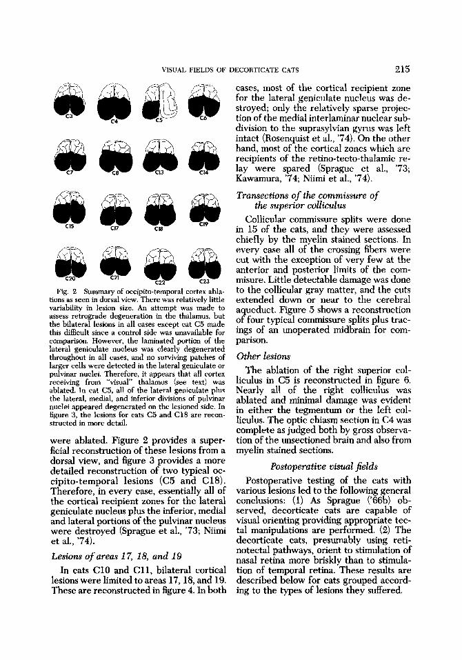
Occipito- temporal cortical lesions Sixteen of the cats underwent large oc-
cipito-temporal cortical lesions (table 1). In every case, all of the cortex on the medial surface above the splenial sulcus was ablated with variable damage to the cingu- late gyrus; on the dorsal surface, all of the Dosterior two-thirds of the lateral, suDra-
brrors for each responselevel are 2-3% for iylvian and most of the ectosylvian gyri
VISUAL FIELDS OF DECORTICATE CATS 215
C23
Fig. 2 Summary of occipito-temporal cortex abla- tions as seen in dorsal view. There was relatively little variability in lesion size. An attempt was made to assess retrograde degeneration in the thalamus, but the bilateral lesions in all cases except cat C5 made this difficult since a control side was unavailable for comparison. However, the laminated portion of the lateral geniculate nucleus was clearly degenerated throughout in all cases, and no surviving patches of larger cells were detected in the lateral geniculate or pulvinar nuclei. Therefore, it appears that all cortex receiving from “visual” thalamus (see text) was ablated. In cat C5, all of the lateral geniculate plus the lateral, medial, and inferior divisions of pulvinar nuclei appeared degenerated on the lesioned side. In figure 3, the lesions for cats C5 and C18 are recon- structed in more detail.
were ablated. Figure 2 provides a super- ficial reconstruction of these lesions from a dorsal view, and figure 3 provides a more detailed reconstruction of two typical oc- cipito-temporal lesions (C5 and C18). Therefore, in every case, essentially all of the cortical recipient zones for the lateral geniculate nucleus plus the inferior, medial and lateral portions of the pulvinar nucleus were destroyed (Sprague et al., ’73; Niimi et al., ’74). Lesions of areas 1 7, 18, and 19
In cats C10 and C l l , bilateral cortical lesions were limited to areas 17,18, and 19. These are reconstructed in figure 4. In both
cases, most of the cortical recipient zone for the lateral geniculate nucleus was de- stroyed; only the relatively sparse projec- tion of the medial interlaminar nuclear sub- division to the suprasylvian gyrus was left intact (Rosenquist et al., ’74). On the other hand, most of the cortical zones which are recipients of the retino-tecto-thalamic re- lay were spared (Sprague et al., ’73; Kawamura, ’74; Niimi et al., ’74).
Trunsections of the commissure of the superior colliculus
Collicular commissure splits were done in 15 of the cats, and they were assessed chiefly by the myelin stained sections. In every case all of the crossing fibers were cut with the exception of very few at the anterior and posterior limits of the com- misure. Little detectable damage was done to the collicular gray matter, and the cuts extended down or near to the cerebral aqueduct. Figure 5 shows a reconstruction of four typical commissure splits plus trac- ings of an unoperated midbrain for com- parison.
Other lesions The ablation of the right superior col-
liculus in C5 is reconstructed in figure 6. Nearly all of the right colliculus was ablated and minimal damage was evident in either the tegmentum or the left col- liculus. The optic chiasm section in C4 was complete as judged both by gross observa- tion of the unsectioned brain and also from myelin stained sections.
Postoperative visual fields Postoperative testing of the cats with
various lesions led to the following general conclusions: (1) As Sprague (‘66b) ob- served, decorticate cats are capable of visual orienting providing appropriate tec- tal manipulations are performed. (2) The decorticate cats, presumably using reti- notectal pathways, orient to stimulation of nasal retina more briskly than to stimula- tion of temporal retina. These results are described below for cats grouped accord- ing to the types of lesions they suffered.

216 S. MURRAY SHERMAN
Fig. 3 Reconstruction of cortical ablations in cats C5 (upper) and C18 (lower). These were chosen as representative lesions for the series of figure 2. The cortical ablations for cats C3 and C7 are reconstructed in Sherman (‘74a). Abbreviations: ES, ectosylvian sulcus; LS, lateral sulcus; RS, rhinal sulcus; SpS, splenial sulcus; SuS, suprasylvian sulcus; SyS, sylvian sulcus.

VISUAL FIELDS OF DECORTICATE CATS 217
figure 3.
cut c5 As figure 7 shows, data from cat C5
confirm and somewhat extend Sprague's ('66b) findings. The left occipito-temporal cortical ablation resulted in a right hemi- anopia that persisted stably for seven months (fig. 7B). The cat simply ignored all stimuli to the right of the fixation object.
Following the 7-month period, the cat's right superior colliculus was ablated (fig. 6). During the weeks postoperative to the col- licular lesion, the cat tended to circle no- ticeably to the ri ht (Sprague and Meikle, '65; Sprague, '66b s which made visual field testing extremely difficult. After two to
three months, this circling tendency nearly disappeared, and figure 7C shows the fields after this time. Visual orientation was dra- matically restored for stimuli throughout the right hemifield, although responses to the right were noticeably less brisk and ac- curate than those to the left (cf. Sprague, '66b). Presumably, the right hemifield was now dependent upon retinotectal path- ways; the left, upon cortical pathways,
Figure 7C also demonstrates an interocu- lar asymmetry in the visual fields. The right eye had a full field of view, but the left saw clearly only in its ipsilateral hemifield. That is, the collicular lesion restored vision

218 S. MURRAY SHERMAN
Fig. 5 Reconstruction of collicular commissure transections for cats C4, C15, C19, and C21. The knife cut and lesioned area is in black. These are representative of the transections not illustrated for the other cats receiving such surgery (table 1). For each cat, drawings are from evenly spaced sections moving from posterior at the bottom left to anterior at the top right. Collicular commissure transec- tions for C3 and C7 are reconstructed in Sherman (‘74a). Drawings through the midbrain are shown at the top for C22 to illustrate the unsectioned collicular commissure. CSC indicates the commissure of the superior colliculus, and the collicular layers are shown as I, 11,111, and LV according to Kanaseki and Sprague (‘74). The relationship to older terminology is: I, stratum zonale; 11, stratum griseum superficiale; 111, stratum opticum; IV, stratum griseum intermediale.
for the right nasal retina much more dra- matically than for the left temporal retina. In fact, there was no evidence that with the left eye the cat could orient to stimuli fall- ing upon temporal retina, but there may have been some vision there for the follow- ing reason. With left-monocular viewing, C5 had a much higher spontaneous re- sponse level (MATERIALS AND METHODS and fig. 7C). However, this higher level obtained nearly exclusively because of spontaneous orientations towards the right as if the cat
were scanning the amblyopic field, al- though this could also represent the cir- cling due to the collicular ablation. This high spontaneous level obscured orienta- tions evoked by stimuli presented from 0” to 45” right, and thus a genuine but low re- sponse level might be obscured by the spontaneous orientations (fig. 7D). This point is elaborated again in the next sec- tion.
This asymmetry between monocular vis- ual fields in C5 was seen in another way.

VISUAL FIELDS OF DECORTICATE CATS 219
Fig. 6 Reconstruction of ablation of right superior colliculus in cat C5. Conventions and abbreviations are the same as in figure 5.
During right-monocular viewing, C5 would follow objects circling in either direction around it in an apparently normal fashion (i.e., much like normal cats; see above). However, with left-monocular viewing, the cat would continue tracking only objects circling to the left; for a rightward moving stimulus, C5 soon lost it, apparently when
the stimulus fell onto the temporal retina. Note that this asymmetry cannot be ex- plained by residual circling caused by the collicular lesion, since such circling was di- rected to the right.
Cats C6, C14, C15, C22 The first operation in each of these cats
was a large bilateral occipito-temporal cor- tex ablation. All four cats reacted in an identical fashion: they appeared blind on the visual field test, although C14, C15, and C22 were able to learn a brightness dis- crimination (Loop and Sherman, '77, sub- mitted).2 C22 appeared blind on the visual field test for the remaining 12 months, at which time it was sacrificed. C6, C14, and C15 showed no signs of orienting responses throughout the 8 to 9-month period be- tween operations (fig. 8B). At this point, the collicular commissure was split during a second operation in these three cats, and within the first four postoperative weeks each of these cats showed dramatic recov- ery of orienting responses. Figure 8C shows this for C15, and virtually identical results obtained in C6 and C14. The responses, while clearly evident, were sluggish and relatively poorly directed; they in all ways resembled the final responses of C5 to stimuli in its right hemifield after its col- licular ablation. With monocular viewing, figure 8C shows that C15 clearly oriented only to stimuli in the ipsilateral hemifield (i.e., to stimuli falling on the nasal retina), but again responses to stimuli in the con- tralateral 45" of visual field could have been obscured by the relatively high rate of spontaneous orientation towards the amblyopic, contralateral hemifield (fig. 8D). As was seen for C5, most (>907) of the spontaneous responses were in this di- rection, but this cannot be explained by an overall tendency for the cat to orient spon- taneously left or right: with the right eye these spontaneous turnings were to the left; with the left eye, to the right. C6 and
It is emphasized that cats which fail to demonstrate visual orientations on this visual field task may well demonstrate visually guided behavior, such as brightness discrimination, on other tasks with different visuo-motor requirements.

220 S. MURRAY SHERMAN
RIGHT EYE BIN 0 C U 1 A R LEFT EYE
Fig. 7 Visual fields for cat C5. The conventions for the polar plots are the same as in figure 1. A, B, C represent normalized scoring (MATERIALS AND METHODS) and the number next to each origin indi- cates the percent level of spontaneous orientation. A. Preoperative fields. B. Fields following the left cortical ablation. C5 now had a stable; right hemianopia. C. Fields following the right collicular abla- tion. Orienting to the right hemifield returned, but not for left monocular viewing. D. Same fields as in C, but without normalized scoring. The dashed semicircle for the left eye field indicates the level of spontaneous orientations, nearly all of which were directed to the right (see text). The spontaneous level was too low (3%) to be indicated in binocular viewing, and with the right eye, there were no spontaneous orientations.

VISUAL FIELDS OF DECORTICATE CATS
BINOCULAR LEFT EYE RIGHT EYE
221
A
NO VISUAL ORIENTING
Fig. 8 Visual fields for cat C15. Conventions as in figure 7. A, B, C, are normalized scoring; D is not. A. Preoperative fields. B. After the bilateral cortical ablation, the animal failed to orient to visual stimuli. C. Fields after the subsequent collicular commissure transection. Visual orienting returned, but mainly or only to stimulation of nasal retina. D. Same fields as in C, but without normalized scor- ing.
C14 showed the same pattern. Also, all three cats with monocular viewing con- tinuously followed objects moving only in the imiversive direction (i.e., movine: in a
spontaneous turning in that direction. The monocular visual fields for C6, C14, and C15 were consequently in all ways like the left-monocular field for C5.
temporal direction in the hemifieldr, but lost them if they moved across the midline in the other direction. As in cat C5. movine:
cuts c37 c7, c137 c28j c20, C22, C23
targets could not be consistently -trackea into the amblyopic hemifield despite the
Each of these cats had a one stage opera- tion in which a large bilateral occipito-

222 S. MURRAY SHERMAN
BINOCULAR 96 1
A 0-
LEFT EYE
9 RIGHT EYE
Fig. 9 Visual fields for cat C13. Conventions as in figure 7. A, B are normalized scoring; C is not. A. Preoperative fields. B. Fields after the bilateral cortex ablation plus a transection of the collicular commissure. Orienting was limited mainly or only to stimulation of nasal retina. C. Same fields as in B. - but without normalized scoring.
temporal cortex ablation and a split of the commissure of the superior colliculus were made. All of these cats reacted identically, and figure 9 illustrates the typical and rep- resentative results from C13. Within three to eight weeks postoperatively, these cats showed behavior which was indistin- guishable from the performance of C14, C15, and C22 after their collicular com- missure transection (fig. 9B). Even the pat- tern of postoperative spontaneous orienta- tions with monocular viewing was the same (fig. 9C), as was the following of moving targets.
Cut c17 This cat had the same surgery as the pre-
vious group of eight cats (i.e., one stage
cortical ablation plus collicular commissure transection) but its postoperative behavior differed. For the entire 16-month postop- erative period before sacrifice, C17 could not be tested for its visual fields because it would not consistently fixate forward. Overall, its postoperative visual ability seemed considerably poorer than that of the above eight cats. It did, however, occa- sionally demonstrate, with either eye, the ability to locate and even briefly follow objects moving in its presumptive field of view. In addition to its apparently poorer visual ability, this cat showed neurological signs not evident in any other cat: namely, it was hyperexcitable and had several grand muE seizures. On the basis of availa- ble histology, however, the cortical lesions

VISUAL FIELDS OF DECORTICATE CATS 223 BINOCULAR
90" L
B 0
I
L,
LEFT EYE
9 9
19 c- 9 ,/"
RIGHT EYE
r l Fig. 10 Visual fields for cat C8; conventions as in figure 7 with normalized scoring throughout. A.
Preoperative fields B. Fields following the midsagittal transection of the commissure of the superior colliculus. C. Fields after the subsequent bilateral cortical ablation.
were within the limits of the other occip- ito-temporal decorticate cats, and no obvious damage was seen in the midbrain other than the transection of the collicular commissure. The poor vision of this cat rel- ative to the others with similar lesions could be due to two factors: (1) There may be considerable variability among cats in the potential to demonstrate the Sprague effect, but even if so, certainly the great preponderance of cats shows the effect. (2) A more likely explanation is that other neu- ral tissue (i.e., cortical, midbrain, etc.) was damaged but such damage could not be as- certained with the available histological material.
Cut C8 Since a split of the collicular commissure
after cortical lesions greatly enhanced
visually guided behavior, this lesion alone could affect visual fields in a cat. Thus cat C8 had only its collicular commissure tran- sected during its first operation. Figure 10B shows that this surgery had no observable effect on the visual fields, nor did it affect any obvious aspect of the cat's behavior (including visual discrimination learning; Loop and Sherman, '77, submitted). During the second operation seven months later, occipito-temporal cortex was bilaterally removed. Figure 1OC shows that the final fields were identical in every respect to those of other cats with such cortical and midbrain lesions, and thus the order of the lesions is not crucial.
Cut 4 From the above, it appears that cats de-
pendent upon retinotectal pathways de-

224 S. MURRAY SHERMAN
BINOCULAR
c PO* 1
A 3 0”
9 B
LEFT EYE
9 NO VISUAL ORIENTING
RIGHT EYE
3
Fig. 11 Visual fields for cat C4. Conventions as in figure 7 with normalized scoring throughout, A. Preoperative fields. B. Fields following the midsaggital transection of the optic chiasm. The cat oriented only to stimulation of temporal retina. C. After the subsequent bilateral cortical ablation plus a transection of the collicular commissure, the cat failed to orient to any visual stimulus.
velop fairly good visual responses for stim- uli falling upon the nasal retina and poor or no responses to stimulation of the temporal retina. Responses related to the temporal retina were further studied in C4 after first destroying nasal retinal connections by a midsagittal transection of the optic chiasm. This led to the classic bitemporal hemi- anopia illustrated in figure 11B. A second operation two months later removed occip- ito-temporal cortex bilaterally and split the collicular commissure. For the remaining 15-month postoperative period, C4 ap- peared blind on the behavioral tests ap- plied in this study. It attended to the fixation object, presumably due to the
auditory cues, and would move toward it when unrestrained, but the cat consistently ignored all of the visual test stimuli. Stimuli placed in its path yielded a distinct startle response when the cat contacted them. Un- like the previously described cats with sim- ilar cortical and midbrain lesions, this cat showed very few spontaneous orientations (7%). c10, c11
All of the above results suggest that a cat dependent upon retinotectal pathways re- sponds much better to stimulation of nasal retina than to stimulation of temporal reti- na. Evidently the visual fields normally

VISUAL FIELDS OF DECORTICATE CATS 225
@9 c 10
c11
BINOCULAR Po- L
LEFT EYE
l i b
0 c10
c11
Fig. 12 Visual fields for cats C10 and (211; conventions as in figure 7 with normalized scoring throughout. A. PreoDerative fields. B. Fields following the bilateral cortical lesion of areas 17, 18 and most i f 19.
mapped for temporal retina depend upon retinofugal pathways other than retinotec- tal, and the most likely are the retino-ge- niculo-cortical. For C10 and C11, areas 17, 18 and most of 19 were removed bilateral- ly to disrupt this pathway. Thus the re- maining visual cortex was mostly depen- dent on retinotectal pathways via the infe- rior, medial and lateral divisions of the pulvinar (Sprague et al., '73; Kawamura, '74; Niimi et al., '74). Figure 12B indicates that these cats postoperatively showed
clear visual fields for nasal retina and much reduced responsiveness to stimuli imping- ing upon temporal retina. Like the pre- viously described cats, C10 and C11 during monocular viewing showed more spontan- eous orientations into the amblyopic re- gion. Unlike the cats with larger cortical lesions, C10 and C11 showed normally brisk and accurate responses to stimulation of nasal retina. With monocular viewing they could follow fast stimuli only if moved ipsiversively. They could, however, follow

226 S. MURRAY SHERMAN
very slowly moving targets (<5"/sec) in the opposite direction.
DISCUSSION
With the possible exception of cat C17, these cats provided data consistent with two conclusions. First, decorticate cats have considerable visually guided behavior if intercollicular influences are abolished, either by ablation of one colliculus or tran- section of the collicular commissure. With- out such tectal surgery, a permanent and total deficit in orienting ensues. This is pre- cisely the effect that Sprague ('66b) de- scribed. Second, while dependent upon retinotectal pathways, the cat sees much better with nasal than with temporal reti- na. With intact geniculo-cortical pathways, no such difference between nasal and tem- poral retina is evident.
Basis for the Sprague effect Given the potential importance of this
effect, it is somewhat surprising that so lit- tle has been written about it in the past ten years (e.g., for the cat: Wood, '73, '75; for the rat: Cooper et al., '70; Goodale, '73). At the single neuron level, many changes occur among collicular neuronal properties after cortical ablations (e.g., Rosenquist and Palmer, '71; Wickelgren and Sterling, '69). Perhaps the most striking result of de- cortication is the total loss of visual re- sponses among multimodal neurons in deeper collicular layers (Schiller et al., '74; Stein and Arigbede, '72). It is possible that some of these changes underly the Sprague effect, but if so, then a collicular com- missure split or contralateral collicular lesion should reduce or reverse these changes. Unfortunately, such data are as yet unavailable, and the specific neuronal correlates of the Sprague effect remain un- known. Our understanding of the phe- nomenon consequently'remains at the level of Sprague's ('66b) general suggestion that collicular function is somehow affected by the balance of global facilitatory (cor- ticotectal) and suppressive (colliculo-col- licular) influences. In this context, the pres-
ent results add some details to our knowl- edge of the phenomenon.
A decorticate cat will demonstrate clear visually guided orienting behavior provid- ing the colliculi are functionally discon- nected. The order of these lesions is irrel- evant (see, however, Wood, '75). In cats C5, C6, C14, and C15, collicular disconnec- tion followed decortication (in C5 this dis- connection was achieved by removing a source, the right colliculus). In cats C3, C7, C13, C18, C19, C20 and C23, the cortical ablation and collicular commissure transec- tion were done simultaneously. In cat C8, the collicular commissure transection pre- ceded decortication. Yet all of these cats provided consistent behavioral data, and the final behavior in no way correlated with the order of the lesions.
If, as Sprague ('66b) suggested, collicular function is depressed by decortication, then one might expect the depressed col- liculus to be unable to suppress the other colliculus. If so, then one might expect a balanced, bilateral cortical ablation (with- out transection of the collicular com- missure) to permit better visually guided behavior than seen in the hemifield con- tralateral to a unilateral cortical ablation. This was not the case. After bilateral, occipito-temporal cortical ablations, cats C6, C14, C15 and C22 appeared just as blind as did cat C5 in its right hemifield after its left cortical ablation. Therefore, any colliculo-collicular suppression cross- ing in the commissure is not dependent upon facilitatory influences descending from cortex.
A final point, illustrated by cat C5 and also made by Sprague ('66b), is that a cat has the potential for good visually guided orienting behavior providing either the cortical or collicular pathways are intact. That is, after the collicular ablation {in C5, orientations to targets in the right hemi- field presumably depended upon retinotec- tal pathways while those in the left hemi- field depended upon retino-geniculo-corti- cal pathways. This would seem different from conclusions in the hamster (Schnei-
VISUAL FIELDS OF DECORTICATE CATS 227
der, '69), rat (Kirvel et al., '74), and tree shrew (Casagrande and Diamond, '74). In these animals, visually guided orienting appears dependent upon retinotectal path- ways. In the absence of these pathways, but with functional retino-geniculo-cortical pathways, these animals cannot visually orient. Differences between the cat and the other mammalian species regarding po- tential cortical participation in visual orienting could be an important inter-spe- cies difference. On the other hand, these differences could obtain from different testing procedures.
Contents of the collicular commissure The commissure of the superior col-.
liculus is anatomically complex. Not only does it contain fibers interconnecting the colliculi, many in a homo-typical fashion, but it also includes many decussating fibers heading to the superior colliculus from other regions or vice-versa (Antonetty and Webster, '75; Edwards, '77; Powell, '76). Therefore, the commissure transections described in this study probably interfered with many or all of the above-mentioned fiber systems, and at present it is not possi- ble to determine which of these systems is related to the Sprague effect.
Consistency of the Sprague efect Clear evidence for the Sprague effect
was seen in 13 cats (C3, C5, C6, C7, C8, C13, C14, C15, C18, C19, C20, C21, and C23), and only one cat, C17, failed to dem- onstrate the effect. Of the cats that did show it, an impressive consistency of final behavior was noted. Given the difficulty of the surgery, especially the commissure transection, this consistency is surprising and suggests that the phenomenon is a powerful one that can survive minor surgi- cal variations. While it is possible that not every cat has the potential for the full- blown phenomenon, certainly the over- whelming majority do. It is most likely that the one failure, C17, was due to extra, un- controlled neural damage.
Temporal versus nasal retinal stimulation
The behavior of cats showing the Sprague effect seems largely dependent on retinotectal pathways (Sprague and Mie- kle, '65; Sprague, '66a). Whereas a normal cat demonstrates good visually guided orienting behavior for stimulation of the entire retina (fig. 1, and Hughes, '761, cats without cortex demonstrate good vision on these tests for nasal retina but poor or no vision for temporal retina. The question as to whether any orienting behavior in de- corticate cats is specifically evoked by stimulation of temporal retina is not possi- ble to answer from these data because of the curious spontaneous scanning of this re- gion of visual field; but clearly, stimulation of nasal retina is considerably more effec- tive in eliciting fairly accurate orienting than is stimulation of temporal retina. It should be noted that the spontaneous scan- ning of amblyopic portions of visual .field seen in these cats was not seen in other cats with partial field defects. Thus, optic chiasm section (C4, unilateral cortical lesion (C5), and monocular or binocular deprivation (Sherman, '73, '74a; see also the following paper, Sherman, '77), do not produce this scanning in the region of the field defect.
This retinal asymmetry would suggest that the retino-geniculo-cortical pathways necessary for this behavior receive roughly equal functional inputs from corresponding parts of nasal and temporal retina; but the analogous retino-tectal pathways receive functional inputs predominantly from nasal retina. This suggestion has an anatomical basis. In the cat, the retino-geniculate pathways representing binocular portions of the visual fields are nearly equal in terms of fibers from nasal and temporal retina, whereas the retinotectal pathways are pre- dominantly crossed from nasal retina (Sprague, '66b; Wickelgren and Sterling, '69; Rosenquist and Palmer, '71; Sterling, '73; see, however, Graybiel, '75; Harting and Guillery, '76). Some fibers from tem-
228 S. MURRAY SHERMAN
poral retina also cross to terminate an- teriorly in the superior colliculus (Harting and Guillery, '76), but their function is un- clear. Presumably this asymmetry between nasal and temporal retinal projections to colliculus underlies the behavioral asym- metry seen in the decorticate cats. Cat C5 clearly exemplifies this. With the right eye, it has a full field of view, because the right temporal retina projects into the undam- aged geniculocortical system, and the right nasal retina projects to the undamaged left colliculus. With the left eye, the field of view is largely limited to the left hemifield. This presumably obtains because the nasal retina projects contralaterally to the intact geniculocortical system while temporal fibers have only a small projection to ipsi- lateral colliculus and their large input through the lateral geniculate nucleus to the cortex has been destroyed by the corti- cal lesion. Good visual responses for stimu- lation of temporal retina, then, depend upon the integrity of the retino-geniculo- cortical pathways, and destruction of these (as in cats C10 and C l l ) can lead to the rel- atively poor, or absent, behavioral re- sponses for the temporal retinal stimulation described in this paper.
This postoperative asymmetry related to nasal and temporal retina raises a question about the virtue of the split-brain prepara- tion (i.e., after transections of both optic chiasm and commissures such as the corpus callosum). The preparation is frequently used for intrasubject control: each eye is connected strictly to the ipsilateral half of brain; and if lesions are limited to one side, the behavioral capacity related to the le- sioned eyebrain combination is compared to that of the other, control combination (cf. Berlucchi et al., '72; Wood, '73, '75). However, the transected optic chiasm it- self largely denervates the superior col- liculus since only ipsilaterally directed fibers from temporal retina survive the transection. Studies of functions in any way dependent upon retinotectal pathways in split-brain animals could consequently pro- vide misleading data.
CONCLUSIONS
On the basis of the behavior described in this paper, it is clear that the Sprague effect is a robust phenomenon in cats. That is, visual behavior dependent upon retino- tectal pathways after large cortical lesions can be unmasked if the superior colliculi are functionally disconnected. In such a preparation, one could perhaps more accu- rately assess the visual capacity of decorti- cate cats. For instance, it may be that, in a wide range of testing conditions, transec- tion of the collicular commissure allows for visual behavior which is considerably bet- ter than has been previously reported for decorticate cats, and erhaps other mam- mals (Kirvel et al., '74 P .
ACKNOWLEDGMENTS
The expert technical assistance of Lodi Smith and Sally Gibson is gratefully ac- knowledged. This research was supported by Grants BMS 73-06938 from the National Science Foundation and EY01565 from the Public Health Service as well as a Research Career Development Award (EY00020) from the Public Health Service.
LITERATURE CITED Antonetty, C. M., and K. E. Webster 1975 The
organization of the spinotectal projection. An experimental study in the rat. J. Comp. Neur., 163:
Berlucchi, G., J. M. Sprague, J. Levy and A. C. DiBerardino 1972 Pretectum and superior col- liculus in visually guided behavior and in flux and form discrimination in the cat. J. Comp. & Physiol. Psychol. Monogr., 78: 123-172.
Casagrande, V. A., and I. T. Diamond 1974 Ablation study of the superior colliculus in the tree shrew (Tupuiu glis). J. Comp. Neur., 156: 207-238.
Cooper, R. M., B H. Bland, L. A. Gillespie and R. H. Whittaker 1970 Unilateral posterior cortical and
'unilateral collicular lesions and visually guided be- havior in the rat. J. Comp. Physiol. Psychol., 72:
Edwards, S. B. 1977 The commissural projection of the superior colliculus in the cat. Submitted to J. Comp. Neur., in press.
Goodale, M. A. 1973 Cortico-tectal and intertectal modulation of visual responses in the rat's superior colliculus. Exp. Brain Res., 17: 75-86.
Graybiel, A. M. 1975 Anatomical organization of reti- notectal afferents in the cat: an autoradiographic study. Bruin Res., 96: 1-23.
449-466.
286-295.

VISUAL FIELDS OF DECORTICATE CATS 229
Harting, J. K., and R. W. Guillery 1976 Organization of the retinocollicular pathways in the cat. J. Comp. Neur., 166: 133-144.
Hughes, A. 1976 A supplement to the cat schematic eye. Vision Res., 16: 149-154.
Kanaseki, T., and J. M. Sprague 1974 Anatomical organization of pretectal nuclei and tectal laminae in the cat. J. Comp. Neur., 158: 319-338.
Kawamura, S. 1974 Topical organization of the extra- geniculate visual system in the cat. Exp. Neurol., 45:
Kirvel, R. D., R. A. Greenfield and D.R. Meyer 1974 Multimodal sensory neglect in rats with radi- cal unilateral isocortical and superior collicular ablations. J. Comp. Physiol. Psychol., 87: 156-162.
Loop, M. S., and S. M. Sherman (1977, submitted) Visual discriminations of cats with cortical and tec- tal lesions. J, Comp. Neur.
Niimi, K., M. Kadota and Y. Matsushita 1974 Cortical projections of the pulvinar nuclear group of the thalamus in the cat. Brain, Behav. and Evol., 9: 422- 457.
Powell, T. P. S. 1976 Bilateral cortico-tectal projec- tion from the visual cortex in the cat. Nature, 260:
Rosenquist, A. C. , S. B. Edwards and L. A. Palmer 1974 An autoradiographic study of the projections of the dorsal lateral geniculate nucleus and the pos- terior nucleus in the cat. Brain Res., 80: 71-93.
Rosenquist, A. C., and L. A. Palmer 1971 Visual re- ceptive field properties of cells of the superior col- liculus after cortical lesions in the cat. Exp. Neurol.,
Schiller, P. H., M. Stryker, M. Cynader and N. Berman 1974 Response characteristics of single cells in the monkey superior colliculus following ablation or cooling of visual cortex. J. Neurophysiol., 37: 181- 194.
Schneider, G. E. 1969 Two visual systems. Science,
451-461.
526-527.
33: 629-652.
163: 895-902.
larly and binocularly deprived cats. Brain Res., 49:
- 1974a Permanence of visual perimetry deficits in monocularly and binocularly deprived cats. Brain Res., 73: 491-501. - 1974b Visual fields of cats with cortical and
tectal lesions. Science, 185: 355-357. 1977 The effect of cortical and tectal lesions
on the visual fields of binocularly deprived cats. J. Comp. Neur., 172: 231-246.
Sprague, J. M. 1966a Visual, acoustic and somesthetic deficits in the cat after cortical and midbrain lesions. In: The Thalamus. D. P. Purpura and M. D. Yahr, eds. New York, Columbia University Press,
- 1966b Interaction of cortex and superior col- liculus in mediation of visually guided behavior in the cat. Science, 153: 1544-1547.
Sprague, J. M., G. Berlucchi and G Rizzolatti 1973 The role of the superior colliculus and pretectum in vision and visually guided behavior. In: Handbook of Sensory Physiology. Vol. VII/3B. R. Jung, ed. Springer-Verlag, Berlin, pp. 27-101.
Sprague, J. M., and T. H. Miekle, Jr. 1965 The role of the superior colliculus in visually guided behavior. Exp. Neurol., 11: 115-146.
Stein, B. E., and M. 0. Argibede 1972 Unimodal and multimodd response properties of neurons in the cat's superior colliculus. Exp. Neurol., 36: 179-196.
Sterling, P. 1973 Quantitative mapping with the elec- tron microscope: retinal terminals in the superior colliculus. Brain Res., 54: 347-354.
Wickelgren, B. G., and P. Sterling 1969 Influence of visual cortex on receptive fields in the superior col- liculus of the cat. J. Neurophysiol., 32: 16-23.
Wood, B. S. 1973 Monocular relearning of a dark- light discrimination after large unilateral cortical lesions. Brain Res., 53: 428-434. - 1975 Monocular relearning of a dark-light discrimination bv cats after unilateral cortical and
25-45.
pp. 391-417.
Sherman, S. M. 1973 Visual field defects in monocu- collicular lesion;. Brain Res., 83: 156-162.