Embed Size (px)
Citation preview

1
The effect of Q-Switched Nd:YAG 1064nm/532 nm laser
in the treatment of onychomycosis in vivo Kostas Kalokasidis1, Meltem Onder2,3, Myrto-Georgia Trakatelli,4
Bertrand Richert5, Klaus Fritz2,6,7
1.Dermatology and Laser Clinic Thessaloniki, GREECE
2.Dermatology and Laser Center, Landau-GERMANY 3.Gazi University Medical Faculty Emeritus Professor, Ankara-TURKEY 4.Aristotle University Medical Faculty, Thessaloniki, GREECE
5.Universite Libre de Bruxelles, Brussels, BELGIUM 6.Associate Professor Carol Davila University – Bucharest-ROMANIA
7.Consultant Dermatologist in Osnabrück University Dermatology-GERMANY
and BernUniversity Dermatology -SWITZERLAND Kostas Kalokasidis
88 Tsimiski Street , 54622
Thessaloniki- Greece e- mail:[email protected] Meltem Onder
Reduiststr.13
76829Landau-Germany e-mail: [email protected] Myrto-Georgia Trakatelli
Second Department of Dermatology and Venereology
Papageorgiou Hospital; Aristotle University School of Medicine Thessaloniki, Greece. e-mail:[email protected]
Bertrand Richert
Clinical Professor Dermatology Department CHU Brugmann - Saint Pierre - HUDERF Université Libre de Bruxelles, Bruselles-Belgium
e-mail: [email protected] Klaus Fritz
Reduiststr.13 76829Landau-Germany
e-mail: [email protected]

2
Correspondence Author and Address
Dr. Klaus Fritz Reduiststr.13 76829Landau-Germany
Conflict of Interest
Kostas Kalokasidis, Myrto-Georgia Trakatelli, Bertrand Richert performed this clinical trial. Meltem Onder and Klaus Fritz did scientific evaluation.
The Authors declare that they have no conflict of interest.
3
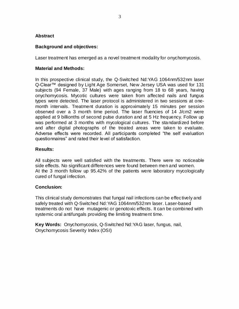
Abstract
Background and objectives:
Laser treatment has emerged as a novel treatment modality for onychomycosis.
Material and Methods:
In this prospective clinical study, the Q-Switched Nd:YAG 1064nm/532nm laser Q-Clear™ designed by Light Age Somerset, New Jersey USA was used for 131 subjects (94 Female, 37 Male) with ages ranging from 18 to 68 years, having
onychomycosis. Mycotic cultures were taken from affected nails and fungus types were detected. The laser protocol is administered in two sessions at one-
month intervals. Treatment duration is approximately 15 minutes per session observed over a 3 month time period. The laser fluencies of 14 J/cm2 were applied at 9 billionths of second pulse duration and at 5 Hz frequency. Follow up
was performed at 3 months with mycological cultures. The standardized before and after digital photographs of the treated areas were taken to evaluate.
Adverse effects were recorded. All participants completed “the self evaluation questionnaires” and rated their level of satisfaction. Results:
All subjects were well satisfied with the treatments. There were no noticeable side effects. No significant differences were found between men and women. At the 3 month follow up 95.42% of the patients were laboratory mycologically
cured of fungal infection. Conclusion:
This clinical study demonstrates that fungal nail infections can be effec tively and
safely treated with Q-Switched Nd:YAG 1064nm/532nm laser. Laser-based treatments do not have mutagenic or genotoxic effects. It can be combined with
systemic oral antifungals providing the limiting treatment time. Key Words: Onychomycosis, Q-Switched Nd:YAG laser, fungus, nail,
Onychomycosis Severity Index (OSI)

4
Introduction
Onychomycosis is defined as a fungal infection of the nail that expands slowly, and if left untreated, leads to complete destruction of the nail plate. Onychomycosis can be dermatophytic (99%) and/or non-dermatophytic (1%)
(including yeasts) infections of the nail plate.
The dermatophytes Trichophyton rubrum and Trichophyton mentagrophytes are the most common causative pathogens responsible for up to 90% of all cases. (1) Onychomycosis represents about 30% of all dermatophyte infections and
accounts for 18%-40% of all nail disorders. The prevalence of onychomycosis ranges between 2%-28% of the general population and it is estimated to be
significantly higher in specific populations such as in diabetes mellitus, the immunosuppressed and elderly. (2, 3)
Among the non-dermatophytes, the yeast Candida albicans, Candida tropicalis, aspergillus and other molds may be responsible. It usually represents
contamination and is an emerging problem in HIV patients. Toenails are far more likely to be involved than fingernails. Initially solitary nails
are involved; later, many may be infected, but often one or more can stay disease free. Onychomycosis has no tendency for spontaneous remission and
should be considered as a problem with serious medical, social and emotional extensions, not solely a cosmetic problem. The primary concerns of the patients are the risk of spread to other nails or to people in their environment. Others
consider their deformed nails as unattractive to other people, which may lead to lower self-esteem, a sense of inadequacy and even depression. (4, 5) In addition
to these social and emotional problems, onychomycosis is a serious medical problem that can be the source of further fungal infections to surrounding tissues. Also, it may predispose patients to secondary bacterial infections leading to
localized paronychia and perhaps worse and deeper infections such as erysipelas-cellulitis, especially in the high-risk groups such as diabetics. (6, 7)
Clinically it can cause varying degrees of pain or discomfort (especially in walking) and problems in cutting nails.
Classical treatment options include mechanical and chemical debridement, topical antifungal lacquers, systemic antifungal drugs and finally various
combinations of the above. Most effective as mono-therapy are the systemic antifungal agents that have been the gold standard and form the mainstay of therapy, however they require long treatment periods (approximately 6 months
for toenails and up to 4 months for fingernails). This requires liver function-transaminases, and kidney function blot test control. Patients may also receive

5
concomitant medications for comorbidities, so there is also the issue of drug interactions. Additionally, long lasting treatment means high treatment costs for
both the patient and health insurers. Finally, high recurrence rates have been described, 22% three years after completion of treatment and higher recurrence rates at five years follow-up. (8, 9, 10)
Recently, lasers have emerged as potential new treatment modalities. These
treatments offer the advantage of having few contraindications, and minimal side effects. (11,12,13) Laser energy has the potential to eliminate microorganisms.
Vural E. et al. recently demonstrated direct inhibitory activity of laser energy on T. rubrum isolates in vitro. (14) Manevitch Z. et al. recently published the direct
antifungal effect of the femtosecond laser on T. rubrum onychomycosis as well (7). The laser must have the ability to penetrate under the nail plate in order to
reach the fungi colonies of the nail bed and nail matrix. When it gets to that point
it should selectively deliver laser energy to fungi while respecting the surrounding healthy tissues.
In this study we planned to evaluate the effect of the neodymium: yttrium-aluminum-garnet (Nd:YAG) 1064-nm/532 nm laser in the treatment of
onychomycosis in vivo.
Material and Methods
Nail sampling and fungal cultures
Nail cuttings sized 2 x 3 mm were obtained from patients with clinical suspicion of onychomycosis. After direct microscopy to observe spores, hyphae, mycelia and
colonies of the latter, samples were plated on Sabouraud glucose agars with cyclohexamide to select for dermatophytes, in order to verify fungal infection.
Cultures were incubated at 28oC for 3 weeks until fungal colonies developed.
Evaluation of fungal elimination
Before the treatment culture was performed, and 4 weeks after the second
treatment session (8 weeks after the first positive culture), culture was repeated. Mycological cure is defined as negative microscopy and culture. Clinical cure is
associated with the alteration of the percentages of disease free nails. Complete cure is accepted as the combination of mycological and clinical cure. Three months after the first treatment session, laser treatment was evaluated. (15, 16)
Inclusion criteria
To take part in the study each patient had to have one or more toenail and/or fingernail fungal infections of the follow types: distal subungual onychomycosis,
proximal subungual onychomycosis, superficial white onychomycosis or total dystrophic type onychomycosis. Patients with diabetes mellitus,
6
immunocompromised patients and organ transplant patients were also included,
although we considered these patient groups success rates could be considerably less.
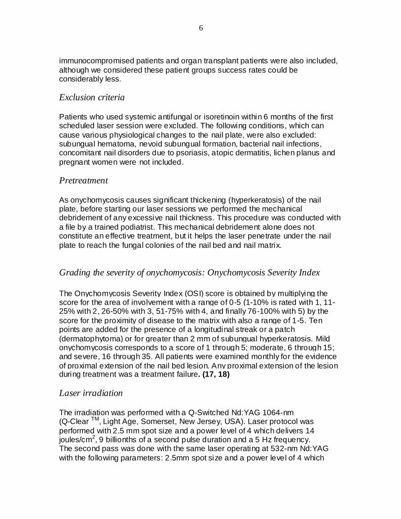
Exclusion criteria
Patients who used systemic antifungal or isoretinoin within 6 months of the first scheduled laser session were excluded. The following conditions, which can
cause various physiological changes to the nail plate, were also excluded: subungual hematoma, nevoid subungual formation, bacterial nail infections, concomitant nail disorders due to psoriasis, atopic dermatitis, lichen planus and
pregnant women were not included.
Pretreatment
As onychomycosis causes significant thickening (hyperkeratosis) of the nail plate, before starting our laser sessions we performed the mechanical debridement of any excessive nail thickness. This procedure was conducted with
a file by a trained podiatrist. This mechanical debridement alone does not constitute an effective treatment, but it helps the laser penetrate under the nail
plate to reach the fungal colonies of the nail bed and nail matrix.
Grading the severity of onychomycosis: Onychomycosis Severity Index The Onychomycosis Severity Index (OSI) score is obtained by multiplying the score for the area of involvement with a range of 0-5 (1-10% is rated with 1, 11-25% with 2, 26-50% with 3, 51-75% with 4, and finally 76-100% with 5) by the
score for the proximity of disease to the matrix with also a range of 1-5. Ten points are added for the presence of a longitudinal streak or a patch
(dermatophytoma) or for greater than 2 mm of subungual hyperkeratosis. Mild onychomycosis corresponds to a score of 1 through 5; moderate, 6 through 15; and severe, 16 through 35. All patients were examined monthly for the evidence
of proximal extension of the nail bed lesion. Any proximal extension of the lesion during treatment was a treatment failure. (17, 18)
Laser irradiation
The irradiation was performed with a Q-Switched Nd:YAG 1064-nm (Q-Clear TM, Light Age, Somerset, New Jersey, USA). Laser protocol was
performed with 2.5 mm spot size and a power level of 4 which delivers 14 joules/cm2, 9 billionths of a second pulse duration and a 5 Hz frequency.: The second pass was done with the same laser operating at 532-nm Nd:YAG
with the following parameters: 2.5mm spot size and a power level of 4 which
7
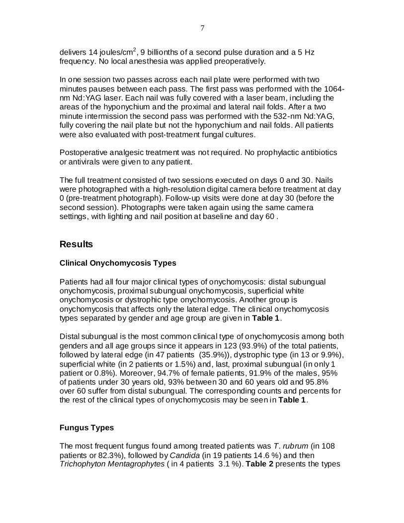
delivers 14 joules/cm2, 9 billionths of a second pulse duration and a 5 Hz frequency. No local anesthesia was applied preoperatively.
In one session two passes across each nail plate were performed with two
minutes pauses between each pass. The first pass was performed with the 1064-nm Nd:YAG laser. Each nail was fully covered with a laser beam, including the areas of the hyponychium and the proximal and lateral nail folds. After a two
minute intermission the second pass was performed with the 532-nm Nd:YAG, fully covering the nail plate but not the hyponychium and nail folds. All patients
were also evaluated with post-treatment fungal cultures.
Postoperative analgesic treatment was not required. No prophylactic antibiotics
or antivirals were given to any patient.
The full treatment consisted of two sessions executed on days 0 and 30. Nails were photographed with a high-resolution digital camera before treatment at day 0 (pre-treatment photograph). Follow-up visits were done at day 30 (before the
second session). Photographs were taken again using the same camera settings, with lighting and nail position at baseline and day 60 .
Results Clinical Onychomycosis Types
Patients had all four major clinical types of onychomycosis: distal subungual onychomycosis, proximal subungual onychomycosis, superficial white onychomycosis or dystrophic type onychomycosis. Another group is
onychomycosis that affects only the lateral edge. The clinical onychomycosis types separated by gender and age group are given in Table 1.
Distal subungual is the most common clinical type of onychomycosis among both
genders and all age groups since it appears in 123 (93.9%) of the total patients, followed by lateral edge (in 47 patients (35.9%)), dystrophic type (in 13 or 9.9%),
superficial white (in 2 patients or 1.5%) and, last, proximal subungual (in only 1 patient or 0.8%). Moreover, 94.7% of female patients, 91.9% of the males, 95% of patients under 30 years old, 93% between 30 and 60 years old and 95.8% over 60 suffer from distal subungual. The corresponding counts and percents for the rest of the clinical types of onychomycosis may be seen in Table 1.
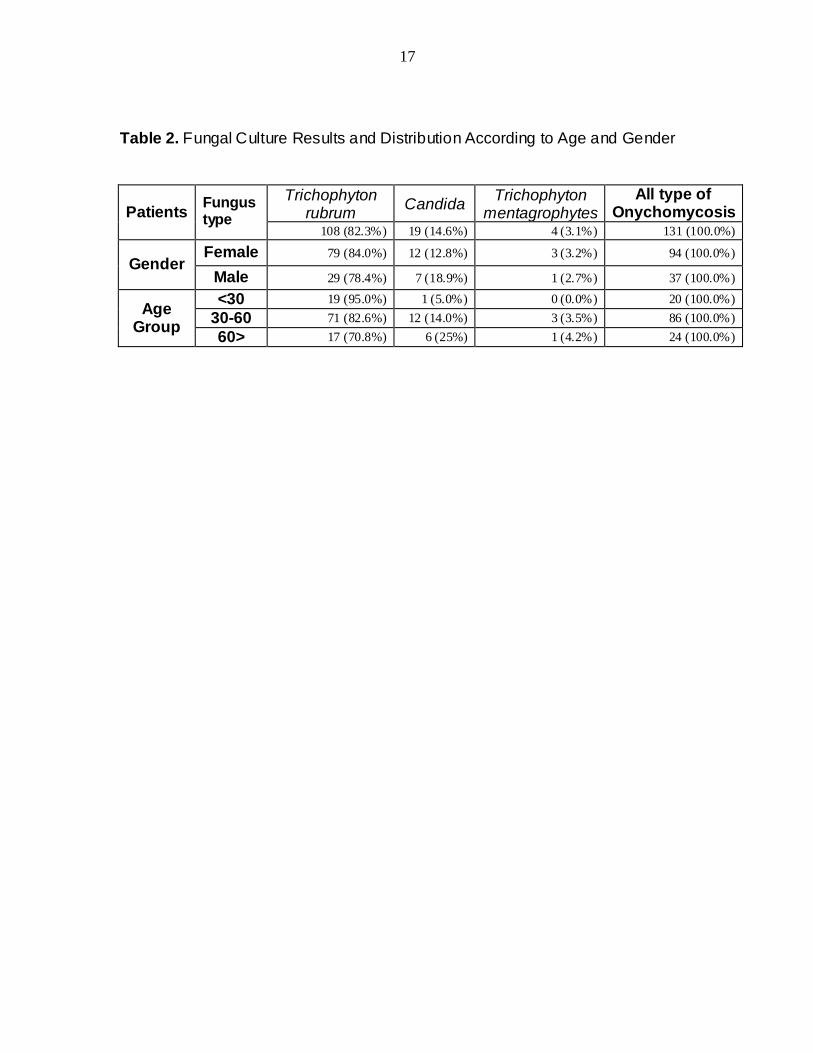
Fungus Types
The most frequent fungus found among treated patients was T. rubrum (in 108
patients or 82.3%), followed by Candida (in 19 patients 14.6 %) and then Trichophyton Mentagrophytes ( in 4 patients 3.1 %). Table 2 presents the types
8
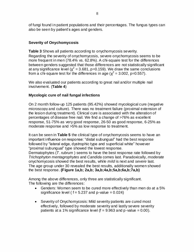
of fungi found in patient populations and their percentages. The fungus types can
also be seen by patient’s ages and genders.
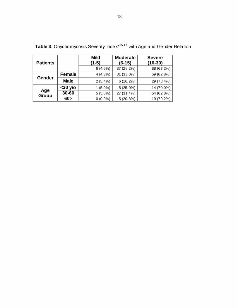
Severity of Onychomycosis
Table 3 Shows all patients according to onychomycosis severity.
Regarding the severity of onychomycosis, severe onychomycosis seems to be more frequent in men (78.4% vs. 62.8%). A chi-square test for the differences
between genders suggested that those differences are not statistically significant at any significance level (χ2 = 3.681, p=0.159). We draw the same conclusions from a chi-square test for the differences in age (χ2 = 3.002, p=0.557).
We also evaluated our patients according to great nail and/or multiple nail involvement. (Table 4) Mycologic cure of nail fungal infections
On 2 month follow-up 125 patients (95.42%) showed mycological cure (negative
microscopy and culture). There was no treatment failure (proximal extension of the lesion during treatment). Clinical cure is associated with the alteration of percentages of disease free nail. We find a change of >76% as excellent
response, 51-75% as very good response, 26-50 as good response, 6-25% as moderate response and >5% as low response to treatment.
It can be seen in Table 5 the clinical type of onychomycosis seems to have an
important influence on response: “distal subungual” had the best response
followed by “lateral edge, dystrophic type and superficial white” however “proximal subungual” type showed the lowest response.
Dermatophytes (T. rubrum ) seems to have the best response rate followed by Trichophyton mentagrophytes and Candida comes last. Paradoxically, moderate onychomycosis showed the best results, while mild is next and severe last.
The age group under 30 revealed the best results, additionally women showed the best response. (Figure 1a,b; 2a,b; 3a,b;4a,b;5a,b;6a,b;7a,b)
Among the above differences, only three are statistically significant. The following are the differences:
Genders: Women seem to be cured more effectively than men do at a 5% significance level ( f = 5.237 and p-value = 0.024)
Severity of Onychomycosis: Mild severity patients are cured most
effectively, followed by moderate severity and lastly severe severity patients at a 1% significance level (f = 9.963 and p-value = 0.00).

9
The responsible nail fungi: T. rubrum recede more quickly after the cure, followed by trichophyton mentagrophytes and Candida at a 1%
significance level ( f= 15.347 and p-value = 0.00).
Adverse event evaluation
Most patients, 94, (83.21%) reported mild pain, 22 patients (16.79%) reported no pain. This “pain” sensation was described as ”a stinging” during the 1064nm pass
and as “burning” during the 532 nm pass. None of the patients treated had severe or intolerable pain. No postoperative analgesic treatment was required. Interestingly many of patients developed a kind of pain resistance during the
therapy, meaning they reported the highest level of pain during the first session. We believe this suggests the patients knew what to expect or that the fear of an
unknown treatment no longer existed. Patients were also asked to report all possible adverse events that could be
related to our treatment. There were no reports of any other side effects.
Discussion Treatment of onychomycosis is difficult. Laser treatment is considered by some authors to be a promising new method. Our study population comprised of 131
individuals. 15.3% of the participants in the study were below 30 years of age, 65.6 % between 30-60 years, and finally 18.3 %, were over 60 years old. These
groups allowed us to maintain a large enough sample within each group to compare the effectiveness of the laser treatment on different age groups. Women
were the 71.8% of our patient sample. This does not mean that onychomycosis
occurs more frequently in women but that men may be more negligent in matters relating to the cosmetic appearance and hygiene of their feet.
In a recent paper Vural, et al. showed that 1064nm and 532nm Q-Switched
Nd:YAG laser systems had significant inhibitory effect upon T. rubrum isolates and caused colony growth inhibition in vitro (14). It is well known that the efficacy
of laser energy depends on the light-tissue interaction which is a function of wave-length, fluence, as well as tissue optics (7). We have used various spot
sizes in all power levels with our system. This can provide combinations which
deliver different energy fluence. We found the most powerful treatment was 14 joules/cm2, additionally, the 7.5 joules/cm2 (3.5mm spot size and a power level of 4) was also effective. Since the treatment session is very well tolerated in the
maximum energy fluence, we used these settings. We have noticed a significant improvement in the proximal portion of the nail where there was mild initial
mycotic involvement. Our results were better especially in moderate severity patients. That seems reasonable as severe cases are accompanied by dermatophytoma or significant subungual hyperkeratosis, which require more

10
time for the nail plate to restore. Poor prognostic indicators are the total dystrophic onychomycosis, the involvement of the lateral edge of the nail plate,
and the involvement of the matrix (18, 19, 20, 21). The thick plate or subungual
hyperkeratosis >2mm, histologically contain numerous air-filled spaces in which
fungal spores can survive for weeks or months. These resting a rthrospores do not form hyphae, so various antifungal agents have proven ineffective. This phenomenon, termed as dermatophytoma, can be seen as linear streaks or
rounded white areas in the nail plate. The fungal elements are believed to be forming a biofilm, making them refractory to therapy (15, 22, 23). Laser therapy
seems not to be affected of this biofilm formation, this may explain why we achieved very good and good response in 67% of our severe cases. Moreover, old age, presence of immunosuppression, poor peripheral circulation and non-
responsive organisms (non-dermatophyte molds), other dermatoses (e.g. nail psoriasis), and drug resistance are poor prognostic indicators (15, 23, 24). With
the laser we solve the problem of resistance. We suggest that we don’t have non-responsive cases but some poor responding fungi. As another example, occupational factors, as well as occlusive and prolonged contact with water can contribute to poor response of treatment (21).
On the contrary, superficial white onychomycosis is associated with the best therapeutic response to antifungal drugs, and our results seem to agree with this (16,19). Our distal subungual clinical cases had good results as well. Even
dystrophic types showed a very good and good response in 66% of the cases. This supports laser treatment efficacy. Laser treatment seems to outweigh
classical treatments where involvement of the matrix, a thick plate or subungual hyperkeratosis >2mm are factors associated with poor outcome (15, 21).
The Q-Clear™ Laser System, in differentiation to other laser treatments, provides a selective, both thermal (photothermolytic) and mechanical (photomechanical),
effect on the fungi. The mechanism of this fungal destruction may offer some differences. The inhibitory effect is likely due to more than simple nonspecific thermal damage. Denaturing one or more of the molecules within the pathogen
may deactivate the fungi. Vural, et al. discusses that 532 nm setting, which is well absorbed by red pigment in canthomegnin in T. Rubrum, this wavelength generates mechanical damage in the irradiated fungal colony (14).
The 1064 nm setting is beyond the absorption spectrum of xanthomegnin, but i ts effectiveness is due to another absorbing chromophore, perhaps melanin, which is present in the fungal cell wall (14). Melanin is an essential inhabitant of the
fungal cell wall and has been described in many pathogenic species. The type of
melanin varies, although it is commonly Dopa or pentaketide melanin. Moreover, the laser beam may initiate a photo-biological or photochemical reaction that attacks the pathogen cell. We can also suggest a multi-photon dielectric
breakdown at the fungal target as the cause of their destruction, while depth-selective thermal effects by the laser could also be occurring (7).

11
Another possible scenario is by inducing an immune response that attacks the organism. All of the above hypotheses explain how the surrounding host tissue
cells are protected from this attack, with little or no collateral damage. The amount of energy delivered by our treatment session may serve as a
deactivating dose. That amount of energy can deactivate 80-99% of the organisms present in an affected nail without instantly ki lling the fungal colonies but it can disable their ability to replicate or survive according to an apoptotic
mechanism. Apoptosis, a physiological type of cell death, plays an important role in the selective deletion of cells in divergent situations of various tissues (25).
Induced apoptosis may cause direct DNA damage e.g. strand breaks, chromosomal aberrations, induction by transduced signals e.g. FAS/APO-1 transmembrane signals, and stress (heat) mediated death. Hyperthermia, a
typical environmental stress, has long been known as toxic to cells. It has been recognized the mode of cell killing to be influenced by the severity of the heat treatment (26). A number of reports have been published to demonstrate the induction of apoptosis by mild hyperthermia (27, 28).
We are waiting to assess our results following twelve months since the completion of treatment, which is the time required for complete regeneration of
the nail plate. Additionally, we will follow the patients at greater time intervals to assess the occurrence of relapse. Zaias N recommended that the treatment of onychomycosis with oral antifungals should be continued until the mycotic nail
bed had been completely replaced by a new mycotic bed (that requires about 12 months for toenails). With this treatment the authors achieved significantly better cure rates (18). It may be that this maintenance therapy will provide a safety net
for those at risk of relapse after the discontinuation of laser treatments.
In contrast to our results, recently Carney, et al. evaluated thermal response and optical effects of a sub millisecond neodymium: yttrium-aluminum-garnet
(Nd:YAG) 1064-nm laser on common fungal nail pathogens, and the clinical efficacy and safety laser of onychomycotic toenails. A fungicidal effect for T.
rubrum was seen at 50°C after 15 minutes, and for Epidermophyton floccosum at 50°C after 10 minutes. No inhibition was observed after laser treatment of fungal colonies or suspensions. In vivo treatment of toenails showed no improvement in
Onychomycosis Severity Index score. They discussed that laser treatment of onychomycosis was not related to thermal damage or direct laser effects (29).
Similarly Hees, et al. were also unable to show the effect of Nd:YAG laser on T.
rubrum colonies. They assumed that the effect could be due to unspecific tissue heating with a subsequent increase in circulation and stimulation of immunologic process. They also discussed that the associated risks of laser treatment with the use of higher densities (30). Laser systems vary widely and it is understandable
there are differeing results. The Q-Clear's™ 1064/532 nm wavelengths and
unique time-structured pulse profile specifically target the fungal elements, inducing a progressive and eventually lethal temperature increase. At the same time the low-absorption, high water content tissues (dermal), and vascular flow,

12
allow rapid dissipation of absorbed energy, thus "antitargeting" the nail bed and other dermal tissues.
Competing "long pulse" systems presumably relay on bulk heating of fungal
colonies in-situ on the nail bed with the associated discomfort which necessitate multiple treatments, and a high treatment failure rate. Some of the papers in the literature calling laser a fai lure were also only Petri dish studies which cannot
replicate these in-vitro applications.
Although some studies have yielded conflicting results, other studies like ours have shown some promise (31, 32, 33, 34).
Zhang et al. had satisfactory results with the Nd:YAG without significant
complications. They discussed that the thicker the nail plates the higher the laser energy needed to be. Different fungal strains may also have different sensitivities (32). Bornstein et al. described the formation of free radicals as well as the influence of the laser on cellular reaction (33, 34). These results support our
study.
Finally, we find the treatment of onychomycosis with this specific Q-Switched
Nd:YAG, 1064nm/532 nm laser in vivo as extremely promising and efficient. In addition, laser-based treatments have the advantage of a regimen that is devoid
of mutagenic and genotoxic effects. They could be combined with systemic oral antifungals providing the benefit of limiting treatment time.
Weaknesses of the research
Whereas the present study demonstrates the efficacy of the specific laser in the treatment of onychomycosis, we should keep in mind that negative cultures i.e.
mycological cure, do not always constitute proof of clinical cure due to the well-known high rate of false-negative culture results.
13
References
1. Ghannoum MA, Hajjeh RA, Scher R, Konnikov N, Gupta AK, Summerbell R, et al. A large-scale North American study of fungal isolates from nails: the
frequency of onychomycosis, fungal distribution, and antifungal susceptibility patterns. J Am Acad Dermatol. 2000;43(4):641-648.
2. Gupta AK, Humke S. The prevalence and management of onychomycosis in diabetic patients. Eur J Dermatol. 2000;10(5):379-384.
3. Roberts DT. Prevalence of dermatophyte onychomycosis in the United Kingdom: results of an omnibus survey. Br J Dermatol. 1992;126 Suppl 39:23-27.
4. Schlefman BS. Onychomycosis: a compendium of facts and a clinical
experience. J Foot Ankle Surg. 1999;38(4):290-302. 5. Szepietowski JC, Reich A, for the National Quality of Life in Dermatology
Group. Stigmatisation in onychomycosis patients: a population-based study. Mycoses 2009;52(4):343-349.
6. Szepietowski JC, Reich A, Pacan P, Garlowska E, Baran E, Polish Onychomycosis Study Group. Evaluation of quality of life in patients with toenail
onychomycosis by Polish version of an international onychomycosis-specific questionnaire. J Eur Acad Dermatol Venereol. 2007;21(4):491-496.
7. Manevitch Z, Lev D, Hochberg M, Palhan M, Lewis A, Enk CD. Direct antifungal effect of femtosecond laser on Trichophyton rubrum onychomycosis.
Photochem Photobiol 2010;86(2):476-479.
8. Warshaw EM. Evaluating costs for onychomycosis treatments: a practitioner's perspective. J Am Podiatr Med Assoc. 2006;96(1):38-52.
9. Tosti A, Piraccini BM, Stinchi C, Colombo MD. Relapses of onychomycosis
after successful treatment with systemic antifungals: a three-year follow-up. Dermatology 1998;197(2):162-166.
10. Sigurgeirsson B, Olafsson JH, Steinsson JB, Paul C, Billstein S, Evans EG. Long-term effectiveness of treatment with terbinafine vs itraconazole in
onychomycosis: a 5-year blinded prospective follow-up study. Arch Dermatol. 2002;138(3):353-357.
11. Jelinkova H, Dostalova T, Duskova J, Kratky M, Miyagi M, Shoji S, et al.
Er:YAG and alexandrite laser radiation propagation in root canal and its effect on bacteria. J Clin Laser Med Surg. 1999;17(6):267-272.

14
12. Frucht-Pery J, Mor M, Evron R, Lewis A, Zauberman H. The effect of the ArF
excimer laser on Candida albicans in vitro. Graefes Arch Clin Exp Ophthalmol. 1993;231(7):413-415.
13. Keates RH, Drago PC, Rothchild EJ. Effect of excimer laser on microbiological organisms. Ophthalmic Surg. 1988;19(10):715-718.
14. Vural E, Winfield HL, Shingleton AW, Horn TD, Shafirstein G. The effects of
laser irradiation on Trichophyton rubrum growth. Lasers Med Sci. 2008 ;23(4):349-353.
15. Scher RK, Tavakkol A, Sigurgeirsson B, Hay RJ, Joseph WS, Tosti A, et al.
Onychomycosis: diagnosis and definition of cure. J Am Acad Dermatol. 2007;56(6):939-944.
16. Grover C, Khurana A. An update on treatment of onychomycosis. Mycoses 2012;55:541-51.
17. Carney C, Tosti A, Daniel R, Scher R, Rich P, DeCoster J, et al. A new classification system for grading the severity of onychomycosis: Onychomycosis
Severity Index. Arch Dermatol. 2011;147(11):1277-1282.
18. Zaias N, Rebell G, Zaiac MN, Glick B. Onychomycosis treated until the nail is replaced by normal growth or there is failure. Arch Dermatol. 2000;136(7):940.
19. Lecha M, Effendy I, Feuilhade de Chauvin M, Di Chiacchio N, Baran R, Taskforce on Onychomycosis Education. Treatment options--development of consensus guidelines. J Eur Acad Dermatol Venereol. 2005;19 Suppl 1:25-33.
20. Baran R, de Doncker P. Lateral edge nail involvement indicates poor
prognosis for treating onychomycosis with the new systemic antifungals. Acta Derm Venereol. 1996 ;76(1):82-83.
21. Scher RK, Baran R. Onychomycosis in clinical practice: factors contributing to recurrence. Br J Dermatol. 2003;149 Suppl 65:5-9.
22. Burkhart CN, Burkhart CG, Gupta AK. Dermatophytoma: Recalcitrance to treatment because of existence of fungal biofilm. J Am Acad Dermatol.
2002;47(4):629-631.

15
23. Gupta AK, Drummond-Main C, Cooper EA, Brintnell W, Piraccini BM, Tosti A. Systematic review of nondermatophyte mold onychomycosis: diagnosis, clinical
types, epidemiology, and treatment. J Am Acad Dermatol. 2012;66(3):494-502.
24. Sarifakioglu E, Seckin D, Demirbilek M, Can F. In vitro antifungal susceptibility patterns of dermatophyte strains causing tinea unguium. Clin Exp Dermatol. 2007;32(6):675-679.
25. White E. Life, death, and the pursuit of apoptosis. Genes Dev. 1996
1;10(1):1-15. 26. Armour EP, McEachern D, Wang Z, Corry PM, Martinez A. Sensitivity of
human cells to mild hyperthermia. Cancer Res. 1993 15;53(12):2740-2744.
27. Cuende E, Ales-Martinez JE, Ding L, Gonzalez-Garcia M, Martinez C, Nunez G. Programmed cell death by bcl-2-dependent and independent mechanisms in B lymphoma cells. EMBO J. 1993;12(4):1555-1560.
28. Deng G, Podack ER. Suppression of apoptosis in a cytotoxic T-cell line by
interleukin 2-mediated gene transcription and deregulated expression of the protooncogene bcl-2. ProcNatl Acad Sci.1993 15;90(6):2189-2193.
29.Carney C, Cantrell W, Warner J, Elewski B Treatment of onychomycosis
using a submillisecond 1064-nm neodymium:yttrium-aluminum-garnet laser. J Am Acad Dermatol. 2013. [Epub ahead of print]
30. Hees H, Raulin C, Bäumler W. Laser treatment of onychomycosis: an in vitro pilot study. J Dtsch Dermatol Ges. 2012;10(12):913-8
31. Ledon JA, Savas J, Franca K, Chacon A, Nouri K. Laser and light therapy for onychomycosis: a systematic review.Lasers Med Sci. 2012 Nov 20. [Epub ahead of print] 2013 Oct;69(4):578-82. doi: 10.1016/j.jaad.2013.04.054. Epub 2013 Jul 13
32. Zhang RN, Wang DK, Zhuo FL, Duan XH, Zhang XY, Zhao JY.Long-pulse Nd:YAG 1064-nm laser treatment for onychomycosis.Chin Med J (Engl).
2012;125(18):3288-91.
33. Hochman LG. Laser treatment of onychomycosis using a novel 0.65-millisecond pulsed Nd:YAG 1064-nm laser. J Cosmet Laser Ther.2011;13(1):2-5.
34.Bornstein E,Hermans W,Gridley S;Manni J.Near infrared photo inactivation of bacteria and fungi at physiologic temperatures. Photochem Photobiol.
2009;85:1364-74.

16
Table 1. Clinical Onychomycosis Types
Patients Total Distal
Subungual
Proximal
Subungual
Superficial
White
Dystrophic
Type
Lateral
Edge
131 (100.0%)
123 (93.9%) 1 (0.8%) 2 (1.5%) 13 (9.9%) 47 (35.9%)
Gender Female 94 (71.8%) 89 (94.7%) 0 (0.0%) 2 (2.1%) 9 (9.6%) 26 (27.7%)
Male 37 (28.2%) 34 (91.9%) 1 (2.7%) 0 (0.0%) 4 (10.8%) 24 (56.8%)
Age Group
<30 20 (15.3%) 19 (95.0%) 0 (0.0%) 1 (5.0%) 4 (20.0%) 1 (5.0%)
30-60 86 (65.6%) 80 (93.0%) 0 (0.0%) 1 (1.2%) 7 (8.1%) 31 (36.0%)
60> 25 (18.3%) 23 (95.8%) 1 (2.7%) 0 (0.0%) 2 (8.3%) 14 (58.3%)
.
17
Table 2. Fungal Culture Results and Distribution According to Age and Gender
Patients Fungus type
Trichophyton rubrum
Candida Trichophyton
mentagrophytes
All type of Onychomycosis
108 (82.3%) 19 (14.6%) 4 (3.1%) 131 (100.0%)
Gender Female 79 (84.0%) 12 (12.8%) 3 (3.2%) 94 (100.0%)
Male 29 (78.4%) 7 (18.9%) 1 (2.7%) 37 (100.0%)
Age Group
<30 19 (95.0%) 1 (5.0%) 0 (0.0%) 20 (100.0%)
30-60 71 (82.6%) 12 (14.0%) 3 (3.5%) 86 (100.0%)
60> 17 (70.8%) 6 (25%) 1 (4.2%) 24 (100.0%)
18
Table 3. Onychomycosis Severity Index*15,17 with Age and Gender Relation
Patients
Mild (1-5)
Moderate (6-15)
Severe (16-30)
6 (4.6%) 37 (28.2%) 88 (67.2%)
Gender Female 4 (4.3%) 31 (33.0%) 59 (62.8%)
Male 2 (5.4%) 6 (16.2%) 29 (78.4%)
Age Group
<30 y/o 1 (5.0%) 5 (25.0%) 14 (70.0%)
30-60 5 (5.8%) 27 (31.4%) 54 (62.8%)
60> 0 (0.0%) 5 (20.8%) 19 (79.2%)

19
Table 4 Evaluation of patients to Multiple Nail Involvements
Patients Great nail
involvement Multiple nail involvement
74 (56.5%) 33 (25.2%)
Gender Female 58 (61.7%) 27 (28.7%)
Male 16 (43.2%) 6 (16.2%)
Age Group
<30 y.o 7 (35%) 5 (25.0%)
30-60 54 (62.8%) 23 (26.7%)
60> 12 (50%) 5 (20.8%)

20
Table 5. Laser Treatment Response according to Age, Gender, Type of Fungi,
Clinical Type of Onychomycosis and Location
Patients
Excellent
response
(>75%)
Very
good
response
(50-74)
Good
response
(25-49)
Moderate
response
(10-24%)
Low
Response
(>9%)
No
Response
(0%)
Gender Female 10 (10.6%) 44 (46.8%) 25 (26.6%) 10 (10.6%) 0 (0.0%) 5 (5.3%)
Male 2 (5.4%) 9 (24.3%) 16 (43.2%) 9 (24.3%) 0 (0.0%) 1 (2.7%)
Age <30 y.o 3 (15.0%) 4 (20.0%) 10 (50.0%) 2 (10.0%) 0 (0.0%) 1 (5.0%)
30-60 9 (10.5%) 37 (43.0%) 24 (27.9%) 13 (15.1%) 0 (0.0%) 3 (3.5%)
60> 0 (0.0%) 12 (50.0%) 6 (25.0%) 4 (16.7%) 0 (0.0%) 2 (8.3%)
Onychomycosis
Severity Mild 3 (50.0%) 2 (33.3%) 1 (16.7%) 0 (0.0%) 0 (0.0%) 0 (0.0%)
Moderate 5 (13.5%) 23 (62.2%) 9 (24.3%) 0 (0.0%) 0 (0.0%) 0 (0.0%)
Severe 4 (4.5%) 28 (31.8%) 31 (35.2%) 19 (21.6%) 0 (0.0%) 6 (6.8%)
Types of Fungi
T. rubrum 10 (9.3%) 51 (47.2%) 38 (35.2%) 8 (7.4%) 0 (0.0%) 1 (0.9%)
Candida 1 (5.3%) 1 (5.3%) 3 (15.8%) 10 (52.6%) 0 (0.0%) 4 (21.1%)
Non dermatophytes
1 (25.0%) 1 (25.0%) 0 (0.0%) 1 0 (0.0%) 1 (25.0%)
T. mentographytes 9 (9.4%) 48 (50.0%) 35 (36.5%) 4 (4.2%) 0 (0.0%) 0 (0.0%)
Clinical Type
of
Onychomycosis
Distal
Subungual 9 (7.3%) 50 (40.7%) 40 (32.5%) 18 (14.6%) 0 (0.0%) 6 (4.9%)
Proximal
Subungual 0 (0.0%) 1 (100.0%) 0 (0.0%) 0 (0.0%) 0 (0.0%) 0 (0.0%)
Superficial
White 1 (50.0%) 0 (0.0%) 0 (0.0%) 1 (50.0%) 0 (0.0%) 0 (0.0%)
Dystrophic 2 (4.3%) 14 (29.8%) 17 (36.2%) 11 (23.4%) 0 (0.0%) 3 (6.4%)
Lateral Edge 2 (4.3%) 5 (38.5%) 5 (38.5%) 0 (0.0%) 0 (0.0%) 1 (7.7%)
Location Hand 0 (0.0%) 1 (9.1%) 3 (27.3%) 6 (54.5%) 0 (0.0%) 1 (9.1%)
Feet 9 (9.9%) 38 (41.8%) 27 (29.7%) 14 (15.4%) 0 (0.0%) 3 ()3.3%
21
Figure Legends
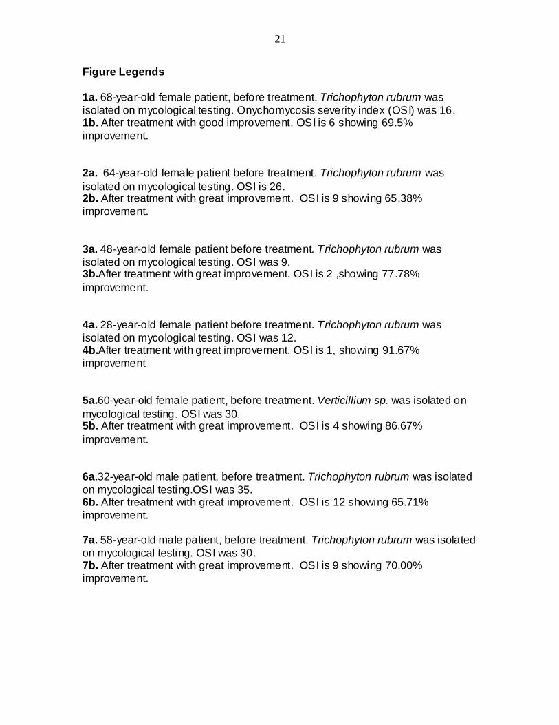
1a. 68-year-old female patient, before treatment. Trichophyton rubrum was
isolated on mycological testing. Onychomycosis severity index (OSI) was 16. 1b. After treatment with good improvement. OSI is 6 showing 69.5%
improvement.
2a. 64-year-old female patient before treatment. Trichophyton rubrum was
isolated on mycological testing. OSI is 26. 2b. After treatment with great improvement. OSI is 9 showing 65.38%
improvement.
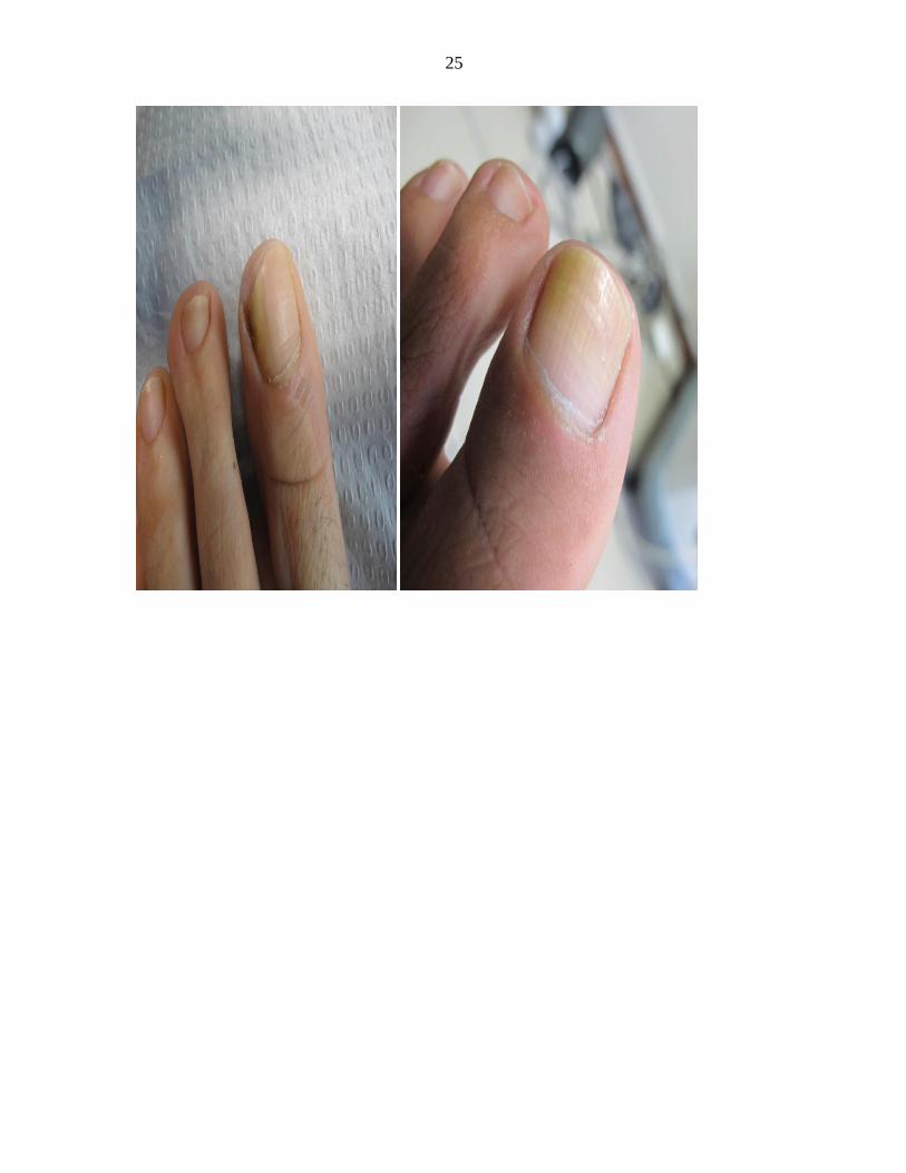
3a. 48-year-old female patient before treatment. Trichophyton rubrum was
isolated on mycological testing. OSI was 9. 3b.After treatment with great improvement. OSI is 2 ,showing 77.78%
improvement.
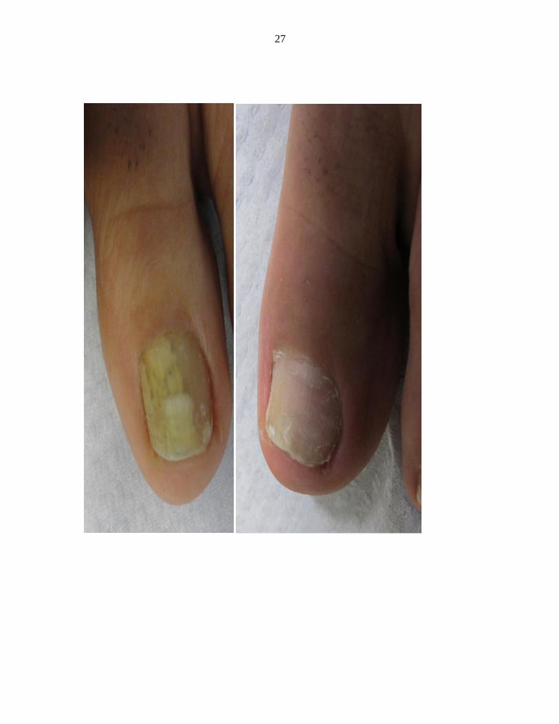
4a. 28-year-old female patient before treatment. Trichophyton rubrum was
isolated on mycological testing. OSI was 12. 4b.After treatment with great improvement. OSI is 1, showing 91.67%
improvement
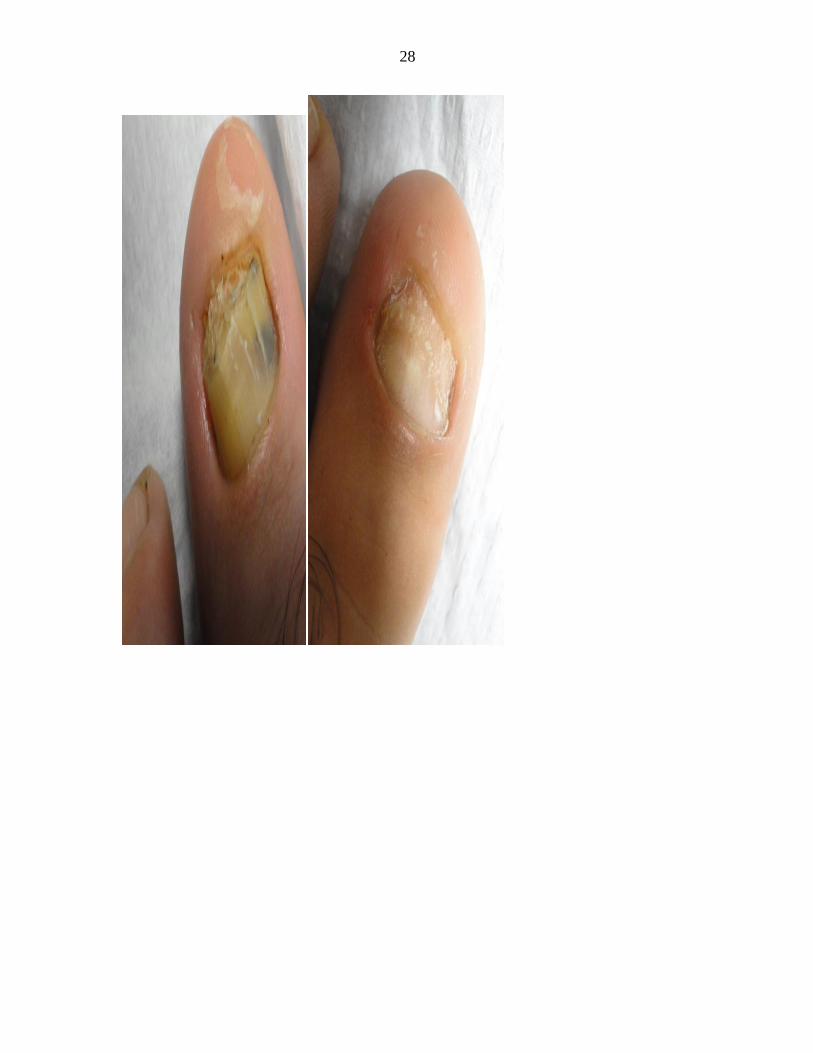
5a.60-year-old female patient, before treatment. Verticillium sp. was isolated on
mycological testing. OSI was 30. 5b. After treatment with great improvement. OSI is 4 showing 86.67%
improvement. 6a.32-year-old male patient, before treatment. Trichophyton rubrum was isolated
on mycological testing.OSI was 35. 6b. After treatment with great improvement. OSI is 12 showing 65.71%
improvement.
7a. 58-year-old male patient, before treatment. Trichophyton rubrum was isolated
on mycological testing. OSI was 30. 7b. After treatment with great improvement. OSI is 9 showing 70.00%
improvement.

22
Figure 1a,b Figure2a,b
Figure3a.b Figure 4a,b
Figure 5a,b Figure6a,b Figure7a,b

23

24
25

26
27
28
29