Embed Size (px)
Citation preview
THE EFFECT OF PRETERM BIRTH ON THE DEVELOPMENT OF THE DENTITION
VIRPIHARILA
Department of Oral Developmentand Orthodontics,
Institute of Dentistry,University of Oulu
OULU 2004

VIRPI HARILA
THE EFFECT OF PRETERM BIRTH ON THE DEVELOPMENT OF THE DENTITION
Academic Dissertation to be presented with the assent ofthe Faculty of Medicine, University of Oulu, for publicdiscussion in Auditorium 1 of the Institute of Dentistry(Aapistie 3), on September 25th, 2004, at 12 noon.
OULUN YLIOPISTO, OULU 2004
Copyright © 2004University of Oulu, 2004
Supervised byProfessor Lassi AlvesaloDoctor Tuomo Heikkinen
Reviewed byDocent Anna-Liisa JärvenpääDocent Marjatta Nyström
ISBN 941-42-7438-5 (nid.)ISBN 951-42-7439-3 (PDF) http://herkules.oulu.fi/isbn9514274393/
ISSN 0355-3221 http://herkules.oulu.fi/issn03553221/
OULU UNIVERSITY PRESSOULU 2004

Harila, Virpi, The effect of preterm birth on the development of the dentition Department of Oral Development and Orthodontics, Institute of Dentistry, University of Oulu,P.O.Box 5281, FIN-90014 University of Oulu, Finland 2004Oulu, Finland
AbstractThe aim of this study was to examine the effect of preterm birth on the development of the dentition.The spesific aims were to examine the effect on deciduous and permanent tooth crown dimensions,the eruption of permanent teeth and the sagittal occlusal relationships within the dentition.
The subjects consisted of 328 prematurely born (< 37 gestational weeks ) white and black childrenand 1804 control children, who participated in the cross-sectional study of the Collaborative PerinatalProject (USA) in the 1960's and 1970's.
Dental examinations, including dental casts were performed at the age of 6–12 years. Tooth crownsize measurements, recording of the sagittal occlusal relationships and tooth eruption stages wereperformed by examining the dental casts.
In general larger permanent tooth crown dimensions were found in preterm white boys and blackgirls and smaller permanent tooth crown dimensions in preterm white girls and black boys. Therewere both increased and decreased deciduous tooth crown dimensions in preterm children comparedto controls, but no significant differences were found. Boys had larger tooth crown sizes than girlswithin all preterm and control groups showing sexual dimorphism. The results showed earliereruption of permanent incisors and first molars in all preterm children compared to controls and alsoaccording to sex and race. Concerning the sagittal occlusal relationships, the results showed greaterprevalence of prenormal canine relationships in preterm group than in the controls. When the molarrelationships were concerned, the prevalence of mesial molar occlusion was greater in the pretermgroup. The incidence of bilateral symmetrical canine relationship was the same in both preterm andcontrol groups, but inside the preterm group the girls had better symmetry than the boys.
The findings of this research suggest that short gestation is not associated with reduced permanentand deciduous tooth crown dimensions in prematurely born children and also confirm the presenceof the sexual dimorphism in tooth crown size. The studies also indicate that the clinical tooth eruptionis accelerated in all observable permanent teeth in prematurely born children. The findings of occlusalmorphology indicated that premature birth may effect the sagittal occlusal development. Generalhealth condition, neonatal and postnatal factors like intubation, postnatal molding of head shape andthe importance of catch-up growth and early functional activity should be considered as possibleinfluencing factors. Preterm birth may also interfere with the development of symmetry andlateralization.
Keywords: dental occlusion, dentition, preterm birth, tooth crown, tooth eruption


Acknowledgements
This work was carried out at the Department of Oral Development and Orthodontics, Institute of Dentistry, University of Oulu.I wish to express my respect for Emeritus Professor Richard H.Osborne, by whom the large and valuable material of the Genetic-Odontometric Study project as a part of the Collaborative Perinatal Study, was collected and organized. I also want to express my sincere gratitude and thanks to the Head of the Department of Oral Development and Orthodontics, Professor Lassi Alvesalo, who was also actively involved in the collection and the analyses of the data, for providing me with his expert quidance and support and enthusiasm, which have been extremely valuable during these years.
My deepest and warmest thanks go to my other supervisor Tuomo Heikkinen, D.D.S., Ph.D., for his encouragement and endless support, and for all his help to achieve this goal. He introduced me to the subject and never lost his belief in me.
I am very grateful to the official referees, Docent Anna-Liisa Järvenpää, M.D. and Docent Marjatta Nyström, D.D.S., PhD, for their valuable comments and careful revisions of the manuscript. Their constructive criticism developed and improved the content of the thesis.
I also wish to thank the other members of the Genetic Odontometric Study team, my co-author Mathias Grön, D.D.S., PhD and the other staff of the project in 1972–1974, Ms Sirkka Alvesalo, Ms Gisela Nass, Ms Helen Bennett, Mr Russel Spry and others.
I am also greatful to Professor Juha Tienari, Ms Päivi Laukkanen, Ms Anna-Kaisa Rimpiläinen, Mr Ari Sarpola and Mr Ahti Niinimaa for their statistical expertice and advise analysing the data and to Malcolm Hicks M.A., for revision of the language of this thesis.
My sincere thanks go to my present and former colleagues and other staff in the Department of Oral Development and Orthodontics and in the Institute of Dentistry for their encouragement and friendship during these years. I also want to thank all my friends for their support during these quite busy years.You have all reminded me of the other important things in life.
I wish to express my loving thanks to my parents, my brother and his family, who have always encouraged me to study hard and supported me whatever I have done.

Finally, I express my most loving thanks to my dear daughter Laura. I am privileged to have a lovely daughter and I dedicate this work to her.
This work was supported by contract NO1-NS-2-2302 from the U.S.National Institute of Neurological Disorders and Stroke, by the Academy of Finland, by the University of Oulu and grants from Finnish Dental Society and Orthodontic Society.
Oulu, August 2004

List of original publications
This thesis is based on the following original publications, which are refered to in the text by Roman numerals I to IV. Some additional data is also presented.
I Harila V, Heikkinen T & Alvesalo L (2003) Deciduous tooth crown size in prematurely born children. Early Human Development 75(1-2):9–20.
II Harila-Kaera V, Heikkinen T, Alvesalo L & Osborne RH (2001) Permanent tooth crown dimensions in prematurely born children. Early Human Development 62:131–147.
III Harila-Kaera V, Heikkinen T & Alvesalo L (2003) The eruption of permanent incisors and first molars in prematurely born children. European Journal of Orthodontics 25:293–299.
IV Harila-Kaera V, Grön M, Heikkinen T & Alvesalo L (2002) Sagittal occlusal relationships and asymmetry in prematurely born children. European Journal of Orthodontics 24:615–625.

Contents
Abstract Acknowledgements List of original publications Contents 1 Introduction ................................................................................................................... 11 2 Review of the literature .................................................................................................13
2.1 Tooth crown size.....................................................................................................13 2.2 Tooth eruption ........................................................................................................15 2.3 Occlusal relationships and asymmetry ...................................................................18 2.4 The prevalence of dental defects in prematurely born children..............................20
3 Aim of the study ............................................................................................................22 4 Subjects and methods ....................................................................................................23
4.1 Tooth crown measurements ....................................................................................24 4.2 Permanent tooth eruption .......................................................................................25 4.3 Measurements of occlusal variables .......................................................................26
5 Results ...........................................................................................................................27 5.1 Deciduous tooth crown size (Study I) ....................................................................27 5.2 Permanent tooth crown size (Study II) ...................................................................28 5.3 Eruption of the permanent incisors and first molars (Study III).............................30 5.4 Sagittal occlusal relationships and asymmetry (Study IV) .....................................31
6 Discussion .....................................................................................................................35 6.1 General ...................................................................................................................35 6.2 Development of the dentition .................................................................................35
6.2.1 Tooth crown size..............................................................................................35 6.2.2 Tooth eruption .................................................................................................36 6.2.3 Occlusion.........................................................................................................37 6.2.4 Asymmetry ......................................................................................................38
6.3 Growth....................................................................................................................39 6.4 View for the future..................................................................................................39
7 Conclusions ...................................................................................................................41 References Appendix Original publications

1 Introduction
Prematurely born infants have a short prenatal development period and are predisposed to various neonatal complications and developmental problems. According to the World Health Organization definition, a delivery is preterm when it occurs before the 37th completed week of pregnancy. Previously the limit for prematurity had been defined by birth weight under 2500 g (Ylppö 1919), but it was reported by Lubchenco et al. (1963) that birthweight is also determined by the foetal growth rate in addition to gestational age and this led to the new definition.
Prematurity naturally includes a wide variation of gestational ages and birthweights (Cartlidge et al. 1997). In the 1940’s and 1950’s, very few low birth weight (VLBW, < 1500g) infants survived and almost half of the survivors had abnormal developmental outcomes (Lee et al. 1980). With the introduction of intensive medical care in the 1960’s the increased use of antenatal corticosteroids and the recent routine administration of surfactant replacement therapy, survival of prematurely born infants has improved markedly (Cartlidge et al. 1997). The mortality rate has fallen with time, and smaller and younger neonates are surviving. Thus mortality among VLBW infants had fallen from 50 % in the 1960’s to 15–20% by the early 1990’s (Lee et al. 1995, Tommiska et al. 2001). The immaturity of many organs nevertheless makes a prematurely born infant vulnerable to developmental problems. Many aetiological factors for premature birth exist, many of which are associated with maternal and foetal diseases, but often the cause remains obscure. Despite modern perinatal technology and knowledge, morbidity rates are still high (Cartlidge et al. 1997) and the rates of neurodevelopmental disability have in general remained unchanged (Hack & Fanaroff 2000).
There are several systemic derangements that can interfere with the developing teeth, especially teeth that are at a critical stage of development at the time of the insult and are not calcified or dimensionally complete at the time of birth.Variations in tooth crown size can be compounded by genetic and environmental factors, including maternal influence, and there is a considerable range of dental variability within and between populations. Permanent incisors and first molars are at the premineralization and pre-eruptive stages, and enter the first phases of eruption before or soon after birth, when systemic and environmental factors are likely to influence tooth development. Adverse perinatal

12
factors, premature birth and exceptional early adaptation to extra-uterine life and functional activity may influence dental occlusal development and symmetry in the jaws.
According to earlier studies it is known that preterm birth may lead to defective development of the dentition. The goal of the present study is to elucidate further the effect of preterm birth on the development of the dentition.

2 Review of the literature
2.1 Tooth crown size
The development of the dentition begins at the age of 4 weeks in utero and continues for about 20 years postnatally. The mitotic activity of the cells of the inner enamel epithelium and the deposition of enamel are factors that increase the overall crown dimensions. The sequence of initial calcification of the deciduous dentition can be established as beginning with the central incisors and then proceeding to the first molars, lateral incisors, canines and second molars with the mandibular teeth usually preceding their maxillary antagonists in the case of the incisors and first molars. Initial calcification of the central deciduous incisors begins at the 12th to16th gestational week, and the last deciduous tooth to develop, the second primary molar, starts its calcification from the first cusp around the 18th or the 20th gestational week. The intercuspal distances do not increase after coalescence between the cusps, which is completed by 36 gestational weeks (Kraus 1959). Not all deciduous teeth calcify at the same rate in either the mesiodistal or vertical dimension, and the maxillary central incisors appear to calcify at a faster rate than the other deciduous teeth in both dimensions (Kraus 1959). Cell proliferations increase the crown size during this developmental process and as calcification continues, the increase in the mesiodistal dimension of the tooth crown diminishes and enamel formation finally determines the contour of the crown. After the 19th week of gestation, for example, there is a decrease in the rate of mesiodistal growth in the maxillary second deciduous molar. At the time of birth (40 gestational weeks), mineralization of the crowns of the deciduous incisors is almost complete and mineralization of the other deciduous crowns has already started. All the crowns of deciduous incisors and canines are complete by the age of six months after birth and the rest all the mandibular deciduous molars, by the age of one year.
Calcification of the first permanent molar crown begins at the 28th–30th gestational week, and dimensions are completed by the age of three to four years postnatally. The average age of crown completion is 2.7 years for black males, 3.0 years for black females, 3.4 for white males and 3.5 years for white females (Harris & McKee 1990). Fusion of the cusps of the first permanent molar occurs at the age of 6 months postnatally

14
and the intercuspal distances remain unchanged after this (Kraus & Jordan 1965). Completion of the rest of the permanent tooth crowns, excluding the third molars, has been shown to occur by 7 or 8 years of age. Initiation of all permanent teeth begins in utero, but their completion in terms of size extends over a wide period of time and is highly variable.
The variation in tooth crown size is compounded by genetic and environmental conditions, including maternal influence. There are differences in the development of the dentition between the sexes and between ethnic groups, girls preceding boys in reaching the stages of crown mineralization and sexual dimorphism being observed in the size and shape of tooth crowns. Although no significant trend for sexual dimorphism in the mesiodistal and buccolingual dimensions of the deciduous teeth has been observed (Townsend & Garcia-Godoy 1984, Fearne & Brook 1993, Seow &Wan 2000), analysis of the Burlington data revealed significant sexual dimorphism in the 40 deciduous tooth diameters, being even greater than that seen in the permanent dentition of several sample populations. All means of tooth diameters are significantly larger in males than in females, although no figures for the actual tooth crown sizes have been published (DeVito & Saunders 1990). Boys generally have larger teeth than girls, and although the genes affecting tooth size are also situated on both X and Y-chromosomes the influence of the Y-chromosome on phenotypic quantity is different from that of the X-chromosome Alvesalo 1971, 1997). The X-chromosome increases enamel thickness, while the Y-chromosome promotes amelogenesis and dentinogenesis. A major sex difference has been found in the shape of the tooth and cusp, boys having more nearly square dimensions and girls showing greater size reduction buccolingually than mesiodistally (Garn et al. 1966, Mayhall & Alvesalo 1992, Mayhall & Alvesalo 1995). The timing of the initial calcification may be similar in both sexes, but males may have a longer duration of growth and/or a greater rate of growth in their developing teeth than females (Alvesalo 1971, Townsend 1985). In terms of racial differences blacks are found to achieve the mineralization stages significantly earlier than whites (Harris & McKee 1990), and it is also generally assumed that the size, cusp number and growth rate of the first permanent molars will differ between populations (Kraus & Jordan 1965).
Some studies have indicated that genetic factors account for about 60% of the variation in tooth size observed in both the deciduous and permanent dentition (Alvesalo & Tigerstedt 1974, Townsend 1992). The extent of the total genetic variation depends on the particular tooth and its dimension, certain points in the dentition being more variable than others (Moorrees et al. 1957). The canines exhibit relatively high stability from the developmental and evolutionary point of view, and the early-forming teeth are in general under stronger genetic control than later-forming ones. High genetic variability in the mesiodistal dimension of the permanent anterior teeth has been confirmed in previous studies (Osborne et al. 1958). According to the ”field theory” of the human dentition (Dahlberg 1945), the more mesial tooth within a group is subjected to a stronger influence from the same morphogenetic fields than the more distally located ones, with the exception that the lower lateral incisors show higher heritabilities than the central incisors (Alvesalo & Tigerstedt 1974).
Tooth crown dimensions may be affected by maternal and developmental factors, including those teeth neither calcified nor dimensionally completed at birth, and may reflect general disturbances in individual development in both the prenatal and postnatal

15
period. Maternal factors during pregnancy, including hypothyroidism, diabetes, hypertension, smoking etc. can also effect the size of the foetus and infant and the dimensions of the tooth crowns, although the effect has been found to be smaller in girls than in boys (Garn et al. 1979). Maternal and foetal or gestational determinants of both the deciduous and permanent tooth crown dimensions may account for half of the variability in crown size (Garn et al. 1979). Larger tooth crown size is thought to be associated with prolonged gestation, large size at birth and high birthweight and length, and also with maternal hypothyroidism or diabetes, while it has been suggested that smaller teeth are associated with small size at birth, short gestation, low birthweight, low birth length and maternal hypertension (Garn et al. 1979, Garn et al. 1980), and that the trend for smaller odontometric dimensions may derive from the improved survival of smaller and developmentally immature neonates. In the study of Fearne & Brook (1993), low birthweight children had smaller deciduous tooth crown size in all but the first deciduous molar and the correlation between the length of gestation and tooth size was positive but not significant. No association was found between the serious of illness during neonatal period (duration of ventilator support, drugs for apnoea or i.v.alimentation) and deciduous tooth size. Seow et al. (2000) found that the mesiodistal and buccolingual dimensions of the maxillary deciduous central and lateral incisors and the mandibular deciduous central incisors were from 6 to 11% smaller in low birthweight children. Maternal smoking during pregnancy reduces some dimensions of the tooth crowns in both the deciduous and permanent dentition and it can also lead to thinning of some permanent incisors (Heikkinen 1996).
2.2 Tooth eruption
Various theories have been put forward to explain the tooth eruption process, which is still largely unknown. Root growth, dentine formation, proliferation of the dental pulp, the periodontal ligament, the connection between the enamel organ and the oral epithelium and the role of the dental follicle, including its innervation and blood supply, are considered to be essential (Magnusson 1968, Cahill & Marks 1980, Sutton & Graze 1985). The dental follicle is required for tooth eruption and its role may be in part one of local control over alveolar bone formation and resorption in alveolar tooth eruption. The presence of the dental follicle is necessary for the tooth to erupt, as shown by Cahill & Marks (1980) and Marks & Cahill (1984). It has also been proposed that a molecular signal initiates the cellular events of eruption, culminating in alveolar bone resorption to form the eruption pathway. The possible molecular signals initiating and regulating tooth eruption at the cellular level include, for example, epidermal growth factor (Cohen 1962), transforming growth factor –alpha (Tam 1985) and colony-stimulating factor-1 (CSF-1) (Iizuka et al. 1992), which stimulate and accelerate eruption. Cellular events in the follicle just before emergence or at its onset include an influx of monocytes and an increase in the number of osteoclasts in the bony crypt to achieve localized bone resorption (Wise et al. 1985, 1994; Wise 1998). Recent studies in mice have also indicated that parathyroid hormone related protein (PTHrP) is required for tooth eruption to exert a paracrine effect on dental cells of the adjacent dental follicle to create a local
16
environment inductive for osteoclast formation and to initiate eruption (Wise et al. 2000). The hypothesis behind the blood vessel thrust theory emphasizes the blood flow through the vessels of the dental pulp, while the tissues surrounding the tooth produce hydrodynamic and hydrostatic forces towards the tooth within the blood vessels, which may partly cause movement of the tooth through the alveolar bone (Sutton & Graze 1985).
Both genetic and environmental factors acting during odontogenesis are associated with the tooth eruption process. The environment, prenatal and maternal factors, social factors, climate etc. may influence the timing of permanent tooth eruption (Nanda 1960, Friedlander & Bailit 1969, Rantakallio & Mäkinen 1983, Heikkinen et al. 1995), but the determinants of this timing are still thought to be more genetic than environmental (Hatton 1950), although the eruption of teeth includes local controlling factors such as crowding and premature or delayed loss of deciduous teeth. The chronology of the eruption of the deciduous teeth is thought to be more extensively genetically determined than in the case of the permanent teeth (Hatton 1950, Backström et al. 2000). There are differences between the sexes and between ethnic groups in the development of the permanent dentition and in the order of tooth mineralization (Haavikko 1970, Alvesalo 1971, Harris & McKee 1990, Heikkinen 1996) and such differences are thought to be greater in the permanent than in the deciduous dentition. Girls are found to precede boys in tooth mineralization (Harris & McKee 1990) and in root development and tooth eruption (Haavikko1970), while blacks are found to achieve the mineralization stages significantly earlier than whites (Harris & McKee 1990).
In addition to the genetic point in tooth eruption, prenatal and postnatal environmental factors may also affect the eruption process. It has been suggested that an early critical stage in tooth development may exist after which most systemic environmental influences cease to have any effect (Friedlander & Bailit 1969), and this critical time may occur prenatally (Main 1966, Glasstone 1967). The permanent canines and first premolars, which develop later, may reach the susceptible stage during the first postnatal years, while in the second premolars and molars it will be 2 or 3 years later (Heikkinen et al. 1995). It has been suggested that there may be stimulating pressures in mastication that favour the tooth eruption process (Friedlander & Bailit 1969). There are differences in the timing of tooth eruption between the jaws, the mandibular teeth usually erupting earlier than the corresponding maxillary ones (Nanda 1960). Environmental factors such as smoking during pregnancy have been found to accelerate both deciduous and permanent tooth eruption (Rantakallio & Mäkinen 1983, Heikkinen 1996), and an association between birthweight and advanced tooth eruption has been suggested by Bailit et al. (1968) and Bailit & Sung (1968), children who are heavier at birth experiencing to have permanent tooth eruption significantly earlier. Studies on the role of nutrition and vitamin supplementation in the eruption process suggest that early vitamin D intake, for example, has no effect on the maturation of the deciduous dentition in preterm children, although children receiving a higher vitamin D dose in the neonatal period (1000IU/day compared to 500IU/day) have a more mature permanent dentition. Mineral intake, calcium and phosphorus supplemented breast milk, didn’t affect maturation of permanent teeth. Also bone mineral status at 3 months of chronological age was not associated with the maturation of deciduous dentition. (Backström et al. 2000).

17
Natal and neonatal teeth, which are teeth observed at birth or during the first 30 days of life are rare, and are suggested to be associated with many factors like superficial position of the tooth germ, some syndromes, malnutrition, hypovitaminosis, hormonal stimulation accelerating the eruption (Cunha et al. 2001). Populations living in tropical climates are found to be dentally more advanced, and climate may possibly affect dental maturation (Friedlander & Bailit 1969). Tooth emergence has also been assumed to be affected by socio-economic factors, advanced eruption being found among children with superior socio-economic circumstances (Lee et al. 1965). A high correlation has been found between general growth and the eruption of the deciduous dentition (Wedgewood & Holt 1968, Infante & Owen 1973, Fadavi et al. 1992). Dental mineralization has been found to be more related to height and weight than to skeletal mineralization in both sexes, but the stages of tooth development are more related to height than weight in males (Anderson et al. 1975).
The timing of deciduous and permanent tooth eruption has sometimes been found to be delayed in prematurely born children (Wedgwood &Holt 1968, Fadavi et al. 1992, Drummond et al. 1992, Viscardi et al. 1994), and it has also been reported that the maturation of both dentitions does not differ appreciably between preterm and full-term children (Backström et al. 2000). Factors related to delayed tooth eruption are thought to include the general delay in the growth development of preterm babies (Drummond et al. 1992), short gestation (Golden et al. 1981, Seow et al. 1988, Fadavi et al. 1992), low birthweight (Trupkin 1974) and neonatal factors, including complications of prematurity, systemic disorders, duration of oral intubation, average weight gain / day etc.(Viscardi et al. 1994). Nutritional factors, postnatal weight gain and growth of the child may also effect the eruption of the deciduous teeth (Infante & Owen 1973, Delgado et al. 1975). Seow (1996) has reported results showing a delay in the maturity of the permanent dentition as determined from panoramic radiographs among VLBW children compared with normal birth weight (NBW) children. In the study of Harris et al. (1993) on LBW black African-American children, the delay in dental development was limited to the permanent first molars and central incisors, while those with the poorest height-for-age had the greatest delay in tooth formation, but only those teeth undergoing rapid differentiation neonatally were affected systematically.
Preterm infants may experience a period of catch-up growth, even up to 8 years of age among very low birthweight children (Hack et al. 1996). Seow (1996) reported the effect of catch-up growth in the study, where preterm low birthweight infants showed a delay of approximately 3 months in dental maturation under 6 years of age, but no significant differences where found over 9 years of age. The chronological teething age may be delayed in prematurely born children, but when corrected ages are used, tooth eruption is not delayed (Falkner 1957, Golden et al. 1981, Seow et al. 1988, Fadavi et al. 1992). Individual differences may exist, however, and gestational age should be taken into account when estimating the eruption of the dentition in premature infants.

18
2.3 Occlusal relationships and asymmetry
Both genetics and environment influence the development of the occlusion. Various environmental factors, including disturbances in general health and growth in childhood, masticatory muscle activity, dietary factors (Varrela 1990), mouth breathing, oral habits, the mother’s and child’s nutrition and health condition and other perinatal factors, may influence the dentition during the occlusal development period and the growth of the jaws. Individual occlusal relationships have been reported as indicating a dominance of environment over genetic factors, while some combinations of occlusal traits show noticeable genetic influence (Harris & Smith 1982). The genetic contribution to occlusal variation has been thought to be quite low, at least for the molar relationship (Harris & Smith 1980). Generally low heritability estimates have similarly been found for overbite, overjet and the sagittal molar relationship (30%) in the studies of Townsend et al. (1988), emphasizing the importance of environmental influences on occlusal variation, although a change in the sexchromosome balance seems to lead to a typical occlusal morphology (Laine et al. 1986, Alvesalo & Laine 1992, Laine et al. 1992). It has also been suggested, that hereditary factors determine the development of the jaws and the occlusion, and that environmental factors have only a modifying effect (Myllärniemi 1972).
Variations exist between individuals in the onset and direction of changes and in the total increments in arch length, breadth and circumference (Moorrees 1959). The changes in maxillary and mandibular arch length are not continuous during the development of the dentition but occur in the form of growth periods, mainly from 4 to 6 and 10 to 14 years of age. Certain patterns of mean changes in the maxillary and mandibular arch length and breadth are generally associated with eruption of the permanent incisors, canines and premolars, when the changes are greatest (Moorrees 1959). The mandible is composed of different morphogenetic and functional units, among which the condyle is considered to be a growth zone that is affected by functional alterations. The influence of the functioning of the masticatory muscles has been demonstrated in animal studies, which have shown that increased masticatory function may lead to increased sutural growth and bone apposition and reduced muscle function to a decrease in bone mineral mass (Kiliaridis 1995). The mandible is a paired bone throughout foetal life, and small, irregular bones fuse with the mandibular body at the end of the first year, the two halves of the mandibula being united by ossification of the symphyseal fibrocartilage (Bhaskar 1980, Nyström & Ranta 2003). A remodelling process then takes place during postnatal bone growth to maintain a form appropriate to their biomechanical function.
Imbalances in the mechanical forces acting on malleable tissues may result in deformations, and gravitational and positioning forces may lead to deformations and deviation of the cranial and facial bones in immature, prematurely born infants. Postnatal moulding of the soft skull, with side to side flattening of the head shape, is commonly seen in preterm infants, especially during the first few months (Baum & Searls 1971, Marsden 1980, Elliman et al. 1986).This results from the quite long immobile neonatal period, when preterm babies lie with their heads turned on one side due to a relatively large head mass and poor neck muscle tone and prolonged pressure is applied to a small area of the soft skull. The altered head shape of the skull does not appear to be related to developmental delay nor to abnormal cranial ultrasound results (Elliman et al. 1986).

19
Head flattening may resolve with time (Rutter et al. 1993), but adults who were born prematurely have been found to show a persistence of the skull deformation acquired neonatally, with elongated and narrower skulls than normally (Baum & Searls 1971, Elliman et al. 1986). It has been suggested that the way the infant is nursed,e.g. the use of water pillows, may reduce skull deformation in preterm infants (Marsden 1980, Schwirian et al. 1986). Nowadays better nutrition and supplement therapy in preterm infants has resolved this problem quite well.
According to the limited number of reports available on occlusal relationships in preterm infants, very low birth weight infants do not differ from matched controls with respect to the deciduous dentition, both groups having a majority of normal occlusal relationships and equal numbers of palatal cross-bites. It has been suggested by Seow (1997) in an uncontrolled study that a high prevalence of Class II malocclusions and a high, narrowed palate may be found in intubated low birth weight preterm children. In the early stages of the development of the oral cavity, the soft bones of the palate are malleable and pressure from any object can easily mould its shape (Palmer 1998). A significant increase in the growth of the craniofacial and palatal widths and the palatal area has been observed in very low birthweight infants wearing specially designed pressure-dispersing pads during hospitalization compared with those not wearing such pads (Morris & Burns 1994). Preterm infants often require neonatal oral intubation for resuscitation, and prolonged intubation has been associated with narrowing and deepening of the palate and with palatal asymmetry (Ash & Moss 1987, Macey-Dare et al. 1999). Also, gravitational forces may cause deviation of the cranial, facial and palatal bones in preterm infants. Pressures from laryngoscopy and continual trauma from orotracheal tubes may alter the palatal configuration, and palatal grooves and deeper palates have been observed in intubated children (Erenberg & Novak 1984, Fadavi et al. 1992). Kraus & Jordan (1965) reported the development of an indentation on the anterior ridge in children receiving mechanical ventilation, but the effects of endotracheal intubation on the palatal and dental arch configuration may not be persistent, due to growth changes and the palatal remodelling procedure (Seow et al. 1985). A narrow, high-vaulted palate may also be simply an oral manifestation of the narrow, elongated head shape (Ash & Moss 1987).
Oral habits such as thumb sucking, mouth breathing or tongue thrusting, and also oromuscular forces affecting the developing dentition in formative periods, are important as aetiological factors for malocclusions (Nanda et al. 1972, Larsson 1994). Large tonsils can reduce the space in the mouth and cause the tongue to be low and pushed forward, pressing against the teeth of the lower jaw (Ricketts 1968, Myllärniemi 1972). Children with a mouth breathing habit have been shown to have a greater prevalence of Class III canine and molar relationships and greater overjet than others, while tongue thrust is an infantile habit that is apt to lead to an increased prevalence of a Class III canine and molar relationship and open bite (Nanda et al. 1972). An association between postural disorders, head posture and dental occlusion has also been reported (Huggare 1998, Solow & Sonnesen 1998). Impaired nose breathing triggers an increase in craniocervical angulation (Solow & Sierbaek-Nielsen 1984) and sagittal dentofacial development has been found to be restrained by an increased craniocervical angle and released by a reduction in this angle (Solow & Sonnesen 1998).

20
Perinatal stress during development has been reported to increase the magnitude of fluctuating asymmetry in the dentition, long bones and membranous bones (Siegel & Mooney 1987). According to some studies, preterm delivery and adverse perinatal factors may interrupt the normal development of brain asymmetry and lead to an atypical direction of lateralization (Powls et al. 1996). Prematurity has been suggested as influencing expression of the right-shift factor and leading to a reduced left hemisphere advantage (Annett 1985). Procedures undertaken during delivery and postnatally that cause the moulding of head shape may also influence the development of symmetry in the jaws and the occlusion (Pirttiniemi et al. 1994). Also a sex difference in asymmetry has been reported, with girls generally having greater symmetry than boys, e.g. in permanent tooth eruption (Heikkinen et al. 1999). General directional asymmetry and left-side dominance in the mandible has also been reported by Huggare and Houghton (1995). Males seem to be more sensitive and more likely to become left-handed than females (Coren & Porac 1980). There are also studies, however, in which no associations with handedness or specific perinatal difficulties such as foetal distress, long labour, premature delivery and intrauterine growth retardation have been found (Smart et al. 1980). An increased prevalence of non-right-handedness has been found among very low and extremely low birth weight children (Ross et al. 1987, Marlow et al. 1989, Powls et al. 1996), as also among mentally handicapped individuals (Batheja & McManus 1985).
2.4 The prevalence of dental defects in prematurely born children
Studies of preterm children have shown that the developing dentition may be affected at time of an exceptionally early delivery. The prevalence of enamel defects in the deciduous (Grahnen & Larsson 1958; Funakoshi et al. 1981; Mellander et al. 1982; Pimlott et al. 1985, Seow 1986, 1996, 1997, Drummond et al. 1992, Lai et al. 1997) and permanent dentition (Nikiforuk & Fraser 1979, Seow 1997, Aine et al. 2000) has been reported to be higher in preterm infants and may vary between studies. The earlier studies were mostly carried out on higher birthweight and more mature infants than recent ones, when the advances in medical care has made possibilities for studying lower birthweights and very immature neonates.
Dental defects include various enamel hypoplasias, deficiency of enamel in the form of pits, grooves or other quantitative surface loss and enamel hypocalcifications and opacities, i.e. qualitative changes in the translucency of the enamel without loss of the enamel surface in the form of stained or white opaque flecks. In the deciduous dentition enamel defects are found from 21% of cases (enamel hypoplasias)(Grahnen & Larsson 1958) to as much as 96% of cases (enamel hypoplasias and opacities combined)(Lai et al. 1997). It has been hypothesized that metabolic derangements in the postnatal period affect the mineralization of the first developing permanent teeth, which has already commenced. In the study of Aine et al. (2000) the prevalence of enamel defects (hypoplasias and opacities combined) in preterm children was 78% in the deciduous dentition, 83% in the permanent dentition. Neither the mineral supplementation nor a vitamin D dose of 1000IU/day as compared with 500IU/day used in preterm infants, reduced the prevalence of enamel defects in the deciduous or permanent dentitions.

21
The possible pathogenesis of the dental defects associated with preterm birth may be related to direct damage to the ameloblasts as in maternal infections (rubella, cytomegalovirus). Complications of pregnancy that reduce maternal serum calcium concentrations like maternal toxaemia and diabetes, hyperparathyroidism, maternal calcium and vitamin D deficiencies, are often associated with preterm birth. Similarly, neonatal hypocalcaemia is often associated with traumatic delivery, Ceasarean section, birth asphyxia and cerebral injuries (Seow 1986). Various complications associated with prematurity predispose these infants to severe metabolic derangements and hypocalcaemia, and these conditions may result in disturbed enamel formation. The problem of deranged calcium metabolism occurs to varying degrees in most premature infants, because two-thirds of the individual’s stores of calcium and phosphorus accumulate during the last trimester of pregnancy and preterm infants miss much of this mineral accretion (Tsang et al. 1976).
Systemic insults to the developing dentition may occur prenatally, neonatally or postnatally. Enamel defects related to systemic factors are usually symmetrical and involve those teeth undergoing development and enamel formation at that time, but local tissue variability in development must be taken into consideration. Hypomineralization of the teeth has been thought to be associated with systemic derangements of preterm birth such as nutritional problems, hypocalcaemia (Stimmler et al. 1973, Nikiforuk & Fraser 1979, Noren 1983, Seow et al. 1984), hypophosphataemic rickets (Seow et al. 1984) neonatal infections and sepsis (Funakoshi et al. 1981), metabolic disorders, neonatal asphyxia (Grahnen et al. 1974, Fearne et al. 1990), respiratory distress syndrome (RDS)(Mellander et al. 1982, Johnsen et al. 1984), and hyperbilirubinaemia (Grahnen & Granath 1962). Mellander et al. (1982) concluded that mineralization defects in the deciduous dentition in low birth weight infants seem to be dependent on the amount of breast milk given during the neonatal period, and also found a seasonal variation, in that the highest prevalence of defects occured in children born during the winter months.
Dental defects arising from local causes tend to affect only single teeth or groups of teeth. Local traumatic factors associated with endotracheal intubation and laryngoscopy during a critical period of tooth amelogenesis may also contribute to dental defects in the deciduous dentition (Seow 1997, Moylan et al. 1980, Fadavi et al. 1992, Noren et al. 1993) and compound the high predisposition of preterm infants to developmental enamel defects in as many as 85% of cases (Seow et al. 1984). The distribution of the defects within the dentition is usually localized to the anterior region of the maxillary anterior teeth, and it has been suggested that this is better explained by the laryngoscopy procedure than by orotracheal intubation (Seow et al. 1984). Cases have been reported in which concavity in the alveolar ridge outlining the orotracheal tube and severe disruption of the developing enamel organ have been found (Boice et al. 1976). A close correlation exists between birth weight, dental defects and the intubation period, the prevalence of dental defects generally increasing with a longer period of orotracheal intubation (Fadavi et al. 1992).

3 Aim of the study
The purpose of the present work was to examine the influence of preterm birth on the development of the dentition. The specific aims were to investigate:
− deciduous and permanent tooth crown dimensions − the eruption of the permanent teeth − the sagittal occlusal relationship in prematurely born children.

4 Subjects and methods
The subjects consisted of 328 preterm and 1804 control children, giving a total of 2132. There were 60 white children, 40 boys and 20 girls, and 268 black children, 140 boys and 128 girls in the preterm group. The control group included 803 white children, 408 boys and 395 girls, and 1001 black children, 477 boys and 524 girls. The limit for prematurity was placed at 36 gestational weeks in whites and 35 gestational weeks in blacks, in order to maintain practical proportions of preterm / control children in the statistical comparisons. The average length of gestation was approximately 9 days shorter for blacks than for whites (Hardy et al. 1979). The mean gestational age was 33.7 weeks for the white preterm boys and 40.4 weeks for the controls, with corresponding figures of 34.6 weeks and 40.6 weeks for the white girls, 31.7 and 39.8 weeks for the black boys and 32.2 and 39.9 weeks for the black girls (Table 1).
Table 1. Mean gestational ages (sd) and numbers of subjects of premature and control children by sex and race.
Preterm gest.wk (sd)
Controls gest.wk (sd)
White boys 33.7 (3.4) 40.4 (1.9) N 40 408 White girls 34.6 (1.7) 40.6 (1.9) N 20 395 Black boys 31.7 (3.0) 39.8 (2.5) N 140 477 Black girls 32.2 (3.0) 39.9 (2.4) N 128 528 The children were among the 60 000 participants in the Collaborative Perinatal Study conducted by the National Institute of Neurological Disorders and Stroke (NINDS) in the 1960’s. The dental examinations were performed in six collaborating medical centres (Buffalo, NY; Richmond, VA; Portland, OR; Philadelphia, PA; Providence, RI and Johns Hopkins, MD) in the 1972–1974 under the direction of Professor Richard H.Osborne. Prospective medical background data were obtained from the first registration of the

24
pregnancy up to the seventh year of age, including anamnestic information on the mother’s health and background (Hardy et al. 1979). Postnatal longitudinal growth data up to the seventh year of age were also available, including about 1500 variables. The duration of gestation was taken to be the time elapsing between the first day of the last menstrual period (LMP) reported by the woman and delivery, computed in days and then transposed to weeks and rounded to the nearest week. The date of LMP was ascertained by a specially trained interviewer. Information pertaining to the LMP was also collected independently by the hospital staff and by the obstetrician, who was required to provide an estimate of the duration of pregnancy based on physical findings and anamnestic information. This estimate was confirmed at each prenatal visit. The gestational age data obtained in the Collaborative Perinatal Study were found to be valid and reasonably accurate, and to be in general agreement with the medical diagnosis despite possible errors in dating and obtaining the information (Cushner & Mellits,1971). Data concerning the delivery, including birthweight, birth length, head circumference, placental weight, etc., were obtained within 1 hour of delivery by the observer of labour and delivery, using calibrated scales. The determinations of relevant variables were repeated at the ages of 4, 8 and 12 months and then at 3, 4 and 7 years of age.
4.1 Tooth crown measurements
The dental examinations were carried out cross-sectionally, with dental casts made and photographs taken in a standardized fashion at ages varying from 6 to 12 years in 95% of the subjects. Alginate impressions were taken at each co-operating centre and plaster casts were made as soon as practicable (Hunter & Priest, 1960). All the casts were checked and trimmed at the University of Wisconsin. Tooth crown size measurements were performed on the dental casts with an electronic measuring device which employed a Helios needle point automated caliper with a 0.05 mm readout by two experienced, calibrated observers. The measurement procedure was supervised by Professor Lassi Alvesalo. Blind replicate measurements of every 15th case and cross-determinations by different observers showed 0.05–0.15mm differences depending on the tooth and dimension concerned. The intra-examiner error (Sm) was determined from the formula
2
2m
dS
n= ∑
where d is the difference between the two measurements and n is the number of double determinations.
The inter-examiner error (Si) was determined from the formula
21 ( )1i jS Z z
n= −
− ∑

25
where Zj is the difference between two determinations made by different observers, and z the mean of the difference.
In the permanent dentition both maxillary and mandibular first permanent molars were measured mesiodistally (MD) parallel to the occlusal and labial surfaces, and labiolingually (LL) in a plane perpendicular to the above, and intercuspal distances between the mesiobuccal and distobuccal cusps (1 CD) and between the mesiobuccal and mesiopalatinal / mesiolingual cusps (2 CD) were measured from the tips of the cusps, all as maximal dimensions. The MD dimension was also measured for the incisors. The canines, premolars and second molars were left out of the comparisons because of reduced numbers of teeth on account of the ages of the subjects. In the deciduous dentition both maxillary and mandibular teeth were measured on both sides of the jaws. Deciduous cuspids and molars were measured mesiodistally and second deciduous molars labiolingually as well. Labiolingual dimensions of deciduous canines and first molars were not measured because of technical difficulties encountered in determining the exact sites of the measurements, and incisors were not measured because of considerable attrition and loss of teeth in these age groups. Both the permanent and deciduous tooth crown dimensions were compared between the premature and control groups, each divided by race and sex. The groups were not matched by age because of the early and final determination of the tooth crown dimensions. Teeth with attrition, decay or filling etc. at the measurement points were not measured.
The statistical method used was Mann-Whitney’s U-test (Willcoxon Rank-Sums test).
4.2 Permanent tooth eruption
The eruption stages were: tooth not erupted, occlusal surface recently emerged, tooth crown half erupted, and eruption of tooth complete or nearly complete. The tooth antimeres for each child were compared in terms of a four-grade eruption scale, each tooth separately. Defective teeth that could not be readily assigned to an eruption stage were excluded from the analysis. The determinations were made by Professor Lassi Alvesalo and the reliability of staging was determined by performing duplicate blind determinations in 200 cases. Inter-observer reproducibility of the stages was moderately good, with an average of 85%. When pairing the dental casts, the total data were used in order to achieve the maximum number of antimeric pairs. Teeth in the category of not erupted can occasionally be unilaterally missing in varying proportions. It was not possible to ascertain hypodontia by means of radiographs in these series (Heikkinen et al. 2001).
The age of the child was taken as that at which the dental impression had been obtained, and the chronological age was then transposed to the conceptional age in both the preterm and control groups by reference to the information on the gestational age. Pairs were matched for sex, race and age + / - two months of the conceptional age, and the preterm child was compared with the control child in terms of the four-grade eruption scale for each of the eight incisors and the four first molars, considering each tooth separately. Proportions expressing the binary distribution of antimeric tooth pairs into

26
advanced eruption or equal eruption, were calculated for each tooth. The hypothesis of binomial frequencies for discordant pairs was tested by the method of McNemar (1947).
4.3 Measurements of occlusal variables
The molar and canine sagittal relationships were recorded by examining and measuring the hard stone casts. The mean chronological age at which the dental casts were taken was 8.8 years (sd 1.7) in the preterm group and 8.5 (sd 1.8) years in the controls, and the conceptional ages of the groups were also quite similar. The occlusal variables were compared between the preterm and control groups according to sex and race, the canine relationship being recorded in the sagittal plane with the hard stone casts in a centric occlusion looking along the tangent to the upper incisors on both sides of the face. The points at which the measurements were taken in the primary, mixed and permanent dentitions were the mesial prominences of the canines, the approximal points of the lower canines, and the lower second incisors in the mandible (Grön & Alvesalo 1997). The canine relationship was classified as prenormal, normal, cusp/cusp, or post-normal.
The sagittal occlusion of the permanent molars was determined in a corresponding manner, using Angle’s classification and the method of Björk et al. (1964) and Laine & Alvesalo (1986) as guides in recording the results. A classification of the occlusion on both sides of the dental arch to an accuracy of half a cusp was thus obtained. The occlusion of the molars was classified as either neutral, mesial or distal (Grön & Alvesalo 1997).
In subjects without eruption of the permanent first molars, the molar sagittal relationship was recorded from the deciduous second molars. Teeth with attrition, decay or filling etc. at the measurement points, were not measured.
The sagittal asymmetry of the bilateral canine relationships were classified as either symmetrical (left and right side gradings equal) or asymmetrical: either left side ahead (the canine relationship on the left side more mesial than on the right side), or right side ahead (the reverse).
The level of intra-examiner error in the analysis of molar and canine occlusions was estimated as the percentage reproducibility of the same occlusal status in double determinations performed on 70 dental casts. The reproducibility was 95% in the analysis of molar occlusions, and 83% in that of canine occlusions.
The statistical method used was chi-square analysis and directional asymmetry was explored using the binomic test for asymmetrical cases.

5 Results
5.1 Deciduous tooth crown size (Study I)
Both an increase and a decrease in deciduous tooth crown dimensions was observed in the prematurely born children relative to their full-term controls, but the results showed no significant (p < 0.05) differences between the groups. A trend for increased crown dimensions was found in the white preterm boys relative to the controls. Only the mesiodistal dimension of the upper right first deciduous cuspid and the mesiodistal dimension of the lower left second deciduous molar were decreased. In the white preterm girls there were both decreased and increased dimensions, as also in the black preterm boys. In this study the preterm white girls and boys formed smaller groups than the black preterm children. In the black preterm girls the mesiodistal dimensions of all four deciduous canines and the upper first deciduous molars were decreased relative to the full-term controls, the sizes of the other deciduous tooth crowns being increased. Considering the tooth size difference between the ethnic groups, the blacks had larger teeth than the whites, the differences varying from one to eight percent in both sexes. A clear sexual dimorphism was found in all the comparisons and tooth dimensions, the boys having from one to five percent larger teeth than the girls.
To summarize, there were both increased and decreased deciduous tooth crown dimensions in the prematurely born children relative to their controls, but no significant differences was found. The boys had larger tooth crown sizes than the girls in all the preterm and control groups, pointing to sexual dimorphism. (Table 2.)

28
Table 2. Summary of the comparison of mean deciduous tooth crown dimensions between the preterm and control children.
Tooth Dim White boys White girls Black boys Black girls 1DM2 2DM2 3DM2 4DM2 1DM2 2DM2 3DM2 4DM2 1DM1 2DM1 3DM1 4DM1 1DC 2DC 3DC 4DC
MD MD MD MD LL LL LL LL MD MD MD MD MD MD MD MD
( + ) =
( - ) ( + ) ( + ) ( + )
+ +
( + ) +
( + ) + -
( + ) ( + ) ( + )
( + ) ( + ) ( + ) ( - ) ( - ) ( + ) ( - ) ( - ) ( - ) ( + ) ( - ) ( + ) ( - ) ( - ) ( + ) ( + )
( + ) ( + ) ( - ) ( - ) ( + ) ( - ) ( - ) = +
( + ) ( + ) ( + ) ( - ) ( + ) ( - ) ( - )
+ +
( + ) +
( + ) ( + ) ( + ) ( + ) ( - )
- =
( + ) ( - ) ( - ) ( - ) ( - )
White boys White girls Black boys Black girls Total number of + 4 0 1 3 8 number of (+) 9 8 7 6 30 number of = 1 0 1 1 3 number of - 1 0 0 1 2 number of (-) 1 8 7 5 21 + = 0.1mm or larger dimension in prematurely born children, - = 0.1mm or smaller dimension in prematurely born children, = = equal dimensions, ( )= difference between means < 0.1mm, 1DM2 = upper right second deciduous molar, 2DM2 = upper left second deciduous molar, 3DM2 = lower left second deciduous molar, 4DM2 = lower right second deciduous molar, 1DM1 = upper right first deciduous molar etc., 1DC= upper right deciduous canine etc., MD= mesiodistal dimension, LL = labiolingual dimension.
5.2 Permanent tooth crown size (Study II)
Both increased and decreased tooth crown dimensions were found in the preterm children relative to the controls, and significant increases in the means for some mesiodistal and intercuspal distances of the first permanent molars and the mesiodistal dimension of the incisors in the prematurely born white boys and black girls. The means for the intercuspal dimension 2CD (dimension between the mesiobuccal and mesiolingual/mesiopalatal cusps) of the upper first permanent molars in white preterm boys were increased by 3.6% (p < 0.022) on the right side and 4.7% (p < 0.004) on the left side, with corresponding increases of 6.4% (p < 0.002) on the lower right side and 5.6% (p < 0.003) on the left side. The intercuspal distance 1CD (dimension between the mesiobuccal and distobuccal cusps) of the first permanent molar was also increased in the preterm white boys, but the results were not statistically significant. The increase in
29
the 1CD dimension of the lower permanent first molar on the left side was 3.4% (p < 0.068). As in the preterm white boys, larger permanent tooth crown dimensions were also found in the preterm black girls. The mesiodistal dimension of the lower lateral incisors was increased by 2% (p < 0.021 on the right side) and the mesiodistal dimension of the lower first central incisor was also increased, but this was not a statistically significant result (p < 0.10). The mesiodistal dimension in the upper first molars was increased by 1.7%, a statistically significant difference on the left side (p < 0.022).
Decreased tooth crown dimensions were found in some cases, especially in the preterm black boys and white girls. The reduction in the labiolingual dimensions of the lower first molars was 2% in the preterm black boys, and reached statistical significance on the right side (p < 0.005). In the preterm white girls, which formed the smallest group, the reduction in the upper first molar mesiodistal dimension was 3.3% to 4.1% on both sides (p < 0.010 on the right side), and the labiolingual dimension of the lower first molar was also reduced by 5.4% on the right side (p < 0.035), while the upper first molar intercuspal distance 1CD was reduced by 5% on the right side (p < 0.047).The mesiodistal dimension of the upper and lower central incisors showed a reduction of 3.7% to 5.3% on both sides (p < 0.008 on the lower left side) and the mesiodistal dimension of the upper and lower lateral incisors was reduced by 10% on the upper right side (p < 0.028) and by 4.4% on the lower left side (p < 0.014). The mesiodistal dimension of the lower lateral left incisor in the preterm white boys, was also significantly reduced, by 4.4% (p < 0.006).
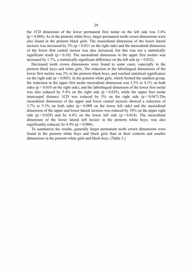
To summarize the results, generally larger permanent tooth crown dimensions were found in the preterm white boys and black girls than in their controls and smaller dimensions in the preterm white girls and black boys. (Table 3.)
30
Table 3. Summary of the comparison of mean permanent tooth crown dimensions between the preterm and control children.
Tooth Dim. White boys White girls Black boys Black girls 1M1 2M1
1 CD 1 CD
= ( + )
- * -
( - ) ( - )
= ( + )
3M1 4M1 1M1
1 CD 1 CD 2 CD
( + ) +
+*
( - ) ( - )
-
( - ) ( + ) ( - )
( + ) =
( + ) 2M1 3M1 4M1
2 CD 2 CD 2 CD
+ ** + ** + **
- ( - ) =
( - ) ( - ) =
( + ) ( + ) ( - )
1M1 2M1 3M1 4M1
MD MD MD MD
( + ) ( - )
- ( - )
- * - * - -
( - ) = - -
+ +* ( - ) ( - )
1M1 2M1 3M1 4M1
LL LL LL LL
( + ) ( - ) +
( + )
- -
- * ( + )
- ( - ) - * ( - )
( + ) ( + ) ( - ) ( - )
1 i1 2i1 3i1 4i1 1i2 2i2 3i2 4i2
MD MD MD MD MD MD MD MD
( + ) ( + ) ( - )
- ( - ) ( + ) ( - ) -**
- * - * - * - ** - * - -
- **
( - ) ( - ) ( - ) ( - ) ( - ) ( - ) ( - ) ( - )
( + ) ( + ) ( + ) ( + ) ( + )
+ +* +*
number of + 6 0 0 5 number of (+) 8 1 1 12 number of = 1 1 2 2 number of - 3 19 4 0 number of (-) 6 3 17 5 + = 0.1mm or larger dimension in prematurely born children, - = 0.1mm or smaller dimension in prematurely born children, = =equal dimensions, ( ) = difference between means < 0.1mm, 1M1 = upper right first permanent molar, 2M1 = upper left first permanent molar, 3M1 = lower left first permanent molar, 4M1 = lower right first permanent molar, 1i1 = upper right first permanent incisor etc., 1i2 = upper right second permanent incisor etc., MD = mesiodistal dimension, LL = labiolingual dimension.
5.3 Eruption of the permanent incisors and first molars (Study III)
The results showed earlier eruption of the first permanent molars and permanent incisors in the prematurely born children than in the full-term children of similar conceptional age. Generally the eruption was earlier for all permanent incisors and first molars and for all four preterm groups, although differences appeared between the groups in the
31
maxillary and mandibular teeth, and between the teeth on the left and right sides. Eruption of all the permanent teeth was earlier in the prematurely born black boys, significant results being found for the upper first permanent molar on the right side (p < 0.04), the upper central incisors (p < 0.005) and all four permanent lateral incisors (p < 0.05). In the preterm black girls the eruption of the upper first permanent molars (p < 0.05), the upper central permanent incisors (p < 0.005) and all four permanent lateral incisors (p < 0.05) was also significantly advanced. In all the comparisons eruption was advanced in the preterm groups relative to the controls.
In the preterm white boys, significantly advanced eruption was found in the case of the upper and lower central permanent incisors on the left side (p < 0.05 in each case), while in the preterm white girls the eruption of the upper lateral permanent incisor on the right side was significantly earlier (p < 0.05).
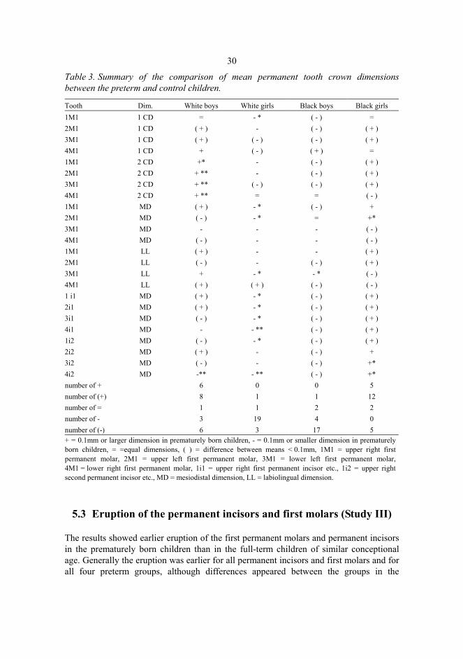
To summarize, the results showed earlier eruption of the permanent incisors and first molars in all the preterm children compared with the controls, even when grouped according to sex and race. The age of the child taken as the conceptional age. (Table 4.)
Table 4. Comparison of permanent tooth eruption in discordant pairs of preterm and control white boys, white girls, black boys and black girls matched by conceptional age.
Tooth white boys white girls black boys black girls 1M1 (+) (+) +* +* 2M1 (+) (+) (+) +* 3M1 (+) (+) (+) (+) 4M1 (+) (-) (+) (+) 1I1 (+) (+) +** +** 2I1 +* (+) +** +** 3I1 +* (+) (+) 4I1 (+) (+) (+) (+) 1I2 (+) +* +* +* 2I2 (-) (+) +** +* 3I2 (+) (+) +** +* 4I2 (+) (+) +** +* 1M1 = upper right first permanent molar; 2M1 = upper left first permanent molar; 3M1 = lower left first permanent molar; 4M1 = lower right first permanent molar;1I1 = central permanent incisor; 1I2 = lateral permanent incisor, etc., *P < 0.05; **P < 0.01., + = significantly advanced eruption in premature children, (+) = advanced eruption in premature children, but not significant, (-) = delayed eruption in premature children, but not significant.
5.4 Sagittal occlusal relationships and asymmetry (Study IV)
The prevalence of a mesial sagittal occlusion was greater in the prematurely born children than in the control children. When all the preterm infants were compared with all the controls a prenormal canine relationship was found on the right side in 42.9% of the subjects in the preterm group and in 33.0% of the controls [95% confidence interval
32
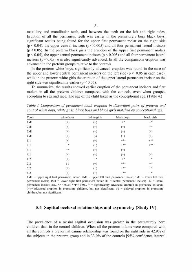
(CI) 3.86,16.0] and on the left side in 48.6% of the preterm group and in 34.8% of the controls (95% CI 7.55, 20.0) There were less cusp/cusp and postnormal canine relationships in the preterm group, and the prevalence of a normal canine relationship was reduced by 5.2% on the right side and 7.9% on the left side compared with the fullterm children.(Fig.1.)
Fig. 1. The canine relationship on the right (R)and left (L) side in preterm (Case) and control children.
The same trend existed, when the groups were divided by sex and race, the prevalence of a prenormal canine relationship on the left was greater in the preterm black girls than in their controls, the difference being 14.6%, which was a statistically significant result (95% CI 4.17, 25.0). The prevalence of a normal canine relationship was lower relative to the controls, the difference being 11.8% and that of a cusp/cusp relationship was also lower, the difference being 6.1%. A greater proportion of prenormal canine relationships was also found in the preterm white boys and white girls, the difference being 13.3% in the white boys and 1.3% in white girls on the left side, while the results on the right side showed an 8.5% greater prevalence in the preterm white boys and 5.8% in the preterm black girls, 0.2% in the preterm white girls and 0.4% in the preterm black boys, although these results were not statistically significant.
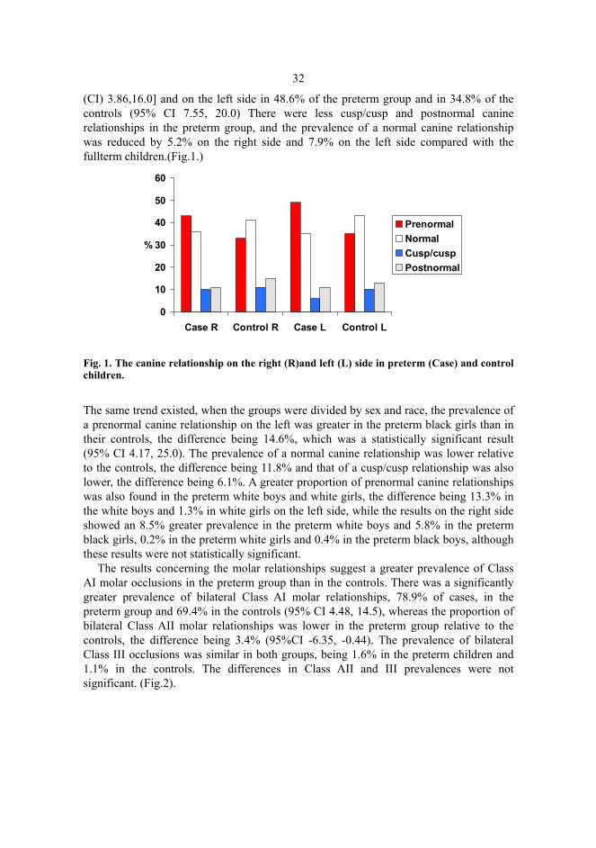
The results concerning the molar relationships suggest a greater prevalence of Class AI molar occlusions in the preterm group than in the controls. There was a significantly greater prevalence of bilateral Class AI molar relationships, 78.9% of cases, in the preterm group and 69.4% in the controls (95% CI 4.48, 14.5), whereas the proportion of bilateral Class AII molar relationships was lower in the preterm group relative to the controls, the difference being 3.4% (95%CI -6.35, -0.44). The prevalence of bilateral Class III occlusions was similar in both groups, being 1.6% in the preterm children and 1.1% in the controls. The differences in Class AII and III prevalences were not significant. (Fig.2).
0
10
20
30
40
50
60
Case R Control R Case L Control L
%
PrenormalNormalCusp/cuspPostnormal
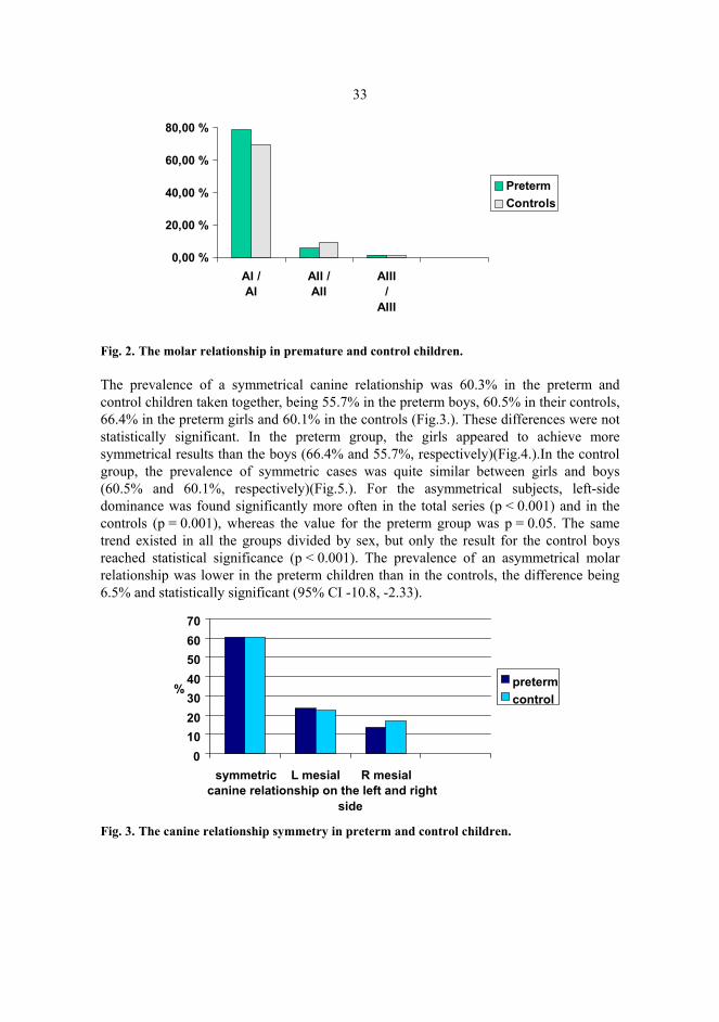
33
Fig. 2. The molar relationship in premature and control children.
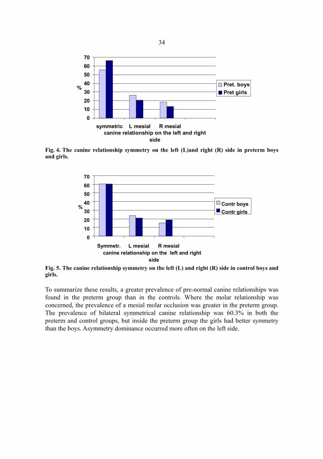
The prevalence of a symmetrical canine relationship was 60.3% in the preterm and control children taken together, being 55.7% in the preterm boys, 60.5% in their controls, 66.4% in the preterm girls and 60.1% in the controls (Fig.3.). These differences were not statistically significant. In the preterm group, the girls appeared to achieve more symmetrical results than the boys (66.4% and 55.7%, respectively)(Fig.4.).In the control group, the prevalence of symmetric cases was quite similar between girls and boys (60.5% and 60.1%, respectively)(Fig.5.). For the asymmetrical subjects, left-side dominance was found significantly more often in the total series (p < 0.001) and in the controls (p = 0.001), whereas the value for the preterm group was p = 0.05. The same trend existed in all the groups divided by sex, but only the result for the control boys reached statistical significance (p < 0.001). The prevalence of an asymmetrical molar relationship was lower in the preterm children than in the controls, the difference being 6.5% and statistically significant (95% CI -10.8, -2.33).
Fig. 3. The canine relationship symmetry in preterm and control children.
0,00 %
20,00 %
40,00 %
60,00 %
80,00 %
AI /AI
AII /AII
AIII/
AIII
PretermControls
0
10
20
30
40
50
60
70
symmetric L mesial R mesialcanine relationship on the left and right
side
% preterm control
34
Fig. 4. The canine relationship symmetry on the left (L)and right (R) side in preterm boys and girls.
Fig. 5. The canine relationship symmetry on the left (L) and right (R) side in control boys and girls.
To summarize these results, a greater prevalence of pre-normal canine relationships was found in the preterm group than in the controls. Where the molar relationship was concerned, the prevalence of a mesial molar occlusion was greater in the preterm group. The prevalence of bilateral symmetrical canine relationship was 60.3% in both the preterm and control groups, but inside the preterm group the girls had better symmetry than the boys. Asymmetry dominance occurred more often on the left side.
010203040506070
symmetric L mesial R mesialcanine relationship on the left and right
side
% Pret. boysPret girls
0 10 20 30 40 50 60 70
Symmetr. L mesial R mesialcanine relationship on the left and right
side
% Contr boys Contr girls

6 Discussion
6.1 General
The etiology of preterm births is usually multifactorial and may be related to disease in the foetus or mother, although in many cases the aetiology is unknown. A preterm infant usually requires considerable medical support and intervention during neonatal period. Many serious complications are encountered in nearly all major organ systems. Common medical complications of preterm infants include birth asphyxia, apnoea, respiratory distress syndrome, patent ductus arteriosus, intracranial hemorrhage, immaturity of liver and kidney, gastrointestinal intolerance. Also metabolic dysfunction, hypoglycaemia, hypocalcaemia, osteopenia and rickets, anaemia and susceptibility to infections are common problems in prematurely born neonates. With increasing knowledge of neonatal medical care in recents years, the survival rates of the most preterm children have improved dramatically. There may be differences within the preterm group, which may consist of both children with an appropriate birthweight and children with a low birthweight for their gestational age. Low birthweight may occur in infants delivered at or near term, and conversely that some infants delivered early in the third trimester can have relatively high birthweights of the kind usually associated with full term deliveries.
6.2 Development of the dentition
6.2.1 Tooth crown size
The determination of tooth crown size is thought to be multifactorial and to include genetic, maternal and environmental components. Certain points in the dentition are more variable than others, and the total amount of genetic variation depends on the particular tooth and dimension (Moorrees et al. 1957). The canines are known to exhibit high stability from the developmental point of view and the early-forming teeth are under

36
stronger genetic regulation than later-forming ones (Osborne et al. 1958). Our present results concerning permanent tooth crown size partly support previous findings of decreased tooth crown dimensions in preterm infants (Garn et al. 1979, 1980), but the increased crown dimensions found in some of the permanent teeth of preterm white boys and black girls differ from earlier observations. Environmental factors, including maternal influences like hypothyroidism, diabetes, hypertension, smoking etc., nutritional factors and developmental disturbances, may also affect the determination of tooth crown size (Garn et al. 1979).
Ethnic factors and sex differences must also be taken into consideration when evaluating processes affecting the dentition. Male advancement has been observed in the development of the deciduous teeth, notably in first trimester human embryos and at later stages (Garn et al. 1958, Garn & Burdi 1971, Demirjan & Levesque 1980). Girls precede boys in the tooth crown mineralization stages, root development and the tooth eruption process (Nyström et al. 2000), but boys usually have larger permanent teeth, the effect of the Y-chromosome being the decisive factor producing this difference (Alvesalo 1971, 1997). There may also be genetic differences between populations in the magnitude of environmental stress responses to general disturbances and prematurity. Girls have a considerable advantage over boys in terms of perinatal survival, and this holds good at all gestational ages and may be due to certain developmental characteristics that differ between the sexes, e.g. fetal lung maturity (Kline et al 1989). The influence of nutrition and dietary factors is also worth noting. The maintenance of optimal nutrition, mineral and vitamin supplementation during infancy is important for preterm infants and can have a long-term influence on their health, growth and neural development (Yu 1999).
The features observed here, especially the increased intercuspal distances in the first permanent molar in preterm white boys, may reflect the sensitivity of the dentition during the period of final determination of both crown dimensions, differences in the development of the dentition between the sexes and between ethnic groups and also the possible effects of an accelerated growth period, i.e. a period when the general velocity of growth is above normal for that age. This may be consistent with increased cellular activity in the developing tooth germs during the catch-up growth period, producing quantitative changes that are more pronounced in those tooth crown regions, that are at a sensitive stage in development at that time.
6.2.2 Tooth eruption
Like tooth crown size and the occlusal relationship, the tooth eruption process is also influenced by both genetic and environmental factors acting during odontogenesis. The environment, prenatal and maternal factors, diseases, nutrition, socio-economic status, climate etc. may effect the timing of tooth eruption, as previously reported. The eruption of the deciduous teeth has been thought to be more genetically determined than that of the permanent dentition. Our results showed earlier eruption of the first permanent molar and incisors in the prematurely born children than in controls, and this applied generally to all the permanent teeth and to all four preterm groups, white girls and boys and black girls and boys, although differences appeared between the teeth on the left and right sides

37
and between maxillary and mandibular teeth. Ethnic and sexual differences have been found previously in the order of tooth emergence and mineralization, and also differences between individuals in the developmental order of the teeth (Harris & McKee 1990), and the standards for tooth emergence vary greatly between populations and may be influenced by changing environmental conditions (Nanda 1960). The effect of low birthweight and preterm birth on tooth emergence has been found to be uncertain in previous reports.
It should be remembered that the age of the child in our study was calculated by transforming the chronological age to conceptional age by the reference of information on gestational ages in the preterm and control groups. Also the sample size was smaller for the white preterm boys and girls than for the black preterm boys and girls, which should be taken into account when analysing the results.The preterm white girls formed the smallest group, a fact that should be taken into consideration when evaluating the results divided by sex and race. The stimulating pressures of mastication promoting the tooth eruption process have been described by Friedlander & Bailit (1969), and our results seem to support this explanation. Also, the existence of sensitive periods in the tooth emergence process and various postnatal factors including catch-up growth period may influence the eruption of teeth.
6.2.3 Occlusion
The results concerning the sagittal occlusal relationship and the increased prevalence of a prenormal canine relationship and a mesial molar occlusion in prematurely born children focuses attention on the importance of perinatal factors in the development of the dentition. Prematurely born children have to adapt to extra-uterine conditions exceptionally early, and oral nutritive function begins about 1–1.5 months earlier than in full-term children. Nutritive sucking has been found possible from a postmenstrual age of 31 weeks onwards, and full breastfeeding at a mean postmenstrual age of 36 weeks (Nyqvist et al. 1999). Both genetic and environmental factors affect craniofacial morphology, but it has been suggested that the occlusal similarities in families may be more related to common environmental effects than to genetic factors and that the genetic contribution to occlusal variation is quite low, at least where the molar relationship is concerned (Harris & Smith 1980). When the racial differences are concerned, Class II molar relation has been reported to be more prevalent in white children and mesioclusion more frequently appearing in black children. A summary of estimates in the literature ranging from 8–13 per cent for North American black and Indian children and 11–39 per cent for white children. Class III molar relation appear from 1–7 per cent in white, black and Indian children (Infante 1975). The results of our study support these estimates, black children having more prenormal canine relationships than whites both in preterm and control groups. On contrary postnormal canine relationship was more frequent in whites compared to blacks.
Prematurely born children have an increased risk of respiratory problems, infections and systemic diseases in infancy. The most frequent causes of mouth breathing are allergies, adenoids and respiratory infections, and mouth breathing involving a low
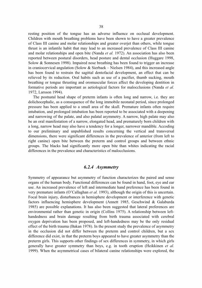
38
resting position of the tongue has an adverse influence on occlusal development. Children with mouth breathing problems have been shown to have a greater prevalence of Class III canine and molar relationships and greater overjet than others, while tongue thrust is an infantile habit that may lead to an increased prevalence of Class III canine and molar relationships and open bite (Nanda et al. 1972). An association has also been reported between postural disorders, head posture and dental occlusion (Huggare 1998, Solow & Sonnesen 1998). Impaired nose breathing has been found to trigger an increase in craniocervical angulation (Solow & Sierbaek – Nielsen 1984), and this increased angle has been found to restrain the sagittal dentofacial development, an effect that can be relieved by its reduction. Oral habits such as use of a pacifier, thumb sucking, mouth breathing or tongue thrusting and oromuscular forces affect the developing dentition in formative periods are important as aetiological factors for malocclusions (Nanda et al. 1972, Larsson 1994).
The postnatal head shape of preterm infants is often long and narrow, i.e. they are dolichocephalic, as a consequence of the long immobile neonatal period, since prolonged pressure has been applied to a small area of the skull. Premature infants often require intubation, and prolonged intubation has been reported to be associated with a deepening and narrowing of the palate, and also palatal asymmetry. A narrow, high palate may also be an oral manifestation of a narrow, elongated head, and prematurely born children with a long, narrow head may also have a tendency for a longer, narrower mandible. Accoding to our preliminary and unpublished results concerning the vertical and transversal dimensions, there were significant differences in the prevalence of anterior (from left to right canine) open bite between the preterm and control groups and between ethnic groups. The blacks had significantly more open bite than whites indicating the racial differences in the prevalence and characteristics of malocclusions.
6.2.4 Asymmetry
Symmetry of appearance but asymmetry of function characterizes the paired and sense organs of the human body. Functional differences can be found in hand, foot, eye and ear use. An increased prevalence of left and intermediate hand preference has been found in very premature infants (O’Callaghan et al. 1993), although the origin of this is uncertain. Focal brain injury, disturbances in hemisphere development or interference with genetic factors influencing hemisphere development (Annett 1985, Geschwind & Galaburda 1985) are possible explanations. It has also been suggested that lateral preferences are environmental rather than genetic in origin (Collins 1975). A relationship between left-handedness and brain damage resulting from birth trauma associated with cerebral oxygen deprivation has been proposed, and left-handedness may be the only residual effect of the birth trauma (Bakan 1978). In the present study the prevalence of asymmetry in the occlusion did not differ between the preterm and control children, but a sex difference did exist, in that the preterm boys appeared to have greater asymmetry than the preterm girls. This supports other findings of sex differences in symmetry, in which girls generally have greater symmetry than boys, e.g. in tooth eruption (Heikkinen et al. 1999). When the asymmetrical cases of bilateral canine relationships were explored, the
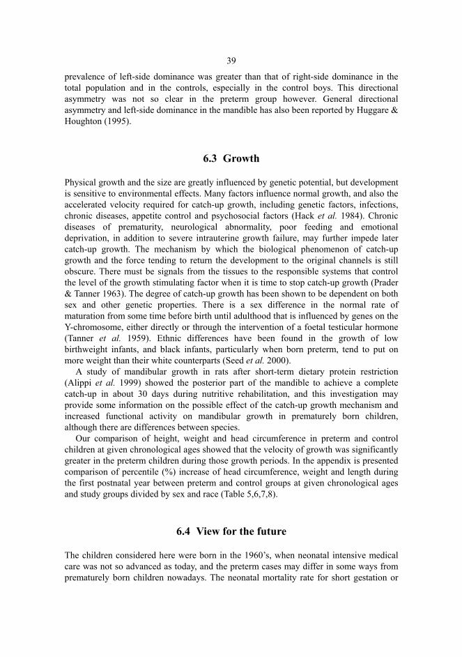
39
prevalence of left-side dominance was greater than that of right-side dominance in the total population and in the controls, especially in the control boys. This directional asymmetry was not so clear in the preterm group however. General directional asymmetry and left-side dominance in the mandible has also been reported by Huggare & Houghton (1995).
6.3 Growth
Physical growth and the size are greatly influenced by genetic potential, but development is sensitive to environmental effects. Many factors influence normal growth, and also the accelerated velocity required for catch-up growth, including genetic factors, infections, chronic diseases, appetite control and psychosocial factors (Hack et al. 1984). Chronic diseases of prematurity, neurological abnormality, poor feeding and emotional deprivation, in addition to severe intrauterine growth failure, may further impede later catch-up growth. The mechanism by which the biological phenomenon of catch-up growth and the force tending to return the development to the original channels is still obscure. There must be signals from the tissues to the responsible systems that control the level of the growth stimulating factor when it is time to stop catch-up growth (Prader & Tanner 1963). The degree of catch-up growth has been shown to be dependent on both sex and other genetic properties. There is a sex difference in the normal rate of maturation from some time before birth until adulthood that is influenced by genes on the Y-chromosome, either directly or through the intervention of a foetal testicular hormone (Tanner et al. 1959). Ethnic differences have been found in the growth of low birthweight infants, and black infants, particularly when born preterm, tend to put on more weight than their white counterparts (Seed et al. 2000).
A study of mandibular growth in rats after short-term dietary protein restriction (Alippi et al. 1999) showed the posterior part of the mandible to achieve a complete catch-up in about 30 days during nutritive rehabilitation, and this investigation may provide some information on the possible effect of the catch-up growth mechanism and increased functional activity on mandibular growth in prematurely born children, although there are differences between species.
Our comparison of height, weight and head circumference in preterm and control children at given chronological ages showed that the velocity of growth was significantly greater in the preterm children during those growth periods. In the appendix is presented comparison of percentile (%) increase of head circumference, weight and length during the first postnatal year between preterm and control groups at given chronological ages and study groups divided by sex and race (Table 5,6,7,8).
6.4 View for the future
The children considered here were born in the 1960’s, when neonatal intensive medical care was not so advanced as today, and the preterm cases may differ in some ways from prematurely born children nowadays. The neonatal mortality rate for short gestation or

40
low birthweight infants was higher, and individual properties were of considerable importance for the survival of preterm neonates, which may be reflected also in the results. Nowadays infants delivered after a very short period of gestation and suffering from immaturity in many organs can still survive without major impairments.
It would be interesting to continue the research by examining the possible effect of improved modern perinatal technology, advances in knowledge and improved educational and socioeconomic level to dental and occlusal development in children born very immature and after very short gestation nowadays.

7 Conclusions
1. The findings indicate that in prematurely born children deciduous and permanent tooth crown size variation remains within normal limits.
2. The results are parallel and confirm earlier findings of the presence of sexual dimorphism in permanent and deciduous tooth crown sizes.
3. The findings indicate that clinical eruption of permanent incisors and first molars is accelerated in prematurely born children.
4. The results indicate that premature birth may affect the development of sagittal occlusal relationships and asymmetry.

References
Aine L, Backström MC, Mäki R, Kuusela A-L, Koivisto A-M, Ikonen R-S & Mäki M (2000) Enamel defects in primary and permanent teeth of children born prematurely. J Oral Pathol Med 29:403–9.
Alippi RM, Meta MD, Boyer PM & Bozzini CE (1999) Catch-up growth in mandibular growth after short-term dietary protein restriction in rats during the post-weaning period. Eur J Oral Sci 107:260–264.
Alvesalo L (1971) The influence of sex-chromosome genes on tooth size in man. Proc Finn Dent Soc 67:3–54.
Alvesalo L & Tigerstedt PMA (1974) Heritabilities of human tooth dimensions. Hereditas 77:311–318.
Alvesalo L & Laine T (1992) Occlusion in 47,XXY (Klinefelter syndrome) men. Am J Phys Anthrop 87:161–165.
Alvesalo L (1997) Sex chromosomes and human growth. A dental approach. Hum Genet 101:1–5. Anderson DL Thompson GW & Popovich F (1975) Interrelationships of dental maturity, skeletal
maturity, height and weight from age 4 to 14 years. Growth 39:453–462. Annett M (1985) Left, Right, Hand and Brain: The Right Shift Theory. London: Erlbaum
Associates. Ash SP & Moss JP (1987) An investigation of the features of the pre-term infant palate and the
effect of prolonged orotracheal intubation with and without protective appliances. Br J Orthod 14(4):253–261.
Backström MC, Aine L, Mäki R, Kuusela A-L, Sievänen H, Koivisto A-M, Ikonen R-S & Mäki M (2000) Maturation of primary and permanent teeth in preterm infants. Arch Dis Child Fetal Neonatal Ed 83(2):104–108.
Bailit HL & Sung B (1968) Maternal effects on the developing dentition. Arch Oral Biol 13:155–162.
Bailit HL, Niswander JD & MacLean CJ (1968) The relationship among several prenatal factors and variation in the permanent dentition in Japanese children. Growth 32:331–345.
Bakan P (1978) Handedness and birth order: a critical note on a critical note. Mot Skills 46:556. Batheja M & McManus IC (1985) Handedness in the mentally handicapped. Dev Med Child
Neurol 27:63–68. Baum JD & Searls D (1971) Head shape and size of pre-term low-birthweight infants. Dev Med
Child Neurol 13:575–581.

43Bhaskar SN (1980) Maxilla and mandible (alveolar process). In: Bhaskar S N (ed.) Orban’s Oral
histology and embryology. Chapter 8, St.Louis, Toronto, London, The C V Mosby Company 243.
Björk A, Krebs A & Solow B (1964) A method for epidemiological registration of malocclusion. Acta Odontol Scand 22:27–41.
Boice JB, Krous HF & Foley JN (1976) Gingival and dental complications of orotracheal intubation. JAMA 236:957–958.
Cahill DR & Marks SC (1980) Tooth eruption: evidence for the central role of the dental follicle. J Oral Pathol 9:189–200.
Cartlidge PHT & Stewart JH (1997) Survival of very low birthweight and very preterm infants in a geographically defined population.Acta Paediatr 86:105–10.
Cohen S (1962) Isolation of a mouse submaxillary gland protein accelerating incisor eruption and eyelid opening in the newborn animal. J Biol Chem 237:1555–1562.
Collins RL (1975) When left-handed mice live in right-handed worlds. Science 187:181–184. Coren S & Porac C (1980) Birth factors and laterality: Effects of birth order, parental age, and birth
stress on four indises of lateral preference. Behav Genet 10(2):123–138. Cunha RF, Boer FAC, Torriani DD & Frossard WTG (2001) Natal and neonatal teeth: review of
the literature. Pediatr Dent 23(2):158–162. Cushner IM & Mellits ED (1971) The relationship between fetal outcome and the gestational age
and birthweight of the fetus. Johns Hopkins Medical Journal 128:252–260. Dahlberg AA (1945) The changing dentition of man. J Am Dent Assoc 32:676–690. Delgado H, Habicht JP, Yarbrough C, Lechtig A, Martorell R, Malina RM & Klein RE (1975)
Nutritional status and the timing of deciduous tooth eruption. Am J Clin Nutr 28:216–24. Demirjan A & Levesque G-J (1980) Sexual difference in dental development and prediction of emergence, J Dent Res 59:1110–1122. DeVito C & Saunders S (1990) A discriminant function analysis of deciduous teeth to determine
sex. J Forensic Sci 35(4):845–858. Drummond BK, Ryan S, O’Sullivan EA, Congdon P & Curzon MEJ (1992) Enamel defects of the
primary dentition and osteopenia of prematurity. Pediatr Dent 14(2):119–121. Elliman AM, Bryan EM, Elliman AD & Starte D (1986) Narrow heads of preterm infants - do they
matter? Dev Med Child Neurol 28:745–748. Erenberg A & Nowak A J (1984) Palatal groove formation in neonates and infants with orotracheal
tubes. AJDC 138: 974–975. Fadavi S, Punwani IC, Adeni S & Vidyasagar D (1992) Eruption pattern in the primary dentition of
premature low-birth-weight children. J Dent Child March-April 120–122. Fadavi S, Adeni S, Dziedzic K, Punwani I &Vidyasagar D (1992) The oral effects of orotracheal
intubation in prematurely born preschoolers. J Dent Child. November-December 420–424. Falkner F (1957) Deciduous tooth eruption. Arch Dis Child 32:386–391. Fearne JM, Bryan EM, Elliman AM, Brook AH &Williams DM (1990) Enamel defects in the
primary dentition of children born weighing less than 2000g. Br Dent J 168(11):433–7. Fearne JM & Brook AH (1993) Small primary tooth-crown size in low birth-weight children. Early
Hum Dev 33:81–90. Friedlander JS & Bailit HL (1969) Eruption times of the deciduous and permanent teeth of natives
on Bougainville island, territory of New Guinea. A study of racial variation. Hum Biol 41:51–65.
Funakoshi Y, Kushida Y & Hieda T (1981) Dental observations of low birth weight infants. Pediatr Dent 3(1):21–25.
Garn SM, Lewis AB, Koski K & Polacheck DL (1958) The sex difference in tooth calcification, J. Dent.Res.37:561–567.

44Garn SM, Lewis AB & Kerewsky RS (1966) The meaning of bilateral asymmetry in the permanent
dentition. Angle Orthod 36:55–62. Garn SM & Burdi AR (1971) Prenatal ordering and postnatal sequence in dental development. J
Dent Res 50:1407–1414. Garn SM (1977) Genetics of dental development. In: McNamara JA,editor. The biology of occlusal
development. Ann Arbor, Michigan: Craniofacial Growth Series 61–88. Garn SM, Osborne RH & McCabe KD (1979) The effect of prenatal factors on crown dimensions.
Am J Phys Anthrop 51(4):665–678. Garn SM, Osborne RH, Alvesalo L & Horowitz SL (1980) Maternal and gestational influences on
deciduous and permanent tooth size. J Dent Res 59(2):142–143. Geschwind N & Galaburda AM (1985) Cerebral lateralization. Biological mechanisms,
associations, and pathology:1.A hypothesis and program of research. Arch Neurol 42:428–654. Glasstone S (1967) Development of teeth in tissue culture. J Dent Res 46:858–861. Golden NL, Takieddine F & Hirsch VJ (1981) Teething age in prematurely born infants. AJDC
135:903–904. Grahnen H & Larsson PG (1958) Enamel defects in the deciduous dentition of prematurely born
children. Odontol Revy 9:193–204. Grahnen H & Granath L-E (1962) The effect of hyperbilirubinemia on primary teeth. Odontol
Revy 13:337–43. Grahnen H, Sjölin S & Stenström A (1974) Mineralization defects of primary teeth in children
borm preterm. Scand J Dent Res 82:396–400. Grön M & Alvesalo L (1997) Dental occlusion and arch size and shape in karyotype 46,XY
females. Eur J Orthod 19:329–335. Haavikko K (1970) The formation and the alveolar and clinical eruption of the permanent teeth
(Dissertation). Suomen Hammaslääkäriseuran toimituksia 66:103–170. Hack M, Merkatz IR, McGrath SK, Jones PK & Avroy AF (1984) Catch-up Growth in Very-Low-
Birth-Weight Infants. AJDC 138:370–375. Hack M, Weissman B & Borawski-Clark E (1996) Catch-up growth during childhood among very
low birth weight children. Arch Pediatr Adolesc Med 150(11):1122–1129. Hack M & Fanaroff AA (2000) Outcomes of children of extremely low birthweight and gestational
age in the 1990’s. Semin Neonatol 5:89–106. Hardy JB, Drage JS & Jackson EC (1979) Intrauterine growth. In: Hardy J B, Drage J S, Jackson E
C (editors) The First Year of Life. Chapter 7, Baltimore and London, The Johns Hopkins University Press 38–51.
Harris EF & Smith RJ (1980) A study of occlusion and arch widths in families. Am J Orthod 78(2):155–163.
Harris EF & Smith RJ (1982) Occlusion and arch size in families. A principal components analysis. Angle Orthod 52(2):135–143.
Harris EF & McKee JH (1990) Tooth mineralization standards for blacks and whites from the middle southern United States.J Forensic Sci 35:859–872.
Harris EF, Barcroft ED, Haydar S & Haydar B (1993) Delayed tooth formation in low birthweight African-American children. Pediatr Dent 15:30–4.
Hatton ME (1950) A measure of the effects of heredity and environment on eruption of the deciduous teeth. J Dent Res 34:397–401.
Heikkinen T, Alvesalo L, Osborne RH & Sarpola A (1995) Clinical eruption of permanent incisors and first molars after maternal smoking in pregnancy. Acta Med Auxol 27(2-3):83–96.
Heikkinen T (1996) The effect of maternal smoking during pregnancy on the teeth of the child. Thesis Acta Univ Oul D 373.
45Heikkinen T, Alvesalo L, Osborne RH, Tienari J & Virtanen JI (1999) The effect of sex and race
on the symmetry of the permanent tooth clinical eruption. In: Mayhall JT and Heikkinen T (editors) Dental Morphology 1998. Proceedings of the 11th International Symposium on Dental Morphology, Oulu University Press 473–482.
Heikkinen T, Alvesalo L, Osborne R H & Tienari J (2001) Tooth eruption symmetry in functional lateralities. Arch Oral Biol 46:609–617.
Huggare J & Houghton P (1995) Asymmetry in the human skeleton. A study on prehistoric Polynesians and Thais. Eur J Morphol 33:3–14.
Huggare J (1998) Postural disorders and dentofacial morphology. Acta Odontol Scand 56:383–386. Hunter WS & Priest WR (1960) Errors and discrepancies in measurement of tooth size. J Dent Res
39:405–414. Iizuka T, Cielinski M, Aukerman SL & Marks SCJr (1992) The effects of colony-stimulating
factor-1 (CSF-1) on tooth eruption in the toothless (osteopetrotic) rat illustrate the critical periods for bone resorption during tooth eruption. Arch Oral Biol 37:831–840.
Infante PF & Owen GM (1973) Relation of chronology of deciduous tooth emergence to height, weight and head circumference in children. Arch Oral Biol 18:1411–17.
Infante PF (1975) Malocclusion in the deciduous dentition in white, black and Apache Indian children. Angle Orthodont 45:213–218.
Johnsen D, Krejci C, Hack M & Faranoff A (1984) Distribution of enamel defects and the association with respiratory distress in very low birth-weight infants. J Dent Res 63(1):59–64.
Kiliaridis S (1995) Masticatory muscle influence on craniofacial growth. Acta Odontol Scand 53:196–202.
Kline J, Stein Z & Susser M (1989) Conception to Birth. Epidemiology of Prenatal Development. Monogr Epidemiol Biostatist 14:212.
Kraus BS Calcification of the human deciduos teeth (1959) J Am Dent Assoc 74:1128–36. Kraus BS & Jordan R (1965) Pattern and Rates of Growth In: The Human Dentition Before Birth.
Philadelphia: Lea & Febiger 143. Lai PY, Seow WK, Tudehope DI & Rogers Y (1997) Pediatr Dent 19(1):42–49. Laine T, Alvesalo L, Savolainen A & Lammi S (1986) Occlusal morphology in 45,X females. J
Craniofac Genet Dev Biol 6:351–355. Laine T & Alvesalo L. (1986) Size of the alveolar arch of the mandible in relation to that of the
maxilla in 45,X females. J Dent Res 65:1432–34. Laine T, Alvesalo L & Lammi S (1992) A study in 47,XYY men of the expression of sex-
chromosome anomalies in dental occlusion. Arch Oral Biol 37(11):923–928. Larsson E (1994) Artificial Sucking Habits: Etiology, Prevalence and Effect on Occlusion. Int J
Orofacial Myology 20:10–21. Lee MMC, Low WD& Chang KSF (1965) Eruption of permanent dentition of Southern Chinese
children in Hong Kong. Arch Oral Biol 10:849–861. Lee KS, Paneth N & Gartner LM (1980) Neonatal mortality: an analysis of recent improvement in
the United States. Am J Public Health 70:15–21. Lee K, Kim B, Khosnood B, Hsieh H, Chen T-J, Herschel M & Mittendorf R (1995) Outcome of
very low birth weight infants in industrialized countries: 1947-1987. Am J Epidemiol 141:1188–1193.
Lubchenco LO, Hansman C, Dressler M & Boyd E (1963) Intrauterine growth as estimated from liveborn birth-weight data at 24-42 weeks of gestation. Pediatrics 32:793–800.
Macey-Dare LV, Moles DR, Evans RD & Nixon F (1999)Long-term effect of neonatal endotracheal intubation on palatal form and symmetry in 8-11-year-old children. Eur J Orthod 21:703–710.
Magnusson B (1968) Tissue changes during molar tooth eruption. Transactions of the Royal School of Dentistry, Stockholm and Umeå 13:1–122.

46Main JHP (1966) Retention of potential to differentiate in long-term cultures of tooth germs.
Science 152:778–779. Marks SCJr & Cahill DR (1984) Experimental study in the dog of the nonactive role of the tooth in
the eruptive process. Arch Oral Biol 29:311–22 Marlow N, Roberts BL & Cooke RWI (1989) Laterality and prematurity. Arch Dis Child 64:1713–
6. Marsden DJ (1980) Reduction of head flattening in preterm infants. Dev Med Child Neurol
22:507–509. Mayhall JT & Alvesalo L (1992) Dental morphology of 45,XO human females:molar cusp area,
volume, shape and linear measurements. Arch Oral Biol 37(12):1039–1043. Mayhall JT & Alvesalo L (1995) The effects of the sex chromosomes on molar morphology In:
Aspects of dental biology:palaeontology, anthropology and evolution. J.Moggi-Cecchi, ed. Florence:International Institute for the Study of Man.
McNemar Q (1947) Note on the sampling error of the difference between correlated proportions or percentages. Psychometrika 12:153–157
Mellander M, Noren JG, Freden H & Kjellmer I (1982) Mineralization defects in deciduous teeth of low birthweight infants Acta Paediatr Scand 71:727–733.
Moorrees CFA, Thomsen SO, Jensen E & Yen PK (1957) Mesio-distal crown diameters of the deciduous and permanent teeth in individuals. J Dent Res 36:39–47.
Moorrees CFA (1959) The dentition of the growing child. Harvard University Press, Cambridge, Massachusetts 88–101.
Morris KM & Burns YR (1994) Reduction of craniofacial and palatal narrowing in very low birthweight infants. J Paediatr Child Health 30:518–522.
Moylan FMB, Seldin EB, Shannon DC & Todres ID (1980) Defective primary dentition in survivors of neonatal mechanical ventilation. Pediatr 96(1):106–8.
Myllärniemi S (1972) Malocclusion and childhood health. Proc Finn Dent Soc 68:286–291. Nanda RS (1960) Eruption of human teeth. Am J Orthod 46:363–378 Nanda RS, Khan I & Anand R (1972) Effect of oral habits on the occlusion in preschool children. J
Dent Child 39:449–452. Nikiforuk G & Fraser D (1979) Chemical Determinants of enamel hypoplasia in children with
disorders of calcium and phospate homeostasis. J Dent Res 58(B):1014–1015. Noren JG (1983) Enamel structure in deciduous teeth from low birth weight infants. Acta Odontol
Scand 41:355–362. Noren JG, Ranggård L, Klingberg G & Persson C (1993) Intubation and mineralization
disturbances in the enamel of primary teeth. Acta Odontol Scand 51:271–275. Nyqvist KH, Sjoden PO & Ewald U (1999) The development of preterm infant’s breastfeeding
behavior. Early Hum Dev 55(3):247–64. Nyström M, Aine L, Peck L, Haavikko K & Kataja M (2000) Dental maturity in finns and the
problem of missing teeth. Acta Odontol Scand 58:49–56. Nyström M & Ranta H (2003) Tooth formation and the mandibular symphysis during the first five
postnatal months. J Forensic Sci 48(2):1–6. O’Callaghan MJ, Burn YR, Mohay HA, Rogers Y & Tudehope DI (1993) Handedness in
extremely low birthweight infants:aetiology and relationship to intellectual abilities, motor performance and behaviour at four and six years. Cortex 29:629–637.
Osborne RH, Horowitz SL & DeGeorge FV (1958) Genetic variation in tooth dimensions. A twin study of the permanent anterior teeth. Am J Hum Genet 10(3).
Palmer B (1998) The influence of breastfeeding on the development of the oral cavity :a commentary. J Hum Lact 14:93–98.
Pimlott JFL, Howley TP, Nikiforuk G & Fitzhardinge PM (1985) Enamel defects in prematurely born, low birthweight infants. Pediatr Dent 7:218–223.

47Pirttiniemi P, Grön M, Alvesalo L, Heikkinen T & Osborne RH (1994) Relationship of difficult
forceps delivery to dental arches and occlusion. Pediatr Dent 16(4):289–293. Powls A, Botting N, Cooke RWI & Marlow N (1996) Handedness in very-low-birthweight
(VLBW) children at 12 years of age: relation to perinatal outcome variables. Dev Med Child Neurol 38:594–602.
Prader A, Tanner JM & von Harnack GA (1963) Catch-up growth following illness or starvation. J Pediatr 62(5):646–659.
Proffit WR (ed) (1986) Malocclusion and dentofacial deformityin contemporary society. In: Contemporary orthodontics. CV Mosby Co., St Louis 1–5.
Rantakallio P & Mäkinen H (1983) The effect of maternal smoking on the timing of deciduous tooth eruption.Growth 47:122–128.
Ricketts RM (1968) Respiratory obstruction syndrome. Am J Orthod 54:495–507. Ross G, Lipper EG & Auld PAM (1987) Hand preference in four-year-old children: its relationship
to premature birth and neurodevelopmental outcome. Dev Med Child Neurol 29:615–622. Rutter N, Hincliffe W & Cartlidge PHT (1993) Do preterm infants always have flattened heads?
Arch Dis Child 68:606–607. Schwirian PM, Eesley T & Cuellar L (1986) Use of water pillows in reducing head shape distortion
in preterm infants. Res Nurs Health 9:203–207. Seed PT, Ogundipe EM & Wolfe CD (2000) Ethnic differences in the growth of low-birthweight
infants. Paediatr Perinat Epidemiol 14(1):4–13. Seow WK, Brown JP, Tudehope DA & O’Callaghan M (1984) Dental defects in the deciduous
dentition of premature infants with low birthweight and neonatal rickets. Pediatr Dent 6(2):88–92.
Seow WK, Brown JP, Tudehope DI & O’Callaghan M (1985) Effect of neonatal laryngoscopy and endotracheal intubation on palatal symmetry in two to five-year -old children. Pediatr Dent 7:30–6.
Seow WK (1986) Oral complications of premature birth. Aust Dent J 31(1):23–29. Seow WK, Humphrys C, Mahanonda R & Tudehope DI (1988) Dental eruption in low birth-weight
prematurely born children: a controlled study. Pediatr Dent 10(1):39–42. Seow WK (1996) A study of the development of the permanent dentition in very low birthweight
children. Pediatr Dent 18(5):379–84. Seow WK (1997) Effects of preterm birth on oral growth and development. Aust Dent J 42:(2):85–
91. Seow WK & Wan A (2000) A controlled study of the morphometric changes in the primary
dentition of pre-term, very-low-birthweight children, J Dent Res 79(1):63–9. Siegel MI & Mooney MP (1987) Perinatal stress and increased fluctuating asymmetry of dental
calcium in the laboratory rat. Am J Phys Anthropol 73:267–270. Smart JL, Jeffery C & Richards B (1980) A retrospective study of the relationship between birth
history and handedness at six years Early Hum Dev 4(1):79–88. Solow B & Sonnesen L (1998) Head posture and malocclusions. Eur J Orthod 20:685–693. Solow B & Siersbaek-Nielsen S (1984) Airway adequacy, head posture, and craniofacial
morphology. Am J Orthod 86:214–23. Stimmler L, Snodgrass GJAI & Jaffe E (1973) Dental defects associated with neonatal
symptomatic hypocalcaemia Arch Dis Child 48:217–220. Sutton RN & Graze HR (1985) The blood vessel thrust theory of tooth eruption and migration.
Med Hypotheses 18:289–295. Tam JP (1985) Physiological effects of transforming growth factor in the newborn mouse. Science
229:673–675.
48Tanner JM, Prader A, Habich H & Ferguson-Smith MA (1959) Genes on the Y-chromosome
influencing rate of development in man: skeletal age studies in Klinefelter’s (XXY) and Turner’s (XO) syndromes. Lancet 2:141.
Tommiska V, Heinonen K, Ikonen S, Kero P, Pokela M-L, Renlund M, Virtanen M & Fellman V (2001) A national short-term follow-up study of extremely low birth weight infants born in Finland in 1996-1997. Pediatrics 107(1):1–9.
Townsend GC & Garcia-Godoy F (1984) Fluctuating asymmetry in the deciduous dentition of Dominican Mulatto children. Arch Oral Biol 29(7):483–486.
Townsend GC (1985) Intercuspal distances of maxillary pre-molar teeth in Australian aboriginals. J Dent Res 64(3):443–446.
Townsend GC, Corruccini RS, Richards LC & Brown T (1988) Genetic and environmental determinants of dental occlusal variation in South Australian twins. Aust Orthod J 10(4):231–235.
Townsend GC (1992) Genetic and environmental contributions to morphometric dental variation. J Hum Ecol special Issue 2:61–72.
Trupkin DP (1974) Eruption patterns of the first primary tooth in infants who were underweight at birth. ASDC J Dent Child 41:279–82
Tsang RC, Donovan EF & Steichen JJ (1976) Calcium physiology and pathology in the neonate. Pediatr. Clin N Am 23:611–626.
Varrela J (1990) Occurrence of malocclusion in attritive environment: a study of a skull sample from southwest Finland. Scand Dent Res 98:242–7.
Viscardi RM, Romberg E & Abrams RG (1994) Delayed primary tooth eruption in premature infants: relationship to neonatal factors. Pediatr Dent 16(1):23–28.
Wedgwood M & Holt KS (1968) A longitudinal study of the dental and physical development of 2-3 year-old children who were underweight at birth. Biol Neonat 12:214–232.
Wise GE, Marks SCJr & Cahill DR (1985) Ultrastructural features of the dental follicle associated with formation of the tooth eruption pathway in the dog. J Oral Pathol 14:15–26.
Wise GE, Lin F, Marks SCJr & Gorski JP (1994) The molecular basis of tooth eruption. In: Davidovitch Z (Ed), The Biological Mechanisms of Tooth Eruption, Resorption and Replacement by Implants. Harvard Society for the Advancement of Orthodontics, Boston, Massachusetts, USA 383–389.
Wise GE (1998) Cell and molecular biology of tooth eruption. In: Davidovitch Z and Mah J (Eds), Biological Mechanisms of Tooth Eruption, Resorption and Replacement by Implants. Harvard Society for the Advancement of Orthodontics, Boston, Massachusetts, USA 1–8.
Wise GE, Que BG, Huang H & Lumpin SJ (2000) Enhancement of gene expression in rat dental follicle cells by parathyroid hormone-related protein. Arch Oral Biol 45:903–909.
Ylppö A (1919) Zur Physiologie, Klinik und zum Schicksal der Fruhgeborenen. Z Kinderhlkd 24:1–110.
Yu VYH (1999) Enteral feeding in the preterm infant. Early Hum Dev 56(2-3):89–115.

Appendix 1
Table 5. Comparison of the percentile increase of head circumference, weight and length between preterm and control groups in black boys.
Preterm Control Growth (%) N md
(%) mean (%)
sd N md (%)
mean (%)
sd p-value difference of medians (%)
Hcf 0–4 months 124 23.5 24.2 7.3 431 20.5 21.2 5.1 0.000*** +3.0 4–8 months 82 9.8 9.8 3.5 272 8.0 8.8 5.1 0.007** +1.8 8–12 months 81 4.5 4.2 2.3 269 4.2 3.7 3.6 0.056 +0.2
Weight 0–4 months 129 112.4 128.0 49.5 433 103.1 106.3 32.0 0.000*** +9.3 4–8 months 82 38.1 43.6 21.9 265 35.3 36.3 11.8 0.053 +2.8 8–12 months 82 15.3 17.6 11.0 266 14.3 14.9 7.5 0.080 1.0
Length 0–4 months 124 26.0 27.1 7.6 420 25.5 25.8 6.2 0.198 +0.5 4–8 months 82 13.9 14.2 5.0 268 12.5 12.1 6.4 0.009** +1.4 8–12 months 81 7.2 7.6 3.9 269 7.1 7.9 10.6 0.8 +0.1
Mann-Whitney U-test *p < 0.05 ** p < 0.01 *** p < 0.001, md = (median), N = (number of cases), sd = (standard deviation), Hcf (Head circumference).
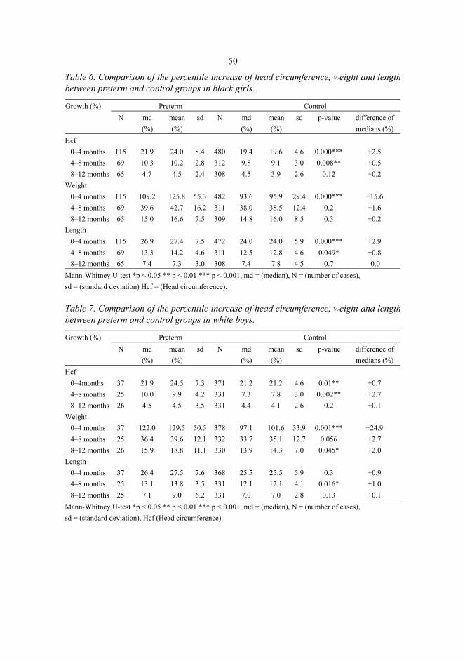
50
Table 6. Comparison of the percentile increase of head circumference, weight and length between preterm and control groups in black girls.
Preterm Control Growth (%) N md
(%) mean (%)
sd N md (%)
mean (%)
sd p-value difference of medians (%)
Hcf 0–4 months 115 21.9 24.0 8.4 480 19.4 19.6 4.6 0.000*** +2.5 4–8 months 69 10.3 10.2 2.8 312 9.8 9.1 3.0 0.008** +0.5 8–12 months 65 4.7 4.5 2.4 308 4.5 3.9 2.6 0.12 +0.2
Weight 0–4 months 115 109.2 125.8 55.3 482 93.6 95.9 29.4 0.000*** +15.6 4–8 months 69 39.6 42.7 16.2 311 38.0 38.5 12.4 0.2 +1.6 8–12 months 65 15.0 16.6 7.5 309 14.8 16.0 8.5 0.3 +0.2
Length 0–4 months 115 26.9 27.4 7.5 472 24.0 24.0 5.9 0.000*** +2.9 4–8 months 69 13.3 14.2 4.6 311 12.5 12.8 4.6 0.049* +0.8 8–12 months 65 7.4 7.3 3.0 308 7.4 7.8 4.5 0.7 0.0
Mann-Whitney U-test *p < 0.05 ** p < 0.01 *** p < 0.001, md = (median), N = (number of cases), sd = (standard deviation) Hcf = (Head circumference).
Table 7. Comparison of the percentile increase of head circumference, weight and length between preterm and control groups in white boys.
Preterm Control Growth (%) N md
(%) mean (%)
sd N md (%)
mean (%)
sd p-value difference of medians (%)
Hcf 0–4months 37 21.9 24.5 7.3 371 21.2 21.2 4.6 0.01** +0.7 4–8 months 25 10.0 9.9 4.2 331 7.3 7.8 3.0 0.002** +2.7 8–12 months 26 4.5 4.5 3.5 331 4.4 4.1 2.6 0.2 +0.1
Weight 0–4 months 37 122.0 129.5 50.5 378 97.1 101.6 33.9 0.001*** +24.9 4–8 months 25 36.4 39.6 12.1 332 33.7 35.1 12.7 0.056 +2.7 8–12 months 26 15.9 18.8 11.1 330 13.9 14.3 7.0 0.045* +2.0
Length 0–4 months 37 26.4 27.5 7.6 368 25.5 25.5 5.9 0.3 +0.9 4–8 months 25 13.1 13.8 3.5 331 12.1 12.1 4.1 0.016* +1.0 8–12 months 25 7.1 9.0 6.2 331 7.0 7.0 2.8 0.13 +0.1
Mann-Whitney U-test *p < 0.05 ** p < 0.01 *** p < 0.001, md = (median), N = (number of cases), sd = (standard deviation), Hcf (Head circumference).

51
Table 8. Comparison of the percentile increase of head circumference, weight and length between preterm and control groups in white girls.
Preterm Control Growth (%) N md
(%) mean (%)
sd N md (%)
mean (%)
sd p-value difference of medians (%)
Hcf 0–4 months 20 23.4 23.4 7.6 364 20.6 20.6 5.0 0.048* +2.8 4–8 months 11 10.0 9.5 3.1 324 7.5 7.5 2.9 0.031* +2.5 8–12 months 11 4.8 5.0 1.7 323 4.5 4.2 2.6 0.17 +0.3
Weight 0–4 months 20 111.6 116.0 48.9 369 92.6 94.4 29.9 0.026* +19.0 4–8 months 11 35.5 41.5 20.2 326 33.0 34.0 12.3 0.2 +2.5 8–12months 11 21.2 22.1 6.5 326 14.7 15.0 7.0 0.045* +6.5
Length 0–4 months 19 27.1 27.7 8.7 358 24.0 24,2 5.8 0.052 +3.1 4–8 months 11 12.9 12.8 3.8 325 11.9 11.8 6.0 0.6 +1.0 8–12 months 10 8.1 8.9 4.9 325 7.4 8.6 17.5 0.6 +0.7
Mann-Whitney U-test *p < 0.05 ** p < 0.01 *** p < 0.001, md = (median), N = (number of cases), sd = (standard deviation), Hcf = (Head circumference)