Embed Size (px)
Citation preview

R ESEARCH ARTICLE
doi: 10.2306/scienceasia1513-1874.2013.39.356ScienceAsia 39 (2013): 356–362
The effect of mirtazapine on methotrexate-inducedtoxicity in rat liverBunyami Ozogula, Abdullah Kisaoglua, Mehmet Ibrahim Turanb,∗, Durdu Altunerc, Ebru Senerd,Nihal Cetine, Cengiz Ozturke,f
a Department of Surgery, Faculty of Medicine, Ataturk University, 25240, Erzurum, Turkeyb Department of Paediatrics, Faculty of Medicine, Ataturk University, 25240, Erzurum, Turkeyc Department of Pharmacology, Faculty of Medicine, Recep Tayyip Erdogan University, 53100, Rize, Turkeyd Department of Pathology, Erzurum Region Education and Research Hospital, Erzurum, Turkeye Department of Pharmacology-Toxicology, Faculty of Veterinary Medicine, Ataturk University, 25240,
Erzurum, Turkeyf Department of Family Medicine, Erzurum State Hospital, Erzurum, Turkey
∗Corresponding author, e-mail: [email protected] 18 Dec 2012Accepted 20 Mar 2013
ABSTRACT: Methotrexate is used as a chemotherapeutic agent and its anti-oxidant activity is used to treat many cancertypes. This study conducts a biochemical and histopathological investigation into whether mirtazapine has a protective effecton methotrexate-induced hepatotoxicity in rats. Distilled water was given to a healthy group intraperitoneally. Methotrexatealone was injected in the control group, again intraperitoneally. Mirtazapine and, 1 h later, methotrexate were given tothe rats in the final group. This procedure was repeated over 7 days. In the control group rats receiving methotrexate,blood AST, ALT, and LDH levels were 227± 3 µmol/l, 85± 2 µmol/l, and 357± 13 µmol/l, respectively. In the ratsreceiving mirtazapine and methotrexate, these values were 152± 3 µmol/l, 25± 1 µmol/l, and 141± 15 µmol/l. In thehealthy rat group, AST, ALT, and LDH levels were 136 µmol/l, 20 µmol/l, and 133 µmol/l, respectively. Histopathologically,apoptotic bodies with condensed cytoplasm, peripheral, and pyknotic nuclei in the hepatocytes, focal necrosis and intenseinflammation in the interstitial areas were present in the control group. In the methotrexate and mirtazapine group, therewere no apoptotic bodies or inflammation, only isolated necrosis in the hepatocytes. In conclusion, mirtazapine protectedthe liver against methotrexate toxicity.
KEYWORDS: drug, hepatotoxicity, oxidant/anti-oxidant
INTRODUCTION
Methotrexate, a folic acid anti-metabolite, is usedas a chemotherapeutic agent for many cancer types(leukaemia, lymphoma, osteosarcoma, head and necktumours, lung cancer, breast cancer, etc.). Methotrex-ate is also used for the treatment of multiple scle-rosis, dermatomyositis, sarcoidosis, psoriasis, andrheumatoid arthritis, disorders causing inflammation.However, the use of high-dose methotrexate (as inleukaemia) or prolonged use may result in hepato-toxicity that may lead to progressive fibrosis andcirrhosis1. Clinically, hepatotoxicity, which occursin long-term use of methotrexate, remains one of thesignificant restrictions on its use in the doses desired2.Methotrexate inhibits the formation of tetrahydrofo-late from folic acid. The inhibition of tetrahydrofolateformation is responsible for both the therapeutic andtoxic effects of methotrexate3. Although these dele-
terious toxic effects of methotrexate can theoreticallybe reduced or prevented with the addition of folic acidto the treatment, there is as yet no consensus on thesubject. On the contrary, there are studies showingthat with the addition of folic acid, the therapeuticeffectiveness of methotrexate decreases4. This indi-cates that the mechanism of action of methotrexatehepatotoxicity has not yet been fully explained5.
Toxicity studies with methotrexate highlight therole of oxidative stress in causing toxicity on the liverand other organs. Levels of both enzymatic and non-enzymatic anti-oxidants are inhibited and the levelsof oxidants increase in the liver, kidney, and guttissues of laboratory animals given methotrexate6. Itis therefore thought that anti-oxidant therapy may beuseful in preventing or reducing hepatotoxicity due tomethotrexate. Studies have demonstrated that variousanti-oxidants are protective against methotrexate hep-atotoxicity.
www.scienceasia.org

ScienceAsia 39 (2013) 357
Mirtazapine, which we tested for methotrexatehepatotoxicity in our study, is an anti-depressant drugused for the treatment of major depression. Mir-tazapine has been shown to inhibit the production ofenzymatic and non-enzymatic oxidant parameters, butto increase anti-oxidants in gastric tissue7. Our reviewof the literature elicited no information or interven-tions regarding the protective effects of mirtazapine onmethotrexate-induced oxidative stress in rats. The aimof this study was therefore to investigate, biochem-ically and histopathologically, whether mirtazapinehas a protective effect against methotrexate-inducedoxidative stress in the rat liver.
MATERIALS AND METHODS
Animals
Thirty male albino Wistar rats weighing between220 g and 235 g, provided by the Ataturk UniversityMedical Experimental Practice and Research Centre,were used in the study. Before the experiments, theanimals were housed and fed in groups at room tem-perature (22 °C). Animal experiments were performedin accordance with national guidelines for the use andcare of laboratory animals and approved by the localanimal care committee of Ataturk University (proto-col number: B.30.2.ATA.0.01.02/5388, approval date:17.11.2011).
Chemical substances
Of the chemical substances used for the experi-ments, thiopental sodium was provided by IE Ulagay-Turkey and mirtazapine was obtained from OrganonPharmaceuticals-USA.
Experimental procedure
The rats to be used in the experiment were dividedinto three groups: a control group given methotrexate(MTXC), a group given mirtazapine and methotrexate(MMTX), and a healthy (H) group.
MTXC group rats (n = 10) were injected in-traperitoneally with 5 mg/kg methotrexate. TheMMTX group (n = 10) was given mirtazapine30 mg/kg by the oral route; 1 h after administrationof mirtazapine 5 mg/kg methotrexate was injectedintraperitoneally. The H group was given an equiv-alent volume of distilled water, using the same tech-nique, by the oral route. Methotrexate was givenin a single dose to the MTXC and MMTX groups.The MMTX group was given mirtazapine for 7 days,and the H group distilled water for 7 days. At theend of this period, all animals were sacrificed usinghigh-dose anaesthesia, their livers were removed and
histopathological and biochemical examinations wereperformed. Histopathological and biochemical datafrom the mirtazapine group were assessed in compar-ison with those obtained from the control and healthygroups.
Biochemical analysis
AST, ALT, and LDH measurements: Venous bloodsamples were collected into tubes without anticoag-ulant. Serum was separated by centrifugation afterclotting and stored at −80 °C until assay. Serum ASTand ALT activities as liver function tests, and LDHactivity as a marker of tissue injury, were measuredspectrophotometrically on a Cobas 8000 (Roche) au-toanalyser using commercially available kits (RocheDiagnostics, GmBH, Mannheim, Germany).
MDA, MPO, GSH, and SOD measurements: Fol-lowing macroscopic analyses, glutathione (GSH), su-peroxide dismutase (SOD), myeloperoxidase (MPO),and malondialdehyde (MDA) enzyme activities andlevels were determined in rat liver tissues. Ratlivers were frozen at −80 °C until biochemical in-vestigations. To prepare the tissue homogenates,liver tissues were ground with liquid nitrogen in amortar. The ground tissues (0.5 g each) were thentreated with 4.5 ml of buffers (consisting of 0.5%HDTMAB [0.5% hexadecyl tri methyl ammoniumbromide] pH: 6 potassium phosphate buffer for MPOanalysis, 1.15% potassium chloride solution for MDAanalysis, and pH: 7.5 phosphate buffer for the SOD,GSH analysis). The mixtures were homogenized onice using an Ultra-Turrax homogenizer for 15 min.Homogenates were filtered and centrifuged using arefrigerated centrifuge at 4 °C. The supernatants wereused for the determination of the enzymatic activities.All assays were carried out at room temperature intriplicate.
Total GSH determination
The amount of GSH in the hepatic mucosa was mea-sured according to the method described by Sedlakand Lindsay apoptotic8. The mucosal surface of theliver was collected by scraping, weighed, and thenhomogenized in 2 ml 50 mM Tris-HCl buffer contain-ing 20 mM EDTA and 0.2 mM sucrose, pH 7.5. Thehomogenate was immediately precipitated with 0.1 mlof 25% trichloroacetic acid, and the precipitate wasremoved by centrifugation at 1380g for 40 min at 4 °C.The supernatant was used to determine GSH using5,5′-dithiobis(2-nitrobenzoic acid). Absorbance wasmeasured at 412 nm using a spectrophotometer. GSH
www.scienceasia.org

358 ScienceAsia 39 (2013)
levels in the mucosa were expressed as nanomoles permilligram of tissue (nmol/mg tissue).
MPO activity
MPO activity was measured according to the modifiedmethod described by Bradley et al9. The homoge-nized samples were frozen and thawed three times andcentrifuged at 1500g for 10 min at 4 °C. MPO activityin the supernatant was determined by adding 100 ml ofthe supernatant to 1.9 ml of 10 mM phosphate buffer(pH 6.0) and 1 ml of 1.5 mM o-dianisidine hydrochlo-ride containing 0.0005% (wt/vol) hydrogen peroxide.The changes in absorbance at 450 nm of each samplewere recorded on a UV-Vis spectrophotometer. MPOactivity in liver tissues was expressed as millimolesper minute per milligram of tissue (mmol min−1 (mgtissue)−1).
Determination of lipid peroxidation or MDAformation
Concentrations of hepatic mucosal lipid peroxidationwere determined by estimating MDA using the thio-barbituric acid test10. Briefly, the rat livers werepromptly excised and rinsed with cold saline. To mini-mize the possibility of haemoglobin interference withfree radicals, any blood adhering to the mucosa wascarefully removed. The corpus mucosa was scraped,weighed and homogenized in 10 ml of 100 g/l KCl.The homogenate (0.5 ml) was added to a solution con-taining 0.2 ml of 80 g/l sodium lauryl sulphate, 1.5 mlof 200 g/l acetic acid, 1.5 ml of 8 g/l 2-thiobarbiturate,and 0.3 ml distilled water. The mixture was thenincubated at 98 °C for 1 h. Upon cooling, 5 ml ofn-butanol:pyridine (15:1) was added. The mixturewas vortexed for 1 min and centrifuged for 30 minat 4000 rpm. The absorbance of the supernatant wasmeasured at 532 nm. A standard curve was generatedusing 1,1,3,3-tetramethoxypropane. The recovery wasover 90%. The results were expressed as nanomolesMDA per milligram wet tissue (nmol/mg tissue).
Histopathological examination
The livers removed from the rats were fixed in10% formaldehyde. After routine tissue preparation,haematoxylin and eosin sections 5 µm in thicknesswere obtained. Masson’s trichrome and reticulin wereapplied to all sections. Liver lobules and portal areaswere examined histopathologically. Representativephotomicrographs demonstrate the liver histopathol-ogy of test animals.
MTXC MMTX H0
2
4
6
8
10 GSH nmol/g protein SOD u/g protein
Fig. 1 The GSH level and SOD activity in MMTX, MTXC,and SG rat groups. Results expressed as mean± standarderror of the mean (n= 10).
Statistical analysis
All data were analysed by one-way ANOVA usingSPSS 13.0. Differences among groups were calcu-lated using the least significant difference option, andsignificance was set at p< 0.05.
RESULTS
Biochemical results
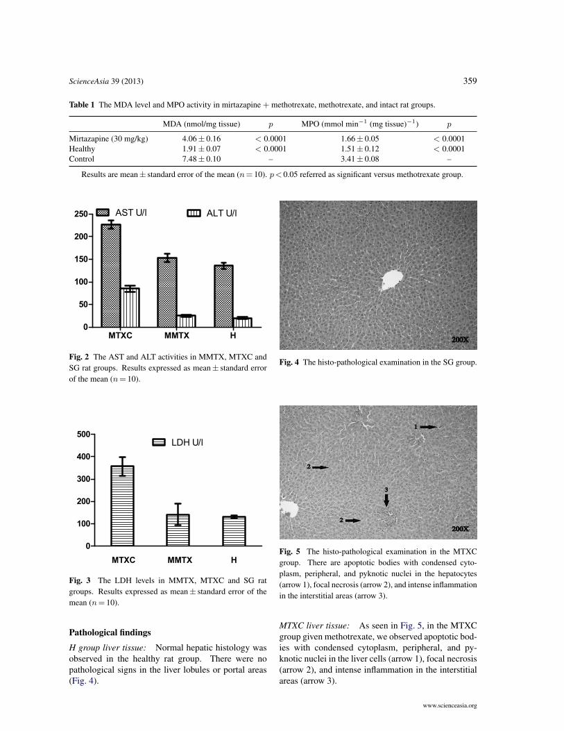
MDA, MPO, GSH, SOD results: As shown inTable 1 and Fig. 1, MDA, MPO, GSH, and SOD levelsin the liver tissues of the rats receiving methotrex-ate in the MTXC group were 7.5± 0.1 µmol/g pro-tein, 3.4± 0.1 µmol/g protein, 2.4± 0.1 nmol/g pro-tein, and 5.5± 0.2 µmol/g protein, respectively. Inthe MMTX group, MDA, MPO, GSH, and SODvalues were 4.1± 0.2 µmol/g protein (p < 0.001),1.7± 0.1 µmol/g protein (p< 0.001), 4.6± 0.1 nmol/gprotein (p < 0.001), and 6.4± 0.1 µmol/g protein(p < 0.001), respectively. In the H group, MDA,MPO, GSH, and SOD levels were 1.9± 0.1 µmol/gprotein (p < 0.001), 1.5± 0.1 U/g protein (p <0.001), 4.9± 0.1 nmol/g protein (p < 0.001), and8.3± 0.2 U/g (p< 0.001), respectively.
AST, ALT, LDH results: As shown in Fig. 2 andFig. 3, blood AST, ALT, and LDH levels in theMTXC group were 227± 3 U/l, 84.8± 2.3 U/l, and357± 13 U/l, respectively, compared to 153± 3 U/l(p < 0.0001), 25.1± 0.8 U/l (p < 0.0001), and142± 15 U/l (p < 0.0001) in the MMTX group.In the H group, AST, ALT, and LDH levels were135.8± 2.1 U/l (p < 0.001), 20.4± 0.9 U/l (p <0.001), and 131.4± 1.6 U/l (p< 0.001), respectively.
www.scienceasia.org
ScienceAsia 39 (2013) 359
Table 1 The MDA level and MPO activity in mirtazapine + methotrexate, methotrexate, and intact rat groups.
MDA (nmol/mg tissue) p MPO (mmol min−1 (mg tissue)−1) p
Mirtazapine (30 mg/kg) 4.06± 0.16 < 0.0001 1.66± 0.05 < 0.0001Healthy 1.91± 0.07 < 0.0001 1.51± 0.12 < 0.0001Control 7.48± 0.10 – 3.41± 0.08 –
Results are mean± standard error of the mean (n= 10). p< 0.05 referred as significant versus methotrexate group.
MTXC MMTX H0
50
100
150
200
250 AST U/l ALT U/l
Fig. 2 The AST and ALT activities in MMTX, MTXC andSG rat groups. Results expressed as mean± standard errorof the mean (n= 10).
MTXC MMTX H
0
100
200
300
400
500LDH U/l
Fig. 3 The LDH levels in MMTX, MTXC and SG ratgroups. Results expressed as mean± standard error of themean (n= 10).
Pathological findings
H group liver tissue: Normal hepatic histology wasobserved in the healthy rat group. There were nopathological signs in the liver lobules or portal areas(Fig. 4).
Fig. 4 The histo-pathological examination in the SG group.
Fig. 5 The histo-pathological examination in the MTXCgroup. There are apoptotic bodies with condensed cyto-plasm, peripheral, and pyknotic nuclei in the hepatocytes(arrow 1), focal necrosis (arrow 2), and intense inflammationin the interstitial areas (arrow 3).
MTXC liver tissue: As seen in Fig. 5, in the MTXCgroup given methotrexate, we observed apoptotic bod-ies with condensed cytoplasm, peripheral, and py-knotic nuclei in the liver cells (arrow 1), focal necrosis(arrow 2), and intense inflammation in the interstitialareas (arrow 3).
www.scienceasia.org

360 ScienceAsia 39 (2013)
Fig. 6 The histo-pathological examination in the MMTXgroup. There was isolated necrosis (arrow).
MMTX liver tissue: No apoptotic bodies or inflam-mation were observed in the MMTX group, onlyisolated necrosis (Fig. 6).
DISCUSSION
This study was a biochemical and histopatholog-ical investigation of the effect of mirtazapine onmethotrexate hepatotoxicity in rats. The results ofbiochemical tests showed a significant increase in theliver tissue of the MTXC group in the levels of oxidantparameters, e.g., MDA and MPO, and a decrease inanti-oxidant parameters, e.g., GSH and SOD. Oxida-tive stress is implicated in methotrexate toxicity in theliver11. Oxidative stress is an indicator of the damagethat results from a change in the balance betweenoxidants and anti-oxidants in favour of oxidants. Mir-tazapine, which we used in our experiment, has beenobserved to significantly inhibit MDA increase dueto methotrexate in the liver. MDA is a 3-carbonaldehyde, the end-product of lipid peroxidation, and isused as a marker of oxidative stress12. MDA releasedafter peroxidation of lipids causes irreversible damageto cells and organelle contents13. Increased levels ofplasma MDA may be attributed to overproduction ofreactive oxygen species or a deficiency of antioxidantdefence14. There are studies showing that MDAlevels increase in hepatic injury caused by oxidativestress15. The high level of MDA in the MTXC groupliver tissue indicates that methotrexate gives rise tooxidative stress in hepatic tissue.
MPO activity in our study was higher in com-parison with the H and MMTX groups in the livertissues of the animals receiving methotrexate. MPOis released from activated neutrophils and is generallyused as a major marker of inflammation16. Previ-
ous experimental studies with rats have reported thatmethotrexate increased levels of MPO, a marker ofneutrophil infiltration5. Another study showed thatmethotrexate increased MPO activity, a marker ofinflammatory response in live and other tissues, inrats6. Free radicals lead to activation of neutrophils.Activated neutrophils release extensive amounts ofMPO in the tissue areas damaged by free radicals.This situation leads to further exacerbation of tissuedamage17.
If the delicate balance between oxidants andanti-oxidants cannot be maintained in tissues, manypathological changes extending to cellular damageoccur. GSH is an endogenous anti-oxidant, whichexists within many cells of the body and protects thefunctional proteins of the cell against oxidant agents.In our study, mirtazapine significantly eliminatedthe inhibitor effect of methotrexate on GSH produc-tion. Studies in the literature have demonstrated thatmethotrexate reduces the levels of glutathione in livercells18. A methotrexate-associated fall in glutathionelevels leads to hepatocyte injury.
In the present study, SOD activity was signifi-cantly lower in the MTXC group in comparison withthe MMTX and H groups. The function of SODis to catalyse the dismutation of O2 and to protectthe tissue against the harmful effects of toxic oxygenradicals19. Yagmurca et al demonstrated that SODactivity reduced doxorubicin hepatotoxicity in rats20.If oxidative damage is involved in the beginningor pathology of the disease, successful anti-oxidanttherapy may prevent the disease occurring or delay itsonset.
Our study also ascertained the effects ofmethotrexate on liver enzymes, e.g., AST, ALT, andLDH. These are associated with hepatocellular injury.ALT and AST are of particular importance in thedetermination of liver damage. AST, ALT and LDHlevels were higher in the MTXC group blood samplesin comparison with the MMTX and H groups. Previ-ous studies have showed that methotrexate increasesliver enzymes. The serum transaminases have highsensitivity in demonstrating hepatocyte injury. Oneof the most reliable parameters showing cell destruc-tion in the liver is ALT level21. The group withthe highest ALT level in this study was the MTXCgroup, while the MMTX group had the lowest level.AST and ALT have been confirmed as markers ofhepatic cell injury22. Antioxidants have been shownto prevent severe increases in LDH, AST, ALT andantioxidant parameters in liver injury. In the MTXCgroup, in which LDH, AST, ALT activities and ox-idant parameters were higher, there were apoptotic
www.scienceasia.org

ScienceAsia 39 (2013) 361
bodies, focal necrosis and intense inflammation in theinterstitial areas. In contrast, in the MMTX groupthere were only a few necrotic cells. Previous studieshave also shown that methotrexate intensifies apopto-sis23. Caspase is a gene that controls cell apoptosis.Lipid peroxidation established by free radicals hasbeen shown to increase apoptosis by stimulating thecaspase gene. Cell apoptosis intensified in hepaticinjury induced with methotrexate in rats has beenreported24. Apoptosis and necrosis may be activatedwith the same stimuli25. In necrosis, cytoplasmicand nuclear contents are released into the intercellularspace. Release of cellular contents into intercellu-lar space leads to inflammation. The distinguishingcharacteristic of this phenomenon is that macrophagesand neutrophils migrate to necrotic tissue. Thesemigrating cells cause phagocytosis in necrotic tissues.Inflammation is therefore a significant indicator ofnecrosis26. These findings from the literature arecompatible with our own histopathological findings.Mirtazapine blocks 5-HT2 and 5-HT3 receptors withits anti-oxidant property27. Stimulation of both 5-HT2and 5-HT3 receptors is known to be associated withtoxic adverse effects28.
CONCLUSIONS
Methotrexate led to oxidative stress in the rat liver,while mirtazapine significantly prevented methotrex-ate-induced oxidative stress. This shows that thedesired dose of methotrexate can safely be used withmirtazapine in the treatment of cancer and non-cancerdiseases.
Acknowledgements: This study was supported by theScientific Research Project of Ataturk University-BAP2005/160-2008/126. We thank Prof. Fatih Akcay for hiscontributions to this study.
REFERENCES1. Hytiroglou P, Tobias H, Saxena R, Abramidou M,
Papadimitriou CS, Theise ND (2004) The canals ofhering might represent a target of methotrexate hepatictoxicity. Am J Clin Pathol 121, 324–9.
2. Sener G, Eksioglu-Demiralp E, Cetiner M, Ercan F,Yegen BC (2006) Beta-glucan ameliorates methotrex-ate-induced oxidative organ injury via its antioxidantand immunomodulatory effects. Eur J Pharmacol 542,170–8.
3. Jolivet J, Cowan KH, Curt GA, Clendeninn NJ, Chab-ner BA (1983) The pharmacology and clinical use ofmethotrexate. New Engl J Med 309, 1094–104.
4. Whittle SL, Hughes RA (2004) Folate supplementationand methotrexate treatment in rheumatoid arthritis: areview. Rheumatology 43, 267–71.
5. Cetinkaya A, Bulbuloglu E, Kurutas EB, KantarcekenB (2006) N-acetylcysteine ameliorates methotrexate-induced oxidative liver damage in rats. Med Sci Monit12, 274–8.
6. Jahovic N, Cevik H, Sehirli AO, Yegen BC, Sener G(2003) Melatonin prevents methotrexate-induced hepa-torenal oxidative injury in rats. J Pineal Res 34, 282–7.
7. Bilici M, Ozturk C, Dursun H, Albayrak F, Saglam MB,Uyanik A, Gulaboglu M, Tekin SB (2009) Protectiveeffect of mirtazapine on indomethacin-induced ulcer inrats and its relationship with oxidant and antioxidantparameters. Dig Dis Sci 54, 1868–75.
8. Sedlak J, Lindsay RH (1968) Estimation of total,protein-bound, and nonprotein sulfhydryl groups in tis-sue with Ellman’s reagent. Anal Biochem 25, 192–205.
9. Bradley PP, Priebat DA, Christensen RD, Rothstein G(1982) Measurement of cutaneous inflammation: esti-mation of neutrophil content with an enzyme marker.J Investig Dermatol 78, 206–9.
10. Ohkawa H, Ohishi N, Yagi K (1979) Assay for lipidperoxides in animal tissues by thiobarbituric acid reac-tion. Anal Biochem 95, 351–8.
11. Devrim E, Cetin R, Kilicoglu B, Erguder BI, Avci A,Durak I (2005) Methotrexate causes oxidative stress inrat kidney tissues. Ren Fail 27, 771–3.
12. Cighetti G, Duca L, Bortone L, Sala S, Nava I, FiorelliG, Cappellini MD (2002) Oxidative status and malon-dialdehyde in β-thalassaemia patients. Eur J Clin In-vestig 32, Suppl 1, 55–60.
13. Gutteridge JM (1995) Lipid peroxidation and antioxi-dants as biomarkers of tissue damage. Clin Chem 41,1819–28.
14. Amirkhizi F, Siassi F, Minaie S, Djalali M, RahimiA, M, C (2008) Assessment of lipid peroxidationand activities of erythrocyte cytoprotective enzymes inwomen with iron deficiency anemia. J Res Med Sci 13,248–54.
15. Matos HR, Capelozzi VL, Gomes OF, Di Mascio P,Medeiros MHG (2001) Lycopene inhibits DNA dam-age and liver necrosis in rats treated with ferric nitrilo-triacetate. Arch Biochem Biophys 396, 171–7.
16. Song M, Song Z, Barve S, Zhang J, Chen T, Liu M,Arteel GE, Brewer GJ, McClain CJ (2008) Tetrathio-molybdate protects against bile duct ligation-inducedcholestatic liver injury and fibrosis. J Pharmacol ExpTherapeut 325, 409–16.
17. Sullivan GW, Sarembock IJ, Linden J (2000) The roleof inflammation in vascular diseases. J Leukoc Biol 67,591–602.
18. Babiak RM, Campello AP, Carnieri EG, OliveiraMB (1998) Methotrexate: pentose cycle and oxidativestress. Cell Biochem Funct 16, 283–93.
19. Fridovich I (1995) Superoxide radical and superoxidedismutases. Annu Rev Biochem 64, 97–112.
20. Yagmurca M, Bas O, Mollaoglu H, Sahin O, NacarA, Karaman O, Songur A (2007) Protective effectsof erdosteine on doxorubicin-induced hepatotoxicity in
www.scienceasia.org

362 ScienceAsia 39 (2013)
rats. Arch Med Res 38, 380–5.21. Hamanoue M, Kawaida K, Takao S, Shimazu H,
Noji S, Matsumoto K, Nakamura T (1992) Rapid andmarked induction of hepatocyte growth factor duringliver regeneration after ischemic or crush injury. Hepa-tology 16, 1485–92.
22. Desai SP, Isa-Pratt S (2004) Clinician’s Guide toLaboratory Medicine. Chapter 66. Lexi-Comp Inc.pp 612–3.
23. Horie T, Li T, Ito K, Sumi S, Fuwa T (2006) Aged garlicextract protects against methotrexate-induced apoptoticcell injury of IEC-6 cells. J Nutr 136, 861S–3S.
24. Demiryilmaz I, Sener E, Cetin N, Altuner D, SuleymanB, Albayrak F, Akcay F, Suleyman H (2012) Bio-chemically and histopathologically comparative reviewof thiamine’s and thiamine pyrophosphate’s oxidativestress effects generated with methotrexate in rat liver.Med Sci Monit 18, 475–81.
25. Bonfoco E, Krainc D, Ankarcrona M, Nicotera P,Lipton SA (1995) Apoptosis and necrosis: two distinctevents induced, respectively, by mild and intense in-sults with N-methyl-D-aspartate or nitric oxide/super-oxide in cortical cell cultures. Proc Natl Acad Sci UnitStates Am 92, 7162–6.
26. Schwartzman RA, Cidlowski JA (1993) Apoptosis: thebiochemistry and molecular biology of programmedcell death. Endocr Rev 14, 133–51.
27. Agargun MY, Ebrinc S (1998) Mirtazapine: a review.Bull Clin Psychopharmacol 8, 59–68.
28. Frazer A (1994) Antidepressant drugs. Depression 2,1–19.
www.scienceasia.org


![Research Paper The association between RFC 1 G80A ...[17], and RFC1 gene variation can affect the outcome and toxicity of methotrexate (MTX) therapy in leu-kemia [18]. RFC1 G80A polymorphism](https://img.dokumen.tips/doc/110x75/5ed05af527e719381c0e844e/research-paper-the-association-between-rfc-1-g80a-17-and-rfc1-gene-variation.jpg)