Embed Size (px)
Citation preview

1
The impact of food components on the intrinsic dissolution rate of ketoconazole
Authors
Heba S. Ghazal a, A. Mark Dyas b, James L. Ford a and Gillian A. Hutcheon a*
a School of Pharmacy and Biomolecular Sciences, Liverpool John Moores University, Byrom
St., Liverpool L3 3AF, UK
b Novaliq GmbH, Im Neuenheimerfeld 515, .D-69120 Heidelberg, Germany
* Corresponsing author
Dr Gillian A Hutcheon
School of Pharmacy and Biomolecular Sciences
Byrom Street
Liverpool
L3 3AF, UK
Email: [email protected]
Phone: 0151 2312130
Keywords: Solubility, dissolution, bioavailability, biorelevant dissolution media, gastric
fluid, food additives.
2
Abstract
To accurately predict the in vivo performance of drugs from an in vitro dissolution test, the
dissolution conditions used are supposed to be similar to those present in the gastrointestinal
milieu. Post-prandial gastric fluid contains partially digested food mixtures consisting of fat,
protein and carbohydrate. Despite this, the compendial dissolution medium recommended to
simulate the gastric fluid is still composed of simple solution of hydrochloric acid and sodium
chloride with or without the addition of pepsin. Therefore, in this investigation, biorelevant
dissolution media were developed to evaluate the impact of food constituents; milk with
different fat contents, egg albumin, gelatin, casein, gluten, carbohydrates and amino acids on
the intrinsic dissolution behaviour of ketoconazole. Most of the food additives that were
evaluated enhanced the apparent solubility of the drug but to different extents. The greatest
enhancement in dissolution was observed in media containing either neutral amino acids or
media based on milk mixtures. The formation of complexes between the drug and the
additives most likely accounted for the solubilising effect and in milk-containing media the
effect was attributed to the whole complex structure of milk rather than simply its fat content.
These results highlight the potential effect of the type of ingested meal on drug dissolution
and subsequent bioavailability.

3
1. Introduction
Dissolution testing is a potential predictive tool for the oral absorption of drugs but, to
achieve this, test conditions need to simulate the physiological conditions prevailing in the
intraluminal environment. Therefore, efforts have been made to develop biorelevant media
which represent gastrointestinal fluids at fed and fasting states.
The composition of the gastric fluid in post-prandial conditions is variable according to the
type of the administered meal (1). Therefore, to tackle the problem of variability in gastric
fluid composition, biorelevant dissolution media were suggested to standardize the
composition of simulated gastric fluids at fed state. Milk was utilised as it is a nutrient that
contains the three basic components of fat, protein and carbohydrates in a ratio resembling a
typical diet (2). In 2008, Jantratid et al. (3) recommended the use of a mixture of milk and
acetate buffer and subsequently a formulation of milk digested with pepsin and lipase was
proposed to represent the changes in intra-gastric composition according to the digestion
stage (4). Artificial liquid meals, such as Ensure™ and Intralipid™ were recommended as
they are specifically designed to reflect the composition of a standard meal and can be well
standardised (5).The use of Ensure Plus™ containing a viscosity enhancer, 0.45% pectin (6) or
medium containing 1.4% HPMC in acetate buffer (7) were suggested as an alternative media
to the homogenised standard breakfast. A medium containing two types of vegetable oils,
sucrose and surfactants was used to mimic fed gastric conditions (8). Media based on mono-
components of proteins, carbohydrates or amino acids were utilised as simple dissolution
media that allowed the investigation of the effect of these individual components on the
dissolution of drugs (9, 10).
Drugs present in the gastric fluids are exposed to mixtures of foodstuffs made up of protein,
carbohydrates, fats and amino acids and dissolution in the intra-gastric fluid is essential for
4
the subsequent absorption of basic lipophilic drugs, in particular. Hence, we previously
evaluated the dissolution of itraconazole in biorelevant media (10). Interestingly, the inclusion
of food additives, such as milk and egg albumin, induced a pronounced increase in the
dissolution of itraconazole, which suggested that the type of diet can influence drug
dissolution in vivo and its subsequent bioavailability.
In the present investigation, these studies were extended to examine and compare the impact
of similar biorelevant dissolution media on the dissolution behaviour of a second basic poorly
soluble azole anti-fungal drug, ketoconazole. The drug is classified as class II according to
the Biopharmaceutics Classification System (BCS) so it is more likely to exhibit dissolution-
rate-limited absorption problems (2). Clinical studies showed that when ketoconazole oral
dosage forms were given with a breakfast meal the bioavailability of the drug increased (11).
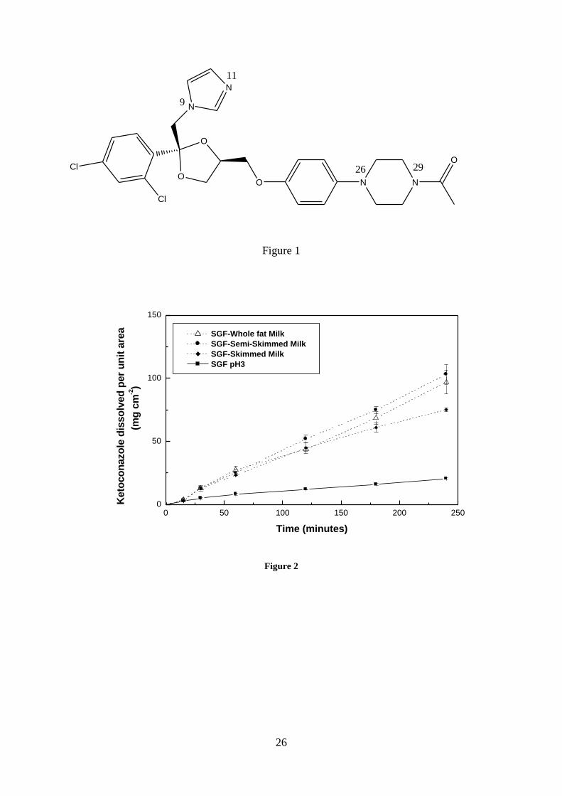
The structure of ketoconazole (log P is 3.73) presents two basic groups, a piperazine ring and
an imidazole ring, with pKa values of 2.94 and 6.51, respectively (Figure 1). Thus, there are
two basic centres for protonation, N26 of the piperazine ring and N11 of the imidazole ring.
The solubility of ketoconazole has previously been studied in human gastric fluid (12, 13) and
the dissolution of its oral dosage forms was investigated in biorelevant media based on milk
(2). However, studying the intrinsic dissolution of a drug through the determination of the
intrinsic dissolution rate (IDR) from constant surface area of compact drug powder is of
particular interest since it is independent of the formulation, excipient effects and the size of
the particles (14).
Thus, the objective of the present work was to characterise the intrinsic dissolution
performance of ketoconazole in biorelevant dissolution media containing food constituents as
a representative for gastric fluid at fed state.

5
2 Materials and methods
2.1 Chemicals and reagents
Ketoconazole was obtained from Medichem (Shenzhen, Guangdong, China). Casein from
bovine milk, gluten from wheat, gelatin from bovine skin, glycine (GLY), leucine (LYS),
aspartic acid (ASP) were purchased from Sigma-Aldrich (Steinheim, Germany). Albumin
from hen egg white, alanine (ALA) and lysine (LYS) were purchased from Fluka (Sigma-
Aldrich, Steinheim, Germany). Glucose and starch were purchased from BDH (Poole, UK)
and lactose was obtained from Foremost (Baraboo, Wisconsin, USA). Three types of fresh
pasteurised bovine milk, whole milk, semi-skimmed milk and skimmed milk (Express
dairies) with fat contents of 3.6%, 1.7% and 0.1%, respectively were purchased from local
retail outlets (Liverpool, UK).
Sodium hydroxide pellets, potassium dihydrogen phosphate, sodium chloride and
hydrochloric acid and as well acetonitrile were all purchased from BDH (Poole, UK).
Triethylamine and n-butyl chloride were purchased from Fluka (Sigma-Aldrich, Steinheim,
Germany).
2.2 Composition of dissolution media
Simulated gastric fluid (SGF) pH 1.2 without enzymes contained 2g/L NaCl and 7mL/L HCl
(to adjust the pH to 1.2) in deionised water. SGF pH 3 also contained 2g/L NaCl in deionised
water and the pH adjusted to 3 using 0.1M HCl.
The composition and preparation of the dietary media were as previously described (10). SGF
media containing milk were prepared using equal volumes of milk and SGF pH 1.2. The final
pH was adjusted to 3 using either 0.1M HCl or 0.1M NaOH. Media containing a single
dietary component were prepared by dissolving or dispersing the substance in SGF then the

6
final pH was adjusted to 3 with a solution of HCl (1 or 0.1M). Four proteins were chosen as
models of proteins available in common diet. Albumin (0.01, 0.1, 0.5%, 1%, and 3%, w/v)
and gelatin (1%, 2% and 4%, w/v) were directly added to SGF. A saturated solution of gluten
was prepared by adding of an excess amount of gluten to deionised water and stirring
overnight before filtration through Whatman filter paper type: 1 and the addition of NaCl (2g
L-1). A filtered Casein solution was also prepared following same procedure and further
dilutions (0.005, 0.0038, 0.0025 and 0.0013%, w/v) were prepared using SGF (pH 3). The
final pH of all media was 3.
Amino acids were chosen to represent basic (LYS at 1%, w/v), neutral (GLY, ALA and LEU,
each at 1%, w/v) and acidic amino acids (saturated solution of ASP). Three different
carbohydrates were chosen as examples of monosaccharides (glucose), disaccharides
(lactose) and polysaccharides (starch), each at 1%, w/v.
Simulated intestinal fluid (SIF) was prepared as described in the USP 32 (14). Phosphate
buffer pH (7.5) and acetate buffer (pH 5) were prepared according to the BP (2012) (15).
2.3 Determination of saturation solubility
The solubility of ketoconazole in each dissolution medium was determined using a modified
‘shake-flask method’ (16). Each experiment was performed in triplicate. Medium (10mL) and
approximately 300mg of drug powder were transferred into closed-cap vials, shaken gently
for 24h at 37 °C in a shaking incubator (Model AM89B, Dynex Technologies Ltd). After this
time, the suspension was centrifuged at 4000rpm for 10 min (Centaur 2, MSE, Fisons).
The supernatants were collected and filtered through a 0.2μm PVDF syringe with the first
portion of each filtrate being discarded to circumvent any initial adsorption of the drug to the
filters. Dissolution media which contained particulate matter (milk and albumin containing-
7
media) clogged these filters which required the use of filters with a bigger pore size (17).
Filters were validated prior to their use to verify that they did not adsorb the drug and hence
affect the results of analysis. For the albumin-containing media, 1m glass filters (Gelman
Sciences) were used and 5m Acrodisc Versapor filters (Gelman Sciences, medium: acrylic
polymer) were used for the milk-containing samples. Ketoconazole filtrates were diluted as
required with a mixture of acetonitrile-water (50:50, v/v) and then submitted to HPLC
analysis.
2.4 Intrinsic dissolution disks and dissolution conditions
Intrinsic dissolution rate (IDR) testing was performed using a stationary disk apparatus. A
hardened polished steel plate covered with aluminium foil was attached to the steel die and
the drug powder (approximately 200mg) was inserted into the cavity of the steel die (9.5mm
diameter). The steel plate was occasionally re-polished to enhance the smoothness of the
surface and so allow the dissolution to take place evenly across the surface (18). A hardened-
steel punch was inserted to die cavity then the whole arrangement was transferred to a
hydraulic compressor and compressed under a pressure of 1000 p.s.i. for 30s yielding a disk
of circular surface area. The disks were blown with compressed air to remove any loose
particles.
Dissolution studies were carried out using USP apparatus II (paddle) (Pharmatest PTW S3C,
Pharmatest GmbH). The die, containing the compressed drug powder, was positioned disk-up
at the bottom of a flat-bottom dissolution vessel containing 500mL of the dissolution
medium. The temperature was set at 37 ± 0.5C and a rotation speed of 100rpm was applied.
Samples (3mL) were withdrawn periodically from the dissolution vessels through 0.45m
filters except for particulate complex dissolution media where samples were withdrawn
through 20μm filters.

8
Compression is a high energy process which may affect the crystallinity of substances and
different polymorphs may exhibit different solubility behaviours. Therefore, studies were
carried out to determine whether compression of ketoconazole into IDR disks led to any
change in polymorphic form. Differential scanning calorimetry, infra-red spectroscopy and
powder x-ray diffractometry were employed and no evidence that compressing the
ketoconazole powder affected the solid-state of the drug was observed.
2.5 Extraction of ketoconazole from IDR sample solutions
The drug was extracted from the complex dissolution media by liquid-liquid extraction at
alkaline pH. Samples (1mL) removed from the dissolution media were alkalinised with 1mL
of 0.05M NaOH and shaken mechanically for 10 min. 5mL of a mixture of acetonitrile:n-
butyl chloride (1:4, v/v) was added, shaken for 1 min and then centrifuged at 4000 rpm for 10
min. 2mL aliquots were collected from the upper layer and evaporated to dryness with
nitrogen at 60C. The residues were reconstituted in 2mL of eluent (mobile phase used for
HPLC) by sonication for 2 min and subsequently analysed using HPLC.
2.6 Analytical method
Drug quantitation was carried out using a HPLC separation module Waters Alliance 2695
chromatograph using a Waters 996 Photodiode Array Detector (PAD). Ketoconazole samples
(20L) were eluted using a mobile phase consisting of acetonitrile-water-triethylamine
(50:50:0.1 v/v/v), with a flow rate of 1.1mL min-1. A 5μm Hypersil BDS C18 column (150
mm x 4.6mm) (Thermo Electron Corporation) fitted with a Phenomenex C18 guard cartridge
(4 mm x 3mm) was used and eluting peaks were detected at a wavelength of 254nm.
Quantitation was based on the use of an external ketoconazole calibration standard and peak
area measurement. A plot of the amount dissolved per unit area versus time was constructed

9
and the slope of the fitted linear regression represented the IDR. The initial dissolution rate
referred to the period 0-15min and the subsequent rate referred to the period 15-240min.
3 Results and discussion
3.1 Dissolution and saturation solubility in conventional media
Ketoconazole solubility was highly dependent on the pH of the medium. At pH 3 all the
imidazole moieties (pKa1=6.5) and nearly 50% of the piperazine moieties (pKa2= 2.9) were
protonated. By lowering the pH further to 1.2, both base moieties were protonated leading to
a significant increase in solubility under the more acidic conditions (Table 1).
The IDR of the drug could not be determined at pH 1.2 due to its high solubility at that pH
whereby the disks entirely dissolved within 15min. The IDR data at pH 3 and at pH 6.8
presented in Table 2 indicate the profound effect of the pH of the dissolution medium on the
rate and extent of drug dissolution. The dissolution data in SGF (pH 3) were considered as a
reference and employed for subsequent comparison with the dissolution profiles in more
complex media.
The increase in the dissolution rate and solubility at pH 1.2 confirms the importance of the
gastric acidity on the bioavailability of ketoconazole. Clinical studies reported that the
bioavailability of the drug decreased when the gastric pH increased by administering the drug
concomitantly with antacids or H2 blockers (19).
3.2 Dissolution and saturation solubility in milk-containing media
The solubility of ketoconazole in milk-SGF mixtures (1:1) was approximately 6 times greater
than the solubility in SGF alone (Table 3). No significant difference between the solubility
10
values for the three different milk-containing media was observed, according to the one-way
analysis of variance (ANOVA) at significance level of 0.05.
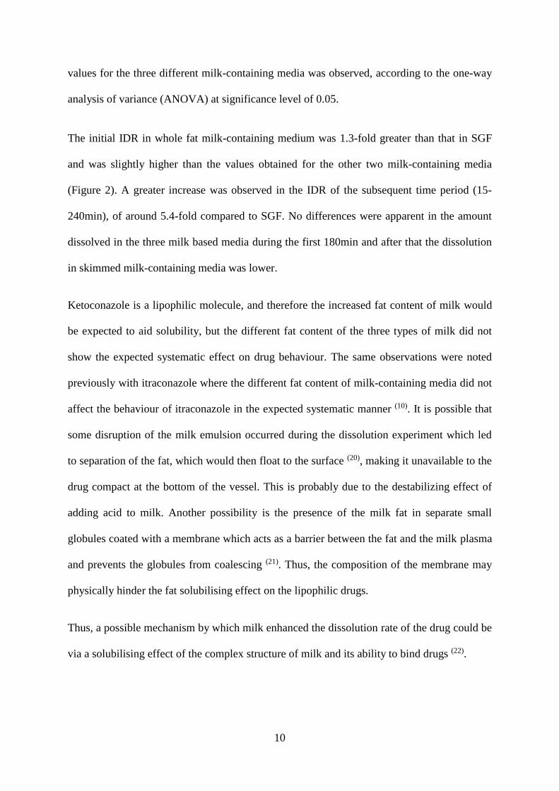
The initial IDR in whole fat milk-containing medium was 1.3-fold greater than that in SGF
and was slightly higher than the values obtained for the other two milk-containing media
(Figure 2). A greater increase was observed in the IDR of the subsequent time period (15-
240min), of around 5.4-fold compared to SGF. No differences were apparent in the amount
dissolved in the three milk based media during the first 180min and after that the dissolution
in skimmed milk-containing media was lower.
Ketoconazole is a lipophilic molecule, and therefore the increased fat content of milk would
be expected to aid solubility, but the different fat content of the three types of milk did not
show the expected systematic effect on drug behaviour. The same observations were noted
previously with itraconazole where the different fat content of milk-containing media did not
affect the behaviour of itraconazole in the expected systematic manner (10). It is possible that
some disruption of the milk emulsion occurred during the dissolution experiment which led
to separation of the fat, which would then float to the surface (20), making it unavailable to the
drug compact at the bottom of the vessel. This is probably due to the destabilizing effect of
adding acid to milk. Another possibility is the presence of the milk fat in separate small
globules coated with a membrane which acts as a barrier between the fat and the milk plasma
and prevents the globules from coalescing (21). Thus, the composition of the membrane may
physically hinder the fat solubilising effect on the lipophilic drugs.
Thus, a possible mechanism by which milk enhanced the dissolution rate of the drug could be
via a solubilising effect of the complex structure of milk and its ability to bind drugs (22).
11
3.3 Dissolution and saturation solubility in the presence of proteins
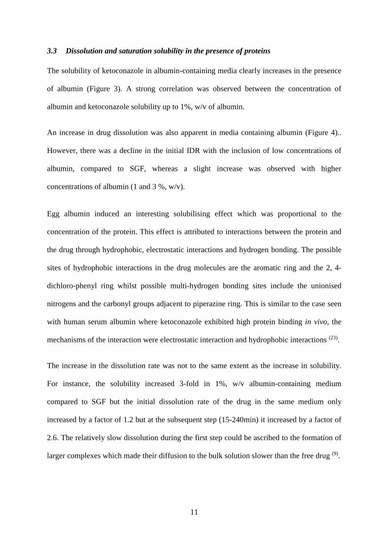
The solubility of ketoconazole in albumin-containing media clearly increases in the presence
of albumin (Figure 3). A strong correlation was observed between the concentration of
albumin and ketoconazole solubility up to 1%, w/v of albumin.
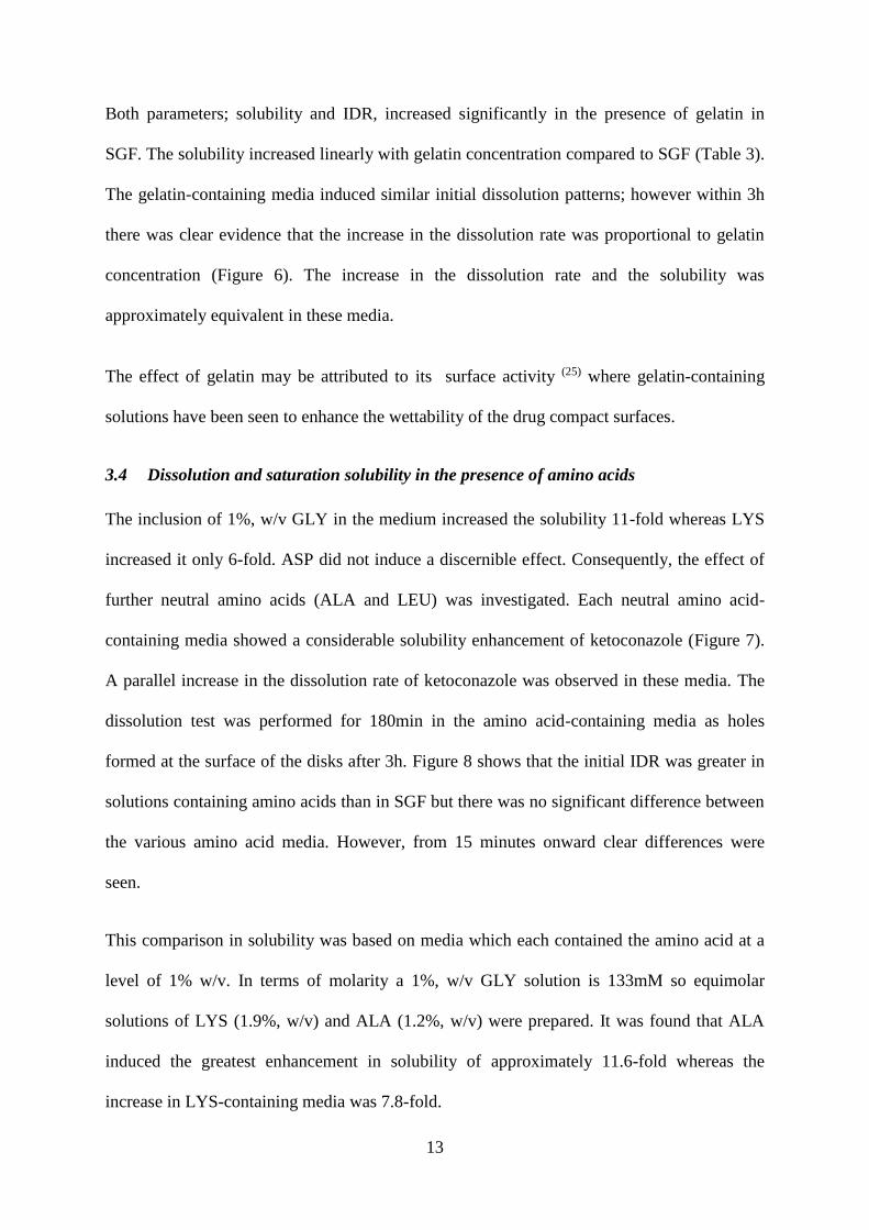
An increase in drug dissolution was also apparent in media containing albumin (Figure 4)..
However, there was a decline in the initial IDR with the inclusion of low concentrations of
albumin, compared to SGF, whereas a slight increase was observed with higher
concentrations of albumin (1 and 3 %, w/v).
Egg albumin induced an interesting solubilising effect which was proportional to the
concentration of the protein. This effect is attributed to interactions between the protein and
the drug through hydrophobic, electrostatic interactions and hydrogen bonding. The possible
sites of hydrophobic interactions in the drug molecules are the aromatic ring and the 2, 4-
dichloro-phenyl ring whilst possible multi-hydrogen bonding sites include the unionised
nitrogens and the carbonyl groups adjacent to piperazine ring. This is similar to the case seen
with human serum albumin where ketoconazole exhibited high protein binding in vivo, the
mechanisms of the interaction were electrostatic interaction and hydrophobic interactions (23).
The increase in the dissolution rate was not to the same extent as the increase in solubility.
For instance, the solubility increased 3-fold in 1%, w/v albumin-containing medium
compared to SGF but the initial dissolution rate of the drug in the same medium only
increased by a factor of 1.2 but at the subsequent step (15-240min) it increased by a factor of
2.6. The relatively slow dissolution during the first step could be ascribed to the formation of
larger complexes which made their diffusion to the bulk solution slower than the free drug (9).

12
The solubility studies indicated a slight enhancement in solubility in 0.0038 and 0.005%, w/v
casein-containing solutions whereas the more dilute solutions of casein did not induce a
significant effect (Table 3). The data presented in (Figure 5) demonstrate that there was an
increase in dissolution rate of ketoconazole in casein saturated solution compared to the SGF.
The initial IDR was 1.5-fold greater than its counterpart in SGF however the IDR
representing the subsequent stage in casein solution was not determined due to its poor
linearity.
It was reported previously that the mechanism by which casein affected drug dissolution was
via micelle formation (9). Casein molecules have a strong tendency to self-assemble into
micelles because of their amphiphilic nature in aqueous solution. Various models of the
assembly and structure of the casein have been suggested in the literature of which the sub-
unit model for casein aggregation was the most widely accepted. This model suggests that
15–20 molecules of casein aggregate via hydrophobic interactions and form sub-units where
the hydrophobic core is surrounded by a polar portion. These sub-units form the building
units of the micelles (24). Casein monomers and sub-units exist in the solution when the casein
concentration is below the CMC, whereas above the CMC casein micelles are combined with
monomers and sub-micelles. Based on this structure, casein is able to encapsulate
hydrophobic compounds into the hydrophobic core even at the sub-micellar level.
Consequently, ketoconazole could be solubilised through these sub-units.
The inclusion of gluten in the media did not induce a discernible effect on either the solubility
or the dissolution rate of ketoconazole (Figure 5). This indicated that this protein could not
solubilise ketoconazole which agrees with the previous finding regarding itraconazole
dissolution in this medium (10).
13
Both parameters; solubility and IDR, increased significantly in the presence of gelatin in
SGF. The solubility increased linearly with gelatin concentration compared to SGF (Table 3).
The gelatin-containing media induced similar initial dissolution patterns; however within 3h
there was clear evidence that the increase in the dissolution rate was proportional to gelatin
concentration (Figure 6). The increase in the dissolution rate and the solubility was
approximately equivalent in these media.
The effect of gelatin may be attributed to its surface activity (25) where gelatin-containing
solutions have been seen to enhance the wettability of the drug compact surfaces.
3.4 Dissolution and saturation solubility in the presence of amino acids
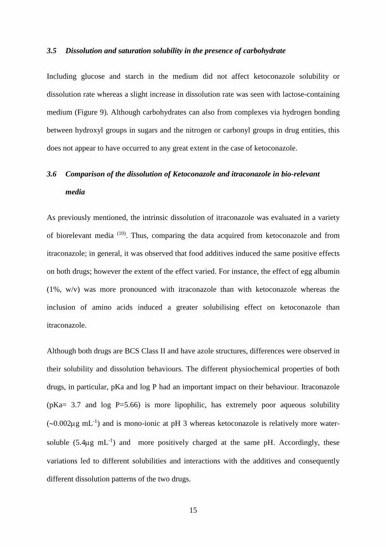
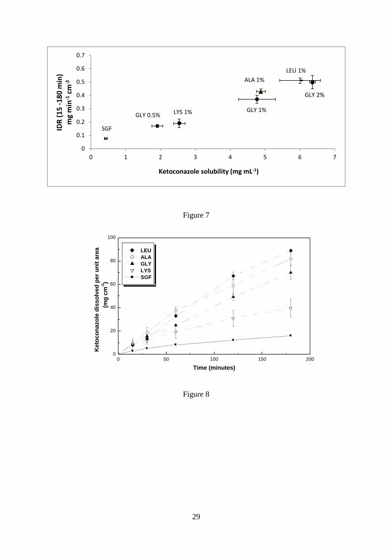
The inclusion of 1%, w/v GLY in the medium increased the solubility 11-fold whereas LYS
increased it only 6-fold. ASP did not induce a discernible effect. Consequently, the effect of
further neutral amino acids (ALA and LEU) was investigated. Each neutral amino acid-
containing media showed a considerable solubility enhancement of ketoconazole (Figure 7).
A parallel increase in the dissolution rate of ketoconazole was observed in these media. The
dissolution test was performed for 180min in the amino acid-containing media as holes
formed at the surface of the disks after 3h. Figure 8 shows that the initial IDR was greater in
solutions containing amino acids than in SGF but there was no significant difference between
the various amino acid media. However, from 15 minutes onward clear differences were
seen.
This comparison in solubility was based on media which each contained the amino acid at a
level of 1% w/v. In terms of molarity a 1%, w/v GLY solution is 133mM so equimolar
solutions of LYS (1.9%, w/v) and ALA (1.2%, w/v) were prepared. It was found that ALA
induced the greatest enhancement in solubility of approximately 11.6-fold whereas the
increase in LYS-containing media was 7.8-fold.

14
Thus, it could be said that the inclusion of neutral amino acids induced a significant increase
in dissolution and solubility. Since these amino acids have identical isoelectric points (PI=6);
the greatest effect being seen with LEU- then ALA- and finally GLY-containing media,
suggest the increase in solubility and dissolution was possibly proportional to the length of
side chain of the amino acids and consequently their overall hydrophobicity.
This effect of the amino acids could be attributed to the formation of a soluble complex with
the drug. At pH 3, ketoconazole molecules coexist in the two ionised forms: mono-protonated
and di-protonated. The acidic groups of the amino acids have a pKa1 of 2.3 and thus at pH 3,
the majority of these acidic moieties are deprotonated. Consequently, ionic interactions may
occur between the negatively charged carboxylic acid of the amino acid and the positively
charged drug molecules. Furthermore, hydrogen bonding may also contribute to the
interactions where the uncharged nitrogen or carbonyl groups of the drug are possible sites
for hydrogen bonds with the carboxylic acid groups of the amino acid. In addition, the
solubility of ketoconazole increased with an increase in the hydrophobic character of the
amino acid in the dissolution medium suggesting that hydrophobic interactions could be an
important force in the interactions.
To evaluate the effect of the pH of the medium on the amino acid-solubilising effect,
dissolution was further investigated in SIF (pH 6.8) containing 1%, w/v GLY. In this instance
GLY only increased the dissolution rate 1.8-fold and the solubility 1.3-fold, compared to SIF.
This indicated that the solubilising effect of GLY at pH 6.8 (SIF) was clearly less than at pH
3 (SGF), presumably because ketoconazole has lost most of its positive charge at this pH and
is therefore less able to complex with the amino acid anions.
15
3.5 Dissolution and saturation solubility in the presence of carbohydrate
Including glucose and starch in the medium did not affect ketoconazole solubility or
dissolution rate whereas a slight increase in dissolution rate was seen with lactose-containing
medium (Figure 9). Although carbohydrates can also from complexes via hydrogen bonding
between hydroxyl groups in sugars and the nitrogen or carbonyl groups in drug entities, this
does not appear to have occurred to any great extent in the case of ketoconazole.
3.6 Comparison of the dissolution of Ketoconazole and itraconazole in bio-relevant
media
As previously mentioned, the intrinsic dissolution of itraconazole was evaluated in a variety
of biorelevant media (10). Thus, comparing the data acquired from ketoconazole and from
itraconazole; in general, it was observed that food additives induced the same positive effects
on both drugs; however the extent of the effect varied. For instance, the effect of egg albumin
(1%, w/v) was more pronounced with itraconazole than with ketoconazole whereas the
inclusion of amino acids induced a greater solubilising effect on ketoconazole than
itraconazole.
Although both drugs are BCS Class II and have azole structures, differences were observed in
their solubility and dissolution behaviours. The different physiochemical properties of both
drugs, in particular, pKa and log P had an important impact on their behaviour. Itraconazole
(pKa= 3.7 and log P=5.66) is more lipophilic, has extremely poor aqueous solubility
(0.002g mL-1) and is mono-ionic at pH 3 whereas ketoconazole is relatively more water-
soluble (5.4g mL-1) and more positively charged at the same pH. Accordingly, these
variations led to different solubilities and interactions with the additives and consequently
different dissolution patterns of the two drugs.

16
4 Conclusions
Although food has a buffering effect on the acidity of the gastric fluid which in turn is
thought to suppress the bioavailability of the two basic drugs; itraconazole and ketoconazole,
clinically an increase in the bioavailability of the drugs was reported. In the present study it
was observed that the dissolution of ketoconazole increased with the presence of most of the
dietary additives investigated. Consequently, increased drug dissolution could be identified as
a reason for the increasing bioavailability of this poorly soluble drug at the post-prandial
state, in addition to the physiological changes in response to food ingestion such as
promotion in the bile secretion and extending the gastric residence time. Since additives such
as milk, egg albumin and gelatin increased the solubility of the drug, it might be useful to
employ these substances in developing drug formulations as the use of such natural vehicles
in appropriately designed formulations may enhance the solubility of the poorly soluble drugs
and provide safe and cheap alternative for the synthetic polymers.
In conclusion, biorelevant media based on food constituents are recommended for the
prediction of the behaviour of the drugs in the gastrointestinal tract as they appear as more
realistically representative of the intraluminal fluids than the simple compendial dissolution
media. Such media have the potential to reflect changes in the performance of BCS Class II
drugs due to fed and fasting conditions and, even more specifically, due to the type of the
meal which in turn can affect drug bioavailability. Clinicians can benefit from such
information when prescribing drugs to achieve the desired bioavailability and therapeutic
efficacy.

17
Acknowledgements
The financial support of Damascus University is acknowledged.
Declarations of Interest
The authors report no declarations of interest.
18
References
1. Koziolek M, Garbacz G, Neumann M, Weitschies W. Simulating the postprandial
stomach: biorelevant test methods for the estimation of intragastric drug dissolution. Mole
Pharm, 2013;10:2211-21.
2. Galia E, Nicolaides E, Hoerter D, Loebenberg R, Reppas C, Dressman JB. Evaluation
of various dissolution media for predicting in vivo performance of class I and II drugs. Pharm
Res, 1998;15:698-705.
3. Jantratid E, Janssen N, Reppas C, Dressman J. Dissolution media simulating
conditions in the proximal human gastrointestinal tract: an update. Pharm Res, 2008;25:1663-
76.
4. Diakidou A, Vertzoni M, Abrahamsson B, Dressman J, Reppas C. Simulation of
gastric lipolysis and prediction of felodipine release from a matrix tablet in the fed stomach.
Eur J Pharm Sci, 2009;37:133-40.
5. Buckton G, Beezer AE, Chatham SM, Patel KK. In vitro dissolution testing of oral
controlled release preparations in the presence of artificial foodstuffs. II. Probing drug/food
interactions using microcalorimetry. Int J Pharm, 1989;56:151-7.
6. Klein S, Butler J, Hempenstall JM, Reppas C, Dressman JB. Media to simulate the
postprandial stomach I. Matching the physicochemical characteristics of standard breakfasts.
J Pharm and Pharmacol, 2004;56:605-10.
7. Radwan A, Amidon GL, Langguth P. Mechanistic investigation of food effect on
disintegration and dissolution of BCS class III compound solid formulations: the importance
of viscosity. Biopharm Drug Dispos, 2012;33:403-16.
8. Muschert S, Siepmann F, Leclercq B, Carlin B, Siepmann J. Simulated food effects
on drug release from ethylcellulose: PVA–PEG graft copolymer-coated pellets. Drug Dev Ind
Pharm, 2010;36:173-9.

19
9. Macheras P, Reppas C. Dissolution and in vitro permeation behaviours of dicumarol
nitrofurantoin and sulfamethizole in the presence of protein. Int J Pharm, 1987;37:103-12.
10. Ghazal HS, Dyas AM, Ford JL, Hutcheon GA. In vitro evaluation of the dissolution
behaviour of itraconazole in bio-relevant media. Int J Pharm, 2009;366:117-23.
11. Gascoigne EW, Barton GJ, Michaels M, Meuldermans W. The kinetics of
ketoconazole in animals and man. Clin Res Rev, 1981;1:177-87.
12. Vertzoni M, Pastelli E, Psachoulias D, Kalantzi L, Reppas C. Estimation of
intragastric solubility of drugs: in what medium? Pharm Res, 2007;24:909-17.
13. Diakidou A, Vertzoni M, Dressman J, Reppas C. Estimation of intragastric drug
solubility in the fed state: comparison of various media with data in aspirates. Biopharm Drug
Dispos, 2009;30:318-25.
14. USP. US Pharmacopeia 32. Rockville, MD, USA: US Pharmacopeial Convention;
2009.
15. BP. British Pharmacopoeia: The Stationary Office, London, 2012.
16. Sunesen VH, Pedersen BL, Kristensen HG, Mullertz A. In vivo in vitro correlations
for a poorly soluble drug, danazol, using the flow-through dissolution method with
biorelevant dissolution media. Eur J Pharm Sci, 2005;24:305-13.
17. Nicolaides E, Galia E, Efthymiopoulos C, Dressman JB, Reppas C. Forecasting the in
vivo performance of four low solubility drugs from their in vitro dissolution data. Pharm Res.
1999;16:1876-82.
18. Ghazal H, Ford JL, Dyas AM. Morphological changes observed during intrinsic
dissolution rate testing of itraconazole. J Pharma Pharmacol, 2006;58; SUPP(10;
SUPP/1):138.

20
19. Van Der Meer JWM, Keuning JJ, Scheijgrond HW, Heykants J, Van Cutsem J,
Brugmans J. The influence of gastric acidity on the bio-availability of ketoconazole. J
Antimicrob Chemother, 1980;6:552-4.
20. Fox PF, McSweeney PLH. Stability of the milk fat emulsion. Dairy Chemistry and
Biochemistry. New York: Kluwer Academic / Plenum Publishers; 1998. 104-108.
21. Walstra P, Wouters JM, Geurts TJ. Colloidal particles of milk. Dairy Science and
Technology. 2nd ed. Boca Raton: Taylor & Francis; 2006. 109-157.
22. Macheras PE, Koupparis MA, Antimisiaris SG. Drug binding and solubility in milk.
Pharm Res, 1990;7:537-41.
23. Guo Q-L, Li R, Zhou X, Liu Y. Study on the interaction of ketoconazole with human
and bovine serum albumins by fluorescence Spectroscopy. Chin J Chem, 2008;26:2207-15.
24. McMahon DJ, Rodney JB. Composition, structure, and integrity of casein micelles: A
review. J dairy sci, 1984;67:499-512.
25. Acarturk F, Kislal O, Celebi N. The effect of some natural polymers on the solubility
and dissolution characteristics of nifedipine. Int J Pharm, 1992;85:1-6.

21
Table 1 Solubility of Ketoconazole in SGF, SIF, acetate buffer, phosphate buffer and
deionised water. Each data point represents the mean ± SD of 3 measurements
Medium
Ketoconazole
Solubility (mg mL-1)
SGF (pH 1.2) 20.33 ± 3.26
SGF (pH 3) 0.43 ± 0.05
Acetate buffer (pH 5) 0.1 ± 0.009
SIF (pH 6.8) 0.007 ± 0.001
Phosphate buffer (pH 7.5) 0.006 ± 0.001
Deionised water 0.0054 ± 0.0005

22
Table 2 Intrinsic dissolution rate (IDR) data of ketoconazole in simulated gastric fluid
(SGF), simulated intestinal fluid (SIF) and in SGF containing milk or proteins
(albumin, gelatin, casein or gluten). Each data point represents the mean ± S.D. of 3
measurements
Medium
Initial IDR
(0-15min)
(g min-1 cm-2)
Subsequent IDR
(15-240min)
(g min-1 cm-2)
SGF pH 3 187.3 ± 27.3 75.4 ± 3.5
SIF pH 6.8 43.4 ± 7.9 1.4 ± 0.1
SGF-Whole fat milk 247.7 ± 40.5 407.7 ± 37.5
SGF-Semi-skimmed
milk
242.7 ± 39.8 433.7 ± 58.5
SGF-Skimmed milk 206.4 ± 13.7 311.3 ± 6.8
Albumin 1% 217.6 ± 6.3 194.8 ± 9.3
Casein filtered solution 284.0 ± 45.4 -
Gluten filtered solution 207.0 ± 11.4 71.6 ± 13.1
Gelatin 1% 332.0 ± 47.8 250.8 ± 8.8
23
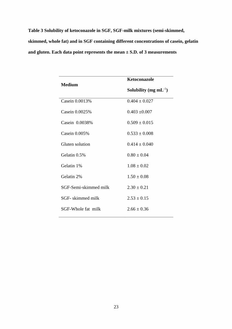
Table 3 Solubility of ketoconazole in SGF, SGF-milk mixtures (semi-skimmed,
skimmed, whole fat) and in SGF containing different concentrations of casein, gelatin
and gluten. Each data point represents the mean ± S.D. of 3 measurements
Medium
Ketoconazole
Solubility (mg mL-1)
Casein 0.0013% 0.404 ± 0.027
Casein 0.0025% 0.403 ±0.007
Casein 0.0038% 0.509 ± 0.015
Casein 0.005% 0.533 ± 0.008
Gluten solution 0.414 ± 0.040
Gelatin 0.5% 0.80 ± 0.04
Gelatin 1% 1.08 ± 0.02
Gelatin 2% 1.50 ± 0.08
SGF-Semi-skimmed milk 2.30 ± 0.21
SGF- skimmed milk 2.53 ± 0.15
SGF-Whole fat milk 2.66 ± 0.36
24
Figure Captions
Figure 1 Chemical structure of ketoconazole
Figure 2 The intrinsic dissolution profile of ketoconazole in simulated gastric fluid (SGF) (■)
and in SGF-milk mixtures (1:1) (whole fat () semi-skimmed (●) or skimmed (♦). Each
data point represents the mean ± S.D. of 3 measurements
Figure 3 The effect of egg albumin concentration on the solubility of ketoconazole
Figure 4 The intrinsic dissolution profile of ketoconazole in simulated gastric fluid (SGF) (■)
and in SGF containing egg albumin in concentrations of 0.01% (), 0.1% (▲), 0.5%
(□), 1% (▼) and 3% (○), w/v. Each data point represents the mean ± S.D. of 3
measurements
Figure 5 The Intrinsic dissolution profile of ketoconazole in simulated gastric fluid (SGF) (■)
and in SGF containing casein () or gluten (▲) filtered solutions. Each data point
represents the mean of 3 measurements ± S. D.
Figure 6 The intrinsic dissolution profile of ketoconazole in simulated gastric fluid (SGF) (■)
and in SGF containing gelatin in concentrations of 1%(●), 2%() and 4%(▼), w/v. Each
data point represents the mean ± S. D. of 3 measurements
Figure 7 Solubility and IDR of ketoconazole in simulated gastric fluid (SGF) and SGF
containing amino acids. Each data point represents the mean ± S.D. of 3 measurements
25
Figure 8 The intrinsic dissolution profile of ketoconazole in simulated gastric fluid (SGF) (■)
containing 1%, w/v amino acids LYS(), GLY(▲), ALA(○)and LEU(♦). Each data
point represents the mean ± S.D. of 3 measurements
Figure 9 The intrinsic dissolution profile of ketoconazole in SGF (■) and SGF containing 1%
carbohydrates (glucose (▲), lactose () and starch ()). Each data point represents the
mean ± S.D. of 3 measurements
26
Figure 1
0 50 100 150 200 2500
50
100
150
SGF-Whole fat Milk
SGF-Semi-Skimmed Milk
SGF-Skimmed Milk
SGF pH3
Keto
co
nazo
le d
isso
lved
per
un
it a
rea
(
mg
cm
-2)
Time (minutes)
Figure 2
O
NNOO
O
N
N
Cl
Cl
9
11
26 29

27
0.0 0.5 1.0 1.5 2.0 2.5 3.0
0.4
0.6
0.8
1.0
1.2
1.4
1.6
1.8
Keto
co
nazo
le s
olu
bilit
y (
mg
mL
-1)
Albumin concentrations (%, w/v)
Figure 3
0 50 100 150 200 2500
20
40
60
80
Keto
co
nazo
le d
isso
lved
per
un
it a
rea
mg
cm
-2
Time (minutes)
Albumin3%
Albumin1%
Albumin0.5%
Albumin0.1%
Albumin0.01%
SGFpH3
Figure 4

28
0 50 100 150 200 2500
10
20
30
40
Keto
co
nazo
le d
isso
lved
per
un
it a
rea
(mg
cm
-2)
Time (minutes)
Casein Filtrate
Gluten Filtrate
SGFpH3
Figure 5
0 50 100 150 200 2500
20
40
60
80
100
Keto
co
nazo
le d
isso
lved
per
un
it a
rea
(
mg
cm
-2)
Time (minutes)
Gelatin 4%
Gelatin 2%
Gelatin 1%
SGF
Figure 6
29
Figure 7
0 50 100 150 2000
20
40
60
80
100
LEU
ALA
GLY
LYS
SGF
Keto
co
nazo
le d
isso
lved
per
un
it a
rea
(
mg
cm
-2)
Time (minutes)
Figure 8
GLY 2%
ALA 1%
LYS 1%
LEU 1%
GLY 0.5%GLY 1%
SGF
0
0.1
0.2
0.3
0.4
0.5
0.6
0.7
0 1 2 3 4 5 6 7
IDR
(1
5 -
18
0 m
in)
mg
min
-1cm
-2
Ketoconazole solubility (mg mL-1)

30
0 50 100 150 200 2500
10
20
30
40
SGF
Glucose
Lactose
Starch
Keto
co
nazo
le d
isso
lved
per
un
it a
rea
(
mg
cm
-2)
Time (minutes)
Figure 9

31