Embed Size (px)
Citation preview

Pertanika J. Trop. Agric. Sci. 36 (2): 145 - 160 (2013)
ISSN: 1511-3701 © Universiti Putra Malaysia Press
TROPICAL AGRICULTURAL SCIENCEJournal homepage: http://www.pertanika.upm.edu.my/
Article history:Received: 18 May 2011Accepted: 2 November 2011
ARTICLE INFO
E-mail addresses: [email protected] (Loh, S. P.), [email protected] (Lee, S. P.)* Corresponding author
The Effect of Extraction Methods on Fatty Acid and Carotenoid Compositions of Marine Microalgae Nannochloropsis oculata and Chaetoceros gracilis
Loh, S. P.1,2* and Lee, S. P.1
1Department of Nutrition and Dietetics, Faculty of Medicine and Health Sciences, Universiti Putra Malaysia, 43400 Serdang, Selangor, Malaysia2Institute of Bioscience, Universiti Putra Malaysia, 43400 Serdang, Selangor, Malaysia
ABSTRACT
This study was conducted to assess three extraction methods for the determination of fatty acid compositions and carotenoids (lutein, zeaxanthin, β-carotene, and α-carotene) from marine microalgae, Nannochloropsis oculata (NO) and Chaetoceros gracilis (CG). For this purpose, three different extraction methods for the determination of fatty acids (dichloromethane:methanol, water:propan-2-ol:hexane and direct saponification-ethanol KOH) and carotenoids (hexane:ethanol:acetone:toluene, methanol:chloroform and methanol:tetrahydrofulran) were used. Two derivatization methods using different types of catalyst (acetyl chloride and boron trifluoride) were also used for the transmethylation of the fatty acids into corresponding methyl esters. The results of the fatty acid compositions showed that NO had a higher amount of n-3 and n-6 polyunsaturated fatty acid (PUFA), particularly eicosapentaenoic acid (EPA) (C20:5). CG was predominantly high in palmitic acid (C16:0) and palmitoleic acid (C16:1). The extraction method 1 (dichloromethane:methanol) and extraction method 2 (water: propan-2-ol: hexane) with acetyl chloride-catalyzed transmethylation were found to be the best methods for the determination of fatty acid compositions in NO and CG, respectively. A significantly higher (P<0.05) amount of carotenoids was found in NO as compared to CG using different extraction methods. Extraction method 1 (involving saponification procedure) yielded the best result for NO while extraction method 3 (methanol: tetrahydrofuran with no
saponification procedure) generated higher amounts of carotenoids in CG. Overall, this study has shown that significantly high amounts of fatty acids and carotenoids could be obtained from these microalgae using these methods.
brought to you by COREView metadata, citation and similar papers at core.ac.uk
provided by Universiti Putra Malaysia Institutional Repository

Loh, S. P. and Lee, S. P.
146 Pertanika J. Trop. Agric. Sci. 36 (2): 146 - 160 (2013)
Keywords: Carotenoids, Chaetoceros gracilis,
extraction, fatty acid composition, Nannochloropsis
occulata, microalgae
INTRODUCTION
Microalgae are prokaryotic or eukaryotic photosynthetic microorganisms unified primarily by the lack of roots, leaves, and stems that characterize higher plants. They can be found almost anywhere, with water and sunlight as their fundamental requirements, including lakes, soils, rivers, hot springs, and the ocean. Microalgae contain high value compounds like fatty acids [γ- linolenic acid, arachidonic acid, eicosapentaenoic acid (EPA), docosahexaenoic acids (DHA), etc.], pigments (chlorophyll and carotenoids), vitamins (biotin, vitamins C and E, and others) (Converti et al., 2009). Chaetoceros gracillis, a diatom in the class of Bacillariophycae and Nannochloropsis occulata, a unicellular green alga with spherical shape of the Eustigmatophyceae class plays an important role in the food chain system and it is also commonly used as live feed; thus, it is widely cultivated in fish hatcheries and shrimp farms (Gwo et al., 2005).
DHA and EPA are constantly an area of interest in nutrition because they are essential for optimizing human health. DHA is important for the development of the brain and eyes in pre-term and young infants, as well as for supporting cardiovascular health in adults, whereas EPA is essential for the human metabolism and involved in the blood lipid equilibrium
that prevents hypertriglyceridemia and anti-inflammatory activities (Kroes et al., 2003; Ward & Singh, 2005; Fajardo et al., 2007). Previously, fish was the principal dietary source of DHA and EPA. However, due to the serious environmental consequences and continuous exploitation, the declining sources of marine fish stocks and fish oil have prompted research into new sources of polyunsatured fatty acids (PUFAs) (Burja et al., 2007). In addition, certain disadvantages of fish oil, such as the unpleasant odour, possible pollutants, and mixed fatty acid properties have also encouraged the search for alternative sources of PUFAs (Pulz & Gross, 2004).
In addition, microalgae contain a multitude of pigments, particularly chlorophyll and carotenoid. Carotenoids are essential to human health and important in commercial applications (Felti et al., 2005). For example, β-carotene acts as pro-vitamin A and has been proven to prevent xeropthlamia (Puah et al., 2005); astaxanthin acts as a natural colorant for muscle in marine fish and crustaceans; lutein, zeaxantin and canthaxantin for chicken skin coloration, pharmaceutical purposes, and also as a natural food additive (Del Campo et al., 2000; Pulz & Gross, 2004).
Although microalgae contain important bioactive components (particularly PUFA and carotenoids), the extraction methods, especially for algae, are not well established, as there are no standard extraction methods for the determination of the fatty acid content or carotenoids in microalgae (Wiltshire et

Fatty Acid and Carotenoids from Two Marine Microalgae
147Pertanika J. Trop. Agric. Sci. 36 (2): 147 - 160 (2013)
al., 2000). Lipids are mainly a mixture of esters, and therefore, the preparation of fatty acid methyl esters (FAME) consists essentially on the conversion of one ester to another (i.e. transesterification) by cleavage of an ester bond via an alcohol; when such an alcohol is methanol, the reaction is referred to as methanolysis or transmethylation (Liu, 1994). Transmethylation are reversible reactions which are normally accomplished in the presence of a catalyst, either an acid or a base. Reactions involving acidic catalysts require heat to accelerate the process. Commonly used acidic catalysts are (the Brønsted-Lowry acid) HCl, H2SO4, acetyl chloride and (the Lewis acid) BF3. Base-catalyzed methanolysis proceeds much more rapidly under mild temperature conditions than acid-catalyzed reactions. However, bases cannot catalyze the esterification of FFAs.
The nutritional value of microalgae is strongly dependent on its bioactive profile; therefore, methods that could get the highest value is preferred. Felti et al. (2005) suggested that the absence of a standard extraction method for carotenoid is actually attributed to the wide spectrum of the analyzed materials (foodstuff, plant, animal, and human samples) and the wide range of the carotenoids present. As a result, three extractions methods of fatty acid and carotenoids from marine microalgae, Nannochloropsis oculata and Chaetoceros gracilis were evaluated in the current study. The criteria used in choosing these methods were maximum extraction efficiency, ease of handling, and use of solvents of low toxicity.
MATERIAL AND METHODS
Microalgae Samples
Both the Nannochloropsis oculata and Chaetoceros gracilis samples were purchased from Reed Mariculture Inc. USA. The samples were collected using non-probability, convenient sampling method. Both the microalgae were purchased in two batches and prior to the extraction, these samples were freeze-dried, homogenized, and kept at -20oC until further use.
Oil Extraction
The first extraction method used was adopted from Cequier-Sanchez et al. (2008). First, 500 mg of the samples were extracted by mixing 15 ml of dichloromethane-methanol 2:1 (v/v) contained in a beaker. The mixing was performed with occasional gentle hand agitation for 2 hours. Subsequently, the samples were filtered and transferred into a new test tube to which 3.13ml of an aqueous solution of potassium chloride (0.88%, w/v) was added with strong agitation, followed by centrifugation (Universal 320/320R Benchtop Centrifuges, Hettich Instruments, Germany) at 350g at 40C for 5 minutes. The aqueous upper phase was discarded and the organic phase was evaporated using a rotary evaporator (Büchi Rotavapor R-200, Switzerland).
The second extraction method was adopted from Schlechtriem et al. (2003) with a slight modification, in which hexane was used to substitute the cyclohexane. First, 500 mg samples were weighed into the Falcon tubes and mixed with 10ml of

Loh, S. P. and Lee, S. P.
148 Pertanika J. Trop. Agric. Sci. 36 (2): 148 - 160 (2013)
propan-2-ol and 12.5 ml of hexane using a vortex for 30s. The tubes were placed in an ultrasonic bath at room temperature for 15 minutes. Then, 13.75ml of water was added to obtain a mixture of water: propan-2-ol:hexane (11:8:10 v/v/v). The mixture was mixed again using the vortex for 30s. The different phases were separated by centrifugation at 1800g for 10 minutes and the organic phase was transferred into a pre-weighed Flacon tube with a dropper. The organic phase containing the lipid fraction was separated at the top of the extraction mixture (the hexane phase). Subsequently, the second extraction with 12.5ml of hexane containing 13% v/v propan-2-ol was done. The mixture was vortexed and placed into the ultrasonic bath for another 15 minutes. After centrifugation, the hexane phase was added to the first extract. The tubes were placed in a water bath (500C) for about 15 minutes and the solvent was evaporated to dryness using a rotary evaporator.
The third extraction method was adopted from Burja et al. (2007) with a minor modification, in which 95% ethanol was used instead of 96% ethanol. First, the 500mg samples were weighed. Then, 38ml of 3mM potassium hydroxide in ethanol (95%) was added into a 150ml beaker. The beaker was passed under the flow of nitrogen, and shaken for 1 hour in a water bath set at 60°C. Thereafter, the samples were cooled to room temperature and filtered through filter paper. The biomass was washed with 10ml of ethanol and transferred into a new beaker, to which, 10ml of water was added. Unsaponifiables
were extracted by adding 20ml of hexane and gently mixing twice. After the layers were separated, the pH was adjusted to 1 (from pH 13-14) by the addition of hydrochloric acid:water (1:1, v/v). The top layer, containing the fatty acid fraction, was recovered by two rounds of the addition of 10ml of hexane and a gentle mixing. Lastly, the solvent at the top layer was evaporated to dryness using a rotary evaporator.
Preparation of Fatty Acid Methyl Esters (FAMEs)
T h i s a c e t y l c h l o r i d e - c a t a l y z e d transmethylation method was adopted from Carvalho and Malcata (2005). The lipid extracts (2mg) were subjected to acid-catalyzed transesterification by dissolving them in 2ml of a freshly prepared mixture of acetyl chloride and methanol at a ratio of 5:100 (v/v), together with 1mg of tricosanoic acid as an internal standard. The reagents were placed in Teflon-capped Pyrex tubes, and the reaction continued at 100°C for 1 hour under pure nitrogen and darkness. After cooling to 30~40°C, 1ml of the extracting solvent (isooctane containing 0.01% butylated hydroxytoluene, BHT) was added, and the FAME solvent solution was mixed using a vortex for between 5 to 30s. The purification of the solution was achieved by adding 1ml of water, causing the formation of two immiscible phases, which were then allowed to separate. Subsequently, the upper extracted solvent phase was recovered and stored in sealed glass vials at -200C until GC analysis.
The BF3-catalyzed transmethylation
Fatty Acid and Carotenoids from Two Marine Microalgae
149Pertanika J. Trop. Agric. Sci. 36 (2): 149 - 160 (2013)
method was adopted from Carvalho and Malcata (2005), which was modified by the use of 10% (v/v) of BF3 in methanol instead of 12% (v/v) of BF3 in methanol. First, all the lipid extracts (2mg) were subjected to a preliminary alkaline hydrolysis with 0.5M sodium hydroxide at 1000C for 5 minutes. Subsequently, it was dissolved in 2ml of 10% (v/v) BF3 in methanol, together with 1mg of tricosanoic acid as an internal standard. The reagents were placed in the Teflon-capped Pyrex tubes, and the reaction was allowed to continue at 100°C for 30 minutes under pure nitrogen and darkness. The subsequent procedure was similar to the acetyl chloride-catalyzed transmethylation procedure, as described above.
Gas Chromatography Analysis
The assay of FAME was analyzed using gas chromatography (Agilent 6890, ISA Agilent Tech, USA) equipped with a split/splitless injector, and Hewlett Packard EL-980 flame ionization detector (FID). The FID system was used to separate and quantify each FAME component. FAME was separated using DB-23 column (60m x 0.25mm ID, and 0.15 µm). The chromatography data were recorded and integrated using the chemistation software (version 6). The oven temperature was programmed to hold at 500C for 1 min, before it was increased to 1750C with 250C/min, held for 4 minutes, and lastly increased to 2300C with 40C/min and held for 5 mins. The temperature for the injector and detector was set at 2500C and 2800C, respectively. One microlitre of the sample volume was injected with a split
ratio of 1:50µl and a column temperature of 1100C. The carrier gas was helium gas (1.0 ml/min) which was controlled at 123.4kPa/Hz and the air used for FID was held at 275.6kPa.
Calculation of Fatty Acid
The identification of the fatty acid compositions for the sample was made by comparing the retention time of the sample FAMEs with those of Supelco 37 component FAMEs mixture (Sigma-aldrich, USA) for each chromatography peak. The quantification of the fatty acid was done using tricosanoic acid (C23:0) as an internal standard. The amount of the individual fatty acid was calculated using the expression: Ci = Cp (Ai/Ap), where A is the chromatographic area units and C is the amount of fatty acid. Subscript p represents the internal standard and i refers to any fatty acid. The percentage of the individual fatty acid in the total amount of fats used was calculated as Ci/total amounts of fat x 100%.
Carotenoids Extraction
The first extraction method was adopted from Inbaraj et al. (2006). First, 400mg of freeze-dried microalgae was mixed with 12 ml hexane-ethanol-acetone-toluene (10:6:7:7 v/v/v/v) in a volumetric flask. After shaking for 1 hour, 0.8ml 40% methanolic potassium hydroxide was added and the solution was saponified at 250C in the dark for 16 hours. Then, 12ml of hexane was added to partition the carotenoids. The mixture was shaken for 1 min and 10% sodium sulfate solution was added. After

Loh, S. P. and Lee, S. P.
150 Pertanika J. Trop. Agric. Sci. 36 (2): 150 - 160 (2013)
shaking for 1 minute, the upper layer was collected and the lower layer was repeatedly extracted twice with hexane. Finally, the upper extracts were pooled and evaporated to dryness using a rotary evaporator.
The second extraction method was adopted from Reboul et al. (2006). First, 400mg of frozen-dried microalgae was added into 8ml of methanol containing 0.57% magnesium carbonate. Subsequently, the samples were homogenised for 30s using a vortex. Then, 8ml of chloroform, containing 0.005% butylated hydroxyl toluene (BHT), was added. The samples were homogenized for 30s more in the vortex blender. After a rest of 15 minutes, 8ml of distilled water was added into the samples and centrifuged (2000g for 10 minutes). The lower phases of the samples were collected and the remaining upper phases were extracted by the addition of 6ml of tetrahydrofuran. After that, the mixture was vortexed for 30s, and 6 ml of dichloromethane was also added. It was then vortexed for another 30s, after which 4ml of distilled water was added and the mixture was further re-vortexed for 30s. After centrifugation (2000g for 10 minutes at room temperature), the lower phase was collected and pooled with the previously collected phase. Lastly, the collected lower phase solvent was evaporated to dryness using a rotary evaporator.
The final extraction method was adopted from Marinova and Ribarova (2007). First, the pigments were extracted from a 400mg sample to which 0.04 g magnesium carbonate was added, with 6ml extraction
solvent methanol:tetrahydrofuran (1:1, v/v) containing 0.1% butyl hydroxytoluene (BHT). The mixture was vortexed for about 3 minutes, and then centrifuged for 3 minutes at 1400g and the supernatant was collected. The pellet was re-extracted following the same procedure until the supernatant became colourless. The combined supernatants were evaporated to dryness using a rotary evaporator.
All the extraction procedures were performed under subdued light to avoid degradation loss of the pigments. The residue was dissolved in methanol at a concentration of 100mg/ml. Prior to the HPLC analysis, the sample solution was filtered using a Whatman polytetrafluroethylene (PTFE) 0.22µm syringe filter and the filtrate was injected into a HPLC valve with a 1ml syringe.
Analysis of Carotenoids
The carotenoids were analyzed by using HPLC (Agilent Series 1100, Model G131 3A, Agilent Technologies, Germany) equipped with degasser, quaternary pump, auto sampler and photodiode array detector. The carotenoids were separated by HPLC using a 150 × 4.6 mm, 3 µm C30 analytical column (Waters Co., Milford, MA, USA). The mobile phase system comprised methanol–methyl tert-butyl ether (MTBE) – water (81:15:4 v/v/v) (A) and methanol/MTBE (10:90 v/v) (B) in the following gradient conditions 100% of A and 0% B, to 50% A and 50% B in 45 minutes, followed by 100% B within 15 minutes. The column temperature was set at 250C. The volume

Fatty Acid and Carotenoids from Two Marine Microalgae
151Pertanika J. Trop. Agric. Sci. 36 (2): 151 - 160 (2013)
injected into the HPLC was set as 20µl and the flow rate during the separation was set as 1ml/min. The wavelength used for the photodiode array detector in measuring the carotenoids was 450nm. The elution time was 45 minutes for a sample and the post time was 5 minutes.
The standard for the carotenoids were prepared from a stock solution of β-carotene, α-carotene, zeaxanthin and lutein. The identification of carotenoids was made by comparing with these standards, and the spiking test was also carried out to confirm the identification of certain peaks. The carotenoids were quantified using a calibration curve that was prepared using pure standards in the range of 0.025-5µg/ml.
Data Analysis
The computer software Statistical Package for Social Sciences version 16 (SPSS 16) was used to analyze the data in this study. The analysis was done in triplicates and the results were expressed as mean ± standard deviation. Meanwhile, the two-way ANOVA was used to compare the differences in the mean amounts of fatty acid and carotenoid from the microalgae, Nannochloropsis
oculata and Chaetoceros gracilis using various extraction methods and also two different transmethylation methods (fatty acid only). The analysis was considered at a significance value of p<0.05.
RESULTS AND DISCUSSION
Percentage of Extraction Yields (Total Oil)
As shown in Table 1, extraction method 1 (dichloromethane:methanol) showed the highest amount of the extraction yields for both NO and CG, with the mean values of 48.61% and 36.81%, respectively. Extraction method 3 using direct saponification with ethanolic KOH gave the least amount of extraction yields for both NO and CG, with the percentage value of 10.45% and 14.67%, respectively. A two-way ANOVA was conducted to examine the sample differences and the extraction methods on the extraction yields. There was a significant interaction between the various methods used and the extraction yields, p = .027. However, no significant differences were seen between the two microalgae in the extraction yield. The higher extraction yields of method 1 (dichloromethane: methanol) might be attributed to the presence of methanol,
TABLE 1 Total oil yield using different extraction methods from microalgae Nannochloropsis oculata (NO) and Chaetoceros gracilis (CG)
Extraction methods Nannochloropsis oculata (NO) Chaetoceros gracilis (CG)Method 1(Dichloromethane:methanol) 486 ± 236a 368 ± 56a
Method 2(Water: propan-2-ol: hexane) 366 ± 151a 316 ±18a
Method 3 (Direct saponification-ethanol) 105 ± 14b 147 ± 21a
Each value is the mean ± standard deviation of triplicates expressed as g kg-1dry weight. Within a column, means followed by the same letter are not significantly different (p>0.05).No significant differences were observed between NO and CG.

Loh, S. P. and Lee, S. P.
152 Pertanika J. Trop. Agric. Sci. 36 (2): 152 - 160 (2013)
a primary alcohol with the most active hydroxyl group (highly polar), which could stimulate the disruption of hydrogen bonds between lipid carbonyl, hydroxyl, and the amino groups, and the compounds of the nonextractable residue (Ruiz-lopez et al., 2003).
Fatty Acid Composition of Nannochloropsis Oculata (NO) and Chaetoceros Gracilis (CG) Using Different Extraction Methods
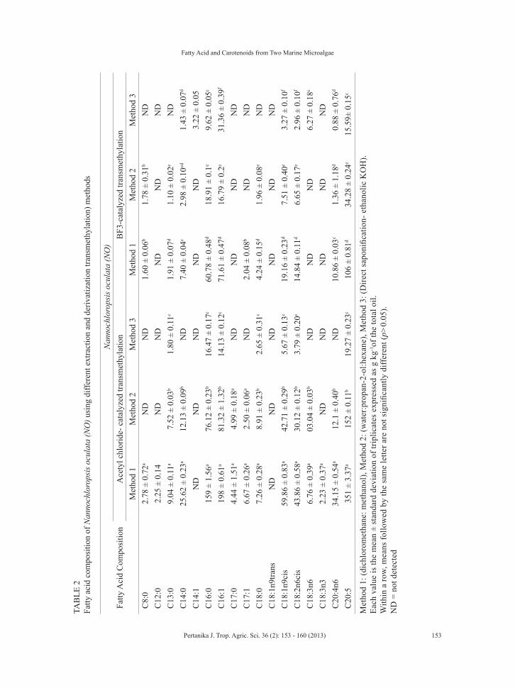
In terms of the extraction efficiency of the fatty acid content (weight %), extraction method 1 (dichloromethane: methanol) coupled with acetyl chloride catalyzed transmethylation appeared to be the most efficient method for NO, as compared to other methods (Table 2). This was because this particular method could generate higher fatty acid content than other methods using both the acetyl chloride and BF3-catalyzed transmethylation methods. Since NO consists of a polysaccharide cell wall, the solubility of its cell matrix towards the non-polar solvent may enable the penetration of solvents into it and subsequently allow the oil to dissolve and be extracted for transmethylation. In addition, this method is also simpler and easier in its procedure as compared to two other methods. Indeed, a previous study has shown that dichloromethane, a less hazardous solvent, was an effective extraction solvent for fatty acid research (Cequier-Sanchez, 2008).
Nevertheless, extraction method 2 (water:propan-2-ol:hexane), coupled with acetyl chloride catalyzed transmethylation, appeared to be the most suitable method
for the determination of fatty acid for CG, particularly C16 and C18 fatty acids (Table 3). The use of additional cell disruption treatment (ultrasonic bath) in this extraction procedure was notably useful in CG, a genus of diatoms, as they have the unique characteristic of a silica-based rigid cell wall, which may be difficult to break (Scala et al., 2002). The use of an ultrasonic bath was related to the destruction of cell walls and the enhancement of mass-transfer through the cell wall due to the collapse of the bubbles produced by cavitation (Macias-Sanchez et al., 2009). In this way, its extraction efficiency could be enhanced. Moreover, the use of this particular combination of solvents was also highly recommended in terms of its safety, low toxicity, and low cost (Smedes, 1999). Although hexane was used to substitute cyclohexane in this method, their almost similar properties would not create much difference in the result.
Among the three extraction methods used, method 3, which involved a direct saponification using ethanolic potassium hydroxide, gave the least number and amount (weight %) of fatty acid compositions in both the acetyl chloride and BF3-catalyzed transmethylation methods. This result disagrees with previous study which claimed that this method was an efficient technique to increase the extraction of fatty acid from biomass (Burja et al., 2007). However, a study by Wang et al. (2000) found a lower concentration of fatty acids on chicken egg yolk by using the direct saponification extraction method compared to other methods (direct-methylation, chloroform-
Fatty Acid and Carotenoids from Two Marine Microalgae
153Pertanika J. Trop. Agric. Sci. 36 (2): 153 - 160 (2013)
TAB
LE 2
Fa
tty a
cid
com
posi
tion
of N
anno
chlo
rops
is o
cula
ta (N
O) u
sing
diff
eren
t ext
ract
ion
and
deriv
atiz
atio
n tra
nsm
ethy
latio
n) m
etho
ds
Fatty
Aci
d C
ompo
sitio
nN
anno
chlo
rops
is o
cula
ta (N
O)
Ace
tyl c
hlor
ide-
cat
alyz
ed tr
ansm
ethy
latio
nB
F3-c
atal
yzed
tran
smet
hyla
tion
Met
hod
1M
etho
d 2
Met
hod
3M
etho
d 1
Met
hod
2M
etho
d 3
C8:
02.
78 ±
0.7
2aN
DN
D1.
60 ±
0.0
6b1.
78 ±
0.3
1bN
DC
12:0
2.25
± 0
.14
ND
ND
ND
ND
ND
C13
:09.
04 ±
0.1
1a7.
52 ±
0.0
3b1.
80 ±
0.1
1c1.
91 ±
0.0
7d1.
10 ±
0.0
2eN
DC
14:0
25.6
2 ±
0.23
a12
.13
± 0.
09b
ND
7.40
± 0
.04c
2.98
± 0
.10cd
1.43
± 0
.07d
C14
:1N
DN
DN
DN
DN
D3.
22 ±
0.0
5C
16:0
159
± 1.
56a
76.1
2 ±
0.23
b16
.47
± 0.
17c
60.7
8 ±
0.48
d18
.91
± 0.
1e9.
62 ±
0.0
5c
C16
:119
8 ±
0.61
a81
.32
± 1.
32b
14.1
3 ±
0.12
c71
.61
± 0.
47d
16.7
9 ±
0.2e
31.3
6 ±
0.39
f
C17
:04.
44 ±
1.5
1a4.
99 ±
0.1
8aN
DN
DN
DN
DC
17:1
6.67
± 0
.26a
2.50
± 0
.06a
ND
2.04
± 0
.08b
ND
ND
C18
:07.
26 ±
0.2
8a8.
91 ±
0.2
3b2.
65 ±
0.3
1c4.
24 ±
0.1
5d1.
96 ±
0.0
8eN
DC
18:1
n9tra
nsN
DN
DN
DN
DN
DN
DC
18:1
n9ci
s59
.86
± 0.
83a
42.7
1 ±
0.29
b5.
67 ±
0.1
3c19
.16
± 0.
23d
7.51
± 0
.40e
3.27
± 0
.10f
C18
:2n6
cis
43.8
6 ±
0.58
a30
.12
± 0.
12b
3.79
± 0
.20c
14.8
4 ±
0.11
d6.
65 ±
0.1
7e2.
96 ±
0.1
0f
C18
:3n6
6.76
± 0
.39a
03.0
4 ±
0.03
bN
DN
DN
D6.
27 ±
0.1
8c
C18
:3n3
2.23
± 0
.37a
ND
ND
ND
ND
ND
C20
:4n6
34.1
5 ±
0.54
a12
.1 ±
0.4
0bN
D10
.86
± 0.
03c
1.36
± 1
.18d
0.88
± 0
.76d
C20
:535
1 ±
3.37
a15
2 ±
0.11
b19
.27
± 0.
23c
106
± 0.
81d
34.2
8 ±
0.24
e
15.5
9± 0
.15c
Met
hod
1: (d
ichl
orom
etha
ne: m
etha
nol),
Met
hod
2: (w
ater
:pro
pan-
2-ol
:hex
ane)
, Met
hod
3: (D
irect
sap
onifi
catio
n- e
than
olic
KO
H).
Each
val
ue is
the
mea
n ±
stan
dard
dev
iatio
n of
trip
licat
es e
xpre
ssed
as
g kg
-1of
the
tota
l oil.
With
in a
row
, mea
ns fo
llow
ed b
y th
e sa
me
lette
r are
not
sig
nific
antly
diff
eren
t (p>
0.05
).N
D =
not
det
ecte
d

Loh, S. P. and Lee, S. P.
154 Pertanika J. Trop. Agric. Sci. 36 (2): 154 - 160 (2013)
TAB
LE 3
Fa
tty a
cid
com
posi
tion
of C
haet
ocer
os g
raci
lis (C
G) u
sing
diff
eren
t ext
ract
ion
and
deriv
atiz
atio
n (tr
ansm
ethy
latio
n) m
etho
ds
Fatty
Aci
d C
ompo
sitio
nC
haet
ocer
os g
raci
lis (C
G)
Ace
tyl c
hlor
ide
trans
met
hyla
tion
BF3
-cat
alyz
ed tr
ansm
ethy
latio
nM
etho
d 1
Met
hod
2M
etho
d 3
Met
hod
1M
etho
d 2
Met
hod
3C
8:0
3.12
± 0
.62a
ND
ND
1.41
± 0
.27b
0.67
± 0
.59b
0.87
± 0
.76b
C12
:02.
03 ±
2.0
4N
DN
DN
DN
DN
DC
13:0
5.83
± 1
03a
14.4
6 ±
0.07
b0.
93 ±
0.8
1c2.
26 ±
0.0
9d1.
50 ±
0.2
2dN
DC
14:0
77.0
3 ±
0.10
a80
.70
± 0.
55a
11.4
5 ±
5.57
b19
.73
± 0.
10c
47.3
4 ±
0.19
d4.
04 ±
0.1
1e
C14
:12.
42 ±
0.1
1a2.
33 ±
0.0
2aN
D0.
93 ±
0.0
4b1.
60 ±
0.1
0cN
DC
16:0
60.6
0 ±
1.78
a12
9 ±
0.47
b13
.52
± 7.
60c
12.1
3 ±
0.12
d70
.38
± 0.
13e
8.50
± 0
.10c
C16
:110
7 ±
0.28
a10
3 ±
0.88
a19
.29
± 7.
38b
25.7
1 ±
0.55
c92
.70
± 0.
09d
6.54
± 0
.17e
C17
:020
.38
± 0.
34a
18.8
9 ±
0.29
ab3.
50 ±
1.5
9c4.
80 ±
0.0
6d17
.95
± 0.
07b
1.33
± 0
.14e
C17
:128
.72
± 0.
22a
25.5
5 ±
4.34
a6.
28 ±
2.3
6b6.
55 ±
0.2
5c22
± 0
.20b
2.14
± 0
.06c
C18
:08.
13 ±
0.0
4a20
.95
± 0.
43b
ND
1.64
± 0
.03c
12.5
2 ±
0.12
dN
DC
18:1
n9tra
nsN
D5.
68 ±
0.2
8aN
DN
D6.
62 ±
0.1
4bN
DC
18:1
n9ci
s11
.80
± 0.
18a
173
± 0.
29b
5.79
± 0
.70c
ND
3.38
± 0
.19d
2.42
± 0
.04e
C18
:2n6
cis
7.76
± 0
.09a
121
± 0.
21b
3.08
± 0
.18c
1.05
± 0
.02d
5.13
± 0
.15e
1.59
± 0
.11d
C18
:3n6
ND
ND
ND
ND
1.22
± 0
.05a
ND
C18
:3n3
ND
4.64
± 0
.10a
ND
ND
ND
ND
C20
:4n6
7.83
± 8
.74a
ND
ND
ND
2.44
± 0
.11b
ND
C20
:518
.05
± 0.
35a
14.9
3 ±
0.28
ab3.
82 ±
3.3
2c3.
52 ±
0.0
6d17
.45
± 0.
12b
ND
Met
hod
1: (d
ichl
orom
etha
ne: m
etha
nol),
Met
hod
2: (w
ater
:pro
pan-
2-ol
:hex
ane)
, Met
hod
3: (D
irect
sap
onifi
catio
n- e
than
olic
KO
H).
Each
val
ue is
the
mea
n ±
stan
dard
dev
iatio
n of
trip
licat
e ex
pres
sed
as g
kg-1
of to
tal o
il.W
ithin
a ro
w, m
eans
follo
wed
by
the
sam
e le
tter a
re n
ot s
igni
fican
tly d
iffer
ent (
p>0.
05).
ND
= n
ot d
etec
ted.

Fatty Acid and Carotenoids from Two Marine Microalgae
155Pertanika J. Trop. Agric. Sci. 36 (2): 155 - 160 (2013)
methanol extraction, and postextraction saponification), although the reason was unknown.
A c e t y l c h l o r i d e - c a t a l y z e d transmethylation generated a higher amount (weight %) of fatty acid compared to BF3-catalyzed transmethylation in both the microalgae. This might be due to the highly basic condition of acetyl chloride, which could cause severe disruption to cell integrity, making in situ methyl ester derivation efficient (Tran et al., 2009). Furthermore, the use of acetyl chloride-catalyzed transmethylation procedure has several advantages as compared to the most commonly performed methanolic BF3 method, such as longer shelf-life (without the need for refrigeration), lower cost, and smaller amount of catalyst required (5% acetyl chloride versus 10% BF3) (Carvalho & Malcata, 2005).
Percentage of Extraction Yields (Carotenoids)
Table 4 shows that extraction method 1 (hexane:ethanol:acetone:toluene) generated the
highest extraction yields for both NO (74.54 ± 4.75%) and CG (69.28 ± 14.71%). This was probably due to the longer period of contact time (1 hour) between the cellular component to be extracted and the solvent mixtures in extraction method 1 as compared to the other two methods (Henriques et al., 2007). The two-way ANOVA showed significant differences between the samples (p = 0.022) on the extraction yields. Overall, it could be seen that all the extraction methods used generated higher extraction yields in NO than in CG. However, the difference in the extraction yields was small among these microalgae, particularly between extraction methods 2 and 3.
Carotenoids Concentration of the Different Extracts of Nannochloropsis Oculata (NO) and Chaetoceros Gracilis (CG)
As shown in Table 5, β-carotene was found to be the highest, followed by zeaxanthin, α-carotene, and lutein in the NO using different extraction methods. However, extraction methods 1 and 2 were the only methods that could detect lutein
TABLE 4 Extraction yield of carotenoids using different extraction methods from microalgae Nannochloropsis oculata (NO) and Chaetoceros gracilis(CG)
Extraction methods Nannochloropsis oculata (NO) Chaetoceros gracilis (CG)Method 1 (Saponification)(hexane:ethanol:acetone:toluene)
745 ± 48a 693 ± 147a
Method 2 (No saponification)(methanol:chloroform)
682 ± 25a 515 ±12b
Method 3 (No saponification)(methanol:tetrahydrofuran)
636 ± 39a 502 ± 25a
Each value is the mean ± standard deviation of triplicates expressed as g kg-1dry weight.
Within a row, means followed by the same letter are not significantly different (p>0.05).No significant differences were observed between the 3 extraction methods.

Loh, S. P. and Lee, S. P.
156 Pertanika J. Trop. Agric. Sci. 36 (2): 156 - 160 (2013)
and α–carotene in NO, respectively, while zeaxanthin was not detected in extraction method 2. For zeaxanthin and β-carotene contents in NO, extraction method 1, which involved the saponification step, generated the highest concentration (µg/100g dry weight) as compared to the other methods. The functions of saponification include hydrolyzing the carotenoid esters and removing the chlorophyll and unwanted lipids on microalgae, which may interfere with chromatographic separation (Howe et al., 2006). Since microalgae were high in their lipid content, saponification was necessary to achieve better results (better identification and higher concentration) as
compared to the other two methods, which do not employ saponification.
Just like NO, CG was found to be the highest in the amount (g/kg dry weight) of β-carotene, followed by lutein and zeaxanthin using different extraction methods. However, α-carotene was not detected in CG with either of these extraction methods. This does not indicate the absence of α-carotene in CG because the failure to detect it might be due to other possible reasons such as the presence of light and oxygen while handling the samples or storing that would have contributed to its degradation. As shown in Table 6, extraction method 2 was the only method that could not
TABLE 5 Carotenoid concentrations (g kg-1 dry weight) of Nannochloropsis oculata (NO) using different extraction methods
CarotenoidsMethod 1
( Saponification)(hexane:ethanol:acetone: toluene)
Method 2(No saponification)
(methanol : chloroform)
Method 3(No saponification)
(methanol: tetrahydrofuran)
Lutein 1.55 ±0.01 ND NDZeaxanthin 3.74 ± 0.03a ND 2.67 ± 0.08b
β-carotene 9.58 ± 0.002a 9.46 ± 0.05b 8.69 ± 0.04c
α-carotene ND 2.16 ± 0.004 ND
Each value is the mean ± standard deviation of triplicates expressed as g kg-1 dry weight.Within a row, means followed by the same letter are not significantly different (p>0.05).ND = not detected.
TABLE 6: Carotenoid concentrations (g kg-1dry weight) of Chaetoceros gracilis (CG) using different extraction methods
CarotenoidsMethod 1
( Saponification)(hexane:ethanol:acetone: toluene)
Method 2(No saponification)
(methanol:chloroform)
Method 3(No saponification)
(methanol: tetrahydrofuran)
Lutein 1.33 ± 0.003a ND 1.57 ± 0.02b
Zeaxanthin 0.58 ± 0.01a 0.75 ± 0.003b 8.68 ± 0.02c
β-carotene 7.945 ± 0.002a ND 8.08 ± 0.01b
α-carotene ND ND ND
Each value is the mean ± standard deviation of triplicates expressed as g kg-1dry weight.Within a row, means followed by the same letter are not significantly different (p>0.05).ND = not detected.
Fatty Acid and Carotenoids from Two Marine Microalgae
157Pertanika J. Trop. Agric. Sci. 36 (2): 157 - 160 (2013)
detect the presence of lutein and β-carotene in CG. Hence, extraction method 2 was less suitable for the determination of carotenoid for CG as compared to extraction methods 1 and 3. However, extraction method 3 could generate a higher concentration of carotenoids (g/kg dry weight) compared to extraction method 1. In the present study, the saponification step in extraction method 1 might not have much impact on carotenoids determination of CG since the absence of the saponification step in extraction method 3 yielded a better result for carotenoids.
Moreover, the different cell matrix of these microalgae might have contributed to the difference in the concentrations of carotenoid in them. As described earlier in the determination of the fatty acid composition, the cellular structure of NO was distinctly different to CG. Hence, CG with its unique characteristic of a silica-based rigid cell wall might cause incomplete extraction of the biochemical compounds by the solvents alone, without any additional treatment (e.g. ultrasound bath, enzymes, microwave-assisted, etc.). This is in accordance with a published study, whereby an efficient disruption treatment of the membrane was required in order to achieve the efficient extraction of carotenoids as there was no standard technique can guarantee a maximization of the extraction yield (Valduga et al., 2009). Unlike CG, the polysaccharide cell wall of NO might also be easier to penetrate using compatible solvents, and subsequently allow the extraction of the desired biochemical compounds.
CONCLUSION
The comparison of various extraction methods on both fatty acids and carotenoids revealed that they produced extracts with different characteristics as well as quantitative differences. For fatty acid determination, the utilization of method 1 (dichloromethane:methanol) appeared to be the most efficient method for NO. Nevertheless, extraction method 2 (water:propan-2-ol:hexane), which involved additional treatment (ultrasonic bath), appeared to be a more suitable method for fatty acid determination in CG. As for carotenoids, extraction method 1, which uses the saponification step to remove chlorophyll, unwanted lipids and the involvement of more solvent mixtures (2 polar and 2 non-polar solvents), generated the highest concentration (µg/100g dry weight) in NO. However, extraction method 3 generated the highest concentrations in CG. Overall, this study has shown that using the right extraction method, high amounts of fatty acids and carotenoids could be obtained from the microalgae.
This work was supported by Agri-Science Fund Grant from the Ministry of Agriculture, Malaysia (Project No. 5450384).
REFERENCESBurja, A. M., Armenta R. E., Radianingtyas, H., &
Barrow, C. J. (2007). Evaluation of fatty acid extraction methods for Thraustochytrium sp. ONC-T18. Journal of Agricultural and Food Chemistry, 55, 4795-4801.
ACKNOWLEDGEMENTS

Loh, S. P. and Lee, S. P.
158 Pertanika J. Trop. Agric. Sci. 36 (2): 158 - 160 (2013)
Carvalho, A. P., & Malcata, F. X. (2005). Preparation of fatty acid methyl esters for gas chromatography analysis of marine lipids: insight studies. Journal of Agricultural and Food Chemistry, 53, 5049-5059.
Cequier-Sanchez, E., Rodriguez, C., Ravelo, A. G., & Zarate, R. (2008). Dichloromethane as a solvent for lipid extraction and assessment of lipid classes and fatty acids from samples of different natures. Journal of Agricultural and Food Chemistry, 56, 4297-4303.
Converti, A., Casazza, A. A., Ortiz, E. Y., Perego, P., & Borghi, M. D. (2009). Effect of temperature and nitrogen concentration on the growth and lipid content of Nannochloropsis oculata and chlorella vulgaris for biodiesel production. Chemical Engineering and Processing, 48, 1146-1151.
Del Campo, J. A., Moreno, J., Rodriguez, H., Vargas, M. A., Rivas, J., & Guerrero, M. G. (2000). Carotenoid content of chlorophycean microalgae: factors determining lutein accumulation in Muriellopsis sp. (Chrolophyta). Journal of Biotechnology, 76, 51-59.
Fajardo, A. R., Cerdan, L. E., Medina, A. R., Acien Fernandez, F. G., Gonzalez Moreno, P. A., & Molina-Grima, E. (2007). Lipid extraction from the microalgae Phaeodactylum tricornutum. European Journal of Lipid Science Technology, 109, 120-126.
Felti, L., Pacakova, V., Stulik, K., & Volka, K. (2005). Reliability of carotenoid analyses: A review. Current Analytical Chemistry, 1, 93-102.
Gwo, J. C., Chiu, J. Y., Chou, C. C., & Chen H. Y. (2005). Cryopreservation of marine microalga, Nannochlorosis oculata (Eustigmatophyceae). Cryobiology, 50, 338-343.
Henriques, M., Silva, A., & Rocha, J. (2007). Extraction and quantification of pigments from a marine microalga: a simple and reproducible method. In
A. Mendez-Vilas (Ed.). Communicating current research and educational topics and trends in applied microbiology, vol. 2 (pp. 586-593). Badajoz, Spain: Formatex.
Howe, J. A., & Tanumihardjo, S. A. (2006). Evaluation of analytical methods for carotenoid extraction from biofortified maize (Zea mays sp.). Journal of Agricultural and Food Chemistry, 54, 7992-7997.
Inbaraj, B. S., Chien, J. T., & Chen, B. H. (2006). Improved high performance liquid chromatographic method for determination of carotenoids in the microalga Chlorella pyrenoidosa. Journal of Chromatography A, 1102, 193-199.
Kroes, R., Schaefer, E. J., Squire, R. A., & William, G. M. (2003). A review of safety of DHA45-oil. Food Chemistry and Toxicology, 41, 1433-1446.
Liu, K. S. (1994). Preparation of fatty acid methyl esters for gaschromatographic analysis of lipids in biological materials. Journal of American Oil and Chemical Society, 71, 1179-1187.
Macias-Sanchez, M. D., Mantell, C., Rodriguez, M., Martinez de la Ossa, E., Lubian, L. M., & Montero, O. (2009). Comparison of supercritical fluid and ultrasound-assisted extraction of carotenoids and chlorophyll a from Dunaliella. Talanta, 77, 948-952.
Marinova, D. , & Ribarova. (2007). HPLC determination of carotenoids in Bulgarian berries. Journal of Food Composition and Analysis, 20, 370-374.
Puah, C. W., Choo, Y. M., Ma, A. N., & Chuah, C. H. (2005). Supercritical fluid extraction of palm carotenoids. American Journal of Environmental Sciences, 1, 264-269.
Pulz, O., & Gross, W. (2004). Valuable products from biotechnology of microalgae. Applied Microbiological and Biotechnology, 65, 635-648.
Fatty Acid and Carotenoids from Two Marine Microalgae
159Pertanika J. Trop. Agric. Sci. 36 (2): 159 - 160 (2013)
Reboul, E., Richelle, M., Perrot, E., Desmoulins-malezet, C., Pirisi, V., & Borel, P. (2006). Bioaccessibility of carotenoids and vitamin E from their main dietary sources. Journal of Agricultural and Food Chemistry, 54, 8749−8755.
Ruiz-Lopez, N., Martinez-Force, E., & Garces, R. (2003). Sequential one-step extraction and analysis of triacylglycerols and fatty acids in plant tissues. Analytical Biochemistry, 317, 247–254.
Scala, N., Carels, A., Falciatore, M. L., & Chiusano, C. (2002). Bowler, genome properties of the diatom Phaeodactylum tricornutum. Plant Physiology, 129, 993–1002.
Schlechtriem, C. H., Focken, U., & Becker, K. (2003). Effect of different lipid extraction methods on δ13 C of lipid and lipid-free fraction of fish and different fish feeds. Isotopes Environmental Health Studies, 39, 135-140.
Smedes, F. (1999). Determination of total lipid using non-chlorinated solvents. Analys., 124, 1711–1718.
Tran, H. L., Hong, S. J., & Lee, C. G. (2009). Evaluation of extraction methods for recovery of fatty acids from Botryococcus braunii LB572 and Synechocystis sp. PCC6803. Biotechnology and Bioprocess Engineering, 14, 187-192.
Valduga, E., Valerio, A., Tatsch, P. O., Treichel, H., Jr, A. F., & Luccio, M. D. (2009). Assessment of cell disruption and carotenoids extraction from Sporidiobolus salmonicolor (CBS 2636). Food and Bioprocess Technology, 2, 234–238.
Wang, Y., Sunwoo, H., Cherian, G., & Sim, J. S. (2000). Fatty acid determination in chicken egg yolk: a comparison of different methods. Poultry Science, 79, 1168–1171.
Ward, O. P., & Singh, A. (2005). Omega-3/6 fatty acids: alternative sources of production. Process Biochemistry, 40, 3627-3652.
Wiltshire, K. H., Boersman, M., Moller, A., & Buhtz, H. (2000). Extraction of pigments and fatty acids from the green alga Scenedesmus obliquus (chlorophyceae). Aquatic Ecology, 34, 119-126.