Embed Size (px)
Citation preview

Portland State University Portland State University
PDXScholar PDXScholar
University Honors Theses University Honors College
6-16-2021
The Effect of Diazepam on Early Neural Stem Cells The Effect of Diazepam on Early Neural Stem Cells
Proliferative Activity and Hippocampal-Dependent Proliferative Activity and Hippocampal-Dependent
Memory after Traumatic Brain Injury Memory after Traumatic Brain Injury
Van Khanh Le Doan Portland State University
Follow this and additional works at: https://pdxscholar.library.pdx.edu/honorstheses
Part of the Biology Commons, Cognitive Neuroscience Commons, and the Other Chemicals and Drugs
Commons
Let us know how access to this document benefits you.
Recommended Citation Recommended Citation Doan, Van Khanh Le, "The Effect of Diazepam on Early Neural Stem Cells Proliferative Activity and Hippocampal-Dependent Memory after Traumatic Brain Injury" (2021). University Honors Theses. Paper 1097. https://doi.org/10.15760/honors.1124
This Thesis is brought to you for free and open access. It has been accepted for inclusion in University Honors Theses by an authorized administrator of PDXScholar. Please contact us if we can make this document more accessible: [email protected].
The effect of diazepam on early neural stem cells proliferative activity and hippocampal
dependent memory after traumatic brain injury
by
Van Khanh Doan
An undergraduate honors thesis submitted in partial fulfillment of the
requirements for the degree of
Bachelor of Science
in
University Honors
and
Biochemistry
Thesis Adviser
Dr. Laura Villasana
Portland State University
2021
Abstract
Traumatic brain injury (TBI) induces an upregulation of neurogenesis in the brain
specifically in the hippocampus, an area pertaining to learning and memory formation.
Although this upregulated response is intuitively thought to be restorative, previous
studies show that the nascent neurons generated after TBI exhibit abnormalities, such
as aberrant morphologies and enhanced migrations, which could suggest to be
maladaptive. The GABA-A agonist diazepam has been shown to inhibit this
upregulation in neurogenesis and normalizes dendrites after TBI. To determine whether
modulation of neurogenesis with diazepam benefits or hinders cognitive recovery,
C57BI/6J wild-type mice received a sham or controlled cortical impact (CCI) injury and
were administered diazepam for one week. Mice underwent the Morris Water Maze
(MWM) and Reversal Morris Water Maze testing one month later to examine
hippocampal-dependent memory. Diazepam was found to attenuate
neurogenesis-dependent memory deficits after CCI in the reversal MWM test. We also
examined whether modulation of post-TBI neurogenesis by diazepam is associated with
attenuation of hippocampal neural stem cells (NSCs). The percent of proliferating
hippocampal NSCs was not significantly different in any experimental group. Therefore,
proliferative activity of hippocampal neural stem cells by diazepam was inconclusive.
Further studies are needed to examine proliferating NSCs by diazepam.
Introduction
In 2014, approximately 2.87 million people in the United States alone had
traumatic brain injury (TBI)-related emergency department visits, hospitalization, and
deaths (Centers for Disease Control and Prevention [CDC], 2014). Fortunately, there
were only 56,800 TBI-related deaths (CDC, 2014). However, this means that the
majority of the survivors have to possibly live with the lasting consequences of TBI,
such as permanent cognitive impairments (Richardson et. al., 2007). A better
understanding of the impact of TBI is needed to alleviate neurological consequences
and improve cognitive recovery through new clinical treatments.
Neurogenesis, the regeneration of neurons, occurs throughout the lifespan of
humans and is necessary for the formation of specific memories (Clelland et. al., 2009).
Importantly, TBI increases neurogenesis primarily in the dentate gyrus of the
hippocampus (Dash et. al., 2001; Chirumamilla et. al., 2002; Villasana et. al., 2014).
The upregulation of neurogenesis after TBI could be a beneficial mechanism in which
the brain attempts to repair neuronal damage sustained during TBI (Richardson et. al.,
2007). However, newly immature neurons after TBI exhibit abnormal properties, such as
aberrant dendritic morphology and enhanced migration within the hippocampus (Ibrahim
et. al., 2016; Villasana et. al., 2015). Additionally, post-TBI neurogenesis results in
long-term deficits in neural progenitor cell (NPC) proliferation and immature granule
cells (IGC) (Neuberger et. al., 2017). The reduction in neurogenesis and NPC
proliferative capacity in the long term, along with the aberrant characteristics of the
nascent neurons, foster the importance of determining whether attenuating increases in

post-traumatic neurogenesis potentially improves or worsens recovery of
hippocampal-dependent memory after TBI.
In an attempt to answer the question of whether post-TBI neurogenesis is
beneficial or detrimental, post-TBI neurogenesis is commonly manipulated to measure
its cellular effect in the hippocampus and its cognitive function and recovery (Ngwenya
& Danzer, 2019). The gamma-aminobutyric acid type A (GABA-A) agonist diazepam,
which is a benzodiazepine, reduces post-TBI neurogenesis and normalizes the aberrant
dendritic morphology of new neurons in mice when administered for one week
immediately after a controlled cortical impact (CCI) injury in mice (Villasana et. al.,
2019). In addition, the NMDA-R antagonist ketamine also reduces post-TBI
neurogenesis and in conjunction improves strategy flexibility in the
hippocampal-dependent water maze test (Peters et. al., 2018). Therefore, the ability of
diazepam to inhibit post-TBI neurogenesis and rectify the aberrant dendritic morphology
makes it a potentially relevant tool in post-TBI cognitive recovery, as seen with
ketamine. However, neither the association between diazepam-induced neurogenesis
reduction and cognitive recovery after TBI nor its mechanism of reduction have yet to be
examined.
The change in neurogenesis after TBI could be due to many factors including
neural stem cell (NSC) proliferation, cell survival, and differentiation, as well as changes
in the neural environment. Massive change in the extracellular flow of ions and
neurotransmitters following a TBI could affect cell survival by inducing an excitotoxicity
environment, which can cause metabolic stress and cause excitotoxic neuronal death
(Katayama et. al., 1990). NSCs reside in the dentate gyrus of the hippocampus and can

proliferate and differentiate into two cell types: astrocytes or neurons (Bond et. al.,
2015). It is possible that the differentiation of NSC could change in the presence of
diazepam where neuronal differentiation is suppressed or the level of differentiation of
other cell types is increased. Diazepam has also been shown to block hippocampal
activity and NSC proliferation after a middle cerebral artery occlusion (MCAO), a stroke
injury model, which also increases neurogenesis in mice (Nochi et. al., 2013).
Therefore, similar to MCAO, in the CCI injury model, diazepam treatment may inhibit the
transient post-TBI neurogenesis via the inhibition of NSC proliferation.
Benzodiazepines are often administered to severe-TBI patients upon
hospitalization for injury management (Flower & Hellings, 2012). The effects of
benzodiazepines act transiently to reduce the consciousness of the patients, elevated
intracranial pressure, and the seizure susceptibility (Flower & Hellings, 2012). As a
possible agent employed to provide early medical care, it is additionally important to
determine the mechanism of diazepam on post-TBI neurogenesis and whether the
administration of diazepam can play a role in long-term cognitive recovery. In this study,
the effects of diazepam administration after CCI on hippocampal-dependent memory
were examined. We also examined whether diazepam administration immediately after
CCI attenuated the proliferation of NSCs.
Method
Animals
All mouse handling and procedures were performed in accordance with National
Institutes of Health guidelines and were in compliance with Oregon Health and Science
and Legacy Research Institute Institutional Animal Care and Use Committee approved

protocols. Seven-week old male and female C57Bl/6J wild-type mice were purchased
from Jackson Laboratories and allowed to acclimate one week prior to surgeries. A total
of 28 surviving mice were used for the behavioral assays. For the analysis of NSC
proliferation, brain tissue from 16 mice from the previous study (Villasana et al., 2019)
were used.
Controlled cortical impact injury and osmotic pump implantation
Controlled cortical impact (CCI) was used as the mouse TBI model. There were
four groups of mice: sham/vehicle (4 males, 4 females), CCI/vehicle (3 males, 3
females), sham/diazepam (4 males, 3 females), and CCI/diazepam (4 males, 3
females). Littermates were used for each group. Four mice had to be euthanized due to
suture complications (28 mice remaining). Approximately an hour before surgery, all
mice received 0.2 mL of Children’s Tylenol (orally). During the injury surgery, all mice
were anesthetized with 2% isoflurane (continuously inhaled) and placed on a heated
stereotaxic apparatus.Their heads and left shoulder blades were shaved and sterilized
with 10% betadine followed by 2% lidocaine. An incision was made along the midline of
the scalp followed by a 4mm lateral (left) craniotomy between bregma and lambda
leaving the dura mater intact. At the exposed area, a 3 mm stainless steel piston
attached to an electromagnetic impactor (Leica Microsystems, Germany) was used to
make a 0.9mm deformation at a 4.67 m/s velocity with a dwell time of 300ms. Sham
mice received the same procedure minus the craniotomy and injury. The incision was
sutured and osmotic pumps (Alzet; Cupertino, CA) containing either vehicle (1:1
DMSO/propylene glycol) or diazepam (15 mg/kg/day) were subcutaneously implanted
over the left shoulder blade immediately after the sham or CCI injury. Mice were ear
punched for identification and allowed to recover in a warm padded chamber for 45
minutes prior to conducting a brief neurologic severity assessment. All mice were given
Tylenol twice daily for 2 days after surgery. Osmotic pumps were removed one week
later via the removal and reinstatement of sutures at the incision and saved to confirm
the delivery rate.
BrdU protocol
Behavioral mice received 3 intraperitoneal injections (2 hours apart) of BrdU
(50mg/kg dissolved in saline), beginning 2 days after CCI or sham. BrdU provides the
labeling of proliferating cells (Cameron & McKay, 2001). Their histological examination
of NSCs using BrdU is not included in this study.
Assessment of neural stem cell proliferation
Tissue from sham or CCI treated mice with or without diazepam was obtained
from a previous study conducted at Oregon Health and Science University (Villasana et
al., 2019). In that study, proliferation in general was assessed with only BrdU or Ki67 in
response to diazepam 3 days after the injury. In this study, with the same samples, the
proliferation of NSCs was assessed using Ki67 coupled with GFAP and Sox2. The
number of mice brain samples used in the following experimental group were 4
sham/veh, 4 CCI/veh, 4 sham/dzp, 4 CCI/dzp from the previous study (Villasana et. al.,
2019).

Immunohistochemistry
Immunohistochemistry (IHC) was performed as previously described (Villasana
et. al., 2019) with some modifications. To perform IHC with Ki67/Sox2/GFAP/DAPI, four
brain sections containing the hippocampus (two dorsal, two ventral) were obtained from
each mouse. The brain sections were washed with 0.5 mL PBST for 10 minutes five
times. The sections were blocked with 5% normal donkey serum (NDS) in PBST for 60
minutes then incubated at 4℃ overnight with primary antibodies diluted with 1.5% NDS.
The primary antibodies used were rabbit anti-Ki67 (1:200), rat anti-Sox2 (1:400), and
mouse anti-GFAP (1:400). The sections were washed with PBST for 10 minutes three
times and incubated with the secondary antibodies for 2-3 hours. The secondary
antibodies used were donkey anti-rabbit (1:500, Alexa fluor 488), donkey anti-rat (1:500,
Alexa fluor 555), and anti-mouse (1:500, Alexa fluor 647). The sections were then
washed with PBST for 10 minutes three times and incubated in DAPI in PBST
(1:10,000) for 20 minutes. Finally, the sections were washed with PBST for 10 minutes
two times and mounted on slides with Fluoromount G (Southern Biotech).
Confocal microscopy and imaging analyses
The dentate gyrus of the hippocampus was imaged with a Leica TCS SPE-II
confocal microscope using a 20x0.8 lens. All subsequent analyses were performed on
coded images with ImageJ software. The targeted cells were only counted in the
granular cell layer (GCL) and the subgranular zone (SGZ) of the dentate gyrus.
Specifically, Ki67 labels cells undergoing G1, S, G2, and M phase (Tanaka et. al., 2011).
Sox2 labels neural stem cells (NSCs) and astrocytes. GFAP labels the processes of the
astrocytes and NSCs. In Ki67/Sox2/GFAP stained sections, NSCs were identified by the

colabeling of Sox2 and GFAP with a singular process from the cell body (versus
astrocytes with multiple processes). Of these NSCs, proliferation was identified by the
additional labeling with Ki67. The volume of the GCL was measured by collecting the
depth (z-stack) of the image (ranges from 13-26 µm) and the area of the GCL. The cell
density was measured by taking the number of cells over the volume of GCL
(#cells/mm3). The percentage of proliferating NSCs were calculated by taking the
number of proliferating NSCs (KI67/Sox2/GFAP) over the total number of NSCs
(Sox2/GFAP).
Behavioral analyses
Behavioral testing began 4 weeks after surgeries. One hour prior to conducting
the water maze test, mice were temporarily singly housed in cages, which sat halfway
on top of heating pads. A 122 cm circular pool was filled with water (20°C + 1) and
made opaque using white chalk. The water maze test was conducted with 2 sessions of
training each day with each session consisting of 3 trials. The first three days out of 7
days, mice were trained to locate the platform within the pool. Distal cues were placed
around the room, and consisted of evenly distanced black rectangle boards with either a
circle, a horizontal line, a vertical line, or no line through the middle. Mice were placed in
a pseudorandom location within the pool and were given 60 seconds to swim and locate
the “hidden” platform which sat 3 cm below the water. If they did not find the platform
within 60 seconds, they were hand guided to the platform location and allowed to sit on
it for 3 seconds before removing them. If they did find the platform within 60 seconds,
they were allowed to remain on it for 3 seconds as well before removing them. On the
4th day (7th session), a probe trial was conducted to assess the spatial memory

retention of the mice. In this trial, the platform was removed, and the bias for the
location of the platform was assessed. To mitigate any potential extinction effects of the
probe trial, a reinforcement session with the platform returned to its original location was
conducted immediately after the probe trial.
Reversal Water Maze
On day 5 and 6 (session 8-11), mice were trained to locate a new location of the
platform in a new quadrant of the pool. A probe trial was conducted 24 hours after each
of these training days. Mice were euthanized 90 minutes after the last reversal probe
trial.
Statistical analyses
For all analyses, the data was first assessed for normality to determine use of
parametric versus non-parametric tests. For water maze analyses, a 3-way repeated
measures ANOVA with injury drug and sex as between subject variables and session as
the within subject factor was used to assess the learning curves. For the probe trials, a
3-way ANOVA was used to compare group differences in the cumulative distance swam
from the platform. The proliferation of NSCs was compared between groups using a
3-way ANOVA. Fisher’s post-hoc test was used where appropriate. Figures were
created using Prism GraphPad Software, La Joylla, CA) and statistics were conducted
using SPSS Software (IBM SPSS, Armonk NY).
Results
Diazepam improves spatial acquisition during the learning phase of the water
maze test after CCI

To determine the effect of diazepam after CCI on cognitive recovery, mice
underwent Morris water maze behavioral testing 4 weeks after injury. The performance
on the Morris water maze test depends on hippocampal function, which allows the mice
to form a spatial map of the room using visual cues in order to locate the escape
platform hidden just underneath the water (Morris, 1984). Changing the location of the
hidden platform (reversal task) requires the mice to integrate overlapping context and
change search strategies. This type of learning and memory is sensitive to changes in
neurogenesis (Garthe & Kempermann, 2013). Mice were first trained to locate the
platform in one quadrant and the distance from the target platform was calculated to
measure their performance (Figure 1A). As the number of sessions increased, the
distance from the target platform decreased for all groups of mice, illustrating they were
all able to learn the hidden platform location. On average across all training sessions,
however, CCI/vehicle treated mice performed significantly worse (p < 0.05). In contrast,
the CCI/Diazepam(dzp) treated mice showed no differences from the sham/veh or the
sham/dzp group. However, by the end of the last session, all mice were able to locate
the hidden platform without group differences.
In session 7, a probe trial was conducted in which the platform was removed and
the distance the mice swam from the learned location of the platform was calculated
(Figure 1B). The CCI/veh group appeared to perform worse compared to the sham/veh
group but group differences did not reach statistical differences. When mice cross the
learned location of the platform, a platform crossing was recorded (Figure 1C). Higher
the platform crossings are an indication of retained memory. Likewise, the
CCI/veh-treated mice showed a similar trend to perform worse with the least number of

platform crossings compared to the rest of the groups but this did not reach statistical
significance. Because all the mice groups were able to perform similarly to each other
with no statistical significance at the end of training and the probe trial, they were
qualified to be tested in the reversal water maze.
Diazepam ameliorates the CCI-induced memory impairment in the water maze
reversal
In conjunction with the first platform location during the water maze test, mice
were in addition subjected to a reversal Morris water maze test. In the reversal Morris
water maze test, mice had to learn a new platform location in the opposite quadrant
from the first. The cues around the room remained the same, but mice had to integrate
the overlapping cues for the 2nd platform from the first. This type of integration and
memory is neurogenesis-dependent (Garthe & Kempermann, 2013).
In Figure 2 (D), the training for the reversal water maze was conducted from
session 8 to session 11. As the number of sessions increased, the distance from
platform decreased, showing that the mice were able to learn the new location of the
second platform. However, the CCI/veh-treated mice performed significantly worse than
all other groups in sessions 10 and 11 (p < 0.05, session x drug x injury interaction). In
contrast, the CCI/dzp group had similar distances from the platform as that of sham/veh
and sham/dzp mice.
In session 12, a probe trial for the reversal maze water test was conducted to
examine the ability of mice to retain the spatial memory for the 2nd platform. The
memory retention for the second platform was measured by the distance swam from the
2nd platform (Figure 2E). The distance swam from the 1st platform was also measured.

The CCI/veh mice were impaired compared to the other groups. (drug x injury
interaction, p < 0.05). They performed significantly worse than sham/veh mice (p = 0.01)
and CCI/dzp (p = 0.01) by swimming the farthest from the location of the platform. In the
same figure, there was no statistical significance among the groups in distance swam
from the 1st platform. Similar group trends were observed on the platform crossings,
although these did not reach statistical significance (Figure 2F). Sham/veh, sham/dzp,
and CCI/dzp groups showed both platform crossings for the 1st platform and the 2nd
platform, but the platform crossings were greater for the 2nd platform. Meanwhile, the
CCI/veh group had the least platform crossings for the 2nd platform and no platform
crossings for the 1st platform. Surprisingly, the CCI/dzp group showed the most platform
crossings between the 1st platform and the 2nd platform, being able to locate the 2nd
platform and retain the memory of the 1st platform the most, but these did not reach a
statistical difference and was considered a trend only.
Diazepam does not inhibit the proliferation of neural stem cells after CCI
To investigate the mechanism in which diazepam reduces neurogenesis, the
proliferation of neural stem cells was examined using cell markers Sox2,GFAP, and
Ki67. A reduction in the proliferation of NSCs could result in the reduced production of
neurons. Figure 3 (Right) shows a slight increase in NSC proliferation in CCI/veh group
compared to sham/veh group, which is congruent to the upregulation of neurogenesis,
but this did not reach significance as neither did the CCI/dzp group. In addition, the
figure shows a slight decrease of proliferating NSCs between CCI/veh and CCI/dzp,
which could explain the inhibition of post-TBI neurogenesis by diazepam, but this did
not reach statistical significance.
Discussion
Diazepam, a GABA-A agonist, plays an inhibitory role in the central nervous
system by increasing the opening of chloride channels (Study & Barker, 1981) and thus
increasing the likelihood of hyperpolarization (inhibition) of neural cells (Pontes et. al.,
2013). GABAergic regulation within the brain modulates the proliferation of cells as well
as neuronal maturation and dendritic development (Ge et. al., 2005; Duveau et. al.,
2011), and the GABA-A agonist diazepam rectifies the aberrant dendritic morphologies
associated with post-TBI neurogenesis (Villasana et. al., 2019). The administration of
diazepam has previously been shown to block hippocampal activity as well as NSC
proliferation but in a different injury model (Nochi et. al., 2013). Therefore, to observe
the inhibitory nature of diazepam in reducing neurogenesis after CCI, the level of NSCs
proliferation was histologically examined in this study as the suspected inhibitory
mechanism. To gain a better understanding of how modulation of post-traumatic
neurogenesis by diazepam may affect recovery, behavioral outcomes of the water maze
test were also examined.
We found that the administration of diazepam after CCI prevented deficits in the
learning acquisition during standard reference memory in the water maze test. We also
found that it prevented deficits in strategy flexibility during the reversal water maze test.
Although we found group trends in the proliferative behavior of the NSCs, differences
were not enough to reach significance, and therefore no conclusion could be made.
Rescued spatial memory acquisition with diazepam
Despite all groups improving their ability to locate the first platform, CCI/veh
treated mice performed worse across the average of the training sessions whereas

CCI/dzp treated mice did not and emulated results similar to the control groups. This
suggests that administering diazepam after injury is beneficial for spatial acquisition.
However, the difference in neurogenesis between groups at this point was not
histologically assessed in this study, and therefore how this protection is associated with
post-TBI neurogenesis has yet to be determined.
In the second training for the reversal water maze test, mice treated with vehicle
after CCI performed worse in learning the location of the second platform over repeated
sessions. Mice treated with diazepam after CCI however performed as well as the
sham/veh treated mice. The difference between the first training for the first platform
and the second training for the 2nd platform is that CCI/veh treated mice were able to
retain memories of the first platform at the end despite difficulty at first, whereas
CCI/veh treated mice in the reversal learning phase stayed impaired throughout. This
suggests that the CCI injury did not impair the hippocampal-dependent ability to form a
spatial map but did impair the neurogenesis-dependent memory to integrate
overlapping information associated with the same spatial map. The administration of
diazepam after CCI rectified these learning deficits in both trainings and returned
learning to a normal level seen in the non-injured mice treated with vehicle.
Normal spatial memory retention during the 1st probe trial with and without
diazepam
Before the 1st probe trial, mice had learned the location of the first platform with
associated cues around the pool. In the probe trial, the platform is removed, and the
mice must be able to orient themselves spatially using the associated cues to find the
location of the platform. Although no statistical difference was found in the distance to

platform and platform crossings data, CCI/veh treated mice tended to perform the worst,
and mice treated with diazepam after CCI tended to perform better. No statistical
differences among the groups suggest that hippocampal-dependent spatial memory
retention during this probe trial was not affected by CCI and diazepam, even if initial
memory acquisition was negatively impacted during training by CCI and improved with
diazepam.
Improved neurogenesis-dependent memory during the 2nd probe trial with
diazepam
The reversal Morris water maze is used to further test neurogenesis-sensitive
memory (Clelland et. al., 2009; Garthe et. al., 2013). The test requires integration
between similar cues in the new (2nd) platform versus cues in the old (1st) platform.
The CCI injury impacted the ability to integrate the memory of the 2nd platform after
memory of the 1st platform. The CCI only mice performed the worst in the reversal
water maze test, but the mice treated with diazepam after CCI performed better and
similarly to the control sham/veh mice. Interestingly, the non-injured mice treated with
diazepam did not show a significant difference in the distance from platform 2 compared
to the CCI only group. They had a higher distance swam than sham/veh and CCI/dzp
but lower than CCI only group. This could suggest that diazepam hindered memory
integration of the second platform in non-injured mice, but this did not reach statistical
significance. One experimental problem could be the rigorousness of the contextual
cues. The cues employed for both water maze tests may have been so similar that the
mice had trouble differentiating the cues themselves and the cues associated with each
platform. This was done intentionally to make the test more challenging by requiring

pattern separation, a neurogenesis-sensitive type of learning (Clelland et. al., 2009;
Garthe et. al., 2013)
The link between NSCs proliferative activity and modulation of post-CCI
neurogenesis by diazepam requires further testing
Neural stem cells are responsible for the production of astrocytes and neurons
within the subgranular zone (SGZ) of the dentate gyrus (Bond, 2015). Therefore, it was
suspected that the reduction in hippocampal-neurogenesis by diazepam was due to the
reduction in the proliferative activity of SGZ NSCs. In this study, no links were observed.
However, this aspect of the study warrants further investigation. Because of differences
in tissue preservation, we were restricted to a limited number of samples. A greater
sample size is needed to definitively investigate NSCs. In addition, positive proliferating
NSCs stipulated the triple-labeling of Ki67, Sox2, and GFAP. Better utilization of different
antibodies to improve the identification of proliferating and non-proliferating NSCs may
reduce cell counting errors by the researcher.
It is also possible that diazepam may act through on another stage of
neurogenesis to inhibit post-TBI neurogenesis. Before NSCs differentiate into a
neuronal cell, they give rise to intermediate progenitor cells (IPCs) which will then
become neurons (Kriegstein & Alvarez-Buylla, 2011; Harris et. al., 2016). Thus, it is
possible that the effect of diazepam could be at the level of IPCs. Further IHC studies
can investigate IPCs proliferative activity with diazepam to determine the mechanism by
which diazepam reduces post-CCI neurogenesis.
Conclusion:
The administration of diazepam after traumatic brain injury rectifies learning
deficits in the Morris water maze test and the reversal water maze test one month after
injury. This is followed up with an improvement in neurogenesis-dependent memory in
the reversal water maze probe trial. Additional studies focusing on the cellular
mechanism of diazepam in brain injury are needed to gain a better understanding of the
physiological interaction between the TBI and the diazepam. In addition, behavioral
studies including other cognitive domains are needed to better understand how
diazepam affects cognitive recovery. These results provide a foundation for further
examining the clinical utility of benzodiazepines on cognitive recovery after brain injury.
Acknowledgements
I would like to thank Dr. Laura Villasana, my thesis advisor, for the project
guidance and for being the most influential person in my undergraduate career; Ariel
Weingarten, a research assistant in the Villasana lab, for her hard work in conducting
the experiments in place of me during the Covid-based lab restrictions; and SreeNeha
Yeturu, a fellow research intern, for her assistance in data analysis.
The material in this study is supported and conducted at Oregon Health and
Science University and Legacy Research Institute.

References
Bond, A., Ming, G., & Song H. (2015) Adult Mammalian neural stem cells and neurogenesis:Five Decades later. Cell Stem Cell. NIH DOI: https://pubmed.ncbi.nlm.nih.gov/26431181/
Centers for Disease Control and Prevention (2014). Surveillance report of traumatic braininjury-related emergency visits, hospitalizations, and deaths. U.S. Department of Health andHuman Services. DOI: https://www.cdc.gov/traumaticbraininjury/data/
Chirumamilla, S., Sun, D., Bullock, M., & Colello, R. (2002). Traumatic brain injury induced cellproliferation in the adult mammalian central nervous system. Journal of Neurotrauma. NIH. DOI:https://pubmed.ncbi.nlm.nih.gov/12165131/
Clelland, C., Choi M., Romberg C., Clemenson, G., Fragniere, A., Tyers, P., Jessberger, S.,Saksida, L., Barker, R., & Bussey, T. (2009).A functional role for adult hippocampalneurogenesis in spatial pattern separation. National Institutes of Health. Science. DOI:https://www.ncbi.nlm.nih.gov/pmc/articles/PMC2997634/pdf/nihms-243000.pdf
Dash, P., Mach S., and Moore A. (2001). Enhanced neurogenesis in the rodent hippocampusfollowing traumatic brain injury. Journal of Neuroscience Research. Wiley Online Library. DOI:https://onlinelibrary.wiley.com/doi/abs/10.1002/1097-4547(20010215)63:4%3C313::AID-JNR1025%3E3.0.CO;2-4
Duveau, V., Laustela, S., Gianolini, F., Vogt, K., Keist, C., Homanics, G., Rudolph, U., & Fritschy,J. (2011). Spatiotemporal specificity of GABAA receptor-mediated regulation of adulthippocampal neurogenesis. European Journal of Neuroscience. NIH. DOI:https://pubmed.ncbi.nlm.nih.gov/21722213/
Flower, O. & Hellings S. (2012). Sedation in traumatic brain injury. Emergency MedicineInternational. NIH. DOI: https://pubmed.ncbi.nlm.nih.gov/23050154/
Garthe, A. & Kempermann G. (2013). An old test for new neurons: refining the Morris watermaze to study the functional relevance of adult hippocampal neurogenesis. Frontiers inNeuroscience. NIH. DOI: https://pubmed.ncbi.nlm.nih.gov/23653589/
Ge, S., Goh, E., Sailor, K., Kitabatake, Y., Ming, G., & Song, H. (2005). GABA regulates synapticintegration of newly generated neurons in the adult brain. Nature. DOI:https://www.nature.com/articles/nature04404
Harris, L., Zalucki, O., Gobius, I., McDonald, H., Osinki, J., Harvey, T., Essebier, A., Vidovic, D.,Brune, T., Heng, J., Richards, L., Gronostajski, R., & Piper, M. (2016). Transcriptional regulationof intermediate progenitor cell generation during hippocampal development. Development. DOI:https://journals.biologists.com/dev/article/143/24/4620/47628/Transcriptional-regulation-of-intermediate

Ibrahim, S., Hu, W., Wang, X., Xiang, G., He, C., & Chen, J. (2016). Traumatic brain injurycauses aberrant migration of adult-born neurons in the hippocampus. Nature. Scientific Reports.DOI: https://www.nature.com/articles/srep21793
Katayama, Y., Becker, D., Tamura, T., & Hovda, D. (1990). Massive increases in extracellularpotassium and the indiscriminate release of glutamate following concussive brain injury. Journalof Neuroscience. NIH. DOI: https://pubmed.ncbi.nlm.nih.gov/1977896/
Kriegstein, A. & Alvarez-Buylla A. (2011). The glial nature of embryonic and adult neural stemcells. Annual Review of Neuroscience. NCBI. DOI:https://www.ncbi.nlm.nih.gov/pmc/articles/PMC3086722/
Morris, R. (1984). Development of a water-maze procedure for studying spatial learning in therat. Journal of Neuroscience Methods. Science Direct. DOI:https://www.sciencedirect.com/science/article/abs/pii/0165027084900074?via%3Dihub
Neuberger, E., Swietek, B., Corrubia, L., Prasanna, A., & Santhakumar, V. (2017). EnhancedDentate neurogenesis after brain injury undermines long-term neurogenic potential andpromotes seizure susceptibility. Stem Cell Reports. DOI:https://www.sciencedirect.com/science/article/pii/S2213671117303259
Ngwenya, L., & Danzer, S. (2019). Impact of Traumatic Brain Injury on Neurogenesis. Frontiersin Neuroscience. DOI: https://www.frontiersin.org/articles/10.3389/fnins.2018.01014/full
Nochi, R., Kaneko, J., Okada, N., Terazono, Y., Matani, A., & Hisatsune, T. (2013). Diazepamtreatment blocks the elevation of hippocampal activity and the accelerated proliferation ofhippocampal neural stem cells after focal cerebral ischemia in mice. Journal of Neuroscience.Wiley Online Library. DOI: https://onlinelibrary.wiley.com/doi/epdf/10.1002/jnr.23264
Peters, A., Villasana, L., & Schnell, E. (2018). Ketamine alters hippocampal cell proliferation andimproves learning in mice after traumatic brain injury. OHSU. DOI:https://ohsu.pure.elsevier.com/en/publications/ketamine-alters-hippocampal-cell-proliferation-and-improves-learn
Pontes, A., Zhang, Y., & Hu, W. (2013). Novel functions of GABA signaling in adultneurogenesis. Frontiers in Biology. DOI:https://link.springer.com/article/10.1007/s11515-013-1270-2#citeas
Richardson, R., Sun, D., & Bullock M. (2007). Neurogenesis After Traumatic Injury.Neurosurgery Clinics of North America. DOI:https://www.researchgate.net/publication/6557822_Neurogenesis_After_Traumatic_Brain_Injury

Study, R., & Barker, J. (1981). Diazepam and (--)-pentobarbital: fluctuation analysis revealsdifferent mechanisms for potentiation of gamma-aminobutyric acid responses in cultured centralneurons. PNAS. NCBI. DOI: https://www.ncbi.nlm.nih.gov/pmc/articles/PMC349220/
Tanaka, R., Tainaka, M., Ota, T., Mizuguchi, N., Kato, H., Urabe, S., Chen, Y., Fustin, J.M.,Yamaguchi Y., Doi, M., Hamada, S., & Okamura, H. (2011). Accurate determination of S-phasefraction in proliferative cells by dual fluorescence and peroxidase immunohistochemistry with5-bromo-2’-deoxyuridine (BrdU) and Ki67 antibodies. Journal of Histochemistry &Cytochemistry. NIH. DOI: https://pubmed.ncbi.nlm.nih.gov/21551319/
Villasana, L., Westbrook, G., & Schnell, E. (2014). Neurologic impairment following closed headinjury predicts post-traumatic neurogenesis. OHSU. DOI:https://ohsu.pure.elsevier.com/en/publications/neurologic-impairment-following-closed-head-injury-predicts-post--2
Villasana, L., Peters, A., McCallum, R., Liu, C., & Schnell, E. (2019). Diazepam inhibitspost-traumatic neurogenesis and blocks aberrant dendritic development. OHSU. DOI:https://ohsu.pure.elsevier.com/en/publications/diazepam-inhibits-post-traumatic-neurogenesis-and-blocks-aberrant

Figure 1:Diazepam improved performance of CCI treated mice during spatial
acquisition. (A) Learning curves during spatial acquisition of the first hidden platform as
measured how close mice swam to the location of the platform. CCI/vehicle-treated
mice performed significantly worse across the average of the sessions(p < 0.05). (B-C)
Probe trial performance 24-hour after session 6. Group trends show no significance.
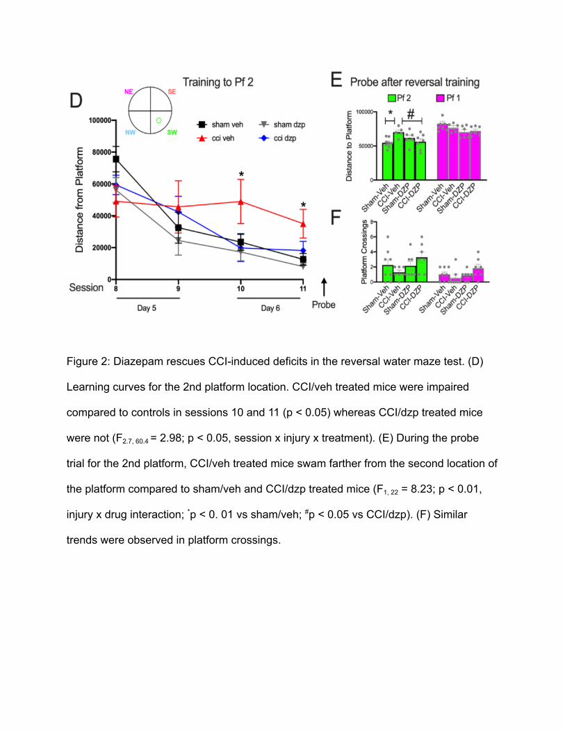
Figure 2: Diazepam rescues CCI-induced deficits in the reversal water maze test. (D)
Learning curves for the 2nd platform location. CCI/veh treated mice were impaired
compared to controls in sessions 10 and 11 (p < 0.05) whereas CCI/dzp treated mice
were not (F2.7, 60.4 = 2.98; p < 0.05, session x injury x treatment). (E) During the probe
trial for the 2nd platform, CCI/veh treated mice swam farther from the second location of
the platform compared to sham/veh and CCI/dzp treated mice (F1, 22 = 8.23; p < 0.01,
injury x drug interaction; *p < 0. 01 vs sham/veh; #p < 0.05 vs CCI/dzp). (F) Similar
trends were observed in platform crossings.
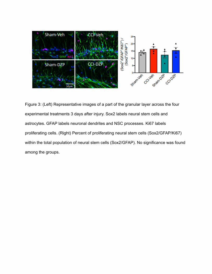
Figure 3: (Left) Representative images of a part of the granular layer across the four
experimental treatments 3 days after injury. Sox2 labels neural stem cells and
astrocytes. GFAP labels neuronal dendrites and NSC processes. Ki67 labels
proliferating cells. (Right) Percent of proliferating neural stem cells (Sox2/GFAP/Ki67)
within the total population of neural stem cells (Sox2/GFAP). No significance was found
among the groups.