Embed Size (px)
Citation preview

The Early Development of the Chondrocraniumof the Lizard.
By
G. R. de Beer. M.A., B.Sc, F.L.S., C.M.Z.S.,
Fellow of Merton College, Lecturer of Magdalen College, Jenkinson Lecturerin Embryology, and University Demonstrator in Zoology, Oxford.
With 28 Text-figures.
C O N T E N T S .PAGE
I. INTRODUCTION 707
II . DESCRIPTION OF STAGES 709
EXPLANATION OF LETTERING . . . . . . 710
I I I . DISCUSSION 730
A. The nasal capsule . . . . . . . 731B. The fenestra ovalis and the basicapsular fenestra . . 733C. The planum supraseptale, ala orbitalis, and interorbital
septum . . . . . . . . . 734D. The phylogenetic position of the L a c e r t i l i a i n the light
of the chondrocranium . . . . . . 735
IV. SUMMARY 737
V. LIST OV LITERATURE CITED 773
I. INTRODUCTION.
THE embryonic skull of the lizard was one of the first to bestudied by modem critical methods, and now the morphologyof the chondrocranium of the Lace r t i l i a may be regarded aswell known, thanks to the work of Gaupp on L a c e r t aagil is (31 mm. stage (1900) and 47 mm. stage (1906)), ofEice (1920) on Eumeces (various stages), and of Pearson(1921) on Lygosoma. However, with the exception ofLeydig's (1872) work on L a c e r t a and Anguis , Parker's(1879) on L a c e r t a , and Sewertzoff's paper (1900) on Asca-l a b o t e s , none of which are very detailed, practically no
NO. 292 3 A

708 G. H. DE BEER
investigations have been made into the embryology of thelacertilian chondrocranium. The present paper is an attempt tofill this gap, as a result of a study of some two dozen embryos ofL a c e r t a agi l is of varying stages of development, preparedaccording to van Wijhe's method (1902), as amended (1922),using victoria blue.
The material was obtained and preserved by Professor E. S.Goodrich in Naples, and I wish here to record my gratitude tohim for very kindly turning some of it over to me for this work.The embryos were removed from their shells, and only thosewhich were living and healthy were fixed and used. Unfor-tunately, it was impossible to determine the respective ages ofembryos of the different stages, and it was necessary to haverecourse solely to measurements. The shape of the lacertilianembryo is such that the body from the tip of the snout to thetip of the tail is coiled twice on itself, with the result that theso-called ' greatest length ', from the prominence of the mid-brain to the root of the tail, is very variable, and depends onthe degree of tightness of the coil. For comparative purposes,therefore, measurements of greatest length are of little use, andthe investigator is driven to adopting the head-length as hisstandard of comparison. The various stages figured anddescribed in this paper are enumerated below.
Stage. Embryo. Head-length.
123456789
10
XAGHIBCDEF
2-252-53-5444-555-255-255-5
The most advanced stage described in this paper leads on con-veniently to the younger of those which Gaupp worked at.Gaupp's model, reproduced in wax by Ziegler, was used asa standard of comparison and as a check to the interpretation

CHONDEOCRANIUM OF THE LIZARD 709
of the drawings which were made directly from the preparationsunder a camera lucida. In addition, a number of sets of serialsections of L a c e r t a at various stages were used to confirmthe reconstructions. It should be remembered that the victoria-blue method of van Wihje is specific for chondrin to the exclusionof procartilage, with the result that embryos prepared by thismethod may appear to be unduly delayed in their chondrifica-tion as compared with reconstructions made from sections ofthe same stages. In the latter case, it almost always happensthat the line of demarcation between cartilage and procartilageis interpreted very liberally for the cartilage. In any case, thedifferences are slight, and the matter is of little importance.
The work was done in the Department of Zoology and Com-parative Anatomy of the Oxford University Museum, in which Ienjoyed the unfailing encouragement of Professor Goodrich.
II. DESCRIPTION OB' STAGES.
Stage 1 (embryo X, H.L. 2-25 mm., Text-fig. 1).—The firstpart of the cartilaginous skeleton to chondrify is Meckel'scartilage, and some of the embryos of this stage show no othervisible skeleton. In others, however, it is possible to make outa thin film of cartilage on each side of the notochord, beneaththe hind-brain, and continuous posteriorly with a small uprisingoccipital arch on each side. All the roots of the hypoglossalnerve emerge freely in front of the occipital arch of their ownside, passing over the parachordal. In such embryos, the chondri-flcation of Meckel's cartilage is obviously more advanced thanthat of the parachordal and occipital arch. The lizard musttherefore be counted among those forms in which the splanchno-cranium develops before the neurocranium.
Stage 2 (embryo A, H.L. 2-5 mm., Text-figs. 2 and 3).—Thechief difference between this and the previous stage is the factthat the auditory capsule has put in its appearance, as a thinfilm of cartilage moulded round the lateral surface of the utricle.It must also be noticed that the hindmost of pair roots of thehypoglossal nerve has been enclosed in a foramen by a bar ofcartilage which projects sideways and upwards from the para-
3A2

710 G. R. DE BEER
TEXT-FIG. 1.
I mmFig. 1.—Lateral view from the left sid
H.L. 2-25 mm.of embryo X, stage 1,
EXPLANATION OF LETTERING.
a, aditus conchae ; ac, auditory capsule; be, basicapsular commis-sure ; bd, basidorsal cartilage; bf, basicapsular fenestra ; bt, basi-trabecular process; c, eye; ca, columella auris; cbl, cornubranchiate primum (1st ceratobranchial); cb2, cornu branchialesecundum (2nd ceratobranchial, distal portion); d>2a, 2nd cerato-branchial, proximal portion; cc, cavum conchale; ch, cornu hyale(eeratohyal); co, concha nasalis ; cp, crista parotica; cr, cristasellaris; cs, sphenethmoid commissure; dgl, duct of lateral nasalgland; dl, dental lamina; era, external nostril ; fa, foramenapicale; fb, basicranial fenestra; fbr, fore-brain; fe, foramenepiphaniale; ftp, fenestra epioptica; ff, foramen faciale; fl,fenestra lateralis nasi; fin, fissura metotica; fmo, fenestrametoptica; jo, fenestra optica; fol, fenestra olfactoria; fp,fenestra prootica; fs, fenestra septalis; fsu, fenestra superior nasi;g, gland ; gl, lateral nasal gland ; he, hypochordal commissure ;hf, hypoglossal foramen or foramina ; hh, hypohyal cartilage ; hy,hypophysial fenestra ; in, internal nostril; ip, incisura prootica ;Jo, Jacobson's organ; Ita, lamina transversalis anterior; Me,Meckel's cartilage ; nvp, meniscus pterygoideus ; n, notochord ;oa, occipital arch; oc, cavity of olfactory sac; on, olfactorynerve; p, parachordal cartilage; pa, processus ascendens;pac, pila accessoria ; pan, planum antorbitale ; poo, pila antotica(prootica); pc, paraseptal cartilage ; pe, processus entoglossus ;pm, pila metoptica; pmp, processns maxillaris posterior; pp,

CHONDKOCRANIUM OF THE LIZARD 711
chordal to join the occipital arch behind it, on each side. Thebar of cartilage in question may be regarded as a preoccipitalarch, similar to that which has been demonstrated in Scyl l iumand in Amblys toma by Goodrich (1911 and 1918), and inLepus by de Beer and Woodger (in the press). In L a c e r t a
TEXT-FIGS. 2, 3.
hf O3ac
2
Me
Fig. 2.—Lateral view from left side of the skull.Fig. 3.—Dorsal view of embryo A, stage 2, H.L. 2-5 mm.
there are eventually (see Text-figs. 7 and 8) three such archeson each side, in front of the occipital arch. Between them thesearches will enclose the three roots of the hypoglossal nerves in
processus paroticus; pr, processus retroarticularis of Meckel'scartilage ; ps, planum supraseptale ; pt, parietotectal cartilage ofnasal capsule; q, quadrate cartilage; r, raphe between lateral andmedial nasal processes, leading to aperture of Jacobson's organ;re, recessus extraconchalis ; ret, ramus ethmoidalis of profundusnerve ; rl, ramus lateralis of ethmoid nerve; mm, ramus medialisof ethmoid nerve; si, interorbital septum; sin, subieuluminfundibuli; sn, nasal septum ; t, trabecula cranii; tc, trabeculacommunis; tl, true lateral wall of nasal capsule, forming innerwall of cavum conchale; im, taenia marginalis; tee, taeniamedialis; Is, tectum synoticum.

712 G. R. DB BEER
separate foramina. This condition is similar to that whichSewertzoff (1897) observed i n A s c a l a b o t e s . At the stage inquestion in L a c e r t a (stage 2), the two anterior pairs of rootsof the hypoglossal nerve are still free.
S tage 3 (embryo G, H.L. 3-5 mm., Text-figs. 4, 5, and 6).—TEXT-FIGS. 4-6.
Kg. 4.—Lateral view from left side of the skull.Fig. 5.—Dorsal view of embryo G, stage 3, H.L. 3-5 mm.Kg. 6.—Ventral view of hyoid and branchial arches of embryo G.
At this stage, the trabeculae cranii have made their appearanceas a pair of bars of cartilage, in front of the parachordals.Posteriorly, the trabeculae diverge from one another, butanteriorly they converge and fuse to form the trabecula corn-munis, which extends forwards for a little way. It has notbeen possible to find a stage in cartilage at which the trabeculaewere not already fused to form a trabecula communis. The

CHONDROCRANIUM OP THE LIZARD 713
auditory capsule shows a fine process directed downwards, andslightly towards the lateral edge of the parachordal of its ownside. In front of this process is the quadrate, which is not, asyet, in contact with either Meckel's cartilage or the auditorycapsule. Posteriorly, the enclosure of the hypoglossal root isproceeding, but unequally on the two sides. On the left, thesecond root is definitely enclosed, while, on the right, it runsthrough a deep notch. In some embryos at this stage theincipient chondrification of the columella auris may be observed.
The so-called ' hyoid ' skeleton at this and the subsequentstages is in an interesting condition. There is a median processusentoglossus (or processus lingualis) corresponding to a basihyaland chondrifying independently. On each side of the posteriorend of this basihyal is a pair of separate nodules of cartilagewhich represent the hypohyals, and lateral to them are theceratohyals in the form of thin rods extending backwards andoutwards. Between the ceratohyals are two more pairs of rodsof cartilage, chondrifying independently. These are the firstand second ceratobranchials. Whereas at these stages all thesecartilages are still separate, they eventually all fuse together,as shown in fig. 386 on p. 771 of Gaupp's (1906) description ofa 47 mm. embryo. The hypohyal and ceratohyal of each sidethen form the cornu hyale or anterior horn of the hyoid. Simi-larly, the first ceratobranchial gives rise to the cornu branchialeprimum and the second ceratobranchial to the cornu branchialesecundum. The latter structure represents a chondrification ofthe skeletal elements of the fourth visceral arch, and it would seemto be incomplete, for, as will be seen below, another cartilagebelonging apparently also to the fourth visceral arch appears atlater stages.
S tage 4 (embryo H, H.L. 4 mm., Text-figs. 7 and 8).—Thetrabecula communis has extended forward between the eyestowards the septum separating the nasal sacs, but posteriorly,the hind ends of the trabeculae cranii are still free from thefront of the parachordals. Where the anterior edge of theparachordals touches the notochord there has been no advance,but farther to the side a cartilaginous process is directed for-

714 G. K. DE BEER
ward towards the hind ends of the trabeculae cranii. In thisway, the front of the parachordals comes to present a hemispheri-cal concavity from the centre of which the notochord projects.This concavity marks the hind border of the future fenestrabasicranialis. The lateral edge of the parachordal shows a littleprominence which is directed towards the downward projection
TEXT-FIGS. 7, 8.
Pig. 7.—Lateral view from the left side of the skull.Kg. 8.—Dorsal view of embryo H, stage 4, H.L. 4 mm.
from the auditory capsule of its own side, but as far as can bemade out is still free from it. Behind this prominence all threeroots of the hypoglossal nerve are now enclosed in separateforamina on each side. The occipital arches have extendedupwards behind the auditory capsules, and it is now possibleto outline a space comprised between the auditory capsule,the lateral edge of the parachordal, and the occipital arch, whichspace will eventually become the fissura metotica. The hind-

CHONDROCBANIUM OF THE LIZARD 715
most portions of the parachordal of each side are now extendingtowards one another beneath the notochord, which will resultin the formation of the hypochordal commissure. The chondri-fication of the auditory capsule is farther advanced, and portionsof the roof as well as the septa of the semicircular canals are
TEXT-FIGS. 9, 10.
be
.-hF
-P-
Fig. 9.—Lateral view from the left side of the skull.Fig. 10.—Dorsal view of embryo I, stage 5, H.L. 4 mm.
present. The walls of the cochlear portion of the auditorycapsule are, however, still membranous. Part of the columellaauris is now present and can be seen as an independent nodule ofcartilage situated behind the quadrate, in the middle of thatpersistently membranous portion of the side wall of the auditorycapsule which will eventually become the fenestra ovalis. Thisnodule represents the proximal portion of the columella auris.

716 G. R. DB BEER
the so-called otostapes. The quadrate is now articulated withMeckel's cartilage ventrally and with the side wall of theauditory capsule dorsally. No c a r t i l a g i n o u s connexionbetween the quadrate and the columella auris was observed atthis or any other stage.
S tage 5 (embryo I, H.L. 4 mm., Text-figs. 9 and 10.)—Twonew features have appeared at this stage, viz. the interorbitalseptum and the processus ascendens. The interorbital septumhas begun to chondrify as a little strip of cartilage dorsal to thetrabecula communis. Each processus ascendens is a bar ofcartilage in a more or less vertical position, in front of theauditory capsules on each side, and quite free from any othercartilage. The most interesting features of this stage concernthe relations of the auditory capsule to the parachordal, andhere, unfortunately, the results are not as definite as in otherregions. It may be stated at once that the investigation of theembryology of the chondrocranium of the lizard by means ofthe van Wijhe technique has been more difficult than in the caseof any other type of vertebrate. The extreme fineness of thestrips of cartilage and the difficulties attending the dissectionof the preparations prior to mounting have been a handicapto the interpretation of the relations in the more complicatedregions. This is especially the case in the region in question,principally owing to the apparent superposition of structuresseen in a total preparation. However, by careful comparisonbetween several preparations and reference to serial sections, thefollowing points can be made out. The cochlear portion of theauditory capsule becomes chondrified (in continuity with therest of the capsule) and acquires a connexion with the para-chordal. From the earliest of these stages it seems that thefacial nerve is enclosed in its facial foramen, and therefore theconnexion between the cochlear part of the auditory capsuleand the parachordal must be composed of a prefacial commissureas well as an anterior basicapsular commissure.
Farther back, the process which at earlier stages was describedas projecting towards the prominence on the lateral edge ofthe parachordal now comes into contact with the latter. It is
CHONDKOCRANIUM OF THE LIZARD 717
very important to define this process, and it may be describedas the strip of cartilage which forms the lateral and anteriorborders of the foramen perilymphaticum of the definitiveauditory capsule.
The result of these relations is that between the auditorycapsule and its process described in the previous paragraph, thelateral edge of the parachordal, and the anterior basicapsularcommissure, there is a gap which at this stage is free fromcartilage. Into the upper portion of this gap the proximal endof the columella auris projects, and so this portion of the gapmay be regarded as the fenestra ovalis. The lower portion ofthe gap, however, owes its existence solely to the delay inchondrification of the floor of the cochlear portion of theauditory capsule. While the van Wijhe preparations show aclear space in this region, sections reveal the presence of pro-cartilage in an early stage of histological differentiation, and thisprocartilaginous floor of the auditory capsule is in contact withthe lateral edge of the parachordal. These relations are ofimportance in view of the question of the relation of the fenestraovalis of the auditory capsule to the so-called basicapsularfenestra of other forms. At the present stage in the developmentof the lizard, the fenestra ovalis is present, and it is continuouswith a space which may be called the basicapsular fenestra andwhich represents merely the as yet unchondrified floor of theauditory capsule.
Anteriorly, the parachordals are still free from the trabeculae,while posteriorly they have met beneath the notochord to forma hypochordal commissure.
Stage 6 (embryo B, H.L. 4-5 mm., Text-figs. 11 and 12).—The hind ends of the trabeculae cranii have now establishedconnexion with the anterior projections of the parachordals,with the result that a large pear-shaped gap is enclosed in thefloor of the skull. This gap represents the conjoined fenestrabasicranialis and fenestra hypophyseos of later stages, whichhave not yet become separated from one another. Opposite thebase of each processus ascendens is a small lateral projectionfrom the hindmost region of each trabecula cranii, forming the

718 G. R. DE BEER
basitrabecular process. Farther forward, the interorbital septumis now more extensive and has become connected with thetrabecula communis below, and with a new structure, the
TEXT-FIGS. 11, 12.
tc
cs
\
M'c
f!s
Me .:t
pe \ ;Fb
_—i ^
^ ^
h. — Fm\ —hF
T'-P
'•03
ac
12
Fig. 11.—Lateral view from the left side of the skull.Fig. 12.—Dorsal view of embryo B, stage 6, H.L. 4-5 mm.
planum, supraseptale above. The latter is really a paired struc-ture of which the two members have met in the middle linedorsal to the interorbital septum and beneath the brain.Anteriorly the planum supraseptale is continuous with a pairof processes which project to each side: the rudiments of thesphenethmoid commissures. In front of this again, the roof ofthe nasal capsule is beginning to chondrify in the form of theparietotectal cartilage. The trabecula communis now extends

CHONDROCEANIUM OF THE LIZARD 719
between the paired nasal sacs and gives rise in this region tothe nasal septum.
As regards the auditory capsule, the floor has now becomecartilaginous, with the result that the fenestra ovalis has a
TEXT-FIGS. 13, 14.
tme
cp
Fig. 13.—Lateral view from the left side of the skull.Fig. 14.—Dorsal view of embryo C, stage 7, H.L. 5 mm.
median border. The walls and roof of the capsule are now wellformed, and the quadrate abuts against a prominence formed bythe lateral semicircular canal.
It is interesting to note that at this stage the trabeculae makea fairly sharp angle with the parachordals.
S tage 7 (embryo C, H.L. 5 mm., Text-figs. 13 and 14).—The nasal and interorbital septa are now more extensive,

720 G. R. DE BEER
although unchondrified gaps remain in the form of septalforamina. Anteriorly, the parietotectal cartilages, which formthe roof and part of the side of the nasal capsule, grow out of thenasal septum. The sphenethmoid commissures still end freelyin front, but the planum supraseptale has enlarged to form aplate immediately underlying the end-brain. In the orbito-temporal region, the rudiments of the side wall of the skull areappearing in the form of a few struts, in which part of the taeniamedialis (taenia parietalis media) can be recognized. The basi-trabecular processes are further developed and immediatelyabove them is the small rudiment of the pila antotica.
At the place where the trabeculae and parachordals met,there is from the point of junction on each side a process pro-jecting inwards towards its fellow of the opposite side, andtending to divide the original large pear-shaped gap in thefloor of the skull into an anterior fenestra hypophyseos and aposterior fenestra basicranialis. These processes form the rudi-ment of the crista sellaris, and its paired origin is unexpected.As far as can be made out, the space between the two halves ofthe crista sellaris in the middle line is occupied by the anteriorend of the notochord. The angle between the planes of thetrabeculae and of the parachordals, which was marked at theprevious stage, has now been smoothened out to a considerableextent.
The side wall of the auditory capsule bears a projection whichjuts out from the prominence for the lateral semicircular canal,behind the head of the quadrate. This is the crista parotica,to which, as Gaupp (1900 and 1906) showed, a structure whichwas in blastematous continuity with the columella auris andknown as the processus paroticus, becomes attached. Thisprocessus paroticus eventually chondrifies, as will be seen instage 9.
As regards the splanchnocranium, the posterior or dorsalportion of the skeleton of the fourth visceral arch has nowchondrified, as a pair of rods lying between the hind ends of thefirst ceratobranchials.
S tage 8 (embryo D, H.L. 5-25 mm., Text-figs. 15 and 16).—

CHONDBOCEANIUM OF THE LIZARD 721
The chief advance which this stage shows concerns the sidewall of the skull in the orbitotemporal region. The rudiment ofthe taenia medialis of the previous stage is now connected with
TEXT-FIGS. 15, 16.
tme
Fig. 15.—Lateral view from the left side of the skull.Fig. 16.—Dorsal view of embryo D, stage 8, H.L. 5*25 mm.
the trabecula communis by means of the pila metoptica, anddorsally it is continuous by means of the pila accessoria with thetaenia marginalis which extends forward as a slender strip ofcartilage from the roof of the auditory capsule. Neither thetaenia marginalis nor the taenia medialis have yet established cartilaginous connexions with the planum supraseptale,

722 G. E. DE BEER
although the position of these future connexions is evident fromthe appearance of projections from the planum supraseptaleitself. The hinder portion of the interorbital septum projectsupwards and backwards towards the hinder part of the planumsupraseptale, with which however it does not fuse.
In the nasal capsule the parietotectal cartilages are now moreextensive, while the auditory capsules are now joined to oneanother above the brain by means of a slender bar of cartilageforming the tectum synoticum. This structure passes imme-diately behind the large and prominent endolymphatic sacs.
The crista sellaris is now a complete bar, separating thefenestra hypophyseos from the fenestra basicranialis. The distalend of the columella auris bears a process which extends for-ward in the tympanum, and which corresponds to the parsinferior of the hyostapes of Versluys's (1898) descriptions. Atthis stage, at least on one side of embryo D, this parsinferior is separate from the conical proximal portion of thecolumella auris, which confirms Versluys's statement (1903) thatthe proximal and distal ends of the columella auris have separatecentres of chondrifieation.
The wall of the auditory capsule has now closed in round thefootplate of the proximal end of the columella auris, with theresult that the fenestra ovalis is reduced to its definitive sizeand that its aperture is blocked by the above-mentioned foot-plate. It is, however, important to remember that the columellaauris arose as a separate cartilage without any connexion withthe wall of the auditory capsule.
Stage 9 (embryo E, H.L. 5-25 mm., Text-figs. 17 and 18).—This is the last stage of which a complete description of thechondrocranium will be given, for it may be compared directlywith the earlier of the stages described by Gaupp (1900). Theside wall of the skull in the orbitotemporal region is now ascomplete as it will ever be. The taenia marginalis extends fromthe planum supraseptale to the roof of the auditory capsule;the pila metoptica joins the taenia medialis, which in turn isconnected with the planum supraseptale in front, the pilaaccessoria above, and the pila antotica behind and beneath.
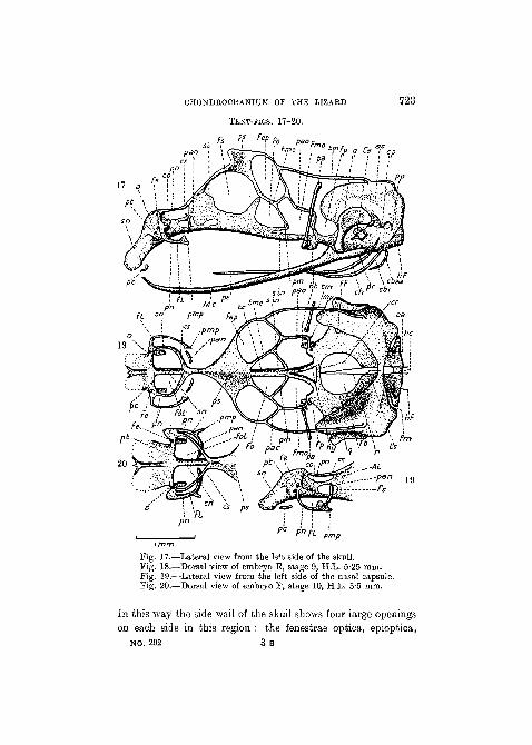
CHONDROCRANIUM OF THE LIZARD
TEXT-FIGS. 17-20.
Fs p's FePFo P.
728
pmp
Fig. 17.—Lateral view from the left side of the skull.Fig. 18.—Dorsal view of embryo E, stage 9, H.L. 5-25 mm.Fig. 19.—Lateral view from the left side of the nasal capsule.Fig. 20.—Dorsal view of embryo F, stage 10, H.L. 5*5 mm.
In this way the side wall of the skull shows four large openingson each side in this region : the fenestrae optica, epioptica,
NO. 292 3 B

724 G. K. DB BEER
metoptica, and prootica. The relations of the various nerves tothese different apertures have been described by Gaupp (1900).
The basitrabecular processes are now large structures, anda feature of considerable interest is the appearance of a smallindependent piece of cartilage between the basitrabecular pro-cess and the base of the processus ascendens. This is the so-called meniscus pterygoideus of Howes and Swinnerton's (1901)description of Sphenodon , and the cartilago articularis ossispterygoidei of Gaupp's (1900) description of L a c e r t a . Asimilar structure has been reported in E my s by Kunkel (1912).In his earlier work, Gaupp (1891) showed that the cartilagoarticularis was connected with the base of the processus ascen-dens in early stages, and subsequently (1902) he regarded it asthe representative of the basal process of the palatoquadrate.This is probably correct. The processus ascendens is still anisolated cartilage, and it is to be noticed that the pterygoidprocess at its base has not yet developed (cf. stages describedby Gaupp). In sections of an embryo slightly older than thathere described it has been possible to confirm Broom's (1924)observations (on the lacertilians Zonurus , E r e m i a s , andMabuia) that the strand of dense tissue which connects thebase of the processus ascendens with the quadrate (describedby Gaupp, 1891) sometimes undergoes chondrification. Thequadrate cartilage of L a c e r t a is therefore not always freeat this stage.
As regards the columella auris, the pars inferior of the distalend is now fairly well developed, and the proximal and distalends have joined to form a single rod, expanded at eachextremity. Dorsal to the columella a small cartilage is seenlodged between the head of the quadrate and the crista parotica.Sections show that this little cartilage is in blastematous con-nexion with the columella auris, of which it represents theprocessus dorsalis, or intercalary, or processus paroticus. Asalready mentioned, this structure eventually becomes joined onto the crista parotica, as Gaupp (1900), Versluys (1903), andGoodrich (1916) described.
The fissura metotica is still open posterodorsally, for the
CHONDROCRANIUM OF THE LIZARD 725
occipital arches have not yet fused on to the hind wall of theauditory capsules. Otherwise, the fissura metotica has assumedits definitive form and bears the usual relations to the foramenperilymphaticum of the auditory capsule (de Beer, 1929). Theforamen perilymphaticum is merely an unchondrified portion ofthe hinder part of the floor of the auditory capsule.
Perhaps the most interesting features which this stage pre-sents are connected with the nasal capsule. The parietotectalcartilages have extended right and left of the nasal septum, andby now a considerable portion of the roof and side walls of thecapsule have been formed. Attached to the posterolateralcorners of the parietotectal cartilages a new element has ap-peared on each side. This is the paranasal cartilage.
The paranasal cartilage is shaped like a crescent with theconvex side turned forward, and the two horns pointing back-ward, one above and the other below. The upper horn isattached to the free anterior end of the sphenethmoid commis-sure, and, slightly in front of this, the paranasal cartilage isattached to the parietotectal cartilage. In this way a pair ofapertures is enclosed. Each of these apertures, which are thefenestrae olfactoriae, is bounded medially by the nasal septum,posterolaterally by the planum supraseptale and the spheneth-moid commissure, anterolaterally by the paranasal cartilage,and anteriorly by the parietotectal cartilage.
Near the point of attachment of the paranasal cartilage tothe parietotectal cartilage is the foramen epiphaniale, throughwhich the lateral branch of the ethmoid nerve leaves the cavityof the nasal capsule, and the position of this foramen is ofimportance. Immediately beneath the foramen epiphaniale theparanasal cartilage forms as it were a duplication of the sidewall of the capsule, for it is situated laterally to that hindmostportion of the side wall which is formed by the parietotectalcartilage. Between these two cartilaginous walls there is there-fore a space, the cavum conchale, which ends blindly behind,but opens forward by the so-called aditus conchae, and lodgesthe lateral nasal gland. The whole structure, which is shapedsomewhat like a cone with the point directed backward, is
3 B 2

726 G. R. DE BEER
known as the concha, and it projects into the cavity of thedefinitive nasal capsule. The lower horn of the paranasalcartilage projects freely backward as the processus maxillarisposterior.
The hind.wall of the nasal capsule is represented by an inde-pendent paired cartilage, theplanum antorbitale, situated atthe side of the nasal septum, between the root of the spheneth-moid commissure and the processus maxillaris posterior. Thefloor of the nasal capsule is represented only by the isolatedrudiments of the paraseptal cartilages, which extend for onlya short way beneath the ventral edge of the nasal septum.
Stage 10 (embryo F, H.L. 5-5 mm., Text-figs. 19 and 20).—The main portions of the skull of this stage show no appreciableadvance over that of the previous one ; only in the nasal capsulehave important advances been made. There, the lower pos-teriorly directed horns of the paranasal cartilages have becomeattached to the planum antorbitale of their own side. The latterhas, however, not yet established connexion with the spheneth-moid commissure. When that has happened, there will still bea large lateral opening in the hinder part of the side wall of thenasal capsule : the fenestra lateralis, through which it is possibleto see the concha as it projects backward into the cavity of thenasal capsule. This condition is illustrated in figs. 3 and 13 ofGaupp's (1900) description of the skull of an embryo of L a c e r ta31 mm. in length, and Bom's (1879) fig. 1, PI. VI, of an embryoof L a c e r t a ready to hatch.
Although Gaupp's descriptions of the nasal capsule are excel-lent, they are somewhat difficult to follow, and it is not easyto visualize the geometrical relations without a series of trans-verse sections. For this reason, a few sections through selectedregions of the nasal capsule of an embryo of L a c e r t a slightlyyounger than the earliest of Gaupp's have been added (Text-figs. 21-8).
Text-fig. 21 passes through the external nostril and thefenestra narina through which the nasal sac communicates withthe exterior. The roof of the capsule is formed by the front partof the parietotectal cartilage, and the section passes through the

CHONDROCRANIUM OF THE LIZARD
TEXT-FIGS. 21-6.
727
22
25 26
Selected transverse sections. Series Jenkinson D.Fig. 21, section 1-2-2 ; fig. 22, section 1-3-14; fig. 23, section
1-4-5 ; fig. 24, section 2-1-4; fig. 25, section 2-2-7 ; fig. 26,section 2-2-15.
small foramen apicale, through which the medial branch of theethmoid nerve emerges.
Text-fig. 22 shows the hinder portion of the fenestra narina

728 G. R. DE BEER
through which the duct of the lateral nasal gland leaves theolfactory sac to run back laterally to the wall of the capsule.The roof of the capsule is here perforated by the fenestra superior,which appears to have no morphological significance.
The floor is formed by the lamina transversalis anterior,which is connected with the ventral edge of the parietotectalcartilage and was connected with the ventral edge of the nasalseptum a few sections farther forward.
Text-fig. 23 is behind the lamina transversalis anterior, andJacobson's organ is seen descending towards its opening intothe buccal cavity. The fenestra superior is still shown, as arethe paraseptal cartilages. Lateral to the true side wall of thecapsule (formed by the parietotectal cartilage) may be seen thelateral branch of the ethmoid nerve and the duct of the lateralnasal gland.
Text-fig. 24 is behind the fenestra superior, and so the roofof the capsule is complete. The section passes through theanterior acini of the lateral gland, which are accommodated ina groove in the wall of the capsule. The lateral branch of theethmoid nerve is still lateral to the side wall of the capsule.
Text-fig. 25 passes through the anterior region of the paranasalcartilage. It is lateral to the lateral nasal gland, which in turn is(together with the lateral branch of the ethmoid nerve) lateralto the true side wall. The gland therefore finds itself enclosedby cartilage in the cavum conchale, which the duct and thenerve have entered by its anterior opening : the aditus conchae.
In Text-fig. 26 the paranasal cartilage has become attachedto the parietotectal cartilage, but it does not form a completecartilaginous wall because of the large fenestra lateralis. Thelateral branch of the ethmoid nerve is now median to the para-nasal cartilage, and has entered the cavity of the nasal capsule(through its own small foramen epiphaniale). It is importantto realize that the cavity of the concha (the cavum conchale)is really a part of extracapsular space, and it is lined throughoutby what are really external capsular walls. The concha may bedescriptively regarded as having been pushed into the cavityof the capsule at the spot where the paranasal cartilage becomes

CHONDROCBANIUM OF THE LIZARD 729
attached to the parietotectal. Or, alternatively (and with agreater degree of probability), it may be said that the conchaowes its existence to the fact that the paranasal cartilage hasbeen reflexed forward, outside the side wall of the capsule
TEXT-FIGS. 27, 28.
3
27
Fig. 27, section 2-4-4 ; fig. 28, section 2-5-8.
formed by the parietotectal cartilage. The result is that thecavity of the capsule bulges out and forward over the concha,and this bulge accommodates the recessus extraconchalis. Thecause of this bulge is probably associated with the enormoussize of the eyes, which press on the hind wall of the nasal

780 G. R. DB BEER
capsule, causing it to find the necessary accommodation for itscontents in the manner described.
In Text-fig. 27 the recessus extraconchalis is still open to theside through the fenestra lateralis. The lateral and medianbranches of the ethmoid nerve are now united, dorsal to thebranches of the olfactory nerve, forming the ramus ethmoidalisof the profundus bran<5h of the trigeminal nerve.
In Text-fig. 28 the fenestra lateralis is no longer seen, for theplanum antorbitale (itself attached to the lower horn of theparanasal cartilage) has established connexion with the spheneth-moid commissure (itself attached to the upper horn of theparanasal cartilage). The paraseptal cartilage has also run intothe median portion of the planum antorbitale, and, a few sectionsfarther back, the planum antorbitale will be seen forming acomplete hind wall to the cavity of the nasal capsule.
III. DISCUSSION.
The only other lacertilian of which the earliest stages ofdevelopment have been studied is A s c a l a b o t e s , by Sewert-zoff (1900), and it must be noted that there are considerabledifferences between the conditions which he describes and thosewhich are given in this paper. According to Sewertzoff, thetrabeculae in Asca labo te s appear wide apart and separatefrom one another, whereas in L a c e r t a I have found them tobe joined anteriorly to form a trabecula communis from theirearliest appearance. Then Sewertzoff describes the appearanceat an early stage of a crista sellaris, connected on each side withan ' alisphenoid ' (meaning the pila antotica) and unconnectedeither with the trabeculae in front or the parachordals behind.In L a c e r t a , however, as described in this paper, the chondri-fication of the crista sellaris and pila antotica occurs com-paratively late, and in continuity with the cartilages of the floorof the skull. In this respect, my observations are partly inagreement with Parker's (1879) on L a c e r t a . His earliestfigured stage (PL XXXIX, fig. 1) shows the trabeculae attachedto the parachordals, and diverging freely anteriorly. On theother hand, in his next stage (PI. XXXIX, fig. 2) the trabeeulae

CHONDBOCRANIUM OF THE LIZARD 731
have fused to form a trabecula cornmunis but there is no cristasellaris.
The comparison between the chondrocraniurn of La cer t aand that of other reptiles so far known is reserved for a latersection of this discussion. It may, however, be noted here thatShaner (1926), who studied early stages of development ofChrysemys , found conditions comparable to those describedby Sewertzoff for A s c a l a b o t e s ; the trabeculae were freeanteriorly and a structure was present corresponding to thecrista sellaris.
A discussion of the relations of the cartilages to the nervesand blood-vessels is unnecessary here, for they have beensummarized in a previous work (de Beer, 1926).
A. The nasa l capsu le .
The nasal capsule of the lacertilian is a complicated structure,and a welcome light on its interpretation has been, thrown bythe recently acquired knowledge of the development of the nasalcapsule in certain mammals. It has been shown by Terry (1917)in the cat, and confirmed by de Beer and Woodger in the rabbit,that three elements take part in the formation of the nasalcapsule. There is (1) the parietotectal cartilage, which is con-tinuous with the dorsal edge of the nasal septum, and forms theanterior part of the roof and side wall of the capsule; (2) theparanasal cartilage, which forms the hind part of the side wall;and (3) the planum antorbitale which forms the hind wall. In themammals mentioned these elements chondrify independentlyof one another, and subsequently become connected. Where theparanasal cartilage joins the parietotectal, the foramen epi-phaniale remains as a witness of the former space separatingthem. Further, as the paranasal cartilage overlaps the parieto-tectal, the posterior edge of the side wall formed by the latterprojects into the cavity of the nasal capsule as the so-calledcrista semicircularis. Now, although in the lizard it has notbeen possible to find a stage at which the paranasal cartilagewas separate from the parietotectal, yet the position of theforamen epiphaniale may be taken as an indication of the line

732 G. E. DE BEEB
of demarcation between these two elements. Further, this lineis also that of the aditus conchae. Allowance has of course tobe made for the fact that the paranasal cartilage is widelyfenestrated (by the fenestra lateralis) in L a c e r t a , but thisis not the case in Eumeces (Eice, 1920) or in Lygosoma(Pearson, 1921), where this cartilage forms an unbroken wall.Now the median wall of the concha of the lacertilian (represent-ing the posterior portion of the side wall of the capsule formedby the parietotectal cartilage) bears relations which are verycomparable to those of the crista semicircularis of the mammal,and the mammalian condition would be still further approachedif the cavity of the concha of the reptilian nasal capsule wereobliterated by the approximation and fusion of its median andlateral walls, or if the lateral wall of the concha disappearedand the aditus conchae were closed by the paranasal car-
It is not proposed to homologize the reptilian concha withthe mammalian crista semicircularis, but an investigation intothe causes contributory to the formation of the former mightthrow some light on the interpretation of the latter. Seydel(1896) considered the concha as an inpushing of the capsularwall due to the development of the lateral nasal gland. Gaupp(1900), however, inclined to the view that the accommodationof the gland in the concha is a passive result of another process,viz. the expansion of the cavity of the nasal capsule resultingin a bulging outward and forward of the side wall over theconcha. It seems further not improbable that this process mayhave been associated with the huge size of the eye in thelacertilian, which presses on the capsule from behind.
However, the latter factor is probably less important in thecase of the other reptiles which possess a concha : crocodiles andsnakes; and it can hardly be appealed to in the case of themammals.
The formation of the concha is therefore probably associatedwith an expansion of the cavity of the nasal capsule. Therecessus extraconchalis of the lizard may be regarded as com-parable to the mammalian recessus anterior : but the concha

CHONDBOCRANIUM OF THE LIZARD 733
would not correspond to the mammalian maxilloturbinal asGaupp (1900) supposes.
Among other reptiles the presence of a concha is reportedby Peyer (1912) for V i p e r a ; by Brock (1929) for Lepto-d e i r a ; and by Shiino (1914) for Crocodi lus . In birdsTonkoff (1900) has shown for Gallus that a structure correspond-ing to the reptilian concha is present.
B. The fenes t ra oval is and the b a s i c a p s u l a rf enes t r a .
In many animals the cartilaginous auditory capsule becomesattached to the lateral edge of the parachordal by two com-missures known respectively as the anterior and posterior basi-capsular commissures (e.g. in the trout, de Beer, 1927). A gapis thus formed between the capsule laterally and the para-chordal medially known as the fenestra basicapsularis, andwhich ultimately becomes obliterated as the chondrification ofthe floor of the auditory capsule is completed. Now, in severalplaces in his treatise, Gaupp (1906) states that this basicapsularfenestra becomes the fenestra ovalis ( = fenestra vestibuli) ofthe auditory capsule of tetrapods, while it becomes obliteratedin the fish (cf. loc. cit., pp. 583,725). As it stands, this statementis slightly misleading, for it would lead one to suppose that themedial border of the fenestra ovalis (into which the footplateof the columella auris fits) is formed of parachordal and thereforeaxial cartilage. There is no doubt that the medial border of theforamen ovale is formed of true capsular cartilage. However,as in many forms the chondrification of the floor of the capsuleis delayed, the fenestra ovalis has for a time no medial border,and is therefore confluent with the basicapsular genestra. Itis therefore hardly legitimate to say that the fenestra ovalis isa remnant of the basicapsular fenestra itself.
When the condition in the tetrapod is compared with that ofSelachii, it is clear that the basicapsular fenestra of the latterhas no claim to represent any part of the fenestra ovalis. Theposition of the fenestra ovalis is morphologically indicated bythe point of articulation of the hyomandibula, which is a

734 G. E. DE BEER
considerable distance lateral to the basicapsular fenestra. In thisconnexion it is of the greatest interest to note that van Wijhe(1924) has actually found a small fenestra ovalis in H e p t a n -chus , situated where one would expect it, viz. ' in the underpart of the fossa for the hyomandibula '.
C. The p l anum s u p r a s e p t a l e , ala o r b i t a l i s , andi n t e r o r b i t a l s e p t u m .
Much of the peculiarity of the skull of the lizard is due to thefact that the floor of the cranial cavity in the orbital region islifted high above the level of the trabecula communis, a tallvertical interorbital septum being intercalated in between. Itis clear that this modification is directly related to the large sizeof the eyes in these animals.
As regards the interorbital septum itself, the conditions inSphenodon as reported by Schauinsland (1900) and byHowes and Swinnerton (1901) lead to the conclusion that itis really a distinct element, separate from the trabecula com-munis. Fuchs (1915) approaches the matter from a differentpoint of view in his study of 0 h e 1 o n e, for he denies that thetrabecula communis extends as far forward as this. The condi-tions in L a c e r t a here described lend support to the view thatthe interorbital septum is an independent structure, whichrapidly acquires connexion with the trabecula. At all events thematter seems to be of little importance. The membranous skull(dura mater) is stretched up from the trabecula (as the fore-brain is lifted dorsally) and gives rise to a vertical wall betweenthe orbits, in which chondrification sets in.
The formation of the interorbital septum results in importantmodifications of the orbital cartilage. This structure no longersprings up from two pairs of roots direct from the trabecula.The posterior pair (the pila metoptica) is present, but theanterior pair has been lost. Further, the orbital cartilages intheir anterior portion have been pressed together between theorbits so that their medial borders meet in the middle line,forming the planum supraseptale. This explains why thesphenethmoid commissures leave the planum supraseptale near

CHONDROCRANIUM OF THE LIZARD 735
the middle line, and diverge as they run forward. It may benoted that an interorbital septum is present in certain mammals(Primates, Eodents), and in these the orbital cartilage (alaorbitalis) springing from the dorsal edge of the septum approxi-mates to the form of a planum supraseptale.
The posterior portion of the orbital cartilage is in a reducedcondition, being represented only by the taenia medialis, taeniamarginalis, pila accessoria, pila metoptica, and pila antotica,which form a very slender framework. This region of the skullpresents features of interest for comparison with other reptiles,from which interesting conclusions as to the affinities of thelacertilians may be drawn.
D. The phy logene t i c pos i t ion of the L a c e r t i l i ain the l ight of the c h o n d r o c r a n i u m .
The affinities of the L a c e r t i l i a have been a matter ofcontroversy for some time. Long ago Huxley (1871) consideredthat the Lace r t i l i a had lost the lower temporal bar which isstill preserved in Sphenodon . Whether they are regarded, asHuxley would have and as Broom (1924) does, as members ofthe D i a p s i d a in which the lower temporal fossa has been lost,or, with Williston (1925), as P a r a p s i d a which have neverpossessed a lower temporal fossa, the fact remains that thesedistinctions are based primarily on the configurations of thetemporal arches formed by the dermal bones in the osteocranium.It becomes of interest to inquire whether any independentindications of affinity are given by the chondrocrania ofLace r t i l i a and other reptiles. An attempt at a comparisonof this kind is now possible, since investigations have been madeby modern methods into the structure of the chondrocranium ofall the principal groups of surviving reptiles.
It must be said at once that the snakes must be excludedfrom any such comparison, for their skulls are so peculiar andspecialized as to supply little material for profitable comparison.As Brock (1929) has pointed out, the evidence in favour of thelacertilian origin of the 0 p h i d i a is debatable. There remains,then, Sphenodon, the Crocodi l ia , and the Chelonia ,

736 G. R. DE BEER
with which the L a c er t i 1 i a may be compared. In all of thesethere is the same general form of a tropitrabic skull with atrabecula comrnuhis, an interorbital septum, and a planumsupraseptale, and in all the posterior portion of the orbitalcartilage is more or less reduced. This reduction of the orbitalcartilage follows definite lines, and it is interesting to note thatthe condition in the L a c e r t i l i a (Lacer ta , Gaupp, 1900;Eumeces , Eice, 1920) is identical in plan with that foundin Crocodi l ia (Shiino, 1914) and in Sphenodon (Schauins-land, 1900 ; Howes and Swinnerton, 1901). In each case it iseasy to identify the four fenestrae : optica, metoptica, epioptica,and prootica; separated from one another by the varioustaeniae and pilae already described. These cartilaginous strutsare more slender in the L a c e r t i l i a than in the others,indicating a greater degree of specialization. In the C h e 1 o n i athe conditions are more variable. In Dermochelys (Nick,1912) the side wall of the skull in this region is fairly substantial,while in Chelone (Fuchs, 1915) it has reached approximatelythe same degree of reduction as in the crocodile. Emys(Kunkel, 1912), Chrysemys (Shaner, 1926), and Chelydra(Nick, 1912) show still greater reduction, but the same generalplan of structure can be recognized. The fact that the lacertiliansconform to this plan suggests that their affinities lie with theseanimals.
This conclusion is strengthened by a consideration of twoother regions of the skull. The concha of the nasal capsule,present in lacertilians, is also present in a precisely comparablecondition in the crocodile (Shiino, 1914). (The concha is presentin the snakes: Vipera , Peyer, 1912; L e p t o d e i r a ,Brock, 1929.) The pterygoquadrate presents great similaritiesin Sphenodon , Crocodi lus , and Chelonia , consist-ing of an otic process connected to the base of an ascendingprocess, from which a pterygoid process projects forward. Inthe L a c e r t i l i a all these elements are present in preciselycomparable conditions, although the connexion between thebases of the ascending and otic processes is slender and transient.
As far as the evidence from the chondrocranium goes, it

CHONDROCRANIUM OF THE LIZARD 737
shows that the L a c e r t i l i a have several points of similaritywith Sphenodon and the crocodiles, that is to say, withDiaps ida , and it supports Broom's views concerning thederivation of the L a c e r t i l i a from this group.
IV. SUMMARY.
1. The embryology of the chondrocranium has been studied inten stages of the development of L a c e r t a ag i l i s .
2. The splanchnocranium chondrifies before the neuro-cranium.
3. The crista sellaris has a paired and belated origin.4. The constituents of the nasal capsule of L a c e r t a agree
generally with those of mammals.5. It is pointed out that the foramen ovale is not a derivative
of the fenestra basicapsularis.6. The otic process of L a c e r t a has a temporary cartila-
ginous connexion with the processus ascendens.7. Points of similarity between the chondrocrania of Lacer -
t i l ia and of other reptiles lead to the conclusion that theL a c e r t i l i a are derived from Diaps id reptiles.
V. LIST OF LITERATURE CITED.Born, G. (1879).—" Die Nasenhohlen und der Thranennasengang der
amnioten Wirbelthiere ", ' Morph. Jahrb.', 5.Brock, G. T. (1929).—" On the development of the skull of Leptodeira
hotamboia ", ' Quart. Journ. Micr. Soi.', 73.Broom, R. (1924).—" On the classification of the Reptiles ", ' Bull. Amer.
Mus.Nat. Hist.1, 51.de Beer, G. R. (1926).—" Studies on the vertebrate head. II. The orbito-
temporal region of the skull", ' Quart. Journ. Micr. Sci.', 70.(1927).—"The early development of the chondrocranium of Salmo
fario ", ibid., 71.(1929).—" The development of the skull of the shrew ", ' Phil. Trans.
Roy. Soc.', B, 217.de Beer, G. R., and Woodger, J. H.—(In the press)' The early development
of the skull of the rabbit.'Fuchs, H. (1915).—" t)ber den Bau und die Entwieklung des Schadels der
Chelone imbricata ", ' Voeltzkow : Reise in Ostafrika'.Gaupp, E. (1891).—" Die Columella der Kionokranen Saurier ", ' Anat.
Anz.', 6.

738 G. R. DE BEER
Gaupp, E. (1900).—" Das Chondrocranium von Lacerta agilis ", ' Anat.Hefte', 15.
(1902).—" tJber die Ala temporalis des Siiugerschadels und dieRegio orbitalis einiger anderen Wirbeltierschadel", ibid., 19.
(1906).—" Die Entwicklung des Kopfskelettes ", Hertwig's ' Hand-buch d. verg. u. exp. Entwick. d. Wirb.', 3.
Goodrich, E. S. (1911).—" On the segmentation of the occipital regionof the head in the Batrachia Urodela ", ' Proc. Zool. Soc'
(1916).—" The Chordatympani and middle ear in Reptiles, Birds,and Mammals " , ' Quart. Journ. Micr. Sci.', 61.
(1918).—" On the development of the segments of the head inScyllium ", ibid., 63.
Howes, G. B., and Swinnerton, H. H. (1901).—" On the development ofthe skeleton of the tuatara ", ' Trans. Zool. Soc.', 16.
Huxley, T. H. (1871).—' A manual of the Anatomy of Verfcebrated Animals.'Kunkel, B. W. (1912).—" The development of the skull of Emys " , ' Journ.
Morph.', 23.Leydig, IP. (1872).—' Die in Deutschland lebenden Arten der Saurier.'Nick, L. (1912).—"Das Kopfskelett von Dermochelys coriacea ", ' Zool.
Jahrb. Abt. f. Anat.', 33.Parker, W. K. (1879).—" On the structure and development of the skull in
the Lacertilia ", ' Phil. Trans. Roy. Soc.', 170.Pearson, H. S. (1921).—" The skull and some related structures of a late
embryo of Lygosoma ", ' Journ. Anat.', 56.Peyer, B. (1912).—" Die Entwickelung des Schadelskelettes von Vipera
aspis ", ' Morph. Jahrb.', 44.Rice, E. L. (1920).—" The development of the skull in the Skink, Eumeces
quinquelineatus ", ' Journ. Morph.', 34.Schauinsland, H. (1900).—" Weitere Beitrage zur Entwicklungageschichte
der Hatteria ", ' Arch. f. mikr. Anat;', 56. (Also : 1903.—'Zoologica.')Sewertzoff, A. N. (1897).—" Beitrag zur Entwicklungsgeschichte des
Wirbeltierschadels ", ' Anat. Anz.', 13.(1900).—" Zur Entwioklungsgeschichte von Ascalabotes fasoicularis ",
ibid., 18.Seydel, O. (1896).—" tjber die NasenhShle und das Jacobson'sche Organ
der Land- und Sumpfschildkroten ", ' Festschr. f. Gegenbaur ', 2.Shaner, R. F. (1926).—" The development of the skull of the turtle, with
remarks on fossil reptile skulls ", ' Anat. Rec.', 32.Sh-ino, K. (1914).—"Das Chondrocranium von Crocodilus mit Beriick-
sichtigung der Gehirnnerven und Kopfgefasse ", ' Anat. Hefte', 50.Terry, R. J. (1917).—" The primordial cranium of the cat", ' Journ.
Morph.', 29.Tonkoff, W. (1900).—" Zur Entwickelungsgeschichte des Hiihnerschadels :',
' Anat. Anz.', 18.

CHONDROCEANIUM OF THE LIZARD 739
van Wijhe, J. W. (1902).—" A new method for demonstrating cartilaginousmikroskeletons ", ' Kon. Akad. v. Wetenso. t. Amsterdam.'
(1922).—" Friihe Entwicklungsstadien des Kopf- und Rumpf-skeletts von Aoanthias vulgaris ", ' Bijdr. t. d. Dierk.', 22.
(1924).—" Thymus, spiracular sense organ and fenestra vestibuli(ovalis) in a 63 mm. long embryo of Heptanohua einereus " , ' Kon. Akad.v. Wetenso. t. Amsterdam. Proc.', 26.
Versluys, J. (1898).—" Die mittlere und aussere Ohrsphare der Lacertilierund Rhynchocephalier ", ' Zool. Jahrb. Abt. f. Anat.', 12.
(1903).—"Entwickelung der Columella auris bei den Lacertiliern",ibid., 19.
Williston, S. W. (1925).—' The osteology of the Reptiles.' Harvard.
NO. 292 3 C