Embed Size (px)
Citation preview

© 2017. Published by The Company of Biologists Ltd.
The DUX4 homeodomains mediate inhibition of myogenesis and are
functionally exchangeable with the Pax7 homeodomain
Darko Bosnakovski1,2
, Erik A. Toso2, Lynn M. Hartweck
2, Alessandro Magli
3, Heather A.
Lee2, Eliza R. Thompson
2, Abhijit Dandapat
2, Rita C. R. Perlingeiro
3, and Michael Kyba
2,*
1Faculty of Medical Sciences, University Goce Delcev - Stip, 2000 Stip, R. Macedonia
2Lillehei Heart Institute, Department of Pediatrics, University of Minnesota, Minneapolis,
MN, 55014
3Lillehei Heart Institute, Department of Medicine, University of Minnesota, Minneapolis,
MN, 55104
Key words: Facioscapulohumeral muscular dystrophy, DUX4, myogenesis, homeodomain,
Pax3 and Pax7
Summary Statement: The facioscapulohumeral muscular dystrophy-associated protein
DUX4 inhibits myogenic differentiation via its homeodomains, which bear similarity to, and
can be exchanged with, those of the muscle stem cell master regulator, Pax7.
*Corresponding author: Michael Kyba, PhD,
Lillehei Heart Institute NHH4.126,
University of Minnesota, 312 Church St SE,
Minneapolis, MN, 55014
612.626.5869
Jour
nal o
f Cel
l Sci
ence
• A
ccep
ted
man
uscr
ipt
JCS Advance Online Article. Posted on 21 September 2017

ABSTRACT
Facioscapulohumeral muscular dystrophy (FSHD) is caused by inappropriate expression of
the double homeodomain protein, DUX4. DUX4 has bimodal effects, inhibiting myogenic
differentiation and blocking MyoD at low levels of expression, and killing myoblasts at high
levels. Pax3 and Pax7, which contain related homeodomains, antagonize the cell death
phenotype of DUX4 in C2C12 cells, suggesting some type of competitive interaction. Here,
we show that effects on differentiation and MyoD expression require the homeodomains but
do not require the C-terminal activation domain of DUX4. We test the set of equally related
homeodomain proteins; Pax6, Pitx2c, OTX1, Rax, Hesx1, MIXL1 and Tbx1; and find that
only Pax3 and Pax7 display phenotypic competition. Domain analysis on Pax3 reveals that
the Pax3 homeodomain is necessary for phenotypic competition, but not sufficient;
competition also required the paired and transcriptional activation domains of Pax3.
Remarkably, substitution mutants in which DUX4 homeodomains are replaced by Pax7
homeodomains retain the ability to inhibit differentiation and to induce cytotoxicity.
Jour
nal o
f Cel
l Sci
ence
• A
ccep
ted
man
uscr
ipt

INTRODUCTION
Facioscapulohumeral muscular dystrophy (FSHD) is a dominant inherited myopathy caused
by mutations leading to loss of repeat-induced silencing of the D4Z4 repeat array on
chromosome 4 (Gabellini et al., 2002; Lemmers et al., 2012; van Overveld et al., 2003;
Wijmenga et al., 1992). This in turn provides favorable conditions for expression of DUX4, a
gene embedded within each D4Z4 repeat unit (Gabriels et al., 1999). In several independent
studies, DUX4 mRNA or protein was detected at extremely low levels, specifically in
myoblasts from FSHD patients (Block et al., 2013; Dixit et al., 2007; Jones et al., 2012;
Kowaljow et al., 2007; Snider et al., 2010). DUX4 functions as a transcriptional activator,
inducing expression of hundreds of target genes (Bosnakovski et al., 2008b; Geng et al.,
2012) through a mechanism involving p300/CBP (Choi et al., 2016).
Cultured myoblasts from FSHD patients have been found to exhibit greater sensitivity to
oxidative stress and reduced levels of expression of MyoD and downstream target genes,
compared to controls (Celegato et al., 2006; Krom et al., 2012; Rahimov et al., 2012; Tassin
et al., 2012; Tsumagari et al., 2011; Winokur et al., 2003a; Winokur et al., 2003b). In our
previous work, we demonstrated that DUX4, when expressed at low levels in C2C12
myoblasts, recapitulates aspects of this FSHD myoblast phenotype, namely that it sensitizes
cells to oxidative stress and severely reduces MyoD mRNA and protein levels (Bosnakovski
et al., 2008b). High-level DUX4 expression caused cell death (Bosnakovski et al., 2008b). In
addition to these effects, myoblasts expressing low levels of DUX4 had diminished
differentiation potential, presumed to be due to dysregulation of myogenic regulatory factors
(MRFs), including MyoD (Bosnakovski et al., 2008b), and the transcriptional profile of
DUX4 has been described as characteristic of a less differentiated state (Knopp et al., 2016).
Similar assays performed on DUX4c, a gene encoded by a satellite repeat 42 kb centromeric
to the D4Z4 repeat array, and lacking the 82 C-terminal amino acids of DUX4 due to a
frameshift also downregulated MyoD and inhibited myogenic differentiation, but was not
cytotoxic (Bosnakovski et al., 2008a) suggesting that the C-terminus was not necessary for
effects on myogenesis.
Jour
nal o
f Cel
l Sci
ence
• A
ccep
ted
man
uscr
ipt

The N-terminal part of DUX4 contains its only highly conserved and recognizable domains:
two paired-class homeodomains. Both homeodomains bear significant similarity to the
homeodomains of PAX3 and PAX7, two paired-class homeodomain proteins that act at the
apex of the myogenic regulatory hierarchy, and are expressed in adult satellite cells
(Buckingham et al., 2003; Montarras et al., 2005; Seale et al., 2000). We hypothesized that
DUX4 might impair myogenesis, and therefore muscle regeneration in FSHD, through
interference with PAX3 and/or PAX7 or misregulation of their homeodomain-dependent
target genes in satellite cells or their activated progeny. Consistent with the idea that DUX4
and Pax3/7 can compete with one another, perhaps for regulation of critical target genes, both
Pax3 and Pax7 acted as dose-dependent suppressors of DUX4-induced cytotoxicity when
overexpressed in C2C12 cells (Bosnakovski et al., 2008b). However, although the
homeodomains of Pax3 and Pax7 are highly related to those of DUX4, a number of other
non-myogenic proteins have homeodomains of equal or greater similarity. These are Pax6,
OTX1, Rax, Hesx, and MIXL1. It is unclear whether the competition between DUX4 and
Pax3/7 is due to competition between their homeodomains, and if so, whether this is a
generic feature of the paired class of homeodomains vs. a specific feature of certain members,
and whether proteins with homeodomains of greater similarity than Pax3 and Pax7 compete
more effectively with DUX4. In this study, we investigate the homeodomains of DUX4 and
their competitive interactions with other homeodomain-containing proteins, by co-
overexpression, domain analysis, and homeodomain substitutions. The results point to a
unique relationship between the homeodomains of DUX4 and those of Pax3/Pax7.
RESULTS
Generation of inducible myoblasts bearing DUX4 deletions.
We previously showed that DUX4 expressed at high levels in different cell types induces
rapid cell death and, at low levels, interferes with master myogenic transcription factors
MyoD and Myf5, strongly inhibiting myogenesis. In order to investigate which domains
within DUX4 are necessary to induce these phenotypes, we made a series of deletion
constructs and generated doxycycline-inducible mouse C2C12 myoblast cell lines by
inducible cassette exchange (Bosnakovski et al., 2008b). As a template for these deletion
constructs, we began with a DUX4 construct derived from the terminal D4Z4 repeat of a
4qA161 allele (Gabriels et al., 1999), which contained 3’UTR sequence up to the EcoRI site
Jour
nal o
f Cel
l Sci
ence
• A
ccep
ted
man
uscr
ipt

(Fig. 1A). We wished to determine the role of the N-terminus (the homeodomains), the C-
terminus (missing in DUX4c), and whether D4Z4 RNA or some additional elements from the
3’ UTR play a role in the DUX4 phenotypes described above. To explore the necessity of the
homeodomains, we made a construct initiating precisely at homeodomain 2 (ΔHD1(81-424),
amino acids listed in parentheses) and a construct initiating where HD2 ends, ie. lacking both
homeodomains (ΔHD(1+2)(157-424)) with an ATG added to start translation. To analyze the
role of the C-terminus, a deletion series was made (Fig. 1A). To evaluate activity of the
RNA, or some additional unknown product that might be transcribed from the D4Z4
sequence, we made constructs in which we deleted the ATG (∆ATG) of DUX4, or created a
sequence that initiated at an internal Bsu 36I site (∆5'+3'UTR), or deleted the entire internal
sequence between the first and last Pvu II sites ((ATG(1-75)+∆3'UTR) (Fig. 1A). The
possibility of an activity being encoded by the antisense strand was tested by placing the
D4Z4 sequence in reverse orientation with respect to the inducible promoter (DUX4-opp,
Fig. 1A). All constructs were integrated into the unique inducible cassette exchange locus in
iC2C12 cells by ICE recombination, and inducible cell lines resistant to G418 were generated
as previously described (Bosnakovski et al., 2008b). Each construct is expressed from the
same locus and can be regulated in a dose-responsive manner with doxycycline. RNA for all
constructs was detected by RTPCR, and proteins for each construct were evaluated by
western blot using antibodies that recognize N-terminal (P2G4), central (9A12) or C-terminal
(E5-5) epitopes of DUX4 (Fig. 1B) (Dixit et al., 2007; Geng et al.). With the 9A12 antibody,
we were able to detect all of the deletion proteins except DUX4(1-217), DUX4(1-
75)+∆3'UTR, and ∆5'+3'UTR. The DUX4(1-217) deletion (but not ∆DUX4(1-75)+ ∆3'UTR
or ∆5'+3'UTR) was recognized by P2G4. By combining the 9A12 and P2G4 antibodies we
could evaluate relative sizes of all constructs for which antibodies exist; these were as
expected and ranged from ~30 kDa (DUX4(1-217)) to ~51 kDa (DUX4+3'UTR). Notably,
the protein levels of all constructs lacking the C-terminus (DUX4c DUX4(1-377), and
DUX4(1-399)), were increased relative to those that have a full C-terminus (Fig. 1B).
Interestingly, in using the 9A12 antibody to detect the construct in which the start codon was
removed (∆ATG), we saw a smaller band of approximately 40 kDa, suggesting the presence
of a cryptic internal translation initiation site (Fig. 1B). This protein is likely initiated from
an internal leucine (as described in: Snider et al., 2010), is apparently inactive and resembles
the HD deletions. To confirm that deletion constructs were homogenously inducible in all
cells of the generated cell lines, we performed immunostaining after 20 hours doxycycline
induction (Fig. 1C).
Jour
nal o
f Cel
l Sci
ence
• A
ccep
ted
man
uscr
ipt

Both homeodomains and C-terminus are necessary for cytotoxicity.
We assayed all of the deletion cell lines for cell viability by plating cells at equal density
(2000/well in 96-well plates), inducing the following day with various doses of doxycycline,
and quantifying viability 48 hrs post-induction. Cell death was observed at 24 hours only in
cell lines expressing full-length DUX4 constructs, ie. DUX4(1-424) and DUX4+3'UTR; the
same effect was evident at 48 or 72 hrs post-induction (data not shown). However, in the
construct with the smallest 3’ deletion (DUX4(1-399)), we observed decreased confluency
but obvious cell death by visual inspection (Fig. 2A). Measurement of ATP content confirmed
the previously noticed cell death induced by full length DUX4 (Fig. 2B). In addition,
significantly decreased cell viability of DUX4(1-399) expressing cells was detected (Fig.
2B). On the other hand, ATP content in other cell lines was similar regardless of the levels
and duration of expression (Fig. 2B). This experiment clearly shows that, in addition to the
N-terminal homeodomains, the C-terminus of DUX4 is necessary to cause toxicity, as had
been described (Bosnakovski et al., 2008a). To further clarify the effect of DUX4(1-399), we
analyzed the levels of apoptosis and proliferation in induced cells and found a 1.6 fold
increase in apoptotic cells in cultures that expressed DUX4(1-399) for 18 hours (Fig. 2C, D).
This construct also affected the proliferation rate, seen by a 30% decreased incorporation of
EdU compared to the control uninduced cells (Fig2. E, F). The other constructs, which
showed no effect on ATP content, did not induce apoptosis or affect cell cycle (Fig. 2C-F).
The homeodomains alone are sufficient to alter MRF expression and to block
differentiation.
DUX4 and DUX4c interfere with myogenic regulators, predominantly with MyoD and its
downstream targets (Bosnakovski et al., 2008a; Bosnakovski et al., 2008b). We previously
reported that DUX4 and DUX4c rapidly suppress MyoD within 2 hours of induction, even at
low doses. To identify domains essential for this phenotype, we induced DUX4 constructs in
proliferating cells and analyzed the expression levels of MyoD and Myf5 by RT-qPCR. After
a short 6 hour induction, we found that every construct retaining both homeodomains
provoked a reduction in MyoD (Fig. 3A), like DUX4 and DUX4c. However, for Myf5, the
constructs bearing larger C-terminal deletions (DUX4(1-217) downregulated its expression,
like DUX4c, while the shortest C-terminal deletion (DUX4(1-399)) moderately increased
Jour
nal o
f Cel
l Sci
ence
• A
ccep
ted
man
uscr
ipt

Myf5, similar to WT DUX4 (Fig. 2A). Constructs with N-terminal deletions had no
influence on MyoD or Myf5 (not shown).
To test the functional relevance of interactions between deletion constructs and myogenic
regulators, we induced myogenic differentiation and analyzed the levels of myotube
formation. All of the cell lines were cultured to 80% confluence in proliferation medium, at
which point they were differentiated with myogenic differentiation medium containing horse
serum (HS) and insulin and induced with doxycycline at low (25 ng ml-1
) and high doses (250
ng ml-1
). Formation of terminally-differentiated, multinucleated myotubes was evaluated by
staining for myosin heavy chain (MHC) and calculating myotube fusion index. Consistent
with their ability to repress MyoD, only those constructs that contained intact homeodomains
inhibited myogenic differentiation: only sporadic MHC-positive cells were found in the
induced cells, compared to robust differentiation observed in uninduced cells (Fig. 3B-D).
Interestingly, the effect on differentiation was partially dependent on the amount of C-
terminus remaining. At the low doxycycline level (25 ng ml-1
) the shorter constructs were less
effective at inhibiting differentiation compared to the longer ones (Fig. 3C). Using
immunostaining, we confirmed that the cells that failed to differentiate still expressed the
deletion construct and have decreased MyoD expression (Fig. 3D, E). This analysis clearly
shows that the homeodomains of DUX4 are sufficient for repressing MyoD and Myf5, and
inhibiting differentiation, while the C-terminus leads directly or indirectly to transcriptional
activation of Myf5 (but not MyoD), which overrides the repression that would be imparted by
the homeodomains alone.
MYOD expression restores differentiation.
To test whether downregulation of MyoD is one of the reasons for DUX4 inhibition of
myogenic differentiation we overexpressed MYOD in DUX4(1-377) cells using MSCV-
MYOD-ires-GFP vector. As a control we used empty MSCV-ires-GFP. Successful integration
of the vectors was monitored by detection of GFP by FACS (Fig. 4A). For this experiment we
used human MYOD to be able easily to distinguish expression of the transgene from
endogenous MyoD (Fig 4B). While we could detect ectopic expression of human MYOD,
endogenous MyoD was still suppressed by DUX4(1-377) at 14 hours post induction (Fig 4B).
Restoring MYOD was sufficient to revert the effect of DUX4(1-377) on differentiation. The
Jour
nal o
f Cel
l Sci
ence
• A
ccep
ted
man
uscr
ipt

iC2C12-DUX4(1-377) & MYOD cell line induced with high level of doxycycline (250 ng
ml-1
) differentiated normally as uninduced control cell line (Fig. 4C, D). Thus we concluded
that downregulation of MyoD by DUX4 and DUX4 deletion constructs is one of the reasons
for inhibition of myogenesis.
Screening related homeodomain proteins for suppression of DUX4 toxicity.
Previously, we proposed that DUX4 and Pax3/Pax7 might compete for a subset of target
genes based on the similarity of their homeodomains (Bosnakovski et al., 2008b). This could
impair satellite cells because they express Pax7 when quiescent (Seale et al., 2000) and Pax3
transiently when activated (Conboy and Rando, 2002). In agreement with that hypothesis, we
showed that overexpression of the Paired-class homeodomain proteins, Pax3 or Pax7, but not
the Antennapedia-class homeodomain protein, HoxB4, inhibited DUX4 toxicity. The extent
of rescue was dependent on the relative levels of Pax3/Pax7 and DUX4. This competitive
interaction is striking but it is not clear whether it is specific to Pax3 and Pax7, or to the
Paired class generally. Database searches identified 5 proteins whose homeodomains are of
equal or greater similarity to those of DUX4 compared to Pax3 or Pax7. These are Pax6,
Otx1, Rax, Hesx1 and Mixl1 (Fig. 5A, B). We overexpressed each of these genes in iC2C12-
DUX4 cells using an MSCV-ires-GFP vector and, by FACS sorting of GFP-positive cells,
derived stable, homogeneous cell lines expressing the retroviral constructs (Fig.5C). Besides
testing the set of genes with homeodomains most similar to DUX4, we also analyzed the
competitive potential of two other homeobox genes, Tbx1 and Pitx2c, which are known for
their involvement in the myogenesis of facial muscle (Sambasivan et al., 2009). The
rationale of this approach was that facial muscles are among the muscle groups most affected
in FSHD, and the consequent possibility of a competitive interaction between these two
myogenic transcription factors and DUX4. Because iC2C12 cells can drift phenotypically
with passage, this panel of cell lines was generated concurrently and tested immediately, ie.
we repeated the generation of Pax3- and Pax7-expressing cells along with the others and an
empty vector control. Virus was titred in order to achieve similar transduction rates.
Competition was monitored by evaluating cell survival and measuring cell viability (ATP
content) in response to a dose-series of DUX4 induction over 48 hours. From all tested cell
lines, only the cells expressing Pax3 or Pax7 were protected from the acute toxic effect of
DUX4 expression. Remarkably, in spite of having homeodomains more closely related in
sequence, no other proteins were able to competitively inhibit the cytotoxicity of DUX4.
Jour
nal o
f Cel
l Sci
ence
• A
ccep
ted
man
uscr
ipt

This result demonstrates that the competition between DUX4 and Pax3/7 is not a general
feature of highly-related Paired-class homeodomain proteins.
We next generated a panel of Pax3 deletion mutants to determine which domains were
required for effective competition with DUX4 (Fig. 5D). We tested each of these for rescue
of DUX4-induced toxicity, and found that the octapeptide, paired c-terminal RED domain,
and aa352-391 within the transactivation domain were dispensible but that mutants lacking
the paired, homeo- or complete transactivation domain were not effective at competing with
DUX4 (Fig. 5E). These data clearly show that while the homeodomain of Pax3 is essential
for competition, it is not sufficient.
Toxicity of DUX4(Pax7) homeodomain substitution mutants.
To rigorously test whether the homeodomains of DUX4 and the myogenic Pax factors are
functionally equivalent, we generated three hybrid proteins in which either the first, second,
or both DUX4 homeodomains were substituted with the mouse Pax7 homeodomain (Fig.
6A). The Pax3 and Pax7 homeodomains are highly similar, with perfect identity through all
DNA-interacting amino acids (Figure S1). iC2C12 cell lines expressing the hybrid constructs
were generated, and inducible protein expression and nuclear localization were confirmed
(Fig. 6B, C). We measured toxicity for all 3 substitution mutant cell lines and WT DUX4
side-by-side. After 48 hours of high level induction, cell death was clearly observed in
response to all substitution mutants (Fig. 6D). Immunostaining demonstrated that reduced
toxicity in some of the cell lines was not due to non-expressing escapers (Fig. 6C).
Measuring viability and apoptosis clearly showed significant cell death in all 3 HD
substitution cell lines, comparable to that observed in iC2C12-DUX4 cells (Fig. 6E-G). In
addition, the effect of hybrid proteins with substituted homeodomains on DNA synthesis and
cell proliferation were similar to that of WT DUX4, seen by evaluation of EdU incorporation
(Fig. 6H), although the HD1+HD2 double substitution showed a weaker effect (Fig. 6 I).
Thus, we conclude that for effects on viability and apoptosis the DUX4 and Pax7
homeodomains are in fact functionally interchangeable.
Myogenic effects of DUX4-Pax7 homeodomain substitution mutants.
We extended the analysis of the HD substitution mutants to MRF expression and myogenesis.
When these mutants were cultured under proliferation conditions and induced for 8 hours
Jour
nal o
f Cel
l Sci
ence
• A
ccep
ted
man
uscr
ipt

with 250 ng ml-1
doxycycline, MyoD RNA levels were decreased significantly in all three
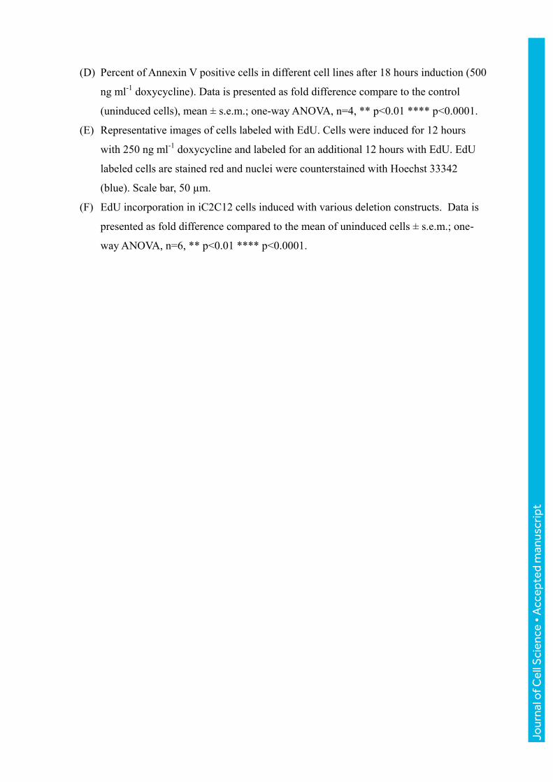
hybrid constructs (Fig. 7A). Interestingly, only the construct in which both homeodomains
were substituted showed significant upregulation of Myf5 (Fig. 7A). To analyze their effects
on myogenic differentiation, cells were cultured to confluence, subsequently switched to
medium promoting differentiation, and induced with doxycycline at low and high
concentrations (25 and 250 ng ml-1
) for 4 days. All 3 hybrid proteins at high levels showed
impaired differentiation, as indicated by an absence of distinct myotube formation and by a
decreased myotube fusion index (Fig. 7B and C). Notably, hybrid proteins were not as
effective as WT DUX4 when induced al low levels (Fig. 7C), and the double substitution was
weakest, however they all clearly showed a statistically significant inhibition of
differentiation when comparing no dox to 250 ng ml-1
dox. Taken together, we conclude that
the DUX4 homeodomains can be substituted for those of Pax7 to generate a substitution
mutant that broadly retains the characteristics of WT DUX4.
Discussion
In this study, we probe the function of various domains of the DUX4 protein by testing a
series of deletion and substitution mutants for effects on viability, myogenesis, and gene
expression. Importantly, the inducible cassette exchange system used here targets constructs
into the same doxycycline-regulated genomic locus (Bosnakovski et al., 2008b). This offers
numerous advantages compared to transient gene expression with plasmid DNA or over-
expression approaches using integration of viral constructs: gene expression from a single
integration site in a non-silencing locus eliminates variation due to integration site and copy
number differences. In addition, selection for integration is independent of gene expression.
This means that until doxycycline is added, cells are unaffected by the various constructs
inserted into their genome, an important consideration for toxic genes such as DUX4.
Our previous work with DUX4c, which is not toxic (Bosnakovski et al., 2008a), had
indicated that a domain within the C-terminal 80 amino acids of DUX4 was necessary for
toxicity. In the present work, we show that C-terminal mutations increase stability but
decrease activity in most assays. In particular, the DUX4(1-399) deletion highlights the
importance of the last 25 amino acids for toxicity. Loss of this region disrupts a weakly
conserved sequence motif that defines the DUXC family (Leidenroth and Hewitt, 2010).
Jour
nal o
f Cel
l Sci
ence
• A
ccep
ted
man
uscr
ipt

Humans and rodents have lost the canonical DUXC gene, but based on the presence of this
C-terminal sequence, DUX4 is evidently a retrotransposed copy that has subsumed the
function of DUXC in humans, while the mDux gene on murine chromosome 10 has done the
same in rodents. Interestingly, DUXC members, DUX4 and murine Dux, are also present in
tandem repeats (Leidenroth and Hewitt, 2010). The results of our study define several
phenotypic features of this C-terminal DUXC signature motif: 1) the presence of this motif
predicts toxicity of the DUX protein bearing it, 2) the presence of this motif limits
accumulation of the DUX protein, most likely by reducing stability, and 3) this motif is
important for activation and suppression of various downstream target genes of DUX4,
including MyoD and Myf5.
N-terminal deletions that remove one or both homeodomains resulted in completely inactive
proteins. A previous study supports this result by showing that a version of DUX4 with 5
alanine substitutions in HD1 (predicted to abolish the domain) is no longer toxic (Wallace et
al., 2011). Whereas the complete DUX4 protein is necessary to induce cell toxicity, the
homeodomains alone are sufficient to interfere with myogenesis. These effects may involve
changes in MRF expression, most likely MyoD expression, as both full length and
homeodomain-only versions of DUX4 repressed MyoD, but had opposite effects on Myf5.
C-terminal deletion studies confirm that intact DUX4 induces Myf5, and with successive
deletions of C-terminal sequence, this induction is lost and switches to repression. The
DUX4(1-377) and DUX4(1-399) constructs, which are shorter than DUX4 but longer than
DUX4c, are on the transition threshold: DUX4(1-399) activates weakly while DUX4(1-377)
represses weakly. DUX4-mediated induction of Myf5 is unlikely to be an indirect
consequence of MyoD diminution, since it was also observed in cells that normally do not
express MyoD or Myf5, such as fibroblasts and ES cells (Bosnakovski et al., 2008b).
Besides DUX4, various sense and antisense transcripts as well as potential siRNA or miRNA
fragments originating from D4Z4 but not specific to FSHD have been described (Snider et
al., 2009). A similar bidirectional product found using RT-PCR and RNA-FISH was reported
for mouse Dux (Clapp et al., 2007). We tested whether the transcript originating from D4Z4
had any activity on viability or differentiation separate from the DUX4 ORF using several
approaches. We eliminated the start codon of DUX4 from the full length transcript, we
Jour
nal o
f Cel
l Sci
ence
• A
ccep
ted
man
uscr
ipt

generated inactive mutants of DUX4 through N-terminal deletions that deleted one or both
homeodomains, and we placed the whole DUX4 sequence in reverse orientation behind the
promoter. Proteins or transcripts were detected from all of these constructs, but none of them
exhibited functionality in cell viability, apoptosis, cell cycle or myogenesis assays. When we
deleted the ATG, we detected a protein product using the antibodies that recognize the central
and C-terminal parts of DUX4, an observation made previously by Snider et al. (Snider et al.,
2009). Smaller bands on the western blot were not present when the constructs with intact
ATG start codons were expressed. This suggests that the DUX4 sequence contains
alternative internal initiation sites, which in some situations might be engaged. The
functional relevance of these potential alternative transcripts to FSHD remains to be
explored, although the small ~40 kDa protein that we observed did not induce an effect on
cell viability or interfere with myogenesis. We conclude that the principle deleterious factor
within the transcript is the DUX4 ORF.
Because the homeodomains were essential for activity in both toxicity and differentiation
assays, and because we had previously shown that Pax3 and Pax7 can act as dominant
suppressors of the gene expression changes and of toxicity induced by DUX4, we
investigated the relationship between DUX4 and these homeodomain-containing proteins in
more depth by testing a family of highly related homeodomain-containing proteins.
Remarkably, from the set of homeodomain proteins most similar to DUX4, only Pax3 and
Pax7 were epistatic to DUX4-induced toxicity. This emphasizes the specificity of the
Pax3/7-DUX4 interaction and its relevance to the myogenic lineage, including potentially the
postnatal cells in which Pax3/7 are expressed. Because of the crucial role that Pax3/7 play in
embryonic myogenesis and postnatal physiology of the muscle (Bajard et al., 2006; Bober et
al., 1994; Braun and Gautel; Buckingham and Relaix, 2007; Oustanina et al., 2004; Relaix et
al., 2005; Seale et al., 2000), this competitive interaction is intriguing. If operative in satellite
cells, the presence of DUX4 would be predicted to interfere with Pax3/7-mediated
regenerative regulatory pathways.
In addition, we show that Pitx2 and Tbx1, genes involved in embryonic development of head
muscles, are not able to revert DUX4 toxicity. This is also intriguing because facial muscle,
and the facial muscle satellite cell pool, is derived in embryogenesis from Pax3-negative
Jour
nal o
f Cel
l Sci
ence
• A
ccep
ted
man
uscr
ipt

progenitors, while limb and body wall muscle are derived from Pax3+ progenitors. Pax3
expression during development might limit the insult caused by low level DUX4 expression
during establishment of the satellite cell pool. If so, the facial muscle founders, which lack
Pax3, might be more affected, as indeed is the case in FSHD.
We confirmed the necessity of the Pax3/7 homeodomain for phenotypic competition with
DUX4 by deleting it. As expected, Pax3 lacking its homeodomain was not able to compete
with DUX4. Unexpectedly however, the Paired and activation domains were also found to be
necessary. Whether this means that competition is not at the level of the homeodomain, for
example through upregulation of a Pax3/7 target that inhibits the toxic effects of DUX4, or
whether the Paired and activation domains somehow facilitate direct competition of the
Pax3/7 homeodomain with that of DUX4 is at present unclear. If the former, one would
expect that replacing the DUX4 homeodomains with that of Pax7 would abolish DUX4
toxicity as the optimal Pax7 and DUX4 DNA recognition sequences are distinct (Geng et al.,
2012; Soleimani et al., 2012; Zhang et al., 2016). To evaluate this, we generated hybrid
proteins in which the DUX4 homeodomains were substituted with that of Pax7. Although
there were some differences between the constructs, with the double homeodomain
substitution being somewhat weaker than each individual substitution, all of these hybrid
proteins were toxic and inhibited differentiation, like DUX4, showing that in spite of having
different optimal DNA recognition motifs, the DUX4 and Pax7 homeodomains are
functionally interchangeable.
Two possibilities may explain this perplexing result. First, the majority of genomic sites
bound by these transcription factors do not have the “optimal” DNA recognition motif, but
variants of it, and it is possible that although most DUX4 sites are distinct from those of
Pax3/7, there may be key toxicity-related targets with a motif recognized by both DUX4 and
Pax3/7. A second possibility is that the homeodomains are competing for a homeodomain-
interacting protein, rather than for binding to a specific target sequence. Overexpression of
Pax3 or 7 would thus deplete the DUX4 complex of a key cofactor necessary for full DUX4
activity. Further investigation to address this issue will lead to a better understanding of the
toxic activity of DUX4.
Jour
nal o
f Cel
l Sci
ence
• A
ccep
ted
man
uscr
ipt

MATERIALS AND METHODS
D4Z4 deletion constructs. The terminal D4Z4 repeat (2.7 kb) from pCIneo-DUX4 was used
as a template for generating all of the deletion constructs (Gabriels et al., 1999). In order to
recombine the DUX4 deletion constructs into the inducible cassette exchange (ICE) locus in
iC2C12 myoblasts, they were cloned into p2Lox, the targeting recombination plasmid
(Iacovino et al., 2011; Kyba et al., 2002). By directional XhoI/NotI cloning, a portion of the
last D4Z4 repeat was inserted into p2Lox to generate the p2Lox-DUX4+3'UTR construct
(Bosnakovski et al., 2008b). The DUX4 ORF (named DUX4(1-424)) construct was generated
by PCR using a forward primer (CTCGAGATGGCCCTCCCGAC) to introduce an XhoI
cutting site and reverse primers at the end of the sequence. ATG deletion constructs were
made by PCR by omitting the ATG start codon in the forward amplification primer
(CTCGAGGCCCTCCCGACACC). Deletion of the first, second or both homeodomains was
accomplished by PCR with specific forward primers with ATG added to start translation. The
∆5'+3'UTR construct was made by cutting the p2Lox-DUX4+3'UTR plasmid with XhoI and
Bsu36I, and blunt end religation. Additional 3’ deletions of DUX4 were generated by cutting
p2Lox-Dux4 with NotI and StuI for DUX4(1-399), NotI and SacI for DUX4(1-377), and NotI
and PstI for DUX4(1-217) followed by blunt end religation of the plasmid. ∆DUX4(1-75)+
∆3'UTR was generated by removing the PvuII flanked sequence in ∆Dux4+3'UTR. For
DUX4 opposite (DUX4 opp), the whole D4Z4 sequence was cloned into the opposite
orientation in p2Lox. The integrity of each deletion construct was confirmed by sequencing.
Cell culture. iC2C12 cells bearing different deletion constructs were expanded in
proliferation medium consisting of high glucose Dulbecco's Modified Eagle Media (DMEM)
supplemented with L-glutamine, sodium pyruvate, penicillin and streptomycin (P/S, all from
Gibco, Invitrogen, Carlsbad, CA) and 10% fetal bovine serum (FBS, HyClone,
Thermoscientific, Logan, UT) at 37 °C in 5% CO2. Myogenic differentiation was induced in
confluent cells cultured on gelatin-coated dishes with DMEM supplemented with 2% horse
serum (HS, Sigma, St. Louis, MO) and insulin (Sigma) for 4 days. Cells were authenticated
by PCR for the unique ICE locus and by western blot for size of induced proteins, and have
tested negative for contamination.
Generating the iC2C12-DUX4 deletion cell lines. Inducible cell lines with the various
constructs were made as we previously described (Bosnakovski et al., 2008b). Briefly,
iC2C12 myoblasts were induced to express Cre recombinase with 500 ng ml-1
doxycycline
one day before targeting the ICE locus. The recombination plasmid, p2Lox carrying the
Jour
nal o
f Cel
l Sci
ence
• A
ccep
ted
man
uscr
ipt

deletion constructs, was transfected using FUGEN 6 (Roche, Indianapolis, IN). On the
following day, selection with 800 µg ml-1
G418 was initiated. Within 2 weeks recombinant
G418 resistant clones were generated and tested for inducibility of the constructs. For the
deletion constructs which could be detected by DUX4 antibody, the expression of the protein
was tested by immunostaining and western blotting (see below). For the constructs for which
we did not have suitable antibody we used RT-PCR followed by sequencing.
Cloning of retroviral constructs and virus production. MSCV-IRES-GFP retroviral
expression plasmids containing the various genes of interest were constructed using the
Gateway recombination system (Invitrogen). Human MIXL1 was amplified from pCR4-
TOPO-MIXL1, mouse Hesx1 from pCR4-TOPO-Hesx1, mouse Rax from pCMV-SPORT6-
Rax, human OTX1 from pOTB7-OTX1, mouse Pitx2 from pYX-Asc-Pitx2, human PAX6
from pCMV-SPORT6-PAX6 (all from Open Biosystems, Thermoscientific). Mouse Pax3
and Pax7 were generated from previously described pcDNA3.1-Pax3 and pBRIT-Pax7
(McKinnell et al., 2008). Mouse Tbx1 was amplified from cDNA reverse-transcribed from
RNA isolated from the pharyngeal arch of mouse embryos (E9.5); all other genes were
amplified from plasmid templates encoding their respective cDNAs. Mouse Pax3 deletions
were constructed as described (Magli et al., 2013). The human MYOD expression construct
was previously described by (Bosnakovski et al., 2008a). PCR products were purified using
the Promega (Madison, WI) Wizard SV Gel & PCR Clean-Up System, then inserted into the
pDONR-221 plasmid using the BP Clonase II enzyme mix (Invitrogen). Clones were
sequenced by the Iowa State University DNA Facility to ensure sequence integrity, and
correct entry clones were subsequently recombined into the MSCV-IRES-GFP destination
vector using the LR Clonase II enzyme mix (Invitrogen). Expression clones were screened
for proper gene insertion, sequenced, and large-scale DNA preps were generated with the
Nucleobond Xtra Midi Plus DNA purification kit (Macherey-Nagel, Duren, Germany).
Retroviral supernatants were produced in 293T viral packaging cells. Retroviral constructs
were co-transfected with pCL-Eco packaging constructs using FUGENE 6 (Roche). Viral
supernatant was collected at 48 hours post-transfection and iC2C12-DUX4 cells were
infected by spin-infection (2000 g at 33 °C for 90 min).
ATP assay. iC2C12 carrying DUX4 deletions were plated in a 96 well plate (2000
cells/well). Cells were induced the following day with various doxycycline concentrations for
24 or 48 hours. ATP assays were performed following the manufacturer's instructions
(Promega) by lysing the cells with 100 ul ATPlite and analyzing the luminescence on a
Jour
nal o
f Cel
l Sci
ence
• A
ccep
ted
man
uscr
ipt

POLARstar Optima Microplate Reader (BMG Labtech, Offenburg, Germany). Data was
presented as fold difference compared to the control (uninduced cells) as mean ± s.d. (n=8)
calculated with Xcel software (Microsoft, Redmond, WA).
Annexin V/7-AAD staining. Cells were cultured in proliferation conditions and induced for
18 hours with doxycycline. Cells were trypsinized and stained with Annexin V and 7-AAD
using APC Annexin V staining kit (BioLegend) according to the manufacturer’s instructions.
Stained cells were measured on a FACSARIA II (BD) and analyzed using FlowJo (FlowJo,
LLC).
EdU incorporation. Cells were plated at low density (1000 cells per well of 96 well plate)
in proliferating medium. EdU labeling and visualization were done using the Click-iT®
EdU Imaging Kit (Thermo Fisher Scientific). 12 hours post induction cells were treated with
EdU (1 uM) for an additional 12 hours. At 24 hours cells were fixed with 10% formalin and
stained following the manufacturer’s instructions. Microscopic fluorescent images of each
cell line in the presence and absence of doxycycline were taken with Zen Pro at 10X
magnification (6 images per cell line). ImageJ was used to calculate the proportion of nuclei
with positive EdU staining (Alexa Fluor 555). Images with the Hoechst 33342 channel and
Alexa Fluor 555 were loaded into ImageJ separately. Each image was rendered into a 16 bit
image and then thresholded for maximum clarity. To calculate the proportion of nuclei with
positive EdU staining, the number of nuclei counted in the Alexa Fluor 555 channel image
was divided by the number of nuclei counted in the Hoechst 33342 channel image, which
represent total nuclei present.
Western blotting. Cells were washed twice with PBS, lysed with RIPA buffer (Santa Cruz
Biotechnology, Santa Cruz, CA), mixed with loading buffer (BioRad, Hercules, CA) and
boiled for 5 min. Proteins were separated by electrophoresis on 10% PAGE gels. The gels
were then transferred to PVDF membranes (BioRad), and blocked in 5% skim milk in TBS-T
(Tris buffered saline with 0.01% Tween 20) for 1 h at RT. Primary antibodies
that recognize
N-terminal E5-5 (Abcam) and RD247c (R&D Systems), central (9A12) or C-terminal (P2G4)
epitopes of DUX4 (Dixit et al., 2007; Geng et al.) were diluted in blocking mixture and blots
were incubated overnight at 4°C. After washing, secondary anti mouse/rabbit horseradish
peroxidase-conjugated (1:3000, Santa Cruz) antibodies were applied for 1 h at room
temperature in blocking mixture. Signal was detected by ECL Plus (GE Healthcare,
Piscataway, NJ) with X-ray film exposure.
Jour
nal o
f Cel
l Sci
ence
• A
ccep
ted
man
uscr
ipt

Immunofluorescence. Cells were fixed with 4% paraformaldehyde for 20 min,
permeabilized by 0.3%Triton X-100 for 30 min and blocked by 3% BSA/PBS for 1 hour at
room temperature. Primary antibodies were incubated in 3% BSA/PBS at 4°C overnight,
followed by secondary antibodies at room temperature for 45 min. Nuclei were visualized
using 4',6-diamidino-2-phenylindole (DAPI, Invitrogen). The following antibodies were
used: mouse anti-MHC (MF20, 1:20, Developmental Studies Hybridoma Bank), rabbit anti-
MyoD (1:200, Santa Cruz), rabbit anti-DUX4 (1:50, R&D Systems), Alexa fluor 555 Goat
Anti-Mouse, Alexafluor 555 or 488 Goat Anti-Rabbit (1:500, Invitrogen).
Quantitative Real Time RT-PCR (RT-qPCR). RNA was extracted with Trizol (Invitrogen)
and cDNA was made using 0.5 µg total RNA with oligo-dT primer and ThermoScript
following manufacturer’s instructions (Invitrogen). PCR was performed by using TagMan
Real Time PCR premixture and premix probes (Myod1 Mm00440387_m1, Myf5
Mm00435125_m1, human MYOD1 Hs00159528_m1) on a 7500 real time PCR System
(Applied Biosystems, Carlsbad, CA). Glyceraldehyde phosphate dehydrogenase (GAPDH,
Mm99999915_g1) was used as the internal standard. All reactions were performed at least in
triplicate and the data was normalized and analyzed by 7500 System Software using the
∆∆CT method (Applied Biosystems). Data is presented as mean ± standard deviations
calculated with Prism (GraphPad, San Diego, CA) software.
Calculating Fusion Index with G-Tool. G-Tool, a previously developed open-source
algorithm, was used to quantify myogenic differentiation. MHC-stained fluorescent images
were analyzed and the average number of nuclei per MHC positive myotube was calculated.
Microscopic fluorescent images were taken with Zen Pro at 10X magnification. Merged
images containing both the DAPI (nuclei) and the Alexa Fluor 555 channels (MHC) were
input into the G-Tool user platform. Images were then processed according to default
sensitivity and contrast settings. The G-Tool algorithm was then calibrated to the nuclear size
in the image library by manual adjustment of DAPI sensitivity and contrast. Once calibrated,
the G-Tool algorithm analyzed all images according to this calibration and calculated fusion
index of each condition and replicate, in a method described by (Ippolito et al., 2012).
Statistical analyses. All experiments were repeated in at least three biological replicates.
Significance of the paired differences were calculated by T-test, one- or two-way ANOVA
with GraphPad. **** indicates p<0.0001, *** p<0.001 ** p<0.01, and * p<0.05.
Jour
nal o
f Cel
l Sci
ence
• A
ccep
ted
man
uscr
ipt

ACKNOWLEDGMENTS
We thank the Dr. Bob and Jean Smith Foundation for their generous support. We thank Dr.
Daniel Miller for the suggestion to test related homeodomain proteins Hesx1, etc. We thank
L. Geng and S. Tapscott for sharing E5-5 and P2G4 antibodies prior to publication and A.
Belayew for the 9A12 antibody. The monoclonal antibody against MHC was obtained from
the Developmental Studies Hybridoma Bank, developed under the auspices of the NICHD
and maintained by the University of Iowa.
COMPETING INTERESTS
The authors declare no competing financial, personal or professional competing interests.
FUNDING
This work was funded by the NIH (AR055685). DB was supported by a Muscular Dystrophy
Association Development Grant (MDA 4361), Marjorie Bronfman Research Fellowship from
the FSH Society and by the Children’s Cancer Research Fund.
Jour
nal o
f Cel
l Sci
ence
• A
ccep
ted
man
uscr
ipt

REFERENCES
Bajard, L., Relaix, F., Lagha, M., Rocancourt, D., Daubas, P. and Buckingham,
M. E. (2006). A novel genetic hierarchy functions during hypaxial myogenesis: Pax3 directly
activates Myf5 in muscle progenitor cells in the limb. Genes Dev 20, 2450-64.
Block, G. J., Narayanan, D., Amell, A. M., Petek, L. M., Davidson, K. C., Bird, T.
D., Tawil, R., Moon, R. T. and Miller, D. G. (2013). Wnt/beta-catenin signaling suppresses
DUX4 expression and prevents apoptosis of FSHD muscle cells. Hum Mol Genet 22, 4661-
72.
Bober, E., Franz, T., Arnold, H. H., Gruss, P. and Tremblay, P. (1994). Pax-3 is
required for the development of limb muscles: a possible role for the migration of
dermomyotomal muscle progenitor cells. Development 120, 603-12.
Bosnakovski, D., Lamb, S., Simsek, T., Xu, Z., Belayew, A., Perlingeiro, R. and
Kyba, M. (2008a). DUX4c, an FSHD candidate gene, interferes with myogenic regulators
and abolishes myoblast differentiation. Exp Neurol 214, 87-96.
Bosnakovski, D., Xu, Z., Gang, E. J., Galindo, C. L., Liu, M., Simsek, T., Garner,
H. R., Agha-Mohammadi, S., Tassin, A., Coppee, F. et al. (2008b). An isogenetic myoblast
expression screen identifies DUX4-mediated FSHD-associated molecular pathologies. EMBO
J 27, 2766-79.
Braun, T. and Gautel, M. Transcriptional mechanisms regulating skeletal muscle
differentiation, growth and homeostasis. Nat Rev Mol Cell Biol 12, 349-61.
Buckingham, M., Bajard, L., Chang, T., Daubas, P., Hadchouel, J., Meilhac, S.,
Montarras, D., Rocancourt, D. and Relaix, F. (2003). The formation of skeletal muscle:
from somite to limb. J Anat 202, 59-68.
Buckingham, M. and Relaix, F. (2007). The role of pax genes in the development of
tissues and organs: pax3 and pax7 regulate muscle progenitor cell functions. Annu Rev Cell
Dev Biol 23, 645-73.
Celegato, B., Capitanio, D., Pescatori, M., Romualdi, C., Pacchioni, B., Cagnin,
S., Vigano, A., Colantoni, L., Begum, S., Ricci, E. et al. (2006). Parallel protein and
transcript profiles of FSHD patient muscles correlate to the D4Z4 arrangement and reveal a
common impairment of slow to fast fibre differentiation and a general deregulation of MyoD-
dependent genes. Proteomics 6, 5303-21.
Choi, S. H., Gearhart, M. D., Cui, Z., Bosnakovski, D., Kim, M., Schennum, N.
and Kyba, M. (2016). DUX4 recruits p300/CBP through its C-terminus and induces global
H3K27 acetylation changes. Nucleic Acids Res 44, 5161-73.
Clapp, J., Mitchell, L. M., Bolland, D. J., Fantes, J., Corcoran, A. E., Scotting, P.
J., Armour, J. A. and Hewitt, J. E. (2007). Evolutionary conservation of a coding function
for D4Z4, the tandem DNA repeat mutated in facioscapulohumeral muscular dystrophy. Am J
Hum Genet 81, 264-79.
Conboy, I. M. and Rando, T. A. (2002). The regulation of Notch signaling controls
satellite cell activation and cell fate determination in postnatal myogenesis. Dev Cell 3, 397-
409.
Jour
nal o
f Cel
l Sci
ence
• A
ccep
ted
man
uscr
ipt

Dixit, M., Ansseau, E., Tassin, A., Winokur, S., Shi, R., Qian, H., Sauvage, S.,
Matteotti, C., van Acker, A. M., Leo, O. et al. (2007). DUX4, a candidate gene of
facioscapulohumeral muscular dystrophy, encodes a transcriptional activator of PITX1. Proc
Natl Acad Sci U S A 104, 18157-62.
Gabellini, D., Green, M. R. and Tupler, R. (2002). Inappropriate gene activation in
FSHD: a repressor complex binds a chromosomal repeat deleted in dystrophic muscle. Cell
110, 339-48.
Gabriels, J., Beckers, M. C., Ding, H., De Vriese, A., Plaisance, S., van der
Maarel, S. M., Padberg, G. W., Frants, R. R., Hewitt, J. E., Collen, D. et al. (1999).
Nucleotide sequence of the partially deleted D4Z4 locus in a patient with FSHD identifies a
putative gene within each 3.3 kb element. Gene 236, 25-32.
Geng, L. N., Tyler, A. E. and Tapscott, S. J. Immunodetection of human double
homeobox 4. Hybridoma (Larchmt) 30, 125-30.
Geng, L. N., Yao, Z., Snider, L., Fong, A. P., Cech, J. N., Young, J. M., van der
Maarel, S. M., Ruzzo, W. L., Gentleman, R. C., Tawil, R. et al. (2012). DUX4 activates
germline genes, retroelements, and immune mediators: implications for facioscapulohumeral
dystrophy. Dev Cell 22, 38-51.
Iacovino, M., Bosnakovski, D., Fey, H., Rux, D., Bajwa, G., Mahen, E.,
Mitanoska, A., Xu, Z. and Kyba, M. (2011). Inducible cassette exchange: a rapid and
efficient system enabling conditional gene expression in embryonic stem and primary cells.
Stem Cells 29, 1580-1588.
Ippolito, J., Arpke, R. W., Haider, K. T., Zhang, J. and Kyba, M. (2012). Satellite
cell heterogeneity revealed by G-Tool, an open algorithm to quantify myogenesis through
colony-forming assays. Skelet Muscle 2, 13.
Jones, T. I., Chen, J. C., Rahimov, F., Homma, S., Arashiro, P., Beermann, M. L.,
King, O. D., Miller, J. B., Kunkel, L. M., Emerson, C. P., Jr. et al. (2012).
Facioscapulohumeral muscular dystrophy family studies of DUX4 expression: evidence for
disease modifiers and a quantitative model of pathogenesis. Hum Mol Genet 21, 4419-30.
Knopp, P., Krom, Y. D., Banerji, C. R., Panamarova, M., Moyle, L. A., den
Hamer, B., van der Maarel, S. M. and Zammit, P. S. (2016). DUX4 induces a
transcriptome more characteristic of a less-differentiated cell state and inhibits myogenesis. J
Cell Sci 129, 3816-3831.
Kowaljow, V., Marcowycz, A., Ansseau, E., Conde, C. B., Sauvage, S., Matteotti,
C., Arias, C., Corona, E. D., Nunez, N. G., Leo, O. et al. (2007). The DUX4 gene at the
FSHD1A locus encodes a pro-apoptotic protein. Neuromuscul Disord 17, 611-623.
Krom, Y. D., Dumonceaux, J., Mamchaoui, K., den Hamer, B., Mariot, V.,
Negroni, E., Geng, L. N., Martin, N., Tawil, R., Tapscott, S. J. et al. (2012). Generation of
Isogenic D4Z4 Contracted and Noncontracted Immortal Muscle Cell Clones from a Mosaic
Patient: A Cellular Model for FSHD. Am J Pathol 181, 1387-401.
Kyba, M., Perlingeiro, R. C. and Daley, G. Q. (2002). HoxB4 confers definitive
lymphoid-myeloid engraftment potential on embryonic stem cell and yolk sac hematopoietic
progenitors. Cell 109, 29-37.
Leidenroth, A. and Hewitt, J. E. (2010). A family history of DUX4: phylogenetic
analysis of DUXA, B, C and Duxbl reveals the ancestral DUX gene. BMC Evol Biol 10, 364.
Jour
nal o
f Cel
l Sci
ence
• A
ccep
ted
man
uscr
ipt

Lemmers, R. J., Tawil, R., Petek, L. M., Balog, J., Block, G. J., Santen, G. W.,
Amell, A. M., van der Vliet, P. J., Almomani, R., Straasheijm, K. R. et al. (2012). Digenic
inheritance of an SMCHD1 mutation and an FSHD-permissive D4Z4 allele causes
facioscapulohumeral muscular dystrophy type 2. Nat Genet 44, 1370-4.
Magli, A., Schnettler, E., Rinaldi, F., Bremer, P. and Perlingeiro, R. C. (2013).
Functional dissection of Pax3 in paraxial mesoderm development and myogenesis. Stem Cells
31, 59-70.
McKinnell, I. W., Ishibashi, J., Le Grand, F., Punch, V. G., Addicks, G. C.,
Greenblatt, J. F., Dilworth, F. J. and Rudnicki, M. A. (2008). Pax7 activates myogenic
genes by recruitment of a histone methyltransferase complex. Nat Cell Biol 10, 77-84.
Montarras, D., Morgan, J., Collins, C., Relaix, F., Zaffran, S., Cumano, A.,
Partridge, T. and Buckingham, M. (2005). Direct isolation of satellite cells for skeletal
muscle regeneration. Science 309, 2064-7.
Oustanina, S., Hause, G. and Braun, T. (2004). Pax7 directs postnatal renewal and
propagation of myogenic satellite cells but not their specification. EMBO J 23, 3430-9.
Rahimov, F., King, O. D., Leung, D. G., Bibat, G. M., Emerson, C. P., Jr., Kunkel,
L. M. and Wagner, K. R. (2012). Transcriptional profiling in facioscapulohumeral muscular
dystrophy to identify candidate biomarkers. Proc Natl Acad Sci U S A 109, 16234-9.
Relaix, F., Rocancourt, D., Mansouri, A. and Buckingham, M. (2005). A
Pax3/Pax7-dependent population of skeletal muscle progenitor cells. Nature 435, 948-53.
Sambasivan, R., Gayraud-Morel, B., Dumas, G., Cimper, C., Paisant, S., Kelly,
R. G. and Tajbakhsh, S. (2009). Distinct regulatory cascades govern extraocular and
pharyngeal arch muscle progenitor cell fates. Dev Cell 16, 810-21.
Seale, P., Sabourin, L. A., Girgis-Gabardo, A., Mansouri, A., Gruss, P. and
Rudnicki, M. A. (2000). Pax7 is required for the specification of myogenic satellite cells.
Cell 102, 777-86.
Snider, L., Asawachaicharn, A., Tyler, A. E., Geng, L. N., Petek, L. M., Maves, L.,
Miller, D. G., Lemmers, R. J., Winokur, S. T., Tawil, R. et al. (2009). RNA transcripts,
miRNA-sized fragments and proteins produced from D4Z4 units: new candidates for the
pathophysiology of facioscapulohumeral dystrophy. Hum Mol Genet 18, 2414-30.
Snider, L., Geng, L. N., Lemmers, R. J., Kyba, M., Ware, C. B., Nelson, A. M.,
Tawil, R., Filippova, G. N., van der Maarel, S. M., Tapscott, S. J. et al. (2010).
Facioscapulohumeral dystrophy: incomplete suppression of a retrotransposed gene. PLoS
Genet 6, e1001181.
Soleimani, V. D., Punch, V. G., Kawabe, Y., Jones, A. E., Palidwor, G. A., Porter,
C. J., Cross, J. W., Carvajal, J. J., Kockx, C. E., van Ijcken, W. F. et al. (2012).
Transcriptional dominance of pax7 in adult myogenesis is due to high-affinity recognition of
homeodomain motifs. Dev Cell 22, 1208-20.
Tassin, A., Laoudj-Chenivesse, D., Vanderplanck, C., Barro, M., Charron, S.,
Ansseau, E., Chen, Y. W., Mercier, J., Coppee, F. and Belayew, A. (2012). DUX4
expression in FSHD muscle cells: how could such a rare protein cause a myopathy? J Cell
Mol Med.
Jour
nal o
f Cel
l Sci
ence
• A
ccep
ted
man
uscr
ipt

Tsumagari, K., Chang, S. C., Lacey, M., Baribault, C., Chittur, S. V., Sowden, J.,
Tawil, R., Crawford, G. E. and Ehrlich, M. (2011). Gene expression during normal and
FSHD myogenesis. BMC Med Genomics 4, 67.
van Overveld, P. G., Lemmers, R. J., Sandkuijl, L. A., Enthoven, L., Winokur, S.
T., Bakels, F., Padberg, G. W., van Ommen, G. J., Frants, R. R. and van der Maarel, S.
M. (2003). Hypomethylation of D4Z4 in 4q-linked and non-4q-linked facioscapulohumeral
muscular dystrophy. Nat Genet 35, 315-7.
Wallace, L. M., Garwick, S. E., Mei, W., Belayew, A., Coppee, F., Ladner, K. J.,
Guttridge, D., Yang, J. and Harper, S. Q. (2011). DUX4, a candidate gene for
facioscapulohumeral muscular dystrophy, causes p53-dependent myopathy in vivo. Ann
Neurol 69, 540-552.
Wijmenga, C., Hewitt, J. E., Sandkuijl, L. A., Clark, L. N., Wright, T. J.,
Dauwerse, H. G., Gruter, A. M., Hofker, M. H., Moerer, P., Williamson, R. et al. (1992).
Chromosome 4q DNA rearrangements associated with facioscapulohumeral muscular
dystrophy. Nat Genet 2, 26-30.
Winokur, S. T., Barrett, K., Martin, J. H., Forrester, J. R., Simon, M., Tawil, R.,
Chung, S. A., Masny, P. S. and Figlewicz, D. A. (2003a). Facioscapulohumeral muscular
dystrophy (FSHD) myoblasts demonstrate increased susceptibility to oxidative stress.
Neuromuscul Disord 13, 322-33.
Winokur, S. T., Chen, Y. W., Masny, P. S., Martin, J. H., Ehmsen, J. T., Tapscott,
S. J., van der Maarel, S. M., Hayashi, Y. and Flanigan, K. M. (2003b). Expression
profiling of FSHD muscle supports a defect in specific stages of myogenic differentiation.
Hum Mol Genet 12, 2895-907.
Zhang, Y., Lee, J. K., Toso, E. A., Lee, J. S., Choi, S. H., Slattery, M., Aihara, H.
and Kyba, M. (2016). DNA-binding sequence specificity of DUX4. Skelet Muscle 6, 8.
Jour
nal o
f Cel
l Sci
ence
• A
ccep
ted
man
uscr
ipt

Figures
Figure 1. DUX4 deletion constructs and their inducibility.
(A) Schematic diagram of the DUX4 deletion constructs. 2.7 kb of DNA sequence
containing the DUX4-ORF from the last full D4Z4 repeat followed with 3’UTR
including the remainder of the D4Z4 repeat, the terminal partial repeat, and a portion
of the β satellite sequence was used as the template to generate deletions. Deletions
Jour
nal o
f Cel
l Sci
ence
• A
ccep
ted
man
uscr
ipt

were named with the amino acids of DUX4 listed as numbers in parentheses. HD
denotes homeodomains. “DUX4” used elsewhere in the manuscript may refer to
either DUX4-ORF or DUX4+UTR.
(B) Western blotting demonstrates the expression and relative mass of each construct after
12 hours of induction with 250 ng ml-1
of doxycycline. Protein expression was
detected by DUX4 specific antibodies recognizing the N-terminus (P2G4), central
(9A12) or C- terminus (E5-5) of DUX4 and ß-actin levels are also shown.
(C) Immunostaining with DUX4 antibody (RD247c) and DAPI (nuclei, blue) on cells
induced for 20 hours with 250 ng ml-1
doxycycline in selected cell lines. Note that
almost all of the nuclei are positively stained. Scale bar, 50 um.
Jour
nal o
f Cel
l Sci
ence
• A
ccep
ted
man
uscr
ipt

Figure 2. Effect of DUX4 deletion constructs on cell viability and proliferation.
(A) Representative images of cell morphology after 24 hours induction with 500 ng ml-1
doxycycline. From all of the tested constructs only the two encoding an intact full
length DUX4 protein induced rapid cell death. Cells expressing DUX4(1-399)
construct were less confluent. Scale bar, 100 um.
(B) ATP assay for quantification of cell viability after 48 hours of induction with 500 ng
ml-1
doxycycline. The mean of the uninduced (control) cell line was scaled to one and
fold difference for each experimental cell line was calculated. Data is presented as
mean ± s.e.m.; one-way ANOVA, n=8, **** p<0.0001.
(C) Representative FACS analyses of cells induced with 500 ng ml-1
doxycycline for 18
hours and stained with Annexin V and 7-AAD.
Jour
nal o
f Cel
l Sci
ence
• A
ccep
ted
man
uscr
ipt
(D) Percent of Annexin V positive cells in different cell lines after 18 hours induction (500
ng ml-1
doxycycline). Data is presented as fold difference compare to the control
(uninduced cells), mean ± s.e.m.; one-way ANOVA, n=4, ** p<0.01 **** p<0.0001.
(E) Representative images of cells labeled with EdU. Cells were induced for 12 hours
with 250 ng ml-1
doxycycline and labeled for an additional 12 hours with EdU. EdU
labeled cells are stained red and nuclei were counterstained with Hoechst 33342
(blue). Scale bar, 50 µm.
(F) EdU incorporation in iC2C12 cells induced with various deletion constructs. Data is
presented as fold difference compared to the mean of uninduced cells ± s.e.m.; one-
way ANOVA, n=6, ** p<0.01 **** p<0.0001.
Jour
nal o
f Cel
l Sci
ence
• A
ccep
ted
man
uscr
ipt

Figure 3. Effect of DUX4 deletions on myogenesis during proliferation and terminal
differentiation.
(A) RT-qPCR analyses for MyoD and Myf5 expression in cell lines expressing various
deletion constructs induced with 250 ng ml-1
doxycycline for 6 hours in proliferation
medium and compared to uninduced controls. Gene expression was normalized to the
levels of GAPDH. Data is presented as mean ± s.e.m, one-way ANOVA, n=4, *
p<0.05; *** p<0.001, **** p<0.0001.
(B) Analyses of myogenic differentiation of the deletion constructs by
immunofluorescence for MHC (red). Nuclei were visualized by DAPI staining (blue).
Doxycycline (250 ng ml-1
) was added for 4 days while cells were cultured in
differentiation media.
(C) Fusion index analyses for evaluation of levels of myogenic differentiation. Cell lines
were differentiated with 25 and 250 ng ml-1
doxycycline for 4 days. Values of induced
Jour
nal o
f Cel
l Sci
ence
• A
ccep
ted
man
uscr
ipt

cells are normalized to the uninduced group and data is presented as fold difference;
mean ± s.e.m, two-way ANOVA, n=6, * p<0.05; *** p<0.001, **** p<0.0001.
(D) Immunostaining for MHC (red), DUX4 (green) and DAPI (blue staining of the nuclei)
in iC2C12-DUX4 (1-399) at day 4 of differentiation. Cells were induced with 25 ng
ml-1
doxycycline. Note that all of the cells in the doxycycline treated groups are
expressing DUX4 deletion constructs.
(E) Immunostaining for MyoD (red) and counterstaining of the nuclei with DAPI (blue)
in iC2C12-DUX4 (1-399) at day 4 of differentiation. Note that staining for MyoD is
decreased in cells induced with 25 ng ml-1
doxycycline. Scale bar is 50 um in all of
the images.
Jour
nal o
f Cel
l Sci
ence
• A
ccep
ted
man
uscr
ipt

Figure 4. MYOD overexpression suppresses DUX4 dependent inhibition of
differentiation.
(A) FACS analyses of iC2C12-DUX4 (1-377) infected with retrovirus carrying GFP or
human MYOD-ires-GPP expression vectors.
(B) RT-qPCR for human MYOD (MYOD) and endogenous MyoD expression in DUX4
C-terminal deletion , i.e. DUX4(1-377), expressing cell lines transduced with either
GFP control (labelled “&GFP”) or human MYOD1 (labelled “&MYOD”). Human
MYOD1 is shown relative to GAPDH control, while endogenous murine MyoD1 is
shown relative to its expression in the absence of Dox. Cells were induced with 250
ng ml-1
doxycycline for 14 hours. Data is presented as fold difference compared to the
control (uninduced cells), mean ± s.e.m.; one-way ANOVA, n=4, ** p<0.01 ****
p<0.0001. Note that only DUX4 (1-377) & MYOD cell line expresses MYOD.
(C) Immunostaining for MHC (red) and DAPI (blue, nuclei) in differentiating cells at day
4 post induction (doxycycline 250 ng ml-1
). Scale bar, 50 um.
(D) Evaluation of the level of differentiation by fusion index analyses in the cells in
differentiation for 4 days induced with 25 and 250 ng ml-1
doxycycline. Fusion index
represents the ratio of nuclei within myotubes to the total number of nuclei; mean ±
s.e.m.; two way ANOVA, n=6, **** p<0.0001.
Jour
nal o
f Cel
l Sci
ence
• A
ccep
ted
man
uscr
ipt

Figure 5. Assaying homeodomain proteins for dominant suppression of DUX4-
induced toxicity.
(A) Unrooted dendrogram indicating maximum likelihood sequence relationships between
homeodomains of related proteins.
(B) Alignment of homeodomain sequences shown in A. Disruptive substitutions (those
that change charge, hydrophobicity or conformational freedom) between a given
homeodomain and either DUX4HD1 or HD2 are highlighted in red; conservative
substitutions are highlighted in yellow. Sequences are ordered from least to greatest
number of disruptive substitutions. Abbreviations of human genes are written in
capital letters.
(C) Viability assay on induced iC2C12-DUX4 cells constitutively expressing various
genes. ATP analyses was done on cells induced with 25 and 100 ng ml-1
doxycycline
for 48 hours. Note that from of all of the tested candidate genes, only Pax3 and Pax7
Jour
nal o
f Cel
l Sci
ence
• A
ccep
ted
man
uscr
ipt

were able to act as dominant suppressors by rescuing the DUX4 expressing cells.
Each doxycycline-induced cell line was normalized to the control uninduced cells.
Data is presented as mean ± s.e.m, one way ANOVA, n=8, **** p<0.0001. Analyses
indicated that all samples were different from their respective controls (i.e.
experiencing loss of viability) except Pax3 and Pax7.
(D) Schematic diagram of proteins produced from the Pax3 deletion constructs created to
map functional domains of the Dux4/Pax3 phenotypic interaction. Domain
abbreviations (PD: paired domain; RED: terminal part of the paired domain; 8:
octapeptide; HD: homeodomain; TAD: c-terminal activation domain.
(E) ATP assay 48 hours post induction to map Pax3 domains needed for dominant
suppression of doxycycline-induced DUX4 lethality. Each Dox-induced cell line is
normalized to control uninduced cells. Data is presented as mean ± s.e.m, one way
ANOVA, n=8, **** p<0.0001. Note that Pax3, Δ8, ΔRED and Δ352-391 have rescue
effects compare to other domain deletion constructs.
Jour
nal o
f Cel
l Sci
ence
• A
ccep
ted
man
uscr
ipt

Jour
nal o
f Cel
l Sci
ence
• A
ccep
ted
man
uscr
ipt

Figure 6. Toxicity of DUX4-Pax7 homeodomain substitutions.
(A) Schematic diagram of DUX4-Pax7 homeodomain substitutions. In the construct
DUX4- Pax7(HD1) the first DUX4 homeodomain was substituted, in DUX4-
Pax7(HD2) the second, and in DUX4-Pax7(HD1+2) both homeodomains were
substituted with the mouse Pax7 homeodomain.
(B) Western blotting with RD247c antibody shows the protein expression from the hybrid
constructs.
(C) Immunostaining revealed nuclear localization of hybrid proteins (red). Cells were
induced for 20 hours with 250 ng ml-1
doxycycline. Scale bar, 50 um.
(D) Cell morphology after 48 hours induction with 250 ng ml-1
doxycycline. Obvious cell
death was observed with induction of all 3 hybrid constructs. Scale bar, 100 um.
(E) ATP assay for cell viability after 48 hours of induction with 500 ng ml-1
doxycycline.
Data is presented as fold difference compared to control (uninduced cells), mean ±
s.e.m.; one-way ANOVA, n=8, **** p<0.0001.
(F) FACS analyses of cells induced with 500 ng ml-1
doxycycline for 18 hours and stained
with Annexin V/7-AAD.
(G) Percent of Annexin V positive cells after 18 hours induction (500 ng ml-1
doxycycline). Data is presented as fold difference compared to control (uninduced
cells), mean ± s.e.m.; one-way ANOVA, n=4, ** p<0.01 **** p<0.0001.
(H) Representative images of cells labeled with EdU. Cells were induced for 12 hours
with 250 ng ml-1
doxycycline and labeled for an additional 12 hours with EdU. EdU
labeled cells are stained with red and nuclei were counterstained with Hoechst 33342
(blue). Scale bar, 50 µm.
(I) EdU incorporation evaluation of the cells presented in H. Data is presented as fold
difference compare to uninduced cells; mean ± s.e.m, one-way ANOVA, n=6, ***
p<0.001 **** p<0.0001.
Jour
nal o
f Cel
l Sci
ence
• A
ccep
ted
man
uscr
ipt
Figure 7. Effect of homeodomain substitution mutants on myogenesis.
(A) RT-qPCR analyses for MyoD and Myf5 expression in the cells induced with 250 ng
ml-1
doxycycline in proliferation medium for 8 hours. Gene expression was
normalized to GAPDH expression and presented as fold difference compare to the
control samples. Error bar represents mean ± s.e.m, one-way ANOVA, n=4, ***
p<0.001 **** p<0.0001.
Jour
nal o
f Cel
l Sci
ence
• A
ccep
ted
man
uscr
ipt

(B) Immunofluorescence for MHC (red), DUX4 (green) and nuclear staining with DAPI
(blue) in cells induced with 250 ng ml-1
doxycycline for 4 days during myogenic
differentiation.
(C) Fusion index in cells induced with 25 and 250 ng ml-1
doxycycline for 4 days during
differentiation. Data is presented as fold difference of the mean ± s.e.m, two-way
ANOVA, n=6, *** p<0.001 **** p<0.0001.
Jour
nal o
f Cel
l Sci
ence
• A
ccep
ted
man
uscr
ipt

Pax-3 vs. Pax-7 [Mus musculus]
Range 1: homeodomain of Pax7 - 177 to 274
Score Expect Method Identities Positives Gaps
177 bits(450) 4e-59 Compositional matrix adjust. 85/100(85%) 93/100(93%) 2/100(2%)
Pax3 21 EREKKAKHSIDGILSERASAPQSDEGSDIDSEPDLPLKRKQRRSRTTFTAEQLEELERAF 80 + EKKAKHSIDGIL ++ + + DEGSD++SEPDLPLKRKQRRSRTTFTAEQLEELE+AF
Pax7 177 DGEKKAKHSIDGILGDKGN--RLDEGSDVESEPDLPLKRKQRRSRTTFTAEQLEELEKAF 234
Pax3 81 ERTHYPDIYTREELAQRAKLTEARVQVWFSNRRARWRKQA 120
ERTHYPDIYTREELAQR KLTEARVQVWFSNRRARWRKQA Pax7 235 ERTHYPDIYTREELAQRTKLTEARVQVWFSNRRARWRKQA 274
Figure S1. Comparison of Pax3 and Pax7 homeodomains. Alignment of the homeodomains shows the high level of conservation of this domain, with perfect identity of all DNA-interacting residues. Yellow indicates amino acids that make specific base contacts. Green indicates amino acids that make other DNA contacts. Middle row indicates sequence identities/similarities. Source: ncbi.nlm.nih.gov/Structure/
J. Cell Sci. 130: doi:10.1242/jcs.205427: Supplementary information
Jour
nal o
f Cel
l Sci
ence
• S
uppl
emen
tary
info
rmat
ion


![Duality Symmetric String and M-Theory - arXivarXiv:1306.2643v2 [hep-th] 9 Dec 2014 Duality Symmetric String and M-Theory David S. Berman1 ∗ and Daniel C. Thompson2 † 1 Queen Mary](https://img.dokumen.tips/doc/110x75/60467ca3bb30f474836ba8d2/duality-symmetric-string-and-m-theory-arxiv-arxiv13062643v2-hep-th-9-dec-2014.jpg)