Embed Size (px)
Citation preview
1
The Double Helix Structure of DNA
by Jean-Marc Victor Research director at CNRS
Laboratory of Theoretical Condensed Matter Physics (CNRS UMR 7600)
Pierre-and-Marie-Curie University, Paris
If 1905 was the annus mirabilis of physics, with Albert Einstein’s four
historical papers1, then 1953 was undoubtedly the annus mirabilis of biology. In
the 25th April issue of the journal Nature, James D. Watson and Francis H.C. Crick
reveal the double helix structure of DNA (Deoxyribonucleic acid). However, this
“Letter”, of an entire page, only contains the seeds of all molecular biology in the
following fifty years, until the completion of the Human Genome Project in 20032.
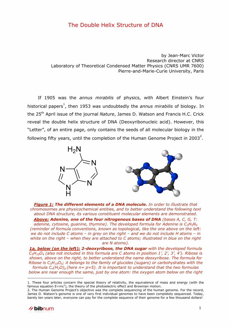
Figure 1: The different elements of a DNA molecule. In order to illustrate that
chromosomes are physicochemical entities, and to better understand the following text about DNA structure, its various constituent molecular elements are demonstrated.
Above: Adenine, one of the four nitrogenous bases of DNA (bases A, C, G, T:
adenine, cytosine, guanine, thymine). The developed formula for Adenine is C5H5N5
(reminder of formula conventions, known as topological, like the one above on the left:
we do not include C atoms – in grey on the right – and we do not include H atoms – in
white on the right – when they are attached to C atoms; illustrated in blue on the right
are N atoms).
1a, below (on the left): 2-deoxyribose, the DNA sugar with the developed formula
C5H10O4 (also not included in this formula are C atoms in position 1’, 2’, 3’, 4’). Ribose is
shown, above on the right, to better understand the name deoxyribose. The formula for
Ribose is C5H10O5; it belongs to the family of glucides (sugars) or carbohydrates with the
formula Cn(H2O)p (here n= p=5). It is important to understand that the two formulas
below are near enough the same, just by one atom: the oxygen atom below on the right
1. These four articles concern the special theory of relativity, the equivalence of mass and energy (with the famous equation E=mc2), the theory of the photoelectric effect and Brownian motion. 2. The Human Genome Project’s objective was the complete sequencing of the human genome. For the record, James D. Watson’s genome is one of very first individual genomes to have been completely sequenced. Today, barely ten years later, everyone can pay for the complete sequence of their genome for a few thousand dollars!

2
(circled in red on the right diagram in position 2’): hence the name deoxyribose (ribose,
from which an oxygen atom is removed). This component, the sugar deoxyribose,
therefore gives its name to DNA (Deoxyribonucleic acid).
1b, below: the third constitutive element type of DNA, the phosphate group. It is
attached to sugars (deoxyribose, figure 1a on the left) at two locations: (see figure 1c,
the left strand) 1°) in position 5’ on the “upper” sugar, the CH2 plays the role of the
radical R below (see figure 1c); 2°) in position 3’ on the “above” sugar, via the O atom
(which loses its H atom during polymerisation, circled in red).
1c, below: actual complete DNA structure with base pairs (cytosine-guanine and
thymine-adenine) between the two complementary strands (taken from the work
Biologie moléculaire et médecine, Jean-Claude Kaplan and Marc Delpech, Médecine-
Sciences, Flammarion, second edition (1996)).
Bases A, T, C or G are always put in position 1’ of deoxyribose.
Each strand is obtained from the alternating polymerisation of sugars and phosphates; in
doing so, there only remains one OH group in each phosphate. The sugars are all
positioned in the same way in a strand, each phosphate thus being surrounded by a
carbon 5’ and carbon 3’.
Each strand is positioned conventionally from end point 5’ towards end point 3’ (top to
bottom on the left, bottom to top on the right, the paired strands are therefore
antiparallel, as shown by the figure’s same construction). The hydrogen bonds are shown
between A and T, and C and G (see upcoming boxed text).
3
PAULING & COREY’S OBJECTIONS TO THE STRUCTURE
Right from the first line, the authors announce their wish to suggest a
structure for the salt of deoxyribonucleic acid. They also warn, in the following
paragraph, that the triple helix structure of the acid itself, proposed by Pauling3
and Corey, is in their opinion irrelevant because the X-ray diagrams4, on which
all previously proposed structures are based, were obtained with acid salt and
not with the acid itself:
We believe that the material which gives the X-ray diagrams is the salt,
not the free acid.
DNA differs from its salt as follows: in DNA, in acid form, the phosphates
(figure 1b above) all have an OH group and are neutral (note that the
phosphates, once polymerised in the sugar-phosphate chain, no longer have only
3. Linus Pauling (1901-1994) is one of the few scholars to have received two Nobel prizes: the Chemistry Prize in 1954, just one year after the discovery of the double helix, and the Peace Prize in 1962 for his campaign against atomic testing. 4. The diffraction of the X-rays by a crystal or a pseudo-crystal (powder of microcrystals or fibres consisting of identical macromolecules, mutually parallel but disorderly positioned) produces what is called an X-ray diagram. In the case of DNA, these are diagrams of fibres, firstly obtained by Rosalind Franklin and secondly by Maurice Wilkins, which allowed Watson & Crick to discover the double helix structure.

4
one OH group); on the contrary, in a DNA salt, these OH groups have lost their
hydrogen (hydrogen known as acidic5) which, when left in H+ saline solution,
leaves a negative charge in the phosphate group (on the O atom which loses its
H atom and becomes O–).
The consequence for Pauling and Corey’s structure is that DNA phosphate
groups – each having a negative charge in the acid salt – are found placed inside
Pauling and Corey’s structure and therefore near the triple helix axis. Yet, these
electric charges strongly repel each other; as a result, it is unclear which forces
in this triple helix structure could hold them in place:
Without the acidic hydrogen atoms it is not clear what forces would hold
the structure together, especially as the negatively charged phosphates
near the axis will repel each other
Consequently, the incorrect structure imagined by Pauling and Corey could
only exist with DNA in acid form. Yet, this is not the case in the X-ray samples.
Additionally, the acidic nature of DNA is often under debate. If words have
any meaning, a deoxyribonucleic acid solution should indeed be acidic! Yet, it is
not DNA that is in the solution but DNA salt, i.e., hydrogen atoms associated with
phosphate groups in acid form are replaced in the salt by sodium ions. So, it is
the base form (with the negatively charged phosphates) that is present in the
solution in usual conditions of extraction (i.e., at physiological PH, close to 7).
THE DOUBLE HELIX STRUCTURE
Back to the first sentence:
We wish to suggest a structure for the salt of DNA.
It sounds retrospectively like the first sentence of Genesis; it is somehow
the “Bereshit” of molecular biology6. Years later, Francis Crick’s wording of the
“central dogma of molecular biology7” also fits into this biblical metaphor.
In the fourth paragraph, the authors resume the opening phrase but in a
more aggressive form:
5. Definition of a hydrogen acid: in a molecule having “an acidic function”, the hydrogen atom is known as
acidic when released into water as an H+
ion. Therefore, it is the H of an AH molecule whose reaction with water
is illustrated by the chemical equation: AH + H2O = A– + H3O+.
6. “Bereshit”, in Hebrew “in the beginning”, is the first word of Genesis. 7. The “central dogma of molecular biology” stated by Francis Crick in 1958 stipulates that DNA is the carrier of genetic information and that this information is transferred to the cell after transcription of DNA into ARN messenger, then translation of ARN messenger into protein. This transfer is irreversible. The information can never be transferred from proteins into DNA. Recent advances in epigenetics have relativized the dogma by showing that biochemical modifications (known as epigenetic) acquired by certain proteins (particularly histones) can be transmitted to the following generations.
5
We wish to put forward a radically different structure for the salt of DNA.
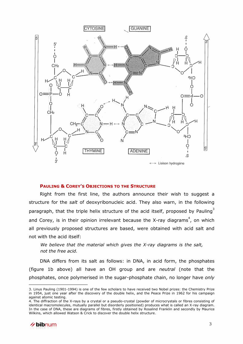
They then reveal what immediately became the icon of all biology: the
figure accompanying the publication, announced as “purely schematic”, is in fact
exceptionally close to the actual atomic structure that was not achieved until
much later on. This fourth paragraph is entirely devoted to the precise
description of the structure’s geometric characteristics. The authors begin by
describing the two sugar-phosphate chains, helically wound around an identical
axis and therefore forming a double helix.
Figure 2: (on the left) illustration from Watson & Crick’s publication. The caption
is from the publication, translated into French. The caption from the English text is
equally refined, but slightly different: “The two ribbons represent the phosphate-sugar
chains and the horizontal rods represent the bonding between the pair of bases.”
(on the right) detail of one of the sugar-phosphate chains (the helix branch)
(illustration taken from James Watson’s account The Double Helix). We see the
bonds of sugars and phosphates. The molecules shown in grey on the right of this figure
are nitrogenous bases, either pyrimidines (bases T or C, cf. figure 1), or purines (bases A
or G). A pyrimidine is always linked to a purine, following only two possible
configurations: A-T and C-G. Remember: the distribution of these pairs in the helix is
completely random: several A-T or C-G, either straight away or not– there is no order
(cf. figure 2 on the left)
Then they announce, given the symmetry of the crystallography images,
that the structure has an axis of order 2, which is not the common axis of the
6
two helices but perpendicular to it8. From this, they deduce that the order of
atoms is the opposite from one chain to another (arrows in opposite directions on
the two helices of the figure 2; see also figure 1c showing the opposite directions
5' -->3' on one strand and 3'-->5' on the other).
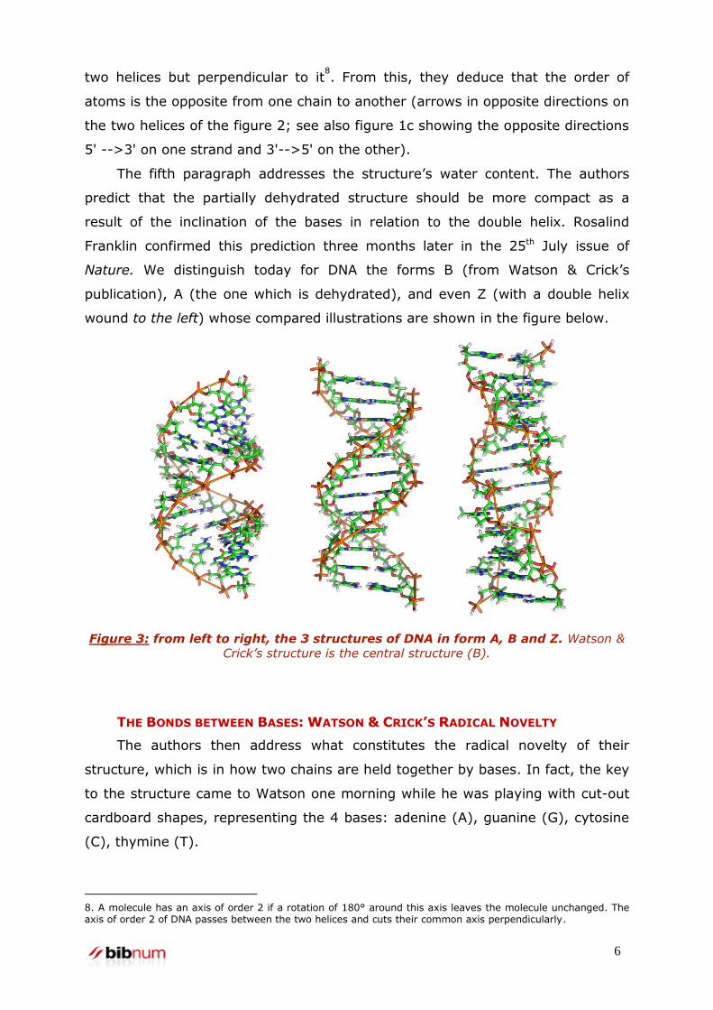
The fifth paragraph addresses the structure’s water content. The authors
predict that the partially dehydrated structure should be more compact as a
result of the inclination of the bases in relation to the double helix. Rosalind
Franklin confirmed this prediction three months later in the 25th July issue of
Nature. We distinguish today for DNA the forms B (from Watson & Crick’s
publication), A (the one which is dehydrated), and even Z (with a double helix
wound to the left) whose compared illustrations are shown in the figure below.
Figure 3: from left to right, the 3 structures of DNA in form A, B and Z. Watson &
Crick’s structure is the central structure (B).
THE BONDS BETWEEN BASES: WATSON & CRICK’S RADICAL NOVELTY
The authors then address what constitutes the radical novelty of their
structure, which is in how two chains are held together by bases. In fact, the key
to the structure came to Watson one morning while he was playing with cut-out
cardboard shapes, representing the 4 bases: adenine (A), guanine (G), cytosine
(C), thymine (T).
8. A molecule has an axis of order 2 if a rotation of 180° around this axis leaves the molecule unchanged. The axis of order 2 of DNA passes between the two helices and cuts their common axis perpendicularly.
7
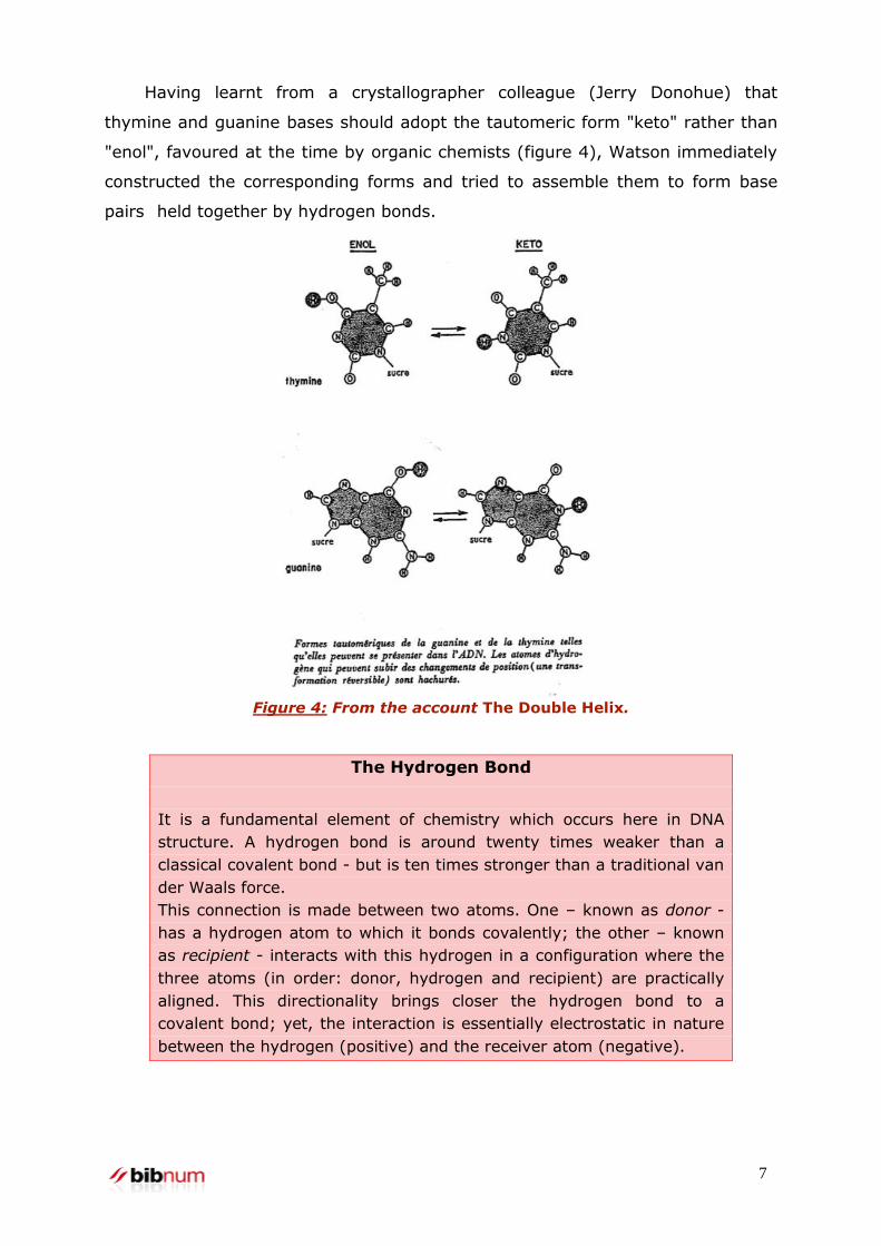
Having learnt from a crystallographer colleague (Jerry Donohue) that
thymine and guanine bases should adopt the tautomeric form "keto" rather than
"enol", favoured at the time by organic chemists (figure 4), Watson immediately
constructed the corresponding forms and tried to assemble them to form base
pairs held together by hydrogen bonds.
Figure 4: From the account The Double Helix.
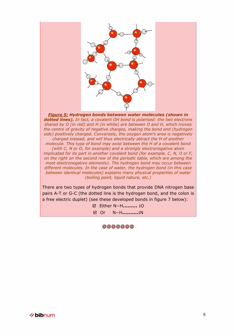
The Hydrogen Bond
It is a fundamental element of chemistry which occurs here in DNA
structure. A hydrogen bond is around twenty times weaker than a
classical covalent bond - but is ten times stronger than a traditional van
der Waals force.
This connection is made between two atoms. One – known as donor -
has a hydrogen atom to which it bonds covalently; the other – known
as recipient - interacts with this hydrogen in a configuration where the
three atoms (in order: donor, hydrogen and recipient) are practically
aligned. This directionality brings closer the hydrogen bond to a
covalent bond; yet, the interaction is essentially electrostatic in nature
between the hydrogen (positive) and the receiver atom (negative).
8
Figure 5: Hydrogen bonds between water molecules (shown in
dotted lines). In fact, a covalent OH bond is polarised: the two electrons
shared by O (in red) and H (in white) are between O and H, which moves
the centre of gravity of negative charges, making the bond end (hydrogen
side) positively charged. Conversely, the oxygen atom’s area is negatively
charged instead, and will thus electrically attract the H of another
molecule. This type of bond may exist between the H of a covalent bond
(with C, N or O, for example) and a strongly electronegative atom
implicated for its part in another covalent bond (for example, C, N, O or F,
on the right on the second row of the periodic table, which are among the
most electronegative elements). The hydrogen bond may occur between
different molecules. In the case of water, the hydrogen bond (in this case
between identical molecules) explains many physical properties of water
(boiling point, liquid nature, etc.)
There are two types of hydrogen bonds that provide DNA nitrogen base
pairs A-T or G-C (the dotted line is the hydrogen bond, and the colon is
a free electric duplet) (see these developed bonds in figure 7 below):
Either N−H……… :O
Or N−H……….:N
@@@@@@@
9
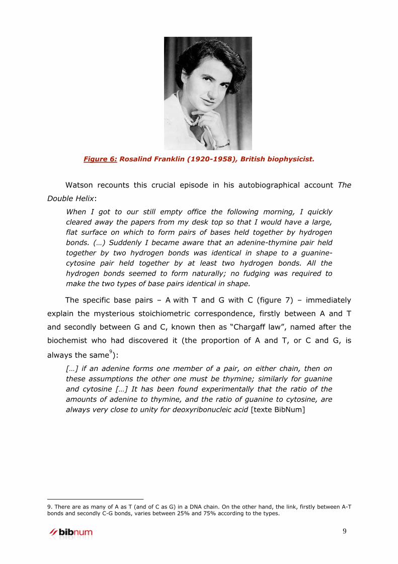
Figure 6: Rosalind Franklin (1920-1958), British biophysicist.
Watson recounts this crucial episode in his autobiographical account The
Double Helix:
When I got to our still empty office the following morning, I quickly
cleared away the papers from my desk top so that I would have a large,
flat surface on which to form pairs of bases held together by hydrogen
bonds. (…) Suddenly I became aware that an adenine-thymine pair held
together by two hydrogen bonds was identical in shape to a guanine-
cytosine pair held together by at least two hydrogen bonds. All the
hydrogen bonds seemed to form naturally; no fudging was required to
make the two types of base pairs identical in shape.
The specific base pairs – A with T and G with C (figure 7) – immediately
explain the mysterious stoichiometric correspondence, firstly between A and T
and secondly between G and C, known then as “Chargaff law”, named after the
biochemist who had discovered it (the proportion of A and T, or C and G, is
always the same9):
[…] if an adenine forms one member of a pair, on either chain, then on
these assumptions the other one must be thymine; similarly for guanine
and cytosine […] It has been found experimentally that the ratio of the
amounts of adenine to thymine, and the ratio of guanine to cytosine, are
always very close to unity for deoxyribonucleic acid [texte BibNum]
9. There are as many of A as T (and of C as G) in a DNA chain. On the other hand, the link, firstly between A-T bonds and secondly C-G bonds, varies between 25% and 75% according to the types.
10
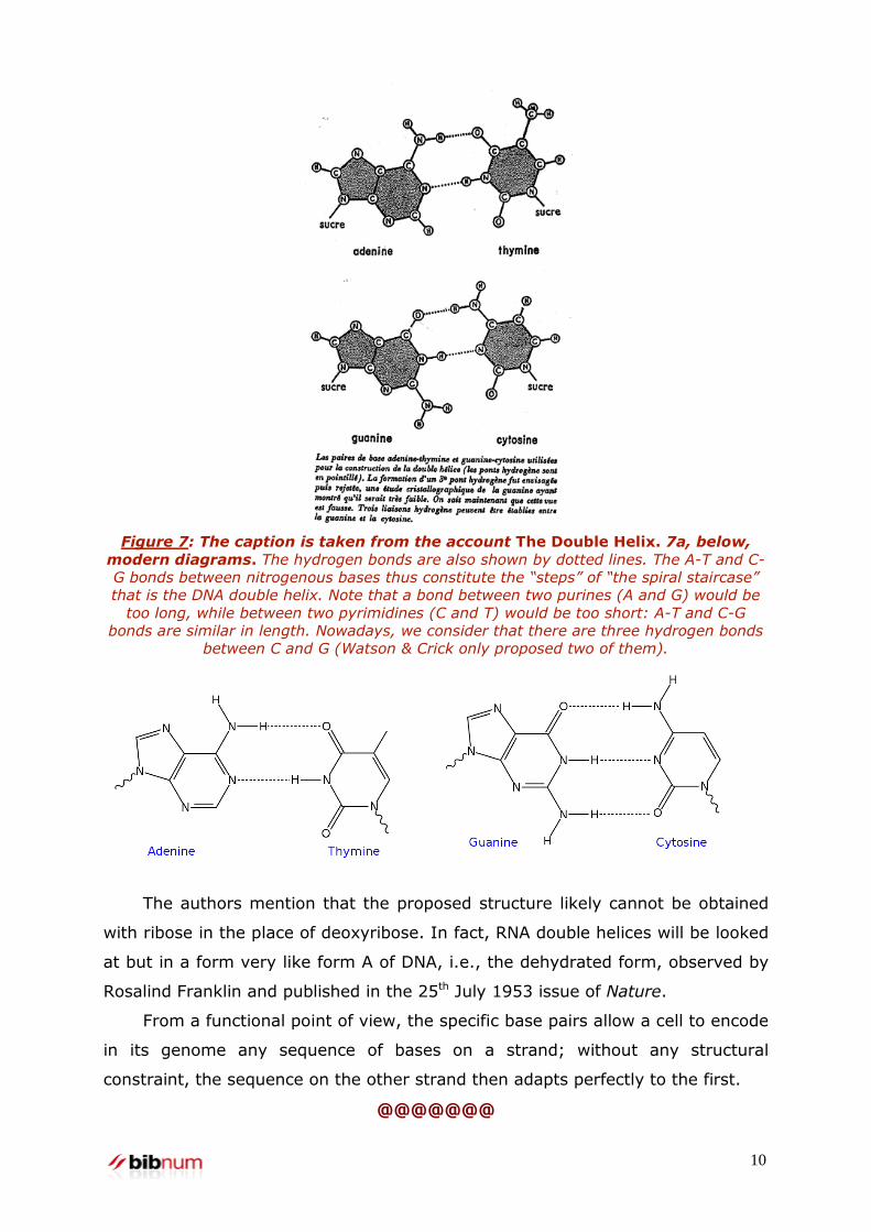
Figure 7: The caption is taken from the account The Double Helix. 7a, below,
modern diagrams. The hydrogen bonds are also shown by dotted lines. The A-T and C-
G bonds between nitrogenous bases thus constitute the “steps” of “the spiral staircase”
that is the DNA double helix. Note that a bond between two purines (A and G) would be
too long, while between two pyrimidines (C and T) would be too short: A-T and C-G
bonds are similar in length. Nowadays, we consider that there are three hydrogen bonds
between C and G (Watson & Crick only proposed two of them).
The authors mention that the proposed structure likely cannot be obtained
with ribose in the place of deoxyribose. In fact, RNA double helices will be looked
at but in a form very like form A of DNA, i.e., the dehydrated form, observed by
Rosalind Franklin and published in the 25th July 1953 issue of Nature.
From a functional point of view, the specific base pairs allow a cell to encode
in its genome any sequence of bases on a strand; without any structural
constraint, the sequence on the other strand then adapts perfectly to the first.
@@@@@@@

11
The experimental confirmation of the double helix structure is provided by
two other published articles in the same 25th April 1953 issue of Nature, placed
immediately after Watson & Crick’s article. The crystallography images, firstly
obtained by Maurice H.F. Wilkins (1916-2004, Nobel Prize in 1962 with Watson &
Crick) and collaborators, and secondly by Rosalind E. Franklin (1920-1958) and
R.G. Gosling (born in 1926) – of a very high quality – permit the identification of
certain characteristic properties of the DNA molecule structure:
- The piling up of the bases like in a pile of coins (“pile of pennies” according to
Wilkins).
- The pace of the helix: 3.4 nm
- The position of the bases inside the double helix
But the limited resolution of these images does not permit the recreation of
the all-atom structure. Watson & Crick’s unpublished exploit – and to this day
still unpublished – has led to the true structure of the DNA double helix via
theoretical arguments10
. Essentially, there are two types:
- Geometric, with the help of models11
;
- Physical: by placing the charged groups on the periphery of the double helix
and not in the centre as Linus Pauling had proposed in his triple helix model –
despite being the greatest chemist of that time; additionally, by fully
exploiting the properties of symmetry: therefore Francis Crick immediately
understands, by seeing the base pairs discovered by Watson, that the two
helices of DNA must be placed in opposite directions to each other (figure 1).
Regarding the in vivo pertinence of the double helix structure proposed by
Watson & Crick, it is given by Maurice Wilkins in the article following Watson &
Crick’s, in which he gives images which are similar but obtained with different
organisms. Maurice Wilkins also pays homage to Watson & Crick for having noted
the first biological importance – the very meaning - of the double helix structure.
At the end of their article, Watson & Crick indeed discuss it:
It has not escaped our notice that the specific pairing we have postulated
immediately suggests a possible copying mechanism12
for the genetic
material.
10. To induce the all-atom structure of a molecule or complex from the poorly resolved X-ray images (when the molecule crystals, or of the molecular complex, are unavailable) or electron microscopy is an on-going problem. The most emblematic example is chromatin fibre’s still unresolved structure: if this fibre’s structural polymorphism is nowadays well established, the different structures are yet to be determined and many competing models continue to exist. 11. The method consisting of building models was very popular at the time. It had already allowed Linus Pauling to discover the fundamental structural units of the protein structure (alpha helix and beta sheet). Today, this method is largely replaced by computer graphics tools to visualise molecule structures in 3 dimensions.
12
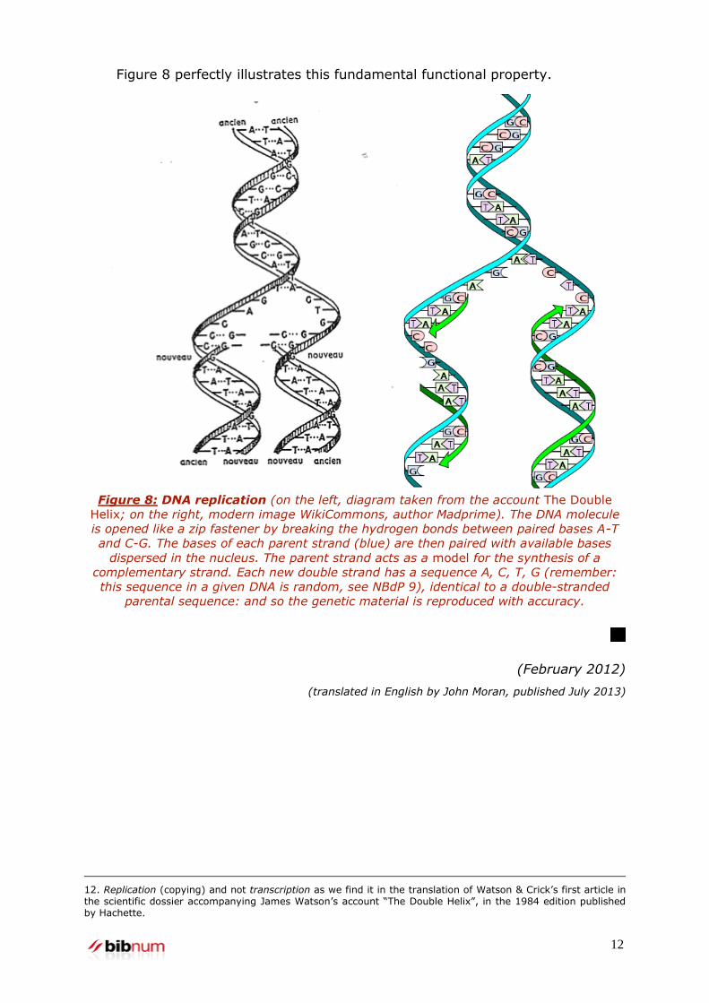
Figure 8 perfectly illustrates this fundamental functional property.
Figure 8: DNA replication (on the left, diagram taken from the account The Double
Helix; on the right, modern image WikiCommons, author Madprime). The DNA molecule
is opened like a zip fastener by breaking the hydrogen bonds between paired bases A-T
and C-G. The bases of each parent strand (blue) are then paired with available bases
dispersed in the nucleus. The parent strand acts as a model for the synthesis of a
complementary strand. Each new double strand has a sequence A, C, T, G (remember:
this sequence in a given DNA is random, see NBdP 9), identical to a double-stranded
parental sequence: and so the genetic material is reproduced with accuracy.
(February 2012)
(translated in English by John Moran, published July 2013)
12. Replication (copying) and not transcription as we find it in the translation of Watson & Crick’s first article in the scientific dossier accompanying James Watson’s account “The Double Helix”, in the 1984 edition published by Hachette.