Embed Size (px)
Citation preview

The Diagnosis and Management of NASH
Philip Wong, MD, MSc (Epid), FRCPC Hepatology & Gastroenterology
February 10, 2014

Accreditation
This event has been approved as an accredited
(Section1) group learning activity as defined by
the Maintenance of Certification program of the
RCPSC. It has been produced under RCPSC
guidelines for the development of co-developed
educational activities between CAG and Gilead
Sciences Canada Inc.

Commercial Interest Relationship*
Astellas Speaker
BMS Consultant, speaker
Gilead Consultant, speaker
Janssen Consultant
Merck Consultant, speaker, investigator
Novartis Speaker, investigator
Roche Consultant, speaker, investigator
Vertex Investigator
3 Products made by these companies will not be discussed in this presentation
Financial Interest Disclosure

Medical Expert (as Medical Experts, physicians integrate all of the CanMEDS Roles,
applying medical knowledge, clinical skills, and professional attitudes in their provision of
patient-centered care. Medical Expert is the central physician Role in the CanMEDS
framework.)
Communicator (as Communicators, physicians effectively facilitate the doctor-patient
relationship and the dynamic exchanges that occur before, during, and after the medical
encounter.)
Collaborator (as Collaborators, physicians effectively work within a healthcare team to
achieve optimal patient care.)
Manager (as Managers, physicians are integral participants in healthcare organizations,
organizing sustainable practices, making decisions about allocating resources, and
contributing to the effectiveness of the healthcare system.)
Health Advocate (as Health Advocates, physicians responsibly use their expertise and
influence to advance the health and well-being of individual patients, communities, and
populations.)
Scholar (as Scholars, physicians demonstrate a lifelong commitment to reflective learning,
as well as the creation, dissemination, application and translation of medical knowledge.)
Professional (as Professionals, physicians are committed to the health and well-being of
individuals and society through ethical practice, profession-led regulation, and high personal
standards of behaviour.)
CANMEDS ROLES

“At the end of this program participants should know”:
OBJECTIVES 1. the factors playing a role in the pathogenesis of NAFLD 2. how to diagnose and stage NAFLD 3. how to provide specific advices regarding diet and exercise 4. what and when to use supplements and drugs

Steatosis by imaging or histology in the absence of secondary causes (alcohol, other drugs, etc)
Histologic spectrum of liver damage
At the cirrhotic stage often “burnt out” or “cryptogenic”
NAFL –steatosis
NASH
Cirrhosis
Non-Alcoholic Fatty Liver Disease (NAFLD)

NASH: CLINICAL IMPACT
• Nonalcoholic Fatty Liver Disease (NAFLD) is the most common liver disease in Western countries
• NAFLD vs. NASH
• 25-46% (general) VS 3%
• 70% (diabetics) VS 22%
• 90% (obese) VS 14-37%
Ratziu et al, J Hepatol 2010

RISK FACTORS ASSOCIATED WITH NAFLD
Conditions with
established association
Conditions with emerging
association
Obesity Polycystic ovary syndrome
Type 2 diabetes mellitus Hypothyroidism
Dyslipidemia Obstructive sleep apnea
Metabolic syndrome Hypopituitarism
Hypogonadism
Pancreato-duodenal
resection

Definitions: Clinical Syndrome
Metabolic syndrome1 Obesity
Diabetes Hypertension Hyperlipidemia
No alcohol: 20 grams vs 40 grams (20-210)2
Absence of other cause of liver disease
1. P Angulo et al. Hepatology 1999;30:1356-62
2. R Saitz. NEJM 2005;352:529-607


Dallas heart study (n=2200)
Liver fat assessed by MR spectroscopy
NAFLD is common and commonly asymptomatic
Liver fat normal (< 5.5%)
Liver fat > 5.5% (31%)
79% with
steatosis
nl enzymes
Hispanics Whites Blacks
45% 33% 24%
Prevalence of Fatty liver
D Browning et al.Hepatology 2004;40:1387-1395 Met synd > in hispanics than whites
Met Synd also common in blacks
NASH in approx 6% overall

National Health and Nutrition Examination Survey (NHANES) III
NAFLD is common and commonly asymptomatic
Normal AST, ALT
5.5%
•Prevalence of elevated
liver enzymes = 8%
•Unexplained in 5.5%
•Many of these had
metabolic syndrome
Clark et al. Am J Gastro 2003; 98:960
Age-adjusted Percentage of U.S. Adults
Who Were Obese or Who Had Diagnosed
Diabetes Obesity (BMI ≥30 kg/m2)
Diabetes
1994
1994
2000
2000
No Data <14.0% 14.0-17.9% 18.0-21.9% 22.0-25.9% >26.0%
No Data <4.5% 4.5-5.9% 6.0-7.4% 7.5-8.9% >9.0%
CDC’s Division of Diabetes Translation. National Diabetes Surveillance System available at
http://www.cdc.gov/diabetes/statistics
2008
2008

Gotay, C. Can J Public Health 2013

Gotay, C. Can J Public Health 2013

Gotay, C. Can J Public Health 2013
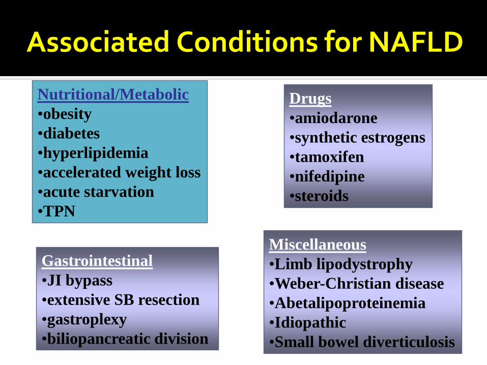
Associated Conditions for NAFLD
Nutritional/Metabolic
•obesity
•diabetes
•hyperlipidemia
•accelerated weight loss
•acute starvation
•TPN
Gastrointestinal
•JI bypass
•extensive SB resection
•gastroplexy
•biliopancreatic division
Drugs
•amiodarone
•synthetic estrogens
•tamoxifen
•nifedipine
•steroids
Miscellaneous
•Limb lipodystrophy
•Weber-Christian disease
•Abetalipoproteinemia
•Idiopathic
•Small bowel diverticulosis

NAFLD in High-Risk Populations
Bariatric surgery candidates
Systematic reviews
High variability in NASH rates
NAFL: 91%
NASH: 37% (24-98%)
Mild fibrosis: 40%
Advanced fibrosis: 10% (cirrhosis 1.4%)
Main association for advanced fibrosis: diabetes
Machado et al. J Hepatol 2006;45(4):600-606 Vernon et al. Alim Pharm Ther 2011; 34:274-85
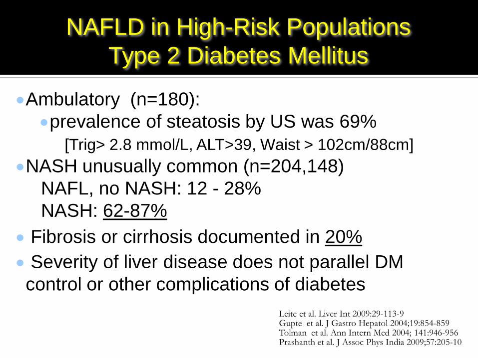
NAFLD in High-Risk Populations
Type 2 Diabetes Mellitus
Ambulatory (n=180):
prevalence of steatosis by US was 69%
[Trig> 2.8 mmol/L, ALT>39, Waist > 102cm/88cm]
NASH unusually common (n=204,148)
NAFL, no NASH: 12 - 28%
NASH: 62-87%
Fibrosis or cirrhosis documented in 20%
Severity of liver disease does not parallel DM
control or other complications of diabetes
Leite et al. Liver Int 2009:29-113-9 Gupte et al. J Gastro Hepatol 2004;19:854-859 Tolman et al. Ann Intern Med 2004; 141:946-956 Prashanth et al. J Assoc Phys India 2009;57:205-10

NAFLD kills

Major risk factors for advanced liver disease
Independent Risk Factors for fibrosis
Older age (>45)
Obesity (BMI > 30)
Long-standing diabetes (> 15 yrs)
Triglycerides > 5.65 (2.8?) mmol/L
AST > ALT
ALT > 2 x ULN and/or AST > normal
1. P Angulo et al. Hepatology 1999;30:1356-62
2. P Sorrentino et al. J Hepatol 2004;41:751
3. V De Ledinghen et al. Eur J Gastro 2004;16:879-83

Major Causes Of Death Are Not Liver
Cause of Death:
Malignancy 28%
Cardiovascular 25%
Liver 13%
For 45 – 54 yr old group CV causes most significant
Standardized mortality ratio all causes: 4.4 (1.2 -13.2)
SMR for CV disease: 8.15 (2 – 33.2)
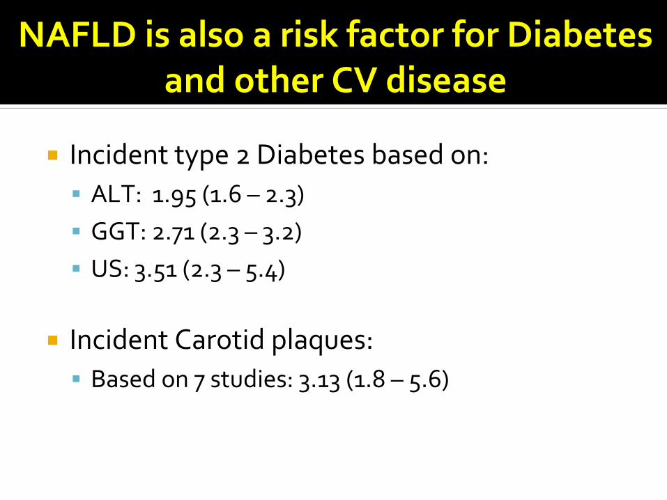
NAFLD is also a risk factor for Diabetes and other CV disease
Incident type 2 Diabetes based on:
ALT: 1.95 (1.6 – 2.3)
GGT: 2.71 (2.3 – 3.2)
US: 3.51 (2.3 – 5.4)
Incident Carotid plaques:
Based on 7 studies: 3.13 (1.8 – 5.6)

Is NAFLD progressive?

PATHOGENESIS : “TWO HITS” OR “DISTINCT HITS” ?
Ylmaz et al, APT 2012
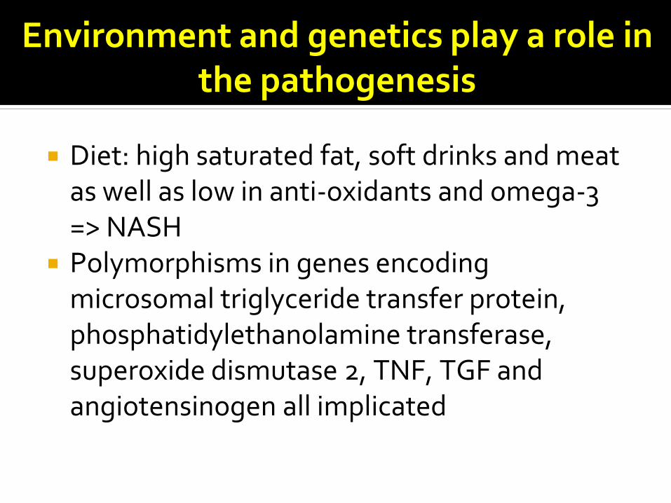
Environment and genetics play a role in the pathogenesis
Diet: high saturated fat, soft drinks and meat as well as low in anti-oxidants and omega-3 => NASH
Polymorphisms in genes encoding microsomal triglyceride transfer protein, phosphatidylethanolamine transferase, superoxide dismutase 2, TNF, TGF and angiotensinogen all implicated
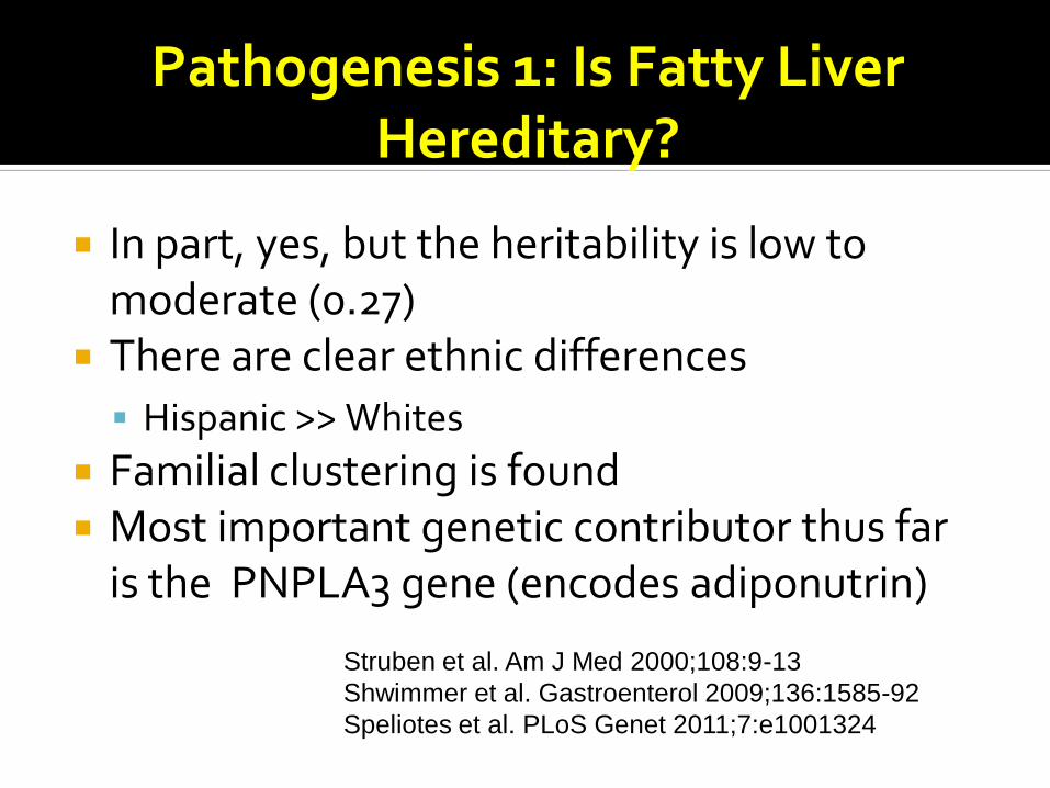
Pathogenesis 1: Is Fatty Liver
Hereditary?
In part, yes, but the heritability is low to moderate (0.27)
There are clear ethnic differences
Hispanic >> Whites
Familial clustering is found Most important genetic contributor thus far
is the PNPLA3 gene (encodes adiponutrin)
Struben et al. Am J Med 2000;108:9-13
Shwimmer et al. Gastroenterol 2009;136:1585-92
Speliotes et al. PLoS Genet 2011;7:e1001324

Hormones vs. Cytokines: Adiponectin and TNF
Adiponectin (decr.)
Anti-inflammatory
Decreases fatty acid uptake
Stimulates fatty acid oxidation / lipid export
Enhances insulin sensitivity
TNF alpha (incr.)
Pro-inflammatory
Pro-apoptotic
Recruits WBCs
Promotes insulin resistance

Mechanisms : Overview
Overview of 2 hit hypothesis of NASH
Steatosis
Hepatic lipogenesis
Fibrosis
Steatohepatitis
Oxidative stress
Increased delivery FFA to liver
Increased lipolysis
Insulin Resistance
Leptin / Angiotensin
TNF Adiponectin
Noradrenaline
PPAR / γ

Be careful when working up “a diagnosis of exclusion”
Elevated serum autoantibodies are common in patients with NAFLD and are generally considered to be an epiphenomenona
Vuppalanchi. Hepatology 2009
NASH Clinical Research Network
+ANA > 1:160 or SMA > 1:40 in 21 % of NAFLD patients without more advanced histologic features
Vuppalanchi R. Hepatol Int. 2011
Chalasani Am J Gastro 2012 Guidelines NAFLD

Which is the best test?
liver enzymes: can be normal in NAFLD and NASH (sensitivity may be poor)
liver US is potentially more sensitive but $$ and not easily done
Histology may be best Look for NASH-related fibrosis when elevated liver
enzymes, presence of insulin resistance (IR) as well as histological ballooning of hepatocytes
Chalasani Am J Gastro 2012 Guidelines NAFLD Lee. J Hepatol 2007 Williams CD. Gastroenterology 2011 Browning JD. Hepatology 2004 Baranova A. BMC Gastroenterol 2011 Younossi Aliment Pharmacol Ther. 2014

EVALUATION OF FIBROSIS: HOW ?
- Liver biopsy (gold standard)
- Blood tests (biomarkers)
- Fibroscan (transient elastography)
Advantages: direct information on fibrosis,
inflammation, steatosis, comorbidities
Limits: semiquantitative, invasive, costly, prone
to sampling errors

SERUM NON-INVASIVE MARKERS for LIVER FIBROSIS
DIRECT MARKERS Matrix Components and
Enzymes regulating
Fibrogenesis / Fibrolysis
INDIRECT
MARKERS Markers of Liver
Inflammation / Function
Combination of Direct / Indirect Tests
Procollagen III, Type IV
collagen, Hyaluronic acid,
YKL-40, Metalloproteinases
and their Inhibitors
AST, ALT, gGT, Platelets,
Bilirubin, Albumin,
Cholesterol, ApoA1, a2-
Macroglobulin, Haptoglobin

Indirect Biomarkers
AUC for >F2 AUC for F4
APRI 0.73-0.87 0.84
Forns’ index 0.60-0.86 NA
Fib-4 0.70-0.85 NA
ALT/AST NA 0.74
BARD score 0.70-0.81 NA
APRI = AST, platelets Forns’ index = gGT, cholesterol, platelets, age Fib-4 = platelets, AST, ALT, age BARD = BMI, AST/ALT and presence of diabetes
Wai et al, Hepatology 2003; Forns et al, Hepatology 2002; Sebastiani et al, CCLM 2011; Sebastiani et al, APT 2011

AST/ALT Ratio 53% sensitive, 100% specific
>1 high = likelihood of cirrhosis Highly predictive of cirrhosis if ratio > 1, 100%
specificity and positive predictive value in distinguishing cirrhotic from noncirrhotic patients, with a 53.2% sensitivity and 80.7% negative predictive value in HCV patients
Baranova BMC Gastroenterol. 2011 Aug 17;11:91
Sheth SG et al. Am J Gastroenterology 1998; 93: 44-8
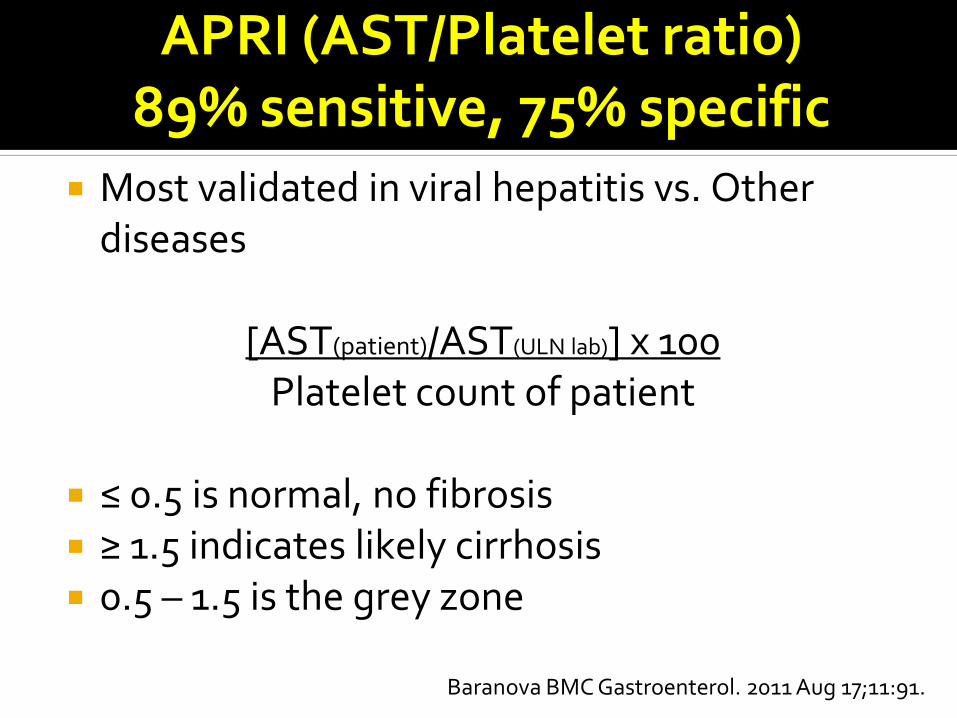
APRI (AST/Platelet ratio) 89% sensitive, 75% specific
Most validated in viral hepatitis vs. Other diseases
[AST(patient)/AST(ULN lab)] x 100
Platelet count of patient
≤ 0.5 is normal, no fibrosis ≥ 1.5 indicates likely cirrhosis 0.5 – 1.5 is the grey zone
Baranova BMC Gastroenterol. 2011 Aug 17;11:91.

SIMPLE BIOMARKERS BASED ON CUT-OFF VALUES: APRI
0.5 1.5
50% of Pts
Wai et al, Hepatology 2003
Absence of significant fibrosis Presence of significant fibrosis
APRI=[AST (/ULN) x 100] / Platelet (109/L)

NAFLD Fibrosis Score
go to gihep.com – formula containing Age, AST, ALT, Platelet Count, BMI, Albumin, Impaired Fasting Glucose/Diabetes
If the NAFLD Score is: < -1.455 = F0-F2 (NPV 88-93% and 88% PPV) > 0.675 = F3-F4 (PPV 82-90%) -1.455 – 0.675 = indeterminate score
a liver biopsy would have been avoided in 75% of
the 733 patients, with 90% accuracy.
Angulo P et al. Hepatology 2007;45(4):846-854

Fib-4
Score = age × AST (IU/l) x √ALT (IU/l) platelet s (×109/L) <1.45 NPV F3-F4 (94.7%) 3.25 PPV F3-F4 (82%) >10 PPV F4 excellent
Vallet-Pichard A et al. Hepatology 2008

Fib-4 70 % sensitive, 74% specific
For HCV with or without HIV: Fib4 score < 1.45 = F0-F1
Fib4 score > 3.25 = F3-F4
For NASH: Fib4 score < 1.30 = F0-F1
Fib4 score > 2.67 = F3-F4 Baranova BMC Gastroenterol. 2011 Aug 17;11:91
Sterling RK et al. HEPATOLOGY 2006; 43: 1317-1325. Martinez, S. M. Et al. Hepatology 2011, 53: 325–335

Direct Biomarkers and Combination Panels
Parameters AUC for >F2 AUC for F4
Fibrotest® gGT, bilirubin,
haptoglobin,
ApoA1, 2M
0.75-0.86 0.75
Hyaluronan Hyaluronan,
TIMP1, 2M
0.73-0.97 0.92
ELF® Hyaluronan,
PIIINP, TIMP1
0.82-0.98 0.90-0.99
Fibrometer® Platelets, PT,
AST, 2M,
hyaluronan,
BUN
0.94 0.92
Imbert-Bismuth et al, Lancet 2001; Patel et al, Clin Gastro 2007; Rosenberg et al, Gastroenterology 2004; Cales et al, Hepatology 2005; Becker et al, Clin Gastro 2009; Sebastiani et al, APT 2011

FibroScan
Painless
Rapid (5 min)
Bedside/Outpatient
Immediate results (3 - 75 kPa)

Successful Fibroscan
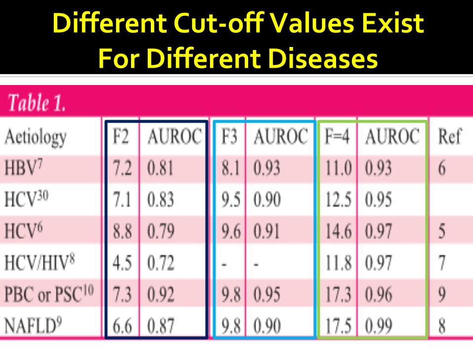
Different Cut-off Values Exist For Different Diseases

Using a cut-off range instead of a single value for each disease is simpler

Fibroscan algorithm

FIBROSCAN FAILURE n=2114
Foucher et al. Eur J Gastroenterol Hepatol 2006; Wong et al, Hepatology 2010; Myers et al, Hepatology 2012
Failure : 4.5 %
BMI > 28
Wong et al: 25.5% if BMI > 30, 2.6% if BMI < 30
Reliable results with the XL probe were obtained in 61% of patients in whom the M probe was unreliable

Composite blood tests may correlate with
NASH and degree of fibrosis
• Steatotest (AUROC =0.8): total bilirubin, GGT, α2-
macroglobulin, apolipoprotein A1, haptoglobin, AST,
serum glucose, triglycerides and cholesterol
• NASH-Test (AUROC=0.8): total bilirubin, GGT, α2-
macroglobulin, apolipoprotein A1, haptoglobin, weight,
height, AST, serum glucose, triglycerides, cholesterol
• Fibrotest (AUROC=0.86-0.9): total bilirubin, GGT, α2-
macroglobulin, apolipoprotein A1, haptoglobin
Poynard et al. BMC Gastroenterology 2006;6:6 and 34
Poynard et al. Comp Hepatol 2005;4:10

Circulating levels of cytokeratin-18 (CK18) fragments may be a useful biomarker for NAFLD
increased in NASH vs. simple steatosis or normal biopsies (P < 0.001)
OR= 1.95 for NASH for every 50 U/ l increase Reproduced in other studies meta-analysis: estimated sensitivity of 78 % ,
specificity of 87 % , and an AUROC of 0.82 (95 % CI: 0.78 – 0.88) for steatohepatitis in patients with NAFLD

How do you assess Steatosis?
Normal liver enzymes do not exclude NASH
No imaging modality distinguishes NAFLD from
NASH or fibrosis
Modality AUROC
Ultrasound 0.87
Xenon133 0.87
CT 0.91
MRI-PDFF* 0.98
Mathiesen et al. Dig Liver dis. 2002;516-22
Albusafi et al. Can J Gastroenterol 2012;155-9
D Joly et al Eur J Gastroenterol Hepatol 2003;15:539-43
Noureddin AASLD 2013, Abstract
Protein Density Fat Fraction
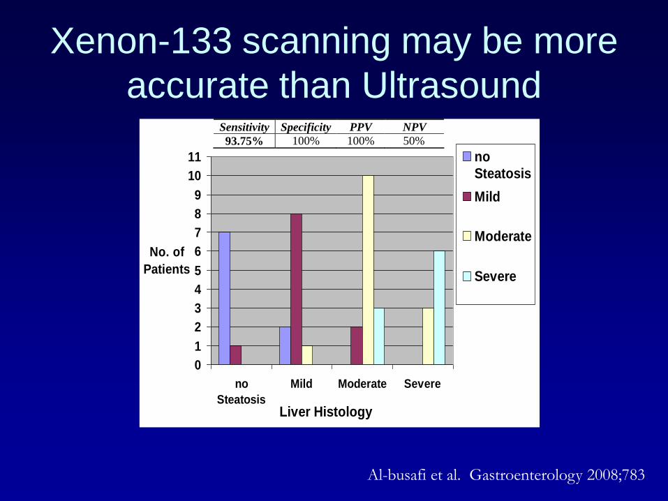
Xenon-133 scanning may be more
accurate than Ultrasound
0
1
2
3
4
5
6
7
8
9
10
11
no
Steatosis
Mild Moderate Severe
Liver Histology
No. of
Patients
no
Steatosis
Mild
Moderate
Severe
Al-busafi et al. Gastroenterology 2008;783
Sensitivity Specificity PPV NPV
93.75% 100% 100% 50%
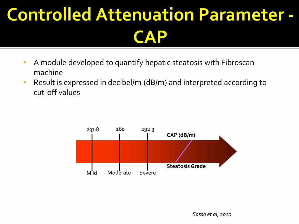
Controlled Attenuation Parameter - CAP
• A module developed to quantify hepatic steatosis with Fibroscan machine
• Result is expressed in decibel/m (dB/m) and interpreted according to cut-off values
Sasso et al, 2010
Steatosis Grade Severe
CAP (dB/m) 260 292.3
Moderate
237.8
Mild

CAP has high accuracy for fat
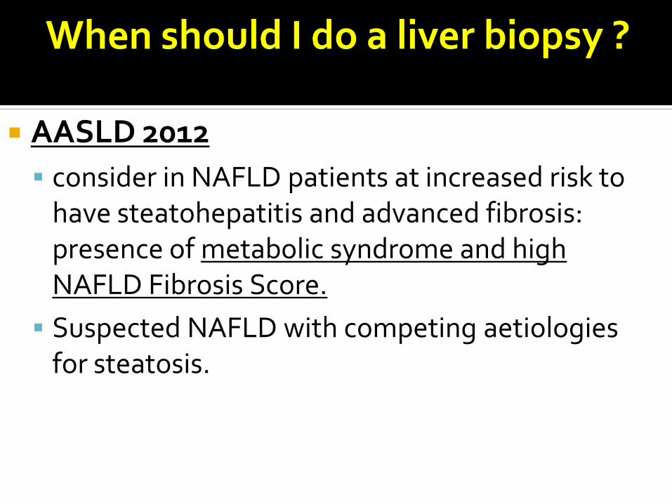
When should I do a liver biopsy ?
AASLD 2012
consider in NAFLD patients at increased risk to have steatohepatitis and advanced fibrosis: presence of metabolic syndrome and high NAFLD Fibrosis Score.
Suspected NAFLD with competing aetiologies for steatosis.
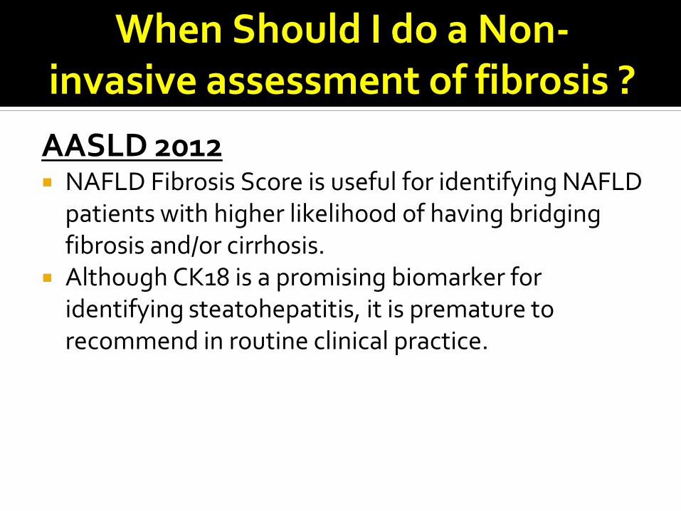
AASLD 2012 NAFLD Fibrosis Score is useful for identifying NAFLD
patients with higher likelihood of having bridging fibrosis and/or cirrhosis.
Although CK18 is a promising biomarker for identifying steatohepatitis, it is premature to recommend in routine clinical practice.
When Should I do a Non-invasive assessment of fibrosis ?
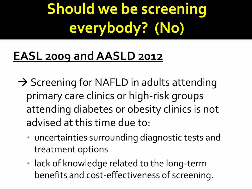
Screening for NAFLD in adults attending primary care clinics or high-risk groups attending diabetes or obesity clinics is not advised at this time due to:
• uncertainties surrounding diagnostic tests and treatment options
• lack of knowledge related to the long-term benefits and cost-effectiveness of screening.
Should we be screening everybody? (No)
EASL 2009 and AASLD 2012

SUMMING-UP and CLINICAL DIRECTIONS
• Liver biopsy remains an important tool to diagnose NASH and to stage liver fibrosis
• The presence of metabolic syndrome is the strongest predictor of NASH
• NAFLD fibrosis score or combination of Fibroscan and a serum test (Fibrotest) can be used to select patients at high risk of advanced fibrosis
• Reliable non-invasive methods to diagnose NASH are highly warranted
• Screening of general population not recommended; but at risk groups ?

Any Questions?
Thank you for your attention
Thank you to Dr.
Peter Ghali and Dr. Giada Sebastiani
for slides and their expertise

Please visit the CAG website at
http://www.cag-acg.org/ to complete
the session evaluation and to print
your certificate of attendance.
Thank you!
Evaluation and Certificate of Attendance