Embed Size (px)
Citation preview

SUMMARY
Palatogenesis is a complex developmentalprocess that requires two main events: elevationand then fusion of the palatal shelves. Thereremains controversy concerning the mecha-nism(s) responsible for palatal shelf elevation, itbeing proposed that an intrinsic shelf elevationforce might be produced either by the genera-tion of a turgor pressure following hydration ofthe extracellular matrix via its glycoconjugatemolecules or by proliferation, migration and/orcontraction of the palatal shelf mesenchymalcells. Recent evidence indicates that the shelfelevation force is related to the presence ofhyaluronan in the extracellular matrix, to an asyet unknown molecule that is packaged in themesenchymal cells’ Golgi complex, and to CD44receptor functioning. For fusion of the palatalshelves to occur, the breakdown of the midlineepithelial seam relates to apoptosis and rediffer-entiation of the epithelial cells and this appearsto be signalled by the synthesis of type IX colla-gen just prior to the breakdown of the basementmembrane around the midline epithelial seam.The events associated with palatogenesis arecontrolled by the palatal shelf mesenchyme,under the influence of a variety of homeoboxgenes and transcription factors and and of sev-eral growth factors (particularly TGF-βs).
Key words: Palatogenesis – Palatal shelf eleva-tion – Hyaluronan – CD44 – TGF-β
INTRODUCTION
Palatogenesis is a complex event and is oftendisturbed to produce the congenital defect
known as cleft palate. Consequently, the eventsand mechanisms responsible for the develop-ment of the palate have been much studied,although some controversy remains.
The definitive palate (or secondary palate)develops in the human fetus between the sixthand eighth week of intra-uterine life (e.g. Fergu-son, 1978a; Johnston and Sulik, 1990; Sadler,2000; Berkovitz et al., 2002). By the sixth weekof development (Fig. 1), the primitive nasal cav-ities are separated by a primary nasal septum
53
The development of the palate – a brief review
B.J. MoxhamCardiff School of Biosciences, Cardiff University, United Kingdom andThe School of Medicine, St. George’s University, Grenada
Correspondence to:Prof. Bernard J. Moxham. Cardiff School of Biosciences, Cardiff University, MuseumAvenue, Cardiff, CF10 3US, United Kingdom. Telephone: +44 (0)29 20874031; Fax: +44(0)29 20875964. E-mail: [email protected] URL: www.cardiff.ac.uk/biosi/
Submitted: March 10, 2003Accepted: June 12, 2003
Figure 1. Diagram showing the state of development of the palateby the sixth week of intra-uterine life. A = primitive nasalcavities; B = primary nasal septum; C = primary palate.
REVIEW Eur J Anat, 7 Suppl. 1: 53-74 (2003)
and are partitioned from the primitive oral cavi-ty by a primary palate. Both the primary nasalseptum and primary palate are derived from thefrontonasal process of the developing face. The
stomodeal chamber is divided at this stage intothe small primitive oral cavity beneath the pri-mary palate, and the relatively large oronasalcavity behind the primary palate. As shown inFigure 2, during the sixth week of development,two palatal shelves develop laterally behind theprimary palate from the maxillary facial process-es. A secondary nasal septum grows down fromthe roof of the stomodeum behind the primarynasal septum, thus dividing the nasal part of theoronasal cavity into two.
Figure 3 shows the developing head duringthe seventh week of development. At this stage,the oral part of the oronasal cavity becomescompletely filled by the developing tongue.Growth of the palatal shelves continues suchthat they come to lie vertically. Two peaks ofDNA synthesis occur as the palatal shelves areformed: during initial shelf outgrowth and duringvertical shelf elongation (Burdett et al., 1988).The reason for mammalian shelves forming witha vertical orientation is unknown. It has beensuggested that the potential space in theoronasal cavity is insufficient because of the evo-lution of a large tongue in mammals (Haywardand Avery, 1957). However, Young et al. (1990)have shown that there is no spatio-temporal rela-tionship between the development of the tongueand the palatal shelves.
During the eighth week of development (Fig.4), the stomodeum enlarges, the tongue ‘drops’
B. J. Moxham
54
Figure 2. Diagram showing the development of the palate duringthe sixth week of intra-uterine life. A = lateral palatalshelves; B = primary palate; C = secondary nasal septum.
Figure 3. Coronal section through the developing head during the seventh week of development showing the palatal shelves (A). B = devel-oping tongue. (Masson’s trichrome). x 55.
A A
B

and the vertically-inclined palatal shelves becomehorizontal. It has been suggested that the descentof the tongue is related to mandibular growthand/or a change in the shape of the tongue (e.g.Humphrey, 1971; Diewert, 1974). On becominghorizontal, the palatal shelves contact each other(and the secondary nasal septum) in the midlineto form the definitive or secondary palate. Theshelves contact the primary palate anteriorly sothat the oronasal cavity becomes subdivided intoits constituent oral and nasal cavities. Figure 5shows a coronal section through the developingoronasal regions following contact of the palatalshelves and secondary nasal septum. After con-tact, the medial edge epithelia of the two shelvesfuse to form a “midline epithelial seam” (MES).Subsequently, this degenerates so that mes-enchymal continuity is established across thenow intact, and horizontal, secondary palate.Fusion of the palatal processes is complete by thetwelfth week of development. Behind the sec-ondary nasal septum, the palatal shelves fuse toform the soft palate and uvula.
Concerning the origin of the mesenchymewithin the fetal processes contributing to thedevelopment of the palate, all of the skeletal andconnective tissues that form the face are derivedfrom neural crest (NC) cells that originate alongthe dorsal margins of the midbrain and rostralhindbrain (Noden, 1978; Couly et al., 1992; Könt-ges and Lumsden, 1996; Le Douarin and Kalcheim,
1999). Indeed, Been and Song (1978) have shownthat localized destruction of midbrain NC inter-
The development of the palate – a brief review
55
Figure 5. Coronal section through developing oronasal regions following contact of the palatal shelves (A) and secondary nasal septum (B);C = midline epithelial seam; D = developing bone of maxilla. (Masson’s trichrome). x 55.
B
A
D
A
C
Figure 4. Diagram showing the state of development of the palateduring the eighth week of intra-uterine life. A = palatalshelves; B = primary palate.
feres with palatal closure. In mammalian fetuses,cranial NC cells do not always migrate before theneural tube closes (e.g. Tan and Morriss-Kay,1986). Furthermore, recent work suggests thatcraniofacial development does not depend on NCpre-programming but is controlled by a complexcombination of cell and tissue interactions involv-ing NC plasticity (Trainor and Krumlauf, 2001).Evidence is also available that suggests that NCcells can be reprogrammed and that their fate andidentity depends upon the cellular signals theyreceive as they migrate to their target tissues (e.g.Schilling et al., 2001). This seems to occur as aresult of alteration in Hox gene identity. However,there is some work indicating that NC cells havesome identity at their place of origin near the neur-al tube; although elaboration of their developmentis reached via integration and interaction with sig-nals from surrounding tissue environmentsthrough which they migrate (Grammatopoulos etal., 2000; Pasqualetti et al., 2000).
Recent research on palatogenesis has concen-trated on two main events: palatal shelf elevationand the initial stage of fusion of the shelves.
PALATAL SHELF ELEVATION
Several mechanisms have been proposed toaccount for the rapid movement (Ferguson, 1978;Brinkley, 1980) of the palatal shelves from the ver-tical to the horizontal position and the source ofthe force(s) responsible for palatal shelf reorienta-tion/elevation is a matter of controversy. Two cat-egories of explanations have been provided: eitherthe forces are extrinsic to the shelves or they aregenerated intrinsically by the shelf mesenchyme.
Those extrinsic forces that have been proposedoften relate to movement of the tongue. Forexample, there have been hypotheses that includedownward movement of the tongue due to amandibular growth spurt clearing a path forpalatal shelf elevation (e.g. Asling et al., 1960;Diewert, 1974), a downward displacement of thetongue by the nasal septum again clearing a pathfor shelf elevation (e.g. Zeiler et al., 1964), and alowering of the tongue due to a fetal mouth open-ing reflex (e.g. Humphrey, 1969, 1971). It has alsobeen suggested that the tongue physically pushesthe palatal shelves upwards (e.g. Walker, 1971).However, it is now generally thought that thepalatal shelf elevation force is not extrinsic in ori-gin. Ferguson (1978a) reviewed the literature relat-ing to extrinsic forces and concluded that thechronology of events extrinsically did not neces-sarily synchronise with shelf elevation. Further-more, following tongue excision during palatoge-nesis, no spatio-temporal relationship exists andaglossia and microglossia in humans does not pre-vent palatal closure (Young et al., 1990). In addi-tion, palatal shelves are seen to elevate in organculture in the absence of a tongue or a lower jaw.
It has been proposed that the intrinsic shelfelevation force might develop as a result ofhydration of extracellular matrix (ECM) compo-nents (principally hyaluronan) in the shelf mes-enchyme (e.g. Larsson et al., 1959; Pratt et al.,1973; Ferguson, 1978a; Brinkley and Morris-Wiman, 1984, 1987; Singh et al., 1994, 1997), oras a result of mesenchymal cell activity (e.g.Shah, 1979, 1980; Innes, 1978; Wee et al., 1979;Zimmerman, 1979; Babiarz et al., 1979; Luke,1984; Bulliet and Zimmerman, 1985; Brinkley andBookstein, 1986; Shah et al., 1989). Of course, theintrinsic shelf elevating force might be multifac-torial, although there is as yet no experimentalevidence to support what otherwise might beconsidered a “commonsense” view.
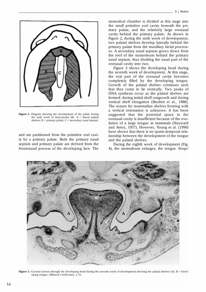
Much recent work has focussed on thechanges occurring in the ECM of the palatalshelf’s mesenchyme during shelf elevation. Thechanging amounts of glycosaminoglycans (GAG)during development of the anterior (presumptivehard) and posterior (presumptive soft) palateshave been reported by Singh et al. (1994) andare illustrated in Fig. 6. The findings show thatthe most significant changes occur after eleva-
B. J. Moxham
56
Figure 6. Graphs illustrating the changing amounts of glycosamino-glycans (GAG) during development of the anterior (pre-sumptive hard) and posterior (presumptive soft) palates.Stage A – prior to shelf elevation; Stage B – after shelf ele-vation; Stage C – during shelf fusion and early histogene-sis; Stage D – a stage of marked histogenesis after fusion.Courtesy of Dr G.D. Singh and B.J. Moxham and the edi-tor of Archives of Oral Biology.
tion and that, during the time of elevation, thereare no differences between the anterior and pos-terior regions of the shelves even though, in thespecies studied here (the rat), the posteriorregion of the shelf does not elevate but growsinitially with a horizontal disposition (Coleman,1965; Cleaton-Jones, 1976a; Singh et al., 1994).Singh et al. (1997) have also reported that, whenpalatal clefts are induced in the rat by 5-fluoro-2-deoxyuridine (FUDR), GAG biosynthesis issuppressed.
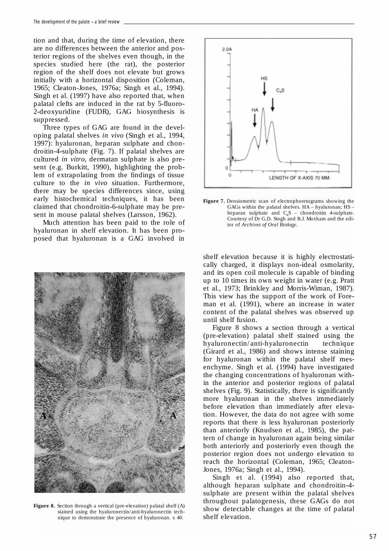
Three types of GAG are found in the devel-oping palatal shelves in vivo (Singh et al., 1994,1997): hyaluronan, heparan sulphate and chon-droitin-4-sulphate (Fig. 7). If palatal shelves arecultured in vitro, dermatan sulphate is also pre-sent (e.g. Burkitt, 1990), highlighting the prob-lem of extrapolating from the findings of tissueculture to the in vivo situation. Furthermore,there may be species differences since, usingearly histochemical techniques, it has beenclaimed that chondroitin-6-sulphate may be pre-sent in mouse palatal shelves (Larsson, 1962).
Much attention has been paid to the role ofhyaluronan in shelf elevation. It has been pro-posed that hyaluronan is a GAG involved in
shelf elevation because it is highly electrostati-cally charged, it displays non-ideal osmolarity,and its open coil molecule is capable of bindingup to 10 times its own weight in water (e.g. Prattet al., 1973; Brinkley and Morris-Wiman, 1987).This view has the support of the work of Fore-man et al. (1991), where an increase in watercontent of the palatal shelves was observed upuntil shelf fusion.
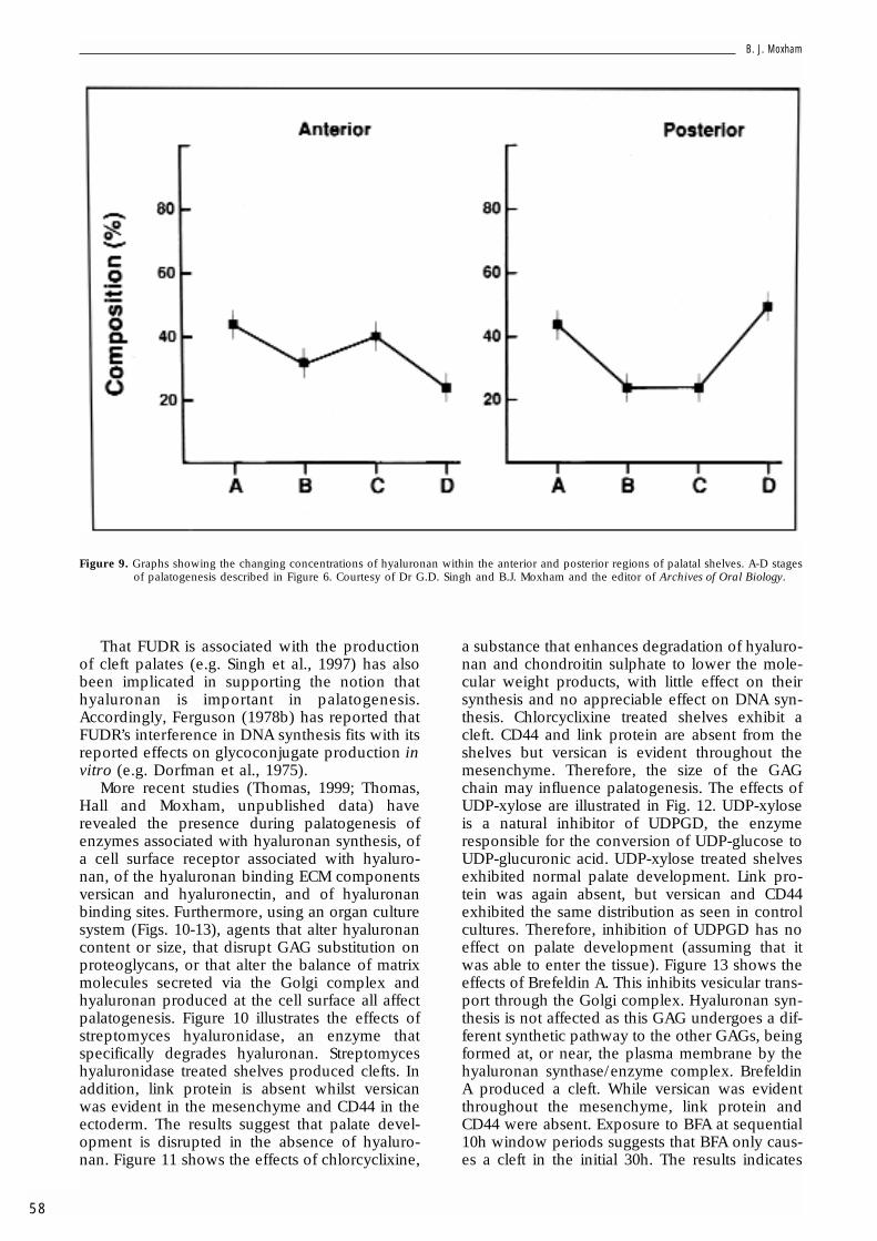
Figure 8 shows a section through a vertical(pre-elevation) palatal shelf stained using thehyaluronectin/anti-hyaluronectin technique(Girard et al., 1986) and shows intense stainingfor hyaluronan within the palatal shelf mes-enchyme. Singh et al. (1994) have investigatedthe changing concentrations of hyaluronan with-in the anterior and posterior regions of palatalshelves (Fig. 9). Statistically, there is significantlymore hyaluronan in the shelves immediatelybefore elevation than immediately after eleva-tion. However, the data do not agree with somereports that there is less hyaluronan posteriorlythan anteriorly (Knudsen et al., 1985), the pat-tern of change in hyaluronan again being similarboth anteriorly and posteriorly even though theposterior region does not undergo elevation toreach the horizontal (Coleman, 1965; Cleaton-Jones, 1976a; Singh et al., 1994).
Singh et al. (1994) also reported that,although heparan sulphate and chondroitin-4-sulphate are present within the palatal shelvesthroughout palatogenesis, these GAGs do notshow detectable changes at the time of palatalshelf elevation.
The development of the palate – a brief review
57
Figure 7. Densiometric scan of electrophoretograms showing theGAGs within the palatal shelves. HA – hyaluronan; HS –heparan sulphate and C4S – chondroitin 4-sulphate.Courtesy of Dr G.D. Singh and B.J. Moxham and the edi-tor of Archives of Oral Biology.
Figure 8. Section through a vertical (pre-elevation) palatal shelf (A)stained using the hyaluronectin/anti-hyaluronectin tech-nique to demonstrate the presence of hyaluronan. x 40.
AA
That FUDR is associated with the productionof cleft palates (e.g. Singh et al., 1997) has alsobeen implicated in supporting the notion thathyaluronan is important in palatogenesis.Accordingly, Ferguson (1978b) has reported thatFUDR’s interference in DNA synthesis fits with itsreported effects on glycoconjugate production invitro (e.g. Dorfman et al., 1975).
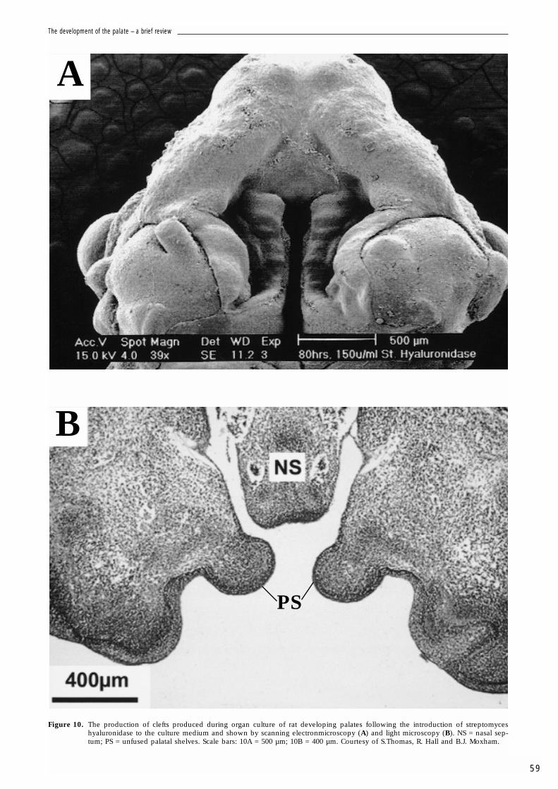
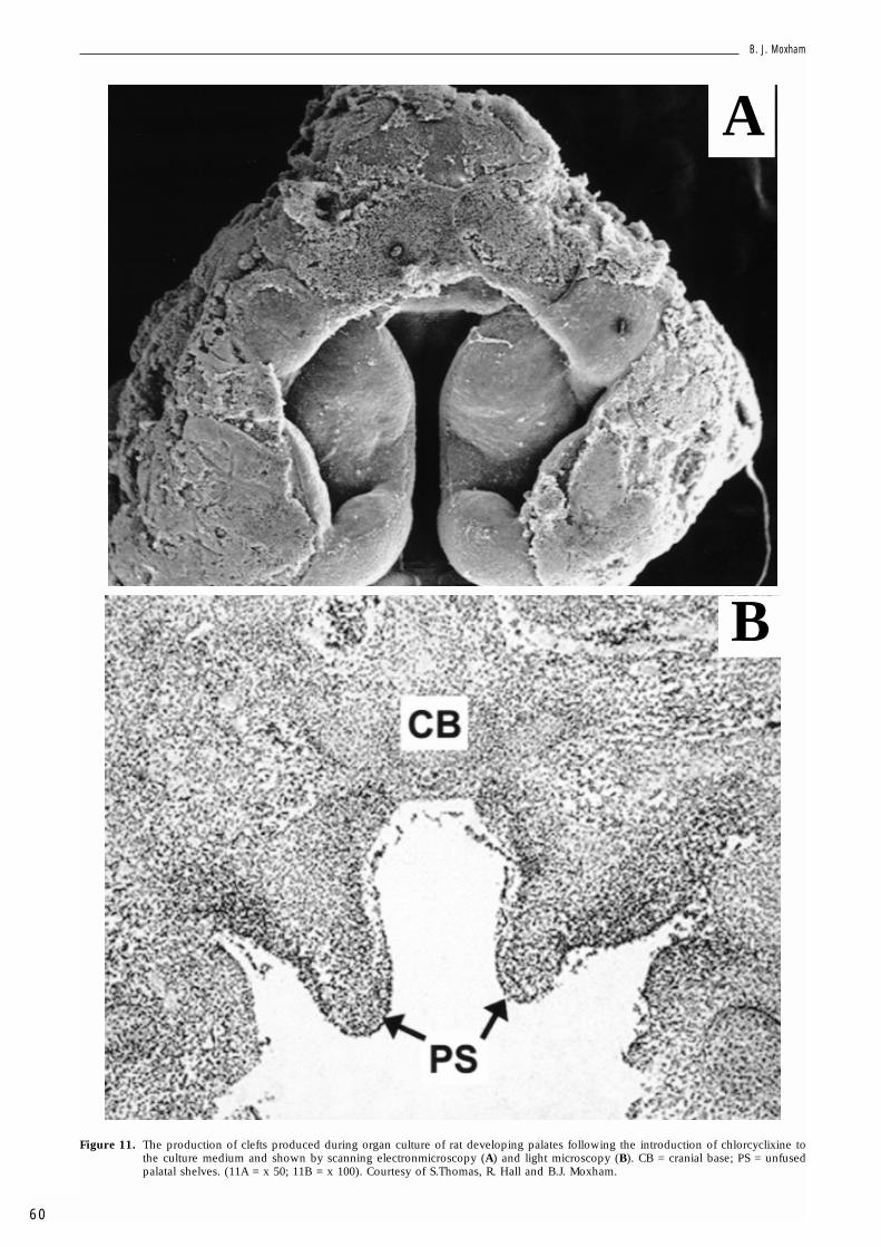
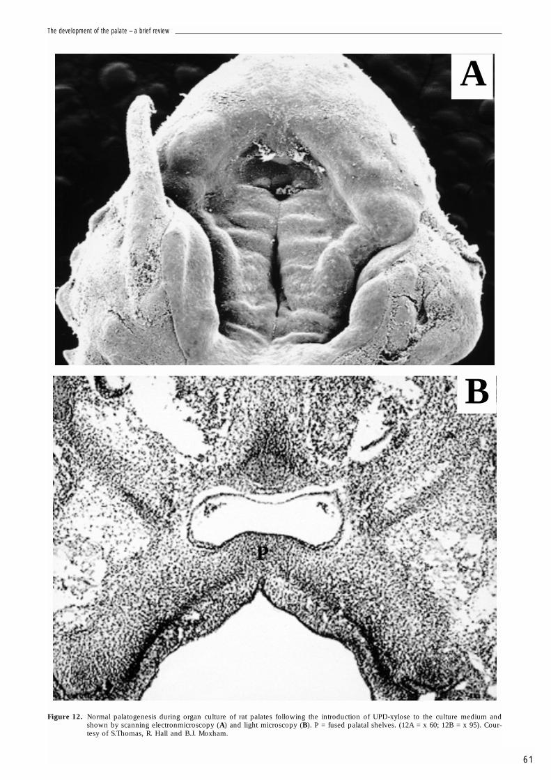
More recent studies (Thomas, 1999; Thomas,Hall and Moxham, unpublished data) haverevealed the presence during palatogenesis ofenzymes associated with hyaluronan synthesis, ofa cell surface receptor associated with hyaluro-nan, of the hyaluronan binding ECM componentsversican and hyaluronectin, and of hyaluronanbinding sites. Furthermore, using an organ culturesystem (Figs. 10-13), agents that alter hyaluronancontent or size, that disrupt GAG substitution onproteoglycans, or that alter the balance of matrixmolecules secreted via the Golgi complex andhyaluronan produced at the cell surface all affectpalatogenesis. Figure 10 illustrates the effects ofstreptomyces hyaluronidase, an enzyme thatspecifically degrades hyaluronan. Streptomyceshyaluronidase treated shelves produced clefts. Inaddition, link protein is absent whilst versicanwas evident in the mesenchyme and CD44 in theectoderm. The results suggest that palate devel-opment is disrupted in the absence of hyaluro-nan. Figure 11 shows the effects of chlorcyclixine,
a substance that enhances degradation of hyaluro-nan and chondroitin sulphate to lower the mole-cular weight products, with little effect on theirsynthesis and no appreciable effect on DNA syn-thesis. Chlorcyclixine treated shelves exhibit acleft. CD44 and link protein are absent from theshelves but versican is evident throughout themesenchyme. Therefore, the size of the GAGchain may influence palatogenesis. The effects ofUDP-xylose are illustrated in Fig. 12. UDP-xyloseis a natural inhibitor of UDPGD, the enzymeresponsible for the conversion of UDP-glucose toUDP-glucuronic acid. UDP-xylose treated shelvesexhibited normal palate development. Link pro-tein was again absent, but versican and CD44exhibited the same distribution as seen in controlcultures. Therefore, inhibition of UDPGD has noeffect on palate development (assuming that itwas able to enter the tissue). Figure 13 shows theeffects of Brefeldin A. This inhibits vesicular trans-port through the Golgi complex. Hyaluronan syn-thesis is not affected as this GAG undergoes a dif-ferent synthetic pathway to the other GAGs, beingformed at, or near, the plasma membrane by thehyaluronan synthase/enzyme complex. BrefeldinA produced a cleft. While versican was evidentthroughout the mesenchyme, link protein andCD44 were absent. Exposure to BFA at sequential10h window periods suggests that BFA only caus-es a cleft in the initial 30h. The results indicates
B. J. Moxham
58
Figure 9. Graphs showing the changing concentrations of hyaluronan within the anterior and posterior regions of palatal shelves. A-D stagesof palatogenesis described in Figure 6. Courtesy of Dr G.D. Singh and B.J. Moxham and the editor of Archives of Oral Biology.
The development of the palate – a brief review
59
Figure 10. The production of clefts produced during organ culture of rat developing palates following the introduction of streptomyceshyaluronidase to the culture medium and shown by scanning electronmicroscopy (A) and light microscopy (B). NS = nasal sep-tum; PS = unfused palatal shelves. Scale bars: 10A = 500 µm; 10B = 400 µm. Courtesy of S.Thomas, R. Hall and B.J. Moxham.
A
B
PS
B. J. Moxham
60
Figure 11. The production of clefts produced during organ culture of rat developing palates following the introduction of chlorcyclixine tothe culture medium and shown by scanning electronmicroscopy (A) and light microscopy (B). CB = cranial base; PS = unfusedpalatal shelves. (11A = x 50; 11B = x 100). Courtesy of S.Thomas, R. Hall and B.J. Moxham.
A
B
The development of the palate – a brief review
61
Figure 12. Normal palatogenesis during organ culture of rat palates following the introduction of UPD-xylose to the culture medium andshown by scanning electronmicroscopy (A) and light microscopy (B). P = fused palatal shelves. (12A = x 60; 12B = x 95). Cour-tesy of S.Thomas, R. Hall and B.J. Moxham.
A
B
P

B. J. Moxham
62
Figure 13. The production of clefts produced during organ culture of rat developing palates following the introduction of brefeldin A to theculture medium and shown by scanning electronmicroscopy (A) and light microscopy (B). CB = cranial base; PS = unfused palatalshelves. (13A = x 50; 13B = x 100). Courtesy of S.Thomas, R. Hall and B.J. Moxham.
A
B
PS

that a set of macromolecules other than hyaluro-nan, and synthesised in the Golgi complex, playsan important role in normal palate development.
Other recent studies at our laboratories atCardiff (Hudson and Hall, unpublished data)have been concerned with the expression ofhyaluronan binding protein splice variants ofCD44, versican and RHAMM and isoforms ofhyaluronan synthases (Has) and hyaluronidases(Hyal) in the developing rat palate. Expressionof CD44 (the major hyaluronan binding protein)was found to be both transient and dynamic dur-ing shelf elevation with differential expression ofCD44 transcripts containing variant exons v1, v2,v8 and v9. It was also noted that larger tran-scripts (containing more variant exons) werepresent after shelf elevation. It can be arguedthat expression of distinct Has and Hyal splicevariants is necessary during palatogenesis inorder for correct tissue formation to occurbecause both enzymes produce different sizes ofhyaluronan, thus promoting distinct cellularresponses depending on cell type (Itano et al.,1999). Small hyaluronan chains can induce geneexpression (McKee et al., 1996), cell signallingresponses and cell differentiation (Termeer et al.,2000), and cell proliferation and growth (Moha-patra et al., 1996, Bourguignon et al., 1997),whereas large hyaluronan chains at high con-centrations inhibit cell growth and induce celladhesion and migration (Noble et al., 1998). Ver-sican splice variants differ in size and GAG chainnumber and are thought to form bridges, help-
ing to stabilize the ECM and create the necessaryturgor pressure to enable shelf elevation.
Other ECM components, including proteogly-cans, are probably of importance to shelf eleva-tion. Versican and decorin (but not biglycan)have been identified at a range of molecularweights corresponding to various processedforms. The extent to which aggregation and dis-aggregation of proteoglycans occurs at differentlocations of the palatal shelf and at differentstages of palatogenesis is unknown; althoughthis could have significant functional implica-tions associated with shelf elevation. The role ofcollagen within the palatal shelves is disputed.Pratt and King (1972) showed that cleft palatescan result from the administration of lathyrogensthat have specific effects on collagen crosslinkformation. Hassell and Orkin (1976) describedcollagen bundles with defined orientation nextto the basement membrane of the palatal shelvesand reported that the rate of collagen synthesiswas greatest just prior to shelf elevation. Indeed,it has been suggested that these collagen fibres“direct” the shelf elevation force (e.g. Bulleit andZimmerman, 1985) and/or contribute to a criticalvolume of the shelves necessary for their re-ori-entation (Ben-Khaial and Sha, 1994). Immuno-histochemically, type 1 collagen can easily beidentified (Fig. 14) (Ferguson, 1988). Indeed,stout bundles of collagen can be seen runningdown the centre of the palatal shelf and theseare orientated from the base towards the tip ofthe shelf.
The development of the palate – a brief review
63
Figure 14. Section of a palatal shelf labelled immunocytochemically with antibodies against type I collagen. A = collagen bundles; B = baseof palatal shelf; C = tip of palatal shelf. x 300. Courtesy of Professor M.W.J. Ferguson.
C
AB

The role of the mesenchymal cells within thepalatal shelves has also been controversial.There is evidence that a critical number of cellsare required for palatal shelf elevation to occur(e.g. Shah et al., 1989) but there is no reliableevidence as yet that these cells, by their rapiddivision and proliferation or by their migrationor contraction, can generate a palatal shelf ele-vation force (particularly in view of the rapidityof shelf elevation). The density of palatal shelfmesenchymal cells appears to change duringpalatogenesis (e.g. Brinkley and Bookstein,1986). This could be the result of variations incell number and/or of cell redistribution. It wasonce believed that differential rates of cellmitoses/proliferation might produce the shelfelevation force (Luke, 1984; Bulliet and Zim-mermann, 1985). 3H-thymidine studies haveshown that there are differential rates of mes-enchymal cell proliferation (e.g. Cleaton-Jones,1976b). However, the differential rates are prob-ably related to histogenic changes and do notnecessarily account for the generation of theshelf elevation force. Brinkley and Bookstein(1986) showed that shelf re-orientation isaccompanied by changes in mesenchymal celldensity and distribution. They suggested thathigh local cell densities were enhanced by celldivision but that decreased cell density (whichcannot be accounted for by an increase in cellsize) was probably related to displacement ofcells by expansion of the ECM. Ferguson(1978a) also noted the closely packed nature ofmesenchymal cells before elevation and com-mented upon the greater cell density within theposterior region of the developing palate (aregion which is the last to fuse).
In addition to mesenchymal cell proliferation,the production of a shelf elevation force mightalso be related to changes, at the critical time, incellular morphology (e.g. Brinkley and Book-stein, 1986) and in particular to changes in theintracellular microfilamentous and microfibrillarsystems (e.g. Kuhn et al., 1980). Babiarz et al.(1979) reported that palatal shelf mesenchymalcells before elevation were elongated and polar-ized, the cells nearest the basement membranebeing perpendicularly aligned to the membrane.After shelf elevation, the cells became morerounded with short cellular projections. Babiarzet al. (1979) considered that these changes wereindicative of cell contraction and that this couldbe the means of generating the shelf elevationforce. Innes (1978) and Shah (1979, 1980)reported that shelf mesenchymal cells possesscontractile, microfilaments. In addition, contrac-tile proteins have been isolated from palatalshelf mesenchymal cells, leading to the claimthat “actin- and myosin-like systems” may beinvolved in shelf elevation (Babiarz, Allenspachand Zimmerman, 1975). Indeed, Babiarz et al.
(1979) reported on the presence of microfila-ments containing actinomyosin and suggestedthat these were associated with cell migrationthat could be responsible for shelf elevation.Wee and Zimmermann (1980) reported thatcytochalasin B inhibits palate shelf elevation bydisrupting actin crosslinking in the cytoskeleton.However, they also found that curare (a micro-filament antagonist) enhanced shelf elevation invitro, thus providing evidence against the notionthat microfilamentous systems are primarilyresponsible for shelf re-orientation. Further-more, it is not clear whether changes in thepalatal shelf mesenchymal cells are primarilyassociated with the re-orientation mechanism orwhether they are the effect of cell displace-ments/cell activities caused by changes in theECM during the period of shelf re-orientation(e.g. Pratt et al., 1973).
There have been many qualitative electron-microscopic investigations of the palatal shelfmesenchymal cells (e.g. De Angelis and Nalban-dian, 1968; Babiarz et al., 1975; Innes, 1978,1981, 1985; Ferguson, 1981a). Essentially, thesestudies show that the mesenchymal cells appearto be very active, possessing many mitochon-dria, abundant cisternae of endoplasmic reticu-lum, a well-developed Golgi complex, and largenumbers of glycogen particles (organelles appro-priate for cells actively synthesising and secret-ing ECM proteins and entirely consistent with theview that the gradual accumulation of GAG iscorrelated with the synthesizing organelles of themesenchymal cells). Shah (1979) described ultra-structural changes occurring during normalpalatogenesis, noting that the cells elongatedafter shelf elevation. Lieb and De Paola (1981)found that the mesenchyme was tightly packedwith polygonal cells possessing centrally placedovoid nuclei with prominent nucleoli. They alsoreported that there was a large complement offree ribosomes and polysomes and very littleintercellular space. Recently, it has been report-ed that filopodia-like structures appear on thesurface of palatal shelf cells at the time of fusion(Taya et al., 1999). Similar events occur duringdevelopment of the intermaxillary segmentwhen the facial processes fuse (Symons andMoxham, 2002). Despite these many studies, todate there have been remarkably few quantita-tive electronmicroscopic studies. Brinkley andBookstein (1986) have undertaken some quanti-tative studies on the development of the mousesecondary palate. The purpose of their investi-gation was to determine differences in cell den-sity at various stages of palatogenesis in vitroand consequently, with the exception of thenuclei, they did not measure the organelles with-in the mesenchymal cells.
It is obvious that, whether or not the palatalshelf mesenchymal cells are involved in the
B. J. Moxham
64

generation of the shelf elevation force, the cellshave to maintain (and control) events takingplace in the shelf ECM. Indeed, using specialsilver staining techniques to highlight nucleolarorganiser regions (NORs), the degree of proteinsynthesising activity of the mesenchymal cellsin the palatal shelf at different stages of palato-genesis has been assessed (Singh and Moxham,1993) (Fig. 15). The number and configurationof “grains” within the NORs reflect the overalldegree of protein synthesis by the cells. Thisstaining procedure confirmed that the rate ofprotein synthesis during palatogenesis is high,is higher before elevation than after elevation,and is higher still during later stages of histoge-nesis. These results accord with the changesoccurring in GAG synthesis at various stages ofpalatogenesis. The AG-NOR staining techniquefurther shows that protein synthesis is severelydepressed during cleft formation, but the tech-nique is unable to demonstrate major differ-ences between anterior and posterior regions.
Although hyaluronan in the palatal shelvesis most often associated with the developmentof a turgor pressure for shelf elevation viaattraction of water molecules, this GAG alsoinfluences cellular activity. For example,hyaluronan produces large intercellular spacesduring early palatogenesis to prevent cell-celland cell-matrix interactions, allowing assemblyof ECM constituents and presentation of growth
factors that in turn influence cell growth anddifferentiation by altering the local concentra-tion of intercellular signals (Toole, 2000). Fol-lowing shelf elevation, there is a decline inhyaluronan shelf content (Singh et al., 1994),probably via CD44 receptor-mediated endocy-tosis of hyaluronan and hyaluronidases thatproduce shorter hyaluronan chains. Thisenables the onset of palatal tissue differentia-tion. Hyaluronan that is taken up into cells canbind to intracellular hyaluronan binding pro-teins, including some RHAMM splice variants.Such binding induces cell signalling pathwaysthat can, in turn, induce changes in thecytoskeleton. During differentiation, the inter-cellular matrix becomes more dense wherehyaluronan is replaced by proteoglycans, butthe remaining hyaluronan binds to such pro-teoglycans (including hyaluronan binding pro-teins such as versican, cell surface RHAMM andCD44) to form a stable ECM.
Finally, although the production of cleftpalates following the administration of FUDR isthought to be related to interference in ECM gly-coconjugate production (e.g. Dorfman et al.,1975; Ferguson, 1978b; Singh et al., 1997), alter-native explanations are possible in terms of cellactivity within the palatal shelves. Indeed,Amwayi and Luke (1990) reported that FUDRproduces a decrease in mesenchymal cell prolif-eration.
The development of the palate – a brief review
65
Figure 15. Silver staining of the palatal shelves (A) to assess the degree of activity of the mesenchymal cells. The black silver grains in the mes-enchymal cell nuclei highlight Nucleolar Organiser Regions (NORs). Silver stain. x 500. Courtesy of Dr. G.D. Singh and B.J. Moxham.
A
FUSION OF THE PALATAL SHELVES
Once the palatal shelves have elevated, theycontact each other (initially in the middle third ofthe palate; Ferguson 1988) and adhere by meansof an “adhesive” glycoprotein that coats the sur-face of the medial edge epithelia of the shelves(Greene and Kochhar, 1974; Pratt and Hassell,1975; Souchon, 1975; Greene and Pratt, 1977).Additionally, the epithelial cells develop desmo-somes (De Angelis and Nalbandian, 1968; Mor-gan and Pratt, 1977) and consequently an epithe-lial seam is formed (Morgan and Pratt, 1977;Ferguson, 1988) (see Fig. 5). The adherence ofthe medial edge epithelia is specific as palatalepithelia will not fuse with epithelia from othersites (e.g. the tongue) (Ferguson et al., 1984).This may be related to the fact that the proteinsassociated with the formation of desmosomes(i.e. desmoplakin) appear specifically on the cellmembranes of the medial edge epithelia justprior to shelf contact (Ferguson, 1988). An intactbasal lamina lies on either side of the epithelialseam.
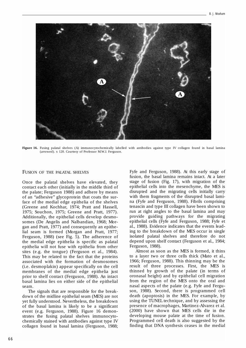
The signals that are responsible for the break-down of the midline epithelial seam (MES) are notyet fully understood. Nevertheless, the breakdownof the basal lamina is likely to be a significantevent (e.g. Ferguson, 1988). Figure 16 demon-strates the fusing palatal shelves immunocyto-chemically stained with antibodies against type IVcollagen found in basal lamina (Ferguson, 1988;
Fyfe and Ferguson, 1988). At this early stage offusion, the basal lamina remains intact. At a laterstage of fusion (Fig. 17), with migration of theepithelial cells into the mesenchyme, the MES isdisrupted and the migrating cells initially carrywith them fragments of the disrupted basal lami-na (Fyfe and Ferguson, 1988). Fibrils comprisingtenascin and type III collagen have been shown torun at right angles to the basal lamina and mayprovide guiding pathways for the migratingepithelial cells (Fyfe and Ferguson, 1988; Fyfe etal., 1988). Evidence indicates that the events lead-ing to the breakdown of the MES occur in singleisolated palatal shelves and therefore do notdepend upon shelf contact (Ferguson et al., 1984;Ferguson, 1988).
Almost as soon as the MES is formed, it thinsto a layer two or three cells thick (Mato et al.,1966; Ferguson, 1988). This thinning may be theresult of three processes. First, the MES isthinned by growth of the palate (in terms oforonasal height) and by epithelial cell migrationfrom the region of the MES onto the oral andnasal aspects of the palate (e.g. Fyfe and Fergu-son, 1988). Second, there is programmed celldeath (apoptosis) in the MES. For example, byusing the TUNEL technique, and by assessing thepresence of macrophages, Martinez-Alvarez et al.(2000) have shown that MES cells die in thedeveloping mouse palate at the time of fusion.Programmed cell death is also suggested by thefinding that DNA synthesis ceases in the medial
B. J. Moxham
66
Figure 16. Fusing palatal shelves (A) immunocytochemically labelled with antibodies against type IV collagen found in basal lamina(arrowed). x 120. Courtesy of Professor M.W.J. Ferguson.
A
A
edge epithelial cells one day prior to shelf con-tact (Hudson and Shapiro, 1973). Furthermore,cyclic AMP increases just before shelf fusion (e.g.Ferguson, 1987) and exogenous cyclic AMP isassociated with precocious cell death in themedial edge epithelia (Pratt and Martin, 1975). Ithas also been shown that epidermal growth fac-tor (EGF) inhibits medial edge cell death (Has-sell, 1975; Pratt et al., 1984; Pratt, 1984) and thatthis inhibition is blocked by exogenous cyclicAMP (Hassell and Pratt, 1977). Care must betaken, however, when interpreting the effects ofcyclic AMP since physiologically it is an intracel-lular messenger and may therefore be mediatingdifferential gene expression triggered by otherevents occurring at the cell surface. Martinez-Alvarez et al. (2000) also suggested that TGF-β3is an inducer of apoptosis during palatal fusion.Third, there is good evidence that some of theepithelial cells migrate from the MES into thepalatal shelf mesenchyme and differentiate intocells indistinguishable from the mesenchymalcells (e.g. Ferguson, 1988). Indeed, it is wellknown that epithelial cells can migrate and dif-ferentiate into mesenchymal-like cells in other
circumstances during development. Althoughlabelling of MES cells with vital lipophilic mark-ers has not clarified whether such cells migrateand/or transform into mesenchyme, in vitrostudies that involve infecting the cells with thereplication-defective helper-free retroviral vectorCXL carrying the Escherichia coli lacZ gene (thusenabling analysis of β-galactosidase activity inthe cells and the determination of cell fate) indi-cate that the cells of the MES transform into mes-enchyme during palatal fusion (Martinez-Alvarezet al., 2000).
There have been many experiments to helpclarify the nature of the epithelial-mesenchymalinteractions during fusion of the palatal shelves.In the main, these experiments have involvedthe separation and then the recombination inculture of the epithelial and mesenchymal com-ponents of the shelves. Overall, these experi-ments have shown that, as with epithelial-mes-enchymal interactions for tooth development, itis the mesenchyme that signals epithelial differ-entiation and behaviour (e.g. Ferguson andHonig, 1984). The nature of this signal is con-troversial. Figure 18 shows the medial edge
The development of the palate – a brief review
67
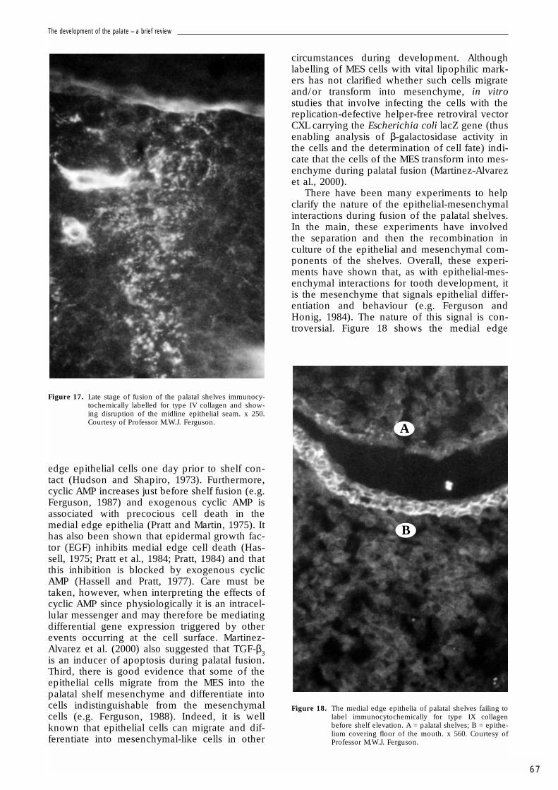
Figure 17. Late stage of fusion of the palatal shelves immunocy-tochemically labelled for type IV collagen and show-ing disruption of the midline epithelial seam. x 250.Courtesy of Professor M.W.J. Ferguson.
Figure 18. The medial edge epithelia of palatal shelves failing tolabel immunocytochemically for type IX collagenbefore shelf elevation. A = palatal shelves; B = epithe-lium covering floor of the mouth. x 560. Courtesy ofProfessor M.W.J. Ferguson.
A
B
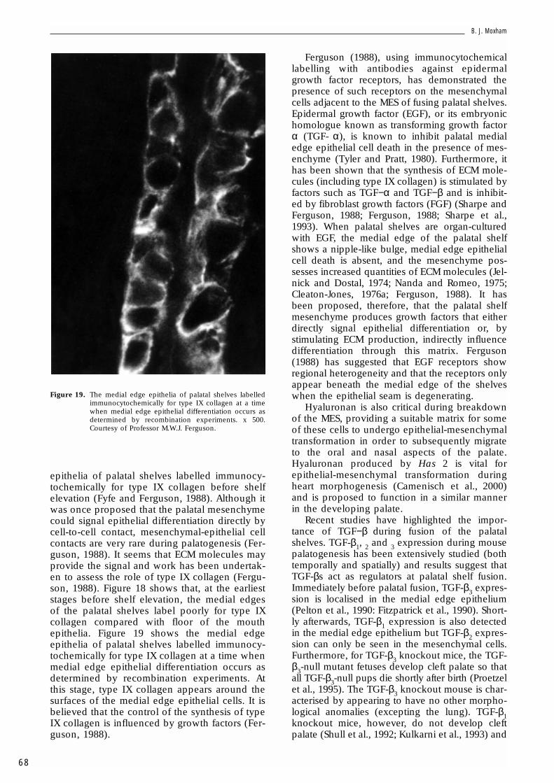
epithelia of palatal shelves labelled immunocy-tochemically for type IX collagen before shelfelevation (Fyfe and Ferguson, 1988). Although itwas once proposed that the palatal mesenchymecould signal epithelial differentiation directly bycell-to-cell contact, mesenchymal-epithelial cellcontacts are very rare during palatogenesis (Fer-guson, 1988). It seems that ECM molecules mayprovide the signal and work has been undertak-en to assess the role of type IX collagen (Fergu-son, 1988). Figure 18 shows that, at the earlieststages before shelf elevation, the medial edgesof the palatal shelves label poorly for type IXcollagen compared with floor of the mouthepithelia. Figure 19 shows the medial edgeepithelia of palatal shelves labelled immunocy-tochemically for type IX collagen at a time whenmedial edge epithelial differentiation occurs asdetermined by recombination experiments. Atthis stage, type IX collagen appears around thesurfaces of the medial edge epithelial cells. It isbelieved that the control of the synthesis of typeIX collagen is influenced by growth factors (Fer-guson, 1988).
Ferguson (1988), using immunocytochemicallabelling with antibodies against epidermalgrowth factor receptors, has demonstrated thepresence of such receptors on the mesenchymalcells adjacent to the MES of fusing palatal shelves.Epidermal growth factor (EGF), or its embryonichomologue known as transforming growth factorα (TGF- α), is known to inhibit palatal medialedge epithelial cell death in the presence of mes-enchyme (Tyler and Pratt, 1980). Furthermore, ithas been shown that the synthesis of ECM mole-cules (including type IX collagen) is stimulated byfactors such as TGF−α and TGF−β and is inhibit-ed by fibroblast growth factors (FGF) (Sharpe andFerguson, 1988; Ferguson, 1988; Sharpe et al.,1993). When palatal shelves are organ-culturedwith EGF, the medial edge of the palatal shelfshows a nipple-like bulge, medial edge epithelialcell death is absent, and the mesenchyme pos-sesses increased quantities of ECM molecules (Jel-nick and Dostal, 1974; Nanda and Romeo, 1975;Cleaton-Jones, 1976a; Ferguson, 1988). It hasbeen proposed, therefore, that the palatal shelfmesenchyme produces growth factors that eitherdirectly signal epithelial differentiation or, bystimulating ECM production, indirectly influencedifferentiation through this matrix. Ferguson(1988) has suggested that EGF receptors showregional heterogeneity and that the receptors onlyappear beneath the medial edge of the shelveswhen the epithelial seam is degenerating.
Hyaluronan is also critical during breakdownof the MES, providing a suitable matrix for someof these cells to undergo epithelial-mesenchymaltransformation in order to subsequently migrateto the oral and nasal aspects of the palate.Hyaluronan produced by Has 2 is vital forepithelial-mesenchymal transformation duringheart morphogenesis (Camenisch et al., 2000)and is proposed to function in a similar mannerin the developing palate.
Recent studies have highlighted the impor-tance of TGF−β during fusion of the palatalshelves. TGF-β1, 2 and 3 expression during mousepalatogenesis has been extensively studied (bothtemporally and spatially) and results suggest thatTGF-βs act as regulators at palatal shelf fusion.Immediately before palatal fusion, TGF-β3 expres-sion is localised in the medial edge epithelium(Pelton et al., 1990: Fitzpatrick et al., 1990). Short-ly afterwards, TGF-β1 expression is also detectedin the medial edge epithelium but TGF-β2 expres-sion can only be seen in the mesenchymal cells.Furthermore, for TGF-β3 knockout mice, the TGF-β3-null mutant fetuses develop cleft palate so thatall TGF-β3-null pups die shortly after birth (Proetzelet al., 1995). The TGF-β3 knockout mouse is char-acterised by appearing to have no other morpho-logical anomalies (excepting the lung). TGF-β1knockout mice, however, do not develop cleftpalate (Shull et al., 1992; Kulkarni et al., 1993) and
B. J. Moxham
68
Figure 19. The medial edge epithelia of palatal shelves labelledimmunocytochemically for type IX collagen at a timewhen medial edge epithelial differentiation occurs asdetermined by recombination experiments. x 500.Courtesy of Professor M.W.J. Ferguson.
cleft palate, along with many other types ofabnormalities, is observed (but at lower incidencerates) in TGF-β2 knockout mice (Sanford et al.,1997). That TGF-β3 plays an important role inpalatal shelf fusion is also shown by the fact thatpalate fusion fails to occur in vitro when the activ-ity of TGF-β3 is inhibited by antisense oligonu-cleotide or by neutralising antibody (Brunet et al.,1995). More recently, Taya et al. (1999) reportedthat mutation of the TGF-β3 gene results in cleftpalate formation and that, when palates fromtransgenic mice with TGF-β3 deletions are grownin organ culture such that shelves were placed inhomologous (+/+ vs +/+, -/- vs -/-, +/- vs +/-) orheterologous (+/+ vs -/-, +/- vs -/-, +/+ vs +/-)paired combinations, pairs of -/- and -/- shelvesfailed to fuse while pairs of +/+ and =/+ shelvesshowed complete disappearance of the MESwhereas -/- and +/+ shelves retained some rem-nants of the MES. They also studied the ability ofTGF-β3 family members to rescue the fusionbetween -/- and -/- palatal shelves in vitro byadding to the culture medium recombinant humanTGF-β1, porcine TGF-β2, recombinant humanTGF-β3, recombinant human activin, or porcineinhibin. It was reported that, for untreated organculture -/- palate pairs that would be expected toshow complete failure to fuse, TGF-β3 treatmentinduced complete palatal fusion whereas TGF-β1or TGF-β2 produced near normal fusion and
activin and inhibin had no effect. The mechanismwhereby TGF-β3 rescued the fusion was claimedto be related to the appearance of filopodia-likeprocess on the surface of the MES cells that arecoated with material resembling proteoglycan.
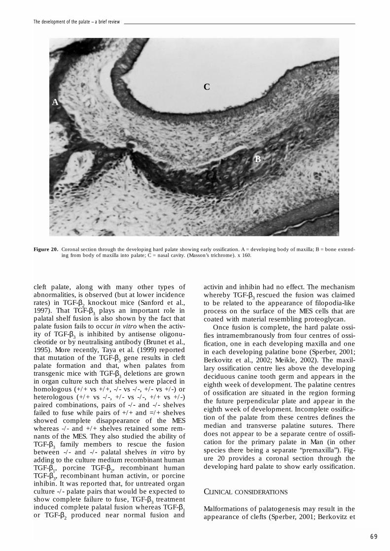
Once fusion is complete, the hard palate ossi-fies intramembranously from four centres of ossi-fication, one in each developing maxilla and onein each developing palatine bone (Sperber, 2001;Berkovitz et al., 2002; Meikle, 2002). The maxil-lary ossification centre lies above the developingdeciduous canine tooth germ and appears in theeighth week of development. The palatine centresof ossification are situated in the region formingthe future perpendicular plate and appear in theeighth week of development. Incomplete ossifica-tion of the palate from these centres defines themedian and transverse palatine sutures. Theredoes not appear to be a separate centre of ossifi-cation for the primary palate in Man (in otherspecies there being a separate “premaxilla”). Fig-ure 20 provides a coronal section through thedeveloping hard palate to show early ossification.
CLINICAL CONSIDERATIONS
Malformations of palatogenesis may result in theappearance of clefts (Sperber, 2001; Berkovitz et
The development of the palate – a brief review
69
Figure 20. Coronal section through the developing hard palate showing early ossification. A = developing body of maxilla; B = bone extend-ing from body of maxilla into palate; C = nasal cavity. (Masson’s trichrome). x 160.
C
A
B
al., 2002; Meikle, 2002). Clefts of the palate, likethose of the lip, are multifactorial malformations,involving both genetic (polygenic) and environ-mental factors. Clefts may result from distur-bances of any of the processes involved duringpalatogenesis, i.e. from defective palatal shelfgrowth (e.g. Abbott et al., 1990); delayed shelfelevation or failure of elevation (e.g. Ferguson,1981a); defective shelf fusion or lack of degen-eration of the MES; or failure of mesenchymalconsolidation and/or differentiation (e.g. Abbottand Birnbaum, 1989).
The mildest form of cleft is that affecting theuvula, such a disturbance occurring relatively latein the process of palatal malfusion. Disturbancesoccurring during the early phases of palatalfusion can result in a more extensive cleft involv-ing most of the secondary palate. Should the cleftinvolve the primary palate, it may extend to theright and/or left of the incisive foramen toinclude the alveolus, passing between the lateralincisor and canine teeth. Cleft palate may beassociated with cleft lip, though the two con-ditions are independently determined. Dental malformations are commonly associated with a cleft involving the alveolus. A submucous cleftdescribes a condition where the palatal mucosa isintact, but the bone/musculature of the palate isdeficient beneath the mucosa. Less problematicthan clefts (but more common) is the retention ofepithelial remnants in the midline that eventuallybecome cystic.
Hypotheses to explain the mechanismsresponsible for cleft palate formation range fromgenetic predisposition (e.g. Bonner and Slavkin,1975) to the administration of teratogens (e.g.Fraser and Fainstat, 1951). Ferguson (1981b) hasalso proposed that the expression of a cleftpalate is a manifestation of phylogeny - birdsdevelop a physiological cleft and the oral andnasal cavities are not separated (e.g. Shah andCrawford, 1980). Recent research indicates thatretinoids in excess have a teratogenic effect, pro-ducing clefts of the palate and the abnormalappearance of “islands” of cartilage in the mes-enchyme (Emmanouil-Nikoloussi et al., 1999;Emmanouil-Nikoloussi et al., 2000). More recent-ly, work by Gunston, Moxham and Emmanouil-Nikoloussi (unpublished data) shows that all-trans retinoic acid (RA) is the most teratogenicisomer of RA in terms of rat palatal abnormalities,that the time of administration of RA is more crit-ical than dose, but that immunohistochemicallabelling for cartilage ECM molecules fails todetect ectopic cartilage within the palates.Ectopic localization of Sonic hedgehog protein(Shh) in the developing rostral neural tube is alsoassociated with craniofacial defects (Nasrallahand Golden, 2001). This is thought to be due todisruption in normal genes expression patterns(e.g. wnt-3a, wnt-4, Pax-6, HNF-3( and Ptc).
Studies using transgenic mice suggest thatmany homeobox genes and transcription factorsare involved in palatogenesis. For example,Satokata and Maas (1994) have highlighted thepossible significance of Msx1 and Winograd etal. (1997) of Msx2. Tissier-Seta et al. (1995) havesuggested a role for Barx 1. Gendron-Maguire etal. (1993) and Rijli et al. (1993) suggest thatHoxa2 is important and Martin et al. (1995) haveimplicated Mhox. Peters et al. (1997) have deter-mined a role for Pax9 and Mo et al. (1997) havereported on the significance of Dli and Dli3.Finally, Takihara et al. (1997) suggest that thereis expression of rae28 during palatogenesis andTakagi et al. (1998) deltaEF1. Additionally, manycytokines (and their receptors) are also involvedin palate development. For example, TGF-alpha/EGF receptors, TGF-β2, and TGF-β3 haveimportant functions (Miettinen et al., 1999; San-ford et al., 1997; Proetzel et al., 1995; Kaartinenet al., 1995; see also above). Furthermore, impor-tance has also been claimed for activin-βA,activin-receptor type II and follistatin (Matzuk etal., 1995a, b, c). Lohnes et al. (1993, 1994) haveshown a role for retinoic acid receptor gammaduring palate development and Kurihara et al.(1994) have reported on endothelin. Orioli et al.(1996) have suggested an involvement ofsek4/nuk1.
ACKNOWLEDGEMENTS
I would like to thank all my colleagues who, atthe Universities of Bristol, Cardiff and Thessa-loniki, have collaborated with me in the study ofpalatogenesis, namely: Dr. G.D. Singh, Dr. W.McLean, Dr. R. Hall, Dr. S. Thomas, Ms L. Hud-son, Ms E. Gunston, Prof. G. Embery, Dr. R.J.Waddington, Dr. M.S. Langley, Dr. E-N.Emmanouil-Nikoloussi. Many thanks for yourhard work and inspiration. I would also like tothank St. George’s University (Grenada), andespecially Prof. R. Jordan, for providing me withfacilities necessary to complete this paper.
REFERENCES
ABBOTT BD and BIRNBAUM LS (1989). TCDD alters medialepithelial cell differentiation during palatogenesis. Toxi-col Appl Pharm, 99: 276-286.
ABBOTT BD, HILL LG and BIRNBAUM LS (1990). Processesinvolved in retinoic acid production of small embryon-ic palatal shelves and limb defects. Teratology, 41: 299-310.
AMWAYI P and LUKE DA (1990). Effects of 5-fluoro-2-deoxyuridine on cell proliferation in the developingmouse palate. Acta Anat, 139: 304-307.
ASLING CW, NELSON MH, DOUGHERTY HL, WRIGHT HV andEVANS HM (1960). The development of cleft palateresulting from maternal pterylglutamic (folic) acid defi-ciency during the latter half of gestation in rats. SurgGynecol Obs, 111: 19-28.
B. J. Moxham
70

BABIARZ BS, ALLENSPACH AL and ZIMMERMAN EF (1975). Ultra-structural evidence of contractile systems in mousepalates prior to rotation. Dev Biol, 47: 32-44.
BABIARZ BS, WEE EL and ZIMMERMAN EF (1979). Palate mor-phogenesis. III. Changes in cell shape and orientationduring shelf elevation. Teratology, 20: 249-278.
BEEN W and SONG SHLK (1978). Harelip and cleft palateconditions in chick embryos following local destructionof the mesencephalic neural crest. A preliminary note.Acta Morphol Neer-scand, 16: 245-255.
BEN-KHAIAL GS and SHAH RM (1994). Effects of 5-fluorouracilon collagen synthesis in the developing palate of ham-ster. Anti-Cancer Drugs, 5: 99-104.
BERKOVITZ BKB, HOLLAND GR and MOXHAM BJ (2002). OralAnatomy, Histology and Embryology. Mosby, Edin-burgh.
BONNER JJ and SLAVKIN HC (1975). Cleft palate susceptibilitylinked to histocompatibility-2 (H-2) in the mouse.Immunogenetics, 2: 213-218.
BOURGUIGNON LYW, ZHU H, CHU A, IIDA N, ZHANG L andHUNG MC (1997). Interaction between the adhesionreceptor, CD44, and the oncogene product, p185HER2,promotes human ovarian tumor cell activation. J BiolChem, 272: 27913-27918.
BRINKLEY LL (1980). In vitro studies of palatal shelf elevation.In: Pratt RM, Christiansen RL (eds). Current ResearchTrends in Prenatal Craniofacial Development. Elsevier,Holland, pp 203-220.
BRINKLEY LL and BOOKSTEIN FL (1986). Cell distribution dur-ing mouse secondary palate closure. II. Mesenchymalcells. J Embryol Exp Morph, 96: 111-130.
BRINKLEY LL and MORRIS-WIMAN J (1984). Role of extracellu-lar matrices in palatal shelf closure. In: Zimmerman EF(ed). Palate Development: Normal and Abnormal Cellu-lar and Molecular Aspects. Academic Press, New York.Current Topics in Developmental Biology, 19: 17-36.
BRINKLEY LL and MORRIS-WIMAN J (1987). Computer-assistedanalysis of hyaluronate distribution during morphogen-esis of the mouse secondary palate. Development, 100:629-636.
BRUNET CL, SHARPE PM and FERGUSON MWJ (1995). Inhibitionof TGF-β3 (but not TGF-β1 or TGF-β2) activity preventsnormal mouse embryonic palate fusion. Int J Dev Biol,39: 345-355.
BULLIET RF and ZIMMERMANN EF (1985). The influence of theepithelium on palate shelf reorientation. J Embryol ExpMorph, 88: 265-279.
BURDETT DN, WATERFIELD JD and SHAH RM (1988). Verticaldevelopment of the secondary palate in hamsterembryos following exposure to 6-mercaptopurine. Ter-atology, 37: 591-597.
BURKITT CL (1990). Insulin-like growth factors affect embry-onic mouse palatal cell proliferation and extracellularmatrix biosynthesis. J Dent Res, 69: 959.
CAMENISCH TD, SPICER AP, BREHM-GIBSON T, BIESTERFELDT J,AUGUSTINE ML, CALABRO A Jr, KUBALAK S, KLEWER SE andMCDONALD JA (2000). Disruption of hyaluronan syn-thase-2 abrogates normal cardiac morphogenesis andhyaluronan-mediated transformation of epithelium tomesenchyme. J Clin Invest,106: 349-360.
CLEATON-JONES P (1976a). A macroscopic and microscopicstudy of the development of the rat palate including thesoft palate. J Dent Res Assoc S Africa, 31: 9-16.
CLEATON-JONES P (1976b). Radioautographic study of mes-enchymal cell activity in the secondary palate of the rat.J Dent Res, 55: 437-440.
COLEMAN RD (1965). Development of the rat palate. AnatRec, 151: 107-118.
COULY GF, COLTEY PM and LE DOUARIN NM (1992). Thedevelopmental fate of the cephalic mesoderm in quail-chick chimeras. Development, 114: 1-15.
DE ANGELIS V and NALBANDIAN J (1968). Ultrastructure ofmouse and rat palatal processes prior to and during sec-ondray palate formation. Archs Oral Biol, 13: 601-608.
DIEWERT VM ( 1974). A cephalometric study of orofacialstructures during secondary palate closure in the rat.Archs Oral Biol, 19: 303-315.
DORFMAN A, LEVITT D, SCHWARTZ NB and HO PL (1975). Stud-ies on cartilage differentiation. In: Slavkin HC, GreulichRC (eds). Extracellular Matrix Influences on GeneExpression. Proceedings of the 2nd Santa Catalina IslandColloquium. Academic Press, New York, pp 19-23.
EMMANOUIL-NIKOLOUSSI E-N, GORET-NICAISE M, FOROGLOU C,DHEM A, DOUROV N, PERSAUD TVN and THLIVERIS JA(1999). Craniofacial abnormalities induced by retinoicacid: a preliminary histological and scanning electronmicroscopic (SEM) study. Exp Toxic Pathol, 52: 445-453.
EMMANOUIL-NIKOLOUSSI E-N, KATSARMA E, GORET-NICAISE M,DHEM A and FOROGLOU C (2000). All trans retinoic acidinterfering with palatal development. Scanning electronmicroscopical and light microscopical observations onembryonic rat palate. Morphologie, 84: 13-21.
FERGUSON MWJ (1978a). Palatal shelf elevation in the Wistarrat fetus. J Anat, 125: 555-577.
FERGUSON MWJ (1978b). The teratogenic effects of 5-fluoro-2-desoxyuridine (F.U.D.R.) on the Wistar rat fetus, withparticular reference to cleft palate. J Anat, 126: 37-49.
FERGUSON MWJ (1981a). The structure and development ofthe palate in Alligator mississippiensis. Archs Oral Biol,26: 427-443.
FERGUSON MWJ (1981b). The value of the American alligator(Alligator mississippiensis) as a model for research incraniofacial development. J Craniofac Genet Devl Biol,1: 123-144.
FERGUSON MWJ (1984). Craniofacial development in Alliga-tor mississippiensis. In: Ferguson MWJ (ed). The Struc-ture, Development and Evolution of Reptiles. AcademicPress, London, pp 223-273.
FERGUSON MWJ (1987). Palate development: mechanismsand malformations. Irish J Med Sci, 156: 309-315.
FERGUSON MWJ (1988). Palate development. Development,103 (Suppl): 41-60.
FERGUSON MWJ and HONIG LS (1984). Epithelial-mesenchy-mal interactions during vertebrate palatogenesis. In:Zimmerman EF (ed). Palate Development: Normal andAbnormal Cellular and Molecular Aspects. AcademicPress, New York. Current Topics in Developmental Biol-ogy, 19: 137-164.
FERGUSON MWJ, HONIG LS and SLAVKIN HC (1984). Differenti-ation of cultured palatal shelves from alligator, chickand mouse embryos. Anat Rec, 209: 231-249.
FITZPATRICK DR, DENHEZ F, KONDAIAH P and AKHURST RJ(1990). Differential expression of TGF-β isoforms inmurine palatogenesis. Development, 109: 585-595.
FOREMAN DM, SHARPE PM and FERGUSON MWJ (1991). Com-parative biochemistry of mouse and chick secondarypalate development in vivo and in vitro with particularemphasis on extracellular matrix molecules and theeffects of growth factors on their synthesis. Archs OralBiol, 36: 457-471.
FRASER FC and FAINSTAT TD (1951). Production of congenitaldefects in the offspring of pregnant mice treated withcortisone. Pediatrics, 8: 527-523.
FYFE DM and FERGUSON MWJ (1988). Immunocytochemicallocalisation of collagen types I-XII, proteoglycans,laminin and fibronectin during mouse secondary palatedevelopment. Quoted by Ferguson MWJ (1988).
FYFE DM, FERGUSON MWJ and CHIQUET-EHRISMANN R (1988).Tenascin immunolocalisation during palate develop-ment in mouse and chicken embryos. Quoted by Fer-guson MWJ (1988).
GENDRON-MAGUIRE M, MALLO M, ZHANG M and GRIDLEY T(1993). Hoxa-2 mutant mice exhibit homeotic transfor-mation of skeletal elements derived from cranial neuralcrest. Cell, 75: 1317-1331.
GIRARD N, DELPECH A and DELPECH B (1986). Characterizationof hyaluronic acid on tissue sections with hyaluronectin.J Histochem Cytochem, 34: 539-541.
The development of the palate – a brief review
71

GRAMMATOPOULOS GA, BELL E, TOOLE L, LUMSDEN A and TUCK-ER AS (2000). Homeotic transformation of branchial archidentity after Hoxa2 overexpression. Development, 127:5355-5365.
GREENE RM and KOCHHAR DM (1974). Surface coat on theepithelium of developing palatine shelves in the mouseas revealed by electron microscopy. J Embryol Exp Mor-phol, 31: 683-692.
GREENE RM and PRATT RM (1977). Inhibition by diazo-oxo-norlucine (DON) of rat palatal glycoprotein synthesisand epithelial cell adhesion in vitro. Exp Cell Res, 105:27-37.
GUNSTON E, EMMANOUIL-NIKOLOUSSI E-N and MOXHAM BJ(unpublished data).
HASSELL JR (1975). The development of rat palatal shelves invitro. An ultrastructural analysis of the inhibition ofepithelial cell death and palatal fusion by the epidermalgrowth factor. Dev Biol, 45: 90-102.
HASSELL JR and ORKIN RW (1976). Synthesis and distributionof collagen in the rat palate during shelf elevation. DevBiol, 49: 80-88.
HASSELL JR and PRATT RM (1977). Elevated levels oc cAMPalters the effect of epidermal growth factor in vitro onprogrammed cell death in the secondary palatal epithe-lium. Exp Cell Res, 106: 55-62.
HAYWARD JR and AVERY JK (1957). A variation in cleft palate.J Oral Surg, 15: 320-324.
HUDSON CD and SHAPIRO BL (1973). An autoradiographicstudy of deoxyribonucleic acid synthesis in embryonicrat palatal shelf epithelium with reference to the con-cept of programmed cell death. Archs Oral Biol, 18: 77-84.
HUDSON L and HALL R (unpublished data).HUMPHREY T (1969). The relation between fetal mouth open-
ing reflexes and closure of the palate. Am J Anat, 125:317-344.
HUMPHREY T (1971). Development of oral and facial motormechanisms in human foetuses and their relation tocranio-facial growth. J Dent Res, 50: 1428-1441.
INNES PB (1978). The ultrastructure of the mesenchymal ele-ment of the palatal shelves of the fetal mouse. J Embry-ol Exp Morphol, 43: 185-194.
INNES PB (1981). The ultrastructure of murine secondarypalatal ectomesenchyme during shelf reorientation. JCraniofac Genet Dev Biol, 1: 359-371.
INNES PB (1985). The ultrastructural effects of prednisoloneon themesenchyme of the palatal shelf in the mouse. JCraniofac Genet Dev Biol, 5: 287-297.
ITANO N, SAWAI T, YOSHIDA M, LENAS P, YAMADA Y, IMAGAWA M,SHINOMURA T, HAMAGUCHI M, YOSHIDA Y, OHNUKI Y,MIYAUCHI S, SPICER AP, MCDONALD JA and KIMATA K(1999). Three isoforms of mammalian hyaluronan syn-thases have distinct enzymatic properties. J Biol Chem,274: 25085-25092.
JELNICK R and DOSTAL M (1974). Morphogenesis of cleftpalate induced by exogenous factors VII. Mitotic activi-ty during formation of themouse secondary palate. FoliaMorphologica (Praha), 22: 94-101.
JOHNSTON MC and SULIK KK (1990). In: Bhaskar SN (ed).Oral Histology and Embryology. Mosby, London, pp 12-16.
KAARTINEN V, VOLCKEN JW, SHULER C, WARBURTON D, BU D,HEISTERKAMP N and GROFFEN J (1995). Abnormal lungdevelopment and cleft palate in mice lacking TGF-β3indicates defects of epithelial-mesenchymal interaction.Nature Genet, 11: 415-421.
KNUDSEN TB, BULLEIT RF and ZIMMERMAN EF (1985). Histo-chemical localisation of glycosaminoglycans duringmorphogenesis of the secondary palate in mice. ActaEmbryol, 173: 137-142.
KÖNTGES G and LUMSDEN A (1996). Rhombencephalic neuralcrest segmentation is preserved throughout craniofacialontogeny. Development, 122: 3229-3242.
KUHN EM, BABIARZ BS, LESSARD JL and ZIMMERMAN EF (1980).Palate morphogenesis I. Immunological and ultrastruc-tural analyses of mouse palate. Teratology, 21: 209-223.
KULKARNI AB, HUH C, BECKER D, GEISER A, LYGHT M, FLANDERS
KC, ROBERTS AB, SPORN MB, WARD JM and KARLSSON S(1993). Transforming growth factor-β1 null mutation inmice causes excessive inflammatory response and earlydeath. Proc Natl Acad Sci USA, 90: 770-774.
KURIHARA Y, KURIHARA H, SUZUKI H, KODAMA T, MAEMURA K,NAGAI R, ODA H, KUWAKI T, CAO WH, KAMADA N, JISHAGE
K, OUCHI Y, AZUMA S, TOYODA Y, ISHIKAWA T, KUMADA Mand YAZAKI Y (1994). Elevated blood pressure and cran-iofacial abnormalities in mice deficient in endothelin-1.Nature, 368: 703-710.
LARSSON KS (1962). Studies on the closure of the secondarypalate, IV. Autoradiographic and histochemical studiesof mouse embryos from cortisone-treated mothers. ActaMorph Neerl Scand, 4: 369-386.
LARSSON KS, BOSTROM H and CARLSOO S (1959). Studies onthe closure of the secondary palate. Exp Cell Res, 16:379-383.
LE DOUARIN NM and KALCHEIM C (1999). The Neural Crest.2nd edition. Cambridge University Press, Cambridge.
LIEB RJ and DE PAOLA DP (1981). Ultrastructural alteration oftrypsin-and pancraetin-separated embryonic rabbitpalate epithelium and mesenchyme. J Dent Res, 60: 164-170.
LOHNES D, MARK M, MENDELSOHN C, DOLLE P, DIERICH A,GORRY P, GANSMULLER A and CHAMBON P (1994). Functionof the retinoic acid receptors (RARs) during develop-ment. (1) Craniofacial and skeletal abnormalities in RARdouble mutants. Development, 120: 2723-2748.
LUKE DA (1984). Epithelial proliferation and development ofrugae in relation to palatal shelf elevation in the mouse.J Anat, 138: 251-258.
MCKEE CM, PENNO MB, COWMAN M, BURDICK MD, STRIETER RMand BAO C (1996). Hyaluronan (HA) fragments inducechemokine gene expression in alveolar macrophages. JClin Invest, 98: 2403-2413.
MARTIN JF, BRADLEY A and OLSEN EN (1995). The paired-likehomeo box gene Mhox is required for early events ofskeletogenesis in multiple lineages. Genes Dev, 9: 1237-1249.
MARTINEZ-ALVAREZ C, TUDELA C, PEREZ-MIGUELSANZ J, O’KANE S,PUERTA J and FERGUSON MWJ (2000). Medial edge epithe-lial cell fate during palatal fusion. Development, 220:343-357.
MATO M, AIKAWA E and KATAHIRA M (1966). Appearance ofvarious types of lysosomes in the epithelium coveringlateral palatine shelves during secondary palate forma-tion. Igunma J med Sci, 15: 46-56.
MATZUK MM, KUMAR TR and BARDLEY A (1995a). Differentphenotypes for mice deficient in either activins oractivin receptor type II. Nature, 374: 356-360.
MATZUK MM, KUMAR TR, VASSALLI A, BICKENBACH JR, ROOP DR,JAENISCH R and BRADLEY A (1995b). Functional analysis ofactivins during mammalian development. Nature, 374:354-356.
MATZUK MM, LU N, VOGEL H, SELLHEYER K, ROOP DR andBRADLEY A (1995c). Multiple defects and perinatal deathin mice deficient in follistatin. Nature, 374: 360-363.
MEIKLE MC (2002). Craniofacial Development, Growth andEvolution. Bateson Publishing, Bressingham.
MIETTINEN PJ, CHIN Jr, SHUM L, SLAVKIN HC, SHULER CF,DERYNCK R and WERB Z (1999). Epidermal growth factorreceptor function is necessary for normal craniofacialdevelopment and palate closure. Nature Genet, 22: 69-73.
MO R, FREER AM, ZINYK DL, CRACKOWER MA, MICHAUD J, HENG
HH, CHIK KW, SHI XM, TSUI LC, CHENG SH, JOYNER ALand HUI C (1997). Specific and redundant function ofGli2 and Gli3 zinc finger genes in skeletal patterningand development. Development, 124: 113-123.
B. J. Moxham
72

MOHAPATRA S, YANG X, WRIGHT JA, TURLEY EA and GREENBERG
AH (1996). Soluble hyaluronan receptor RHAMMinduces mitotic arrest by suppressing Cdc2 and cyclinB1 expression. J Exp Med, 183: 1663-1668.
MORGAN PR and PRATT RM (1977). Ultrastructure of theexpected fusion zone in rat fetuses with diazo-oxo-nor-leucine (D.O.N.) induced cleft palate. Teratology, 15:281-290.
NANDA R and ROMEO D (1975). Differential cell proliferationof embryonic rat palatal processes as determined byincorporation of tritiated thymidine. Cleft Palate J, 12:436-443.
NASRALLAH I and GOLDEN JA (2001). Brain, Eye, and facedefects as a result of ectopic localization of Sonichedgehog protein in the developing rostral neural tube.Teratology, 64: 107-113.
NOBLE PW, MCKEE CM and HORTON MR (1998). Induction ofinflammatory gene expression by low molecular weighthyaluronan fragments in macrophages. In: Laurent TC(ed). The Chemistry, Biology and Medical Applicationsof Hyaluronan and its Derivatives. Portland Press, Lon-don, pp 219-225.
NODEN DM (1978). The control of avian cephalic neuralcrest cytodifferentiation. I. Skeletal and connective tis-sues. Dev Biol, 67: 296-312.
ORIOLI D, HENKEMEYER M, LEMKE G, KLEIN R and PAWSON T(1996). Sek4 and nuk receptors cooperate in guidanceof commissural axons and in palate formation. EMBO J,15: 6035-6049.
PASQUALETTI M, ORI M, NARDI I and RIJLI FM (2000). EctopicHoxa2 induction after neural crest migration results inhomeosis of jaw elements in Xenopus. Development,127: 5367-5378.
PELTON RW, HOGAN BL, MILLER DA and MOSES HL (1990). Dif-ferential expression of genes encoding TGF-β1, β2 andβ3 during murine palate formation. Dev Biol, 141: 456-460.
PETERS H, NEUBUSER A and BALLING R (1997). The role of Pax9during mouse development. Dev Biol, 186: 333.
PRATT RM (1984). Hormones, growth factors and theirreceptors in normal and abnormal prenatal develop-ment. In: Kalter H (ed). Issues and Reviews in Teratol-ogy, vol. 2. Plenum Press, New York, pp 189-217.
PRATT RM, GOGGINS JF, WILK AL and KING CTG (1973). Acidmucopolysaccharide synthesis in the secondary palateof the developing rat at the time of rotation and fusion.Dev Biol, 32: 230-237.
PRATT RM and HASSELL JR (1975). Appearance and distribu-tion of carbohydrate rich macromolecules on theepithelial surface of the rat palatal shelf. Dev Biol, 45:192-198.
PRATT RM and KING CTG (1972). Inhibition of collagencrosslinking associated with β-aminopropionitrile-induced cleft palate in the rat. Dev Biol, 27: 322-328.
PRATT RM, KIM CS and GROOVE RI (1984). Role of glucocor-ticoids and epidermal growth factor in normal andabnormal palatal development. Curr Top Dev Biol, 19:81-101.
PRATT RM and MARTIN GR (1975). Epithelial cell death andcyclic AMP increase during palatal development. ProcNatl Acad Sci USA, 72: 874-877.
PROCTZEL G, PAWLOWSKI SA, WILTS MV, YIN M, BOIVIN GP,HOWLES PN, DING J, FERGUSON MWJ and DOETSCHMAN T(1995). Transforming growth factor –β3 is required forsecondary palate fusim. Nat Genet, 11: 409-414.
RIJLI FM, MARK M, LAKKARAJU S, DIERICH A, DOLLE P and CHAM-BON P (1993). A homtotic transformation is generated inthe rostal branchial region of the head by disruption ofHoxa-Z, which acts as a selector gene. Cell, 75: 1333-1349.
SADLER TW (2000). Langman’s Medical Embryology, eighthedition. Lippincott Williams and Wilkins, Philadelphia.
SANFORD LP, ORMSBY I, GROOT ACG, SARINOLA H, FRIEDMAN R,BOIVIN GP, CARDELL EL and DOETSCHMAN T (1997). TGF-β2 knockout mice have multiple developmental defectsthat are non-overlapping with other TGG-β knockoutphenotype. Development, 124: 2659-2670.
SATOKATA I and MAAS R (1994). Msx-1 deficient mice exhibitcleft palate and abnormalities of craniofacial and toothdevelopment. Nat Genet, 6: 348-355.
SCHILLING TF, PRINCE V and INGHAM PW (2001). Plasticity inZebrafish hox expression in the hindbrain and cranialneural crest. Dev Biol, 231: 201-216.
SHAH RM (1979). Current concepts on the mechanisms ofnormal and abnormal secondary palate formation. In:Persaud TVN (ed). Advances in the Study of BirthDefects. Vol. 1. Teratogenic mechanisms. M.T.P. PressLtd, Lancaster, pp 69-84.
SHAH RM (1980). Ultrastructural observations on the devel-opment of triamcinolone-induced cleft palate in ham-sters. Invest Cell Pathol, 3: 281-294.
SHAH RM, ARCADI F, SUEN R and BURDETT DN (1989). Effectsof cylcophosphamide on the secondary palate develop-ment in Golden Syrian hamster: teratology, morphologyand morphometry. J Craniofac Genet Dev Biol, 9: 381-396.
SHAH RM, CHEN YP and BURDETT DN (1989). Growth of thesecondary palate in the hamster following hydrocorti-sone treatment: shelf area, cell number and DNA syn-thesis. Teratology, 40: 173-180.
SHAH RM and CRAWFORD BJ (1980). Development of the sec-ondary palate in chick embryo: a light and electronmicroscopic and histochemical study. Invest Cell Path, 3:319-328.
SHARPE PM and FERGUSON MWJ (1988). Mesenchymal influ-ences on epithelial differentiation in developing sys-tems. J Cell Sci, 10: 195-230.
SHARPE PM, BRUNET CL, FOREMAN DM and FERGUSON MWJ(1993). Localisation of acidic and basic fibroblast growthfactors during mouse palate development and theireffects on mouse palate mesencymal cells in vitro.Rouxs Arch Dev Biol, 202: 132-143.
SHULL MM, ORMSBY I, KIER AB, PAWLOWSKI S, DIEBOLD RJ, YIN
M, ALLEN R, SIDMAN C, PROETZEL G, CALVIN D, ANNUNZIA-TA N and DOETSCHMAN T (1992). Targeted disruption ofthe mouse transforming growth factor –β, gene resultsin multifocal inflammatory disease. Nature, 359: 639-699.
SINGH GD (1991). Studies on the development of the palate.Ph.D. Thesis. University of Bristol.
SINGH GD and MOXHAM BJ (1993). Cellular activity in thedeveloping palate of the rat assessed by staining ofnucleolar organiser regions. J Anat, 182: 163-168.
SINGH GD, MOXHAM BJ, LANGLEY MS, WADDINGTON RJ andEMBERY G (1994). Changes in the composition of gly-cosaminoglycans during normal palatogenesis in the rat.Archs Oral Biol, 39: 401-407.
SINGH GD, MOXHAM BJ, LANGLEY MS and EMBERY G (1997).Glycosaminoglycan biosynthesis during 5-fluoro-2-deoxyuridine-induced palatal clefts in the rat. ArchsOral Biol, 42: 355-363.
SOUCHON R (1975). Surface coat of the palatal shelf epitheli-um during palatogenesis in mouse embryos. AnatEmbryol, 147: 133-142.
SPERBER GH (2001). Craniofacial Development. Hamilton:BC Decker Inc.
SYMONS D and MOXHAM BJ (2002). Ultrastructural characteri-zation of the mesenchyme of the facial processes dur-ing development of the rat intermaxillary segment.Conn Tiss Res, 43: 238-244.
TAKAGI T, MORIBE H, KONDOH H and HIGASHI Y (1998). DeltaEF1, a zinc finger and homeodomain transcription fac-tor, is required for skeleton patterning in multiple lin-eagis. Development, 125: 21-31.
The development of the palate – a brief review
73
TAKIHARA Y, TOMOTSUNE D, SHIRAI M, KATOH FY, NISHII K,MOTALEB MA, NOMURA M, TSUCHIYA R, FUJITA Y, SHIBATA Y,HIGASHINAKAGAWA T and SHIMADA K (1997). Targeted dis-ruption of the mouse homolog of the Drosophila poly-homeotic gene leads to altered anteroposterior pattern-ing and neural crest defects. Development, 124:3673-3682.
TAN SS and MORRISS-KAY GM (1986). Analysis of cranial neur-al crest cell migration and early fates in postimplanta-tion rat chimaeras J Embryol Exp Morphol, 98: 21-58.
TAYA Y, O’KANE S and FERGUSON MWJ (1999). Pathogenesisof cleft palate in TGF-β3 knockout mice. Development,126: 3869-3879.
TERMEER CC, HENNIES J, VOITH U, AHRENS T, WEISS J, PREHM Pand SIMON JC (2000). Oligosaccharides of hyaluronanare potent activators of dendritic cells. J Immuno, 165:1863-1870.
THOMAS S (1999). Investigations into aspects of mammalianpalatal development. Ph.D. Thesis, Cardiff University.
THOMAS S, HALL R and MOXHAM BJ (unpublished data).TISSIER-SETA JP, MUCCHIELLI ML, MARK M, MATTEI MG, GORIDIS
C and BRUNT JF (1995). Barx, I., a new mouse home-odomain transcription factor expressed in cranio-facialectomesenchyme and the stomach. Mech Dev, 51: 3-15.
TOOLE BP (2000). Hyaluronan is not just a goo! J Clin Invest,106: 335-336.
TRAINOR P and KRUMLAUF R (2001). Hox genes, neural crestcells and branchial arch patterning. Curr Opin Cell Biol,13: 698-705.
TYLER MS and PRATT RM (1980). Effect of epidermal growthfactor on secondary palatal epithelium in vitro: tissueisolation and recombination studies. J Embryol Exp Mor-phol, 58: 93-106.
WALKER BE (1971). Palate morphogenesis in the rabbit.Archs Oral Biol, 16: 275-286.
WEE EL, BARBIARZ BS, ZIMMERMAN S and ZIMMERMAN EF (1979).Palate morphogenesis. IV. Effects of serotonin and itsantagonists on rotation in embryo culture. J Embryol ExpMorphol, 53: 75-90.
WEE EL and ZIMMERMAN EF (1980). Palate morphogenesis. II.Contraction of cytoplasmic processes in ATP-inducedpalate rotation in glycerinated mouse heads. Teratology,21: 15-27.
WINOGRAD J, REILLY MP, ROE R, LUTZ J, LAUGHNER E, XU X, HU
L, ASAKURA T, VAN DER KOLK C, STANDBERG JD and SEMEN-ZA GL (1997). Perinatal lethality and multiple craniofa-cial malformations in MSX2 transgenic mice. HumanMol Genet, 6: 369-379.
YOUNG AV, FEELEY EJE, BENKHAIAL G and SHAH RM (1990).Does the tongue play a role during initial palate devel-opment? J Dent Res, 69: 159.
ZEILER KB, WEINSTEIN S and GIBSON RD (1964). A study of themorphology and the time of closure of the palate in thealbino rat. Archs Oral Biol, 9: 545-554.
ZIMMERMAN EF (1979). Palate morphogenesis: role of con-tractile proteins and neurotransmitters. In: Persaud TVN(ed). Advances in the Study of Birth Defects. Vol. 3.Abnormal embryogenesis, cellular and molecularaspects. M.T.P. Press Ltd, Lancaster, pp 143-159.
B. J. Moxham
74