Embed Size (px)
Citation preview

THE DETERMINATION OF URIC ACID IN HUMAN URINE
BY A. A. CHRISTMAN AND SARAH RAVWITCH
(From the Laboratory of Physiological Chemistry, Medical School, University of Michigan, Ann Arbor)
(Received for publication, November 14, 1931)
Improvements in the methods for the determination of uric acid, during the last 10 years, have been concerned primarily with the determination of uric acid in blood. The last significant con- tribution dealing with the determination of uric acid in urine was made by Benedict and Franke (1) in 1922, with the introduction of a direct calorimetric method. Their procedure involves the production of a color with urine for calorimetric comparison with- out the preliminary separation of the uric acid, as recommended in the earlier methods.
Benedict and Franke (1) compared the uric acid content of 50 samples of human urine as determined by this direct met.hod and the older indirect method’ of Benedict and Hitchcock (2), which involved a preliminary precipitation of the uric acid as a silver salt. Since the agreement in most cases was within 2 or 3 per cent, there seemed to be no objection to the use of the direct method for the analysis of human urine. Apparently many subse- quent workers, regardless of the nature of the experimental work, have employed the direct method without checking the results by one of the older standard methods.
Christman and Mosier (3) have recently demonstrated that uric acid determinations by the direct method of Benedict and Franke are not accurate if the urines contain appreciable amounts of amino nitrogen (e.g., glycine). The presence of amounts of glycine furnishing 0.046 to 0.373 mg. of amino nitrogen in the
1 Hereafter in this paper, methods for uric acid which involve a prelimi- nary separation of the uric acid from other urinary constituents, prior to the calorimetric determination, will be designated as “indirect” methods.
115
by guest on April 26, 2020
http://ww
w.jbc.org/
Dow
nloaded from
116 Uric Acid Determination
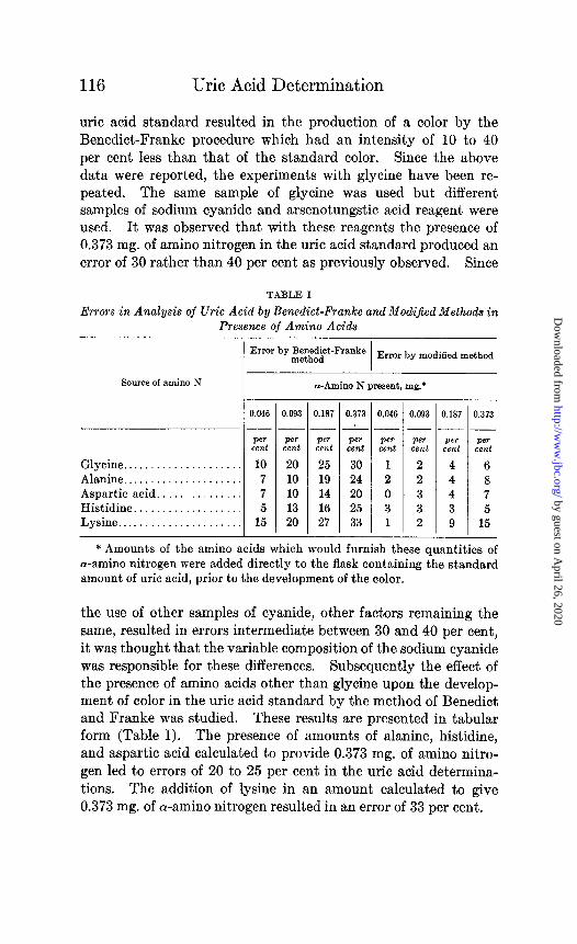
uric acid standard resulted in the production of a color by the Benedict-Franke procedure which had an intensity of 10 to 40 per cent less than that of the standard color. Since the above data were reported, the experiments with glycine have been re- peated. The same sample of glycine was used but different samples of sodium cyanide and arsenotungstic acid reagent were used. It was observed that with these reagents the presence of 0.373 mg. of amino nitrogen in the uric acid standard produced an error of 30 rather than 40 per cent as previously observed. Since
TABLE I
Errors in Analysis of Uric Acid by Benedict-Franke and Modified Methods in Presence of Amino Acids
Error by~~~b$~-Franke Error by modified method
Source of amino N I
a-Amino N present, mg:
0.046 0.093 0.187 0.373 0.046
--...----.----A-
Per Per Pm Per Per cent cent cent cent cent Glycine.. . . . 10 20 25 30 1 Alanine.. . . . . . . . 7 10 19 24 2 Aspartic acid.. 7 10 14 20 0 Histidine.. . 5 13 16 25 3 Lysine.. . . . . . 15 20 27 33 1
0.093 0.187 0.373 ---
Per Per Per cent cent cent 2 4 6 2 4 8 3 4 7 3 3 5 2 9 15
* Amounts of the amino acids which would furnish these quantities of a-amino nitrogen were added directly to the flask containing the standard amount of uric acid, prior to the development of the color.
the use of other samples of cyanide, other factors remaining the same, resulted in errors intermediate between 30 and 40 per cent, it was thought that the variable composition of the sodium cyanide was responsible for these differences. Subsequently the effect of the presence of amino acids other than glycine upon the develop- ment of color in the uric acid standard by the method of Benedict and Franke was studied. These results are presented in tabular form (Table I). The presence of amounts of alanine, histidine, and aspartic acid calculated to provide 0.373 mg. of amino nitro- gen led to errors of 20 to 25 per cent in the uric acid determina- tions. The addition of lysine in an amount calculated to give 0.373 mg. of a-amino nitrogen resulted in an error of 33 per cent.
by guest on April 26, 2020
http://ww
w.jbc.org/
Dow
nloaded from

A. A. Christman and S. Ravwitch 117
It has been noted in this laboratory during the past 4 years that occasionally the analysis of normal human urine by the direct Benedict and Franke procedure yielded results that were definitely lower than those obtained by methods involving a preliminary precipitation of the uric acid, such as the methods of Benedict and Hitchcock (2), Morris and Macleod (4), and Folin and Denis (5). The greatest discrepancy between the results obtained by the direct procedure on the one hand and the indirect procedure on the other hand was observed in the more concentrated urines. If the uric acid content of the volume of urine employed in the analysis by the Benedict and Franke method was such that the calorimetric reading of the unknown was between 13 and 17 mm., with the standard set at 20 mm., results by the direct method were invariably lower than those obtained by the indirect procedures. If the determination by the direct method was then repeated with a smaller volume of urine, so that the calorimetric reading was between 20 and 25 mm., the results more nearly approximated those of the indirect methods.
The results of a typical analysis of a normal urine by the Bene- diet-Franke method are given in Table II (Urine 1). It is to be noted that, when the analysis was made by the method of Bene- dict and Franke, the apparent amount of uric acid per liter of urine varied from 300 to 446 mg. It is to be further noted that, as the volume of urine used in the analysis was decreased, the apparent uric acid content of the urine was increased. The calorimetric readings, ranging from 13.3 to 25.6 mm., are not beyond the limits prescribed by Benedict and Franke for accurate results by this method. Results similar to these have been ob- tained with numerous urines. The analyses of two additional urines are presented in Table II (Urines 2 and 3).
Analyses of several normal human urines by the calorimetric method of Folin (6) showed the content of amino nitrogen to be low, usually less than 0.15 mg. per cc. of urine. Since it has been demonstrated [Table I) that the presence of amino nitrogen even in low concentrations caused considerable error in the analysis for uric acid by the Benedict-Franke method, a modification of the cyanide solution was made to minimize the effect of the pres- ence of amino nitrogen upon the uric acid color reaction. This was accomplished by the inclusion of sodium carbonate in the
by guest on April 26, 2020
http://ww
w.jbc.org/
Dow
nloaded from

118 Uric Acid Determination
TABLE II
Anal& of Normal Human Urine for Uric Acid
Urine No.
kgedk~t- cock
method
Uric acid Volume Colori-
Uric VOlUrn~ Colori-
)f urine metric Uric
reading acid If urine metric readinp acid
T
cc. mm. ml. cc. mm. Tl. per 1. per 1. zg. per 1 og. per Lg. per 1.
1.00 13.3 300 1.00 13.0 461 435 424 404 0.80 14.5 345 0.80 16.5 456 0.50 19.7 406 0.50 26.4 454 0.40 23.6 422 0.35 37.8 451 0.35 25.6 446 l.OOi 14.2 423 0.50t 19.7 406
0.80 17.9 279 2.00 8.0 375 365 372 0.70 18.9 303 1.60 10.0 375 0.50 23.5 340 1.00 15.5 387 0.45 26.0 342 0.70 22.2 386 0.40 28.0 377 0.50 30.5 392
0.40 38.7 388 0.35 43.4 394 1.oot 16.5 364
0.90 16.8 265 1.60 10.3 364 360 348 0.80 17.9 279 1.00 16.4 366 0.60 21.4 311 0.80 20.2 371 0.50 24.5 326 0.60 27.0 370 0.40 28.4 352 0.50 31.7 378 1.oot 14.5 276 0.40 38.9 385 o.sot 17.4 287 1.30t 13.4 345 0.60t 22.0 303 1.oot 17.6 341 0.50t 24.3 330 D.6Ot 28.7 348
D.40t 43.5 345
* In the direct procedures 10 cc. of diluted urine, equivalent to the vol- ume of the original urine indicated in this column, were used in the analysis. In the indirect method, usually 1 cc. of diluted urine equivalent to the amount of urine indicated was used.
t In these determinations the calorimetric estimation was preceded by a precipitation of the uric acid with an ammoniacal silver magnesium
Benedict-Frsnke method Modified method
solution.
by guest on April 26, 2020
http://ww
w.jbc.org/
Dow
nloaded from

A. A. Christman and S. Ravwitch 119
sodium cyanide solution. Since the increased alkalinity of the carbonate-cyanide mixture invariably led to the formation of cloudy solutions during the development of color, urea was added to prevent turbidity in accordance with the experience of Folin (7) in his recent method for the determination of uric acid in blood. The alkaline solution finally adopted to replace the 5 per cent sodium cyanide of the Benedict-Franke method contains 5 per cent of sodium cyanide, 10 per cent of sodium carbonate, and 15 per cent of urea.
Preparation of Reagents
25 gm. of pure sodium cyanide and 50 gm. of anhydrous sodium carbonate are dissolved in 400 cc. of distilled water. If heat has been applied to hasten solution, cool, add 75 gm. of urea, and after solution is complete make to a volume of 500 cc. and filter. A slight precipitate will settle from this solution on standing but the value of the solution for the determination remains unimpaired for several months.
The intensity of the color produced by 5 cc. of this alkaline solution, 0.2 mg. of uric acid, and 1 cc. of Benedict’s arsenotungstic acid reagent is less than that obtained when 5 per cent sodium cyanide furnishes the alkaline medium for the reaction. Accord- ingly, in order to obtain a color intensity comparable to that of the Benedict-Franke procedure, the concentration of the uric acid in the standard solution is increased from 0.2 to 0.3 mg.per 10 cc. of solution. Such a standard solution may be made by the dilution of the Folin (5) or the Benedict-Hitchcock (2) stock uric acid solution. In case the Benedict-Hitchcock stock solution is used, the equivalent of 0.5 cc. of concentrated hydrochloric acid should be present in each liter of the diluted standard. It has been our experience that the Folin stock solution of uric acid is more stable than that of Benedict and Hitchcock. The diluted standard solution made from the Folin stock solution has remained practically constant in value for 1 month.
The arsenotungstic acid reagent used by Benedict and Franke is retained in its original form.
Later in the paper the desirability of a preliminary separation of the uric acid from urine, prior to its calorimetric estimation, will be discussed. For this purpose the ammoniacal silver magnesium
by guest on April 26, 2020
http://ww
w.jbc.org/
Dow
nloaded from

120 Uric Acid Determination
solution recommended by Benedict and Hitchcock (2) is used in conjunction with the reagents just discussed.
Direct Procedure for Determination of Uric Acid in Normal Human Urine
The urine is so diluted that 10 cc. will contain between 0.15 and 0.60 mg. of uric acid. A dilution of 1: 10 is usually satis- factory. 10 cc. portions of the diluted urine are measured into 50 cc. volumetric flasks. In a third flask are placed 10 cc. of the uric acid standard equivalent to 0.3 mg. of uric acid. 5 cc. of the cyanide-carbonate-urea solution are now added to each flask from a burette. After the contents of the flask have been thoroughly mixed, 2 cc. of the uric acid reagent are added to each flask and the contents again mixed. After 5 minutes the volume of liquid in each flask is made to 50 cc. with distilled water, the contents mixed thoroughly, and the color comparison made in the usual manner. The method here outlined will be referred to subse- quently as the modified method.
In order to test the range of proportionality by this modified procedure, 10 cc. portions of uric acid solutions, varying in con- centration from 0.15 to 0.60 mg. of uric acid have been analyzed. The depths of color obtained were found to be almost directly proportional to the concentration of the uric acid. If the color produced by 0.3 mg. of uric acid is set at 20 mm., readings in the calorimeter as high as 40 mm. and as low as 10 mm. are reliable to within 1 or 2 per cent. Experiments similar to those already discussed earlier in the paper were made to ascertain the effect of the presence of amino nitrogen upon the uric acid color develop- ment by the modified method. These results, presented in Table I, may be compared with those obtained by the original Benedict- Franke procedure. It is evident that regardless of the source or the amount of the amino acid, the effect upon the uric acid color reaction was in every case much less marked in the modified pro- cedure as compared to that of Benedict and Franke. Thus in the presence of 0.093 mg. of amino nitrogen (an average amount of amino nitrogen per 1 cc. of normal urine) the error by the Bene- diet-Franke method was from 10 to 20 per cent while the same amount resulted in errors of 2 to 3 per cent by the modified method.
It is gratifying to note that the analysis of normal human urine
by guest on April 26, 2020
http://ww
w.jbc.org/
Dow
nloaded from

A. A. Christman and S. Ravwitch 121
by the direct modified procedure yields consistent results, regard- less of the amount of urine used in the analysis. In the analysis of Urine 1 (Table II) amounts of urine varying from 0.35 to 1.0 cc. were used. The calorimetric readings ranged from 37.8 to 13.0 mm. and the amounts of uric acid per liter of urine calculated from these readings were 451 and 461 mg. or a variation of approxi- mately 2 per cent. These values should be compared to those ob- tained by the Benedict-Franke procedure for the same urine. By the latter method the variation in the apparent uric acid content of the same urine is from 300 to 446 mg. per liter depending upon the amount of urine used in the analysis. The analyses of Urines 2 and 3 (Table II) illustrate again that consistent results are ob- tained by the modified method regardless of the volume of urine used, while the results obtained by the Benedict-Franke proce- dure are subject to wide variations. Although similar results have been obtained by the analysis of many urines, occasionally a urine was analyzed by the Benedict-Franke procedure with ex- cellent agreement regardless of the volume of urine used in the analysis, if the calorimetric readings were between 17 and 25 mm. On the other hand the analysis of urines collected during a high protein diet showed even greater variation in results than those discussed.
It is interesting to note that the values for uric acid by the Bene- diet-Franke method most nearly approximate those of the modified method and of the methods which involve a preliminary precipi- tation of the uric acid (methods of Benedict and Hitchcock, Folin and Denis, and Morris and Macleod), when the calorimetric read- ings by the Benedict-Franke method are between 25 and 30 mm. with the standard set at 20 mm. The proportionality of the color reaction by the Benedict-Franke method for amounts of uric acid which give readings in this range (25 to 30 mm.) has been studied and the r,esults invariably indicated 2 to 3 per cent more uric acid than was actually present. On the other hand concentrations of uric acid which gave readings between 13 and 16 mm. yielded results that were 7 to 10 per cent too low. This lack of propor- tionality in the depth of color with pure uric acid solutions may explain some of the differences in the apparent uric acid content of urines when different volumes are used in analysis but it cannot explain variations as great as 30 per cent.
by guest on April 26, 2020
http://ww
w.jbc.org/
Dow
nloaded from

122 Uric Acid Determination
Although the agreement between the analyses of normal human urine by the modified direct method and the indirect methods is satisfactory for most purposes, it is believed that the determination of uric acid for research data should be made by an indirect met.hod. This would be particularly true, for example, if the urines t,o be analyzed contained reducing sugars or polyphenols in more than normal amounts. Although Benedict and Franke (1) have stated that the presence of glucose in urine did not inter- fere with the determination of uric acid by the direct method, our experiments with their method indicate that the addition of glucose to normal urine to give concentrations of 0.25, 0.5, and 1.0 per cent increased t.he apparent amount of uric acid by 5, 12, and 19 per cent respectively. Somewhat greater errors by the modified method due to the presence of the same concentrations of glucose were observed. It would seem therefore that a preliminary pre- cipitation of the uric acid should precede the colorimet’ric deter- minations for urines which contain more than a normal amount of reducing sugars.
While Benedict (8) reported on the effect of polyphenols in con- nection with the technique for blood analysis, no data were given for the urine method. Since polyphenols are known to occur in normal urines it was thought advisable to secure further data in regard to the influence of resorcinol on the color production for con- centrations of uric acid comparable to those found in urine. Ac- cordingly amounts of resorcinol varying from 0.1 to 1.0 mg. were added to the uric acid standards of the Benedict-Franke and the modified method. The colors were developed in the usual manner and matched against a simultaneously prepared standard contain- ing no resorcinol. By the Benedict-Franke method the addition of 0.1, 0.2, 0.5, and 1.0 mg. of resorcinol to t,he 0.2 mg. of uric acid standard gave results which were too low by 15,22,30, and 50 per cent respectively. The presence of the same amounts of resorcinol in the standard yielded values for uric acid by the modified pro- cedure which were 7, 10, 16, and 17 per cent too low. It is to be noted that the errors due to the presence of resorcinol are less by the modified method than by the Benedict-Franke method. Ap- parently the concentration of polyphenols in normal human urine is not great since the uric acid analyses by the direct modified procedure agree well with those obtained by the indirect methods (Table II).
by guest on April 26, 2020
http://ww
w.jbc.org/
Dow
nloaded from
A. A. Christman and S. Ravwitch 123
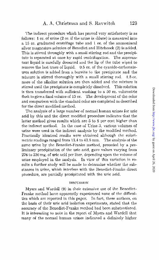
The indirect procedure which has proved very satisfactory is as follows: 1 cc. of urine (2 cc. if the urine is dilute) is measured into a 15 cc. graduated centrifuge tube and 1 cc. of the ammoniacal silver magnesium solution of Benedict and Hitchcock (2) is added. This is stirred throughly with a small stirring rod and the precipi- tate is separated at once by rapid centrifugation. The superna- tant liquid is carefully decanted and the lip of the tube wiped to remove the last trace of liquid. 0.5 cc. of the cyanide-carbonate- urea solution is added from a burette to the precipitate and the mixture is stirred thoroughly with a small stirring rod. 4.5 cc. more of the alkaline solution are then added and the mixture is stirred until the precipitate is completely dissolved. This solution is then transferred with sufficient washing to a 50 cc. volumetric flask to give a final volume of 15 cc. The development of the color and comparison with the standard color are completed as described for the direct modified method.
The analysis of a large number of normal human urines for uric acid by this and the direct’ modified procedure indicates that the latter method gives results which are 5 to 8 per cent higher than the indirect method. In the case of Urine 3, various volumes of urine were used in the indirect analysis by the modified method. Practically identical results were obtained although t,he colori- metric readings ranged from 13.4 to 43.5 mm. The analysis of the same urine by the Benedict-Franke method, preceded by a pre- liminary precipitation of the uric acid, gave values varying from 276 to 330 mg. of uric acid per liter, depending upon the volume of urine employed in the analysis. In view of this variation in re- sult’s a further study will be made to determine whether the sub- stances in urine, which interfere with the Benedict-Franke direct procedure, are partially precipitated with the uric acid.
DISCUSSION
Myers and Wardell (9) in their extensive use of the Benedict- Franke method have apparently experienced none of the difficul- ties which are reported in this paper. In fact, these authors, on the basis of their uric acid isolation experiments, stated that the accuracy of the Benedict-Franke method had been substantiated. It is interesting to note in the report of Myers and Wardell that many of the normal human urines indicated a definitely higher
by guest on April 26, 2020
http://ww
w.jbc.org/
Dow
nloaded from

124 Uric Acid Determination
content of uric acid by the indirect Benedict-Hitchcock method than by the direct method of Benedict and Franke. Similar observations in this laboratory led to the present study of the uric acid methods.
It is possible that the sodium cyanide used by Myers and War- dell in the Benedict-Franke method contained more sodium car- bonate than the cyanide used in this laboratory. If this were the case, the Benedict-Franke method would give more accurate results than reported in this paper. Different lots of pure sodium cyanide (Mallinckrodt) were used throughout this work. The experiments recorded in this paper were practically completed before our attention was called to the fact that various brands of sodium cyanide might contain varying amounts of sodium car- bonate. Consequently no attempt was made to determine the sodium carbonate content of the various sodium cyanide prep- arations.
Benedict (10) has criticized the use of acid silver solutions as precipitants for uric acid because of the lack of specificity as com- pared to ammoniacal silver solutions. Apart from the question of specificity, there is an objection to the use of the acid silver lactate precipitation reagent, as used in the Folin-Wu (11) method for the determination of uric acid in blood and the Folin-Denis method for the determination of uric acid in urine. Rogers (12) reported a number of years ago that serious errors in the determination of uric acid in blood by the Folin-W7u method may result, if the silver precipitate containing the uric acid is exposed for a short time to the ordinary light of a well lighted laboratory. Our experience with the Folin-Denis method for urinary uric acid confirms the observation of Rogers with regard to the rapid loss of uric acid from the silver precipitate on exposure to bright light. Much of the work in regard to this point was done during the late spring and summer months in a well lighted laboratory with a western exposure. It was found that on clear, sunshiny days delay in re- dissolving the silver precipitate containing the uric acid invariably led to low results, even though the precipitates were not exposed to direct sunlight. On dark, cloudy days the uric acid in the precipi- tates remained unchanged for several hours, but if the precipitates were allowed to stand for periods of 12 to 18 hours in compara- tively dark cupboards, errors of 30 to 40 per cent would be noted.
by guest on April 26, 2020
http://ww
w.jbc.org/
Dow
nloaded from

A. A. Christman and S. Ravwitch 125
The change in color of the precipitate from a light gray to a deep brown or black during this period should serve as a warning to a trained analyst. In view of these facts, the desirability of com- pleting the determination as cmickly as possible after the precipi- tation of the uric acid, without undue exposure to light, cannot be overemphasized.
Simultaneously conducted experiments indicate that the uric acid precipitated by ammoniacal silver solutions as used in the proposed indirect method is not affected by an exposure of several hours on a clear day, provided the precipitate is not exposed to direct sunlight.
SUMMARY
Attention is directed to the wide variation in results obtained in the analysis of normal human urine for uric acid by the Benedict- Franke method, depending upon the volume of urine used in the analysis. A modification of the direct procedure which yields more consistent results is proposed. For a more accurate analysis of uric acid in human urine, a modified procedure involving a pre- liminary precipitation of the uric acid prior to its calorimetric estimation is recommended.
Addendum-Since this paper was accepted for publication, the last number of the Biochemical Journal has been received. Salt (13) has modi- fied the method of Folin for the determination of uric acid in urine to pre- vent turbidity during the development of the color. This was accom- plished by the use of a cyanide-urea solution and a 10 per cent rather than a 20 per cent sodium carbonate solution as used in the original procedure. During the past year many determinations of uric acid in urine have been made in this laboratory by the Folin procedure, without the troublesome turbidity during the color development, by the inclusion of urea in the 20 per cent sodium carbonate to the extent of 15 per cent. The results by this modification were identical with those by the original Folin procedure.
BIBLIOGRAPHY
1. Benedict, S. R., and Franke, E., J. Biol. Chem., 62, 387 (1922). 2. Benedict, S. R., and Hitchcock, E. H., J. Biol. Chem., 20, 619 (1915). 3. Christman, A. A., and Mosier, E. C., J. BioZ. Chem., 83, 11 (1929). 4. Morris, J. L., and Macleod, A. G., J. BioZ. Chem., 60, 55 (1922). 5. Folin, O., Laboratory manual of biological chemistry, New York and
London, 4th edition, 141, 251 (1927). 6. Folin, O., J. Biol. Chem., 61, 393 (1922).
by guest on April 26, 2020
http://ww
w.jbc.org/
Dow
nloaded from

Uric Acid Determination
7. Folin, O., J. Biol. Chem., 86, 179 (1930). 8. Benedict, S. R., J. Biol. Chem., 61, 187 (1922). 9. Myers, V. C., and Wardell, E. L., J. Biol. Chem., 77, 697 (1928).
10. Benedict, S. R., J. Biol. Chem., 64, 215 (1925). 11. Folin, O., and Wu, H., J. Biol. Chem., 38, 81 (1919). 12. Rogers, H., J. BioZ. Chem., 66, 325 (1923). 13. Salt, H. B., Biochem. J., 26, 1720 (1931).
by guest on April 26, 2020
http://ww
w.jbc.org/
Dow
nloaded from

A. A. Christman and Sarah RavwitchIN HUMAN URINE
THE DETERMINATION OF URIC ACID
1932, 95:115-126.J. Biol. Chem.
http://www.jbc.org/content/95/1/115.citation
Access the most updated version of this article at
Alerts:
When a correction for this article is posted•
When this article is cited•
alerts to choose from all of JBC's e-mailClick here
ml#ref-list-1
http://www.jbc.org/content/95/1/115.citation.full.htaccessed free atThis article cites 0 references, 0 of which can be
by guest on April 26, 2020
http://ww
w.jbc.org/
Dow
nloaded from