Embed Size (px)
Citation preview

The conserved arginine 380 of Hsp90is not a catalytic residue, but stabilizesthe closed conformation required forATP hydrolysis
Christian N. Cunningham,1,2 Daniel R. Southworth,2 Kristin A. Krukenberg,2,3
and David A. Agard2*
1Graduate Group in Biophysics, University of California, San Francisco, California 941582Department of Biochemistry and Biophysics, Howard Hughes Medical Institute, University of California,San Francisco, California 941583Graduate Program in Chemistry and Chemical Biology, University of California, San Francisco, California 94158
Received 6 March 2012; Revised 22 May 2012; Accepted 23 May 2012
DOI: 10.1002/pro.2103Published online 31 May 2012 proteinscience.org
Abstract: Hsp90, a dimeric ATP-dependent molecular chaperone, is required for the folding and
activation of numerous essential substrate ‘‘client’’ proteins including nuclear receptors, cell cyclekinases, and telomerase. Fundamental to its mechanism is an ensemble of dramatically different
conformational states that result from nucleotide binding and hydrolysis and distinct sets of
interdomain interactions. Previous structural and biochemical work identified a conserved arginineresidue (R380 in yeast) in the Hsp90 middle domain (MD) that is required for wild type hydrolysis
activity in yeast, and hence proposed to be a catalytic residue. As part of our investigations on the
origins of species-specific differences in Hsp90 conformational dynamics we probed the role of thisMD arginine in bacterial, yeast, and human Hsp90s using a combination of structural and functional
approaches. While the R380A mutation compromised ATPase activity in all three homologs, the
impact on ATPase activity was both variable and much more modest (2–7 fold) than themutation of an active site glutamate (40 fold) known to be required for hydrolysis. Single
particle electron microscopy and small-angle X-ray scattering revealed that, for all Hsp90s,
mutation of this arginine abrogated the ability to form the closed ‘‘ATP’’ conformational state inresponse to AMPPNP binding. Taken together with previous mutagenesis data exploring intra-
and intermonomer interactions, these new data suggest that R380 does not directly participate
in the hydrolysis reaction as a catalytic residue, but instead acts as an ATP-sensor to stabilizean NTD-MD conformation required for efficient ATP hydrolysis.
Keywords: Hsp90; conformational dynamics; catalysis; interactions; ATP hydrolysis
Additional Supporting Information may be found in the online version of this article.
Christian N. Cunningham and Daniel R. Southworth contributed equally to this work.
Christian N. Cunningham’s current address is Early Discovery Biochemistry, Genentech, Inc., 1 DNA Way, MS27, SouthSan Francisco, CA 94080.
Daniel R. Southworth’s current address is Department of Biological Chemistry, Life Sciences Institute, University of Michigan,210 Washtenaw Ave, Ann Arbor, MI 48109.
Kristin A. Krukenberg’s current address is Department of Systems Biology, Harvard Medical School, 200 Longwood Ave, WarrenAlpert 536, Boston, MA 02115.
Grant sponsor: UC Discovery Grant; Grant number: bio03-10401/Agard; Grant sponsor: NIH; Grant number: T32 GM08284; Grantsponsor: ACS Postdoctoral Fellowship; NSDEG Graduate Fellowship; Howard Hughes Medical Institute; ARCS Fellowship.
*Correspondence to: Dr. David A. Agard, 600 16th St., MC2240 Room S412D, San Francisco, CA 94158. E-mail: [email protected]
1162 PROTEIN SCIENCE 2012 VOL 21:1162—1171 Published by Wiley-Blackwell. VC 2012 The Protein Society

Introduction
Hsp90 is a member of the molecular chaperone fam-
ily of proteins that facilitate protein folding in the
cell. Well-characterized chaperones including Hsp60
(GroEL/ES) and Hsp70 (DnaK) promote protein fold-
ing by discrete cycles of ATP hydrolysis-driven bind-
ing and release of nascent polypeptides and exposed
hydrophobic regions.1 However, unlike those family
members, Hsp90 acts much later in the folding pro-
cess, interacting with a specific set of client proteins
in a near-native state conformation and promoting
conformational rearrangements required for ligand
binding and downstream signaling events.2–7 In
eukaryotes, these client proteins include many sig-
naling and regulatory proteins such as serine/threo-
nine and tyrosine kinases, telomerase, steroid hor-
mone receptors, and tumor suppressor proteins.8–11
The importance of these client proteins in signaling
and cellular function has made Hsp90 an attractive
target for anticancer therapeutics.12 A majority of
the drugs that inhibit Hsp90 function target the
ATP binding pocket underscoring the importance of
ATP hydrolysis in its chaperoning activity.13,14
Although nucleotide hydrolysis has been shown to
be absolutely required for the in vivo chaperone
function of Hsp9015,16 how the energy from ATP
binding and hydrolysis contributes to both chaper-
one conformational changes and client activation
remains largely unknown.
Hsp90 is a constitutive dimer where each �90
kDa monomer consists of three domains: the N-ter-
minal nucleotide-binding domain (NTD), the middle
domain (MD), and the C-terminal dimerization do-
main (CTD). Hsp90 domain structures,17–19 compari-
sons with other GHKL superfamily members such
as MutL and GyrB,17,19–21 and Hsp90 truncation
studies concluded that the Hsp90 NTD provides all
of the structural requirements for nucleotide bind-
ing5,17,22 and a conserved catalytic glutamate which
activates the attacking water in the ATP hydrolysis
reaction23 [Fig. 1(c)]. However, the NTD alone is not
sufficient for ATP hydrolysis, the MD is required for
even minimal activity and full activity requires both
the MD and dimerization.5
The availability of full-length crystal structures
of the bacterial (apo and ADP states)24 and yeast
Hsp90 (using the nonhydrolyzable ATP analog,
AMPPNP)25 provided important insights into the
relationships between domain structure, organiza-
tion, and ATPase activity. The structures revealed
dramatically different conformational states that are
stabilized by different interactions, each with vary-
ing degree of exposure of conserved hydrophobic
patches on the chaperone, in response to nucleotide
binding [Fig. 1(a)],24,25 suggesting an allosteric cou-
pling between nucleotide and conformational states.
Single particle EM studies confirmed that these
three conformational states (apo, ATP, ADP) are uni-
versally conserved across bacterial, yeast, and
human Hsp90s.26 By contrast, analysis of individual
Hsp90 molecules by EM and population averages by
SAXS revealed that AMPPNP or ADP binding does
not trigger a discrete conformational change but
rather shifts a preexisting equilibrium of states to-
ward the appropriate conformation.26–28
Kinetic studies on Hsp90 homologs from differ-
ent species demonstrated that the chaperone is a
very slow ATPase with activities ranging from 0.1
(human) to 1.2 (yeast) pmol/min/pmol18,19 although
this can be altered by client binding.29–31 Data from
yeast mutants suggests that ATPase rates are pre-
cisely tuned for each organism, as even small
increases or decreases in ATPase rates (T22I 7-fold
increase; T101I 3-fold decrease) will compromise
growth ability.16 From enzyme kinetics, it appears
that the chemical ATP hydrolysis step is orders of
magnitude faster than domain closure, indicating
that conformational changes are rate-limiting.32
This is supported by an observed cross-species corre-
lation between ATPase rates and the population of
closed states in the presence of AMPPNP26 as well
as by direct kinetic observation of domain closure by
either FRET or small angle x-ray scattering
(SAXS).27,28 Given the apparent contradiction
between the required precision of ATPase rates for
in vivo function and the rather weak coupling
between nucleotide and conformation observed in
vitro, we wished to further probe the molecular basis
for nucleotide recognition, hydrolysis, and chaperone
conformation.
Initially identified by comparison with GHKL
family members,33–35 arginine 380 in the yeast
Hsp82 MD (referred to as ArgMD to simplify cross
species comparisons), was shown to have a signifi-
cant role in ATP hydrolysis (related to Lys 307 of
MutL and Lys 33 of GyrB). Mutation of this residue
in Hsp90 resulted in a decrease in ATP hydrolysis
in vitro and a loss of viability in vivo,19 leading to
the suggestion that ArgMD is a catalytic residue
directly participating in the activation and chemis-
try of the hydrolysis reaction. The crystal structure
of Hsp82 in complex with AMPPNP and the co-chap-
erone Sba1 supported this hypothesis, revealing a
direct interaction between the ArgMD and the
AMPPNP c-phosphate in the closed state [Fig.
1(c)].25 By contrast, the E. coli apo N-M structure,
Grp94 N-M structures, and the yeast MD structures
(PDB: 1Y4U, 2O1W, 1HK7) all show a direct interac-
tion of ArgMD with an exposed and conserved
loop on the MD stabilizing the apo state of Hsp90
[Fig. 1(b)].
Previous work from our lab indicated that
ArgMD does not act alone in the activation of ATP
hydrolysis but rather is involved synergistically with
a network of residues from both monomers to
Cunningham et al. PROTEIN SCIENCE VOL 21:1162—1171 1163
stabilize a hydrolysis competent Hsp90 conforma-
tion.36 While investigating this network of interac-
tions we discovered that the equivalent Arg to Ala
mutation in E. coli (R336A) had an unexpectedly
mild affect raising questions about the true role of
this important residue, and indicating that further
investigation was required.
Results
Hydrolysis rates of the middle domain arginine
indicate a noncatalytic role in ATP hydrolysisArgMD was mutated to an alanine in the bacterial
(HtpG�R336A), yeast (Hsc82�R376A), and human
(Hsp90a�R400A) Hsp90 homologs and ATP hydroly-
sis rates were measured by release of radioactive
inorganic phosphate. Previous work has shown that
the affinity of ATP for the NTD alone is essentially
unchanged when compared to the full-length protein
(132 lM vs. 172 lM, respectively), thus residues in
the MD, including the ArgMD, do not contribute sig-
nificantly to nucleotide binding.15,17 The Hsp90-spe-
cific inhibitor, geldanamycin, was used to control for
any small amounts of contaminating ATPases (see
Methods). Mutation of ArgMD resulted in a
decreased ATPase rate in all species, with yeast
showing the most severe effect (a seven-fold reduc-
tion) (Fig. 2; Table I). By contrast, the human and
bacterial Hsp90s, showed only marginal (approxi-
mately two-fold) losses in activity when compared to
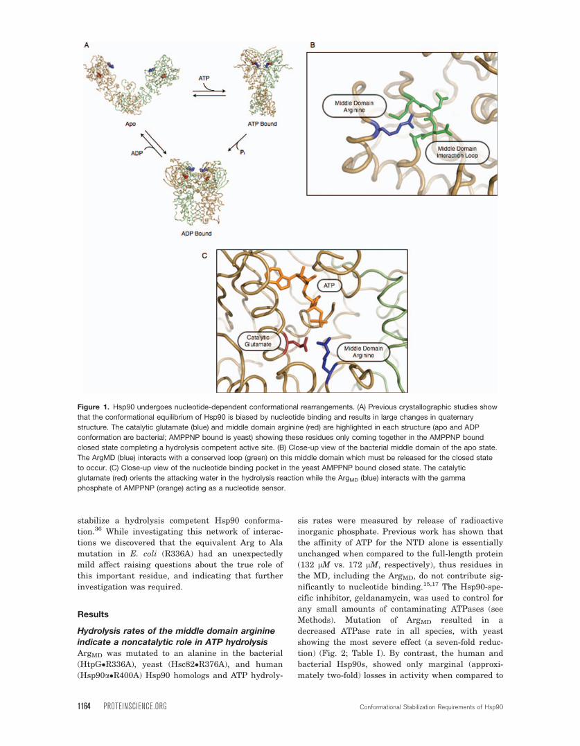
Figure 1. Hsp90 undergoes nucleotide-dependent conformational rearrangements. (A) Previous crystallographic studies show
that the conformational equilibrium of Hsp90 is biased by nucleotide binding and results in large changes in quaternary
structure. The catalytic glutamate (blue) and middle domain arginine (red) are highlighted in each structure (apo and ADP
conformation are bacterial; AMPPNP bound is yeast) showing these residues only coming together in the AMPPNP bound
closed state completing a hydrolysis competent active site. (B) Close-up view of the bacterial middle domain of the apo state.
The ArgMD (blue) interacts with a conserved loop (green) on this middle domain which must be released for the closed state
to occur. (C) Close-up view of the nucleotide binding pocket in the yeast AMPPNP bound closed state. The catalytic
glutamate (red) orients the attacking water in the hydrolysis reaction while the ArgMD (blue) interacts with the gamma
phosphate of AMPPNP (orange) acting as a nucleotide sensor.
1164 PROTEINSCIENCE.ORG Conformational Stabilization Requirements of Hsp90

their respective wild-type constructs. Given the very
high levels of sequence and structural conservation,
this was quite unexpected, although these results
are comparable to those observed for mutations of
the homologous residue in MutL, Lys 307.33 To
ensure that we were mutating the homologous
ArgMD between species we mutated and measured
the ATP hydrolysis rates of several positively
charged residues surrounding ArgMD in the bacterial
homolog. These mutations displayed no change in
ATP hydrolysis (data not shown) when compared to
the wild-type rate indicating that the originally
mutated residues were indeed functionally homolo-
gous equivalents to Arg336 in yeast. Thus while
ArgMD is important for maintaining wild-type levels
of ATPase activity and yeast viability, the very mild
affects observed in both the human and bacterial
enzymes indicate that this residue is not essential
for the ATPase reaction.
As a control for species/variant differences, we
measured the ATP hydrolysis rates of the bacterial
and yeast homologs with the known catalytic gluta-
mate mutated to an alanine (Hsc82�E33A and
HtpG�E34A); due to the very low ATPase basal ac-
tivity of human Hsp90 and the known conserved cat-
alytic role the respective catalytic glutamate muta-
tion was not made. As expected, the bacterial and
yeast mutants were both severely compromised (17-
and 40-fold reductions, respectively) with almost no
ATP hydrolysis measured above the noise level of
this experiment (Fig. 2; Table I). These data support
the known conserved role of the glutamate in the
GHKL family of enzymes coordinating the attacking
water in the hydrolysis reaction.15,21,23,33–35 Thus,
with such a large discrepancy between the hydroly-
sis rates of the arginine and the glutamate muta-
tions observed with both homologs and the overall
modest loss in activity of the ArgMD mutants, our
results suggest that the ArgMD is not required for
catalysis, but instead plays an indirect role in pro-
moting ATP hydrolysis.
Solution X-ray scattering suggests ArgMD
is important for conformational stabilization
We next sought to better define the role of ArgMD.
From crystal structures it is clear that the arginine
residue adopts distinct local interactions in the apo
and ATP states [Fig. 1(b,c)] that appears to stabilize
Hsp90 into one conformation or the other. Given the
unique arrangement of ArgMD in the closed state, an
alternative role could be to stabilize a hydrolysis-
competent Hsp90 conformation in an ATP-specific
manner. To test this, we performed small angle X-
ray scattering (SAXS) on the bacterial and yeast
wild type and mutant constructs in the presence and
absence of AMPPNP. Previous fluorescence studies
have shown that the nonhydrolyzable analog of ATP,
AMPPNP, is able to stabilize the same conformation
of Hsp90 as ATP and is utilized in these structural
studies to prevent conformational cycling of
Hsp90.28,32
Experimental scattering intensity curves (I(q))
of each homolog were measured in the presence and
absence of nucleotide and transformed into inter-
atomic probability profiles, P(r), providing informa-
tion on the overall molecular shape (Fig. 3; left col-
umn). In both wild-type homologs, the apo state
shows a broad distribution of distances in the P(r)
curves representing the very extended open confor-
mation populated in the absence of nucleotide.27
With the addition of saturating amounts of
AMPPNP the bacterial and yeast SAXS curves ex-
hibit a more narrow range of distances in their
P(r) curves with a peak around 55 A indicating a
Figure 2. ATP hydrolysis rates of the bacterial, yeast, and
human Hsp90 homologs. Bacterial, yeast, and human
homologs of Hsp90 were mutated and assayed for ATP
hydrolysis. Wild type rates are shown in black; arginine
mutant rates are shown in gray; catalytic glutamate mutants
are shown in white. All measured hydrolysis rates were
normalized to the wild type yeast homolog rate. The ArgMD
shows a substantial defect in ATP hydrolysis that was not
consistent among all three homologs tested and was not as
deleterious to activity as the catalytic glutamate in all three
cases.
Table I. ATP Hydrolysis Rates for Hsp90 Homologsand Mutants
Organism Wild-typeaArgininemutanta
Glutamatemutanta
Bacteria 0.52 6 0.06 0.21 6 0.03 0.03 6 0.005Yeast 1.2 6 0.05 0.16 6 0.04 0.03 6 0.005Human 0.21 6 0.05 0.11 6 0.03 N/AYeast N599 0.14 6 0.02 0.05 6 0.01 0.008 6 0.005
a Hydrolysis rates are reported as pmol ATP hydrolyzed/min/pmol protein.
Cunningham et al. PROTEIN SCIENCE VOL 21:1162—1171 1165
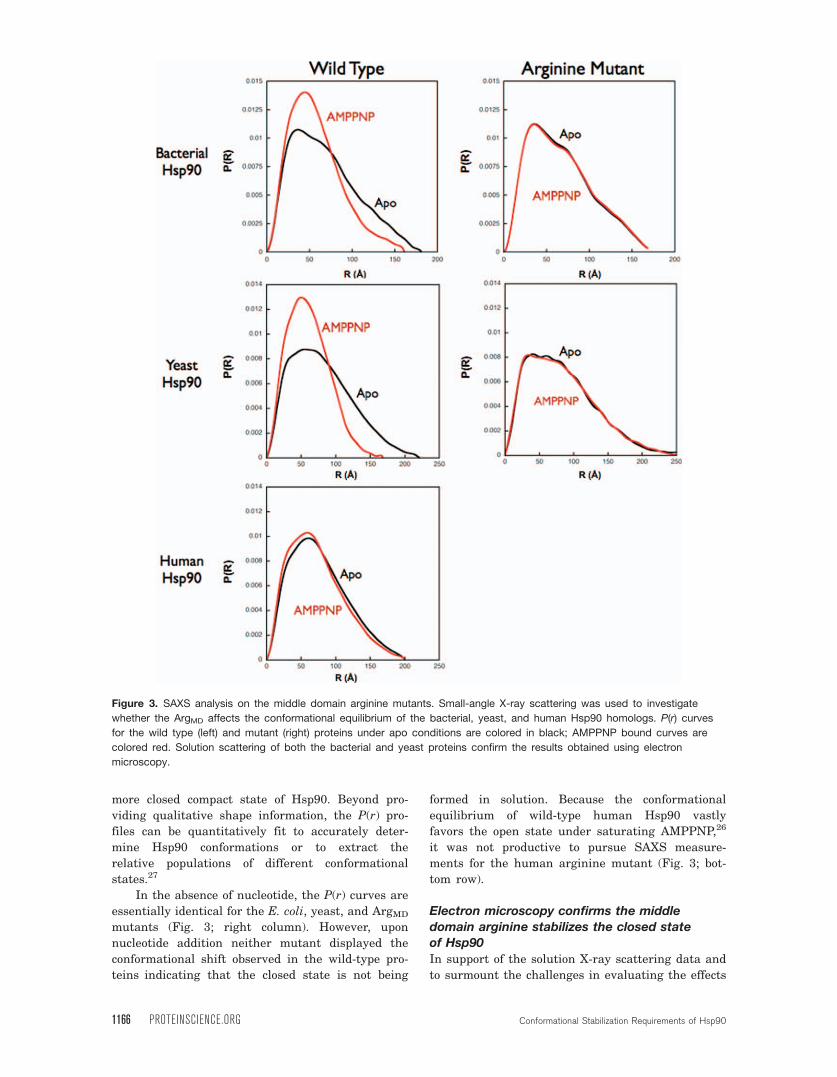
more closed compact state of Hsp90. Beyond pro-
viding qualitative shape information, the P(r) pro-
files can be quantitatively fit to accurately deter-
mine Hsp90 conformations or to extract the
relative populations of different conformational
states.27
In the absence of nucleotide, the P(r) curves are
essentially identical for the E. coli, yeast, and ArgMD
mutants (Fig. 3; right column). However, upon
nucleotide addition neither mutant displayed the
conformational shift observed in the wild-type pro-
teins indicating that the closed state is not being
formed in solution. Because the conformational
equilibrium of wild-type human Hsp90 vastly
favors the open state under saturating AMPPNP,26
it was not productive to pursue SAXS measure-
ments for the human arginine mutant (Fig. 3; bot-
tom row).
Electron microscopy confirms the middle
domain arginine stabilizes the closed stateof Hsp90
In support of the solution X-ray scattering data and
to surmount the challenges in evaluating the effects
Figure 3. SAXS analysis on the middle domain arginine mutants. Small-angle X-ray scattering was used to investigate
whether the ArgMD affects the conformational equilibrium of the bacterial, yeast, and human Hsp90 homologs. P(r) curves
for the wild type (left) and mutant (right) proteins under apo conditions are colored in black; AMPPNP bound curves are
colored red. Solution scattering of both the bacterial and yeast proteins confirm the results obtained using electron
microscopy.
1166 PROTEINSCIENCE.ORG Conformational Stabilization Requirements of Hsp90

of the ArgMD to Ala mutation on the human Hsp90,
we used single particle negative stain electron mi-
croscopy37 of the wild-type and mutant arginine in
all three Hsp90 homologs in the presence and ab-
sence of the nonhydrolyzable ATP analog, AMPPNP.
In the absence of nucleotide, the bacterial, yeast,
and human homologs show a wide range of open
angles between the two monomers similar to what
was observed in our previous electron microscopy
and SAXS studies (Fig. 4, Supporting Information
Fig. 1).26,27 As observed previously,26 upon the addi-
tion of AMPPNP the majority of the bacterial and
yeast Hsp90s form the closed state with the NTDs
dimerized in a conformation equivalent to that
seen in the Hsp82:Sba1:AMPPNP crystal struc-
ture.25 While HtpG�R336A and Hsc82�R376A
looked identical to the wild-type under apo condi-
tions (data not shown), no conversion to the closed
state was observed upon the addition of 5 mM
AMPPNP.
As seen with the human Hsp90 solution scat-
tering results, the closed state is only transiently
sampled despite the presence of saturating
amounts of AMPPNP. Therefore, to address this,
we added a small amount of glutaraldehyde cross-
linker, as previously reported,26 to trap the closed
state in a nucleotide-dependent manner for visual-
ization (Fig. 4, Supporting Information Fig. 2). Fol-
lowing crosslinking, the closed state was not read-
ily observed for human Hsp90a�R400A in the
presence of AMPPNP, while the wild type particles
appeared closed and similar to HtpG and yeast
under the same conditions. Thus, while the affects
of this mutation on ATPase rates are modest and
Figure 4. Electron microscopy single particle analysis of bacterial, yeast, and human Hsp90. Electron microscopy was used
to confirm that the conformational defect observed in solution. Wild type proteins of all homologs displayed the canonical
dynamic open state conformations under apo conditions (left column) and the closed, N-terminal dimerized conformation
when saturating amounts of AMPPNP were added to the solution (middle column). The human protein required the addition of
0.005% glutaraldehyde to detect the closed state in the presence of AMPPNP. In contrast, the arginine mutants of all three
homologs show an inability to stabilize the closed state in the presence of AMPPNP (right column).
Cunningham et al. PROTEIN SCIENCE VOL 21:1162—1171 1167

variable, our EM and SAXS results indicate a con-
served and significant effect on closed conformation
of Hsp90.
The middle domain arginine affects N-M domain
dynamicsWhile mutation of ArgMD to an alanine blocks the
ability to efficiently form the closed, NTD-dimerized
state, it was unclear if ArgMD could directly stabilize
an ATP hydrolysis competent NTD-MD conforma-
tion, or rather acted along only in the context of syn-
ergistic intersubunit interactions36 that together
support the closed NTD-dimerized state. To test this,
we explored the ArgMD–Ala mutation in the context
of a yeast Hsp90 construct lacking the CTD dimeri-
zation domain (Hsc82�N599). In vitro, Hsc82�N599
is a monomer in solution and shows an �10-fold
reduction in ATP hydrolysis (Fig. 5) agreeing with
data published previously for Hsp82.5 As expected
from the full-length results, when the catalytic glu-
tamate was mutated to alanine in the truncated
construct no activity could be measured. Upon
mutation of ArgMD, ATPase activity decreased
three-fold indicating that this arginine helps to sta-
bilize a hydrolysis-competent NTD-MD conforma-
tion even in the absence of NTD dimerization.
Notably, the activity of the Ala mutant in the full-
length protein is greater than the wild type N599
construct suggesting transient N-terminal dimeriza-
tion and ATP hydrolysis in the full-length protein
must be occurring even though it is below the
detection threshold of our structural assays. This is
supported by the contribution of cross-monomer
residues to ATP hydrolysis, even in the context of
the ArgMD mutant.36
DiscussionATP hydrolysis and the conformational changes that
are coupled to this catalytic cycle are central to the
function of Hsp90 by providing the energy to bind,
remodel, and release substrate client proteins.
Although structural studies have shown that all
homologs of Hsp90 appear to undergo similar confor-
mational changes in the presence of nucleotide,
there are substantial species-dependent differences
in the conformational equilibria and kinetics. Muta-
tions made in yeast Hsp82 that either subtly
enhance or decrease ATP hydrolysis showed sub-
stantial growth defects indicating that ATPase rates
and the conformational equilibrium are highly opti-
mized for the specific function of each homolog in
vivo.16
To better understand how conformational equili-
bria and ATP hydrolysis are coupled and to deter-
mine if there is a universal mechanism among
Hsp90 homologs despite the notable differences, we
explored the role of the completely conserved ArgMD
in the bacterial, yeast, and human Hsp90 homologs.
As shown here, the large impact of mutating ArgMD
to Ala on ATPase rate previously observed for yeast
Hsp90, appears to be exclusive to yeast, with only
two fold decreases observed with either the bacterial
or human enzymes. This mild and variable contribu-
tion of ArgMD to ATP hydrolysis rates suggests that
it does not make a critical contribution to chemical
hydrolysis as expected for a conserved catalytic
residue.
By contrast, electron microscopy and SAXS both
indicated that ArgMD does play a conserved role in
stabilizing the closed state in the presence of
AMPPNP. Additionally, mutation of ArgMD has a sig-
nificant affect on the ATPase activity of a monomeric
yeast NTD-MD construct lacking the C-terminal
dimerization domain. Together these data indicate
that ArgMD must contribute directly to stabilization
of a catalysis-competent NTD-MD conformation, in-
dependent of NTD dimerization. The importance of
setting of a defined NTD-MD conformation is under-
scored by the recent discovery that a novel class of
inhibitors that bind at this interface show both se-
lectivity for nucleotide state and function by only in-
hibiting the binding of specific subsets of client and
cochaperone interactions.38,39
While attention has focused on interaction of
ArgMD with the c-phosphate of ATP, several apo
Hsp90 crystal structures reveal that ArgMD directly
interacts with a conserved MD loop in the apo
Figure 5. Middle domain arginine is directly involved in
stabilizing an N-middle conformation that is competent for
hydrolysis. Normalized ATPase activities of the yeast Hsp90
homolog, C-terminal truncation (N599), and the arginine and
glutamate mutants in the C-terminal truncation construct.
There is a substantial loss of activity observed when C-
terminal dimerization is removed. A further loss of activity is
seen when the arginine is mutated indicating its role in
stabilizing a hydrolysis competent conformation of the N-
middle domains. The glutamate mutant in the N599
construct shows no detectable activity as predicted from
the full length activities.
1168 PROTEINSCIENCE.ORG Conformational Stabilization Requirements of Hsp90

state.19,40,41 Thus some of the functional importance
of ArgMD may come from its ability to also stabilize
the apo conformation. From this structural data
and the work presented here we propose a model
where these stabilizing MD interactions in the apo
state (Fig. 6; yellow region) must first be released
followed by a �90 degree rotation of the N-M do-
main42 in order for NTD dimerization and subse-
quently ATP hydrolysis to occur. We propose that
release of these constraints and sampling the N-M
rotated conformation may actually represent the
rate limiting steps in the ATP hydrolysis cycle (Fig.
6; step 2) whereas ATP binding, NTD closure and
chemical hydrolysis are comparatively fast.32 Thus
ArgMD may be playing a role in slowing the for-
ward reaction by stabilizing the apo state while
even more significantly slowing the rate of NTD
rotation and re-opening by recognizing the ATP c-
phosphate. From current and previous work36 we
know that the closed and catalytically active confor-
mation is synergistically stabilized by a combina-
tion of the Arg interactions, a cross-monomer
hydrophobic interaction network, and the NTD
dimerization interface.
Together this work shows that although ArgMD
directly interacts with the c-phosphate, its primary
role is as an ATP sensor that acts to stabilize a spe-
cific NTD-MD conformation rather than playing a
direct catalytic role in the hydrolysis reaction. We
also show that this mechanism of action is universal
among a broad array of Hsp90 homologs even
though ATP hydrolysis rates and conformational
equilibria of the individual species vary greatly. In
conjunction with previous work,36 it is now apparent
that the ATPase active conformation is stabilized by
an array of interdomain and intersubunit interac-
tions that include, but are not limited to NTD dime-
rization that ultimately act to tune the rates of to
match the in vivo requirements for chaperone
function.
Materials and Methods
Hsp90 homolog purificationAll three proteins were purified in a similar manner.
Bacterial and yeast protein was purified from
induced E. coli cultures using Ni-NTA affinity resin
(Qiagen), followed by anion exchange and size exclu-
sion chromatography on a Superdex S200 column
(GE Healthcare). Human Hsp90 was first purified
from induced E. coli cultures using a DEAE column
prior to the Ni-NTA affinity purification and gel fil-
tration on S200. Protein was concentrated in 10 mM
Tris pH 7.5, 100 mM NaCl using Ultrafree Biomax
concentrators (Millipore) to a final concentration of
1–5 mg mL�1 based on UV280 absorption. Protein
was flash frozen in liquid nitrogen and stored at
�80�C until use.
ATPase activity
This assay was adapted from Felts et al.43 2uM pro-
tein was used in each assay with 1 mM ATP and 0.8
pM [32Pc]ATP (6000 Ci mmol�1) in solution. For the
assays using geldanamycin as a control, a final con-
centration of 200 lM was used. Twenty minute time
points were taken over the course of an hour with
the samples shaking and incubating at 37�C. Sepa-
ration of Pi from ATP was performed using the thin
layer chromatography method described.43 Visualiza-
tion of the radiolabeled spots was performed on a
Typhoon Imager (GE Healthcare) and quantification
was performed using the program ImageQuant (GE
Healthcare). The amount of ATP hydrolyzed at each
time point was calculated by taking the ratio of Pi to
ATP in solution. This ratio was then multiplied by
the total amount of ATP added to the reaction and
Figure 6. Model. Stabilization of the hydrolysis competent state of Hsp90 requires a synergistic set of interactions between
the N-terminal domains, a cross monomer hydrophobic network (blue), and the release of the ArgMD from the MD to interact
with the gamma-phosphate of ATP (yellow and red). In the apo state the ArgMD loop interacts with a conserved loop on the
MD. Upon ATP binding (Step 1) the NTD the ArgMD is released from the MD interaction loop and in combination with a �90�
rotation of the NTD-MD interface can interact with the gamma-phosphate of ATP (Step 2) and stabilize the hydrolysis
competent state of Hsp90 (Step 3). We propose that the conformational switching of the ArgMD loop and the rotation of the
NTD-MD domains are slow in comparison to ATP binding and NTD (Step 3).
Cunningham et al. PROTEIN SCIENCE VOL 21:1162—1171 1169

normalized by the total protein in solution. A linear
fit of the time points gave the rate for each reaction.
Activity was plotted on bar graphs using the average
of at least three measurements; error bars indicate
the standard error of the mean.
SAXS data collection and data analysisData reported here was collected at the Advanced
Light Source (ALS) beamline 12.3.1 and the Stan-
ford Synchrotron Radiation Laboratory (SSRL)
beamline 4.2. To minimize aggregation, samples
were spun in a table-top microcentrifuge for 5 min
before data collection. SAXS data was collected at
25�C at 2–5 mg mL�1. At the ALS, the samples were
exposed for 6 and 60 s at a detector distance of 1.6
m. At SSRL samples were exposed for ten 30-s expo-
sures at a detector distance of 2.5 m. Scattering data
was recorded on a Mar165CCD detector. The detec-
tor channels were converted to Q ¼ 4psiny/k, where
2y is the scattering angle and k is the wavelength,
using a silver behenate sample as a calibration
standard. The data was circularly averaged over the
detector and normalized by the incident beam inten-
sity. The raw scattering data were scaled and the
buffers were subtracted. Individual scattering curves
were then merged to provide the final averaged scat-
tering curve. The interatomic distance distribution
functions (P(r)) were then calculated using the pro-
gram GNOM.44 Dmax was determined by constrain-
ing rmin to equal zero and then varying rmax between
150 and 250 A. Rmax was then chosen so that the
P(r) curve smoothly approached zero at the upper
limit. Small changes in rmax (610 A) did not affect
the overall shape of the P(r) curve. Radii of gyration
were calculated from the P(r). Comparable results
were obtained from the scattering curves using the
Guinier approximation as implemented in the pro-
gram PRIMUS,44 however the calculated uncertain-
ties were larger due to limitations in the low angle
data.
Negative-stain electron microscopy
Purified Hsp90 protein was negatively stained with
uranyl formate (pH 5.5–6.0) on thin carbon-layered
(40–50 A thick) 400 mesh copper grids (Pelco) as
described.45 Before staining, protein samples were
incubated for 20 min at 150 nM with or without 5
mM AMPPNP (Sigma–Aldrich) at 37�C for E. coli
and human Hsp90 and 30�C for yeast Hsp90 in
20 mM Tris (pH 7.5), 50 mM KCl, 5 mM MgCl2, and
1 mM DTT. Glutaraldehyde (0.005%) crosslinking
was performed as described (Southworth and Agard,
2008). Samples were imaged using a Tecnai G2 Spi-
rit TEMs (FEI) operated at 120 keV. Micrograph
images were recorded using a 4 k � 4 k CCD camera
(Gatan) at 68,000� magnification with 2.2 A pixel
size.
Acknowledgments
The authors thank the entirety of the Agard Lab for
many helpful discussions and comments.
References
1. Young JC, Agashe VR, Siegers K, Hartl FU (2004)Pathways of chaperone-mediated protein folding in thecytosol. Nat Rev Mol Cell Biol 5:781–791.
2. Freeman BC, Yamamoto KR (2002) Disassembly oftranscriptional regulatory complexes by molecularchaperones. Science 296:2232–2235.
3. Picard D (2002) Heat-shock protein 90, a chaperone forfolding and regulation. Cell Mol Life Sci 59:1640–1648.
4. Pratt WB, Toft DO (2003) Regulation of signalingprotein function and trafficking by the hsp90/hsp70-based chaperone machinery. Exp Biol Med 228:111–133.
5. Richter K, Muschler P, Hainzl O, Buchner J (2001)Coordinated ATP hydrolysis by the Hsp90 dimer. J BiolChem 276:33689–33696.
6. Young JC, Moaerefi I, Hartl FU (2001) Hsp90: a speci-alized but essential protein-folding tool. J Cell Biol 154:267–273.
7. Zhao R, Davey M, Hsu YC, Kaplanek P, Tong A, Par-sons AB, Krogan N, Cagney G, Mai D, Greenblatt J,Boone C, Emili A, Houry WA (2005) Navigating thechaperone network: an integrative map of physical andgenetic interactions mediated by the hsp90 chaperone.Cell 120:715–727.
8. Young JC, Hoogenraad NJ, Hartl FU (2003) Molecularchaperones Hsp90 and Hsp70 deliver preproteins tothe mitochondrial import receptor Tom70. Cell 112:41–50.
9. Pearl LH, Prodromou C (2006) Structure and mecha-nism of the hsp90 molecular chaperone machinery.Annu Rev Biochem 75:271–294.
10. McClellan AJ, Xia Y, Deutschbauer AM, Davis RW,Gerstein M, Frydman J (2007) Diverse cellular func-tions of the hsp90 molecular chaperone uncoveredusing systems approaches. Cell 131:121–135.
11. Neckers L, Neckers K (2005) Heat-shock protein 90inhibitors as novel cancer chemotherapeutics—anupdate. Expert Opin Emerg Drugs 10:137–149.
12. Sharp S, Workman P (2006) Inhibitors of the HSP90molecular chaperone: current status. Adv Cancer Res95:323–348.
13. Jez JM, Chen JC, Rastelli G, Stroud RM, Santi DV(2003) Crystal structure and molecular modeling of 17-DMAG in complex with human Hsp90. Chem Biol 10:361–368.
14. Roe SM, Prodromou C, O’Brien R, Ladbury JE, PiperPW, Pearl LH (1999) Structural basis for inhibition ofthe Hsp90 molecular chaperone by the antitumor anti-biotics radicicol and geldanamycin. J Med Chem 42:260–266.
15. Obermann WM, Sondermann H, Russo AA, PavletichNP, Hartl FU (1998) In vivo function of Hsp90 is de-pendent on ATP binding and ATP hydrolysis. J CellBiol 143:901–910.
16. Hawle P, Siepmann M, Harst A, Siderius M, ReuschHP, Obermann WM (2006) The middle domain ofHsp90 acts as a discriminator between different typesof client proteins. Mol Cell Biol 26:8385–8395.
17. Prodromou C, Roe SM, O’Brien R, Ladbury JE, PiperPW, Pearl LH (1997) Identification and structural char-acterization of the ATP/ADP-binding site in the Hsp90molecular chaperone. Cell 90:65–75.
1170 PROTEINSCIENCE.ORG Conformational Stabilization Requirements of Hsp90

18. Harris SF, Shiau AK, Agard DA (2004) The crystalstructure of the carboxy-terminal dimerization domainof htpG, the Escherichia coli Hsp90, reveals a potentialsubstrate binding site. Structure 12:1087–1097.
19. Meyer P, Prodromou C, Hu B, Vaughan C, Roe SM,Panaretou B, Piper PW, Pearl LH (2003) Structuraland functional analysis of the middle segment ofhsp90: implications for ATP hydrolysis and client pro-tein and cochaperone interactions. Mol Cell 11:647–658.
20. Meyer P, Prodromou C, Liao C, Hu B, Mark Roe S,Vaughan CK, Vlasic I, Panaretou B, Piper PW, PearlLH (2004) Structural basis for recruitment of theATPase activator Aha1 to the Hsp90 chaperone ma-chinery. EMBO J 23:511–519.
21. Dutta R, Inouye M (2000) GHKL, an emergent ATPase/kinase superfamily. Trends Biochem Sci 25:24–28.
22. Prodromou C, Roe SM, Piper PW, Pearl LH (1997) Amolecular clamp in the crystal structure of the N-ter-minal domain of the yeast Hsp90 chaperone. NatStruct Biol 4:477–482.
23. Panaretou B, Prodromou C, Roe SM, O’Brien R, Lad-bury JE, Piper PW, Pearl LH (1998) ATP binding andhydrolysis are essential to the function of the Hsp90molecular chaperone in vivo. EMBO J 17:4829–4836.
24. Shiau AK, Harris SF, Southworth DR, Agard DA(2006) Structural analysis of E. coli hsp90 reveals dra-matic nucleotide-dependent conformational rearrange-ments. Cell 127:329–340.
25. Ali MM, Roe SM, Vaughan CK, Meyer P, Panaretou B,Piper PW, Prodromou C, Pearl LH (2006) Crystal struc-ture of an Hsp90-nucleotide-p23/Sba1 closed chaperonecomplex. Nature 440:1013–1017.
26. Southworth DR, Agard DA (2008) Species-dependentensembles of conserved conformational states definethe Hsp90 chaperone ATPase cycle. Mol Cell 32:631–640.
27. Krukenberg KA, Forster F, Rice LM, Sali A, Agard DA(2008) Multiple conformations of E. coli Hsp90 in solu-tion: insights into the conformational dynamics ofHsp90. Structure 16:755–765.
28. Mickler M, Hessling M, Ratzke C, Buchner J, Hugel T(2009) The large conformational changes of Hsp90 areonly weakly coupled to ATP hydrolysis. Nat Struct MolBiol 16:281–286.
29. McLaughlin SH, Smith HW, Jackson SE (2002) Stimu-lation of the weak ATPase activity of human hsp90 bya client protein. J Mol Biol 315:787–798.
30. Street TO, Lavery LA, Agard DA (2011) Substratebinding drives large-scale conformational changes inthe Hsp90 molecular chaperone. Mol Cell 42:96–105.
31. Motojima-Miyazaki Y, Yoshida M, Motojima F (2010)Ribosomal protein L2 associates with E. coli HtpG and
activates its ATPase activity. Biochem Biophys ResCommun 400:241–245.
32. Hessling M, Richter K, Buchner J (2009) Dissection of
the ATP-induced conformational cycle of the molecular
chaperone Hsp90. Nat Struct Mol Biol 16:287–293.33. Ban C, Junop M, Yang W (1999) Transformation of
MutL by ATP binding and hydrolysis: a switch in DNAmismatch repair. Cell 97:85–97.
34. Ban C, Yang W (1998) Crystal structure and ATPaseactivity of MutL: implications for DNA repair and mu-tagenesis. Cell 95:541–552.
35. Corbett KD, Berger JM (2005) Structural dissection ofATP turnover in the prototypical GHL ATPase TopoVI.Structure 13:873–882.
36. Cunningham CN, Krukenberg KA, Agard DA (2008)Intra- and intermonomer interactions are required tosynergistically facilitate ATP hydrolysis in Hsp90.J Biol Chem 283:21170–21178.
37. McLaughlin SH, Ventouras LA, Lobbezoo B, JacksonSE (2004) Independent ATPase activity of Hsp90 subu-nits creates a flexible assembly platform. J Mol Biol344:813–826.
38. Vasko R, Rodriguez R, Cunningham C, Ardi V, AgardD, McAlpine S (2010) Mechanistic studies of sansalva-mide A-amide: an allosteric modulator of Hsp90. ACSMed Chem Lett 1:4–8.
39. Alexander LD, Partridge JR, Agard DA, McAlpine SR(2011) A small molecule that preferentially binds theclosed conformation of Hsp90. Bioorg Med Chem Lett21:7068–7071.
40. Dollins DE, Warren JJ, Immormino RM, Gewirth DT(2007) Structures of GRP94-nucleotide complexesreveal mechanistic differences between the hsp90 chap-erones. Mol Cell 28:41–56.
41. Huai Q, Wang H, Liu Y, Kim HY, Toft D, Ke H (2005)Structures of the N-terminal and middle domains of E.coli Hsp90 and conformation changes upon ADP bind-ing. Structure 13:579–590.
42. Southworth DR, Agard DA (2011) Client-loading con-formation of the Hsp90 molecular chaperone revealedin the cryo-EM structure of the human Hsp90:Hopcomplex. Mol Cell 42:771–781.
43. Felts SJ, Owen BA, Nguyen P, Trepel J, Donner DB,Toft DO (2000) The hsp90-related protein TRAP1 is amitochondrial protein with distinct functional proper-ties. J Biol Chem 275:3305–3312.
44. Konarev PV, Volkov VV, Sokolova AV, Koch MHJ, Sver-gun DI (2003) PRIMUS: a Windows PC-based systemfor small-angle scattering data analysis. J Appl Cryst36:1277–1282.
45. Ohi M, Li Y, Cheng Y, Walz T (2004) Negative stainingand image classification—powerful tools in modernelectron microscopy. Biol Proced Online 6:23–34.
Cunningham et al. PROTEIN SCIENCE VOL 21:1162—1171 1171


![From a Ratchet Mechanism to Random Fluctuations Evolution ... · The 90‐kDa heat shock proteins [heat shock protein 90 (Hsp90)] are a highly conserved ATP-dependent protein family,](https://img.dokumen.tips/doc/110x75/5c687a8609d3f2f5638b9b33/from-a-ratchet-mechanism-to-random-fluctuations-evolution-the-90kda-heat.jpg)
![Hsp90 Inhibitors Are Efficacious against Kaposi Sarcoma by ......Hsp90 is an emerging therapeutic target for cancer [8,9,10]. The newer class of Hsp90 inhibitors bind to the ATP-binding](https://img.dokumen.tips/doc/110x75/60bea423374b8000d05be373/hsp90-inhibitors-are-efficacious-against-kaposi-sarcoma-by-hsp90-is-an-emerging.jpg)