Embed Size (px)
Citation preview

The clinical relevance of visualising the peripheral retina
Quinn, N., Csincsik, L., Flynn, E., Curcio, C. A., Kiss, S., Sadda, S. R., Hogg, R., Peto, T., & Lengyel, I. (2019).The clinical relevance of visualising the peripheral retina. Progress in Retinal and Eye Research, 68, 83-109.https://doi.org/10.1016/j.preteyeres.2018.10.001
Published in:Progress in Retinal and Eye Research
Document Version:Publisher's PDF, also known as Version of record
Queen's University Belfast - Research Portal:Link to publication record in Queen's University Belfast Research Portal
General rightsCopyright for the publications made accessible via the Queen's University Belfast Research Portal is retained by the author(s) and / or othercopyright owners and it is a condition of accessing these publications that users recognise and abide by the legal requirements associatedwith these rights.
Take down policyThe Research Portal is Queen's institutional repository that provides access to Queen's research output. Every effort has been made toensure that content in the Research Portal does not infringe any person's rights, or applicable UK laws. If you discover content in theResearch Portal that you believe breaches copyright or violates any law, please contact [email protected].
Download date:18. Dec. 2021

Contents lists available at ScienceDirect
Progress in Retinal and Eye Research
journal homepage: www.elsevier.com/locate/preteyeres
The clinical relevance of visualising the peripheral retinaNicola Quinna,1, Lajos Csincsikb,1, Erin Flynnc,1, Christine A. Curciod,1, Szilard Kisse,1,SriniVas R. Saddaf,1, Ruth Hogga,1, Tunde Petoa,1, Imre Lengyelb,∗,1
a Centre for Public Health, Queen's University, Belfast, United Kingdomb Centre for Experimental Medicine, Queen's University, Belfast, United Kingdomc School of Medicine and Health Sciences, The George Washington University, Washington, D.C., United Statesd School of Medicine, University of Alabama at Birmingham, AL, United Statese Department of Ophthalmology, Weill Cornell Medical College, New York, United StatesfDoheny Eye Institute, University of California, Los Angeles, CA, United States
A R T I C L E I N F O
Keywords:Retinal imagingUltra-widefieldPeripheral retinaImage gradingUWFIGrading grid
A B S T R A C T
Recent developments in imaging technologies now allow the documentation, qualitative and quantitative eva-luation of peripheral retinal lesions. As wide field retinal imaging, capturing both the central and peripheralretina up to 200° eccentricity, is becoming readily available the question is: what is it that we gain by imagingthe periphery? Based on accumulating evidence it is clear that findings in the periphery do not always associateto those observed in the posterior pole. However, the newly acquired information may provide useful clues topreviously unrecognised disease features and may facilitate more accurate disease prognostication. In this re-view, we explore the anatomy and physiology of the peripheral retina, focusing on how it differs from theposterior pole, recount the history of peripheral retinal imaging, describe various peripheral retinal lesions andevaluate the overall relevance of peripheral retinal findings to different diseases.
1. Introduction
Since the invention of the first ophthalmoscope in 1851 by Hermannvon Helmholtz, imaging the retina has undergone dramatic improve-ments, not only arousing interest in previously undescribed abnormal-ities but changing our understanding of numerous retinal and choroidaldiseases (Keeler, 2002). The continuous advances in imaging technol-ogies enabled the capture of retinal areas beyond the posterior pole,which were not previously routinely documented digitally. The in-troduction of ultra-widefield imaging (UWFI) led to a renewed interestin lesions in the peripheral retina and a better understanding of theirrelevance to the diagnosis and prognosis of different retinal conditions.To develop appropriate understanding of the images generated by noveltechnologies, several principles need to be considered. An under-standing of the anatomy of the entire neurosensory retina and choroid/retinal pigment epithelium (RPE) interface and the differences and si-milarities between the different geographic retinal locations must bedeveloped. These can help to establish appropriate descriptive ter-minologies to describe and interpret the different UWFI modalities such
as colour imaging, fundus auto fluorescence (FAF), fundus fluoresceinangiography (FFA) and indocyanine green (ICG) angiography.
2. The structure and function of the peripheral retina
2.1. Definition of the peripheral retina
Based on ophthalmoscopic examination a readily recognizable fea-ture of the neurosensory retina is the optic disc, the exit point for theganglion cell nerve fibres and central veins, and entry point for arteriessupplying the inner layers of the neurosensory retina. The arteries formthe so called “arcade” encompassing the macula, including the foveola(Yamada, 1969), fovea, parafovea and perifovea, definitions that areessential for image analysis of fundus photographs and define thewidening areas around the foveola (Bringmann et al., 2018; Proviset al., 2005). The fovea containing the foveola is the place of the highestvisual acuity (Hirsch and Curcio, 1989). Other landmarks of the maculaare the foveal avascular zone and the visually recognizable yellowmacular pigment.
https://doi.org/10.1016/j.preteyeres.2018.10.001Received 24 June 2018; Received in revised form 1 October 2018; Accepted 7 October 2018
∗ Corresponding author. The Wellcome-Wolfson Institute for Experimental Medicine School of Medicine, Dentistry and Biomedical Science, Queen's UniversityBelfast, 97 Lisburn Road, Belfast, BT9 7BL, United Kingdom.
1 Percentage of work contributed by each author in the production of the manuscript is as follows: Nicola Quinn 25%; Lajos Csincsik: 20%; Erin Flynn 5%; ChristineA. Curcio 10%; Szilard Kiss 5%; SriniVas R. Sadda 5%; Ruth Hogg 5% ; Tunde Peto10%; Imre Lengyel 15%.
E-mail address: [email protected] (I. Lengyel).
Progress in Retinal and Eye Research 68 (2019) 83–109
Available online 10 October 20181350-9462/ © 2018 The Authors. Published by Elsevier Ltd. This is an open access article under the CC BY license (http://creativecommons.org/licenses/BY/4.0/).
T

Retinal regions based on gross anatomy including vessels was de-fined in 1961 by Duke-Elder who categorised the peripheral retina intofour zones (Duke-Elder, 1961). These zones are: 1) the near periphery, a1.5 mm ring adjacent to the 6 mm diameter macula (centred on thefoveola and often defined as the posterior fundus); 2) the middle per-iphery, the next 1.5 mm ring; 3) the far-periphery, measuring the next9–10 mm on the temporal and 16 mm on the nasal side; 4) the oraserrata or extreme periphery, measuring the additional 2.1 mm in thetemporal and 0.7 mm on the nasal side. In 1967 Rutnin divided the backof the eye into 3 regions: 1) posterior fundus, a circle centred on thefoveola and including the optic disc; 2) equatorial zone, a ring shapedarea about 5.83 mm wide adjacent to the posterior fundus and borderedby an imaginary circle through the posterior extremities of the vortexveins; 3) and the ora serrata, the area outside of the equatorial zone(Fig. 1) (Rutnin and Schepens, 1967).
Human retinal regions based on the cellular content of the neuro-sensory retina were defined by Polyak (1941), which we will examinein detail in 2.3.3. For the purpose of this review, the peripheral retinawill be considered as area peripheral to the macula, a retinal region thatcan itself be subdivided (see section 2.3).
Grading for retinal abnormalities in clinical studies still use differentdefinitions, evidenced by the use of different grading grids summarizedin Section 4. It is important to recall that grading grids, like all metrics,were developed to investigate specific diseases, for specific purposes, byinvestigators following then-current models of pathogenesis. Thus, it isprudent to periodically ask whether these grids still make sense in thelight of new knowledge.
2.2. Embryological differences between the central and peripheral retina
The embryological development of the retina is a complex process(Oyster, 1999; Provis, 2001; Venters et al., 2013, 2015). At birth, theretinal neuronal cytoarchitecture and physiology is still immature andcontinues to undergo changes for several years (Hildebrand and Fielder,2011). Retinal development is known to begin centrally before ex-tending peripherally, further expansion of the peripheral retina con-tinues and matures before the posterior pole (Hendrickson and Drucker,1992). The initial stage of growth involves the neural tube. As devel-opment proceeds, optic vesicles evaginate, producing a bi-layered opticcup with an inner and outer layer subsequently differentiating into thesensory neural retina and the RPE, respectively (Hendrickson, 2016).Following this initial differentiation, the most central portion of the
neural retina is thought to differentiate early with cells at the peripheralretina margins remaining proliferative and thus differentiating last. Infact the proliferating capacity of the peripheral retina appears to beretained in adults, at least in animal models (Al-Hussaini et al., 2016).
Previous work by Venters et al. (2013) has provided useful insightsinto the derivation of mature eye tissues by a vital labelling strategy ofchick embryos. They demonstrated that significant portions of theperipheral retina is made up of cells that resided at the optic cup lip andthese same cells did not make up any of the central retina, providing thefirst evidence that two distinct sub-domains arise during embryogen-esis, namely the central and peripheral retina (Venters et al., 2013). Thecentral retina has previously been shown to be derived from distal opticvesicles, where upon invagination they displace to the back of the eyeand form the central retina (Shin and O'Brien, 2009). In 2015, Venterset al. further described the subdivision in the neural retina between thecentral and peripheral retina. There is notably a variety of character-istics that distinguish between the central and peripheral retina, with anobvious distinction being the different photoreceptor densities foundwithin the central and peripheral retina, with cones predominantlyfound in the central and rods in the peripheral retina (Venters et al.,2015). In this review we will use the terminology of Polyak (1941) forthe human retinal regions (see later).
2.3. Anatomical differences between the central and peripheral retina: cells,layers, and regions
2.3.1. CellsThe human retina, like all mammalian retinae, is made up of five
major cell classes of neurons: photoreceptors, bipolar cells, ganglioncells, horizontal cells and amacrine cells, which are subdivided intoapproximately 60 molecularly and morphologically distinct types. Thesimplest circuit begins with the photoreceptors, which transduce lightinto electrochemical signal and connect to the bipolar interneurons.These in turn contact ganglion cells, which transmit signal to the brain.Human retina has ∼100 million rods (active in dim light) and 4.6–6million cone photoreceptors (active in bright light) and 1.0 to 1.5million ganglion cells, implying a tremendous level of signal compres-sion. At the same time, there are at least 13 bipolar cell types that splitsignals at the photoreceptor synapse into multiple parallel pathways,each with distinct functions, for transmission to the brain. Horizontalcells and amacrine cells are interneurons that establish lateral interac-tions at the first (photoreceptor → bipolar) and second (bipolar →
Fig. 1. Illustration of the major landmarks in the retina.The left image illustrates the different structures of the retina and their position (Modified from Cybersight, Interpretation of Stereo Ocular Angiography: Retinal andChoroidal Anatomy (Santini, 2017). The right image is an Optomap image taken from the Reykjavik Study and adapted to display the location of the variousstructures on the retina. (MP = Mid-periphery and FP= Far-periphery).
N. Quinn et al. Progress in Retinal and Eye Research 68 (2019) 83–109
84
ganglion cell) synapse. In addition to neurons are glial cells, specificallythe Müller cells that span the retina from the inner surface to externallimiting membrane (a series of junctional complexes), astrocytes in thenerve fibre layer, and microglia that reside in inner retina for surveil-lance. The RPE is simple cuboidal epithelium that provides essentialsupport services to the photoreceptors on its apical aspect and thechoriocapillary (ChC) endothelium on its basal aspect. The retina has adual vasculature, with the attendant vascular endothelium and peri-cytes – the intrinsic retinal circulation supplying the inner retina in-cluding the internal part of the Müller cells, and the choroidal circu-lation supplying the RPE, photoreceptors, and external portion of theMüller cells. Between RPE and ChC is a laminated sub-endothelial ex-tracellular matrix called Bruch's membrane (BrM), which functions as avessel wall laid out flat, in parallel to vascular lumens, rather thancircumferentially around them.
2.3.2. LayersRetinal layers have been named by several schemes; the nomen-
clature of Polyak is widely accepted (Fig. 2) (Polyak, 1941). Retinallayers vary markedly by region. Fig. 2 shows the neurosensory retina atthree locations in the central retina and one in the peripheral retina.The macula, a 6-mm-diameter area centred on the fovea, comprisesnumerous clinically relevant layers, sublayers, and potential spaces. Asclassically described (B. B. Boycott, 1969; Hogan et al., 1971; Polyak,1941), these include (from inner to outer) the RNFL, ganglion cell layer(GCL), inner plexiform (synaptic) layer (IPL), inner nuclear layer (INLwith bipolar, horizontal, amacrine and Müller cell bodies), outerplexiform (synaptic) layer (OPL), outer nuclear layer (ONL, with pho-toreceptor cell bodies), photoreceptor inner and outer segments (IS, OS;Polyak's bacillary layer), and RPE. The macular OPL has sublayers ofbipolar and horizontal cell neurites, cone pedicles and rod spherules,and Henle fibres (photoreceptor axons and Müller cells). PhotoreceptorIS have myoid and ellipsoid compartments. Choroidal layers include the
inner and outer BrM, the ChC, and the choroid, delimited from thesclera by the lamina suprachoroidea. Stereotypic extracellular depositsaccumulate with aging and age-related macular degeneration in thesubretinal space (subretinal drusenoid deposit), between the RPE andits basal lamina (basal laminar deposit), and between the RPE basallamina and the inner collagenous layer of BrM (drusen).
2.3.3. RegionsAccording to the terrain theory of vision (Hughes, 1977), the sam-
pling of visual space by neurons in each species' retina is influenced bythe species’ normal habitat. Mammalian retinae have an area of highneuronal density specialised for visual acuity, onto which images areregistered by coordinated head and eye movements (Wassle andBoycott, 1991). A well-known example of this principle is the visualstreak of rabbits (Vaney, 1980), in which numerous ganglion cells alignhorizontally for optimal surveillance of predators. Conversely, neuronaldensity and spatial resolution is low in peripheral retina, because theperipheral retina has other important functions, i.e., specialization formovement detection and sensitivity, and the part of the visual brainprocessing this information can be limited in size. Further, the level ofconvergence from photoreceptors to ganglion cells is high in the per-iphery, which contributes to lesser acuity and enhances movementdetection.
Polyak (1941) defined the human retinal regions as concentricallyorganized around the visual axis in the fovea and based it on retinalneurobiology in humans and macaques, especially the topography ofretinal ganglion cells and the local rod: cone ratio. The Henle fibre layer(fibres of cones, rods, and Müller cells) is present in para- and perifovea.The given diameter is for the region in humans (of unspecified ages).The radius of the outer boundary is distance from the fovea:
• foveola, 350 μm (175 μm radius), absent ganglion cells;• foveal floor, 400 μm (200 μm radius), absent ganglion cells;
Fig. 2. Thickness of human neurosensory retinavaries by region.N, nasal fovea; T, temporal to fovea. 1, 2, and 3 mm Npertain to parafovea, perifovea, and near periphery,and 13 mm T is extreme periphery, in the termi-nology of Polyak (1941). 88-year-old female donorwith normal retina, < 6 h death-to-preservation,post-fixed with osmium tannic acid para-phenylenediamine, 0.8 μm epoxy sections, stainedwith toluidine blue; original at http://projectmacula.cis.uab.edu/. NFL, nerve fibre layer; GCL, ganglioncell layer; IPL, inner plexiform layer; INL, inner nu-clear layer; OPL, outer plexiform layer; HFL, Henlefibre layer; ONL, outer nuclear layer; ISm, innersegment myoid of photoreceptors; ISe, inner segmentellipsoid of photoreceptors; OS, outer segments ofphotoreceptors. The abundance of ganglion cells andcone photoreceptors in addition to the Henle fibrelayer distinguish the macula from the periphery. Ar-rows indicate a single continuous row of cone nucleiin the parafovea. Some cell bodies in GCL and INL areedematous (vacuolated).
N. Quinn et al. Progress in Retinal and Eye Research 68 (2019) 83–109
85

• parafovea, 2500 μm (1250 μm radius), 5–8 rows of ganglion cells; 1rod between cones
• perifovea, 5500 μm (2750 μm), 4 rows of ganglion cells diminishingto 1 row; 2 rods between cones;
In addition to these macular regions, which are still used today,Polyak also defined regions of peripheral retina on the basis of ganglioncell density. These are not clinically detectable with current technologybut are described here for completeness:
• near periphery, 8500 μm (4250 μm radius); single continuous row ofganglion cells; 2–3 rods between cones; mid-periphery, 14,500 μm(7250 μm radius); discontinuous row of ganglion cells; 3 rods be-tween cones;
• far periphery, 26,000 μm (10,000 μm temporal, 16,000 μm nasal);widely separated ganglion cells; extreme periphery, to the ora ser-rata.
The entire expanse of human retina is ∼43 mm from ora serrata toora serrata. The human macula, 6 mm in diameter, is thus defined as anarea with a continuous layer of ganglion cells (Polyak, 1957) and inepidemiology as the area included in the Early Treatment of DiabeticRetinopathy Study grid for grading colour fundus photographs (EarlyTreatment Diabetic Retinopathy Study Research Group, 1991b). Themacula constitutes ∼3% of the total retina area (of ∼1000 mm2),leaving 97% of retina considered extramacular. Indeed, human per-ipheral retina can be described as a generic mammalian retina thatlacks features that are specific to the macula.
In 1990, comprehensive 2-dimensional maps of cone and rod den-sity in short post-mortem retinae prepared as unstained whole mounts(Curcio et al., 1990) visualized the topography and morphologic detailof inner segments. With accurately localized centres and computerizedmicroscopy, it was possible to determine photoreceptor density over theentire retina (Fig. 3). The human macula was shown to be overall nu-merically rod-dominant, including a rod-dominant perifovea sur-rounding a cone-only foveola (0.8 mm in diameter). The foveola had ahigh peak density of cones (> 150,000 mm2) and a sharp decline witheccentricity. Rods were also numerous (peak > 150,000 mm2) in anelliptical crest at 2–5 mm eccentricity (i.e., along the macular peri-meter) that encircled the optic nerve head nasally, with a hot-spot insuperior retina (Curcio et al., 1990) (Fig. 3). Outside the macula, coneswere 40% more numerous nasally than at corresponding eccentricitiesin temporal retina. The highest ratio of rods to cones was 25–30 at6–14 mm eccentricity, because cones continued to decrease across theretina more rapidly than did rods, except for nasal quadrant, where thisratio was ∼20 due to a less step decline. Cone density is increased atthe nasal ora serrata, as previously seen (Osterberg, 1935).
Similar tissue, microscopy, and computational approaches wererecently applied to cell density and autofluorescence of human macularRPE (Ach et al., 2014). A demonstrated peak cell density of ∼7200 RPEcells/mm2 and a shallow eccentricity-dependent decline was consistentwith previous literature (Gao and Hollyfield, 1992; Snodderly et al.,2002). With photoreceptor and RPE maps represented in a unified co-ordinate system, it is possible to represent the phagocytic load on RPEby a ratio of photoreceptors (cones + rods)/mm2 to RPE/mm2. Thisratio is high (≥28) at the foveal cone peak and lower in the parafovea(13), rising to 29 at the edge of the macula, in human and non-humanprimate (Curcio, 2018; Snodderly et al., 2002). These quantitative datado not support a widely-held belief that higher phagocytic load on RPErenders the macula uniquely vulnerable to age-related disease. In themacula, RPE cell bodies, excluding the apical processes, are ∼14 μmtall and the centre-to-centre spacing of these hexagonal cells is 13, 15,and 16 μm in fovea, perifovea, and near-periphery, respectively (Achet al., 2014; Curcio et al., 2011). At the periphery, cells are flatter andcan be as wide as 60 μm (Harman et al., 1997).
The RPE cells are supported by BrM, an extracellular matrix that can
be defined as penta-laminar (if both basal membranes of the RPE andChC are considered), or trilaminar (if only the inner and outer col-lagenous layer and the elastic layer are considered). Histological ex-amination of BrM of older eyes at the peripheral and central retinashowed that the basal membranes of ChC are similar in the macula andperiphery (Johnson et al., 2007). Both geographic locations showedsignificantly increased collagen fibril density and accumulations ofextracellular lipoprotein-like material and proteins eventually with age(Booij et al., 2010) leading to impaired nutrient flow and clearance ofremnants of the phagocytosed photoreceptor outer segment debris.
Neurons of the inner nuclear and ganglion cell layers are numerousand are displaced from the foveal centre to create the foveal pit (Fig. 3)(Provis et al., 2013). Approximately 95% of macular ganglion cells aremidget cells, i.e., compact neurons responsible for transmitting signalfor high acuity and colour vision to the brain (Dacey, 2000). Mappingstudies using the same methods used for photoreceptors describedabove have shown at least 2 ganglion cells for each foveal cone withinthe central several degrees of vision (Curcio and Allen, 1990; Dacey,1993; Watson, 2014). Each foveal cone contacts two midget bipolarcells, splitting the output signal along “private lines” signifying light ONand light OFF to corresponding midget ganglion cells (Calkins et al.,1994). This unique circuitry means that 40% of human retinal ganglioncells are laterally offset from their corresponding photoreceptors in thecentres of their receptive fields in visual space (Curcio et al., 1990). TheHenle fibre layer thus comprises 14% of retinal thickness in the maculaand contains inner fibres of rod and cone photoreceptors (interleavedwith Müller glia and up to 600 μm long) extending centrifugally tocontact bipolar neurons (Drasdo et al., 2007; Perry and Cowey, 1988;Polyak, 1941). Outside the macula, ganglion cells are reduced in den-sity (by a 40:1 gradient) but are 3 times more numerous in nasalquadrant than in temporal quadrant, resembling a visual streak. Highconcentration of macular ganglion cells is responsible for the highproportion of visual cortex (primary and secondary areas) devoted tocentral vision. Müller glia span the retina between external and internallimiting membranes. In the macula Müller cells equal or exceed thenumber of foveal cones (Burris et al., 2002) and are Z-shaped due to theHenle fibre layer. Extra-macular Müller cells are vertical and out-numbered by photoreceptors.
In the periphery, the retina is thinner and less elaborate in structure,and neurons increase in size and become scarcer (see Fig. 2). Greaterconvergence of photoreceptor signal into interneurons allow summa-tion of signal from more photoreceptors and thus permit a response toweak visual stimuli. The emergence in the late 1980's of cell-typespecific markers and techniques for dye-filling with electrodes meantthat retinal neurons could be studied as entire populations, where newproperties emerged such as cooperative tiling of visual space (i.e., fit-ting dendritic fields together for efficient coverage) (Wassle andBoycott, 1991). These studies showed eccentricity-dependencies thathad implications for spatial vision, e.g., ganglion cells increased indendritic field area with distance from the fovea (Dacey, 1993; Daceyand Petersen, 1992), maintaining the tiling so that spatial density de-creased (cells/mm2). Other studies in macaque retina, which are similarto human in many ways, have shown eccentricity-dependent densitydeclines specific for each cell type as identified with molecular markers(e.g., displaced amacrine cells (Wassle et al., 1989), midget bipolar cells(Wassle et al., 1994), AII amacrine cells (Wassle et al., 1995), andhorizontal cells) (Wassle et al., 2000). Thus the peripheral retina isexemplified by divergence of cone signal to multiple bipolar classes(Chun et al., 1996) whereas the macula includes this level of divergenceplus the private lines of the midget system. Thus, in the fovea centre,there are 3.5 bipolar cells per cone. This ratio is at least 2 out to 5 mmeccentricity (10° in a monkey) (Martin and Grunert, 1992).
2.4. Molecular differences between the central and peripheral retina
Alongside the morphological differences, there is an increasing
N. Quinn et al. Progress in Retinal and Eye Research 68 (2019) 83–109
86

knowledge base about the molecular differences between the centraland peripheral retina (see for reviews (Li et al., 2017; Tian et al.,2015)). Comparison of gene expression between the macula and per-iphery has been investigated by several studies both in human and inanimal models which lack maculae but do have centre-to-peripheralgradients of neuronal densities (Al-Hussaini et al., 2016; Cai et al.,2012; Ishibashi et al., 2004; Kociok and Joussen, 2007; Li et al., 2014).
Li et al. showed that 2000 differentially expressed genes betweenmacular and peripheral retina, using human post-mortem tissues withless than 6 h' post-mortem time (Li et al., 2014). Genes expressed atsignificantly higher levels in the macular area include genes involved inlipid metabolism, ion transport, neuronal differentiation, regulation oftranscription, cell adhesion, cell motility and differentiation. Con-versely genes expressed at significantly higher levels in the peripheralretina were those involved in the Wingless-type MMTV integration sitefamily (Wnt) receptor signalling pathway. Wnt signalling controls manyevents during embryogenesis, exerts significant regulation of cell mor-phology, proliferation, motility and cell fate (Cai et al., 2012). Inhibi-tion of the Wnt pathway is correlated with preventing cells frommoving into a regenerative state, and Wnt signalling is crucial in trans-differentiation of ciliary margin stem cells into neural retina at theciliary marginal zone (Cai et al., 2012). Alongside the transcriptomicchanges, differences in protein levels have also been investigated.Proteomic studies have highlighted the differential protein distributionand the differential change with disease progression (Ethen et al., 2006;Okamoto et al., 2010). A comparison of protein expression between themacula and peripheral retina has concluded that arrestin-C and3′(2′),5′-bisphosphate nucleotidase 1 are highly expressed in the ma-cular region compared to the periphery. This is thought to be due to the
fact arrestin-C is located in photoreceptor outer segments and the outerplexiform layer, while 3′(2′),5′-bisphosphate nucleotidase 1 is highlyexpressed in cone photoreceptors which are most dense in the maculararea (Okamoto et al., 2010). The proteome of the human eye, especiallythe retina, is now under intense investigation and promises to be hottopic for the years to come (Zhang et al., 2015). This is due to theincreasing sensitivity of mass spectrometric approaches, which in turnwill allow a more precise definition of the proteomic landscape of theretinal layers and in fact the whole eye (Zhang et al., 2016).
There are difficulties as well when one wants to compare gene ex-pression with protein expression. For example, when epidermal fattyacid binding protein (E-FABP) gene expression was compared betweenperipheral and macular RPE in humans using a comparative tran-scription study, E-FABP was found to be 6.3 fold higher in the per-ipheral RPE (Umeda et al., 2003). This, however, differs from the in-vestigation of the proteomics of the macular and peripheral retina(Zhang et al., 2015). In addition, immunohistochemical labelling of E-FABP showed that the protein is in the neurosensory retina, but not inthe photoreceptors, while the gene expression change was associatedwith the RPE (Umeda et al., 2003). With improving methodologies andwith the availability of better preserved tissues, analysis is now beingcarried out for lipids (Anderson et al., 2014; Thompson et al., 2015;Zemski Berry et al., 2014), small molecules (Ablonczy et al., 2013),molecular ions (Thomson et al., 2015) and trace elements (Flinn et al.,2014) which will refine the molecular understanding of differencesbetween central and peripheral retina in both health and disease atthese geographic locations.
A key molecular difference between the macula and peripheralhuman retina is the distribution of specialised macular pigments. The
Fig. 3. Photoreceptor and ganglion cell topography in adult human retina. (Modified from Curcio et al. (Curcio and Allen, 1990; Curcio et al., 1990) Maps of cells/mm2
represent a composite of at least 5 donor eyes. Colour bars show cells (x1000) per mm2 in linear scales for A, D, E, and F and in non-linear scales to show peripheralretinal contours in B, C. Increments of colour bars are (A) 12,500; (B) 1000; (C) 500 cells/mm2, then 1000 cells/mm; (D, E) 12,500; (F) 2500.A. Rods are most numerous superior to the macula and in a nasally-extending, horizontally-oriented elliptical ring (rod ring) that extends around the optic nervehead. Rods outnumber cones 20:1 overall. The small map relative to cones and to rods is due to the difficulty of mapping rods in far peripheral retina. B. Cones are40% more numerous in the nasal periphery than at corresponding eccentricities in the temporal periphery. There is a nasal rim of higher cone density. C. Ganglioncells are 3-fold more numerous in the nasal periphery than in corresponding areas of the temporal periphery. D. Rods are absent from the rod-free foveola, (0.350 mmdiameter). E. Cones are predominant in an area 0.8 mm in diameter. The point of highest density is in a < 50 μm-diameter area corresponding to the central bouquet.F. Ganglion cells are most numerous in a horizontally oriented elliptical ring, cresting at 0.8 mm eccentricity. They are largely absent from the foveal floor.
N. Quinn et al. Progress in Retinal and Eye Research 68 (2019) 83–109
87

xanthophyll pigments lutein, zeaxanthin, and meso-zeaxanthin arehighly concentrated in the fovea (Bernstein et al., 2016), decline by anorder of magnitude within 2° of fixation, reach very low levels at theedge of the macula, and remain low throughout the retina (Bone et al.,2001; Hammond et al., 1997; Snodderly et al., 1984). The distributioncan be envisioned as 3 concentric zones centred on the fovea: zeax-anthin is central-most (foveola, 350 μm diameter), overlapped by meso-zeaxanthin (foveal avascular zone, 500 μm diameter); these two sur-rounded by lutein (foveal-parafoveal annulus of outer diameter,2.0 mm) (Nolan et al., 2016). The role of xanthophyll pigments in visionis investigated under several mechanistic hypotheses - anti-oxidantprotection against blue light damage (Loane et al., 2008), enhancedvisual performance and visual comfort by reducing chromatic aberra-tion and light scatter (Nolan et al., 2011; Stringham et al., 2015), andenhanced neural efficiency hypothesis by direct interaction with neu-rons (Renzi and Hammond, 2010). Xanthophyll pigments are also foundin the brain and studied in reference to cognition and aging (Erdmanet al., 2015; Mohn et al., 2017). Dietary supplements containing luteinand zeaxanthin are recommended for patients with intermediate AMD(Age-Related Eye Disease Study 2 Research Group, 2013).
Recent evidence supports macular Müller cells as the principalcellular reservoir of xanthophylls. High concentration in the Henle fibrelayer, foveal centre, and inner plexiform layer is well explained by themorphology of individual Müller glia (Bringmann et al., 2018; Curcio,2018; Snodderly et al., 1984). The finding of rings and shoulders inaddition to a strong central peak is consistent with Müller cell sidebranches in the synaptic layers (Delori et al., 2006; Dietzel et al., 2011).In 1984 Snodderly et al. attributed strong xanthophyll signal in theHenle fibre layer to cone axons, a description which overlooks thenumerous rod and Müller processes also in this layer (Curcio et al.,1990; Perry and Cowey, 1988; Snodderly et al., 1984) and is oftenmistakenly taken to mean that these pigments localize to only coneaxons. In macular telangiectasia type 2, xanthophyll absence is
associated with histologically confirmed degeneration of foveal Müllercells (Powner et al., 2010, 2013). Patients with Sjögren-Larsen syn-drome exhibit loss of clinically detectable xanthophyll and presence ofinner retinal cysts suggestive of Müller cell degeneration (Theelen et al.,2014; van der Veen et al., 2010). Surgically excised lamellar hole as-sociated epiretinal membranes are enriched both in Müller cell markersand in xanthophylls (Obana et al., 2017; Pang et al., 2016). Clinicalimaging suggests that xanthophylls persist in central geographicatrophy (Dysli et al., 2016; Sauer et al., 2018; Sunness et al., 1999),even after photoreceptor death, because Müller cells remain (Li et al.,2018). It has been suggested that HDL-mediated delivery of xantho-phylls through the RPE is a physiologic activity that contributes lipidsto the formation of soft drusen in advanced age (Curcio, 2018).
The ratio of lutein to zeaxanthin within 0.25 mm of the fovea isapproximately 1:2.4 with this reversing in the retinal periphery wherethe ratio is 2:1 between 8.7 and 12.2 mm from the fovea (Bernsteinet al., 2010). There is a 100 fold drop in the concentration of macularpigment in the peripheral retina compared to the macula, althoughlevels vary considerably between individuals (Bartlett et al., 2010).
The principal signal source for clinical autofluorescence imaging isRPE lipofuscin and melanolipofuscin. These are numerous lysosome-related, long-lasting inclusion bodies that are detectable in human RPEstarting in childhood (Feeney, 1978). Throughout adulthood, the RPEcontinues to accumulate lipofuscin both in the central and peripheralretina. The topography of histologic autofluorescence matches that ofphotoreceptors (Ach et al., 2014; Curcio et al., 1990; Wing et al., 1978),i.e., high in a rod-dominated crest at the edge of the macula and presentbut not absent in the fovea. RPE lipofuscin has been postulated ascentral to many deleterious events in AMD, including phototoxicity-mediated cell death and drusen formation (Sparrow et al., 2002; Wuet al., 2010), primarily through studies of cultured cells exposed to A2E,a bisretinoid derivative of vitamin A. A2E was originally isolated fromwhole human eye cups (Eldred and Lasky, 1993), from which it is
Table 1Illustrates the key differences between the central and peripheral retina.
Central Retina Peripheral Retina
Bruch's membrane Thin (2–4 μm), acellular, five-layered extracellular matrix locatedbetween the retina and choroid (Christine and Curcio, 2012; Hildebrandand Fielder, 2011)
Half as much accumulation of lipoprotein-like material than central. Itis approximately 1-2μm in thickness nearing the ora serrata(Hildebrand and Fielder, 2011).
Retinal Pigment Epithelium(RPE)
RPE cells have a peak density of 7500 cells/mm2 at the fovea (Ach et al.,2014)
They have a density of 2000 cells/mm2. (Boulton and Dayhaw-Barker,2001)
Photoreceptors Measures approximately 6 mm in diameter centred on the fovea. Rodsare absent in the fovea and are only present in the central retina in anelliptical ring that passes through the optic nerve. Around 50% of theretinas cone photoreceptors are present in the central 30° of the retina(Curcio et al., 1990; Kolb, 1995).
Stretching to the ora serrata the peripheral retina is approximately21 mm measured from the centre of the fovea. It accounts for around92 million rods. The cone-to-rod ratio in human retinas isapproximately 1:20 (Curcio et al., 1990).
Outer nuclear layer (ONL) Similar thickness in central and peripheral retina. In the central retinacones have oblique axons displacing their cell bodies (Kolb, 2005)
Similar thickness in central and peripheral retina. In the peripheralretina the rod cell bodies outnumber the cone cell bodies while reverseis true for the central retina (Kolb, 2005)
Outer plexiform layer (OPL)and Henle fibre layer
The OPL is thickest in the macula and associates with the Henle fibrelayer (a pale staining fibrous like area present in the central retina due tofibres of cones and rods and accompanying Muller cell processes) (Kolb,1995; Perry and Cowey, 1988; Polyak, 1941)
Thinner and no Henle fibre layer is present.
Inner nuclear layer (INL) Thicker due to greater density of cone-connecting second-order neurons(cone bipolar cells, horizontal cells and amacrine cells) (Kolb, 2005)
Thinner due to the decreased density of cone photoreceptors (Kolb,2005)
Inner plexiform layer (IPL) Thicker due to high number of ganglion cells making connections in thislayer.
Thinner due to the decreased density of the ganglion cells towards theperiphery
Ganglion cell layer (GCL) Densely packed and often numerous layers of ganglion cells. Densitylevels reach 32,000–38,000 cells/mm2 in a horizontal elliptical ring0.4–2 mm from the fovea (Curcio and Allen, 1990).
GCL density drops by 35,000 to 5,000 cells/mm2 within 4 mm of thefovea with this dropping again from 4 mm to the ora serrata by 5000to 200-300 cells/mm2 (Curcio and Allen, 1990)
Nerve fibre layer (NFL) Thicker in the central retina and continues to thicken towards the opticdisc due to convergence of retinal ganglion axon fibres on the optic disc(Kolb, 2005).
Thinner and difficult to recognize. (Kolb, 2005)
Macular Pigment Mass ratio of Lutein to Zeaxanthin is approximately 1:2.4 (0–0.25 mm)(Bernstein et al., 2010)
Mass ratio of Lutein to Zeaxanthin 2:1 (8.7–12.2 mm) (Bernstein et al.,2010).
Gene Expression Neurofilament 3 (NEF3), Light neurofilament (NEFL), Brain-specific α-tubulin (TUBA3), α-tubulin (ᴋ-α-1), Β-tubulin (FKBP1A) (Sharon et al.,2002)
Rhodopsin(RHO), β and γ subunits of transduction (GNB1 andGNGT1), β and γ subunits of rod cGMP-phosphodiesterase (PDE6B andPDE6G), α subunit of the cGMP-gated cation channel (CNGA1), andrecoverin (RCV1) (Sharon et al., 2002)
N. Quinn et al. Progress in Retinal and Eye Research 68 (2019) 83–109
88

difficult to detect macula-specific signals. Recent data using assays withhigher spatial precision have indicated that A2E is sparse in the maculaand abundant in the periphery (Ablonczy et al., 2013; Adler et al.,2015; Anderson et al., 2017; Bhosale et al., 2009; Pallitto et al., 2015;Zemski Berry et al., 2014), in humans and in non-human primates,raising the question of what fluorophore(s) account for macular auto-fluorescence signal. Table 1 shows differences between the central andperipheral retina.
2.5. Functional differences between the central and peripheral retina
When referring to peripheral vision, we refer to the vision mediatedby the photoreceptors within the “peripheral” retina. These specialfeatures of the fovea as outlined earlier (section 2.3) enable high spatialresolution, low spatial summation, poor scotopic vision but excellentphotopic vision, whereas the peripheral retina, which is rod-dominant,enables excellent scotopic vision, high spatial summation but poorspatial resolution.
From a functional/psychophysical perspective, peripheral vision isusually considered to be any point outside the central 1.7° field of vision(i.e. rod-free fovea) (Rosenholtz, 2016). Far peripheral vision has beendesignated as beyond 60° as this is the approximate value for occlusionby the nose and eyebrow. (Simpson, 2017). Other regions such as8°–30° for near peripheral vision and 30°–60° for mid-peripheral visionhave been used in the literature (Simpson, 2017) though these desig-nations are not solidly linked to anatomy (Section 2.3), but rather havedeveloped through convention.
An often-overlooked feature of the retina in the far periphery is theexistence of a cone-rich rim, a 1 mm strip adjacent to the ora serrata inwhich cones predominate rather than rods (Mollon et al., 1998;Østerberg, 1937). Mollon et al., postulated that it may be important fordetecting sudden movements, measuring optic flow, contributing tocolour constancy or involved in modulating circadian rhythms (Mollonet al., 1998). However, given that a similar cone-rich rim is also presentaround the optic disk it could be argued that it should not be assumedthat there is a significant functional role of such anatomical features(Curcio et al., 1990).
Peripheral vision has a different role in visual perception thancentral vision. This is due to the peripheral retina's ability to detectmovements via the wide dendritic trees of the peripheral ganglion cells,while the fovea is poor in movement detection but high in distin-guishing details because of the small dendritic trees of the ganglion cellsat this location (Walls, 1963). Contrast sensitivity beyond 70° has beenshown to the higher to moving stimuli than stationary (To et al., 2011).Peripheral vision compensates for the lack of high acuity by the area itcovers (Larson and Loschky, 2009). Patients more quickly understandand process low-detailed stimuli like a big shape with a uniform“bland” colour when using their peripheral vision than when they werelimited to using their central vision only. Furthermore, blocking outperipheral vision in order to mimic vision loss in the peripheral retinacorrelated to a dramatic slowing of this “low-detail” processing (Larsonand Loschky, 2009).
Peripheral vision also participates in a phenomenon termed “scenegist recognition” or perception without awareness (Larson and Loschky,2009). Peripheral vision has been shown to be more important thancentral vision for an individual's awareness of their surroundings, evenwhen the environment is highly detailed or has high spatial frequency(HSF) information that could not be fully resolved by the peripheralretina. This is extremely interesting, because perception of HSF stimuliis generally associated with central vision. (Musel et al., 2011). Butexperiments illustrate that even in highly detailed environments, in-dividuals primarily rely on peripheral vision for awareness and re-cognition of their settings (Ramanoel et al., 2015). This principle ap-plies both to perception of natural settings containing low spatialfrequencies and urban settings containing higher spatial frequencies(Musel et al., 2013).
The more we understand about peripheral vision, the more we un-derstand its importance in understanding an individual's engagementwith their environment. Numerous studies illustrated the importance ofperipheral vision in attention processing tasks (Desapriya et al., 2014;Ludwig et al., 2014). One study comparing children with normal visionwith those with poor peripheral vision found that the latter group hadslower visual scanning and impaired motion detection, concluding thatreduced peripheral vision in children can have a large impact on normaldevelopment (Huurneman et al., 2014). Most importantly, childrenwith peripheral visual deficits performed significantly worse on tasksthat required visual attention than the normal vision peers and childrenwith poor central vision (Tadin et al., 2012). This correlation betweenperipheral visual deficits and attention tasks isn't surprising. Most stu-dies on elderly also find that peripheral vision is important in drivingand postulate that peripheral visual health is a much better predictor ofdriving ability than visual acuity (Huestegge and Bockler, 2016; Peliet al., 2016). Furthermore, studies have implicated peripheral visualdeficits as a larger cause of pedestrian accidents in dementia patientsthan cognitive impairment alone (Owsley and McGwin, 2010).
Peripheral visual health is vital in the aging population and not onlyfor driving (Fisk and Mennemeier, 2006; Fisk et al., 2002; Wolfe et al.,2017). Peripheral visual function has a more pivotal role than centralvision in mobility, locomotion, and postural stability (Black et al., 2008;Marigold, 2008; Nakayama, 1985; Vargas-Martin and Peli, 2006).Therefore, maintaining and monitoring peripheral vision is likely tobecome a crucial factor for determining if an individual can live in-dependently. Lack of understanding and monitoring peripheral visualfunction both in clinical and in everyday settings can have deleteriousconsequences in a population where the majority is elderly. Despitegrowing evidence of its importance in daily living, very little researchhas been carried out to date to explore the effects of retinal pathologieson peripheral visual function and this should be given more attention.Further consideration of the role of far peripheral vision has been givenin a review by (Simpson, 2017).
2.6. Pathological differences between the central and peripheral retina
A major contribution to the literature on peripheral retina are 17publications of Foos, Lewis, Straatsma, and colleagues (1967–1986).These studies outlined a systematic and quantitative characterization in1191 eyes recovered from 631 individuals at autopsy. Particular at-tention was paid to ex vivo macrophotography prior to paraffin his-tology, and the resulting imaging-histology correlations are valuable forelucidating today's wide-field imaging. Here we summarize findings onpigmentary change and drusen. An early demonstration of the finenessof histologic detail potentially available to clinical imaging, these datarepresent an informative comparison to macular changes associatedwith Age-related Macular Degeneration (AMD). Although AMD affectsprimarily central vision, it is important for understanding the under-lying biology to appreciate the degree to which peripheral retina is alsoaffected.
2.6.1. Pigmentary changesAt the ocular equator and anterior to it, 7 macroscopically defined
patterns of pigmentary change were identified and characterised bytopography and histology (Bastek et al., 1982).
All increased in prevalence across the lifespan. Virtually all oldereyes had one or more of these morphologically distinct degenerations.By affected location, these patterns were divided into 3 groups.Reticular degeneration of RPE is shown in Fig. 4.
Affecting the retina: Linear, a sharply demarcated radial pattern ofblack pigment adjacent to the inner retinal surface at the ora serrata,corresponding to RPE of the pars plana extending along the inner aspectof the retina. Dusting, a coarse, sprinkled pattern of red-brown pigmentin the retina, corresponding to intracellular pigment accumulations ofvarying size, throughout the retinal layers. Spicular, a bone spicule
N. Quinn et al. Progress in Retinal and Eye Research 68 (2019) 83–109
89

pattern of black pigment usually aggregated around blood vessels inmiddle and inner layers (like retinitis pigmentosa (Li et al., 1995)).
Affecting the RPE: Granular, a discrete pattern of small pigmentaggregations, corresponding to variable cell size and pigmentation.Clumping, discrete large pigment aggregations, due to heterogeneity incell size and pigmentation correlating with hyperpigmented and hy-popigmented areas. Reticular, a fishnet-like pattern of linear pigmentaggregations that branched and formed incomplete or complete 5 or 6sided polygons, with internal hypopigmentation, and associated withlines of hard drusen (Fig. 4A–C). Hyperpigmented lines corresponded toRPE cells that were tall and relatively thick, increased in pigmentcontent, and occasionally heaped up into multilayers. Effaced and hy-popigmented RPE overlay drusen, and hyperpigmented and occasion-ally heaped RPE surrounded drusen bases (Fig. 4D–F).
RPE and choroid: Tapetochoroidal hypopigmentation, a distinctband of decreased pigmentation of RPE and choroid, located cir-cumferentially at the equator, corresponding to a diffuse diminution insize and pigmentation of RPE cells.
Reticular degeneration of RPE (RDPE) was first described clinicallyby Haab in 1901 (Lewis et al., 1985), and due to its associations withgenetics and macular pathology, this entity merits re-visiting today.Gass described and illustrated RDPE in a series of 200 older persons,noting that 1/3 of eyes with AMD also had RDPE (Gass, 1972). Thisassociation with AMD was confirmed (Humphrey et al., 1984) andfurther shown to be significantly higher in AMD eyes than in age-matched control eyes. Lewis et al. (1985) showed in a large autopsystudy and a parallel clinical series (n = 50) that RDPE was present in allquadrants, involving the full circumference of the eye, with less in-volvement in temporal retina. The age-increase (4.3% prevalence in
eyes < 50 years; 60% in eyes > 90 years), high degree of bilaterality,and association with AMD changes in the same and fellow eyes werenotable features of RDPE. Lewis et al. speculated that RDPE was a“zonal degeneration related to relative ischemia in the watershed areaof the antero-posterior choroidal circulation and the gradual declinewith aging in the metabolic activities of the RPE.”
Several studies showed that RDPE might signify genetic suscept-ibility for AMD, perhaps even before AMD is visible in the macula.Postel et al. (2005) found that in 411 singletons and 125 multiplexprobands stratified using standardized colour fundus photography,RDPE was almost as common (50.6%, 41.0%, respectively) as RPEhyperpigmentation (59.5%, 58.5%) and large drusen in the macula(49.4%, 47.3%), two well-known ocular risk factors for progression.These same authors (Shuler et al., 2008a) found that the proportion ofeyes with RDPE was significantly higher in carriers of the risk-con-ferring Y402H allele of complement factor H (CFH, n = 796) than innon-carriers (n = 160). RDPE was the only one of 34 characteristicsthat associated with this variant. In a sample of 2103 AMD patients,Seddon et al. found that 25% of eyes had RDPE and that this percentageincreased with AMD severity by standardized colour fundus photo-graphy (Seddon et al., 2009). There was a two-fold greater risk ofmaculopathy in eyes with RDPE, even in those eyes at low AMD grades.Other tested genes did not exhibit this association, and interestingly therisk allele of ARMS2 was inversely related to RDPE.
2.6.2. DrusenS.H. and J.P. Sarks, with M.C. Killingsworth, contributed founda-
tional AMD pathology, including drusen heterogeneity, in studies uti-lizing transmission electron microscopy of affected maculae from
Fig. 4. Reticular degeneration of the retinal pigment epithelium (RDPE).Panels A–C show the RPE layer after the neurosensory retina is removed, in caps sliced off the equator of human globes at autopsy. Panels D–F show 6-μm-thickparaffin sections (D, hematoxylin-eosin stain; E, F, periodic acid-Schiff stain); R, RPE; d, druse; yellow arrows, Bruch's membrane. Panels D, E are more highlymagnified than panel E (compare the sizes of erythrocytes across panels). Adapted with permission from (Lewis et al., 1985)A. Panoramic view. Hyperpigmented lines form a netlike pattern, delineating a region of hypopigmented polygons. Incomplete polygons and branching hy-perpigmented lines contain hard drusen. B. Detailed view. Hyperpigmented lines form a fishnet-like pattern, delineating a region of hypopigmented polygons. C.Close-up view. Drusen (arrows) at the nodes of the fishnet of hyperpigmented lines are hypopigmented in the centre and hyperpigmented around the base. D.Histologic section through a hyperpigmented line. RPE cells are tall and thick, with numerous pigment granules. Bruch's membrane appears thickened. Chor-iocapillaries are intact. E. Dome-shaped hard drusen (d) in linear arrangement with hyperpigmentation and occasional heaping up of RPE cells surrounding them.Choriocapillaries are intact. F. A section through the centre of a hypopigmented polygon shows confluent drusen (called soft) with hypopigmented, flattened, andthinned RPE cells.
N. Quinn et al. Progress in Retinal and Eye Research 68 (2019) 83–109
90

clinically documented eyes of S.H. Sarks' patients. Studies definitivelylocalized drusen in the sub-RPE-basal laminar space and demonstratedthat risk-conferring soft drusen are dome- or mound-shaped withsloping sides and filled with ‘membranous debris’ implying abundantlipids (Sarks et al., 1980, 1994). However, drusen are numerous inperipheral retina (Friedman et al., 1971; Lengyel et al., 2004), which islarge, and thus the majority of drusen are extramacular. Yet the maculais more vulnerable to AMD.
This conundrum might be resolved if macular and extramaculardrusen had different molecular composition. The Sarks stated that softdrusen localized exclusively to central macula (Sarks et al., 1996) butdid not provide supporting data. In particular, data indicating scarcityor absence of soft drusen in the periphery could definitively establish arelationship of these deposits with foveal biology, as postulated (Curcioet al., 2013). Few published studies were designed to test this possibi-lity. Lewis et al. using ex vivo imaging of RPE with retina removed,paraffin histology, and evaluation in 14 defined regions (Lewis et al.,1986a; b) illustrated macular and peripheral drusen in one figure. Fig. 5panel C, H (modified from their papers) shows mound-like, confluent
“serogranular drusen” (a Sarks term) in macula. These were contrastedto globular (‘hard’) drusen (Fig. 5D and E) and other drusen from theperiphery, also called soft (Fig. 5F and G). Although they are lowmounds, it is arguable whether these latter drusen are soft in the Sarks'sense. Rather, the druse bases are not as circular as macular drusen(compare panel C to B), paraffin histology and light microscopy aresuboptimal for assessing lipid, and the druse interior of panel H appearsmostly empty as though it was extracted. In a much smaller comparisonof macula and periphery (Rudolf et al., 2008b), drusen from 9 eyes of 7AMD donors were micro-dissected and pelleted for high resolutionhistology. This study found soft drusen only in the macula, where theywere also more likely to have overlying BLamD, continuous coverage byRPE, and interiors dominated by a friable and homogeneous membra-nous material consistent with the Sarks' descriptions. These suggestionsof regional differences in druse composition should be revisited andtested with new histology and clinical imaging of the entire retina.
The genetic associations of peripheral drusen have been studied,although with variable definitions for the regions, AMD stages, anddruse types. Peripheral drusen, defined as those anterior to the ocular
Fig. 5. Drusen in peripheral retina, in comparison to macula.Retina is completely removed from retinal pigment epithelium (RPE) in A-B and partially in C. A. Temporal equatorial region in an eye with multiple extramaculardrusen, all hard. Soft drusen were not present. The macula also had multiple hard drusen. B. Temporal equator in an eye with multiple extramacular drusen.Numerous hard, discrete soft and confluent soft drusen are present. C. Macular region of the same eye showing discrete and confluent soft drusen (teal arrow). Aremnant of retina remains and is attached at the optic nerve head. D. Contiguous hard drusen with a globular configuration (periodic acid-Schiff). E. Hard druse inthe peripheral fundus with minimal thinning of the overlying RPE and mild hyperpigmented borders (hematoxylin-eosin). F. Discrete soft druse with a dome-shapedconfiguration and lightly staining material (hematoxylin-eosin). G. Confluent soft drusen, with vacuolated granular material between basal laminar deposit (ar-rowhead) and the inner collagenous layer Bruch's membrane (arrow) (hematoxylin-eosin). H. A soft druse with apparent membranous debris (by the Sarks' definition,teal arrow) in the macula. Thick basal laminar deposits are adjacent to the druse (between arrows) and also attached to the inner aspect of Bruch's membrane(arrowhead). A nodular excrescence is noted on the inner side of Bruch's membrane (arrow). Calcification within the druse is also seen (asterisk; hematoxylin-eosin).Adapted with permission from (Lewis et al., 1986b).
N. Quinn et al. Progress in Retinal and Eye Research 68 (2019) 83–109
91

equator, were more significantly common in multiplex than singletonprobands (22.8% vs 7.5%) (Postel et al., 2005). Neither variants in CFHnor ARMS2/HTRA1 were associated with peripheral drusen or extramacular drusen in a clinic-based study of 755 AMD patients; howperiphery and extra-macula were differentiated, however, was not de-fined (Shuler et al., 2008a, 2008b). In contrast, a study of 2103 AMDpatients found a positive association of two CFH variants with periph-eral drusen (i.e., located near the equator) with all severity levels ofAMD but not if only eyes early AMD were analysed (Seddon et al.,2009). In a cohort of adults ≤67 years, the presence of drusen per-ipheral to the vascular arcades was associated with the Y402H variantof CFH, with a fourfold increase in the odds of drusen among subjectshomozygotic for Y402H compared with those in subjects without thispolymorphism. This association also held for macular drusen ≥63 μmbut not for numerous drusen ≤63 μm (Munch et al., 2010).
3. Historical development of peripheral retinal imaging
While imaging the fundus has been a standard for screening, de-tecting and monitoring the presence of ocular diseases including thesystemic diseases that manifest in the retina, obtaining peripheral viewsof the retina remained challenging due to the limitations imposed bythe physical properties of the eye. Since the production of the first re-liable fundus camera by Carl Zeiss in 1926, imaging of the retina hasundergone several significant technical advances. Before the introduc-tion of UWFI, traditional techniques provided adequate imaging of theoptic nerve and posterior pole, but a limited view of the retinal per-iphery. With the introduction of modalities fit to capture the peripheralretina, evaluation and documentation of the retinal periphery has be-come more practical in a clinical environment. Below is a summary ofthe methodologies used to examine and/or record peripheral retinalpathology.
3.1. Retinal drawings
The first known method of wide-angle retinal illustration is thoughtto be retinal drawings. The well-known ophthalmic artist Terry Tarrantsketched retinal maps from 1958 to 1988, producing drawings of theocular fundus (Fig. 6). Drawings took from 30 min to 3 h to mappathologies on the retina, which included tears and retinal detachments(Dvorak and Russell, 2011). He continued retinal drawings, eventhough imaging techniques had been made available, to educate theexaminer in ocular anatomy and the use of imaging.
3.2. Indirect ophthalmoscopy
In 1900s Trantas (1900), used the indirect ophthalmoscope to viewthe peripheral retina by indenting the sclera, through the upper lid overthe limbus, using the nail of his thumb to apply firm pressure. Thisallowed him to visualize wider angle of the eye including examinationof the anterior part of the retina; the view was imperfect and the pro-cedure was highly uncomfortable for the patient. An improvement in-troduced by Schepens 50 years later refined the use of scleral in-dentation while using his newly manufactured binocular indirectophthalmoscope, allowing visualization of the retina to the ora serrataand into the pars plana (Schepens and Bahn, 1950).
3.3. Fundus imaging technique
Advancements in retinal imaging included Jackman and Websterproducing the first published human fundus photographs in the late1800's (Jackman WT, 1886). The first known wide-angle camera, theEquator-Plus camera was developed by Pomerantzeff in the 1970s. Thismethod used scleral trans-illumination coupled with a contact lens toproduce a field of view of approximately 148° (Pomerantzeff, 1975).The introduction of fundus cameras in 1926 revolutionalised imaging of
the eye. Traditional fundus cameras can provide images of the per-ipheral retina covering around 75° field of view by directing the ob-jective lens to the periphery and at the same time asking the patient tofixate eccentrically. Multiple photographs of the posterior pole fromdifferent angles can be taken and subsequently montaged together. Thishas been refined for the purpose of diabetic retinopathy (DR) clinicaltrials in which a seven standard field (7SF) images are captured cov-ering a field of view of approximately 100° (Lotmar, 1977). Montagingimages has several limitations including distortions in alignment,magnification, or brightness between the retinal images. The ability toaccurately register images for longitudinal monitoring is technicallychallenging when using montaged images. This capability is particu-larly important for lesions such as choroidal nevi and tumours, wherecareful longitudinal monitoring and measurements are required.
Fig. 6. Pathology detected on the ocular fundus.A: example of retinal drusen on a retinal drawing; B: Optomap image showingdrusen on the retina; C: retinal drawing showing retinal tears and lattice de-generation; D: Optomap image showing a lattice degeneration; E: retinaldrawing showing retinal haemorrhages and exudates; F: Optomap imageshowing retinal haemorrhages; G: retinal drawing showing a retinal detach-ment; H: Optomap image showing a retinal detachment.
N. Quinn et al. Progress in Retinal and Eye Research 68 (2019) 83–109
92

3.4. Retcam
The year 1997 saw the introduction of another wide field funduscamera known as the RetCam II retinal imaging system (Clarity MedicalSystems, Inc, Pleasanton, CA). This portable camera requires the use ofa contact lens and allows for several different lens attachments. TheRetcam can produce retinal images up to 130° field of view. Thiscamera is primarily used for retinal imaging in children, such asscreening for retinopathy of prematurity, retinoblastoma evaluation,shaken baby syndrome screening and many other paediatric conditions(Dhaliwal et al., 2009). Although this technique was originally regardedas relatively simple and easy, challenges such as media opacities re-sulted in poor image quality. In addition to the Retcam, another systemknown as the Panoret-1000 (CMT Medical Technologies, Inc, ValleyStream, NY), also produces approximately 130° field of view of theretina by using a contact lens system; however, the Panoret uses scleraltransillumination resulting in less difficulty with lens opacities.
3.5. Optomap
In 2000, the Optomap (Optos PLC, Dunfermline, Fife, Scotland, UK)noncontact camera was launched for imaging the eye at an angle of200° field of view, equating to 82.5% of the total retinal surface area.The Optomap allows detection of retinal pathologies at locations whereotherwise these might be missed if indirect ophthalmoscope or standardfundus camera image were utilised. The wide-field view was achievedby employing a scanning laser ophthalmoscope while utilizing an el-lipsoid mirror to obtain images of the retinal periphery. It uses a red(633 nm) and green (523 nm) laser to scan the retina and obtain UWFI.The red and green lasers used to capture an Optomap image produces apseudocolour retinal image, which differs from the “true colour” imageproduced by a traditional fundus camera using a white-light source.Apart from obtaining fundus photographs, the Optomap is also capableof producing high-resolution fluorescein angiogram images of the ret-inal periphery (vascular structure of the retina) and FAF images. FAFimaging uses green laser and reveals defects in the retinal pigmentepithelium which contains fluorophores in lipofuscin that emit light in arange of 540–700 nm range. Advantages of FAF imaging include anincreased depth of the retina captured in a single image, thus over-coming the anatomical curve of the retina, and the capture of 200°including the posterior pole and periphery without requiring pupil di-lation in approximately 0.25 s. Imaging the eye using this device notonly reduces the number of images required to map the fundus, but alsodecreases patient exposure to light and patient imaging and waitingtimes, which is important in a busy clinic. Although these wide-fieldimaging modalities enhanced our understanding of peripheral retinaldiseases and are now valuable research tools, there are still some lim-itations (Bonnay et al., 2011) (Fig. 7). One limitation is the variabilityof the total retinal surface captured between images or imaging ses-sions. Much of this variability can be attributed to the presence ofimage artefacts driven by patient factors such as small pupils, eyelashes,and eyelids; camera factors relating to illumination and physical set-up;and intraocular issues such as the brightness of the reflectance superiorof the optic nerve. In addition, presence of small hard drusen-like“deposits” on the image appearing more frequently in the superior re-tina in red and green laser separation has been found to contribute toimage analysis conundrums. These artefacts may be due to motionduring image capture, the experience of the photographer, patient co-operation, and media opacity (e.g. dense cataract). Especially for noviceusers the pseudo-colour image may challenge the interpretation ofcertain structures (e.g. pigmented choroidal tumours).
Optomap images represent a compromise between area of retinaimaged and resolution required to see relevant retinal changes and sothere is a decreased image resolution(4000 × 4000) when compared toother colour fundus imaging modalities (3456 × 2304) (Witmer andKiss, 2013). The conventional fundus image methods produce a
narrower field of view with good central area resolution whereas theOptomap will produce a simultaneous view of the posterior pole andthe periphery but with adequate resolution for each. In particular, onthe Optomap image, the temporal and nasal peripheral retina have lessresolution resulting in noticeably fewer details. Distortion and de-creased resolution mainly occurs around the far temporal and nasalareas of the periphery. However, there are methods to correct for this(Spaide, 2011). Peripheral distortion occurs as a result of the Optomapusing an ellipsoid mirror in order to produce wide-field image. Thedistortion is particularly apparent in the far temporal and nasal per-iphery. This means that lesions found in the periphery may look biggerthan they truly are and therefore appropriate corrections must be ap-plied when measuring peripheral retinal structures (Witmer et al.,2013).
To address measurement issues, the manufacturer produced a toolthat transform Optomap images into a stereographically projectedimage using assumptions based on a model eye with a diameter of24 mm, and thus reducing the impact of peripheral distortion. This wasachieved by ray-tracing every pixel in the Optomap image through acombined optical model of the Optos 200Tx SLO and the Navarro ultra-widefield model eye. This combined model represented the “projection”utilised by the Optos 200Tx platform to create the two-dimensionalOptomap from scans of a respective eye. By reversing this device-
Fig. 7. Artefacts that can appear on Optomap images.A: the presence of eyelids restricting the view of the retina inferiorly; B: pre-sence of an eyelid and eyelashes that restricts the view of the inferior retina; C:camera reflectance on the image and appears a bright green, the image also haseyelid and camera pad present; D: camera pad present on the temporal retinarestricting the view of the retina. E: pseudocolour Optomap image highlightingthe reflectance that can appear above the optic nerve; F: green separation ofimage E: showing the reflectance above the optic nerve, the image also haseyelid present.
N. Quinn et al. Progress in Retinal and Eye Research 68 (2019) 83–109
93

specific projection, a representation of the image data on a three-di-mensional sphere was created (Croft et al., 2014).
Projection of Optomap images also enables image overlays using theRegistration tool. Registration of images removes any minor shift orrotation between pairs of images taken of the same eye by auto-matically adjusting one or more follow-up image to best fit the layout ofthe baseline image. Appropriate registration is vital in longitudinalstudies where baseline and follow-up images are captured at differenttimes. In addition to these tools, the montaging capability serves thepurpose of being able to stitch together images taken using eye-steeringresulting in the widest field of images being generated.
In early 2014, Croft et al. investigated the quantification andmontaging of the retinal surface area on UWFI (Croft et al., 2014). Foraccurate quantification of retinal surface and precise montaging of thesteered images, UWFI warping, precision and accuracy were assessed asa function of different gaze angles. Precision was tested by a standardretinal surface area being quantified across 10 montages of the sameeye. Once standard areas were measured across the image set, a mean of408.97 mm2 surface area with a standard deviation of 0.7% was found,this reflected a high degree of precision. No major outlier was found inany montages, regardless of gaze angles. Accuracy of measurementswas then assessed by quantifying the mean disc area of 50 patients andcomparing them to standard normative disc area measurements with arange of 1.60–2.63 mm2 mean disc areas (El-Dairi et al., 2009). Theauthors concluded that precise montaging and accurate quantificationof retinal surface area (mm2) can be achieved in UWFI images (Croftet al., 2014). In addition, using an intraocular retinal prosthetic implantof known dimensions as a “ruler” inside the eye, Sagong and colleaguesverified that after stereographic projection (and adjustment for axiallength), retinal measurements could be obtained reliably within 1% ofthe ground truth (Sagong et al., 2015).
3.6. Other wide-field imaging technologies
Heidelberg Engineering's (Heidelberg, Germany) Spectralis® equip-ment produces a 25- and 35- degree retinal field of view with an optionto expand to 55° by using a noncontact lens attachment to attain FAF,FFA and ICG images. Additionally, the Staurenghi contact lens could beused with the Spectralis® to provide a 150-degree view of the retina. In2012, Heidelberg Engineering introduced a non-contact ultra-widefieldangiography (UWFA) module for both the Spectralis® and HeidelbergRetina Angiograph II models, by using an interchangeable non-contactlens attached to the camera head (Fig. 8). It captures a single confocalscanning laser ophthalmoscopy image that is evenly illuminated andhas high contrast across the periphery (Witmer et al., 2013). Fluor-escein and ICG can be performed individually or simultaneously, re-vealing clinically relevant peripheral changes such as but not limited to;
non-perfusion, microaneurysms or neovascularisation. In 2014, Espinaet al. presented data from evaluating the non-contact UWFA and ICGusing a modified Heidelberg Spectralis (Espina et al., 2014). Theyconcluded from examining 35 eyes that a 105-degree retinal field ofview was captured. This device has been utilised for autofluorescence,infrared, intravenous fluorescein angiography, oral fluorescein angio-gram and ICG (Espina et al., 2014).
3.6.1. Zeiss CLARUS 500In late 2017, Zeiss Medical Technology introduced a non-contact,
high-definition UWFI system, the CLARUS 500. The CLARUS utilizesfour LED light sources: Red at 585–640 nm; Green at 500–585 nm; Blueat 435–500 nm; and an infrared laser diode at 785 nm. The fundusimage produced resembles conventional white-flash photography.Using the red, green and blue channels true colour images can be se-parated to help enhance the visual contrast of details in certain layers ofthe retina. In addition blue and green autofluorescence allows the vi-sualization of lipofuscin in the RPE. It also has the capability of pro-viding an external eye image. In the future Zeiss plans to incorporatefluorescein and ICG into CLARUS models. In the widefield single imagemode, the CLARUS captures up to 133° of the retina with a 7.3 μmoptical resolution. In the UWFI mode, which involves capturing twoseparate images, this device can capture up to 200° of the fundus. Athird, montage mode automatically stitches up to six individual images,netting up to 267° of the retina in a single display (Appold, 2017). Themanufacturer asserts that ‘true colour’ images produced by the CLARUSmore closely mimic the colouration of the retina seen during dilatedfundoscopy. This may ultimately assist in a more accurate diagnosis anddocumentation of retina disorders (e.g. choroidal nevi). However, todate, no peer-reviewed studies have substantiated advantages of ‘truecolour’ images, nor to corroborate any deficiencies in accurate identi-fication of retinal pathologies with technologies that blend the red,green and blue channels (e.g. devices from Optos and Heidelberg).Moreover, as with the EIDON (see below), direct comparisons amongthe available UWFI devices is also lacking.
3.6.2. CenterVue EIDONThe EIDON confocal scanner from CenterVue received FDA clear-
ance in December 2014. This device was the first to combine the con-focality of scanning laser ophthalmoscope (SLO) systems with ‘truecolour’ fundus imaging. Unlike other SLO devices with monochromaticlasers, the EIDON utilizes white illumination (440–650 nm) to generatefundus images whose colouration is more similar to that seen on dilatedfundoscopy. In addition to the colour fundus image, the EIDON iscapable of capturing near infra-red (with an 825–870 nm source) aswell as red-free fundus images. The EIDON's optical resolution is ap-proximately 15 μm with an image sensor resolution of 14 megapixels. In
Fig. 8. Fluorescein Angiography images taken at 4 min (A) and at 5 min (B) using the Heidelberg non-contact lens.
N. Quinn et al. Progress in Retinal and Eye Research 68 (2019) 83–109
94

a single image capture, the EIDON camera is capable to capture up to a60° retinal field of view. Using a programmable internal fixation target,along with an auto montage software feature, up to 110° of the fundusmay be imaged. This, however, requires four individual fundus photo-graphs to be taken. Getting closer to the widefield imaging parametersrequires an external fixation and six or more images to be captured, andeven then, only approximately 150° can be montaged into a singlepicture. As is the case for the CLARUS 500, peer-reviewed studiescomparing confocal ‘true colour’ images and blended single channelSLO images are absent, as are comparisons to other widefield imagingdevices.
3.6.3. Wide-field OCTIn 1991, Optical Coherence Tomography (OCT) (Huang et al., 1991)
was first introduced by researchers as a non-invasive, radiation-freediagnostic technique producing high-resolution in vivo imaging of ret-inal, choroidal, and optic nerve head structures (Sung et al., 2012).Following its introduction, OCT has undergone several improvementssuch as increased speed, resolution and depth of image. Recently per-ipheral retinal lesions were imaged with a widefield swept-source OCT(SSOCT) with a field of view of 4.62 ± 0.62 “optic nerve to fovea dis-tance units, increasing the single steer field of view (McNabb et al.,2016). In the same year Choudhry et al. generated far-field OCT imagesthrough eye steering (Choudhry et al., 2016). The field of view of OCTimages are increasing (Shinohara et al., 2018) and it is possible that inthe near future we will be able to capture full-filed OCT withoutsteering. Overall, it is now possible to image the far periphery with OCTand this will deepen our understanding of the lesions identified onUWFI (Choudhry et al., 2016).
4. Comparison of UWFI grading methods
Conventional retinal imaging is well established, and it is im-perative that new imaging modalities are scrutinized for validity, re-liability and clinical utility. As early as 2013, the utility of UWFI image-assisted assessment compared to traditional fundus examination con-sisting of slit-lamp biomicroscopy and binocular indirect ophthalmo-scopy was carried out (Brown et al., 2013). Altogether 170 subjects(339 eyes) were investigated within this study, with image-assistedexamination being superior to examination carried out without ima-ging. Specifically, image-assisted examination detected 90.6% of drusenin the posterior pole area compared with 43.8% using no image-assis-tance. The same was true for the 128 vitreoretinal interface abnorm-alities detected, where out of the 122 white without pressure 92.2%was detected using assistance versus 54.7% without. In cases whereboth techniques disagreed, a retinal specialist performed an adjudica-tion using slit-lamp biomicroscopy, binocular indirect ophthalmoscopy,scleral indentation and a review of UWFI concluded that the image-assisted examination was correct in 75% of cases. The conclusion of thestudy was that non-mydriatic UWFI is useful in detecting and managingocular disease in conjunction with traditional examinations thoughfurther work was required to determine the value of UWFI alone(Brown et al., 2013).
In case of DR clinical studies and research, our understanding ofUWFI is growing steadily as there are now several studies comparingUWFI to standard imaging. Comparing UWF Optomap images to thestandard Early Treatment Diabetic Retinopathy Study (ETDRS) 7SF,resulted in a more severe DR grade in many due to the visibility ofadditional peripheral lesions in areas beyond the 7SF view (Silva et al.,2015; Talks et al., 2015; Wessel et al., 2012a). These studies highlightthe importance of UWFI for more accurate diagnosis of severity of DR,but it requires longitudinal studies to understand the clinical im-portance of this difference. Standard fundus photography has been anintegral part of ocular examination for well over 40 years, and so un-derstanding how UWFI compared to such a well-established techniqueis paramount. It might be that taking UWFI in one disease will have less
utility than in another, and its adoption for the diagnosis, treatment andmonitoring of retinal diseases will develop on the underlying pa-thology.
Objective, valid, reliable and reproducible image analysis is thecornerstone of generating good quality results both for clinical andresearch studies. Grading grids existed since the advent of image ana-lysis using conventional transparencies (film), but these have to be re-established and re-evaluated for the large area covered by UWFI. Giventhe extent of the area available for grading on such UWFI, a grid isusually used to divide up the area into relevant and manageable sec-tions so grading of abnormalities can be carried out. Over the years, anumber of grading grids have been developed and in the section belowthe advantages and disadvantages for these are to be discussed.
It is instructive to review the development and application ofgrading systems for macular disease. The ETDRS grid was first used forfine-grained documentation of vasculopathy and exudation in 7 over-lapping 30° fundus photographs viewed stereoscopically (EarlyTreatment Diabetic Retinopathy Study Research Group, 1991a; b).Drusen were assessed, to differentiate them from hard exudates. Themacula was subdivided into 9 zones, one central encompassing thefovea and foveal avascular zone, and inner (outer diameter at 3 mmradius) and outer rings (outer diameter at 6 mm radius) of 4 subfieldseach. This grid was used for major epidemiologic studies of AMD inEuropean-derived populations (Klein et al., 1991, 1992; Mitchell et al.,1995; Vingerling et al., 1995). Originally designed for inner retinalpathology in diabetes, this grid captures the distribution of inner retinalneurons whose metabolic demands dictate the distribution of vessels ofthe intrinsic retinal circulation. However, the ETDRS grid has importantlimitations for mechanistic insight on outer retinal disease includingAMD. It does capture the division of macula into cone-dominant and rod-dominant sub-regions (central subfield vs inner and outer subfield).However, the grid combines the cone-only foveola and mixed rod-coneparafovea in the central subfield, and it omits altogether a horizontallyoriented elliptical ring of high rod density that localizes to the edge ofthe grid and extends nasally around the optic nerve head.
4.1. Moorfields grid
In 2008, the Moorfields grid was developed and was initially de-rived from the International Classification for AMD system of dividingthe macular area into 3 central zones. The Moorfields grid is loadedonto an individual Optomap image once the user set the location of theoptic disc and fovea manually and then allowed the system to set up thegrid on the image, centred on the macula. The standard Wisconsin age-related maculopathy grading scheme (WARMGS) grid was modifiedwith additional two grading zones added based on the distance betweenthe centres of the optic nerve head and the foveola (defined to be4500 μm). Zone 4 represents the mid periphery, which has a diameter of11000 μm, and zone 5 consists of the far periphery representing allareas outside of zone 4. The Moorfields grid can only be loaded onunprojected Optomap images, without correcting peripheral distortion.Once the concentric rings were fitted, the zones were subdivided intofour quadrants through the centre of the fovea, resulting in superior andinferior nasal quadrants and superior and inferior temporal quadrants(Lengyel et al., 2015). See Fig. 9A.
4.2. Manchester Grid
The Manchester Grid (MG) was initially developed for gradingischemia in fluorescein angiograms for Professor Paulo Stanga at theUniversity of Manchester (Sala-Puigdollers et al., 2013). The grid in-itially utilised three ellipses that divided the retina into three differentzones: posterior pole, mid-periphery and periphery, to facilitate gradingof DR. This was replaced with x- and y-coordinates centred on the fovea(0, 0). The MG covers the retina with approximately 754 squares witheach square being approximately one optic disc area in size (1.77 mm2)
N. Quinn et al. Progress in Retinal and Eye Research 68 (2019) 83–109
95
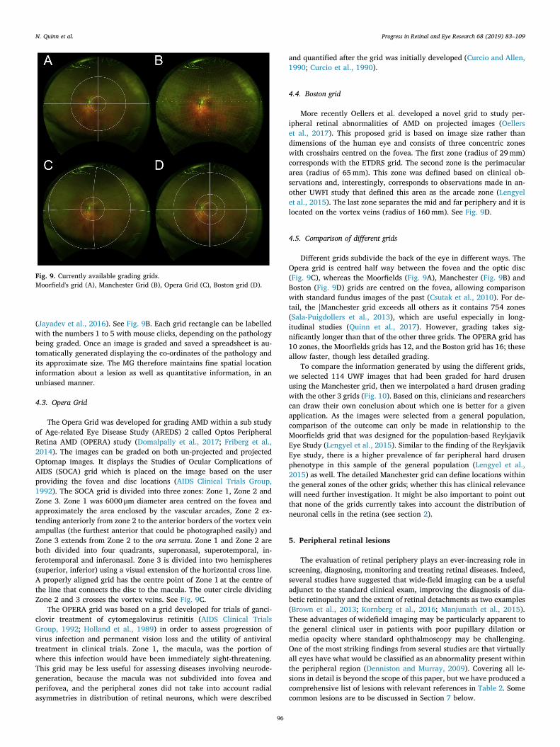
(Jayadev et al., 2016). See Fig. 9B. Each grid rectangle can be labelledwith the numbers 1 to 5 with mouse clicks, depending on the pathologybeing graded. Once an image is graded and saved a spreadsheet is au-tomatically generated displaying the co-ordinates of the pathology andits approximate size. The MG therefore maintains fine spatial locationinformation about a lesion as well as quantitative information, in anunbiased manner.
4.3. Opera Grid
The Opera Grid was developed for grading AMD within a sub studyof Age-related Eye Disease Study (AREDS) 2 called Optos PeripheralRetina AMD (OPERA) study (Domalpally et al., 2017; Friberg et al.,2014). The images can be graded on both un-projected and projectedOptomap images. It displays the Studies of Ocular Complications ofAIDS (SOCA) grid which is placed on the image based on the userproviding the fovea and disc locations (AIDS Clinical Trials Group,1992). The SOCA grid is divided into three zones: Zone 1, Zone 2 andZone 3. Zone 1 was 6000 μm diameter area centred on the fovea andapproximately the area enclosed by the vascular arcades, Zone 2 ex-tending anteriorly from zone 2 to the anterior borders of the vortex veinampullas (the furthest anterior that could be photographed easily) andZone 3 extends from Zone 2 to the ora serrata. Zone 1 and Zone 2 areboth divided into four quadrants, superonasal, superotemporal, in-ferotemporal and inferonasal. Zone 3 is divided into two hemispheres(superior, inferior) using a visual extension of the horizontal cross line.A properly aligned grid has the centre point of Zone 1 at the centre ofthe line that connects the disc to the macula. The outer circle dividingZone 2 and 3 crosses the vortex veins. See Fig. 9C.
The OPERA grid was based on a grid developed for trials of ganci-clovir treatment of cytomegalovirus retinitis (AIDS Clinical TrialsGroup, 1992; Holland et al., 1989) in order to assess progression ofvirus infection and permanent vision loss and the utility of antiviraltreatment in clinical trials. Zone 1, the macula, was the portion ofwhere this infection would have been immediately sight-threatening.This grid may be less useful for assessing diseases involving neurode-generation, because the macula was not subdivided into fovea andperifovea, and the peripheral zones did not take into account radialasymmetries in distribution of retinal neurons, which were described
and quantified after the grid was initially developed (Curcio and Allen,1990; Curcio et al., 1990).
4.4. Boston grid
More recently Oellers et al. developed a novel grid to study per-ipheral retinal abnormalities of AMD on projected images (Oellerset al., 2017). This proposed grid is based on image size rather thandimensions of the human eye and consists of three concentric zoneswith crosshairs centred on the fovea. The first zone (radius of 29 mm)corresponds with the ETDRS grid. The second zone is the perimaculararea (radius of 65 mm). This zone was defined based on clinical ob-servations and, interestingly, corresponds to observations made in an-other UWFI study that defined this area as the arcade zone (Lengyelet al., 2015). The last zone separates the mid and far periphery and it islocated on the vortex veins (radius of 160 mm). See Fig. 9D.
4.5. Comparison of different grids
Different grids subdivide the back of the eye in different ways. TheOpera grid is centred half way between the fovea and the optic disc(Fig. 9C), whereas the Moorfields (Fig. 9A), Manchester (Fig. 9B) andBoston (Fig. 9D) grids are centred on the fovea, allowing comparisonwith standard fundus images of the past (Csutak et al., 2010). For de-tail, the |Manchester grid exceeds all others as it contains 754 zones(Sala-Puigdollers et al., 2013), which are useful especially in long-itudinal studies (Quinn et al., 2017). However, grading takes sig-nificantly longer than that of the other three grids. The OPERA grid has10 zones, the Moorfields grids has 12, and the Boston grid has 16; theseallow faster, though less detailed grading.
To compare the information generated by using the different grids,we selected 114 UWF images that had been graded for hard drusenusing the Manchester grid, then we interpolated a hard drusen gradingwith the other 3 grids (Fig. 10). Based on this, clinicians and researcherscan draw their own conclusion about which one is better for a givenapplication. As the images were selected from a general population,comparison of the outcome can only be made in relationship to theMoorfields grid that was designed for the population-based ReykjavikEye Study (Lengyel et al., 2015). Similar to the finding of the ReykjavikEye study, there is a higher prevalence of far peripheral hard drusenphenotype in this sample of the general population (Lengyel et al.,2015) as well. The detailed Manchester grid can define locations withinthe general zones of the other grids; whether this has clinical relevancewill need further investigation. It might be also important to point outthat none of the grids currently takes into account the distribution ofneuronal cells in the retina (see section 2).
5. Peripheral retinal lesions
The evaluation of retinal periphery plays an ever-increasing role inscreening, diagnosing, monitoring and treating retinal diseases. Indeed,several studies have suggested that wide-field imaging can be a usefuladjunct to the standard clinical exam, improving the diagnosis of dia-betic retinopathy and the extent of retinal detachments as two examples(Brown et al., 2013; Kornberg et al., 2016; Manjunath et al., 2015).These advantages of widefield imaging may be particularly apparent tothe general clinical user in patients with poor pupillary dilation ormedia opacity where standard ophthalmoscopy may be challenging.One of the most striking findings from several studies are that virtuallyall eyes have what would be classified as an abnormality present withinthe peripheral region (Denniston and Murray, 2009). Covering all le-sions in detail is beyond the scope of this paper, but we have produced acomprehensive list of lesions with relevant references in Table 2. Somecommon lesions are to be discussed in Section 7 below.
Fig. 9. Currently available grading grids.Moorfield's grid (A), Manchester Grid (B), Opera Grid (C), Boston grid (D).
N. Quinn et al. Progress in Retinal and Eye Research 68 (2019) 83–109
96

6. New insight into retinal disease from wide-field imaging
As wide-field imaging is increasingly integrated into standardpractice, it has been noted that the presentation of disease in the pos-terior pole does not always mirror the severity of that seen in the retinalperiphery and vice versa. Using UWFI new insights have been gainedinto a number of common retinal conditions:
6.1. Retinal vein occlusion (RVO)
RVO is a common retinal vascular disease; causing vision loss inmany of those affected. RVO occurs more frequently in patients withischaemic heart disease, hypertension and diabetes mellitus. Depending
on the location of the occlusion it can be classified as either central(CRVO) or branch retinal vein occlusion (BRVO). RVO is characterisedby retinal vascular tortuosity, retinal haemorrhages, cotton wool spots,optic disc swelling and macular oedema. In a CRVO retinal haemor-rhages will be seen in all retinal quadrants whereas in a BRVO retinalhaemorrhages will be seen in the area where the vein is occluded(Karia, 2010). Prasad et al. investigated whether peripheral angio-graphic features of BRVO and hemicentral retinal vein occlusion(HRVO) are associated with macular oedema and neovascularisationusing UWFA imaging and concluded that areas of untreated retinalnonperfusion may encourage the production of biochemical mediatorswhich in turn trigger macular oedema and neovascularisation (Prasadet al., 2010). Singer et al. (2014) examined the extent of peripheralretinal nonperfusion in RVO on the effect of macular oedema severityusing UWFA and OCT and reported that people with a higher ischaemicindex correlated with greater macular thickness and worse vision inRVO. Following treatment, OCT imaging showed that the more is-chemic an eye was the greater the decrease in retinal thickness, and thiswas accompanied by improved visual acuity.
6.2. Age-related macular degeneration
AMD is the leading cause of irreversible central vision loss amongthe elderly population. To elucidate the prevalence and significance ofAMD-like lesions in the periphery, several studies used UWFI in patientswith AMD. In 2013, Tan et al. found that peripheral FAF abnormalitiesusing UWFI (Optos 200Tx, Optos, Dunfermline, Scotland) occurred in68.9% of patients with AMD. There were several distinct patterns ofperipheral FAF, including granular (46.2%), mottled (34.0%) andnummular (18.1%). Significant risk factors for FAF abnormalities wereAMD type (neovascular or non-neovascular), older age and female sex.Some colour photography features had a high correlation with specificFAF patterns: these included granular FAF with peripheral drusen andmottled FAF with RPE depigmentation (Tan et al., 2013). Another studyinvestigated peripheral FAF in patients with AMD and found that wide-field FAF imaging revealed a significantly increased central and per-ipheral FAF signal as well as an increased peripheral FAF irregularity inpatients with AMD with or without anti vascular endothelial growthfactor (anti-VEGF) treatment compared with eyes in the control group(Reznicek et al., 2012). In 2015, Lengyel et al. investigated the re-levance of peripheral retinal AMD-like within a population based co-hort. Presence or absence of hard, crystalline, and soft drusen; retinalpigment epithelial changes; choroidal neovascularisation (CNV);atrophy; and hypoautofluorescence and hyperautofluorescence weregraded in the peripheral retina. In 18.9% of eyes examined there was nopathology detected in the macula or periphery; however in 57.4% ofeyes, AMD-like pathology was detected in both the macula and per-ipheral retina. In 13.6% of eyes, AMD like changes were found in themacula alone and in 10.1% of eyes, AMD like lesions were found in theperipheral retina only (Lengyel et al., 2015). More recently, Guduruet al. examined subjective grading of UWFA images in AMD patientsand if there was an association between peripheral FAF abnormalitiesand AMD. They found that there was a moderate agreement in the in-terpretation of peripheral FAF abnormalities. FAF abnormalities ap-peared both in AMD patients and controls; however people with AMDwere at a greater risk of FAF abnormalities (Guduru et al., 2017).
These studies provided interesting insights into the potential asso-ciations between macular and peripheral changes (See section 2.6). It isclear that further work is needed to determine if the development andprogression of retinal pathology related to AMD in the peripheral retinais associated with the development and progression of changes found inthe macula. This should provide both mechanistic insights into thepathophysiology of the condition as well as practical insights intowhether imaging and assessing the peripheral retina should be apriority for clinicians treating AMD patients.
Fig. 10. Spatial distribution of hard drusen using the Moorfields, Opera grid,Boston and Manchester grid respectively.Moorfield's grid: centre ring (black) represents zone 3 (ETDRS equivalent),middle ring represents zone 4 (mid-periphery) and outer represents zone 5 (far-periphery). Opera grid: centre ring represents zone 1, middle ring representszone 2 and outer represents zone 3.Boston grid: centre ring (black) represents zone 1 (ETDRS equivalent), thesecond ring represents zone 2 (perimacular area), the third ring represents zone3(mid-periphery) and outer ring represents zone 5 (far-periphery). The locationof the foveola labelled with “x” and the optic disc labelled with “o”. Red: highprevalence of hard drusen; White: absence of hard drusen; ST-superotemporal;SN-superonasal; IN-inferonasal; IT-inferotemporal.
N. Quinn et al. Progress in Retinal and Eye Research 68 (2019) 83–109
97

Table 2Shows a description of retinal features and diseases and if UWF imaging was used on these retinal lesions and diseases.
Lesion/Disease Description Has it been investigated usingUWF retinal imaging?
Chorioretinal Atrophy A condition where both the choroid and retina are damaged. It can be classified as: diffuse or patchy. It ischaracterised by choroidal depigmentation and thinning of the retina. (Tokoro and SpringerLink, 1998)
Y (Salcedo-Villanueva et al.,2015)
Retinal detachment It is characterised by the separation of the neurosensory retina (NSR) from the RPE. This causes a build-upof subretinal fluid between the NSR and RPE. The key types of detachment are; rhegmatogenous,tractional, exudative and combined tractional-rhegmatogenous (Bowling, 2015f)
Y (Bonnay et al., 2011;Bowling, 2015f)
Congenital Hypertrophy of the RetinalPigment Epithelium (CHRPE)
CHRPE is a benign lesion that appears on the retina as a round usually well-defined lesion that typically isdark grey in colour. It may contain lacunae and a ‘halo’ like lining of hypopigmentation surrounding it.CHRPE can be classified into three entities (solitary, grouped or multiple pigmented fundus lesions) eachwith their own distinct features and implications (Youhnovska et al., 2013).
Y (Shneor et al., 2014; Turgut,2015)
Bear Tracks Known as grouped (multifocal) CHRPE. They appear a lot smaller than regular CHRPE and without thelacunae and ‘haloes’. They appear in groups with a pattern resembling animal footprints hence the name‘Bear Tracks’ (Bowling, 2015c).
Y (Ly et al., 2015)
Peripheral Drusen It is characterised by the gathering of small/yellow deposits on the retina made up of waste products fromdegenerating RPE cells, lipids and some proteins. It is appears similar to the drusen found at the posteriorpole only differing location (Rudolf et al., 2008a).
Y (Aslam et al., 2014)
Choroidal Melanoma It appears as an irregular or oblong feature that has abrupt elevated edges. It can be pigmented or non-pigmented with overlying subretinal fluid and orange pigment (Bowling, 2015a).
Y (Bowling, 2015a; Kerntet al., 2010)
Vitreous Haemorrhage This occurs due to the disruption of healthy or diseased retinal vessels that causes extravasation and/orleakage of blood into the vitreous of the eye. It may also be due to the growth of abnormal retinal vessels(Manuchehri and Kirkby, 2003). It may be diffusely spread throughout the vitreous cavity, or the bloodmay conform to the anatomy of the vitreous. For example, haemorrhage in the subhyaloid space mayresult in a scaphoid (boat-shaped) haemorrhage.
Y (Kim et al., 2014)
Asteroid Hyalosis (AH) AH is a degenerative condition that affects the vitreous humour of the eye. It consists of spherical bodies inthe vitreous cavity most commonly found inferiorly. (Kador and Wyman, 2008)
Y (Ogino et al., 2014)
Retinoschisis This is a vitreo-retinal dystrophy characterised by the splitting of the sensory retina into two layers; innerand outer layer. It is most likely due to vitreous traction where the retina is pulled apart. It may appeareither shallow or bullous and retinal vessels appear dark due to light being blocked (Selaru et al., 2000).
Y (Agarwal et al., 2015)
Sclerosed/Ghost Vessels Vessels become sclerotic when retinal vessels pass through an area of degenerating retina. It is known assheathing of the vessels. When the anterior surface becomes involved, the entire vessel appears opaque.When sheathing encircles the wall it produces a wire like vessel.
N
Snail Tracks This is a vitreoretinal degeneration that is similar to lattice degeneration. It appears like shiny ‘snowflakes’on the retina. It is usually well-defined and oblong in configuration (Bowling, 2015e).
Y (Kothari et al., 2012)
Pavingstone Pavingstone degeneration is characterised by patches of chorioretinal atrophy that coalesce into groups.Pigmentation may surround the lesion. It is more commonly found in the inferior retina (Bowling, 2015c).
Y (Kothari et al., 2012)
Lattice This is where there is a break of the internal limiting membrane with atrophy of the neurosensory retinaapparent. It appears spindle shaped and often sclerosed vessels form. It is commonly bilateral found bothsuperiorly and inferiorly (Bowling, 2015e).
Y (Kothari et al., 2012)
Peripheral Cystoid degeneration This is characterised by the appearance of small vesicles with indistinct boundaries on a white/greyishbackground. It is thought to be present in all adult eyes, increasing in extent with age (Bowling, 2015c).
N
Diabetic Retinopathy It is a microvascular disease that is characterised by the damage of small blood vessels due to high glucoselevels. It is characterised by micoaneursym/macroaneursym, exudates, cotton wool spots, intraretinalmicrovascular abnormality, dot and blot haemorrhages and venous loops or beading (Bowling, 2015b).
Y (Liegl et al., 2014; Silvaet al., 2014)
Gyrate Atrophy (GA) GA is a chorioretinal dystrophy that is relatively rare. It is characterised by well-defined patches ofchorioretinal atrophy in the peripheral retina. GA affects myopes and as people age these lesions tend toincrease in size and number and coalesce throughout the retina (Kaiser-Kupfer Mi, 2002).
Y (Paciuc-Beja et al., 2014)
Choroideremia (CHM) Choroideremia is an X-linked degeneration characterised by atrophy of photoreceptors, RPE and choroid.Areas of RPE atrophy are seen initially in the periphery and progress centrally.
Y (Morgan et al., 2014)
Retinitis pigmentosa (RP) RP isn't a single disease but a group of hereditary disorders. It is characterised by the degeneration of boththe RPE and photoreceptors. (Natarajan, 2011). On the fundus RPE degeneration can be seen throughoutthe retina, pale optic disc and thinner retinal vessels (Jones et al., 2016).
Y (Jones et al., 2016; Oguraet al., 2014)
Retinal Dialysis This is an expression used when describing retinal holes that appear at the ora serrata. In around 5% ofcases dialysis is bilateral most commonly found in the lower temporal area. Demarcation lines and cystsare also associated with retinal dialysis (Tasman, 1974).
N
Choroidal Rupture Choroidal ruptures occur due to ocular trauma. It is characterised by a break in the choroid, Bruch'smembrane and RPE. It may appear as a yellow or white sub-retinal streak, which may be linear or curvedin shape. (Ament et al., 2006)
N
Metastasises of the Choroid It appears as a yellow/orange subretinal mass, colour depending on where the primary tumour is. It isassociated with the accumulation of subretinal fluid and its location again depends of the primary site(Arepalli et al., 2015).
Y (Coffee et al., 2009)
Retinoblastoma This is a rare form of childhood cancer occurring in approximately 1:18000 live births. It is characterisedby the cells of the retina growing rapidly and out of control (Bowling, 2015d). It classically presents withone or multiple nodular, white or cream coloured masses often associated with increased vascularisation.
N
Sickle Cell Retinopathy (SCR) SCR can be classified as either proliferative or non-proliferative. In the non-proliferative stage the mostcommon clinical finding is salmon patched haemorrhages in the peripheral retina. In the non-proliferativestage the most common clinical finding is retinal neovascularisation in the peripheral retina followingvaso-occlusion (Melo, 2014; Mudvari et al., 2010).
Y (Cho et al., 2011)
Cytomegalovirus retinitis It may appear asymptomatic, however if symptoms do arise they include floaters, vision problems andblind spots. It appears irregular in configuration and is characterised by retinal whitening which appearsgranular, haemorrhages may also be present (Heiden et al., 2007).
Y (Mudvari et al., 2010)
Giant Retinal Tear (GRT) GRT is a full-thickness neurosensory retinal break measuring > 90° that extends circumferentially aroundthe retina for three or more clock hours in the presence of a posteriorly detached vitreous (Shunmugamet al., 2014).
Y (Meyer and Saxena, 2010)
(continued on next page)
N. Quinn et al. Progress in Retinal and Eye Research 68 (2019) 83–109
98

Table 2 (continued)
Lesion/Disease Description Has it been investigated usingUWF retinal imaging?
Stargardt disease/FundusFlavimaculatus
It is characterised by the presence of white to yellow dots that surround pigmentary maculopathy and canextend to the midperiphery (Armstrong et al., 1998).
Y (Zein and Brooks, 2012)
Familial Exudative Vitreoretinopathy(FEVR)
This is a rare disorder that can be inherited. It affects retinal angiogenesis thus the vascularisation of theperipheral retina remains incomplete and poor vascular differentiation is visible (Gilmour, 2015). Inserious cases pre-retinal neovascularisation and fibrosis at the junction between vascular and avascularretina can occur that causes traction of the macula and retinal vessels.
Y (Trese et al., 2012)
Susac syndrome (SS) This consists of a triad of symptoms; encephalopathy, branch retinal artery occlusion (BRAO) and hearingloss. It is a very rare disease and can be misdiagnosed due to this (Nazari et al., 2014).
Y (Kumar et al., 2013)
Marfan syndrome This is a connective tissue disorder affecting the skeleton, eyes and cardiovascular system in particular. Inrelation to the eyes the signs and symptoms are myopia, early glaucoma and/or cataracts and detachedretina (Dean, 2002).
Y (Rahmani et al., 2013)
Retinitis Punctata Albescens This is a disorder of the sensory retina and retinal pigment epithelium. It is thought to be congenital andcharacterised by white dot like lesions that are found scattered deep to the retinal vessels (Smith et al.,1959).
N
Chorioretinal coloboma It is often associated with other changes in the eye, these include microphthalmos, high myopia,glaucoma, cataract, and phthisis bulbi. A major risk of coloboma of the choroid is retinal detachment(Jesberg and Schepens, 1961).
Y (Barnard et al., 2012)
Retinal Haemorrhage They can be RNFL haemorrhages arising from pre-capillary arterioles. They can also be intraretinalhaemorrhages that occur at the venous end of the artery that result in a dot/blot appearance. Deeperdarker round haemorrhages occur due to retinal infarcts that happen in the middle retinal layers(Bowling, 2015b).
Y (Nakagawa and Skrinska,2001)
Retinal Hole This is full thickness degeneration of the sensory retina. It appears round to oval in shape and may have awhite collar representing localized detachment.
Y (Kothari et al., 2012)
Floaters They appear as hairlike, dark grey or black in colour due to the shadow they form by blocking exiting laserlight. They take a variation of sizes and appearances on the retina and hide underlying retinal detail.Floaters are thought to be due to an age-related process or due to ocular trauma, asteroid hyalosis, foreignbodies or retinal tears (Sebag et al., 2014).
N
Horseshoe Tear This is due to vitreous traction of the retina. A tear is pulled from the sensory retina in the shape of ahorseshoe. They are most commonly seen between the ora serrata and equator where the retina is thinner.
N
Choroidal Naevus This is a benign lesion that is characterised by the accumulation of melanocytes. They are usuallyasymptomatic and appear black to slate grey in colour and can have pigmentary changes present. It haswell defined edges and can be flat or slightly risen. Over time drusen may be present on the lesion. A nevusis closely followed as it may evolve into a malignant melanoma (Gordon-Shaag et al., 2014).
Y (Gordon-Shaag et al., 2014)
White without Pressure (WWOP) WWOP is a retinal phenomenon that is described as an area of retinal whitening which blocks the view ofthe underlying choroid. It is thought to be due to vitreous traction. WWOP occurs in the far periphery andnever takes the same configuration between eyes (Orlin et al., 2013).
Y (Orlin et al., 2013)
Exudates Exudates appear as waxy yellow lesions that take a ring-like configuration with relatively distinct margins.They are caused by chronic localized retinal oedema (Bowling, 2015b).
N
Cotton Wool Spots They appear as small fluffy whitish lesions that block the view of underlying blood vessels. They are madeup of debris from the nerve fibre layer (Bowling, 2015b).
N
Cholesterol Embolus They are small, yellow reflective lesions. Usually there are multiple present on the retina. They are seen atarteriolar bifurcations and can move or even disappear (Rousseau et al., 2013).
N
Acute posterior multifocal placoidpigment epitheliopathy (APMPPE)
APMPPE is a posterior uveitis. Clinically, it appears as grey-white lesions on the retina. There is usuallymultiple present and appears in both eyes. At the site of APMPPE there is usually pigment clumping(Mrejen et al., 2013).
Y (Mrejen et al., 2013)
Vogt-Koyanagi-Harada (VKH) In most cases VKH is a bilateral disease affecting multiple organs of the body. In the eye it is characterisedby panuveitis with iridocyclitis, serous retinal detachment, diffuse choroidal swelling and optic dischyperaemia (Agrawal and Biswas, 2011).
Y (Heussen et al., 2011)
Birdshot Retinopathy (BSRC) BSRC is an uncommon chronic posterior uveitis. It is characterised by multiple ovoid spots primarily in theposterior pole/mid-periphery. It usually appears bilateral with a distinctive presentation and a stronggenetic association (Gasch et al., 1999).
Y (Piffer et al., 2014)
Central Serous –Chorioretinopathy(CSCR)
CSCR is an acquired chorioretinal disorder. It is due to a serous detachment of the retina that appears likea clear blister. Pigment epithelial detachment and subretinal fluid may also be present (Daruich et al.,2015).
Y (Nam et al., 2015)
Retinopathy of Prematurity (ROP) ROP is a proliferative disorder that affects the developing vessels of the retina in premature infants. Themain cause of ROP is thought to be the abnormal growth of blood vessels. The growth of these vessels canlead to retinal detachments and ultimately blindness (Casteels et al., 2012).
Y (Fung et al., 2014;Theodoropoulou et al., 2013)
Microaneurysms This appears as a tiny area of blood protruding from the retinal capillary wall. They form either by focaldilation or by fusion of two arms of a capillary loop (Bowling, 2015b).
Y (Leicht et al., 2014)
Coats' Disease Coat's disease is characterised by a defect in retinal vascular development which ultimately leads to leakyvessels, subretinal exudation and detachment of the retina. It is thought to be more frequent in men andappears unilateral (Black et al., 1999).
Y (Bass et al., 2011)
Pathological Myopia Myopia is characterised by the extreme increase in axial length of the eye and the formation of posteriorstaphyloma(s). Lesions also may appear in the posterior pole including, myopic choroidalneovascularisation, myopic tractional maculopathy, myopic optic neuropathy and myopic chorioretinalatrophy (Kaneko et al., 2014).
Y (Kaneko et al., 2014)
Acute zonal occult outer retinopathy(AZOOR)
It is an idiopathic syndrome which involves loss of function in one or more zones of the outer retina. It ischaracterised by photopsias and/or scotoma. Some cases present with a grey/white ring surrounding theinvolved part of the retina i.e. acute annular outer retinopathy (AAOR). Chronic stages have signs ofatrophy of the RPE and/or intraretinal pigment migration.
Y (Shifera et al., 2017)
N. Quinn et al. Progress in Retinal and Eye Research 68 (2019) 83–109
99

6.3. Retinopathy of prematurity
Retinopathy of Prematurity (ROP) affects the vasculature of pre-maturely born babies’ eyes. This disease is biphasic: first causing de-layed retinal vascular growth, followed by hypoxia-induced patholo-gical vessel growth (Chen and Smith, 2007). It may be mild with novisual defects, however it may also be aggressive with the formation ofnew vessels, possibly progressing to a retinal detachment. The Optomaphas become increasingly popular with studies reporting its capability ofcapturing clinically useful images documenting retinal features knownto define ROP (Patel et al., 2013), using the “flying baby” technique tofacilitate image acquisition. Historically, Retcam has been the standardimaging modality for monitoring ROP. However, the comparison ofRetcam and Optomap for their ability to capture high quality ROPimages in ROP showed that Retcam takes significantly longer to capturean image and is particularly difficult to use in older babies due to theexcessive head movements. They reported reduced distress responseand found that Optomap could potentially help monitor ROP progres-sion and document regression following treatment more reliably (Patelet al., 2013). Dai et al. evaluated the efficacy of wide field retinalimaging utilizing the Retcam 2 wide field digital camera for screeningof ROP and found a sensitivity and specificity of 100% and 97.9% re-spectively when compared to the gold standard binocular indirectophthalmoscopy in detection of treatment requiring cases. Their con-clusion supported that wide field imaging of ROP infants is accurate,reliable and efficient in detecting treatment requirements. This couldpotentially improve cost, detection rate and accessibility of ROP care(Dai et al., 2011).
6.4. Retinitis pigmentosa
Retinitis pigmentosa refers to a diverse family of inherited eyedisorders characterised by degeneration and dysfunction of rod andcone photoreceptors. Rod photoreceptors are more commonly affectedthan cones; thus retinitis pigmentosa often affects the peripheral retinafirst due to the high density of rod photoreceptors present there com-pared to cones (Hamel, 2006). Most subtypes of retinitis pigmentosa arecharacterised by pigment deposits (bone spicule pigmentation) initiallyin the peripheral retina (Hamel, 2006). There is limited data in theliterature regarding the use of UWFI in retinitis pigmentosa. In 2013Kaufman et al. used the Optomap UWFI and UWFA to report the pre-sence of vascular leakage in the peripheral retina of 15 of 25 patients,suggesting not only vascular dysfunction but also an inflammatorycomponent may play a role in the pathogenesis of this disease (Kaufmanet al., 2013). In 2016 a study used UWFA on a patient with retinitispigmentosa and intermediate uveitis. They picked up peripheral pre-retinal exudates, clinical features of retinitis pigmentosa, disc leakageand cystoid macular oedema identifying both pathologies in a singleinvestigation using UWFI (Tripathy et al., 2016). This further stressesthe advantages of using UWFI techniques for its ability to simulta-neously capture pathology in the central and peripheral retina.
6.5. Uveitis
Patients with uveitis can be challenging to image and manage andaccurate diagnosis of peripheral affection can be vital for diagnosis andmanagement. In the peripheral retina uveitis is characterised by vas-cular leakage, non-perfusion and neovascularisation to name a few(Hamel, 2006). The use of UWFA allowed for more complete evaluationof patients with uveitis showing more excessive disease activity thanapparent from fundus examination, leading to more aggressive andpotentially more efficacious treatment (Kitchens et al., 2009). Campbellet al. investigated whether the use of UWFI influenced the managementof disease in patients with non-infectious posterior uveitis. Managementof disease in clinical judgement alone as well as both clinical judgementand FAF traditional fundus imaging led to an alteration of management
for a total of 17 patients out of 43. However, with the addition of UWFIanalysis, treatment was modified in those 17 patients as well as an extra14 patients out of the 43. This led to a 33% increase in treatmentmodification even in such a small study sample. The study concludedthe use of UWFI can aid in management of non-infectious posterioruveitis; however, further studies are required to determine if thesefindings would improve patient outcomes (Campbell et al., 2012).
6.6. Pathological myopia
Myopia is of significant global public health concern due to its in-creasing prevalence and earlier age-of-onset (Leo and Young, 2011). Inwestern populations, the prevalence of myopia is low in childrencompared to the significantly higher prevalence rates in Asian popu-lations where a prevalence of 6% in 6 year olds rising to a staggering70% by the age of 15 has been reported (Kaneko et al., 2014; Leo andYoung, 2011). Pathological myopia causes several characteristicchanges including an increased axial length of the eye and peripheralretinal thinning. Retinal lesions associated with pathological myopiainclude: myopic choroidal neovascularisation (Avila et al., 1984),myopic maculopathy, myopic optic neuropathy and myopic chorior-etinal atrophy; these have all been detected already using traditionalimaging techniques (Kaneko et al., 2014). A relatively small study ofhighly myopic eyes examined the retinal vasculature using UWFI andfound numerous abnormalities of the retinal vasculature in the farperiphery such as retinal capillary telangiectasia and retinal capillarymicroaneurysms. None of the eyes analysed developed retinal neo-vascularisation. Further research is required to examine if peripheralchanges occur before posterior changes as this could lead to quickerdiagnosis, prognosis and treatment of pathological myopia (Kanekoet al., 2014).
6.7. Cytomegalovirus retinitis
Cytomegalovirus (CMV) retinitis is a retinal inflammation that canultimately lead to blindness. It is primarily seen in patients whose im-mune system is compromised. Lesions associated with CMV may appearin the central and peripheral retina, these include: white granular zonesof retinal necrosis, associated haemorrhages and low-grade iritis andvitritis (Heinemann, 1992). A relatively small study compared Optomapimaging with standard fundus imaging in CMV retinitis patients andfound that 48.3% more retinal area and 40% more retinitis were cap-tured and found with UWFI. In two cases the standard fundus photo-graphy missed peripheral CMV lesions, this could have had disastrousconsequences for the eye and the patient and so they concluded thatOptomap imaging CMV patients is of benefit (Mudvari et al., 2010).
6.8. Ocular markers of disease in the peripheral retina
Due to the ease of examination and the possibilities in imaging theeye, it is sometimes referred to as the “window to the soul”(Foster andKhaw, 2009). The ability to non-invasively image both the retinalvasculature and neural layers provides the opportunity to develop earlymarkers of systemic diseases. Such markers have been extensivelystudied for systemic diseases such as cardiovascular disease and dia-betes, while OCT imaging led to interest in markers of neurologicaldiseases such as multiple sclerosis and Alzheimer's disease (AD). AsUWFI technology developed, markers in the retinal periphery are alsobeing sought and comparisons between presentation in the macular andperiphery undertaken. For some conditions, the initial assumption thatevaluation in the macula is sufficient is gradually being undermined asdata accumulate from wide-field imaging from a variety of systemicdisorders.
6.8.1. Diabetes mellitusIt is understood that the worldwide prevalence of diabetes mellitus
N. Quinn et al. Progress in Retinal and Eye Research 68 (2019) 83–109
100

(DM) was approximately 366 million in 2011, predicted to rise to 552million people by 2030 (Whiting et al., 2011). DM is one of the leadingsystemic diseases affecting people's vision. A variety of ocular diseasesis associated directly with DM; DR, Diabetic Macular Oedema (DMO),cataracts, anterior ischemic optic neuropathy (AION), diabetic pa-pillopathy and ocular movement disorders (Jeganathan et al., 2008). Ofthese, DR is the most commonly diagnosed ocular complication and isthe leading cause of legal blindness in working age adults around theworld, but not in parts of the UK anymore (Liew et al., 2014). Pre-viously, the bulk of DR research concentrated on retinal vascularchanges such as microaneurysms, vascular abnormality and increasedvascular permeability, however more recently degenerative changes arebeing recognised as well (Barber, 2003). Our detailed and systematicunderstanding of features of DR started in 1968, when the Airlie HouseSymposium evaluated knowledge and understanding of DR. The mainoutcome of this meeting was a standardized DR classification (Goldbergand Jampol, 1987). Over the years, DR classification underwentchanges, such as in 2003, when Wilkinson et al. produced a moresimplified classification system (Wilkinson et al., 2003): 1) No apparentretinopathy; 2) Mild non-proliferative DR; 3) Moderate non-pro-liferative DR; 4) Severe non-proliferative DR; 5) Proliferative DR.
Recently several research studies concentrated on UWFA and itsreliability in DR evaluation compared to traditional techniques. UWFAproduces a wider view and has been shown to have better reproduci-bility than standard FA methods for classifying DR (Friberg et al.,2008). Wessel et al. compared UWFA (Optomap Panoramic 200A) andconventional 7SF angiography and found that Optomap captured morenon-perfusion, neovascularisation and panretinal photocoagulationthan 7SF imaging (Wessel et al., 2012b). These findings were largelyreplicated by UPMC Eye Centre, Pittsburgh, showing that retinalischemia was better detected using Optomap (Friberg et al., 2008),confirming that Optomap captured and revealed significantly morepathology in the diabetic eye than the conventional 7SF imaging.
In 2015, three studies evaluated the effect different imaging mod-alities have on detecting DR related retinal abnormalities. Talks et al.(2015) examined identifications of retinal new vessels comparing theuse of the Optomap, the UK's DR screening two field imaging and 7SFphotography and showed that 30% more new vessels were detected onOptomap than on 7SF imaging (Talks et al., 2015). Price et al. foundsubstantial agreement (0.8) between the severity of DR based on Op-tomap and 7SF but noted that a minority of patients were diagnosedwith more severe DR using Optomap. (Price et al., 2015). Manjunathet al. compared OCT and Optomap imaging to clinical examination ofDR on a large sample of 2040 eyes and showed that UWFI was moresensitive when compared to clinical examination. They recommendedthat using both UWFI and OCT deciding on DR severity would lead toenhanced quality of assessment (Manjunath et al., 2015).
Kong et al. carried out a pilot study for evaluating DR by the use ofUWFA. UWFI were obtained from 59 patients with diabetes (118 eyes);11 eyes with no DR, 71 eyes with non-proliferative DR (NPDR) and 36with proliferative DR (PDR). Findings showed that on UWFI micro-aneurysms were seen in more than half (54.5%) of those patients withno apparent DR on conventional imaging; with this percentage pro-gressively increasing with NPDR (84.2%) and PDR (100%) patients. InNPDR and PDR patients new vessels in the periphery and ischemia weredetected using the Optomap FA (Kong et al., 2012). This group con-cluded UWFA had significant advantages in detecting and evaluatingperipheral retinal lesions due to its wider field of view and this wasfound to be especially advantageous in cases where detectable pa-thology was seen on the periphery before any pathology was seen onthe posterior pole.
The relationship between peripheral retinal ischemia and the pre-sence of diabetic macular oedema (DMO) is a topic currently beinginvestigated by several studies. Wessel found that on UWFA, out of the122 treatment-naïve eyes of 70 subjects with DMO, 66 eyes (62%)displayed areas of retinal peripheral ischemia, (p < 0.001). These
findings suggested that UWFI may provide useful information in as-sessing peripheral retinal changes associated with DMO (Wessel et al.,2012b). In analysis of subjects in the Efficacy and Safety Trial of In-travitreal Injections Combined with Panretinal Photocoagulation forClinically Significant Macular Oedema Secondary to Diabetes Mellitus(DAVE) clinical trial, Fan and colleagues observed that the severity ofischemia in eyes with DMO appeared to increase with increasing dis-tance from the fovea (Fan et al., 2017). Non-perfusion in the mid-per-iphery, but not in other regions, was associated with severity of DMO.
A target pattern scan laser (Pascal) retinal photocoagulation (TRP)was designed to treat areas of retinal capillary non-perfusion and in-termediate retinal ischemia regions in PDR. UWFI was used to detectareas of peripheral retinal ischemia and neovascularisation in order toguide TRP laser to the four quadrants of the retina. The researchersfound that UWFI-guided laser treatment was a promising procedure fortreatment of PDR as it destroyed less normal retinal tissue than tradi-tional methods yet obtained favourable disease regression rates at 3–6months (Muqit et al., 2013).
In 2013 Silva et al. assessed DR lesions identified by UWFI andcompared these to ETDRS 7SF. The lesions identified included hae-morrhages and/or microaneurysms, venous beading, intraretinal mi-crovascular abnormality and new vessels elsewhere. UWFI demon-strated substantial agreement with ETDRS film photographs indetermining DR severity. Additional lesions identified by UWFI sug-gested a more severe assessment of DR in 10% of eyes than suggested bylesions found within the ETDRS view (Silva et al., 2013). However, theimplications of peripheral lesions on DR are unknown and furtherstudies will need to be carried out to evaluate this. More recently, Silvaet al. (2015) reported that the presence and increasing extent of per-ipheral lesions were associated with increased risk of DR progressionover 4 years, suggesting that detailed peripheral retinal evaluationprovides important information necessary to assess risk of DR pro-gression (Silva et al., 2015). Currently, there are studies ongoing to testthe clinical significance of peripheral DR lesions in relation to the de-velopment, diagnosis, progression and choice of DR treatment mod-alities. Once these studies report their results, a re-classification of DRmight be required in order to incorporate the peripheral lesion in theclinical pathway.
Most studies exploring visual loss in DR focused on visual acuity;however, numerous studies using perimetry showed DR's detrimentaleffects on peripheral visual function and field (Duh et al., 2017;Nentwich and Ulbig, 2015; Shrestha and Kaiti, 2014). Importantly,these studies noted that using perimetry for tracking peripheral visualfunction captures early diabetes related pathologies better than tradi-tional fundoscopy or visual acuity testing (Greite et al., 1981; Guy,2012; Henricsson and Heijl, 1994; Jackson and Gardner, 2014).
A perimetry study dating back to the 1980s illustrated deficits inperipheral visual functioning before central visual changes could benoted in DR patients. Such visual impairments include visual dysfunc-tion in dim light and difficulty in recognizing contour of objects in lowcontrast conditions even when visual acuity is unaffected (Hyvarinenet al., 1983). However, most research emphasizes central acuity lossover peripheral visual field monitoring due to DMO related visual loss'immediate impact on social functioning. (Duh et al., 2017; Lee et al.,2015). Early perimetry studies illustrated that these peripheral fielddeficits and smaller isopter areas could be observed even in DR patientswhere neovascularisation, haemorrhage, or microaneurysm weren'tobserved (Wisznia et al., 1971).
Recent studies utilizing automated perimetry support these earlyfindings; additional studies have also shown a decreased ability indiabetics with DR to recognize objects with low spatial frequency in theperiphery, similar to studies performed on AD and AMD referenced inthis review. These effects were seen in patients with type 2 diabeteswho were diagnosed for ten years or less and before any vasculopathiescould be visualized in the peripheral retina by imaging (Bell et al.,2010). In agreement, a study from 2008 found that an automated
N. Quinn et al. Progress in Retinal and Eye Research 68 (2019) 83–109
101

Humphrey Perimetry program found reduced peripheral visual func-tioning in those with type 1 diabetes before vascular changes werenoted in both peripheral and central retina. It should be noted thoughthat this study only studied as far out to 30 degrees of visual functioning(Parravano et al., 2008).
A 2013 study on patients with type 1 diabetes illustrated that per-ipheral visual dysfunction could be seen in the early stages of DR beforeimages could capture vascular changes. Disease onset was so early inpatients with diabetes that deficits in perimetric testing of the periph-eral visual fields weren't significant; however, those with diabetesshowed poorer performance and significantly higher variability in totalperformance. Even without visualization of peripheral retinal patholo-gies, perimetry is still highly sensitive at detecting the onset and pro-gression of DR (Isabel Pinilla et al., 2013).
Recently, perimetric studies sought to correlate these peripheralvisual deficits with the onset of retinal pathologies. Some claimed thatdeficits noted on perimetric monitoring of vision might predict futurevasculopathies, but lately it seems that visual peripheral deficits areassociated with areas of lowered autofluorescence, outside the maculaof course (which is protected by an accumulation of luteal pigments).One 2011 study was able to correlate visual deficits, as tracked byHumphrey perimetry, with areas of lowered FAF in DR patients.Although the AF images were taken by traditional fundoscopy (at 40°),they were visualized outside the macula. The study showed that whileno vasculopathies or lesions could be visualized in these areas, the vi-sual dysfunction corresponded to areas of lowered AF, presuming theseareas were vulnerable to ischemia or retinal cell dysfunction (Midenaand Vujosevic, 2011).
Many of these studies suggest that the visual deficits perceived inthe setting of early DR are due to vascular insufficiency and that thesecan be imaged early if the correct imaging methods are used. However,if we were better able to screen for onset and subsequently determinethe progression of DR, it might aid us in treating the disease early andmaintaining visual function for longer in patients with diabetes.
6.8.2. Alzheimer's disease and cognitive decline50 million people living with dementia worldwide and this number
will be 152 by 2050 (Alzheimer's Disease International, 2018). AD is themost common type of senile dementia in aging adults amounting to10% of people aged 65 or over being affected. AD affects 24.3 millionindividuals worldwide, with around 4.6 million new cases reportedeach year (Reitz and Mayeux, 2014). AD clinically manifests by pro-gressive loss of memory, changes in behaviour, mood and thinkingprocesses (McKhann et al., 1984). Diagnosing AD is based on clinicalpresentation and therefore is difficult in any given patient. Definitediagnosis can only be made pathologically by examination of the braintissue at autopsy (Braak and Braak, 1997). In the early stages of diseaseassessments relies on psychological assessments and costly and highlyinvasive clinical tests (Neugroschl and Wang, 2011). Therefore, there isa need to find biomarkers that is objective, could be used repeatedly,could be used for early detection and monitoring progression and aprocedure that patients are comfortable with and can comply with iteasily even at a more advanced stage of disease.
The retina is part of the central nervous system; it shares embry-ological origin and anatomical characteristics with the brain (Byerlyand Blackshaw, 2009; Risau, 1997). As such, it has been proposed thatchanges in the retina may reflect changes in the brain and provideaccessible biomarkers for AD (Colligris et al., 2018; Dehghani et al.,2018; Frost, 2010; Ikram et al., 2012; Liao et al., 2018; Lim et al.,2016). In general, imaging of the retina is non-invasive, well toleratedand readily repeatable in follow up studies. Using OCT/OCTA in AD,several studies report thinning of RNFL, macular volume loss and al-tered retinal vasculature, implying that similar changes take place inthe retina than in the brain (Coppola et al., 2015; Cunha et al., 2016;den Haan et al., 2018; den Haan et al., 2017; Doustar et al., 2017;O'Bryhim et al., 2018; Wang et al., 2018). Quantitative and qualitative
changes in retinal microvasculature using conventional fundus photo-graphs showed an association between cognitive decline/AD and retinalvascular characteristics (McGrory et al., 2017). Further studies sug-gested that there is increased arterial reaction time in people with AD(Mroczkowska et al., 2015) and there is an overall increase in vesselcalibres when peripapillary B-scans are evaluated (Csincsik et al.,2016). However, all these measurements were carried out on the pos-terior pole of the eye without consideration for the peripheral retina.
Based on observations on flat mounted retinas of cadaveric eyes(Lengyel et al., 2004; Ukalovic et al., 2018) it was proposed thatmonitoring the development and progression of pathology in the per-ipheral regions might serve as a valuable tool in detecting and mon-itoring the progression in age-related diseases such as AMD and AD. Ithas been shown that UWFI is comparable to normal fundus imaging(Csutak et al., 2010) and can detect pathological abnormalities at theperipheral retina (Csincsik et al., 2018; Lengyel et al., 2015). Recentpilot study revealed an association between AD and peripheral harddrusen formation and changes to the vasculature beyond the posteriorpole using UWFI (Csincsik et al., 2018). Similar results were found in anindependent study that showed patient with moderate AD has higherburden of peripheral retinal deposits compared to aged matched con-trols or mild cognitive impairment (MCI) (Whitson et al., 2016). Retinalamyloid deposits also been detected in mid-peripheral region of theretina using modified ophthalmoscope after oral administration ofcurcumin (Koronyo et al., 2017). Based on these findings, it is feasibleto speculate that imaging not only the central but the peripheral retinatoo has potentials to be used as biomarkers for progression of disease.To test the validity of retinal imaging as biomarkers several ongoingdementia studies include retinal imaging components. These will ex-amine drusen deposition, vascular and RPE changes and look for ad-ditional pathological changes in the eye that may not have been iden-tified before, especially at the peripheral regions.
Retinal functional changes also have been reported in AD. Studieshave shown that AD patients have difficulty processing and recognizingenvironmental scenes, especially those with low spatial frequency or alack of details. It is worth noting, however, that visual acuity and colourrecognition in AD is generally comparable to age-matched controls (Leeet al., 2015; Wood et al., 1997). This suggests a preservation of aspectsassociated with central vision in AD. Other studies correlated AD withan increased incidence of traffic accidents and implicated deficits inperipheral visual functioning over the degree of cognitive impairmentin the occurrence of these accidents. Motion processing, contrast sen-sitivity, and depth perception, attributes associated with the peripheralvision, have also been shown to be poor in AD patients(Lakshminarayanan et al., 1996; Mendez et al., 1990; Neargarder et al.,2003; Schlotterer et al., 1984).
The observed effects of AD above focus on a characteristic of visionitself because these studies are neurological and approach the visualloss as a result of cortical dysfunction. However, very few studiessought to localize or quantify a loss in the visual field of AD patients. Inthe 1980s (Nissen et al., 1985), there were a few case studies whereGoldmann Perimetry showed dramatic inferior hemifield losses in ADpatients. These results were echoed in a 1995 Humphrey Perimetrystudy which again found inferotemporal and inferonasal losses pre-valent in AD patients (Ceccaldi, 1996; Trick et al., 1995). These studies,though sparse, both agreed that the peripheral inferior visual field wasgreatly upset in the onset and progression of AD (Ceccaldi, 1996; Nissenet al., 1985; Trick et al., 1995).
A 1996 study tried to correlate these perimetric findings in ADpatients by looking at the visual cortices. By measuring the thickness ofthe cuneal and lingual cortex in AD patients, the study hoped to illus-trate the pathway of visual loss through cortical atrophy. The studyfound thinning of the lingual over the cuneal, vice versa, and equalthinning in its AD patients but did not find a direct relationship or adirect trend (Armstrong, 1996). The presence of cortical thinning in ADpatients was not as surprising as the fact that this study could not point
N. Quinn et al. Progress in Retinal and Eye Research 68 (2019) 83–109
102

to a definite trend in the visual association cortices despite such similarattributes observed in peripheral visual functioning in AD. Why AD isassociated with the inferonasal and inferotemporal hemifield losses inperimetry? Why is AD associated with attributes of peripheral visualfunction over central visual function?
Very little new research has been carried out since then in quanti-fying or localizing peripheral visual loss. In contrast, our understandingof retinal nerve ganglion health increased (Berisha et al., 2007; Liuet al., 2015; Marziani et al., 2013). In 2013, frequency-doubling tech-nology (FDT), a form of perimetry that studies flicker sensitivity (anattribute of the retinal nerve ganglion cells) found that AD patients hadpoor results in their inferior visual fields and concluded that the retinalnerve ganglion cells of the superior retina must be pathologic or mustendure the greatest insult in AD. These results might suggest the ae-tiology of the peripheral visual function and observed visual field lossescould directly be due to insult in the retina (Valenti, 2013).
One last attribute to discuss about AD is the presence of halluci-nations (Chapman et al., 1999). Two different commonly reportedfindings in AD that are of interest to the peripheral vision are “illusions”and “misperceptions.” Illusions involve a patient not being able todistinguish between two objects in their environment. Another type iscalled “misperception,” and this emerges when a subject does not re-cognize something and makes a best guess as to what it could be. Mostresearch understands and views these illusions and misperceptions as amanifestation of damage to the visual cycle occurring in the brain. It isworth noting that similar delusions are seen in AMD and correlated tothe degree of vision loss in the central retina. This phenomenon is calledCharles Bonnet Syndrome (CBS), and it is understood as a manifestationof how the brain copes with vision loss, especially in AMD patients(Berrios and Brook, 1982). CBS also occurs in patients with cataract,glaucoma, and DR. Some studies suggest that up to 60% of people whohave dramatic sight loss experience it (RNIB, 2016). It could be perhapsthat hallucinations seen with AD have a similar source to those ex-perienced in patients with other retinal pathologies. These documentedvisual disturbances in AD indicate that more understanding into therelationship between peripheral vision and the progression of AD isnecessary.
7. Future directions & conclusions
In summary, it is evident that the peripheral retina has great im-portance for the prognosis and overall outcome of systemic diseasesaffecting large number of patients. The significance of the appropriateimaging and analysis of the peripheral retina is being shown in thisreview by depicting the various imaging and investigative techniquesclinicians and researcher now have at their disposal. Currently oph-thalmic imaging is undergoing a major revolution due to the avail-ability and utilization of technologies from other discipline. Combiningthese technological advances with a more complete understanding ofretinal morphology, better molecular understanding of diseases led toan increased need for examining the peripheral retina in clinical setting.The need for continued research for the use of UWFI has been high-lighted by the successes of new studies on diabetic retinopathy. Oncethe results of the ongoing studies are fully understood and implementedin clinical practice, these should result in more complete evaluation,earlier diagnosis, better prognostication and more appropriate care ofpatients. Implementation of UWFI systems into clinical practice willhave to be balanced against with increased costs of such systems,though as is the case for most technologies, one might anticipate thatcosts will come down with time. Consensus on imaging and gradingprotocols will be needed for easier comparison of different studies.Ultimately, clinically relevant methods to test functional changes as-sociated with UWFI abnormalities will be required; these will need tofuel wider societal changes leading to better environmental and townplanning taking into account the changes in peripheral vision.
Competing financial interest of the authors
SriniVas R. Sadda is a consultant for Optos, Carl Zeiss Meditec,Centervue, Heidelberg Engineering, Allergan, Roche, Novartis, Iconic,Thrombogenics and receives research support from Optos, Carl ZeissMeditec, Allergan, Genentech. Christine A. Curcio receives researchsupport from Heidelberg Engineering and Hoffman LaRoche. ImreLengyel receives unrestricted research support from Optos Plc. TundePeto is a consultant for Optos Plc, Bayer, Novartis and received un-restricted grant from Allergan and Alimera. Szilard Kiss has served onthe speaker's bureau of Optos, PLC and a consultant for Allergan. Otherauthors do not have any competing financial interest.
Acknowledgements
The research was supported by the Bill Brown Charitable Trust,Moorfields Eye Hospital Special Trustees, Mercer Fund from Fight forSight (I.L). Special thank for The Joint Library of Ophthalmology,Moorfields Eye Hospital & UCL Institute of Ophthalmology for theretinal drawings. Lajos Csincsik is supported by an unrestricted PhDstudentship from Optos Plc. The work was part-supported by the Eye-Risk project funded from the European Union’s Horizon 2020 researchand innovation programme under grant agreement No 634479 (I.L andT.P).
Appendix A. Supplementary data
Supplementary data to this article can be found online at https://doi.org/10.1016/j.preteyeres.2018.10.001.
References
Ablonczy, Z., Higbee, D., Anderson, D.M., Dahrouj, M., Grey, A.C., Gutierrez, D.,Koutalos, Y., Schey, K.L., Hanneken, A., Crouch, R.K., 2013. Lack of correlation be-tween the spatial distribution of A2E and lipofuscin fluorescence in the human retinalpigment EpitheliumLocalization of A2E in the human RPE. Investig. Ophthalmol. Vis.Sci. 54, 5535–5542.
Ach, T., Huisingh, C., McGwin, G., Messinger, J.D., Zhang, T., Bentley, M.J., Gutierrez,D.B., Ablonczy, Z., Smith, R.T., Sloan, K.R., Curcio, C.A., 2014. Quantitative auto-fluorescence and cell density maps of the human retinal pigment epithelium. Investig.Ophthalmol. Vis. Sci. 55, 4832–4841.
Adler, L.t., Boyer, N.P., Anderson, D.M., Spraggins, J.M., Schey, K.L., Hanneken, A.,Ablonczy, Z., Crouch, R.K., Koutalos, Y., 2015. Determination of N-retinylidene-N-retinylethanolamine (A2E) levels in central and peripheral areas of human retinalpigment epithelium. Photochem. Photobiol. Sci.: Off. J. Eur. Photochem. Assoc. Eur.Soc. Photobiol. 14, 1983–1990.
Agarwal, A., Fan, S., Invernizzi, A., Do, D.V., Nguyen, Q.D., Harms, N.V., Sepah, Y.J.,2015. Characterization of retinal structure and diagnosis of peripheral acquired re-tinoschisis using high-resolution ultrasound B-scan. Graefe’s Arch. Clin. Exp.Ophthalmol. 1–7.
Age-Related Eye Disease Study 2 Research Group, 2013. Lutein + zeaxanthin and omega-3 fatty acids for age-related macular degeneration: the Age-Related Eye Disease Study2 (AREDS2) randomized clinical trial. Jama 309, 2005–2015.
Agrawal, A., Biswas, J., 2011. Unilateral vogt-koyanagi-harada disease: report of twocases. Middle East Afr. J. Ophthalmol. 18, 82–84.
AIDS Clinical Trials Group, 1992. Studies of ocular complications of AIDS foscarnet-ganciclovir cytomegalovirus retinitis trial: 1. Rationale, design, and methods. AIDSclinical trials group (ACTG). Contr. Clin. Trials 13, 22–39.
Al-Hussaini, H., Kilarkaje, N., Shahabi, G., Al-Mulla, F., 2016. Proliferation and migrationof peripheral retinal pigment epithelial cells are associated with the upregulation ofwingless-related integration and bone morphogenetic protein signaling in darkAgouti rats. Med. Princ. Pract.: Int. J. Kuwait Univ. Health Science Centre 25,408–416.
Alzheimer's Disease International, 2018. The State of the Art of Dementia Research: NewFrontiers. World Alzheimer Report 2018.
Ament, C.S., Zacks, D.N., Lane, A.M., Krzystolik, M., D'Amico, D.J., Mukai, S., Young,L.H., Loewenstein, J., Arroyo, J., Miller, J.W., 2006. Predictors of visual outcome andchoroidal neovascular membrane formation after traumatic choroidal rupture. Arch.Ophthalmol. 124, 957–966.
Anderson, D.M.G., Ablonczy, Z., Koutalos, Y., Hanneken, A.M., Spraggins, J.M., Calcutt,M.W., Crouch, R.K., Caprioli, R.M., Schey, K.L., 2017. Bis(monoacylglycero)phos-phate lipids in the retinal pigment epithelium implicate lysosomal/endosomal dys-function in a model of Stargardt disease and human retinas. Sci. Rep. 7, 17352.
Anderson, D.M.G., Ablonczy, Z., Koutalos, Y., Spraggins, J., Crouch, R.K., Caprioli, R.M.,Schey, K.L., 2014. High resolution MALDI imaging mass spectrometry of retinal tissuelipids. J. Am. Soc. Mass Spectrom. 25, 1394–1403.
N. Quinn et al. Progress in Retinal and Eye Research 68 (2019) 83–109
103

Appold, K., 2017. New product applications: widening the imaging field. Retin. Physician14 (59), 61.
Arepalli, S., Kaliki, S., Shields, C.L., 2015. Choroidal metastases: origin, features, andtherapy. Indian J. Ophthalmol. 63, 122–127.
Armstrong, J.D., Meyer, D., Xu, S., Elfervig, J.L., 1998. Long-term follow-up of stargardt'sdisease and fundus flavimaculatus1. Ophthalmology 105, 448–457.
Armstrong, R.A., 1996. Visual field defects in Alzheimer's disease patients may reflectdifferential pathology in the primary visual cortex. Optom. Vis. Sci. 73, 677–682.
Aslam, A., Peto, T., Barzegar-Befroei, N., Gregory, S., Morrison, G., Ritchie, C., Lengyel, I.,2014. Assessing peripheral retinal drusen progression in Alzheimer's dementia: a pilotstudy using ultra-wide field imaging. Investig. Ophthalmol. Vis. Sci. 55 659-659.
Avila, M.P., Weiter, J.J., Jalkh, A.E., Trempe, C.L., Pruett, R.C., Schepens, C.L., 1984.Natural history of choroidal neovascularization in degenerative myopia.Ophthalmology 91, 1573–1581.
Boycott, B.B., Dowling, J.E., 1969. Organization of the primate retina: light microscopy,with an appendix: a second type of midget bipolar cell in the primate retina. Philos.Trans. R. Soc. Lond. Ser. B Biol. Sci. 255, 109–184.
Barber, A.J., 2003. A new view of diabetic retinopathy: a neurodegenerative disease ofthe eye. Prog. Neuro-Psychopharmacol. Biol. Psychiat. 27, 283–290.
Barnard, S., Shneor, E., Brauner, J., Millodot, M., Gordon-Shaag, A., 2012. Bilateralchorioretinal coloboma discovered with ultra-wide field retinal imaging. J. opto-metry 5, 150–154.
Bartlett, H., Howells, O., Eperjesi, F., 2010. The role of macular pigment assessment inclinical practice: a review. Clin. Exp. Optom. 93, 300–308.
Bass, S.J., Sherman, J., Giovinazzo, V., 2011. Bilateral Coats' response in a female patientleads to diagnosis of facioscapulohumeral muscular dystrophy. Optometry. J. Am.Optom. Assoc. 82, 72–76.
Bastek, J.V., Siegel, E.B., Straatsma, B.R., Foos, R.Y., 1982. Chorioretinal juncture: pig-mentary patterns of the peripheral fundus. Ophthalmology 89, 1455–1463.
Bell, A., James, A.C., Kolic, M., Essex, R.W., Maddess, T., 2010. Dichoptic multifocalpupillography reveals afferent visual field defects in early type 2 diabetes. Investig.Ophthalmol. Vis. Sci. 51, 602–608.
Berisha, F., Feke, G.T., Trempe, C.L., McMeel, J.W., Schepens, C.L., 2007. Retinal ab-normalities in early Alzheimer's disease. Investig. Ophthalmol. Vis. Sci. 48,2285–2289.
Bernstein, P.S., Delori, F.C., Richer, S., van Kuijk, F.J.M., Wenzel, A.J., 2010. The value ofmeasurement of macular carotenoid pigment optical densities and distributions inage-related macular degeneration and other retinal disorders. Mechanisms inMacular Degeneration 50, 716–728.
Bernstein, P.S., Li, B., Vachali, P.P., Gorusupudi, A., Shyam, R., Henriksen, B.S., Nolan,J.M., 2016. Lutein, zeaxanthin, and meso-zeaxanthin: the basic and clinical scienceunderlying carotenoid-based nutritional interventions against ocular disease. Prog.Retin. Eye Res. 50, 34–66.
Berrios, G.E., Brook, P., 1982. The Charles Bonnet syndrome and the problem of visualperceptual disorders in the elderly. Age Ageing 11, 17–23.
Bhosale, P., Serban, B., Bernstein, P.S., 2009. Retinal carotenoids can attenuate formationof A2E in the retinal pigment epithelium. Arch. Biochem. Biophys. 483, 175–181.
Black, A.A., Wood, J.M., Lovie-Kitchin, J.E., Newman, B.M., 2008. Visual impairment andpostural sway among older adults with glaucoma. Optom. Vis. Sci. 85, 489–497.
Black, G.C.M., Perveen, R., Bonshek, R., Cahill, M., Clayton-Smith, J., Christopher Lloyd,I., McLeod, D., 1999. Coats' disease of the retina (unilateral retinal telangiectasis)caused by somatic mutation in the NDP gene: a role for Norrin in retinal angiogenesis.Hum. Mol. Genet. 8, 2031–2035.
Bone, R.A., Landrum, J.T., Mayne, S.T., Gomez, C.M., Tibor, S.E., Twaroska, E.E., 2001.Macular pigment in donor eyes with and without AMD: a case-control study. Investig.Ophthalmol. Vis. Sci. 42, 235–240.
Bonnay, G., Nguyen, F., Meunier, I., Ducasse, A., Hamel, C., Arndt, C., 2011. Screening forretinal detachment using wide-field retinal imaging. J. Fr. Ophtalmol. 34, 482–485.
Booij, J.C., Baas, D.C., Beisekeeva, J., Gorgels, T.G., Bergen, A.A., 2010. The dynamicnature of Bruch's membrane. Prog. Retin. Eye Res. 29, 1–18.
Boulton, M., Dayhaw-Barker, P., 2001. The role of the retinal pigment epithelium: to-pographical variation and ageing changes. Eye 15, 384–389 (London, England).
Bowling, B., 2015a. Choroidal Melanoma, Kanski's Clinical Ophthalmology: a SystematicApproach, eighth ed. Elsevier, pp. 487–492.
Bowling, B., 2015b. Diabetic Retinopathy, Kanski's Clinical Ophtalmology: a SystematicApproach, eighth ed. Elsevier, pp. 520–526.
Bowling, B., 2015c. Innocuous Peripheral Retinal Degenerations, Kanski's ClinicalOphthalmology: a Systematic Approach, eighth ed. Elsevier, pp. 682–684.
Bowling, B., 2015d. Neural Retinal Tumours: Retinoblastoma, Kanski's ClinicalOphthalmology: a Systematic Approach, eighth ed. Elsevier, pp. 497–504.
Bowling, B., 2015e. Peripheral Lesions Predisposing to Retinal Detachment, Kanski'sClinical Ophthalmology: a Systematic Approach, eighth ed. Elsevier, pp. 689–690.
Bowling, B., 2015f. Retinal Detachment, eighth ed. Elsevier, pp. 701–713.Braak, H., Braak, E., 1997. Frequency of stages of Alzheimer-related lesions in different
age categories. Neurobiol. Aging 18, 351–357.Bringmann, A., Syrbe, S., Gorner, K., Kacza, J., Francke, M., Wiedemann, P., Reichenbach,
A., 2018. The primate fovea: structure, function and development. Prog. Retin. EyeRes. 66, 49–84.
Brown, K., Sewell, J.M., Trempe, C., Peto, T., Travison, T.G., 2013. Comparison of image-assisted versus traditional fundus examination. Eye Brain 5, 1–8.
Burris, C., Klug, K., Ngo, I.T., Sterling, P., Schein, S., 2002. How Muller glial cells inmacaque fovea coat and isolate the synaptic terminals of cone photoreceptors. J.Comp. Neurol. 453, 100–111.
Byerly, M.S., Blackshaw, S., 2009. Vertebrate Retina and Hypothalamus Development,vol. 1. Wiley interdisciplinary reviews. Systems biology and medicine, pp. 380–389.
Cai, H., Fields, M.A., Hoshino, R., Del Priore, L.V., 2012. Effects of aging and anatomic
location on gene expression in human retina. Front. Aging Neurosci. 4.Calkins, D.J., Schein, S.J., Tsukamoto, Y., Sterling, P., 1994. M and L cones in macaque
fovea connect to midget ganglion cells by different numbers of excitatory synapses.Nature 371, 70–72.
Campbell, J.P., Leder, H.A., Sepah, Y.J., Gan, T., Dunn, J.P., Hatef, E., Cho, B., Ibrahim,M., Bittencourt, M., Channa, R., Do, D.V., Nguyen, Q.D., 2012. Wide-field retinalimaging in the management of noninfectious posterior uveitis. Am. J. Ophthalmol.154, 908–911 e902.
Casteels, I., Cassiman, C., Van Calster, J., Allegaert, K., 2012. Educational paper: re-tinopathy of prematurity. Eur. J. Pediatr. 171, 887–893.
Ceccaldi, M., 1996. La vision dans la maladie d'Alzheimer. Rev. Neurol. 152, 441–446.Chapman, F.M., Dickinson, J., McKeith, I., Ballard, C., 1999. Association among visual
hallucinations, visual acuity, and specific eye pathologies in Alzheimer's disease:treatment implications. Am. J. Psychiatry 156, 1983–1985.
Chen, J., Smith, L.E.H., 2007. Retinopathy of prematurity. Angiogenesis 10, 133–140.Cho, M., Aaker, G., D'Amico, D.J., Kiss, S., 2011. Peripheral vascular abnormalities in β-
thalassemia major detected by ultra wide-field fundus imaging. Retin. Cases BriefRep. 5, 339–342.
Choudhry, N., Golding, J., Manry, M.W., Rao, R.C., 2016. Ultra-widefield steering-basedspectral-domain optical coherence tomography imaging of the retinal periphery.Ophthalmology 123, 1368–1374.
Christine, A., Curcio, M.J., 2012. Structure, Function, and Pathology of Bruch'sMembrane, vol. 1. Elsevier Inc, pp. 465–481.
Chun, M.H., Grünert, U., Martin, P.R., Wässle, H., 1996. The synaptic complex of cones inthe fovea and in the periphery of the macaque monkey retina. Vis. Res. 36,3383–3395.
Coffee, R.E., Jain, A., McCannel, T.A., 2009. Ultra Wide-field Imaging of ChoroidalMetastasis Secondary to Primary Breast Cancer, Seminars in Ophthalmology, 1 ed.Informa UK Ltd UK, pp. 34–36.
Colligris, P., Perez de Lara, M.J., Colligris, B., Pintor, J., 2018. Ocular manifestations ofAlzheimer's and other neurodegenerative diseases: the prospect of the eye as a toolfor the early diagnosis of Alzheimer's disease. J. Ophthalmol. 2018, 12.
Coppola, G., Di Renzo, A., Ziccardi, L., Martelli, F., Fadda, A., Manni, G., Barboni, P.,Pierelli, F., Sadun, A.A., Parisi, V., 2015. Optical coherence tomography inAlzheimer's disease: a meta-analysis. PloS One 10, e0134750.
Croft, D.E., van Hemert, J., Wykoff, C.C., Clifton, D., Verhoek, M., Fleming, A., Brown,D.M., 2014. Precise montaging and metric quantification of retinal surface area fromultra-widefield fundus photography and fluorescein angiography. Ophthalmic Surg.Laser. Imag. 45, 312–317.
Csincsik, L., MacGillivray, T.J., Flynn, E., Pellegrini, E., Papanastasiou, G., Barzegar-Befroei, N., Csutak, A., Bird, A.C., Ritchie, C.W., Peto, T., Lengyel, I., 2018. Peripheralretinal imaging biomarkers for Alzheimer's disease: a pilot study. Ophthalmic Res. 59,182–192.
Csincsik, L., Shakespeare, T., Quinn, N., Hogg, R.E., Crutch, S., Craigh, R., Peto, T.,Lengyel, I., 2016. Retinal imaging in early and late Alzheimer's disease. Investig.Ophthalmol. Vis. Sci. 57 3373-3373.
Csutak, A., Lengyel, I., Jonasson, F., Leung, I., Geirsdottir, A., Xing, W., Peto, T., 2010.Agreement between image grading of conventional (45[deg]) and ultra wide-angle(200[deg]) digital images in the macula in the Reykjavik eye study. Eye 24,1568–1575.
Cunha, L.P., Almeida, A.L.M., Costa-Cunha, L.V.F., Costa, C.F., Monteiro, M.L.R., 2016.The role of optical coherence tomography in Alzheimer's disease. Int. J. Retina andVitreous 2, 24.
Curcio, C.A., 2018. Antecedents of soft drusen, the specific deposit of age-related maculardegeneration, in the biology of human macula. Investig. Ophthalmol. Vis. Sci.
Curcio, C.A., Allen, K.A., 1990. Topography of ganglion cells in human retina. J. Comp.Neurol. 300, 5–25.
Curcio, C.A., Messinger, J.D., Sloan, K.R., McGwin, G., Medeiros, N.E., Spaide, R.F., 2013.Subretinal drusenoid deposits in non-neovascular age-related macular degeneration:morphology, prevalence, topography, and biogenesis model. Retina 33, 265–276(Philadelphia, Pa.).
Curcio, C.A., Messinger, J.D., Sloan, K.R., Mitra, A., McGwin, G., Spaide, R.F., 2011.Human chorioretinal layer thicknesses measured in macula-wide, high-resolutionhistologic sections. Investig. Ophthalmol. Vis. Sci. 52, 3943–3954.
Curcio, C.A., Sloan, K.R., Kalina, R.E., Hendrickson, A.E., 1990. Human photoreceptortopography. J. Comp. Neurol. 292, 497–523.
Dacey, D.M., 1993. The mosaic of midget ganglion cells in the human retina. J. Neurosci.13, 5334–5355.
Dacey, D.M., 2000. Parallel pathways for spectral coding in primate retina. Annu. Rev.Neurosci. 23, 743–775.
Dacey, D.M., Petersen, M.R., 1992. Dendritic field size and morphology of midget andparasol ganglion cells of the human retina. Proc. Natl. Acad. Sci. U. S. A. 89,9666–9670.
Dai, S., Chow, K., Vincent, A., 2011. Efficacy of wide-field digital retinal imaging forretinopathy of prematurity screening. Clin. Exp. Ophthalmol. 39, 23–29.
Daruich, A., Matet, A., Dirani, A., Bousquet, E., Zhao, M., Farman, N., Jaisser, F., Behar-Cohen, F., 2015. Central serous chorioretinopathy: recent findings and new physio-pathology hypothesis. Prog. Retin. Eye Res. 48, 82–118.
Dean, J.C., 2002. Management of marfan syndrome. Heart 88, 97–103.Dehghani, C., Frost, S., Jayasena, R., Masters, C.L., Kanagasingam, Y., 2018. Ocular
Biomarkers of Alzheimer's Disease: the Role of Anterior Eye and Potential FutureDirections.
Delori, F.C., Goger, D.G., Keilhauer, C., Salvetti, P., Staurenghi, G., 2006. Bimodal spatialdistribution of macular pigment: evidence of a gender relationship. J Opt Soc Am AOpt Image Sci Vis 23, 521–538.
den Haan, J., Janssen, S.F., van de Kreeke, J.A., Scheltens, P., Verbraak, F.D., Bouwman,
N. Quinn et al. Progress in Retinal and Eye Research 68 (2019) 83–109
104
F.H., 2018. Retinal thickness correlates with parietal cortical atrophy in early-onsetAlzheimer's disease and controls. Alzheimer's Dementia 10, 49–55 (Amsterdam,Netherlands).
den Haan, J., Verbraak, F.D., Visser, P.J., Bouwman, F.H., 2017. Retinal thickness inAlzheimer's disease: a systematic review and meta-analysis. Alzheimer's Dementia:Diagnosis, Assessment & Disease Monitoring 6, 162–170.
Denniston, A.K.O., Murray, P.I., 2009. Oxford Handbook of Ophthalmology. OxfordUniversity Press.
Desapriya, E., Harjee, R., Brubacher, J., Chan, H., Hewapathirane, D.S., Subzwari, S.,Pike, I., 2014. Vision screening of older drivers for preventing road traffic injuries andfatalities. In: The Cochrane Database of Systematic Reviews, Cd006252.
Dhaliwal, C., Wright, E., Graham, C., McIntosh, N., Fleck, B.W., 2009. Wide-field digitalretinal imaging versus binocular indirect ophthalmoscopy for retinopathy of pre-maturity screening: a two-observer prospective, randomised comparison. Br. J.Ophthalmol. 93, 355–359.
Dietzel, M., Zeimer, M., Heimes, B., Pauleikhoff, D., Hense, H.W., 2011. The ringlikestructure of macular pigment in age-related maculopathy: results from the MuensterAging and Retina Study (MARS). Investig. Ophthalmol. Vis. Sci. 52, 8016–8024.
Domalpally, A., Clemons, T.E., Danis, R.P., Sadda, S.R., Cukras, C.A., Toth, C.A., Friberg,T.R., Chew, E.Y., 2017. Peripheral retinal changes associated with age-related ma-cular degeneration in the age-related eye disease study 2: age-related eye diseasestudy 2 report number 12 by the age-related eye disease study 2 Optos PEripheralRetinA (OPERA) study research group. Ophthalmology 124, 479–487.
Doustar, J., Torbati, T., Black, K.L., Koronyo, Y., Koronyo-Hamaoui, M., 2017. Opticalcoherence tomography in Alzheimer's disease and other neurodegenerative diseases.Front. Neurol. 8, 701.
Drasdo, N., Millican, C.L., Katholi, C.R., Curcio, C.A., 2007. The length of Henle fibers inthe human retina and a model of ganglion receptive field density in the visual field.Vis. Res. 47, 2901–2911.
Duh, E.J., Sun, J.K., Stitt, A.W., 2017. Diabetic retinopathy: current understanding, me-chanisms, and treatment strategies. JCI insight 2, e93751.
Duke-Elder, S.W., 1961. System of Ophthalmologyvol11: the Anatomy of the VisualSystem.
Dvorak, L., Russell, S.R., 2011. Retinal drawing: a lost art of medicine. Perm. J. 15, 74–75.Dysli, C., Wolf, S., Zinkernagel, M.S., 2016. Autofluorescence lifetimes in geographic
atrophy in patients with age-related macular degeneration. Investig. Ophthalmol. Vis.Sci. 57, 2479–2487.
Early Treatment Diabetic Retinopathy Study Research Group, 1991a. Classification ofdiabetic retinopathy from fluorescein angiograms. ETDRS report number 11. Earlytreatment diabetic retinopathy study research group. Ophthalmology 98, 807–822.
Early Treatment Diabetic Retinopathy Study Research Group, 1991b. Grading diabeticretinopathy from stereoscopic color fundus photographs–an extension of the mod-ified Airlie House classification. ETDRS report number 10. Early Treatment DiabeticRetinopathy Study Research Group. Ophthalmology 98, 786–806.
El-Dairi, M.A., Asrani, S.G., Enyedi, L.B., Freedman, S.F., 2009. Optical coherence to-mography in the eyes of normal children. Arch. Ophthalmol. 127, 50–58.
Eldred, G.E., Lasky, M.R., 1993. Retinal age pigments generated by self-assembling ly-sosomotropic detergents. Nature 361, 724–726.
Erdman, J.W., Smith, J.W., Kuchan, M.J., Mohn, E.S., Johnson, E.J., Rubakhin, S.S.,Wang, L., Sweedler, J.V., Neuringer, M., 2015. Lutein and brain function. Foods 4,547–564.
Espina, M., Barteselli, G., Ma, F., Arcinue, C.A., Camacho, N., Freeman, W.R., Bartsch, D.-U.G., 2014. Noncontact ultra-wide field lens system by Heidelberg Spectralis. In:ARVO Meeting Abstracts, vol. 55. pp. 1615.
Ethen, C.M., Reilly, C., Feng, X., Olsen, T.W., Ferrington, D.A., 2006. The proteome ofcentral and peripheral retina with progression of age-related macular degeneration.Investig. Ophthalmol. Vis. Sci. 47, 2280–2290.
Fan, W., Wang, K., Ghasemi Falavarjani, K., Sagong, M., Uji, A., Ip, M., Wykoff, C.C.,Brown, D.M., van Hemert, J., Sadda, S.R., 2017. Distribution of nonperfusion area onultra-widefield fluorescein angiography in eyes with diabetic macular edema: DAVEstudy. Am. J. Ophthalmol. 180, 110–116.
Feeney, L., 1978. Lipofuscin and melanin of human retinal pigment epithelium.Fluorescence, enzyme cytochemical, and ultrastructural studies. Investig.Ophthalmol. Vis. Sci. 17, 583–600.
Fisk, G.D., Mennemeier, M., 2006. Common neuropsychological deficits associated withstroke survivors' impaired performance on a useful field of view test. Percept. Mot.Skills 102, 387–394.
Fisk, G.D., Novack, T., Mennemeier, M., Roenker, D., 2002. Useful field of view aftertraumatic brain injury. J. Head Trauma Rehabil. 17, 16–25.
Flinn, J.M., Kakalec, P., Tappero, R., Jones, B., Lengyel, I., 2014. Correlations in dis-tribution and concentration of calcium, copper and iron with zinc in isolated extra-cellular deposits associated with age-related macular degeneration. Metallomics 6,1223–1228.
Foster, P., Khaw, K.-T., 2009. The eye: window to the soul or a mirror of systemic health?Heart 95, 348–349.
Friberg, T.R., Chew, E.Y., Domalpally, A., Sadda, S.R., Clemons, T.E., Danis, R.P., 2014.Peripheral retinal findings in color images in age-related macular degeneration-OPERA, an AREDS 2 Ancillary study. Investig. Ophthalmol. Vis. Sci. 55 4017-4017.
Friberg, T.R., Gupta, A., Yu, J., Huang, L., Suner, I., Puliafito, C.A., Schwartz, S.D., 2008.Ultrawide angle fluorescein angiographic imaging: a comparison to conventionaldigital acquisition systems. Ophthalmic Surg. Laser. Imag.: Off. J. Int. Soc. Imaging inthe Eye 39, 304–311.
Friedman, E., van Buskirk, E.M., Fineberg, E., Messner, K., Schinazi, H., 1971.Pathogenesis of Senile Disciform Degeneration of the Macula, XXI ConciliumOphthalmologicum Mexico. Elsevier Inc, pp. 454–458.
Frost, S., 2010. Ocular biomarkers for early detection of Alzheimer's disease. J. Alzheim.
Dis. 22, 1–16.Fung, T.H., Muqit, M.M., Mordant, D.J., Smith, L.M., Patel, C.K., 2014. Noncontact high-
resolution ultra-wide-field oral fluorescein angiography in premature infants withretinopathy of prematurity. JAMA ophthalmology 132, 108–110.
Gao, H., Hollyfield, J.G., 1992. Aging of the human retina. Differential loss of neurons andretinal pigment epithelial cells. Investig. Ophthalmol. Vis. Sci. 33, 1–17.
Gasch, A.T., Smith, J.A., Whitcup, S.M., 1999. Birdshot retinochoroidopathy. Br. J.Ophthalmol. 83, 241–249.
Gass, J.D., 1972. Drusen and disciform macular detachment and degeneration. Trans. Am.Ophthalmol. Soc. 70, 409–436.
Gilmour, D.F., 2015. Familial exudative vitreoretinopathy and related retinopathies. Eye29, 1–14.
Goldberg, M.F., Jampol, L.M., 1987. Knowledge of diabetic retinopathy before and 18years after the Airlie House Symposium on treatment of diabetic retinopathy.Ophthalmology 94, 741–746.
Gordon-Shaag, A., Barnard, S., Millodot, M., Gantz, L., Chiche, G., Vanessa, E., Ruth, W.,Pinchasov, R., Gosman, Z., Simchi, M., Koslowe, K., Shneor, E., 2014. Prevalence ofchoroidal naevi using scanning laser ophthalmoscope. Ophthalmic Physiol. Optic. 34,94–101.
Greite, J.H., Zumbansen, H.P., Adamczyk, R., 1981. Visual field in diabetic retinopathy(DR). In: Greve, E.L., Verriest, G. (Eds.), Fourth International Visual Field SymposiumBristol, April 13–16,1980. Springer Netherlands, Dordrecht, pp. 25–32.
Guduru, A., Fleischman, D., Shin, S., Zeng, D., Baldwin, J.B., Houghton, O.M., Say, E.A.,2017. Ultra-widefield fundus autofluorescence in age-related macular degeneration.PloS One 12, e0177207.
Guy, S., 2012. Perimetry identifies visual function changes in diabetic retinopathy.Medical Research News 1–2.
Hamel, C., 2006. Retinitis pigmentosa. Orphanet J. Rare Dis. 1, 40.Hammond Jr., B.R., Wooten, B.R., Snodderly, D.M., 1997. Individual variations in the
spatial profile of human macular pigment. J Opt Soc Am A Opt Image Sci Vis 14,1187–1196.
Harman, A.M., Fleming, P.A., Hoskins, R.V., Moore, S.R., 1997. Development and aging ofcell topography in the human retinal pigment epithelium. Investig. Ophthalmol. Vis.Sci. 38, 2016–2026.
Heiden, D., Ford, N., Wilson, D., Rodriguez, W.R., Margolis, T., Janssens, B., Bedelu, M.,Tun, N., Goemaere, E., Saranchuk, P., Sabapathy, K., Smithuis, F., Luyirika, E., Drew,W.L., 2007. Cytomegalovirus retinitis: the neglected disease of the AIDS pandemic.PLoS Med. 4, e334.
Heinemann, M.-H., 1992. Characteristics of cytomegalovirus retinitis in patients withacquired immunodeficiency syndrome. Am. J. Med. 92, S12–S16.
Hendrickson, A., 2016. Development of retinal layers in prenatal human retina. Am. J.Ophthalmol. 161, 29–35 e21.
Hendrickson, A., Drucker, D., 1992. The development of parafoveal and mid-peripheralhuman retina. Behav. Brain Res. 49, 21–31.
Henricsson, M., Heijl, A., 1994. Visual fields at different stages of diabetic retinopathy.Acta Ophthalmol. 72, 560–569.
Heussen, F.M., Vasconcelos-Santos, D.V., Pappuru, R.R., Walsh, A.C., Rao, N.A., Sadda,S.R., 2011. Ultra-wide-field green-light (532-nm) autofluorescence imaging inchronic Vogt-Koyanagi-Harada disease. Ophthalmic Surg. Laser. Imag.: Off. J. Int.Soc. Imaging in the Eye 42, 272–277.
Hildebrand, G.D., Fielder, A.R., 2011. Anatomy and physiology of the retina. In:Reynolds, J., Olitsky, S. (Eds.), Pediatric Retina. Springer Berlin Heidelberg, Berlin,Heidelberg, pp. 39–65.
Hirsch, J., Curcio, C.A., 1989. The spatial resolution capacity of human foveal retina. Vis.Res. 29, 1095–1101.
Hogan, M.J., Alvarado, J.A., Weddell, J.E., 1971. Histology of the Human Eye. An Atlasand Textbook. W. B. Saunders, Philadelphia.
Holland, G.N., Buhles Jr., W.C., Mastre, B., Kaplan, H.J., 1989. A controlled retrospectivestudy of ganciclovir treatment for cytomegalovirus retinopathy. Use of a standardizedsystem for the assessment of disease outcome. UCLA CMV Retinopathy. Study Group.Arch. Ophthalmol. 107, 1759–1766.
Huang, D., Swanson, E.A., Lin, C.P., Schuman, J.S., Stinson, W.G., Chang, W., Hee, M.R.,Flotte, T., Gregory, K., Puliafito, C.A., 1991. Optical coherence tomography. Science254, 1178–1181 (New York, N.Y.).
Huestegge, L., Bockler, A., 2016. Out of the corner of the driver's eye: peripheral pro-cessing of hazards in static traffic scenes. J. Vis. 16, 11.
Hughes, A., 1977. The topography of vision in mammals of contrasting life style: com-parative optics and retinal organisation. In: Crescitelli, F. (Ed.), Handbook of SensoryPhysiology. Springer-Verlag, Berlin 613-756.
Humphrey, W.T., Carlson, R.E., Valone Jr., J.A., 1984. Senile reticular pigmentary de-generation. Am. J. Ophthalmol. 98, 717–722.
Huurneman, B., Cox, R.F.A., Vlaskamp, B.N.S., Boonstra, F.N., 2014. Crowded visualsearch in children with normal vision and children with visual impairment. Vis. Res.96, 65–74.
Hyvarinen, L., Laurinen, P., Rovamo, J., 1983. Contrast sensitivity in evaluation of visualimpairment due to macular degeneration and optic nerve lesions. Acta Ophthalmol.61, 161–170.
Ikram, M.K., Cheung, C.Y., Wong, T.Y., Chen, C.P., 2012. Retinal pathology as biomarkerfor cognitive impairment and Alzheimer's disease. J. Neurol. Neurosurg. Psychiatry83, 917–922.
Isabel Pinilla, A.F., Idoipe, Miriam, Sanchez-Cano, Ana I., Perez-Garcia, Diana, Herrera,Laura X., Pinilla, Maria J., Abecia, Emilio, 2013. Changes in frequency-doublingperimetry in patients with type I diabetes prior to retinopathy. BioMed Res. Int 7pages.
Ishibashi, K., Tian, J., Handa, J.T., 2004. Similarity of mRNA phenotypes of morpholo-gically normal macular and peripheral retinal pigment epithelial cells in older human
N. Quinn et al. Progress in Retinal and Eye Research 68 (2019) 83–109
105

eyes. Investig. Ophthalmol. Vis. Sci. 45, 3291–3301.Jackman WT, W.J., 1886. On photographing the retina of the living human eye. Phila
Photogr 23, 275–276.Jackson, G.R., Gardner, T.W., 2014. Visual fields refine understanding of diabetic re-
tinopathy progression. Diabetes 63, 2909–2910.Jayadev, C., Jain, N., Sachdev, S., Mohan, A., Yadav, N.K., 2016. Utility of noninvasive
imaging modalities in a retina practice. Indian J. Ophthalmol. 64, 940–943.Jeganathan, V.S., Wang, J.J., Wong, T.Y., 2008. Ocular associations of diabetes other than
diabetic retinopathy. Diabetes Care 31, 1905–1912.Jesberg, D.O., Schepens, C.L., 1961. Retinal detachment associated with coloboma of the
choroid. Arch. Ophthalmol. 65, 163–173 (Chicago, Ill.: 1960).Johnson, M., Dabholkar, A., Huang, J.-D., Presley, J.B., Chimento, M.F., Curcio, C.A.,
2007. Comparison of morphology of human macular and peripheral Bruch's mem-brane in older eyes. Curr. Eye Res. 32, 791–799.
Jones, B.W., Pfeiffer, R.L., Ferrell, W.D., Watt, C.B., Marmor, M., Marc, R.E., 2016. Retinalremodeling in human retinitis pigmentosa. Exp. Eye Res. 150, 149–165.
Kador, P.F., Wyman, M., 2008. Asteroid hyalosis: pathogenesis and prospects for pre-vention. Eye 22, 1278–1285 (London, England).
Kaiser-Kupfer Mi, C.R.C.V.D., 2002. Gyrate atrophy of the choroid and retina: furtherexperience with long-term reduction of ornithine levels in children. Arch.Ophthalmol. 120, 146–153.
Kaneko, Y., Moriyama, M., Hirahara, S., Ogura, Y., Ohno-Matsui, K., 2014. Areas ofnonperfusion in peripheral retina of eyes with pathologic myopia detected by ultra-widefield fluorescein AngiographyRetinal vasculature in periphery in pathologicmyopia. Investig. Ophthalmol. Vis. Sci. 55, 1432–1439.
Karia, N., 2010. Retinal vein occlusion: pathophysiology and treatment options. Clin.Ophthalmol. 4, 809–816 (Auckland, N.Z.).
Kaufman, M., Medina-Mendez, C., Friberg, T., Eller, A., 2013. Evaluation of peripheralretinal vasculitis in retinitis pigmentosa using wide-field fluorescein angiography.Investigative Ophtalmol. Visual Sci. 54, 4018.
Keeler, C.R., 2002. The ophthalmoscope in the lifetime of Hermann von Helmholtz. Arch.Ophthalmol. 120, 194–201.
Kernt, M., Schaller, U.C., Stumpf, C., Ulbig, M.W., Kampik, A., Neubauer, A.S., 2010.Choroidal pigmented lesions imaged by ultra-wide-field scanning laser ophthalmo-scopy with two laser wavelengths (Optomap). Clin. Ophthalmol. 4, 829–836(Auckland, N.Z.).
Kim, D.Y., Kim, J.G., Kim, Y.J., Joe, S.G., Lee, J.Y., 2014. Ultra-widefield fluoresceinangiographic findings in patients with recurrent vitreous hemorrhage after diabeticvitrectomy. Investig. Ophthalmol. Vis. Sci. 55, 7040–7046.
Kitchens, J., Stone, T., Westhouse, S., Sommerville, D., Isernhagen, R., Wood, W., 2009.Ultra-widefield Fluorescein Angiography Prompted and Guided Treatment forUveitis.
Klein, R., Davis, M.D., Magli, Y.L., Segal, P., Klein, B.E., Hubbard, L., 1991. The Wisconsinage-related maculopathy grading system. Ophthalmology 98, 1128–1134.
Klein, R., Klein, B.E., Linton, K.L., 1992. Prevalence of age-related maculopathy. Thebeaver dam eye study. Ophthalmology 99, 933–943.
Kociok, N., Joussen, A.M., 2007. Varied expression of functionally important genes of RPEand choroid in the macula and in the periphery of normal human eyes. Graefe’s Arch.Clin. Exp. Ophthalmol. 245, 101–113.
Kolb, H., 1995. Simple anatomy of the retina. In: Kolb, H., Fernandez, E., Nelson, R.(Eds.), Webvision: the Organization of the Retina and Visual System, Salt LakeCity (UT).
Kolb, H., 2005. Simple Anatomy of the Retina. Webvision: The Organization of the Retinaand Visual System.
Kong, M., Lee, M.Y., Ham, D.-I., 2012. Ultrawide-field fluorescein angiography for eva-luation of diabetic retinopathy. Kor. J. Ophthalmol. 26, 428–431.
Kornberg, D.L., Klufas, M.A., Yannuzzi, N.A., Orlin, A., D'Amico, D.J., Kiss, S., 2016.Clinical utility of ultra-widefield imaging with the Optos Optomap compared withindirect ophthalmoscopy in the setting of non-traumatic rhegmatogenous retinaldetachment. Semin. Ophthalmol. 31, 505–512.
Koronyo, Y., Biggs, D., Barron, E., Boyer, D.S., Pearlman, J.A., Au, W.J., Kile, S.J., Blanco,A., Fuchs, D.T., Ashfaq, A., Frautschy, S., Cole, G.M., Miller, C.A., Hinton, D.R.,Verdooner, S.R., Black, K.L., Koronyo-Hamaoui, M., 2017. Retinal amyloid pathologyand proof-of-concept imaging trial in Alzheimer's disease. JCI insight 2.
Kothari, A., Narendran, V., Saravanan, V.R., 2012. In vivo sectional imaging of the retinalperiphery using conventional optical coherence tomography systems. Indian J.Ophthalmol. 60, 235–239.
Kumar, P., Courtney, R., Baynes, K., Lowder, C., Ehlers, J., Rennebohm, R., Srivastava, S.,2013. Novel angiographic findings in Susac syndrome identified by ultra-widefieldimaging technology. Investig. Ophthalmol. Vis. Sci. 54 5853-5853.
Lakshminarayanan, V., Lagrave, J., Kean, M.L., Dick, M., Shankle, R., 1996. Vision indementia: contrast effects. Neurol. Res. 18, 9–15.
Larson, A.M., Loschky, L.C., 2009. The contributions of central versus peripheral vision toscene gist recognition. J. Vis. 9 6-6.
Lee, R., Wong, T.Y., Sabanayagam, C., 2015. Epidemiology of diabetic retinopathy, dia-betic macular edema and related vision loss. Eye and Vision 2, 17.
Leicht, S.F., Kernt, M., Neubauer, A., Wolf, A., Oliveira, C.M., Ulbig, M., Haritoglou, C.,2014. Microaneurysm turnover in diabetic retinopathy assessed by automatedRetmarkerDR image analysis–potential role as biomarker of response to ranibizumabtreatment. Ophthalmologica.Journal international d'ophtalmologie.Internationaljournal of ophthalmology.Zeitschrift fur Augenheilkunde 231, 198–203.
Lengyel, I., Csutak, A., Florea, D., Leung, I., Bird, A.C., Jonasson, F., Peto, T., 2015. Apopulation-based ultra-widefield digital image grading study for age-related maculardegeneration-like lesions at the peripheral retina. Ophthalmology 22, 1340–1347.
Lengyel, I., Tufail, A., Hosaini, H.A., Luthert, P., Bird, A.C., Jeffery, G., 2004. Associationof drusen deposition with choroidal intercapillary pillars in the aging human eye.
Investig. Ophthalmol. Vis. Sci. 45, 2886–2892.Leo, S.-W., Young, T.L., 2011. An evidence-based update on myopia and interventions to
retard its progression. J. Am. Assoc. Pediatr. Ophthalmol. Strabismus 15, 181–189.Lewis, H., Straatsma, B.R., Foos, R.Y., 1986a. Chorioretinal juncture. Multiple extra-
macular drusen. Ophthalmology 93, 1098–1112.Lewis, H., Straatsma, B.R., Foos, R.Y., 1986b. The prevalence of macular drusen in
postmortem eyes. Am. J. Ophthalmol. 102, 801–803.Lewis, H., Straatsma, B.R., Foos, R.Y., Lightfoot, D.O., 1985. Reticular degeneration of the
pigment epithelium. Ophthalmology 92, 1485–1495.Li, M., Huisingh, C., Messinger, J., Dolz-Marco, R., Ferrara, D., Freund, K.B., Curcio, C.A.,
2018. HISTOLOGY OF geographic atrophy secondary to AGE-RELATED maculardegeneration: a multilayer approach. Retina 38, 1937–1953 (Philadelphia, Pa.).
Li, M., Jia, C., Kazmierkiewicz, K.L., Bowman, A.S., Tian, L., Liu, Y., Gupta, N.A.,Gudiseva, H.V., Yee, S.S., Kim, M., Dentchev, T., Kimble, J.A., Parker, J.S., Messinger,J.D., Hakonarson, H., Curcio, C.A., Stambolian, D., 2014. Comprehensive analysis ofgene expression in human retina and supporting tissues. Hum. Mol. Genet. 23,4001–4014.
Li, M., Zauhar, R.J., Grazal, C., Curcio, C.A., DeAngelis, M.M., Stambolian, D., 2017. RNAexpression in human retina. Hum. Mol. Genet. 26, R68–r74.
Li, Z.Y., Possin, D.E., Milam, A.H., 1995. Histopathology of bone spicule pigmentation inretinitis pigmentosa. Ophthalmology 102, 805–816.
Liao, H., Zhu, Z., Peng, Y., 2018. Potential utility of retinal imaging for Alzheimer's dis-ease: a review. Front. Aging Neurosci. 10.
Liegl, R., Liegl, K., Ceklic, L., Haritoglou, C., Kampik, A., Ulbig, M.W., Kernt, M.,Neubauer, A.S., 2014. Nonmydriatic ultra-wide-field scanning laser ophthalmoscopy(Optomap) versus two-field fundus photography in diabetic retinopathy.Ophthalmologica.Journal international d'ophtalmologie.International journal ofophthalmology.Zeitschrift fur Augenheilkunde 231, 31–36.
Liew, G., Michaelides, M., Bunce, C., 2014. A comparison of the causes of blindnesscertifications in England and Wales in working age adults (16-64 years), 1999-2000with 2009-2010. BMJ open 4, e004015.
Lim, J.K.H., Li, Q.-X., He, Z., Vingrys, A.J., Wong, V.H.Y., Currier, N., Mullen, J., Bui,B.V., Nguyen, C.T.O., 2016. The eye as a biomarker for Alzheimer's disease. Front.Neurosci. 10.
Liu, D., Zhang, L., Li, Z., Zhang, X., Wu, Y., Yang, H., Min, B., Zhang, X., Ma, D., Lu, Y.,2015. Thinner changes of the retinal nerve fiber layer in patients with mild cognitiveimpairment and Alzheimer's disease. BMC Neurol. 15, 14.
Loane, E., Kelliher, C., Beatty, S., Nolan, J.M., 2008. The rationale and evidence base for aprotective role of macular pigment in age-related maculopathy. Br. J. Ophthalmol.92, 1163–1168.
Lotmar, W., 1977. A fixation lamp for panoramic fundus pictures (author's transl.Klinische Monatsblatter fur Augenheilkunde 170, 767–774.
Ludwig, C.J.H., Davies, J.R., Eckstein, M.P., 2014. Foveal analysis and peripheral selec-tion during active visual sampling. Proc. Natl. Acad. Sci. U. S. A. 111, E291–E299.
Ly, A., Nivison-Smith, L., Hennessy, M., Kalloniatis, M., 2015. Pigmented Lesions of theRetinal Pigment Epithelium, vol. 92. Optometry and vision science: official pub-lication of the American Academy of Optometry, pp. 844–857.
Manjunath, V., Papastavrou, V., Steel, D.H., Menon, G., Taylor, R., Peto, T., Talks, J.,2015. Wide-field imaging and OCT vs clinical evaluation of patients referred fromdiabetic retinopathy screening. Eye 29, 416–423 (London, England).
Manuchehri, K., Kirkby, G., 2003. Vitreous haemorrhage in elderly patients: managementand prevention. Drugs Aging 20, 655–661.
Marigold, D.S., 2008. Role of peripheral visual cues in online visual guidance of loco-motion. Exerc. Sport Sci. Rev. 36, 145–151.
Martin, P.R., Grunert, U., 1992. Spatial density and immunoreactivity of bipolar cells inthe macaque monkey retina. J. Comp. Neurol. 323, 269–287.
Marziani, E., Pomati, S., Ramolfo, P., Cigada, M., Giani, A., Mariani, C., Staurenghi, G.,2013. Evaluation of retinal nerve fiber layer and ganglion cell layer thickness inAlzheimer's disease using spectral-domain optical coherence tomography. Investig.Ophthalmol. Vis. Sci. 54, 5953–5958.
McGrory, S., Cameron, J.R., Pellegrini, E., Warren, C., Doubal, F.N., Deary, I.J., Dhillon,B., Wardlaw, J.M., Trucco, E., MacGillivray, T.J., 2017. The application of retinalfundus camera imaging in dementia: a systematic review. Alzheimer's Dementia 6,91–107 (Amsterdam, Netherlands).
McKhann, G., Drachman, D., Folstein, M., Katzman, R., Price, D., Stadlan, E.M., 1984.Clinical diagnosis of Alzheimer's disease: report of the NINCDS-ADRDA work group*under the auspices of department of health and human services task force onAlzheimer's disease. Neurology 34, 939.
McNabb, R.P., Grewal, D.S., Mehta, R., Schuman, S.G., Izatt, J.A., Mahmoud, T.H., Jaffe,G.J., Mruthyunjaya, P., Kuo, A.N., 2016. Wide field of view swept-source opticalcoherence tomography for peripheral retinal disease. Br. J. Ophthalmol. 100,1377–1382.
Melo, M.B.d., 2014. An eye on sickle cell retinopathy. Rev. Bras. Hematol. Hemoter. 36,319–321.
Mendez, M.F., Tomsak, R.L., Remler, B., 1990. Disorders of the visual system inAlzheimer's disease. J. Clin. Neuro Ophthalmol. 10, 62–69.
Meyer, C.H., Saxena, S., 2010. Non-mydriatic imaging of a giant retinal tear with theOptos Optomap Panoramic 200MA. Clin. Exp. Ophthalmol. 38 427-427.
Midena, E., Vujosevic, S., 2011. Microperimetry in diabetic retinopathy. Saudi Journal ofOphthalmology 25, 131–135.
Mitchell, P., Smith, W., Attebo, K., Wang, J.J., 1995. Prevalence of age-related maculo-pathy in Australia. The blue mountains eye study. Ophthalmology 102, 1450–1460.
Mohn, E.S., Erdman Jr., J.W., Kuchan, M.J., Neuringer, M., Johnson, E.J., 2017. Luteinaccumulates in subcellular membranes of brain regions in adult rhesus macaques:relationship to DHA oxidation products. PloS One 12, e0186767.
Mollon, J.D., Regan, B.C., Bowmaker, J.K., 1998. What is the function of the cone-rich rim
N. Quinn et al. Progress in Retinal and Eye Research 68 (2019) 83–109
106
of the retina? Eye 12 (Pt 3b), 548–552 (Lond).Morgan, J.I.W., Han, G., Maguire, A.M., Bennett, J., 2014. Retinal structure-function
comparison through adaptive optics imaging reveals disease pathogenesis in chor-oideremia. Investig. Ophthalmol. Vis. Sci. 55 1653-1653.
Mrejen, S., Gallego-Pinazo, R., Wald, K.J., Freund, K.B., 2013. Acute posterior multifocalplacoid pigment epitheliopathy as a choroidopathy: what we learned from adaptiveoptics imaging. JAMA ophthalmology 131, 1363–1364.
Mroczkowska, S., Benavente-Perez, A., Patel, S.R., Negi, A., Sung, V., Bentham, P.,Gherghel, D., 2015. Retinal microvascular dysfunction occurs similarly in Alzheimer'sdisease and primary open angle glaucoma patients. Investig. Ophthalmol. Vis. Sci. 56,2763.
Mudvari, S.S., Virasch, V.V., Singa, R.M., MacCumber, M.W., 2010. Ultra-wide-fieldimaging for cytomegalovirus retinitis. Ophthalmic Surg. Laser. Imag. 41, 311.
Munch, I.C., Ek, J., Kessel, L., Sander, B., Almind, G.J., Brondum-Nielsen, K., Linneberg,A., Larsen, M., 2010. Small, hard macular drusen and peripheral drusen: associationswith AMD genotypes in the Inter99 Eye Study. Investig. Ophthalmol. Vis. Sci. 51,2317–2321.
Muqit, M.M.K., Marcellino, G.R., Henson, D.B., Young, L.B., Patton, N., Charles, S.J.,Turner, G.S., Stanga, P.E., 2013. Optos-guided pattern scan laser (Pascal)-targetedretinal photocoagulation in proliferative diabetic retinopathy. Acta Ophthalmol. 91,251–258.
Musel, B., Bordier, C., Dojat, M., Pichat, C., Chokron, S., Le Bas, J.F., Peyrin, C., 2013.Retinotopic and lateralized processing of spatial frequencies in human visual cortexduring scene categorization. J. Cognit. Neurosci. 25, 1315–1331.
Musel, B., Hera, R., Chokron, S., Alleysson, D., Chiquet, C., Romanet, J.P., Guyader, N.,Peyrin, C., 2011. Residual abilities in age-related macular degeneration to processspatial frequencies during natural scene categorization. Vis. Neurosci. 28, 529–541.
Nakagawa, T.A., Skrinska, R., 2001. Improved documentation of retinal hemorrhagesusing a wide-field digital ophthalmic camera in patients who experienced abusivehead trauma. Arch. Pediatr. Adolesc. Med. 155, 1149–1152.
Nakayama, K., 1985. Biological image motion processing: a review. Vis. Res. 25,625–660.
Nam, K.T., Yun, C.M., Kim, J.T., Yang, K.S., Kim, H.J., Kim, S.W., Oh, J., Huh, K., 2015.Central serous chorioretinopathy fundus autofluorescence comparison with two dif-ferent confocal scanning laser ophthalmoscopes. In: Graefe's archive for clinical andexperimental ophthalmology = Albrecht von Graefes Archiv Fur klinische und ex-perimentelle Ophthalmologie.
Natarajan, S., 2011. Retinitis pigmentosa: a brief overview. Indian J. Ophthalmol. 59,343–346.
Nazari, F., Azimi, A., Abdi, S., 2014. What is Susac syndrome?-A brief review of articles.Iran. J. Neurol. 13, 209.
Neargarder, S.A., Stone, E.R., Cronin-Golomb, A., Oross 3rd, S., 2003. The impact ofacuity on performance of four clinical measures of contrast sensitivity in Alzheimer'sdisease. J. Gerontol. B Psychol. Sci. Soc. Sci. 58, P54–P62.
Nentwich, M.M., Ulbig, M.W., 2015. Diabetic retinopathy - ocular complications of dia-betes mellitus. World J. Diabetes 6, 489–499.
Neugroschl, J., Wang, S., 2011. Alzheimer's disease: diagnosis and treatment across thespectrum of disease severity. MSJM (Mt. Sinai J. Med.) 78, 596–612 New York.
Nissen, M.J., Corkin, S., Buonanno, F.S., Growdon, J.H., Wray, S.H., Bauer, J., 1985.Spatial vision in Alzheimer's disease. General findings and a case report. Arch.Neurol. 42, 667–671.
Nolan, J.M., Loughman, J., Akkali, M.C., Stack, J., Scanlon, G., Davison, P., Beatty, S.,2011. The impact of macular pigment augmentation on visual performance in normalsubjects: COMPASS. Vis. Res. 51, 459–469.
Nolan, J.M., Power, R., Stringham, J., Dennison, J., Stack, J., Kelly, D., Moran, R., Akuffo,K.O., Corcoran, L., Beatty, S., 2016. Enrichment of macular pigment enhances con-trast sensitivity in subjects free of retinal disease: central retinal enrichment sup-plementation trials - report 1. Investig. Ophthalmol. Vis. Sci. 57, 3429–3439.
O'Bryhim, B., Apte, R.S., Kung, N., Coble, D., Van Stavern, G.P., 2018. Association ofpreclinical alzheimer disease with optical coherence tomographic angiographyfindings. JAMA Ophthalmology.
Obana, A., Sasano, H., Okazaki, S., Otsuki, Y., Seto, T., Gohto, Y., 2017. Evidence ofcarotenoid in surgically removed lamellar hole-associated epiretinal proliferation.Investig. Ophthalmol. Vis. Sci. 58, 5157–5163.
Oellers, P., Lains, I., Mach, S., Garas, S., Kim, I.K., Vavvas, D.G., Miller, J.W., Husain, D.,Miller, J.B., 2017. Novel grid combined with peripheral distortion correction forultra-widefield image grading of age-related macular degeneration. Clin.Ophthalmol. 11, 1967–1974.
Ogino, K., Murakami, T., Yoshimura, N., 2014. Photocoagulation guided by wide-fieldfundus autofluorescence in eyes with asteroid hyalosis. Eye 28, 634–635 (London,England).
Ogura, S., Yasukawa, T., Kato, A., Usui, H., Hirano, Y., Yoshida, M., Ogura, Y., 2014.Wide-field fundus autofluorescence imaging to evaluate retinal function in patientswith retinitis pigmentosa. Am. J. Ophthalmol. 158, 1093–1098 e1093.
Okamoto, H., Umeda, S., Nozawa, T., Suzuki, M.T., Yoshikawa, Y., Matsuura, E.T., Iwata,T., 2010. Comparative proteomic analyses of macular and peripheral retina of cy-nomolgus monkeys (Macaca fascicularis). Exp. Anim. 59, 171–182.
Orlin, A., Fatoo, A., Ehrlich, J., D'Amico, D.J., Chan, R.P., Kiss, S., 2013. Ultra-widefieldfluorescein angiography of white without pressure. Clin. Ophthalmol. 7, 959–964.
Osterberg, G., 1935. Topography of the Layer of Rods and Cones in the Human Retina.Copenhagen [Levin & Munksgaard].
Østerberg, G.A., 1937. Topography of the layer of rods and cones in the human retina. J.Am. Med. Assoc. 108 232-232.
Owsley, C., McGwin, G., 2010. Vision and driving. Vis. Res. 50, 2348–2361.Oyster, C.W., 1999. The Human Eye. Sinauer, Sunderland, MA.Paciuc-Beja, M., Salcedo, G., Smith, J.M., Harasawa, M., Velez-Montoya, R., Olson, J.L.,
Oliver, S.C., Mandava, N., Quiroz-Mercado, H., 2014. Measurement of progression ofgyrate atrophy using ultra-wide field fundus photography. Investig. Ophthalmol. Vis.Sci. 55 265-265.
Pallitto, P., Ablonczy, Z., Jones, E.E., Drake, R.R., Koutalos, Y., Crouch, R.K., Donello, J.,Herrmann, J., 2015. A2E and lipofuscin distributions in macaque retinal pigmentepithelium are similar to human. Photochem. Photobiol. Sci.: Off. J. Eur. Photochem.Assoc. Eur. Soc. Photobiol. 14, 1888–1895.
Pang, C.E., Maberley, D.A., Freund, K.B., White, V.A., Rasmussen, S., To, E., Matsubara,J.A., 2016. Lamellar hole-associated epiretinal proliferation: a clinicopathologiccorrelation. Retina 36, 1408–1412 (Philadelphia, Pa.).
Parravano, M., Oddone, F., Mineo, D., Centofanti, M., Borboni, P., Lauro, R., Tanga, L.,Manni, G., 2008. The role of Humphrey Matrix testing in the early diagnosis of re-tinopathy in type 1 diabetes. Br. J. Ophthalmol. 92, 1656–1660.
Patel, C.K., Fung, T.H., Muqit, M.M., Mordant, D.J., Brett, J., Smith, L., Adams, E., 2013.Non-contact ultra-widefield imaging of retinopathy of prematurity using the Optosdual wavelength scanning laser ophthalmoscope. Eye 27, 589–596 (London,England).
Peli, E., Apfelbaum, H., Berson, E.L., Goldstein, R.B., 2016. The risk of pedestrian colli-sions with peripheral visual field loss. J. Vis. 16 5-5.
Perry, V.H., Cowey, A., 1988. The lengths of the fibres of Henle in the retina of macaquemonkeys: implications for vision. Neuroscience 25, 225–236.
Piffer, A.L., Boissonnot, M., Gobert, F., Zenger, A., Wolf, S., Wolf, U., Korobelnik, J.F.,Rougier, M.B., 2014. Relevance of wide-field autofluorescence imaging in Birdshotretinochoroidopathy: descriptive analysis of 76 eyes. Acta Ophthalmol. 92,e463–469.
Polyak, S.L., 1941. The Retina. The University of Chicago Press.Polyak, S.L., 1957. The Vertebrate Visual System. University of Chicago, Chicago.Pomerantzeff, O., 1975. Equator-plus camera. Invest. Ophthalmol. 14, 401–406.Postel, E.A., Agarwal, A., Schmidt, S., Fan, Y.T., Scott, W.K., Gilbert, J.R., Haines, J.L.,
Pericak-Vance, M.A., 2005. Comparing age-related macular degeneration phenotypein probands from singleton and multiplex families. Am. J. Ophthalmol. 139,820–825.
Powner, M.B., Gillies, M.C., Tretiach, M., Scott, A., Guymer, R.H., Hageman, G.S.,Fruttiger, M., 2010. Perifoveal muller cell depletion in a case of macular tel-angiectasia type 2. Ophthalmology 117, 2407–2416.
Powner, M.B., Gillies, M.C., Zhu, M., Vevis, K., Hunyor, A.P., Fruttiger, M., 2013. Loss ofMuller's cells and photoreceptors in macular telangiectasia type 2. Ophthalmology120, 2344–2352.
Prasad, P.S., Oliver, S.C.N., Coffee, R.E., Hubschman, J.-P., Schwartz, S.D., 2010. Ultrawide-field angiographic characteristics of branch retinal and hemicentral retinal veinocclusion. Ophthalmology 117, 780–784.
Price, L.D., Au, S., Chong, N.V., 2015. Optomap ultrawide field imaging identifies addi-tional retinal abnormalities in patients with diabetic retinopathy. Clin. Ophthalmol.9, 527–531 (Auckland, N.Z.).
Provis, J.M., 2001. Development of the primate retinal vasculature. Prog. Retin. Eye Res.20, 799–821.
Provis, J.M., Dubis, A.M., Maddess, T., Carroll, J., 2013. Adaptation of the central retinafor high acuity vision: cones, the fovea and the avascular zone. Prog. Retin. Eye Res.35, 63–81.
Provis, J.M., Penfold, P.L., Cornish, E.E., Sandercoe, T.M., Madigan, M.C., 2005. Anatomyand development of the macula: specialisation and the vulnerability to macular de-generation. Clin. Exp. Optom. 88, 269–281.
Quinn, N.B., Wright, D., Peto, T., Cruise, S.M., Young, I., Kee, F., Chakravarthy, U., Hogg,R.E., 2017. Prevalence and characteristics of peripheral retinal lesions in an ageingpopulation. Investig. Ophthalmol. Vis. Sci. 58 1497-1497.
Rahmani, S., Fawzi, A., Lyon, A., Maumenee, I., Mets, M., 2013. Ultra-widefield retinalimaging documentation and analysis of peripheral retinal disease in Marfan syn-drome: from the Marfan Eye Consortium of Chicago. Investig. Ophthalmol. Vis. Sci.54 5845-5845.
Ramanoel, S., Kauffmann, L., Cousin, E., Dojat, M., Peyrin, C., 2015. Age-related differ-ences in spatial frequency processing during scene categorization. PloS One 10,e0134554.
Reitz, C., Mayeux, R., 2014. Alzheimer disease: epidemiology, diagnostic criteria, riskfactors and biomarkers. Biochem. Pharmacol. 88, 640–651.
Renzi, L.M., Hammond Jr., B.R., 2010. The relation between the macular carotenoids,lutein and zeaxanthin, and temporal vision. Ophthalmic Physiol. Optic. 30, 351–357.
Reznicek, L., Wasfy, T., Stumpf, C., Kampik, A., Ulbig, M., Neubauer, A.S., Kernt, M.,2012. Peripheral fundus autofluorescence is increased in age-related macular de-generation. Investig. Ophthalmol. Vis. Sci. 53, 2193–2198.
Risau, W., 1997. Mechanisms of angiogenesis. Nature 386, 671–674.RNIB, 2016. Charles Bonnet Syndrome (CBS)—visual Hallucinations Associated with Poor
Vision.Rosenholtz, R., 2016. Capabilities and limitations of peripheral vision. Annual review of
vision science 2, 437–457.Rousseau, A., de Monchy, I., Barreau, E., Yahiaoui, Y., M'Garrech, M., Kaswin, G.,
Labetoulle, M., 2013. Retinal emboli in cholesterol crystal embolism. Case reports inophthalmological medicine 2013, 421352.
Rudolf, M., Clark, M.E., Chimento, M.F., Li, C.M., Medeiros, N.E., Curcio, C.A., 2008a.Prevalence and morphology of druse types in the macula and periphery of eyes withage-related maculopathy. Investig. Ophthalmol. Vis. Sci. 49, 1200–1209.
Rudolf, M., Malek, G., Messinger, J.D., Clark, M.E., Wang, L., Curcio, C.A., 2008b. Sub-retinal drusenoid deposits in human retina: organization and composition. Exp. EyeRes. 87, 402–408.
Rutnin, U., Schepens, C.L., 1967. Fundus appearance in normal eyes. II. The standardperipheral fundus and developmental variations. Am. J. Ophthalmol. 64, 840–852.
Sagong, M., van Hemert, J., Olmos de Koo, L.C., Barnett, C., Sadda, S.R., 2015.
N. Quinn et al. Progress in Retinal and Eye Research 68 (2019) 83–109
107
Assessment of accuracy and precision of quantification of ultra-widefield images.Ophthalmology 122, 864–866.
Sala-Puigdollers, A., Caputo, S., Jaberansari, H., Gray, J., D'Souza, Y., Charles, S., Young,L., Henson, D., McLeod, D., Stanga, P., 2013. New software to assess retinal non-perfusion on Optomap® wide-field fundus fluorescein angiography in diabetic ma-cular oedema. Investig. Ophthalmol. Vis. Sci. 54 2410-2410.
Salcedo-Villanueva, G., Paciuc-Beja, M., Villanueva-Mendoza, C., Harasawa, M., Smith,J.M., Velez-Montoya, R., Olson, J.L., Oliver, S.C., Mandava, N., Quiroz-Mercado, H.,2015. Progression of gyrate atrophy measured with ultra-wide-field imaging. Int.Ophthalmol. 36, 111–120.
Santini, B., 2017. Controlling Myopia in Children.Sarks, J.P., Sarks, S.H., Killingsworth, M.C., 1994. Evolution of soft drusen in age-related
macular degeneration. Eye 8 (Pt 3), 269–283.Sarks, S.H., Van Driel, D., Maxwell, L., Killingsworth, M., 1980. Softening of drusen and
subretinal neovascularization. Trans. Ophthalmol. Soc. U. K. 100, 414–422.Sarks, S.H., Arnold, J.J., Sarks, J.P., Gilles, M.C., Walter, C.J., 1996. Prophylactic peri-
foveal laser treatment of soft drusen. Aust. N. Z. J. Ophthalmol. 24, 15–26.Sauer, L., Klemm, M., Peters, S., Schweitzer, D., Schmidt, J., Kreilkamp, L., Ramm, L.,
Meller, D., Hammer, M., 2018. Monitoring foveal sparing in geographic atrophy withfluorescence lifetime imaging ophthalmoscopy - a novel approach. Acta Ophthalmol.96, 257–266.
Schepens, C.L., Bahn, G.C., 1950. Examination of the ora serrata. Its importance in retinaldetachment. A.M.A.archives of ophthalmology 44, 677–690.
Schlotterer, G., Moscovitch, M., Crapper-Mclachlan, D., 1984. Visual processing deficitsas assessed by spatial frequency contrast sensitivity and backward masking in normalageing and alzheimer's disease. Brain: J. Neurol. 107, 309–324.
Sebag, J., Yee, K.M., Wa, C.A., Huang, L.C., Sadun, A.A., 2014. Vitrectomy for floaters:prospective efficacy analyses and retrospective safety profile. Retina 34, 1062–1068(Philadelphia, Pa.).
Seddon, J.M., Reynolds, R., Rosner, B., 2009. Peripheral retinal drusen and reticularpigment: association with CFHY402H and CFHrs1410996 genotypes in family andtwin studies. Investig. Ophthalmol. Vis. Sci. 50, 586–591.
Selaru, D., Ocian, R., Stingu, C., Dragomir, M., Mitulescu, D., 2000. Retinoschisis–theclinical associations and therapeutic possibilities. Oftalmologia 51, 83–86 (Bucharest,Romania: 1990).
Sharon, D., Blackshaw, S., Cepko, C.L., Dryja, T.P., 2002. Profile of the genes expressed inthe human peripheral retina, macula, and retinal pigment epithelium determinedthrough serial analysis of gene expression (SAGE). Proc. Natl. Acad. Sci. U. S. A. 99,315–320.
Shifera, A.S., Pennesi, M.E., Yang, P., Lin, P., 2017. Ultra-wide-field fundus auto-fluorescence findings in patients with acute zonal occult outer retinopathy. Retina 37,1104–1119 (Philadelphia, Pa.).
Shin, S.K., O'Brien, K.M., 2009. Progenitor cells of the rod-free area centralis originate inthe anterior dorsal optic vesicle. BMC Dev. Biol. 9 57-213X-219-257.
Shinohara, K., Tanaka, N., Jonas, J.B., Shimada, N., Moriyama, M., Yoshida, T., Ohno-Matsui, K., 2018. Ultrawide-field OCT to investigate relationships between myopicmacular retinoschisis and posterior staphyloma. Ophthalmology 125, 1575–1586.
Shneor, E., Millodot, M., Barnard, S., Gantz, L., Koslowe, K., Gordon-Shaag, A., 2014.Prevalence of congenital hypertrophy of the retinal pigment epithelium (CHRPE) inIsrael. Ophthalmic Physiol. Optic.: J. Brit. Col. Ophthalmic Opticians (Optometrists)34, 385.
Shrestha, G.S., Kaiti, R., 2014. Visual functions and disability in diabetic retinopathypatients. J. Optom. 7, 37–43.
Shuler Jr., R.K., Schmidt, S., Gallins, P., Hauser, M.A., Scott, W.K., Caldwell, J., Agarwal,A., Haines, J.L., Pericak-Vance, M.A., Postel, E.A., 2008a. Peripheral reticular pig-mentary change is associated with complement factor H polymorphism (Y402H) inage-related macular degeneration. Ophthalmology 115, 520–524.
Shuler Jr., R.K., Schmidt, S., Gallins, P., Hauser, M.A., Scott, W.K., Caldwell, J., Agarwal,A., Haines, J.L., Pericak-Vance, M.A., Postel, E.A., 2008b. Phenotype analysis ofpatients with the risk variant LOC387715 (A69S) in age-related macular degenera-tion. Am. J. Ophthalmol. 145, 303–307.
Shunmugam, M., Ang, G.S., Lois, N., 2014. Giant retinal tears. Surv. Ophthalmol. 59,192–216.
Silva, P.S., Cavallerano, J.D., Haddad, N.M.N., Kwak, H., Dyer, K.H., Omar, A.F., Shikari,H., Aiello, L.M., Sun, J.K., Aiello, L.P., 2015. Peripheral lesions identified on ultra-wide field imaging predict increased risk of diabetic retinopathy progression over 4years. Ophthalmology 122, 949–956.
Silva, P.S., Cavallerano, J.D., Sun, J.K., Soliman, A.Z., Aiello, L.M., Aiello, L.P., 2013.Peripheral lesions identified by mydriatic ultrawide field imaging: distribution andpotential impact on diabetic retinopathy severity. Ophthalmology 120, 2587–2595.
Silva, P.S., Cavallerano, J.D., Tolls, D., Omar, A., Thakore, K., Patel, B., Sehizadeh, M.,Tolson, A.M., Sun, J.K., Aiello, L.M., Aiello, L.P., 2014. Potential efficiency benefits ofnonmydriatic ultrawide field retinal imaging in an ocular telehealth diabetic re-tinopathy program. Diabetes Care 37, 50–55.
Simpson, M.J., 2017. Mini-review: far peripheral vision. Vis. Res. 140, 96–105.Singer, M., Tan, C.S., Bell, D., Sadda, S.R., 2014. Area of peripheral retinal nonperfusion
and treatment response in branch and central retinal vein occlusion. Retina 34,1736–1742 (Philadelphia, Pa.).
Smith, B.F., Ripps, H., Goodman, G., 1959. Retinitis punctata albescens; a functional anddiagnostic evaluation. A.M.A.archives of ophthalmology 61, 93–101.
Snodderly, D.M., Auran, J.D., Delori, F.C., 1984. The macular pigment. II. Spatial dis-tribution in primate retinas. Investig. Ophthalmol. Vis. Sci. 25, 674–685.
Snodderly, D.M., Sandstrom, M.M., Leung, I.Y.F., Zucker, C.L., Neuringer, M., 2002.Retinal pigment epithelial cell distribution in central retina of rhesus monkeys.Investig. Ophthalmol. Vis. Sci. 43, 2815–2818.
Spaide, R.F., 2011. Peripheral areas of nonperfusion in treated central retinal vein
occlusion as imaged by wide-field fluorescein angiography. Retina 31, 829–837(Philadelphia, Pa.).
Sparrow, J.R., Zhou, J., Ben-Shabat, S., Vollmer, H., Itagaki, Y., Nakanishi, K., 2002.Involvement of oxidative mechanisms in blue-light-induced damage to A2E-ladenRPE. Investig. Ophthalmol. Vis. Sci. 43, 1222–1227.
Stringham, J.M., Garcia, P.V., Smith, P.A., Hiers, P.L., McLin, L.N., Kuyk, T.K., Foutch,B.K., 2015. Macular pigment and visual performance in low-light conditions.Investig. Ophthalmol. Vis. Sci. 56, 2459–2468.
Sung, K.R., Wollstein, G., Kim, N.R., Na, J.H., Nevins, J.E., Kim, C.Y., Schuman, J.S.,2012. Macular assessment using optical coherence tomography for glaucoma diag-nosis. Br. J. Ophthalmol. 96, 1452–1455.
Sunness, J.S., Bressler, N.M., Tian, Y., Alexander, J., Applegate, C.A., 1999. Measuringgeographic atrophy in advanced age-related macular degeneration. Investig.Ophthalmol. Vis. Sci. 40, 1761–1769.
Tadin, D., Nyquist, J.B., Lusk, K.E., Corn, A.L., Lappin, J.S., 2012. Peripheral vision ofyouths with low vision: motion perception, crowding, and visual search. Investig.Ophthalmol. Vis. Sci. 53, 5860–5868.
Talks, S.J., Manjunath, V., Steel, D.H., Peto, T., Taylor, R., 2015. New vessels detected onwide-field imaging compared to two-field and seven-field imaging: implications fordiabetic retinopathy screening image analysis. Br. J. Ophthalmol. 99, 1606–1609.
Tan, C.S., Heussen, F., Sadda, S.R., 2013. Peripheral autofluorescence and clinical find-ings in neovascular and non-neovascular age-related macular degeneration.Ophthalmology 120, 1271–1277.
Tasman, W., 1974. Peripheral retinal changes following blunt trauma. Mod. Probl.Ophthalmol. 12, 446–450.
Theelen, T., Berendschot, T.T., Klevering, B.J., Fuijkschot, J., Hoyng, C.B., Willemsen,M.A., 2014. Multimodal imaging of the macula in hereditary and acquired lack ofmacular pigment. Acta Ophthalmol. 92, 138–142.
Theodoropoulou, S., Ainsworth, S., Blaikie, A., 2013. Ultra-wide field imaging of re-tinopathy of prematurity (ROP) using Optomap-200TX. BMJ Case Rep 2013, bcr-2013-200734.
Thompson, R.B., Reffatto, V., Bundy, J.G., Kortvely, E., Flinn, J.M., Lanzirotti, A., Jones,E.A., McPhail, D.S., Fearn, S., Boldt, K., Ueffing, M., Ratu, S.G., Pauleikhoff, L., Bird,A.C., Lengyel, I., 2015. Identification of hydroxyapatite spherules provides new in-sight into subretinal pigment epithelial deposit formation in the aging eye. Proc. Natl.Acad. Sci. U. S. A. 112, 1565–1570.
Thomson, K.L., Yeo, J.M., Waddell, B., Cameron, J.R., Pal, S., 2015. A systematic reviewand meta-analysis of retinal nerve fiber layer change in dementia, using optical co-herence tomography. Alzheimer's Dementia: Diagnosis, Assessment & DiseaseMonitoring 1, 136–143.
Tian, L., Kazmierkiewicz, K.L., Bowman, A.S., Li, M., Curcio, C.A., Stambolian, D.E.,2015. Transcriptome of the human retina, retinal pigmented epithelium and choroid.Genomics 105, 253–264.
To, M.P.S., Regan, B.C., Wood, D., Mollon, J.D., 2011. Vision out of the corner of the eye.Vis. Res. 51, 203–214.
Tokoro, T., SpringerLink, 1998. Atlas of Posterior Fundus Changes in Pathologic Myopia,1 ed. Springer Japan, Tokyo.
Trantas, A., 1900. Moyens d’explorer par l’ophtalmoscope—et par translucidite—Lapartie anterieure du fond oculaire, le cercle ciliaire y compris. Arch.Ophtal.(Paris) 20,314–326.
Trese, M.T., Kashani, A.H., Capone Jr., A., Drenser, K.A., 2012. Wide field angiography indiagnosis and management of familial exudative vitreoretinopathy (FEVR). Investig.Ophthalmol. Vis. Sci. 53 4161-4161.
Trick, G.L., Trick, L.R., Morris, P., Wolf, M., 1995. Visual field loss in senile dementia ofthe Alzheimer's type. Neurology 45, 68–74.
Tripathy, K., Chawla, R., Venkatesh, P., Vohra, R., Sharma, Y.R., Gogia, V., Jain, S.,Behera, A., 2016. Ultra-wide field fluorescein angiography in retinitis pigmentosawith intermediate uveitis. J. Ophthalmic Vis. Res. 11, 237.
Turgut, B., 2015. Congenital hypertrophy of the retinal pigment epithelium in two dif-ferent presentations. EC Ophthal-mology 2, 42–46.
Ukalovic, K., Cao, S., Lee, S., Tang, Q., Beg, M.F., Sarunic, M.V., Hsiung, G.R., Mackenzie,I.R., Hirsch-Reinshagen, V., Cui, J.Z., Matsubara, J.A., 2018. Drusen in the peripheralretina of the Alzheimer's eye. Curr. Alzheimer Res. 15, 743–750.
Umeda, S., Ayyagari, R., Suzuki, M.T., Ono, F., Iwata, F., Fujiki, K., Kanai, A., Takada, Y.,Yoshikawa, Y., Tanaka, Y., Iwata, T., 2003. Molecular Cloning of ELOVL4 Gene fromCynomolgus Monkey (Macaca fascicularis), vol. 52. Experimental animals/JapaneseAssociation for Laboratory Animal Science, pp. 129–135.
Valenti, D.A., 2013. Alzheimer's disease: screening biomarkers using frequency doublingtechnology visual field. ISRN Neurology 2013, 989583.
van der Veen, R.L., Fuijkschot, J., Willemsen, M.A., Cruysberg, J.R., Berendschot, T.T.,Theelen, T., 2010. Patients with Sjogren-Larsson syndrome lack macular pigment.Ophthalmology 117, 966–971.
Vaney, D.I., 1980. A quantitative comparison between the ganglion cell populations andaxonal outflows of the visual streak and periphery of the rabbit retina. J. Comp.Neurol. 189, 215–233.
Vargas-Martin, F., Peli, E., 2006. Eye movements of patients with tunnel vision whilewalking. Investig. Ophthalmol. Vis. Sci. 47, 5295–5302.
Venters, S.J., Mikawa, T., Hyer, J., 2013. Central and peripheral retina arise throughdistinct developmental paths. PloS One 8, e61422.
Venters, S.J., Mikawa, T., Hyer, J., 2015. Early Divergence of Central and PeripheralNeural Retina Precursors during Vertebrate Eye Development, vol. 244.Developmental dynamics: an official publication of the American Association ofAnatomists, pp. 266–276.
Vingerling, J.R., Dielemans, I., Hofman, A., Grobbee, D.E., Hijmering, M., Kramer, C.F.,de Jong, P.T., 1995. The prevalence of age-related maculopathy in the RotterdamStudy. Ophthalmology 102, 205–210.
N. Quinn et al. Progress in Retinal and Eye Research 68 (2019) 83–109
108

Walls, G.L., 1963. The Vertebrate Eye and its Adaptive Radiation. Hafner PublishingCompany.
Wang, L., Murphy, O., Caldito, N.G., Calabresi, P.A., Saidha, S., 2018. Emerging appli-cations of optical coherence tomography angiography (OCTA) in neurological re-search. Eye and Vision 5, 11.
Wassle, H., Boycott, B.B., 1991. Functional architecture of the mammalian retina. Physiol.Rev. 71, 447–480.
Wassle, H., Dacey, D.M., Haun, T., Haverkamp, S., Grunert, U., Boycott, B.B., 2000. Themosaic of horizontal cells in the macaque monkey retina: with a comment on bi-plexiform ganglion cells. Vis. Neurosci. 17, 591–608.
Wassle, H., Grunert, U., Chun, M.H., Boycott, B.B., 1995. The rod pathway of the macaquemonkey retina: identification of AII-amacrine cells with antibodies against calretinin.J. Comp. Neurol. 361, 537–551.
Wassle, H., Grunert, U., Martin, P.R., Boycott, B.B., 1994. Immunocytochemical char-acterization and spatial distribution of midget bipolar cells in the macaque monkeyretina. Vis. Res. 34, 561–579.
Wassle, H., Grunert, U., Rohrenbeck, J., Boycott, B.B., 1989. Cortical magnification factorand the ganglion cell density of the primate retina. Nature 341, 643–646.
Watson, A.B., 2014. A formula for human retinal ganglion cell receptive field density as afunction of visual field location. J. Vis. 14.
Wessel, M.M., Aaker, G.D., Parlitsis, G., Cho, M., D'Amico, D.J., Kiss, S., 2012a. Ultra-wide-field Angiography Improves the Detection and Classification of DiabeticRetinopathy, vol. 32. Retina, Philadelphia, Pa, pp. 785–791.
Wessel, M.M., Nair, N., Aaker, G.D., Ehrlich, J.R., D'Amico, D.J., Kiss, S., 2012b.Peripheral retinal ischaemia, as evaluated by ultra-widefield fluorescein angio-graphy, is associated with diabetic macular oedema. Br. J. Ophthalmol. 96, 694–698.
Whiting, D.R., Guariguata, L., Weil, C., Shaw, J., 2011. IDF Diabetes Atlas: global esti-mates of the prevalence of diabetes for 2011 and 2030. Diabetes Res. Clin. Pract. 94,311–321.
Whitson, H., Farsiu, S., Stinnett, S.S., Kwark, L., Potter, G., Burke, J., Cousins, S.W., Lad,E.M., 2016. Retinal biomarkers of Alzheimer's disease. Investig. Ophthalmol. Vis. Sci.57 3372-3372.
Wilkinson, C.P., Ferris 3rd, F.L., Klein, R.E., Lee, P.P., Agardh, C.D., Davis, M., Dills, D.,Kampik, A., Pararajasegaram, R., Verdaguer, J.T., Global Diabetic RetinopathyProject, G., 2003. Proposed international clinical diabetic retinopathy and diabeticmacular edema disease severity scales. Ophthalmology 110, 1677–1682.
Wing, G.L., Blanchard, G.C., Weiter, J.J., 1978. The topography and age relationship oflipofuscin concentration in the retinal pigment epithelium. Investig. Ophthalmol. Vis.Sci. 17, 601–607.
Wisznia, K.I., Lieberman, T.W., Leopold, I.H., 1971. Visual fields in diabetic retinopathy.Br. J. Ophthalmol. 55, 183–188.
Witmer, M.T., Kiss, S., 2013. Wide-field imaging of the retina. Surv. Ophthalmol. 58,143–154.
Witmer, M.T., Parlitsis, G., Patel, S., Kiss, S., 2013. Comparison of ultra-widefield fluor-escein angiography with the Heidelberg Spectralis((R)) noncontact ultra-widefieldmodule versus the Optos((R)) Optomap((R)). Clin. Ophthalmol. 7, 389–394.
Wolfe, B., Dobres, J., Rosenholtz, R., Reimer, B., 2017. More than the useful field: con-sidering peripheral vision in driving. Appl. Ergon. 65, 316–325.
Wood, S., Mortel, K.F., Hiscock, M., Breitmeyer, B.G., Caroselli, J.S., 1997. Adaptive andmaladaptive utilization of color cues by patients with mild to moderate Alzheimer'sDisease. Arch. Clin. Neuropsychol. 12, 483–489.
Wu, Y., Yanase, E., Feng, X., Siegel, M.M., Sparrow, J.R., 2010. Structural characteriza-tion of bisretinoid A2E photocleavage products and implications for age-relatedmacular degeneration. Proc. Natl. Acad. Sci. U. S. A. 107, 7275–7280.
Yamada, E., 1969. Some structural features of the fovea centralis in the human retina.Arch. Ophthalmol. 82, 151–159.
Youhnovska, P., Toffoli, D., Gauthier, D., 2013. Congenital hypertrophy of the retinalpigment epithelium complicated by a choroidal neovascular membrane. Digit. J.Ophthalmol.: DJO/sponsored by Massachusetts Eye and Ear Infirmary 19, 24–27.
Zein, W.M., Brooks, B.P., 2012. Peripheral color and fundus autofluorescence imaging inpatients with stargardt disease and fundus flavimaculatus. Investig. Ophthalmol. Vis.Sci. 53 2068-2068.
Zemski Berry, K.A., Gordon, W.C., Murphy, R.C., Bazan, N.G., 2014. Spatial organizationof lipids in the human retina and optic nerve by MALDI imaging mass spectrometry.J. Lipid Res. 55, 504–515.
Zhang, P., Dufresne, C., Turner, R., Ferri, S., Venkatraman, V., Karani, R., Lutty, G.A., VanEyk, J.E., Semba, R.D., 2015. The proteome of human retina. Proteomics 15,836–840.
Zhang, P., Kirby, D., Dufresne, C., Chen, Y., Turner, R., Ferri, S., Edward, D.P., Eyk, J.E.,Semba, R.D., 2016. Defining the proteome of human iris, ciliary body, retinal pig-ment epithelium, and choroid. Proteomics 16, 1146–1153.
N. Quinn et al. Progress in Retinal and Eye Research 68 (2019) 83–109
109