Embed Size (px)
Citation preview
Mol. Nutr. Food Res. 2016, 00, 1–11 1DOI 10.1002/mnfr.201500882
RESEARCH ARTICLE
The chelation of colonic luminal iron by a unique sodium
alginate for the improvement of gastrointestinal health
Richard D. Horniblow1, Gladys O. Latunde-Dada2, Stephen E. Harding3, Melanie Schneider1,Fahad M. Almutairi3, Manroy Sahni1, Ahsan Bhatti1, Christian Ludwig1, Ian T. Norton4,Tariq H. Iqbal1 and Chris Tselepis1
1 Institute of Cancer and Genomic Science, University of Birmingham, Edgbaston, Birmingham, UK2 Diabetes and Nutritional Sciences, King’s College London, London, UK3 National Centre for Macromolecular Hydrodynamics, School of Biosciences, The University of Nottingham,
Loughborough, UK4 School of Chemical Engineering, University of Birmingham, Edgbaston, Birmingham, UK
Received: November 12, 2015Revised: March 22, 2016
Accepted: March 28, 2016
Scope: Iron is an essential nutrient. However, in animal models, excess unabsorbed dietary ironresiding within the colonic lumen has been shown to exacerbate inflammatory bowel diseaseand intestinal cancer. Therefore, the aims of this study were to screen a panel of alginates toidentify a therapeutic that can chelate this pool of iron and thus be beneficial for intestinalhealth.Methods and results: Using several in vitro intestinal models, it is evident that only onealginate (Manucol LD) of the panel tested was able to inhibit intracellular iron accumulation asassessed by iron-mediated ferritin induction, transferrin receptor expression, intracellular 59Feconcentrations, and iron flux across a Caco-2 monolayer. Additionally, Manucol LD suppressediron absorption in mice, which was associated with increased fecal iron levels indicating ironchelation within the gastrointestinal tract. Furthermore, the bioactivity of Manucol LD wasfound to be highly dependent on both its molecular weight and its unique compositionalsequence.Conclusion: Manucol LD could be useful for the chelation of this detrimental pool of unab-sorbed iron and it could be fortified in foods to enhance intestinal health.
Keywords:
Absorption / Alginate / Chelation / Intestinal / Iron
� Additional supporting information may be found in the online version of this article atthe publisher’s web-site
1 Introduction
Iron is an essential nutrient with many cellular functionsreliant upon iron-catalyzed processes such as DNA synthesisand ATP generation [1, 2]. Total body iron levels in adultmales are between 3000 and 4000 mg, with a daily nutri-tional need for iron of 20 mg that is mostly required for
Correspondence: Dr. Chris TselepisE-mail: [email protected]
Abbreviations: ADPs, alginate degrade products; AlgE1, algi-nate epimerase 1; AUC, analytical ultracentrifugation; CD, circulardichroism; DGA, D-glucuronic acid; EpLD, epimerized ManucolLD; FCS, fetal calf serum; G, guluronic acid; M, mannuronic acid;TfR1, transferrin receptor 1
erythropoiesis [3]. This daily requirement of iron is ingestedfrom the diet; however, it is known that only 0.7–22.9%of ingested nonheme iron is absorbed within the smallbowel [4]. As a consequence, the remaining dietary ironresides within the large bowel for hours to days. Recentreports have concluded that this “luminal iron” (iron presentwithin the lumen of the colon) has a detrimental effect onintestinal health [5, 6]. Most notably, two recent murinestudies have shown that removal of dietary iron from modelsof inflammatory bowel disease and intestinal cancer resultedin a suppression of disease phenotype, while consumption ofexcess dietary iron exacerbated the conditions [5,6]. How thisexcess iron is mediating these effects is unknown; however,it could be via oncogenic signaling (the Wnt signaling path-way) [7], the generation of reactive oxygen species [8–10], orthrough the modulation of the intestinal microbiome [6, 11].
C© 2016 WILEY-VCH Verlag GmbH & Co. KGaA, Weinheim www.mnf-journal.com

2 R. D. Horniblow et al. Mol. Nutr. Food Res. 2016, 00, 1–11
Iron chelation represents a platform for therapeutic in-tervention, whereby ligated iron would be neutralized andunable to partake in any toxicity-related processes; this hasindeed been demonstrated in the context of chelation of ex-cess systemic iron, where the use of iron chelators has beenshown to have beneficial effects [12]. However, with respect toluminal iron a therapeutic agent must not demonstrate sys-temic iron binding and iron chelation must solely take placewithin the gastrointestinal tract. As such, a compound thatdemonstrates iron-binding potential while having a limitedbioavailability needs to be identified. Sodium alginates are afiber found throughout many foods that demonstrate thesephysicochemical properties [13, 14].
Alginates sourced from algae are formed of unbranched,1–4 linked �-D-mannuronic acid (M) and �-L-guluronic acid(G). The arrangement of G and M residues along the poly-meric backbone sequence is ordered being heteropolymeric(e.g., GMGMGM) or homopolymeric (e.g., MMMGGG) [15].The physical properties of alginate can differ depending ontheir molecular weight and GM sequence; these structuraldifferences give rise to a plethora of possible structural com-positions. How these compositional differences may alter thebiochemical actions of alginates is unknown, and in partic-ular how they influence iron binding in vitro and in vivo isnot known. As such, the impact of a range of alginates (rep-resenting different chemical compositions) on in vitro and invivo cellular iron metabolism was assessed. Further to this,the physicochemical properties of alginate that are crucial forits effects on cellular iron metabolism were examined to re-veal the alginate chemical composition required for luminaliron chelation. Results from these experiments identify analginate that is nutritionally beneficial and likely to be usefulin enhancing intestinal health.
2 Materials and methods
2.1 Cell culture
RKO cells were grown in a growth medium that consisted ofDMEM supplemented with fetal calf serum (FCS, 10 % v/v),penicillin (100 U/mL) and streptomycin (0.1 mg/mL). Caco-2 cells were grown using a similar growth medium that wassupplemented with nonessential amino acids 1% v/v. Bothcell lines were purchased from the American Type CultureCollection (ATCC).
In experiments with iron co-incubation, a standard pro-tocol was employed as previously described [13]. Through-out all experiments, the form of iron used was FeSO4.
To create alginate stimulation media, alginate (2% w/v inDI H2O) was mixed with growth medium with or with-out iron (FeSO4 · 7H2O, 100 �M) to create a resultant0.3% alginate medium, with or without iron supplemen-tation. These stimulation media were cultured with cellsfor 24 h. After this time period, media were removed andcells were washed with PBS and lysed in RIPA lysis buffer
(1% 4-nonylphenyl poly(ethylene glycol), 0.5% sodium deoxy-cholate, 0.1% sodium dodecyl sulfate (w/v) in DI H2O).
2.2 Caco-2 monolayer
Caco-2 cells were seeded into pretreated collagen-coated 6-well Transwell inserts at a concentration of 4 × 10⁵ cells/mL.Cells were grown for 20 days post confluency. Prior to cul-ture with iron and/or alginate, cell medium was changedto FCS-free Minimum Essential Medium as previously re-ported [16]. Subsequently, cells were cocultured with iron(FeSO4 · 7H2O, 100 �M) and/or alginate (0.3 %) as describedin Section 2.2. To create 59Fe iron media, a stock solutionof iron (FeSO4 · 7H2O (108.9 mg, 10 mM) and sodium ascor-bate (396 mg, 500 mM)) into DI H2O (40 mL) was spiked with59FeCl3 to reach a radiation concentration of 10 000 countsper minute per well. This stock was diluted into the mediato create the 100 �M FeSO4 · 7H2O as detailed above. To theapical chamber, FCS-free Minimum Essential Medium with59Fe-spiked iron with or without alginate (2 mL) was added.At 0.5, 4- and 24-h time points, samples were removed fromthe apical chamber. After 24 h, media were removed, and thecells were washed with Versene (0.2 g/L1 EDTA in PBS) andlysed in RIPA buffer. Samples collected were assessed foriron concentration using scintillation counting.
2.3 Western blotting
Cells were incubated in iron (FeSO4 · 7H2O, 100 �M) and/oralginate (0.3% w/v in DI H2O) or D-glucuronic acid (DGA,0.3% w/v in DI H2O) supplemented growth media for 24 hprior to lysis in RIPA buffer. Western blotting was per-formed as previously described, with monoclonal antibodiesto ferritin (1:5000, Abcam, Rabbit AB69090), �-actin (1:5000,Abcam, Mouse AB8226), and transferrin receptor 1 (TfR1;1:1000, Invitrogen, H68.4) [13]. All blots were subject to den-sitometry analysis using ImageJ analyzing software.
2.4 In vivo 59Fe experiments
All in vivo experiments were carried out under Home Officeapproved conditions and animal care and the regulation ofscientific procedures met the criteria laid down by the UnitedKingdom Animals (Scientific Procedures) Act 1986. Male,6-wk-old, CD1 mice (Charles Rivers, UK) in groups of fourto eight were used in 59Fe absorption experiments. All micewere starved 12 h prior to gavage and post administrationmice were given access to water and food ad libitum.
A 59Fe-spiked iron gavage solution was prepared by dilu-tion of a Fe(II) stock solution (FeSO4 · 7H2O, 20 mM in 0.1M HCl) into HEPES, a physiological buffer devoid of divalentmetal cations (HEPES, 16 mM, pH 7.4, NaCl 125 mM) to aconcentration of 250 �M Fe(II). Administration of alginate
C© 2016 WILEY-VCH Verlag GmbH & Co. KGaA, Weinheim www.mnf-journal.com
Mol. Nutr. Food Res. 2016, 00, 1–11 3
solutions (8% w/v in DI H2O, 100 �L) to half of the micewas performed immediately after gavage of the preparedradio-labeled iron (100 �L), whereas the other half receivedan iron-only gavage and no alginate. All mice were housedin metabolic chambers for 48 h prior to culling to allow thecollection of fecal samples. After 48 h, mice were culled anddissected. Intestinal sections (duodenum, jejunum, ileum,and colon) were flushed once with a sodium chloride solution(ca. 15 mL, 0.15 M) and detection of iron concentrationswas performed within these intestinal sections as previouslyreported [17]. Carcass counts per minute measurementswere performed on a whole-body animal counter (LIVE-1,Technical Associates, Canoga Park, CA, USA).
2.5 Heat degradation and viscosity measurements
Aqueous Manucol DH aliquots (10 mL) were heated at100�C for set time points. All viscosity measurements wereperformed at 25�C and alginate concentrations that wereused were within the kinematic range of the viscometer(usually from 0.1 to 1 mg/mL). Viscosities were measuredon a Cannon-Ubbelohde glass viscometer (Cannon instru-ments), size 50, with a kinematic viscosity range of 0.8 to4 mm2/s.
2.6 Analytical ultracentrifugation
Sedimentation velocity and sedimentation equilibrium exper-iments were performed on a Beckman Optima XL-I analyticalultracentrifuge (AUC) equipped with Rayleigh InterferenceOptics and a 30 mW laser wavelength (� = 675 nm) as previ-ously reported [18].
2.7 AlgE1 plasmid extraction and enzyme
production
Agar blocks inoculated with JM 109 containing plasmidpHH1 encoding alginate epimerase 1 (AlgE1) was kindly do-nated from Helga Ertesvag (Norwegian University of Sciencesand Technology). AlgE1 production was performed accordingto the previously published protocol [19].
2.8 Assessing for activity of AlgE1 and preparation
of epimerized LD
To screen the purified enzyme fractions for epimerizationactivity, aqueous Manucol LD (0.1% w/v) in DI H2O wasprepared. Manucol LD solution (2 mL) was mixed with theenzyme fractions (1 mL) and MOPS buffer (80 mM, 1 mL)supplemented with CaCl2 · 2H2O (4 mM). The resultant mix-ture was incubated at 37�C for 2 h. To quench the epimeriza-tion, EDTA (50 mM) was added and the subsequent mixtures
were extensively dialyzed against DI H2O at 4�C. The resul-tant solutions were used directly in the circular dichroism(CD) spectrometer. CD measurements were recorded on aJasco J-810 CD spectropolarimeter using a 1 cm path length,blackened, quartz cell. The selected active fraction was usedto prepare epimerized Manucol LD (EpLD) for cell culture ex-periments. Aqueous Manucol LD (0.2% w/v) was mixed withMOPS buffer (180 mM) supplemented with CaCl2 · 2H2O (40mM) and the active enzyme (20 mL). The resultant solutionwas agitated at 37�C for 24 h, before enzyme deactivation wasinitiated with the addition of EDTA (0.5 M). The mixture wasthen extensively dialyzed at 4�C. The subsequent alginateproduct was pH adjusted (pH 7.4) and concentrated invacuo.
2.9 High-resolution NMR
1H NMR spectra were acquired on a Bruker Avance III 600MHz instrument equipped with a 5 mm TCI Cryoprobe. Theresidual solvent resonance was further suppressed using aNOESY presto pulse sequence. A total of 32 transients and16 steady-state scans were acquired with 16 384 complexdata points. The spectral width was set to 7184 Hz and thesample temperature to 340 K. The free induction decayswere multiplied with a 0.3 Hz broadening exponentialwindow function and zero filled to 32 768 real data pointsprior to Fourier transformation. The spectra were thenmanually phase and baseline corrected, using a splinebaseline correction. Signals for the anomeric protons ofthe different alginate constituents were fitted to Lorentzianlines. All data processing and analysis was performed usingthe Matlab-based MetaboLab software package [20].
2.10 Statistical analysis
All experiments were performed at least in triplicate unlessotherwise stated. Data were processed using Microsoft ex-cel and results are presented as means with standard errorsof the mean (±SEM). The single-factor analysis of variance(ANOVA, Analysis ToolPak, Microsoft Corporation) was usedto determine significant differences between the means ofthree or more independent (unrelated) groups. The unpairedt-test was used to statistically compare mean values betweentwo unrelated datasets within a group. A level of significancewas set to p < 0.05 for both tests of significance.
3 Results
3.1 Manucol LD demonstrates iron chelation in vitro
To screen the iron-binding potential of the range of alginatesused in this study, RKO cells were challenged with iron(100 �M) in the presence or absence of sodium alginates
C© 2016 WILEY-VCH Verlag GmbH & Co. KGaA, Weinheim www.mnf-journal.com
4 R. D. Horniblow et al. Mol. Nutr. Food Res. 2016, 00, 1–11
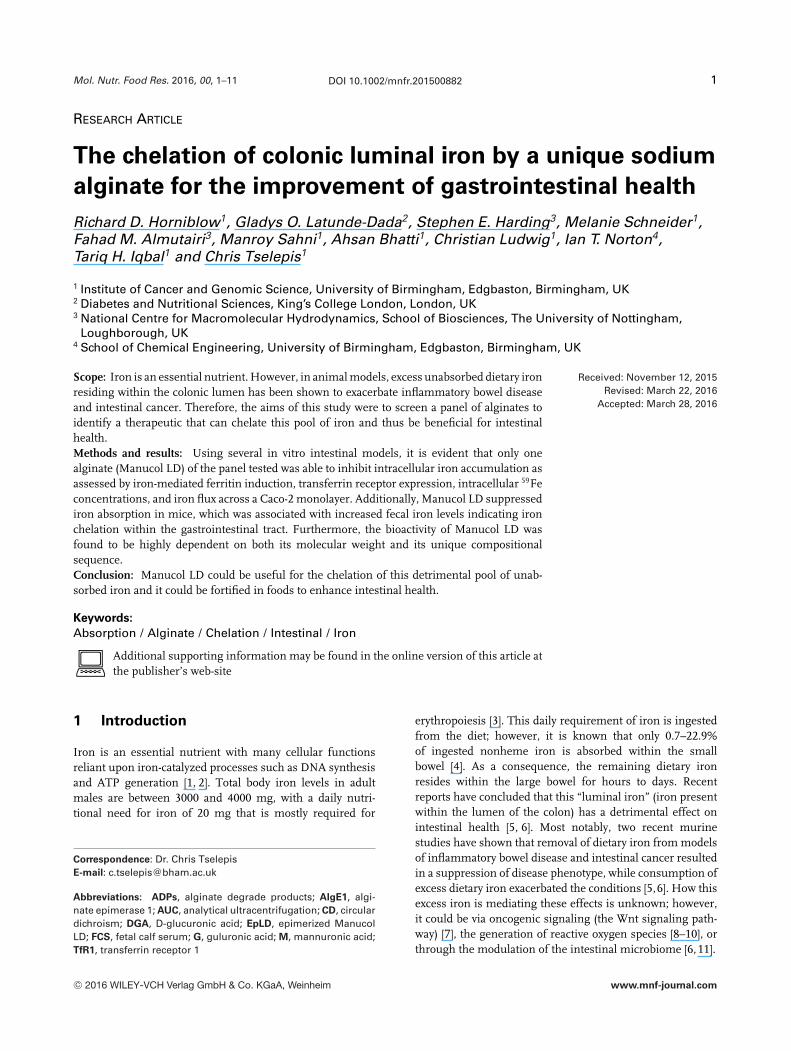
Figure 1. (A) Ferritin protein expression in RKO cells cocultured with iron (Fe) ± sodium alginates (LD, DH, GHB, LFR, KEL, RF, and AFH) for24 h. Data points represent mean fold change in protein expression normalized to �-actin, relative to control. (B) Ferritin protein expressionin RKO cells cocultured with different iron concentrations (Fe) ± LD, DH, GHB, LFR, KEL, RF, and AFH for 24 h. Data points represent meanfold change in protein expression normalized to �-actin, relative to control. (C) Total iron concentrations in RKO cells cultured ± ManucolLD for 24 h. (D) Transferrin receptor expression in RKO cells incubated with iron ± Manucol LD. Data points represent mean fold change inprotein expression, normalized to �-actin, relative to control. Error bars denote ±SEM; *statistical significance p < 0.05 versus iron-onlycontrol and n = 3.
(Manucol LD (LD), Manucol DH (DH), Manugel GHB(GHB), LFR5/60 (LFR), KELTONE (KEL), PROTANALRF6650 (RF) and PROTSEA AFH (AFH), 0.3% w/v) for24 h and cells were then subsequently assayed for ferritinexpression; a surrogate marker for cellular iron levels(Fig. 1A). As expected, treating cells with iron-supplementedmedia alone induced ferritin expression (p < 0.05). Only onealginate (Manucol LD) was able to significantly decrease theiron-induced ferritin response by 60% (p < 0.05). Similarly,culture of RKO cells at lower concentrations of iron (1 and10 �M) in the presence or absence of alginates (LD, DH,GHB, LFR, KEL, RF and AFH, 0.3 % w/v) demonstrated thatManucol LD was the only alginate to significantly reduceferritin expression by 70, 88, and 68% at 1, 10, and 100 �Mconcentrations of iron, respectively, compared with theiron-only control (p < 0.05, Fig. 1B). DH, GHB, and LFRonly inhibited ferritin expression at 10 �M iron by 54, 47,
and 92%, respectively (p < 0.05, Supporting InformationFig. 1). Manucol LD was the only alginate to demonstratebioactivity at all concentrations of iron.
In order to fully verify the iron chelation ability of ManucolLD in vitro, RKO cells were challenged with iron (100 �M)with or without Manucol LD (0.3% w/v) for 24 h before beingassessed for direct intracellular iron concentrations (Fig. 1C)and TfR1 expression (Fig. 1D). RKO cells treated with iron re-sulted in cellular iron loading (ca. 50 nM total cellular iron);the basal levels of iron within the control group were notmeasureable by this assay. It was found that Manucol LD sig-nificantly decreased cellular iron loading by 62% (p < 0.05)(Fig. 1C). This was associated with a statistical increase inTfR1 expression by approximately 50% (p < 0.05) comparedwith iron alone, indicating that Manucol LD is binding thesupplemented iron present in the media hindering its intra-cellular uptake (Fig. 1D).
C© 2016 WILEY-VCH Verlag GmbH & Co. KGaA, Weinheim www.mnf-journal.com

Mol. Nutr. Food Res. 2016, 00, 1–11 5
Figure 2. (A) Iron concentration in Caco-2 cells incubated with iron ± Manucol LD. (B) 59Fe concentrations at 0.5, 4, and 24 h in the apicalchamber following culture with iron ± Manucol LD. Error bars denote ±SEM; *statistical significance, p < 0.05 versus iron-only control.
3.2 Manucol LD demonstrates iron chelation in an
in vitro model of the intestinal lumen
Using radiolabeled iron-spiked cell culture media, ManucolLD significantly decreased intracellular iron concentrationsby 70% (p < 0.05) within the Caco-2 monolayer comparedwith the iron-only control (Fig. 2A). By assessing the levelsof iron within the apical chamber during the experiment, itcould be determined that iron was retained within this com-partment (Fig. 2B). Co-incubation with Manucol LD at both 4and 24 h inhibited cellular iron uptake by the Caco-2 cells by60% (p < 0.05) compared with the iron-only control from theapical compartment. These results validate the observationthat Manucol LD is binding iron in the media, thus prevent-ing its cellular internalization.
3.3 Manucol LD demonstrates iron chelation within
the gastrointestinal tract in vivo
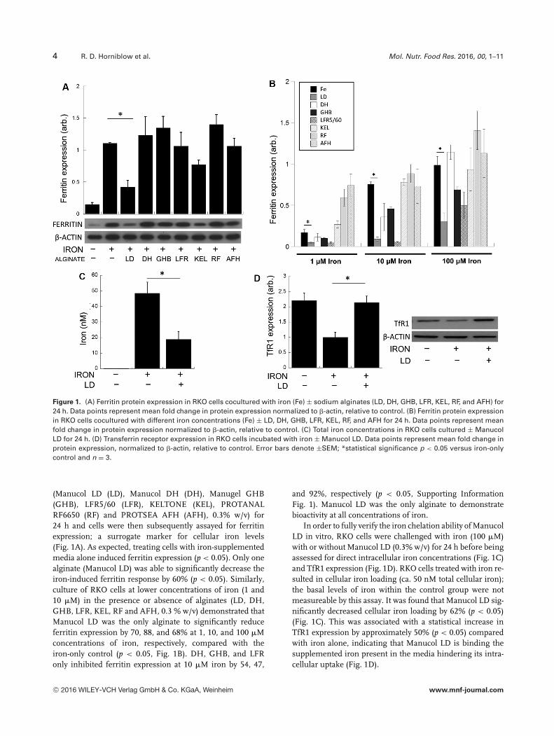
Administration of Manucol LD resulted in significantly de-creased carcass iron concentrations by 71% (p < 0.005) com-pared with an iron-only cohort (Fig. 3A). This equates toonly 13% of the total iron administered being absorbed whenManucol LD was present compared to 46% when it was ab-sent (Supporting Information Fig. 2). In addition, there wasa concomitant increase in fecal iron concentrations of 45%(p < 0.05) in mice administered Manucol LD compared withmice administered iron alone (Fig. 3B). To fully verify theiron chelation potential of Manucol LD throughout the gas-trointestinal tract, the stomach, duodenum, and colon wereassayed for iron concentration and it was found that in all
gastrointestinal tissues there were statistically decreased ironconcentrations when mice were administered Manucol LDcompared with iron alone (Fig. 3C). Specifically, a signifi-cant decrease of 53, 60, and 52% (p < 0.05) in the stomach,duodenum, and colon was found, respectively. Moreover, theiron concentrations significantly increase from the stomachto the duodenum to the colon (stomach → duodenum 73%increase, stomach → colon 83 % increase (p < 0.05)). Allmice received the same concentration of 59Fe radioactive gav-age (Fig. 3D).
3.4 Chemical characterization of alginates
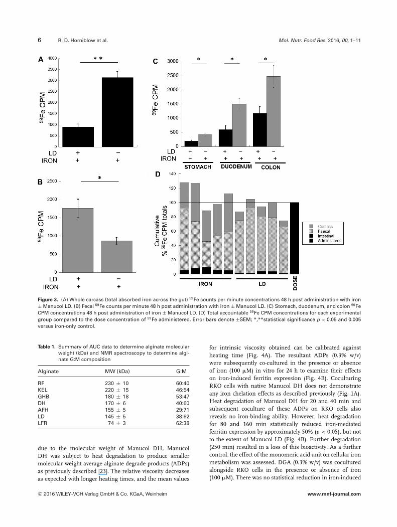
To establish why Manucol LD demonstrated bioactivity andthe other alginates did not, chemical characterization of thealginate series was performed. Both assessment of molecularweight by AUC (Supporting Information Fig. 3) and G:Mcomposition by NMR (data not shown) was performed [21,22].A summary of these results is described in Table 1. ManucolLD was found to have a molecular weight distribution of 145kDa. Manucol LD was also found to have a G:M ratio of 38:62;however, this composition was not unique to Manucol LD asManucol DH had a similar G:M ratio of 40:60.
3.5 Alginate iron chelation is molecular weight
dependent
Since Manucol DH and Manucol LD shared similar G:Mcomposition homology, yet Manucol DH was found to havea higher molecular weight than Manucol LD (cf. 170 vs.145 kDa), to determine if the difference in bioactivity was
C© 2016 WILEY-VCH Verlag GmbH & Co. KGaA, Weinheim www.mnf-journal.com
6 R. D. Horniblow et al. Mol. Nutr. Food Res. 2016, 00, 1–11
Figure 3. (A) Whole carcass (total absorbed iron across the gut) 59Fe counts per minute concentrations 48 h post administration with iron± Manucol LD. (B) Fecal 59Fe counts per minute 48 h post administration with iron ± Manucol LD. (C) Stomach, duodenum, and colon 59FeCPM concentrations 48 h post administration of iron ± Manucol LD. (D) Total accountable 59Fe CPM concentrations for each experimentalgroup compared to the dose concentration of 59Fe administered. Error bars denote ±SEM; *,**statistical significance p < 0.05 and 0.005versus iron-only control.
Table 1. Summary of AUC data to determine alginate molecularweight (kDa) and NMR spectroscopy to determine algi-nate G:M composition
Alginate MW (kDa) G:M
RF 230 ± 10 60:40KEL 220 ± 15 46:54GHB 180 ± 18 53:47DH 170 ± 6 40:60AFH 155 ± 5 29:71LD 145 ± 5 38:62LFR 74 ± 3 62:38
due to the molecular weight of Manucol DH, ManucolDH was subject to heat degradation to produce smallermolecular weight average alginate degrade products (ADPs)as previously described [23]. The relative viscosity decreasesas expected with longer heating times, and the mean values
for intrinsic viscosity obtained can be calibrated againstheating time (Fig. 4A). The resultant ADPs (0.3% w/v)were subsequently co-cultured in the presence or absenceof iron (100 �M) in vitro for 24 h to examine their effectson iron-induced ferritin expression (Fig. 4B). CoculturingRKO cells with native Manucol DH does not demonstrateany iron chelation effects as described previously (Fig. 1A).Heat degradation of Manucol DH for 20 and 40 min andsubsequent coculture of these ADPs on RKO cells alsoreveals no iron-binding ability. However, heat degradationfor 80 and 160 min statistically reduced iron-mediatedferritin expression by approximately 50% (p < 0.05), but notto the extent of Manucol LD (Fig. 4B). Further degradation(250 min) resulted in a loss of this bioactivity. As a furthercontrol, the effect of the monomeric acid unit on cellular ironmetabolism was assessed. DGA (0.3% w/v) was coculturedalongside RKO cells in the presence or absence of iron(100 �M). There was no statistical reduction in iron-induced
C© 2016 WILEY-VCH Verlag GmbH & Co. KGaA, Weinheim www.mnf-journal.com

Mol. Nutr. Food Res. 2016, 00, 1–11 7
Figure 4. (A) Manucol DH relative viscosity (�rel) decreases by heating at 100�C with a plot of intrinsic viscosity against heating time. Errorbars denote ±SEM. (B) Ferritin protein expression in RKO cells incubated with iron ± Manucol DH heat degradation products for 24 h. Datapoints represent mean fold change in protein expression, normalized to �-actin, relative to control. Error bars denote ±SEM, *statisticalsignificance p < 0.05 versus iron-only control and n = 3. (C) 59Fe concentrations in RKO cells incubated with iron challenged ± DGA. Datapoints represent mean fold change in protein expression, normalized to �-actin, relative to control. Error bars denote ±SEM. (D) Ferritinprotein expression in RKO cells incubated with iron ± DGA. Data points represent mean fold change in protein expression, normalized to�-actin, relative to control. Error bars denote ±SEM. NS, no statistical significance.
ferritin expression or 59Fe levels when cells were culturedalongside DGA compared with iron only (Fig. 4C and D).
3.6 Alginate iron chelation is composition
dependent
Manucol LD and Manucol DH have similar G:M chem-ical compositions but this does not preclude differencesin GM sequence structure. To examine this, they wereboth subject to high-resolution NMR to allow calculation oftheir respective monad (G/M), diad (GM/GG/MM), or triad(GMG/GGM/MGM/MMG) concentrations [24]. NMR spec-tra for Manucol LD and Manucol DH were acquired andanalyzed using the correlations described and transformedfree-induction decays were fitted to Lorentzian curves to allowcalculation of the specific monads, diads, and triads (Fig. 5Aand B) according to a standard protocol [24]. Calculation of themajor differences between Manucol LD and Manucol DH re-
vealed that Manucol LD contained 12% more MM diads thanManucol DH. Similarly, Manucol DH contained 8% moreGM diads than Manucol LD.
To interrogate the dependence on G:M compositionand sequence structure, an alginate epimerase enzyme(AlgE1) was produced and utilized for the M→G conversionof Manucol LD. Epimerization of native Manucol LD byAlgE1 resulted in an approximate twofold increase inguluronate residues on EpLD, resulting in a new G:M ratioof 77:23; the highest G-unit concentration alginate out ofthe series as assessed by CD spectroscopy (Fig. 5C). Therewas also very little absorption present at � = 280 nm on theUV-Vis, indicating negligible AlgE1 protein contaminant.To examine the effects of epimerization of Manucol LD oniron-induced ferritin expression, RKO cells were coculturedin the presence or absence of Manucol LD or EpLD with orwithout iron for 24 h before ferritin expression was examined(Fig. 5D). Ferritin expression in RKO cells treated with ironwas significantly higher than that of control media only,
C© 2016 WILEY-VCH Verlag GmbH & Co. KGaA, Weinheim www.mnf-journal.com

8 R. D. Horniblow et al. Mol. Nutr. Food Res. 2016, 00, 1–11
Figure 5. Experimental (red) and simulated (blue) NMR spectra of alginate (A) Manucol DH and (B) Manucol LD for the region of protons1 and 5 of M and glucuronic acid. (C) CD spectra and representative UV-Visible spectra of Manucol LD and EpLD. (D) Ferritin expressionin RKO cells incubated with iron ± Manucol LD and EpLD. Data points represent mean fold change in protein expression, normalized to�-actin, relative to control. Error bars denote ±SEM, *statistical significance p < 0.005 versus iron-only control and n = 3.
as expected. Manucol LD statistically diminished ferritinexpression by 57% compared with iron-only control (cf.60% in previous studies, p < 0.005). EpLD did not reduceferritin expression compared with the iron-only control, andexpression was statistically increased by 63% compared withManucol LD treatment (p < 0.005, Fig. 5D).
4 Discussion
The detrimental effect of excess iron within the colon haspreviously been reported [5, 6, 25, 26]. The ingestion of highamounts of red and processed meats, foods that contain highlevels of iron, have also been implicated in gastrointestinaldisease [27, 28]. Exactly how iron is mediating disease pro-gression within the large bowel is currently unknown. De-spite this, in a range of conditions associated with iron ex-cess, there is evidence that iron chelation is therapeuticallybeneficial [29]. However, in the context of gastrointestinal dis-ease it is specifically an excess of luminal iron within the largebowel that is detrimental and the chelation of this pool of ironpresents itself as a potential therapeutic platform to improveintestinal health.
In order to selectively chelate iron within the lumen ofthe colon, the therapeutic compound must not be absorbable
such that it reaches the large bowel and binds excess iron.Nondigestible fibers have been demonstrated to improvegastrointestinal health [30], yet whether these effects are at-tributed to their iron-binding properties is unknown. Sodiumalginates that are fibers found within the human diet havepreviously been demonstrated to be both nonabsorbable andbind iron [13, 14]. The possible range of alginate composi-tions is huge due to the diversity in polymer length and G:Mchemical composition [31, 32]. How these compositional dif-ferences affect iron-binding potential and subsequent in vitroand in vivo cellular iron modulation is unknown, with previ-ously published reports being inconsistent in their findings[14, 33–35]. This inconsistency is likely due to the fact thatchemically different alginates were used throughout thesestudies, and data presented in this study demonstrate the de-pendence of iron chelation bioactivity on alginate chemicalcomposition.
A range of alginates with different molecular weightsand G:M compositions were primarily screened to assesstheir in vitro iron chelation potential. This initial assessmentidentified only one alginate that was able to modulate ironmetabolism in vitro. Manucol LD decreased ferritin expres-sion by 60% (Fig. 1A and B), decreased total cellular ironby 62% (Fig. 1C), restored TfR1 expression to control levels(Fig. 1D), and decreased absorption of iron in Caco-2 cells by
C© 2016 WILEY-VCH Verlag GmbH & Co. KGaA, Weinheim www.mnf-journal.com

Mol. Nutr. Food Res. 2016, 00, 1–11 9
70% (Fig. 2A and B); this was the only alginate to have con-sistent iron chelation properties (all statistically significant top < 0.05). In vivo, Manucol LD was able to suppress ironabsorption by 71% and increased the iron content found infecal samples by 45% (p < 0.05, Fig. 3). These two results arepivotal in demonstrating the mechanism of iron binding byManucol LD within the gastrointestinal tract, where it can beinferred that Manucol LD binds iron and keeps it chelateduntil defecation. Where the iron is interacting with ManucolLD within the gastrointestinal tract remains unclear; how-ever, as alginates are insoluble in low-pH environments andform insoluble capsules, it is likely that iron chelation wouldnot take place until Manucol LD reached more basic, distal re-gions of the gut [36]. Interestingly, the concentrations of ironwithin the mucosa of gastrointestinal tissues increase fromthe small to the large bowel (stomach < duodenum < colon).This is likely to be a consequence of either (i) more effectiveiron transport across the brush border in the duodenum thanwithin the colon, (ii) the increased length of exposure to ironin the digesta within the colon, or (iii) a combination of both.This finding is supported by studies that have demonstratedthat the colon is able to import iron but there is a lack of reg-ulated iron efflux [37]. Thus unsurprisingly, Manucol LD hadits greatest iron chelation effects within the colon, decreasingiron absorption by 56% (p < 0.05).
Manucol LD has iron chelation effects both in vivo andin vitro; however, what remains unclear is the redox stateand likely species of iron that alginate is chelating to withinthese experimental conditions. Since sodium ascorbate wasincluded within the growth media in cell culture experiments,it is likely that the supplemented ferrous iron will remain inthe “free” ferrous form (“free” referring to its availability tochelation by competing ligands), which would suggest thatManucol LD is certainly binding to free ferrous iron. In ad-dition previous reports have indicated that alginate will alsobind ferric iron [13]. It can be inferred that Manucol LD islikely to have iron chelation ability toward ferrous and ferricions, yet this does not rule out the possibility that iron couldbe forming nanoparticulate species within the gastrointesti-nal tract [38], which alginates have also been documentedto stabilize [39]. If indeed iron in its “free” form is presentwithin the colon, then these data suggest that Manucol LD isable to bind such a form of iron and possibly nanoparticulateforms based on previously published reports [13]. However,the species of iron present within the colon is currently un-known and could be in its “free” state, bound to other dietaryor endogenous intestinal compounds or even as particulatemineralized species [38, 40, 41].
To interrogate the chemical characteristics of alginate re-quired for iron chelation and subsequent modulation of cel-lular iron metabolism, Manucol LD and Manucol DH (thelatter an alginate with similar G:M composition but differentpolymer length) were subject to structural modification byalginate degradation (through heating) and M→G unit con-version (by exposure to AlgE1). Determination of the heatingtime required to reduce molecular weight by specific amount
allowed the calibration of intrinsic viscosity change againstheating time (Fig. 4A). Heat degradation of Manucol DHat 100�C decreased with heating time as previously reported[42]. It was estimated that heating Manucol DH for approxi-mately 3 h would provide an intrinsic viscosity value of 1200mL/g, which would give a molecular weight similar to that ofManucol LD (145 kDa). As such, heating Manucol DH for 180min produced a shorter chain alginate (analogous to Manu-col LD) that when cocultured in vitro with iron decreasediron-induced ferritin expression by 50% (p < 0.05, Fig. 4B).As a control, Manucol DH was heated for longer periods oftime (250 min) to produce shorter molecular weight prod-ucts. Subsequent coculture of these ADPs resulted in dimin-ished iron chelation ability to that observed with the 180-minADPs as ferritin levels were not significantly different to theiron-only control. As a further control, RKO cells were co-cultured with iron in the presence of DGA to examine if theindividual monomeric acid unit was able to modulate cellu-lar iron metabolism; DGA also had no significant effects onferritin expression or intracellular 59Fe concentration. Theseresults demonstrate the importance of polymer length oniron-binding ability, and it has recently been reported howthe polymeric nature of alginate orchestrates iron chelation[13]. Thus, in summary an alginate molecular weight of ap-proximately 145 kDa is required for maximal iron chelationbioactivity.
It is plausible that Manucol LD has a tertiary and sec-ondary structure that forms an iron-binding pocket or cavity;this structure is formed by the specific MG sequence of thealginate. The iron-binding site acts as a nucleation site foriron deposition and such mechanisms have been previouslyreported for other biopolymers [39]. If this were the case, thenalteration of the G:M ratio and thus GM sequence on ManucolLD would disrupt the formation of this binding site. Indeed,data presented in this study have demonstrated this. Alginateepimerase 1 was utilized to convert Manucol LD (G:M 38:62)to EpLD (G:M 77:23). Subsequent coculture of EpLD in thepresence of iron did not reduce ferritin expression as demon-strated for native Manucol LD. Such results demonstrate theimportance of a specific alginate G:M ratio and GM sequencein iron chelation bioactivity. To fully confirm the uniquenessof Manucol LD, high-resolution NMR spectroscopy showedthat Manucol DH and Manucol LD (two alginates with sim-ilar G:M ratio) do indeed have different GM sequence ho-mologies, where differences in MM and GM (or MG) diadfrequencies are most apparent.
In summary, data presented here identify a unique al-ginate, with a specific chemical composition and polymericlength that demonstrate optimal iron chelation propertiesboth in vitro and in vivo. These data not only explain whyinconsistencies have previously been reported on alginatemodulation of cellular iron metabolism but also detail the re-quired chemical characteristics of an alginate for iron chela-tion bioactivity. Importantly, if Manucol LD is to be used tochelate excess colonic luminal iron, it will require formulat-ing so as to ensure colonic delivery. Without employment
C© 2016 WILEY-VCH Verlag GmbH & Co. KGaA, Weinheim www.mnf-journal.com
10 R. D. Horniblow et al. Mol. Nutr. Food Res. 2016, 00, 1–11
of a colonic delivery platform, our data might predict thatchronic consumption of Manucol LD could lead to iron defi-ciency, due to iron chelation within the small bowel. In thecontext of modulating the intestinal microbiome, it is knownthat iron is able to alter microbial colonization to a morepro-inflammatory enterotype and it could be envisaged thatManucol LD could suppress the colonization of these “non-beneficial” bacteria (such as Bacteroides) through the chelationof luminal iron [11,43,44]. Further to this, alginates may haveprebiotic effects acting as a support scaffold for the coloniza-tion of beneficial bacteria and as such further testing of thesepro- and prebiotic effects in man is required.
RH, GOL-D, MS, MS, FMA, AB, and CL performed theexperiments. RH, GOL-D, SEH, TI, IN, and CT conceived anddesigned the experiments. RH, GOL-D, and SEH analyzed thedata. RH and CT wrote the paper.
The following study was supported by the BBSRC casestudentship (STU100020349) and the Experimental CancerMedicine Centre (Birmingham). All animal experiments wereconducted independently of the main funded project by Dr. YemisiLatunde-Dada (King’s College London). We thank Dr. HelgaErtesvag (Norwegian University of Sciences and Technology) forher guidance in alginate epimerization. We are also grateful toTrond Helgerud (FMC Biopolymer, Norway) for supplying thealginates.
The authors have declared no conflict of interest.
5 References
[1] Thelander, L., Graslund, A., Thelander, M., Continual pres-ence of oxygen and iron required for mammalian ri-bonucleotide reduction: possible regulation mechanism.Biochem. Biophys. Res. Commun. 1983, 110, 859–865.
[2] Atamna, H., Walter, P. B., Ames, B. N., The role of Hemeand iron-sulfur clusters in mitochondrial biogenesis, main-tenance, and decay with age. Arch. Biochem. Biophys. 2002,397, 345–353.
[3] Ganz, T., Hepcidin, a key regulator of iron metabolism andmediator of anemia of inflammation. Blood 2003, 102, 783–788.
[4] Collings, R., Harvey, L. J., Hooper, L., Hurst, R. et al., Theabsorption of iron from whole diets: a systematic review.Am. J. Clin. Nutr. 2013, 98, 65–81.
[5] Radulescu, S., Brookes, M. J., Salgueiro, P., Ridgway, R. A.et al., Luminal iron levels govern intestinal tumorigenesisafter Apc loss in vivo. Cell Rep. 2012, 2, 270–282.
[6] Werner, T., Wagner, S. J., Martinez, I., Walter, J. et al., Deple-tion of luminal iron alters the gut microbiota and preventsCrohn’s disease-like ileitis. Gut 2011, 60, 325–333.
[7] Brookes, M. J., Boult, J., Roberts, K., Cooper, B. T. et al., Arole for iron in Wnt signalling. Oncogene 2008, 27, 966–975.
[8] Valko, M., Morris, H., Mazur, M., Rapta, P. et al., Oxygenfree radical generating mechanisms in the colon: do thesemiquinones of vitamin K play a role in the aetiology
of colon cancer? Biochim. Biophys. Acta 2001, 1527, 161–166.
[9] LihBrody, L., Powell, S. R., Collier, K. P., Reddy, G. M. et al.,Increased oxidative stress and decreased antioxidant de-fenses in mucosa of inflammatory bowel disease. Dig. Dis.Sci. 1996, 41, 2078–2086.
[10] Carrier, J., Aghdassi, E., Platt, I., Cullen, J. et al., Effect oforal iron supplementation on oxidative stress and colonicinflammation in rats with induced colitis. Aliment. Pharma-col. Ther. 2001, 15, 1989–1999.
[11] Jaeggi, T., Kortman, G. A., Moretti, D., Chassard, C. et al., Ironfortification adversely affects the gut microbiome, increasespathogen abundance and induces intestinal inflammation inKenyan infants. Gut 2015, 64, 731–742.
[12] Brittenham, G. M., Griffith, P. M., Nienhuis, A. W., McLaren,C. E. et al., Efficacy of deferoxamine in preventing compli-cations of iron overload in patients with thalassemia major.New Engl. J. Med. 1994, 331, 567–573.
[13] Horniblow, R. D., Dowle, M., Iqbal, T. H., Latunde-Dada, G.O. et al., Alginate-iron speciation and its effect on in vitrocellular iron metabolism. PLoS One 2015, 10, e0138240.
[14] Sandberg, A. S., Andersson, H., Bosaeus, I., Carlsson, N. G.et al., Alginate, small-bowel sterol excretion, and absorptionof nutrients in ileostomy subjects. Am. J. Clin. Nutr. 1994, 60,751–756.
[15] Haug, A., Larsen, B., Smidsrod, O., Studies on the sequenceof uronic acid residues in alginic acid. Acta Chem. Scand.1967, 21, 691–704.
[16] Thompson, B., Sharp, P., Elliott, R., Al-Mutairi, S. et al., Devel-opment of a modified Caco-2 cell model system for studyingiron availability in eggs. J. Agric. Food Chem. 2010, 58, 3833–3839.
[17] Powell, J. J., Bruggraber, S. F. A., Faria, N., Poots, L. K. et al.,A nano-disperse ferritin-core mimetic that efficiently cor-rects anemia without luminal iron redox activity. Nanomed.-Nanotech. Biol. Med. 2014, 10, 1529–1538.
[18] Schuck, P., Gillis, R. B., Besong, T. M. D., Almutairi, F. et al.,SEDFIT-MSTAR: molecular weight and molecular weight dis-tribution analysis of polymers by sedimentation equilibriumin the ultracentrifuge. Analyst 2014, 139, 79–92.
[19] Ertesvag, H., Alginate-modifying enzymes: biological rolesand biotechnological uses. Front. Microbiol. 2015, 6, 523.
[20] Ludwig, C., Gunther, U. L., MetaboLab – advanced NMR dataprocessing and analysis for metabolomics. BMC Bioinform.2011, 12, 366.
[21] Davis, T. A., Llanes, F., Volesky, B., Diaz-Pulido, G. et al., H-1-NMR study of Na alginates extracted from Sargassum spp.in relation to metal biosorption. Appl. Biochem. Biotechnol.2003, 110, 75–90.
[22] Holtan, S., Zhang, Q. J., Strand, W. I., Skjak-Braek, G.,Characterization of the hydrolysis mechanism of polyal-ternating alginate in weak acid and assignment of theresulting MG-oligosaccharides by NMR spectroscopy andESI-mass spectrometry. Biomacromolecules 2006, 7, 2108–2121.
[23] Oates, C. G., Ledward, D. A., Studies on the effect of heat onalginates. Food Hydrocoll. 1990, 4, 215–220.
C© 2016 WILEY-VCH Verlag GmbH & Co. KGaA, Weinheim www.mnf-journal.com

Mol. Nutr. Food Res. 2016, 00, 1–11 11
[24] ASTM International, West Conshohocken, Penn., USA 2010.
[25] Seril, D. N., Liao, J., West, A. B., Yang, G. Y., High-iron diet: foeor feat in ulcerative colitis and ulcerative colitis-associatedcarcinogenesis. J. Clin. Gastroenterol. 2006, 40, 391–397.
[26] Siegers, C. P., Bumann, D., Trepkau, H. D., Schadwinkel, B.et al., Influence of dietary iron overload on cell proliferationand intestinal tumorigenesis in mice. Cancer Lett. 1992, 65,245–249.
[27] Chan, D. S. M., Lau, R., Aune, D., Vieira, R. et al., Red and pro-cessed meat and colorectal cancer incidence: meta-analysisof prospective studies. PLoS One 2011, 6, e20456.
[28] Norat, T., Lukanova, A., Ferrari, P., Riboli, E., Meat consump-tion and colorectal cancer risk: dose-response meta-analysisof epidemiological studies. Int. J. Cancer 2002, 98, 241–256.
[29] Mainous, A. G., Tanner, R. J., Hulihan, M. M., Amaya, M.et al., The impact of chelation therapy on survival in trans-fusional iron overload: a meta-analysis of myelodysplasticsyndrome. Br. J. Haematol. 2014, 167, 720–723.
[30] Murphy, N., Norat, T., Ferrari, P., Jenab, M. et al., Dietary fibreintake and risks of cancers of the colon and rectum in theEuropean prospective investigation into cancer and nutrition(EPIC). PLoS One 2012, 7, e39361.
[31] Draget, K. I., Smidsrød, O., Skjak-Bræk, G., Biopolymers On-line, Wiley-VCH Verlag, Weinheim, Germany 2005.
[32] Larsen, B., Smidsrod, O., Painter, T., Haug, A., Calculationof nearest-neighbour frequencies in fragments of alginatefrom yields of free monomers after partial hydrolysis. ActaChem. Scand. 1970, 24, 726–731.
[33] Wawer, A. A., Sharp, P. A., Perez-Moral, N., Fairweather-Tait,S. J., Evidence for an enhancing effect of alginate on ironavailability in Caco-2 cells. J. Agric. Food Chem. 2012, 60,11318–11322.
[34] Wawer, A. A., Harvey, L. J., Dainty, J. R., Perez-Moral,N. et al., Alginate inhibits iron absorption from ferrous
gluconate in a randomized controlled trial and reduces ironuptake into caco-2 cells. PloS one 2014, 9, e112144.
[35] Wolbling, R. H., Becker, G., Forth, W., Inhibition of the intesti-nal absorption of iron by sodium alginate and guar gum inrats. Digestion 1980, 20, 403–409.
[36] Haug, A., Larsen, B., The solubility of alginate at low pH.Acta Chem. Scand. 1963, 17, 1653–1662.
[37] Johnston, K. L., Johnson, D. M., Marks, J., Srai, S. K. et al.,Non-haem iron transport in the rat proximal colon. Eur. J.Clin. Invest. 2006, 36, 35–40.
[38] Pereira, D. I. A., Mergler, B. I., Faria, N., Bruggraber, S. F.A. et al., Caco-2 cell acquisition of dietary iron(III) invokesa nanoparticulate endocytic pathway. PLoS One 2013, 8,e0119747.
[39] Jones, F., Colfen, H., Antonietti, M., Iron oxyhydroxide col-loids stabilized with polysaccharides. Colloid. Polym. Sci.2000, 278, 491–501.
[40] Rudzki, Z., Baker, R., Deller, D., The iron-binding glycoproteinof human gastric juice. II. Nature of the interaction of theglycoprotein with iron. Digestion 1973, 8, 53–67.
[41] Simpson, R. J., Peters, T. J., Forms of soluble iron in mousestomach and duodenal lumen: significance for mucosal up-take. Br. J. Nutr. 1990, 63, 79–89.
[42] Holme, H. K., Foros, H., Pettersen, H., Dornish, M. et al., Ther-mal depolymerization of chitosan chloride. Carbohydr. Poly-mer. 2001, 46, 287–294.
[43] Kortman, G. A. M., Mulder, M. L. M., Richters, T. J. W., Shan-mugam, N. K. N. et al., Low dietary iron intake restrainsthe intestinal inflammatory response and pathology of en-teric infection by food-borne bacterial pathogens. Eur. J. Im-munol. 2015, 45, 2553–2567.
[44] Cherayil, B. J., Ellenbogen, S., Shanmugam, N. N., Iron andintestinal immunity. Curr. Opin. Gastroenterol. 2011, 27, 523–528.
C© 2016 WILEY-VCH Verlag GmbH & Co. KGaA, Weinheim www.mnf-journal.com
View publication statsView publication stats