Embed Size (px)
Citation preview

Research ArticleThe Changes in Rats with Sciatic Nerve Crush InjurySupplemented with Evening Primrose Oil: Behavioural,Morphologic, and Morphometric Analysis
Danial Ramli,1 Izzuddin Aziz,1 Masro Mohamad,2 Dauda Abdulahi,1 and Junedah Sanusi1
1Department of Anatomy, Faculty of Medicine, University of Malaya, Kuala Lumpur, Malaysia2Department of Pharmaceutical Sciences, Faculty of Pharmacy, Cyberjaya University College of Medical Sciences (CUCMS),Cyberjaya, Selangor, Malaysia
Correspondence should be addressed to Junedah Sanusi; [email protected]
Received 27 November 2016; Revised 17 March 2017; Accepted 10 April 2017; Published 23 May 2017
Academic Editor: Jintanaporn Wattanathorn
Copyright © 2017 Danial Ramli et al. This is an open access article distributed under the Creative Commons Attribution License,which permits unrestricted use, distribution, and reproduction in any medium, provided the original work is properly cited.
Nerve crush injuries are commonly used models for axonotmesis to examine peripheral nerve regeneration. As evening primroseoil (EPO) is rich in omega-6 essential fatty acid component and gamma-linolenic acid, studies have shown the potential role of EPOin myelination. Seventy-two healthy adult Sprague-Dawley rats were classified into three groups: normal group, control group, andexperimental group. The result indicates that there was significant difference in toe-spreading reflex between the normal and thecontrol groups (1.9±0.031, 𝑝 < 0.05) and the normal and the EPO groups (0.4±0.031, 𝑝 < 0.05) and significant difference betweenEPO and the control groups (1.5 ± 0.031, 𝑝 < 0.05). Regeneration of axons and myelin in nerve fibre in the EPO-treated groupdeveloped better and faster than in the control group. In the control group, the shape of the axon was irregular with a thinnermyelin sheath. In the experimental group, the shape of the axons, the thickness of the myelin sheath, and the diameter of the axonswere almost the same as in the normal group. In conclusion, EPO supplementation may be beneficial as a therapeutic option fordisturbances of nerve interaction.
1. Introduction
Peripheral nerve encompasses all the nerve trunks andbranches which lie outside the central nervous system.Whena peripheral nerve is injured, the muscles supplied by thatnerve do not receivemessages from the brain.Therefore, theybecomeweakened or paralysed [1, 2]. Traffic collisions usuallyinduce traumatic nerve injuries resulting from disruptionof the intraneural circulation [3, 4]. This condition con-sequently induces demyelinization, remyelinization, axonaldegeneration and axonal regeneration, focal, multifocal, ordiffuse nerve fibre loss, and endoneural edema [4, 5]. Nerveregeneration is a complex phenomenon that has been gaininginterest among scientists for many years. Many experimentalstudies have focused on treatment options to enhance therecovery process of injured peripheral nerves in the ratmodel. This includes the application of an electric field[6], extracts of various natural products, for example, the
medicinal mushroom Hericium erinaceus [7], and surgicalintervention, for example, nerve grafts and transplantingstem cells [8]. Many experimental studies have focused ontreatment options to enhance the recovery process of injuredperipheral nerves in the rat model. This includes the applica-tion of an electric field [6], surgical intervention, for example,nerve grafts and transplanting stem cells [8]. Furthermore,the transplantation of Schwann cells has also been shown toimprove functional recovery and reduce histological deficitsresulting from nerve crush injury [9]. Despite being themajor producer of myelin in the peripheral nervous system,Schwann cells play an important role in promoting axonalregeneration by producing neurotrophic factors such as nervegrowth factor (NGF) and ciliary neurotrophic factor (CNTF)[10]. Commercial drugs such as immunosuppressant andanti-inflammatory drugs may accelerate the rate of nerveregeneration following injury. However, they are associatedwith severe side effects such as high blood pressure, kidney
HindawiEvidence-Based Complementary and Alternative MedicineVolume 2017, Article ID 3476407, 10 pageshttps://doi.org/10.1155/2017/3476407

2 Evidence-Based Complementary and Alternative Medicine
problems, and liver disorders [11]. Therefore, it is importantto search for natural products or substances and possible newdrug treatments that could help improve nerve regeneration.
Evening primrose is a biennial herb originating fromNorth America. Its botanical name is Onoethera biennis.Theseeds of this herb are very valuable as they contain beneficialoil, evening primrose oil. The potential benefits of eveningprimrose oil, or EPO, are derived from the active ingredientcalled gamma-linolenic acid (GLA) (the major commercialsource of GLA), which is an essential fatty acid. GLA isnecessary for human health and may also be obtained fromvegetable oils such as safflower oil (Carthamus tinctorius),blackcurrant seed oil, and borage oil. EPO is one of thesafest sources of GLA [12, 13]. EPO contains 8%–11% GLAand about 70% Linoleic acid such as Eicosanoid Acid (EA)and palmitic, stearic, and oleic acid [14]. Evening primroseis known as King’s Cure-all [15]. This could possibly bedue to its role in treating many diseases including atopicdermatitis, rheumatoid arthritis, mastalgia, and breast cysts[16]. In addition, EPO is also effective in controlling highblood pressure, assisting circulation, andmaintaining healthyarteries, thereby preventing heart disease [17]. EPO hasphytoestrogenic effects and is widely used as a complemen-tary treatment in combating premenstrual tension and alsoas a postmenopausal treatment [18]. Certain studies havereported that consumption of EPOhas been shown to preventand rapidly reverse the nerve conduction velocity (NCV)of deficits found in the rat nerve. Diabetic animals andhumans have a reduced ability to convert dietary linoleic acidinto GLA. GLA and its metabolites are required for normalneuronal structure, function, and a normal microcirculation.A lack of GLA and its metabolites may play a major role inthe development of neuropathy [19].
A study carried out where patients were given 360mgGLA a day for a period of 6 months demonstrated thatthe patients who took GLA showed statistically significantimprovement in neuropathy symptom scores after this timeperiod [20]. The conclusion of the study was that GLAtherapy might have a useful role in the prevention andtreatment of distal diabetic polyneuropathy. Another studywas conducted among patients taking 480mg GLA a dayfor a one-year period concluded that GLA had a beneficialeffect on the course of diabetic neuropathy [21]. In additionother studies have shown that motor and sensory conductiondeficits were largely corrected in an EPO-treated reversalgroup, with the degree of amelioration compared to one-month diabetic control being 99% (𝑝 < 0.001) and 85%(𝑝 < 0.01), respectively [21]. Other researches have beendone concerning peripheral nerve degeneration, as well astreatment and regeneration, from experimental studies insmall animals, especially rats. This is because the speed ofregeneration in these animals is fairly fast [22]. Sciatic nervecrush injuries are an ideal method of studying nerve injuriesand regeneration. This is especially so when a therapeuticapproach is under investigation, as it does not involvecomplete damage to the side of the nerve, for example, theepineurium, as is the case with section and repair [22]. Thepresent study was conducted to observe the morphologicalchanges tomyelin and axons in rat sciatic nerve crush injuries
using Toluidine Blue staining. The aim of this study is todetermine the effect of EPO supplementation on the rateof peripheral nerve regeneration after sciatic nerve injury,through morphological and morphometric analysis of theinjured nerve.
2. Materials and Methods
2.1. Animals and Surgical Procedures. In the present study, 72healthy adult Sprague-Dawley rats that were about 8 weeksold (weighing 250 ± 50 g) were obtained from the animalhouse, Faculty of Medicine, University of Malaya. The careand procedures for animal experiments conformed to theguidelines of the Animal Care and Use Committee (ACUC)[Vot number ANA 14/07/2010/JS (R)], University of Malaya.The rats were classified into three groups.
Group 1. Normal group (𝑛 = 8): the rats did not undergosciatic nerve crush injury and served as control for thehistological study. The rats were fed with a conventional dietad libitum.
Group 2. Control group (𝑛 = 32): the rats underwent sciaticnerve crush injury and were not supplemented with EPO.The rats were divided into four groups (1, 2, 3, and 4 weeks).Each group consisted of 8 rats. The rats were fed with aconventional diet ad libitum.
Group 3. Experimental group (𝑛 = 32): sciatic nerve crushinjury was performed and the rats were supplemented with6000mg/day EPO. The rats were divided into four groups(1, 2, 3, and 4 weeks). Each group consisted of 8 rats. Therats were fed with a conventional diet ad libitum. The ratswere anesthetized with a mixture of ketamine (100mg/kg)and xylazine (10mg/kg). The nerve crush procedure wasperformed on the right hind limb whereas the left side servedas control. The site of the incision was shaved and the skinincised over a length of 2 cm along the proximal half of theline between the trochanter major and the knee joint. Theright sciatic nerve was exposed through a gluteal musclesplitting incision, through which the overlying lateralis andbiceps femoris muscles were separated without cutting themuscle fibres, using a pair of Watchmaker’s forceps.
The visible sciatic nerve was crushed 1 cm proximal tothe division of the sciatic nerve into the tibial and commonperoneal nerves. The crush was achieved by applying 15seconds of consistent pressure with a pair of sterile modifiedWatchmaker’s forceps. A spacer was used at closure point toachieve moderate injury. The nerve was checked to ascertainthat the epineurium was intact and that the nerve wascompletely crushed. This was done by raising the nerveslightly using a microprobe so that a clear area in the nervewas observed, which indicated a complete nerve crush. Theskin incision was sutured and the rat was placed on a heatingpad to keep it warm. Each rat received 1ml of LactatedRinger’s solution subcutaneously and 0.4mg/kg of Baytrilintramuscularly. 10mg/g of Chloramphenicol and 5mg/g ofHydrocortisoneAcetate ointmentwere applied to the incisionarea. The rats were monitored until they recovered from the

Evidence-Based Complementary and Alternative Medicine 3
Table 1: The scoring and grading of the toe-spreading reflex.
Toe-spreading reflex (Gutmann 1942)Grade Clinical symptomsDegree 0 Absence of any abduction (movement) in any digit.Degree I Just visible spreading of the 4th toe alone (or one of the digit)Degree II Slight spreading of all three toesDegree III Spreading of all three toes less forceful than normalDegree IV Full spreading of all three toes (which resembles the unoperated left site)The toe-spreading reflex is an excellent and sensitive indicator of the onset of nerve and motor neuron recovery [24].
anesthesia. After this, the rats were placed in their respectivecages. Each rat was observed postoperatively to confirmcomplete crushing of the nerve shown by paralysis of themuscles to the toes and no spreading of the toes. Rats withmovement in the toes, indicating incomplete nerve crush,were rejected from the group.
2.2. Histological and Morphometric Studies. The rats weresacrificed at different time points (7, 14, 21, and 28 days afteroperation). The point of the nerve crush was determined(1 cm before the bifurcation of the sciatic nerve) and thenerve was harvested for histological preparation. The sciaticnerve was dissected out and fixed in 4% Glutaraldehyde inMillonig’s Phosphate Buffer for 24–72 hours, prior to theembedding process. The embedded sciatic nerve was seriallycross-sectioned at a thickness of 50 𝜇m using a microtome.The sections of the sciatic nerve were stained with ToluidineBlue solution to quantitatively evaluate the regeneration ofthe sciatic nerve. The stained slides were mounted usingDPX and observed under a light microscope equipped witha camera linked to a computer loaded with NIS-Elementsoftware to visualize the axons andmyelinmorphology of thesciatic nerve.
2.3. Dietary Supplementation. After the nerve crush proce-dure, the rats were fed according to the diet assigned fortheir groups for 4 weeks, during which behavioural testingwas performed daily. All group were fed with standard ratchow. In addition, the experimental group were given EPO6000mg/day through an esophageal feeding tube startingfrom day 1 after nerve crush. The EPO administration isconsidered safe as no toxicity, carcinogenicity, or teratogenic-ity has been reported with high oral administration of EPOduring clinical trial [23]. In addition, a higher dose wasselected to determine if a higher dose than that used inSamsinah et al. (1999) would be better for nerve regeneration.
2.4. Functional Assessment. After surgery, the rats wereinspected every single day. During these inspections, eachrat was held by its tail above a surface and then lowereddown onto it and carefully observed for a minute or two[23]. Activities were classified according to the toe-spreadingreflex of the affected right hind limb: no spreading, minimalspreading, average spreading, and normal spreading. Thetoe-spreading reflex was elicited to measure the duration ofcomplete recovery of the nerve. The behavioural observation
was based on comparison of the reflex seen between the rightside of the leg (where the nerve crush was performed) andthe left side of the leg (which served as control). The firstreading was at day 0 (after nerve crush injury) as shown inTable 1. The reflex was checked for 4 weeks. The scoring ofthe toe-spreading reflexwas based on a 5-point scale as shownin Table 1.
2.5. Statistical Analysis. All the data were analyzed andcompared between each group. Statistical significance wasdetermined using one-way analysis of variance (ANOVA)and post hoc analysis (SPSS Version 20). Significant changeswere determined using Tukey’s test formultiple comparisons.Differences were considered to be significant at 𝑝 < 0.05.The normality of all data distributions was checked using theKolmogorov-Smirnov test.
3. Results
3.1. Histological Analysis of Myelin and Axon. The lightmicroscopic sections of sciatic nerve stained with ToluidineBlue at various time points are shown in Figure 1, whereFigure 1(a) represents a photomicrograph of a normal rat. Itshows the normal distribution of axon size, number, and alsomyelin thickness. Few macrophages are visible in the normalgroup. From the histological study, it was clear that the axonsin the normal group were consistently round in shape with athick myelin sheath. The axons consisted of both myelinatedand unmyelinated axons. A myelinated axon has a sheath ofmyelin layer around it while an unmyelinated axon does not.Besides that, the axon and myelin population was notablyhigh. The axon/fibre diameter was also notably high.
When compared to the normal group, the control groupdepicts irregular axon shape with a thinner myelin sheath.The number of axon andmyelin was lower than in the normalgroup. In this group, the diameter of axons was notablysmaller than the normal and experimental group. At Day 7after operation, as seen in Figure 1(b), there was a diffuseloss of large myelinated axons and both the axon number andmyelin thickness had decreased tremendously. Macrophages(arrow in the figure) were visible and their number wasgreater. Starting from Day 14 up to Day 28 after injury,both axon number and myelin thickness increased withtime.
In the experimental group, axonal sprouting was notedwith a population of smaller axons as early as Day 7 after

4 Evidence-Based Complementary and Alternative Medicine
(a) (b)
(c) (d)
(e)
Figure 1:These photographs illustrated sections of sciatic nerve stained with Toluidine Blue at various time points. (a) Normal rat. (b) 7 daysafter operation. (c) 14 days after operation. (d) 21 days after operation. (e) 28 days after operation. Scale bar = 50𝜇m (arrow: macrophages,where the thick arrow indicates macrophage engulfing degenerating myelin sheath).
operation (Figure 2(a)). Evidence of regeneration was notedby the appearance of a large number of macrophages popula-tions (early nerve crush injury) which were swollen with thephagocytosis of degenerative debris, which strongly indicatedthat the nerve was still in the process of degeneration, whileat Day 14 and Day 21 after operation, the population and sizeof the axon and myelin were increased over time (Figures2(b) and 2(c)). In the experimental group, on Day 28 afteroperation (Figure 2(d)), the shape and diameter of the axonsand also the thickness of the myelin sheath were similar to
that of the normal group. The rats in the control group werestill undergoing the regeneration process with the appearanceof macrophages, when the rats in the experimental group hadachieved complete recovery. However, in the control group,the axons were very small and there was no myelin sheathin nearly all of the axons in the nerve (refer to Figures 1(b),1(c), and 1(d)).This indicates that the process ofmyelin sheathrestoration was slow in the control group, as the experimentalgroup had achieved complete recovery by the same timepoint.

Evidence-Based Complementary and Alternative Medicine 5
(a) (b)
(c) (d)
Figure 2:These photographs illustrated sections of sciatic nerve stained with Toluidine Blue at various time points. (a) 7 days after operation.(b) 14 days after operation. (c) 21 days after operation. (d) 28 days after operation (treated with EPO) (arrow: macrophages).
Table 2: Pairwise comparisons of the toe-spreading reflex. Measure: MEASURE_1.
(I) group (J) group Meandifference (I − J)
Std.error Sig.b 95% confidence interval for differenceb
Lower bound Upper bound
Normal Control 1.900∗ .031 .000 1.820 1.980EPO .400∗ .031 .000 .320 .480
Control Normal −1.900∗ .031 .000 −1.980 −1.820EPO −1.500∗ .031 .000 −1.580 −1.420
EPO Normal −.400∗ .031 .000 −.480 −.320Control 1.500∗ .031 .000 1.420 1.580
Based on estimated marginal means. ∗Themean difference is significant at the .05 level. bAdjustment for multiple comparisons: Bonferroni.
3.2. Behavioural Analysis. The pairwise comparison tablesand line graph of the mean score difference of toe-spreadingreflex scores plotted for the normal, control, and experimen-tal group are shown in Table 2 and Figure 3. The pairwisecomparison reflects that there was significant difference intoe-spreading reflex between the normal and the controlgroups (1.9 ± 0.031, 𝑝 < 0.05) and the normal and the EPOgroups (0.4 ± 0.031, 𝑝 < 0.05) and significant differencebetween EPO and the control groups (1.5 ± 0.031, 𝑝 <0.05) as shown in Table 2. The mean difference betweenthe normal and EPO is evidently low, suggesting better TSRrecovery rate. From the graph, a score of 4 (blue line) indicates
the toe-spreading reflex of a normal uninjured hind limbwhile a score of 0 indicates an absence of reflex in any ofthe digits of the hind limb due to the immediate effect ofsciatic nerve crush injury. Seven days after operation, the toe-spreading reflex in the control group (red line) was comparedwith the same reflex in the EPO-treated group (green line),the difference in the scores between these two groups wasstatistically significant (𝑝 < 0.05, two-tailed unpaired 𝑡-Test). In the control group, all rats showed some improvement(recovery process) in the toe-spreading reflex score 7 daysafter operation, but compared to the EPO-treated group itshowed faster recovery as early as day 1 after operation. For

6 Evidence-Based Complementary and Alternative Medicine
EPOControlNormal
Group
Day 28Day 21Day 14Day 7Day 0
Toe-spreading re�ex measure
−0.50
0.000.501.001.502.002.503.003.504.004.50
Mea
n
Figure 3: Graph of mean toe-spreading reflex score versus time.
the EPO-treated group, all the rats showed toe-spreadingreflex score of 4 before surgery as they were in normalcondition. After the sciatic nerve crush injury, the toe-spreading reflex score of the rats in control and experimentalgroups dropped as they lose their function. But, a week afternerve crush injury, the increased toe-spreading reflex scoreindicated that the rats have improved their function. Theefficacy of EPO was seen when the toe-spreading reflex scorefor the experimental group was higher compared to thosein the control group and the differences were significant;difference was seen at day 7 and 14 and 21 days after operation(𝑝 < 0.05, two-tailed unpaired 𝑡-Test).
3.3. Morphometric Evaluation Myelin and Axon. The graphof the mean axon area of the sciatic nerve in the rats ofthe normal, control, and experimental groups is shown inFigure 4. From the data observed, after the nerve crush injury,the size of the axon area was significantly reduced (refer tocontrol group at day 7 after operation, Figure 4) comparedto the normal group, but after that the size of the axon areaincreased gradually over time. In the control group, it wasfound that at day 7 after operation there was a significantlyhigher percentage loss of the axon area (19.6%) comparedto the experimental group, where it was only 15.3%. Thisshows that the EPO supplementation decreased axon arealoss by 4.3%. In the control group at day 14 after operationit was found that there was a 16.8% loss of the axon areacompared to the experimental group which was about 13.4%.At 21 days after operation 16% loss of the axon area was foundcompared to the experimental group which lost about 10.6%.While, in the control group, it was found that at day 28 afteroperation there was 14% loss of the axon area compared to theexperimental group, where it was only 9.6% loss of axon area.
Thus, this data indicates that EPO supplementation maybe related to successful regeneration growth following sciaticnerve crush injury in the rat.
Axon area
Error bars: 95% CI
GroupNormalControlEPO
∗ ∗ ∗
# # # ## # # #
0.00
2.00
4.00
6.00
8.00
10.00
Mea
n
Day 28Day 21Day 14Day 7
Figure 4: Mean axon area. The graph shows the value of the MeanAxon Area (𝜇m2) of the sciatic nerve with standard deviations(vertical bars). ∗𝑝 < 0.05, one-way ANOVA when compared at thesame time point between control and experimental groups. #𝑝 < 0.05,one-way ANOVA when normal group compared with control andexperimental groups at each time point.
The mean axon diameter of the sciatic nerve in rats ofthe normal, control, and experimental groups is shown inFigure 5. From the data observed, after the nerve crush injury,the size of the axon diameter was completely reduced (refer tocontrol group day 7 after operation) compared to the normalgroup but after that the size of the axon diameter increasedgradually over time. In the control group, it was found that,at day 7 after operation, there was a significantly biggerpercentage reduction of the axon diameter (23%) comparedto the experimental group where it was only 13.7%. In thecontrol group at days 14 and 21 after operation 14.8% and10.7% loss of the axon diameter, respectively, were foundcompared to the experimental group which was about 8.6%and 4.1%, respectively, while, in the control group, it wasfound that at day 28 after operation there was 5.15% lossof the axon diameter compared to the experimental group,where it was only 1.03% loss of the axon diameter. Thus, thisdata indicates that EPO supplementation may be related tosuccessful regeneration growth following sciatic nerve crushinjury in the rat.
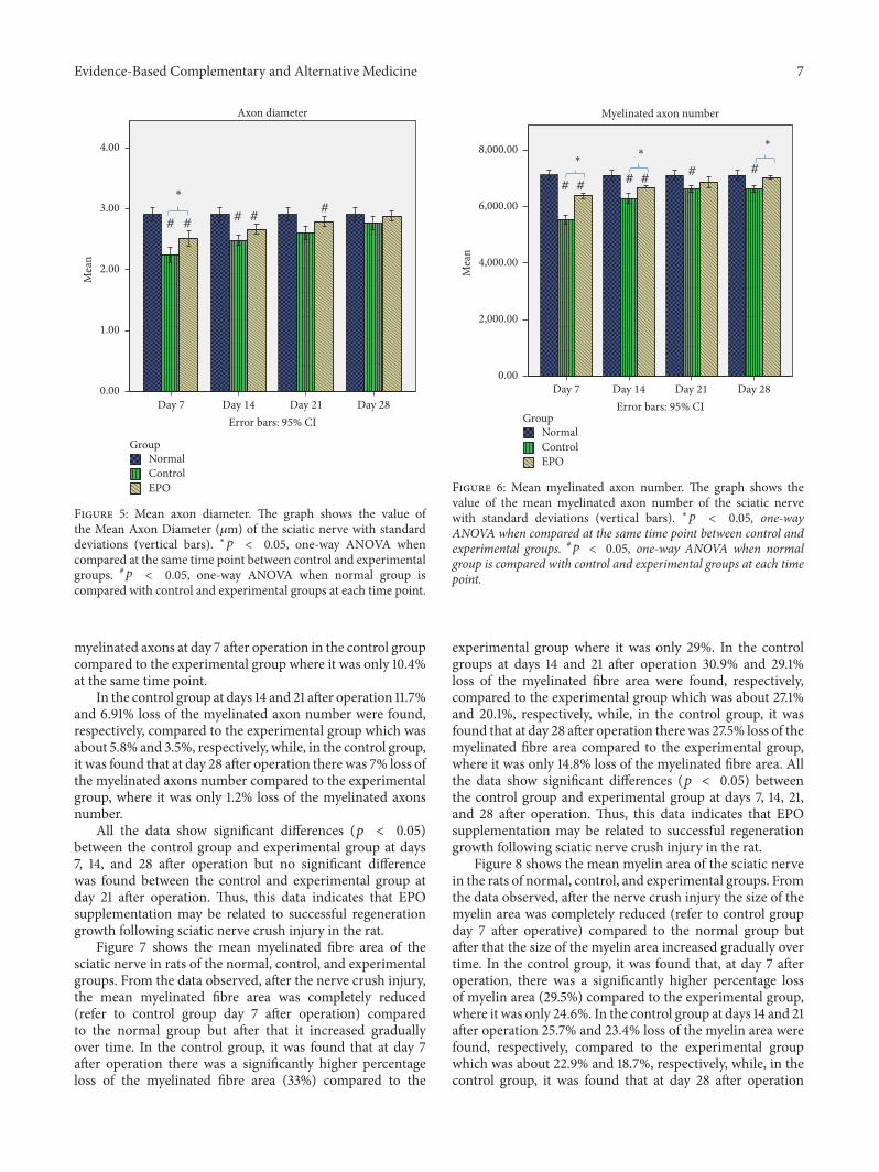
Figure 6 shows the mean myelinated axon number whichwas 7116 for the control group, while for the rats at 7, 14, 21,and 28 days after operation it was 5535, 6281, 6624, and 6620,respectively, with significant differences between the controlgroup and each of the experimental groups. This shows that,28 days after the injury, the mean myelinated axon numberwas still significantly reduced/little compared to that of thecontrol rats. There was a significant (𝑝 < 0.05) 22.2% loss of
Evidence-Based Complementary and Alternative Medicine 7
Axon diameter
Day 14 Day 21 Day 28Day 7Error bars: 95% CI
# ## #
#
GroupNormalControlEPO
∗
0.00
1.00
2.00
3.00
4.00
Mea
n
Figure 5: Mean axon diameter. The graph shows the value ofthe Mean Axon Diameter (𝜇m) of the sciatic nerve with standarddeviations (vertical bars). ∗𝑝 < 0.05, one-way ANOVA whencompared at the same time point between control and experimentalgroups. #𝑝 < 0.05, one-way ANOVA when normal group iscompared with control and experimental groups at each time point.
myelinated axons at day 7 after operation in the control groupcompared to the experimental group where it was only 10.4%at the same time point.
In the control group at days 14 and 21 after operation 11.7%and 6.91% loss of the myelinated axon number were found,respectively, compared to the experimental group which wasabout 5.8% and 3.5%, respectively, while, in the control group,it was found that at day 28 after operation there was 7% loss ofthe myelinated axons number compared to the experimentalgroup, where it was only 1.2% loss of the myelinated axonsnumber.
All the data show significant differences (𝑝 < 0.05)between the control group and experimental group at days7, 14, and 28 after operation but no significant differencewas found between the control and experimental group atday 21 after operation. Thus, this data indicates that EPOsupplementation may be related to successful regenerationgrowth following sciatic nerve crush injury in the rat.
Figure 7 shows the mean myelinated fibre area of thesciatic nerve in rats of the normal, control, and experimentalgroups. From the data observed, after the nerve crush injury,the mean myelinated fibre area was completely reduced(refer to control group day 7 after operation) comparedto the normal group but after that it increased graduallyover time. In the control group, it was found that at day 7after operation there was a significantly higher percentageloss of the myelinated fibre area (33%) compared to the
Myelinated axon number
Day 14 Day 21 Day 28Day 7Error bars: 95% CI
# ##
# ##
∗∗
∗
GroupNormalControlEPO
0.00
2,000.00
4,000.00
6,000.00
8,000.00
Mea
n
Figure 6: Mean myelinated axon number. The graph shows thevalue of the mean myelinated axon number of the sciatic nervewith standard deviations (vertical bars). ∗𝑝 < 0.05, one-wayANOVA when compared at the same time point between control andexperimental groups. #𝑝 < 0.05, one-way ANOVA when normalgroup is compared with control and experimental groups at each timepoint.
experimental group where it was only 29%. In the controlgroups at days 14 and 21 after operation 30.9% and 29.1%loss of the myelinated fibre area were found, respectively,compared to the experimental group which was about 27.1%and 20.1%, respectively, while, in the control group, it wasfound that at day 28 after operation there was 27.5% loss of themyelinated fibre area compared to the experimental group,where it was only 14.8% loss of the myelinated fibre area. Allthe data show significant differences (𝑝 < 0.05) betweenthe control group and experimental group at days 7, 14, 21,and 28 after operation. Thus, this data indicates that EPOsupplementation may be related to successful regenerationgrowth following sciatic nerve crush injury in the rat.
Figure 8 shows the mean myelin area of the sciatic nervein the rats of normal, control, and experimental groups. Fromthe data observed, after the nerve crush injury the size of themyelin area was completely reduced (refer to control groupday 7 after operative) compared to the normal group butafter that the size of the myelin area increased gradually overtime. In the control group, it was found that, at day 7 afteroperation, there was a significantly higher percentage lossof myelin area (29.5%) compared to the experimental group,where it was only 24.6%. In the control group at days 14 and 21after operation 25.7% and 23.4% loss of the myelin area werefound, respectively, compared to the experimental groupwhich was about 22.9% and 18.7%, respectively, while, in thecontrol group, it was found that at day 28 after operation

8 Evidence-Based Complementary and Alternative Medicine
Myelinated Fibre area
# #
∗
# #
∗
0.00
5.00
10.00
15.00
20.00
25.00
Mea
n
Day 14 Day 21 Day 28Day 7Error bars: 95% CI
GroupNormalControlEPO
Figure 7: Mean myelinated fibre area. The graph shows the value ofthe mean myelinated fibre area of the sciatic nerve with standarddeviations (vertical bars). ∗𝑝 < 0.05, one-way ANOVA whencompared at the same time point between control and experimentalgroups. #𝑝 < 0.05, one-way ANOVA when normal group is comparedwith control and experimental groups at each time point.
Myelin area
∗
# ## #
# #
# #
0.00
2.00
4.00
6.00
8.00
10.00
12.00
Mea
n
Day 14 Day 21 Day 28Day 7Error bars: 95% CI
GroupNormalControlEPO
Figure 8: Mean myelin area. The graph shows the value of themeanmyelin area (𝜇m2) of the sciatic nervewith standard deviations(vertical bars). ∗𝑝 < 0.05, one-way ANOVA when compared at thesame time point between control and experimental groups. #𝑝 < 0.05,one-way ANOVA when normal group compared with control andexperimental groups at each time point.
there was 20.2% loss of the myelin area compared to theexperimental group,where it was only 11.6% loss of themyelinarea.Thus, this data indicates that EPO supplementationmaybe related to successful regeneration growth following sciaticnerve crush injury in the rat.
4. Discussion
The results of this study indicate that supplementation withEPO is effective for sciatic nerve crush injury in a variety ofrodentmodels especially the ratmodel.The present studywasconducted to determine the potential effect of EPO supple-mentation on the rate of peripheral nerve regeneration aftersciatic nerve injury, through morphological and morpho-metric analysis of the injured nerve. Oral supplementationof EPO was capable of enhancing nerve regeneration andspeeding up the functional recovery of the nerve after thenerve crush injury in the rat.
Based on both the behavioural and histological outcomes,themost significant finding in this study is that EPOpossessespotential benefits in improving the toe-spreading reflex(behavioural test) and reducing morphological damage tothe sciatic nerve. Many techniques have been developed byresearchers and scientists around the world to induce crushesto the sciatic nerve. The most commonly used model isthe contusion model. This induces a sciatic nerve injury byweight drop and impactor rod [25]. In this study, we usedmodified Watchmaker’s forceps to induce a crush injury tothe sciatic nerve as it is easy to use, is affordable, and hasproven to be clinically relevant. Using forceps with differentseparation distances (calibrated ignition gauge or spacers)at closure point, it is possible to achieve mild, moderate,and severe injuries that can be distinguished histologically orotherwise [26]. However, the forceps induced compressioncannot compute the velocity or force delivered at the injurysite but it has been shown to be widely used and allowscontrol of injury induction [26]. Our results are similar to thefindings of other researchers who have also used forceps toinduce crushes in sciatic nerve models [26]. The functionalrecovery assessment that was used in the present study wasthe toe-spreading reflex. A simple, precise, and inherentlymeaningful measure is the return of toe-spreading [27]. Inthe present study, the findings of functional recovery aresupported by histological analysis of the myelin and axonmorphology and morphometric analysis. It was found thatcomplete nerve recovery was noted as early as day 9 and asalate as day 15 in experimental group. The mean of completenerve recovery was at day 12. EPO treatment, therefore,reduced the time taken for complete nerve recovery by 14 dayscompared to the control group. Furthermore, the result showsthat the mean difference between the normal and the EPOgroup is evidently low compared to the high mean differencebetween the normal and control group. The implication isthat EPO leads to statistically significant TSR recovery at theend experiment (𝑝 < 0.05). This reduction of 53.8% is higherthan the 34.8% reduction observed in the past study [28]where the reduction is statistically significant (𝑝 < 0.005).
Morphologic and morphometric analysis were used toevaluate the condition of the sciatic nerve after nerve crush

Evidence-Based Complementary and Alternative Medicine 9
injury, and the results were compared to those obtained withthe same animals with respect to functional assessment (toe-spreading reflex). In the present study, it should be notedthat (in the histological studies) the epineurium was neverdisrupted in any of the groups, but the perineurium andendoneurium were affected. The histological analysis clearlyshowed that the myelin was well-arranged in the rats fromthe normal group but was scattered and irregularly shapedin the rats from the control group. The micrographs of thesemithin Toluidine Blue staining showed that the axons andmyelin in the experimental group (28 days after operation)had completely recovered or were not markedly differentfrom those of the normal group. At 28 days after operation(experimental group), the axons displayed a rounded struc-ture and the appearance of the myelin sheath became thickercompared to earlier in the nerve crush injury (7 days afteroperation).The observed myelin sheath changes were similarto previous studies as evaluation studies as this parameterallows for discrimination of the pathological condition of aperipheral nerve from a morphologically and physiologicallyintact nerve [29–31]. At 7 days after operation, the axonsdisplayed a population of regenerating axons. The size of theaxons was small and the myelin sheath was not visible.
From the histological analysis, the mean axon area hadsignificantly decreased from 8.95 𝜇m2 in the normal groupto 7.20𝜇m2 7 days after the crush injury (control group).After that, it demonstrated a relative increase 7 days afteroperation up to 28 days after operation. This finding wascomparable with that of a previous study [22] where thevalue of the normal mean axon area was in the range of5.38–9.41 𝜇m2. Meanwhile, the mean axon diameter followedthe same trend as the diameter of the normal axon pro-gressively and significantly decreasing from 2.91𝜇m (normalgroup) to 2.24𝜇m 7 days after the crush injury (controlgroup). This is similar to or not markedly different fromthe results displayed in previous studies, which indicatedthat the diameter of the normal axon was in the rangeof 3.0–3.5 𝜇m and 2.17–2.96𝜇m [29]. Based on our study,the normal mean myelin area was 10.10 𝜇m2. It significantlydecreased to 7.12 𝜇m2 7 days after the crush injury. Thisfinding is supported by the results of previous studies [32]where the value of the normal mean myelin area was 12.75 ±4.37 𝜇m2. Similar behaviour was observed for all of the othermorphometric parameters (namely, the myelinated fibre areaand myelinated axon number), which significantly decreasedin all crush injury groups (control and experimental groups)compared to the normal group. However, since most of thedifferences between the crush injury groups were significant,these findings may be considered normal and indicate thatthe nerve crush injury produced consistent and virtuallysimilar results [22]. It has been reported that EPO supple-mentation was responsible for accelerated axonal regrowthin sciatic nerve injured rats [28, 33]. We also believed thatEPO can improve Schwann cell proliferation. Schwann cellsare responsible for the myelination of peripheral axons. Thisprocess occurs when Schwann cell wrap around the axons,creating a myelin sheath from multiple concentric layers ofcytoplasmic membrane [4]. The EPO acts via GLA which is
a substrate for production of vasodilator prostanoids such asprostacyclin to improve nerve perfusion [34]. Further studiesalso suggests that cyclooxygenase mediated metabolites suchas prostacyclin are vital for EPO’s actions and are necessaryfor maintaining the integrity of sciatic vasa nervorum in rats[35].
5. Conclusion
In conclusion, the injury model that has been used in thisinvestigation is an efficient, cost-effective, and ideal way toinduce sciatic nerve crush injury in rats. Based on the findingsobtained in this study, daily oral EPO supplementation hasbeen shown to enhance the recovery of damaged peripheralnerve. A dose of 6000mg/day of EPO supplementation helpsaccelerate nerve regeneration and, therefore, might have avital role in the therapy of peripheral nerve injury.
Conflicts of Interest
The authors declare that they have no conflicts of interest.
Acknowledgments
This study was supported by Grant nos. PV046/2011A,RG010/09AFR, and CG005-2014 from the University ofMalaya.The authors would like to thank the ElectronMicros-copy Unit technicians for their support and the Departmentof Anatomy staff for their expert technical assistance.
References
[1] D. T. W. Chiu and C. Ishii, “Management of peripheral nerveinjury,” Orthopedic Clinics of North America, vol. 17, no. 3, pp.365–373, 1986.
[2] X. Navarro, M. Vivo, and A. Valero-Cabre, “Neural plasticityafter peripheral nerve injury and regeneration,” Progress inNeurobiology, vol. 82, no. 4, pp. 163–201, 2007.
[3] G. Lundborg, “Intraneural microcirculation,” Orthopedic Clin-ics of North America, vol. 19, no. 1, pp. 1–12, 1988.
[4] D. W. Zachodne and L. T. Ho, “Endoneural microenvironmentand acute nerve crush injury in the rat sciatic nerve,” BrainResearch, vol. 535, pp. 43–48, 1990.
[5] C. Bagdatoglu, A. Saray, H. S. Surucu, H. Ozturk, and L. Tamer,“Effect of trapidil in ischemia/reperfusion injury of peripheralnerves,” Neurosurgery, vol. 51, no. 1, pp. 212–220, 2002.
[6] J. M. Kerns, I. M. Pavkovic, A. J. Fakhouri, K. L. Wickersham,and J. A. Freeman, “An experimental implant for applying aDC electrical field to peripheral nerve,” Journal of NeuroscienceMethods, vol. 19, no. 3, pp. 217–223, 1987.
[7] K.-H. Wong, M. Naidu, P. David et al., “Peripheral nerveregeneration following crush injury to rat peroneal nerve byaqueous extract of medicinal mushroom Hericium erinaceus(Bull.: Fr) Pers. (Aphyllophoromycetideae),” Evidence-BasedComplementary and Alternative Medicine, vol. 2011, Article ID580752, 10 pages, 2011.
[8] A. Kimura, T. Ajiki, K. Takeuchi et al., “Transmigration ofdonor cells involved in the sciatic nerve graft,” TransplantationProceedings, vol. 37, no. 1, pp. 205–207, 2005.

10 Evidence-Based Complementary and Alternative Medicine
[9] N. Kamada, “Transplantation tolerance and immunosuppres-sion following liver grafting in rats,” Immunology Today, vol. 6,no. 11, pp. 336–342, 1985.
[10] F. Reichert, A. Saada, and S. Rotshenker, “Peripheral nerveinjury induces Schwann cells to express two macrophages phe-notypes: phagocytosis and the galactose-specific lectinMAC-2,”The Journal of Neuroscience, vol. 14, no. 5, part 2, pp. 3231–3245,1994.
[11] K. Wierzba, B. Wankowicz, A. Piekarczyk, and A. Danysz,“Cytostatics and immunosuppressive drugs,” Side Effects ofDrugs Annual, vol. 8, pp. 395–425, 1984.
[12] D. F. Horrobin, “Nutritional and medical importance ofgamma-linolenic acid,” Progress in Lipid Research, vol. 31, no.2, pp. 163–194, 1992.
[13] F. D. Gunstone, “Gammar linolenic acid—occurrence andphysical and chemical properties,” Progress in Lipid Research,vol. 31, no. 2, pp. 145–161, 1992.
[14] W.W.Christie, “The analysis of evening primrose oil,” IndustrialCrops and Products, vol. 10, no. 2, pp. 73–83, 1999.
[15] I. Yaychuk-Arabie, “Evening primrose oil: “king’s cure-all”,”Health Naturally, 1993.
[16] J. Kleijnen, “Evening primrose oil: currently used in manyconditions with little justification,” British Medical Journal, vol.309, pp. 824-825, 1994.
[17] P. Mason, “Evening primrose oil,” in Handbook of DietarySupplements: Vitamins and Other Health Supplements, pp. 69–72, Blackwell Science, Oxford, UK, 1995.
[18] D. F. Horrobin, “Essential fatty acids in the managementof impaired nerve function in diabetes,” Diabetes, vol. 46,supplement 2, pp. S90-S93, 1997.
[19] D. F. Horrobin, “The role of essential fatty acids andprostaglandins in the premenstrual syndrome,” Journal Repro-ductive Medicine, vol. 28, no. 7, pp. 465–468, 1983.
[20] G. A. Jamal and H. Carmieheal, “The effect of 𝛾-linolenicacid on human diabetic peripheral neuropathy: a double-blindplacebo-controlled trial,” Diabetic Medicine, vol. 7, no. 4, pp.319–323, 1990.
[21] H. Keen, J. Payan, J. Allawi et al., “Treatment of diabeticneuropathy with 𝛾-linolenic acid,” Diabetes Care, vol. 16, no. 1,pp. 8–15, 1993.
[22] P. Y. C. N. Mazzer, C. H. Barbieri, N. Mazzer, and V. P. S. Fazan,“Morphologic and morphometric evaluation of experimentalacute crush injuries of the sciatic nerve of rats,” Journal ofNeuroscience Methods, vol. 173, no. 2, pp. 249–258, 2008.
[23] Evening Primrose Oil, 2017, http://www.drugs.com/npp/even-ing-primrose-oil.html.
[24] E. Gutmann, “Factors affecting recovery ofmotor function afternerve lesions,” Journal of Neurology, Neurosurgery & Psychiatry,vol. 5, no. 3-4, pp. 81–95, 1942.
[25] H. C. Schmitz and G. M. Beer, “The toe-spreading reflex of therabbit revisited—functional evaluation of complete peronealnerve lesions,” Laboratory Animals, vol. 35, no. 4, pp. 340–345,2001.
[26] A. Raducan, S. Mirica, O. Duicu et al., “Morphological andfunctional aspects of sciatic nerve regeneration after crushinjury,”Romanian Journal ofMorphology& Embryology, vol. 54,pp. 735–739, 2013.
[27] D. Abdullahi, A. A. Annuar,M.Mohamad, I. Aziz, and J. Sanusi,“Experimental spinal cord trauma: a review of mechanicallyinduced spinal cord injury in rat models,” Reviews in theNeurosciences, vol. 28, no. 1, pp. 15–20, 2017.
[28] J. Sanusi,The possible of the afferent activity in motoneurone sur-vival after neonatal target deprivation [Ph.D. thesis], UniversityCollege London, London, UK, 1996.
[29] M. L. Garcia, M. V. Rao, J. Fujimoto et al., “Phosphorylationof highly conserved neurofilament medium KSP repeats is notrequired formyelin-dependent radial axonal growth,” Journal ofNeuroscience, vol. 29, no. 5, pp. 1277–1284, 2009.
[30] H. H. Samsinah, S. Junedah, and A. K. Norazlin, “Effectsof Evening Primrose Oil on the rate of nerve regeneration,”in Proceedings of the 14th Scientific Meeting of the MalaysianSociety of Phamacology and Physiology—Cardiac RehabilitationConference and Cardiovascular CounselingWorkshop, May 1999.
[31] R. Lindemuth, C. Ernzerhof, and K. Schimrigk, “Comparativemorphometry of myelinated nerve fibres in the normal andpathologically altered human sural and tibial nerve,” ClinicalNeuropathology, vol. 21, no. 1, pp. 29–34, 2002.
[32] J. Scharpf, R. Meirer, M. Zielinski et al., “A novel technique forperipheral nerve repair,” Laryngoscope, vol. 113, no. 1, pp. 95–101,2003.
[33] J.M. Schroder, “Altered ratio between axondiameter andmyelinsheath thickness in regenerated nerve fibers,” Brain Research,vol. 45, no. 1, pp. 49–65, 1972.
[34] N. E. Cameron, M. A. Cotter, and S. Robertson, “Essentialfatty acid diet supplementation: effects on peripheral nerve andskeletal muscle function and capillarization in streptozotocin-induced diabetic rats,”Diabetes, vol. 40, no. 5, pp. 532–539, 1991.
[35] N. E. Cameron, M. A. Cotter, K. C. Dines, S. Robertson,and D. Cox, “The effects of evening primrose oil on nervefunction and capillarization in streptozotocin-induced diabeticrats: modulation by the cyclo-oxygenase inhibitor flurbiprofen,”British Journal of Pharmacology, vol. 109, no. 4, pp. 972–979,1993.

Submit your manuscripts athttps://www.hindawi.com
Stem CellsInternational
Hindawi Publishing Corporationhttp://www.hindawi.com Volume 2014
Hindawi Publishing Corporationhttp://www.hindawi.com Volume 2014
MEDIATORSINFLAMMATION
of
Hindawi Publishing Corporationhttp://www.hindawi.com Volume 2014
Behavioural Neurology
EndocrinologyInternational Journal of
Hindawi Publishing Corporationhttp://www.hindawi.com Volume 2014
Hindawi Publishing Corporationhttp://www.hindawi.com Volume 2014
Disease Markers
Hindawi Publishing Corporationhttp://www.hindawi.com Volume 2014
BioMed Research International
OncologyJournal of
Hindawi Publishing Corporationhttp://www.hindawi.com Volume 2014
Hindawi Publishing Corporationhttp://www.hindawi.com Volume 2014
Oxidative Medicine and Cellular Longevity
Hindawi Publishing Corporationhttp://www.hindawi.com Volume 2014
PPAR Research
The Scientific World JournalHindawi Publishing Corporation http://www.hindawi.com Volume 2014
Immunology ResearchHindawi Publishing Corporationhttp://www.hindawi.com Volume 2014
Journal of
ObesityJournal of
Hindawi Publishing Corporationhttp://www.hindawi.com Volume 2014
Hindawi Publishing Corporationhttp://www.hindawi.com Volume 2014
Computational and Mathematical Methods in Medicine
OphthalmologyJournal of
Hindawi Publishing Corporationhttp://www.hindawi.com Volume 2014
Diabetes ResearchJournal of
Hindawi Publishing Corporationhttp://www.hindawi.com Volume 2014
Hindawi Publishing Corporationhttp://www.hindawi.com Volume 2014
Research and TreatmentAIDS
Hindawi Publishing Corporationhttp://www.hindawi.com Volume 2014
Gastroenterology Research and Practice
Hindawi Publishing Corporationhttp://www.hindawi.com Volume 2014
Parkinson’s Disease
Evidence-Based Complementary and Alternative Medicine
Volume 2014Hindawi Publishing Corporationhttp://www.hindawi.com