Embed Size (px)
Citation preview
The Cell-Free Integration of a Polytopic MitochondrialMembrane Protein into Liposomes OccursCotranslationally and in a Lipid-Dependent MannerAshley R. Long, Catherine C. O’Brien, Nathan N. Alder*
Department of Molecular and Cell Biology, University of Connecticut, Storrs, Connecticut, United States of America
Abstract
The ADP/ATP Carrier (AAC) is the most abundant transporter of the mitochondrial inner membrane. The central role thatthis transporter plays in cellular energy production highlights the importance of understanding its structure, function, andthe basis of its pathologies. As a means of preparing proteoliposomes for the study of membrane proteins, several groupshave explored the use of cell-free translation systems to facilitate membrane protein integration directly into preformedunilamellar vesicles without the use of surfactants. Using AAC as a model, we report for the first time the detergent-freereconstitution of a mitochondrial inner membrane protein into liposomes using a wheat germ-based in vitro translationsystem. Using a host of independent approaches, we demonstrate the efficient integration of AAC into vesicles with aninner membrane-mimetic lipid composition and, more importantly, that the integrated AAC is functionally active intransport. By adding liposomes at different stages of the translation reaction, we show that this direct integration isobligatorily cotranslational, and by synthesizing stable ribosome-bound nascent chain intermediates, we show that thenascent AAC polypeptide interacts with lipid vesicles while ribosome-bound. Finally, we show that the presence of thephospholipid cardiolipin in the liposomes specifically enhances AAC translation rate as well as the efficiency of vesicleassociation and integration. In light of these results, the possible mechanisms of liposome-assisted membrane proteinintegration during cell-free translation are discussed with respect to the mode of integration and the role of specific lipids.
Citation: Long AR, O’Brien CC, Alder NN (2012) The Cell-Free Integration of a Polytopic Mitochondrial Membrane Protein into Liposomes Occurs Cotranslationallyand in a Lipid-Dependent Manner. PLoS ONE 7(9): e46332. doi:10.1371/journal.pone.0046332
Editor: Paul Cobine, Auburn University, United States of America
Received June 8, 2012; Accepted August 31, 2012; Published September 25, 2012
Copyright: � 2012 Long et al. This is an open-access article distributed under the terms of the Creative Commons Attribution License, which permitsunrestricted use, distribution, and reproduction in any medium, provided the original author and source are credited.
Funding: This work was funded by a Scientist Development Grant from the American Heart Association to NA (Grant #: 09SDG2380019) (http://www.heart.org).The funder had no role in study design, data collection and analysis, decision to publish, or preparation of the manuscript.
Competing Interests: The authors have declared that no competing interests exist.
* E-mail: [email protected]
Introduction
Membrane proteins constitute roughly one third of all gene
products in any given organism, and over half of all current
pharmaceutical targets [1,2]. However, solution-based biochem-
ical and biophysical studies of membrane proteins are technically
challenging because their hydrophobicity causes them to form
insoluble aggregates in aqueous systems. In vivo expression of such
proteins, while successful in certain cases, can be hampered due to
cell toxicity, misfolding, and aggregation [1,3]. For these reasons,
cell-free synthesis of membrane proteins is becoming recognized as
a powerful technique for studying membrane proteins within
model membrane systems [1,2,4]. By one strategy, cell-free protein
synthesis is conducted in the presence of detergents to maintain the
solubility of the translation product before reconstitution into
liposomes [5,6]. However, this approach can suffer drawbacks
because even mild detergents with a low critical micelle
concentration (cmc) can be inhibitory to the translation apparatus
and can be difficult to remove [1,2]. Recently a number of
independent groups have reported that membrane proteins, even
topologically complex ones, can integrate into pre-formed
unilamellar liposomes during cell-free translation reactions and
fold into a functional state without the inclusion of detergents or a
dedicated complex that mediates integration [1,2,7–9]. Examples
include proteins translated in cell-free systems based on E.coli
lysates (i.e. bacteriorhodopsin [7], connexin-43 [8] and the Fo/F1
ATP synthase [10]) as well as wheat germ lysates (i.e. stearoyl-CoA
desaturase [9] and sphingolipid synthase [11]). By this experi-
mental approach, some polypeptides require a specific lipid
composition for unassisted integration. In the case of bacteriorho-
dopsin, for example, it was shown that the component lipids must
have acyl chain lengths and/or degrees of unsaturation that
maintain the bilayer above the phase transition temperature and
excess lipid with inverted hexagonal (HII) phase propensity may
block insertion [7]. In other cases, it has been suggested that
liposomes must be present in the biosynthetic reaction for insertion
to occur, supporting a cotranslational mode of integration [8]. In
this study we have investigated the cell-free spontaneous integra-
tion of the mitochondrial ADP/ATP Carrier (AAC).
AAC is the most abundant transporter of the mitochondrial
inner membrane (IM) [12]. As a member of the mitochondrial
carrier family, it contains three modular repeats of two
transmembrane segments (TMSs), each connected by matrix-
facing loops with small helical elements [12–14]. As part of the
oxidative phosphorylation system, this carrier mediates adenine
nucleotide transport through the 1:1 electrogenic exchange of
ADP and ATP across the IM [15]. Hence AAC plays a central role
in cellular energy metabolism. During transport AAC is believed
to cycle between two extreme conformers [12,15]. In the c-state
PLOS ONE | www.plosone.org 1 September 2012 | Volume 7 | Issue 9 | e46332

the channel lumen is exposed to the cytosol and in the m-state the
lumen is exposed to the matrix [12,15]. These conformations can
be stabilized by the specific inhibitors carboxyatractyloside (CAT)
and bongkrekic acid, respectively [12,15].
The biogenesis route of AAC within the cell is well established.
AAC is encoded in nuclear DNA, translated on cytosolic
ribosomes, and post-translationally integrated into the mitochon-
drial IM via the TIM22 (Translocase of the Inner Mitochondrial
Membrane) pathway (or Carrier pathway) in a manner dependent
upon the membrane potential of the IM [16–18]. As with many
other mitochondrial membrane proteins, the biogenesis of AAC
requires the presence of the dimeric phospholipid cardiolipin (CL),
which is present in the IM at a concentration of 15–20% total
phospholipid [19–25]. Following integration, AAC interacts with
respiratory complexes as well as other members of the mitochon-
drial carrier family, such as the phosphate carrier, the dicarbox-
ylate transporter and the GDP/GTP transporter in a CL-
dependent fashion [22]. This is consistent with the observed role
of CL in maintaining the association of IM respiratory complexes
as supercomplexes [21,22]. The activity of AAC is directly
dependent on the presence of CL [19,20]. A specific role for CL
in AAC function is also supported by published x-ray structures,
which reveal specific interactions of the CL headgroups and acyl
chains at the matrix-facing regions of AAC [14].
The ability to reconstitute multi-spanning mitochondrial
membrane proteins into liposomes by a detergent-free process
would represent a significant technical advance for structural and
functional studies. In this report, we show that AAC translated in a
wheat germ lysate-based system integrates into small unilamellar
vesicles (SUVs) in the absence of translocons and that it is
functional as an adenine nucleotide transporter. This system does
not recapitulate the in vivo import or folding of AAC, but rather
serves as a suitable mimic for reconstitution into an isolated lipid
environment. Within this system, we thoroughly characterize the
role of CL with respect to both the rate of protein synthesis and the
efficiency of integration. Finally, we provide strong experimental
evidence that spontaneous membrane protein integration by this
process is obligatorily cotranslational.
Results
I. In vitro translated AAC integrates directly intoliposomes as functionally active carriers
A. The experimental system. When synthesized in a cell-
free system, hydrophobic proteins typically result in the formation
of insoluble precipitates. Detergents can, in some cases, be added
to cell-free translation reactions to maintain membrane proteins
soluble in micelles [26–28]. However, we found that nonionic
detergents like n-Dodecyl-b-D-maltoside, even at low concentra-
tions (0.05% [w/v]), were inhibitory to our wheat germ
translational apparatus and resulted in lowered yields. We
therefore adopted the recently-described procedure for spontane-
ous insertion of in vitro translated membrane proteins into
liposomes [7–11,29] to test whether AAC would properly integrate
into SUVs included in our wheat germ lysate system.
In our experimental approach, unilamellar liposomes of a
defined lipid content and size were added directly to the
translation reaction and the resulting proteoliposomes were
subsequently purified by discontinuous sucrose gradient ultracen-
trifugation (SGU) (Fig. 1A). These liposomes contained a
biomimetic blend of 1-palmitoyl-2-oleoyl-sn-glycero-3-phospho-
choline (POPC), 1-palmitoyl-2-oleoyl-sn-glycero-3-phosphoetha-
nolamine (POPE), 19,39-bis[1,2-dioleoyl-sn-glycero-3-phospho]-sn-
glycerol (CL), 1-palmitoyl-2-oleoyl-sn-glycero-3-phosphoserine
(POPS), and 1-palmitoyl-2-oleoyl-sn-glycero-3-phosphate (POPA),
in molar ratios of 54:24:16:4:2, respectively. To confirm that our
translated AAC was integrated into liposomes, SGU was used to
separate proteoliposomes from cell-free extract and any aggregat-
ed proteins (Fig. 1A). This method allows for the isolation of
proteoliposomes from the translation components and unincorpo-
rated protein; proteoliposomes will float to the top of the sucrose
gradient, whereas excess translation machinery will pellet [8]. By
this analysis, [35S]AAC translated in the absence of liposomes
pelleted as water-insoluble aggregates (Fig. 1B, ‘‘no liposomes’’). In
contrast, a fraction of [35S]AAC translated in the presence of
liposomes floated with the vesicles, and the percentage of total
AAC in the soluble liposome fraction increased with increasing
liposome concentration (Fig. 1B, ‘‘liposomes’’). We conclude that
[35S]AAC associated strongly enough with SUVs that some
fraction was recovered as floated proteoliposomes.
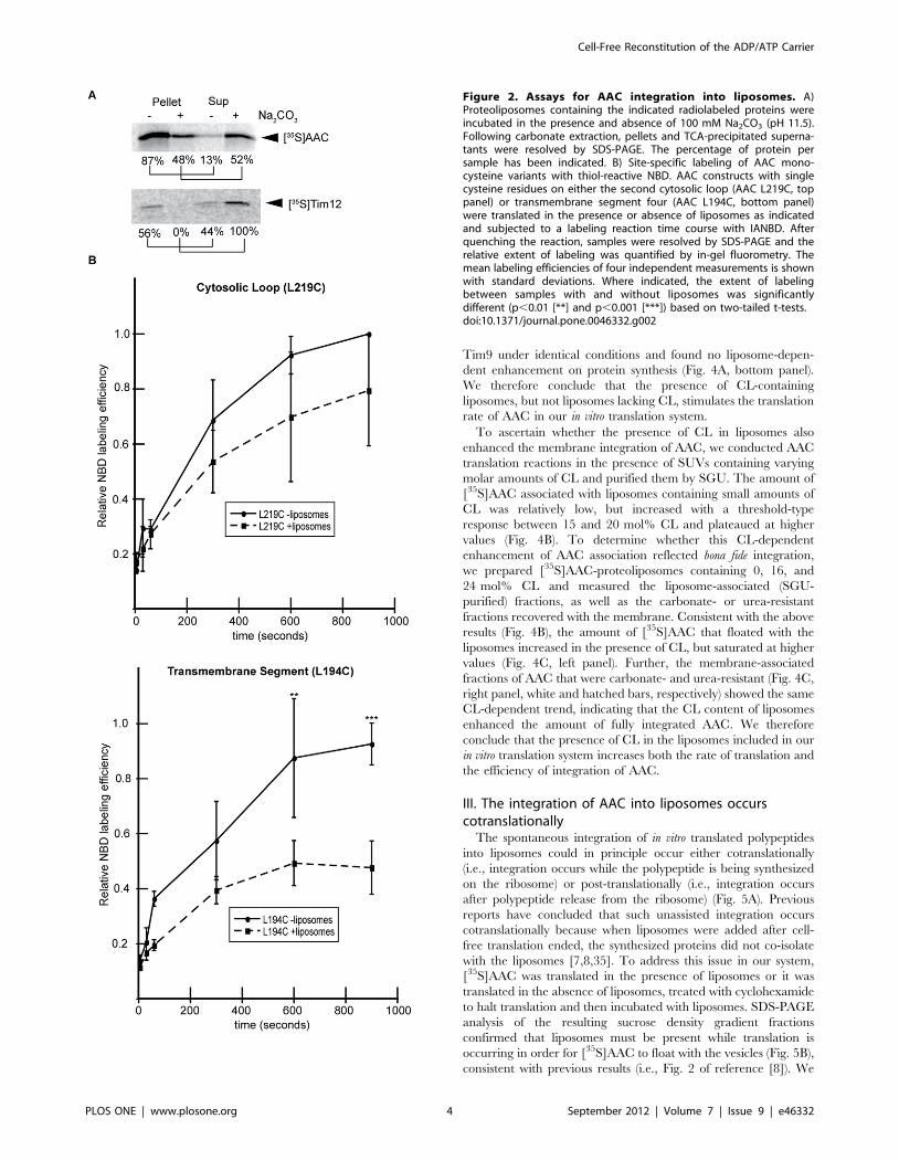
B. Assaying the proper integration of AAC. We conducted
a series of tests to address whether [35S]AAC translated in the
presence of liposomes was properly integrated with transmem-
brane topology or nonspecifically bound to the vesicles. We first
subjected our purified samples to alkaline extraction (pH 11.5),
which will release peripheral membrane proteins from the bilayer
but leave integral membrane proteins associated with the
membrane [5,30]. Following carbonate extraction, approximately
50% of [35S]AAC translated in the presence of SUVs remained
stably associated with the vesicles (Fig. 2A, upper gel, compare
pellet fractions with and without carbonate treatment). As a
control, we translated the peripheral protein [35S]Tim12 in the
presence of liposomes and found that all of the bilayer-associated
protein was removed by carbonate (Fig. 2A, bottom gel, compare
pellet fractions with and without carbonate treatment), confirming
the efficiency of this procedure. These results indicate that a
significant fraction of AAC spontaneously integrated into lipo-
somes is carbonate resistant.
As an independent assay for the proper integration of our
samples, we measured the solvent accessibility of single cysteine
residues within AAC to the thiol-reactive derivative of the
fluorescent probe 7-nitrobenz-2-oxa-1,3-diazolyl (IANBD). This
membrane-impermeable reagent will react with cysteine residues
in a polar environment, but not with those buried within a lipid
bilayer [31]. Thus, we translated AAC variants containing a single
cysteine on an intermembrane space-facing loop (AAC L219C) or
in a transmembrane segment (AAC L194C) in the presence or
absence of liposomes, subjected the samples to IANBD labeling,
and quantified relative labeling efficiencies by in-gel fluorometry.
The cysteine side chain in the loop region was labeled to a similar
extent in the presence and absence of liposomes (Fig. 2B top panel,
compare solid and dashed lines). In contrast, whereas the cysteine
within the TMS was highly accessible to the labeling reagent in the
absence of liposomes (Fig. 2B bottom panel, solid line), when
translated in the presence of liposomes, its labeling decreased
dramatically (Fig. 2B bottom panel, dashed line), consistent with its
presence in a lipid bilayer. The [35S]AAC L194C polypeptides
that were labeled in the presence of liposomes likely represent the
fraction that was not fully integrated and/or misfolded (Fig. 2A).
Taken together, the data from Fig. 2 indicate that a significant
fraction of AAC translated in the presence of SUVs (roughly half,
based on our results) is properly integrated into the membrane
bilayer.
C. AAC is functionally active in cell-free generated
proteoliposomes. To assay the activity of our reconstituted
AAC, we adapted the luciferin-luciferase assay [32] to test the
ability of the carrier to specifically transport ATP out of lipid
vesicles. In our experimental strategy, AAC translation reactions
Cell-Free Reconstitution of the ADP/ATP Carrier
PLOS ONE | www.plosone.org 2 September 2012 | Volume 7 | Issue 9 | e46332

were supplemented with liposomes that were pre-loaded with ATP
and the resulting proteoliposomes were incubated in the presence
of the translation reaction to allow ADP/ATP transport to occur.
Following gel filtration chromatography to remove excess nucle-
otide, the transport competence of AAC was assayed as the
amount of ATP remaining in the liposomes by disrupting the
vesicles with detergent and measuring the total ATP in the sample
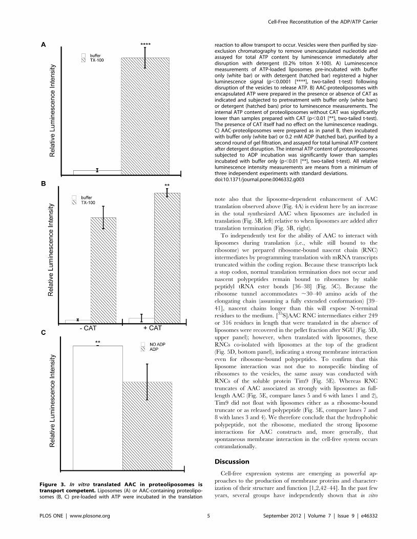
by luminescence. To illustrate the principle behind this strategy,
ATP-loaded, column purified liposomes (not containing AAC) pre-
incubated with buffer only registered a very low luminescence
signal, indicating that there was little free ATP accessible to the
luminescence reagent (Fig. 3A, white bar). However, when parallel
samples of ATP-loaded vesicles were pre-incubated with triton X-
100 just prior to measurement, the luminescence signal increased
dramatically, demonstrating the high ATP content of the vesicle
lumens (Fig. 3A, hatched bar).
We then applied this same analysis to AAC-containing
proteoliposomes that were prepared in the presence and absence
of the specific AAC inhibitor CAT (Fig. 3B). As expected,
detergent treatment resulted in an increased luminescence signal
showing that the purified proteoliposomes contained ATP (Fig. 3B,
compare white and hatched bars for both ‘‘2CAT’’ and ‘‘+CAT’’
samples). However, AAC proteoliposomes prepared in the
presence of inhibitor retained a significantly higher amount of
ATP than those prepared without inhibitor (Fig. 3B, compare
hatched bars of ‘‘+CAT’’ and ‘‘2CAT’’ samples). These results
show that during the incubation process, ATP was specifically
transported out of the proteoliposomes containing non-inhibited
AAC. The decrease in luminal ATP in this assay was likely from
AAC-mediated antiport of [ATP]in (at high concentration) and
[ADP]out (present in the translation reaction), although an AAC-
mediated uniport of ATP out of the liposomes (half-reaction) may
have occurred as well [33].
As an independent means of measuring ADP/ATP antiporter
activity of our reconstituted AAC, we subjected column-purified,
ATP-loaded proteoliposomes to an incubation step with 200 mM
ADP (or buffer only) for 30 min, re-purified them by gel filtration
to remove excess nucleotide, and assayed for encapsulated ATP as
above. Proteoliposomes incubated with ADP contained signifi-
cantly lower ATP concentrations than those incubated with buffer
alone (Fig. 3C, compare white and hatched bars), confirming that
the presence of ADP stimulated the efflux of ATP from the
proteoliposomes. Although these results do not provide an exact
measure of transporter specific activity, they do confirm that our
AAC-containing proteoliposomes support adenine nucleotide
transport and are therefore in a functionally active state.
II. The synthesis and integration of AAC is enhanced inthe presence of CL-containing liposomes
Some reports of cotranslational integration of membrane
proteins into liposomes demonstrate a liposome-dependent
increase in the total amount of protein synthesized [7,34] whereas
others detect no such liposome enhancement of translation rate
[8]. We addressed this issue by conducting cell-free translation
reactions of [35S]AAC in the presence of increasing liposome
concentrations and measuring the total amount of protein
synthesis. We found a marked increase in total [35S]AAC synthesis
with increasing concentrations of inner membrane-mimetic
liposomes (containing CL) but interestingly this stimulating effect
was absent for liposomes lacking CL (Fig. 4A, top panel). As a
control, we measured the rate of translation of the soluble protein
Figure 1. Cell-free translation and purification of AAC-containing proteoliposomes. A) Schematic illustration of the experimental process.[35S]AAC is translated in a wheat germ-based system in the presence of SUVs and the sample is subjected to SGU. Buoyant proteoliposomes remainat the top of the gradient, whereas aggregated protein and ribosomes from the translation reaction pellet. B) The extent of AAC integration dependson liposome concentration. [35S]AAC was translated in the presence of variable SUV concentrations as indicated and samples from each fraction wereresolved by SDS-PAGE. Fractions 1–4 indicate fractions collected from top to bottom of the gradient. Normalized band intensities are shown beloweach lane of the gel.doi:10.1371/journal.pone.0046332.g001
Cell-Free Reconstitution of the ADP/ATP Carrier
PLOS ONE | www.plosone.org 3 September 2012 | Volume 7 | Issue 9 | e46332
Tim9 under identical conditions and found no liposome-depen-
dent enhancement on protein synthesis (Fig. 4A, bottom panel).
We therefore conclude that the presence of CL-containing
liposomes, but not liposomes lacking CL, stimulates the translation
rate of AAC in our in vitro translation system.
To ascertain whether the presence of CL in liposomes also
enhanced the membrane integration of AAC, we conducted AAC
translation reactions in the presence of SUVs containing varying
molar amounts of CL and purified them by SGU. The amount of
[35S]AAC associated with liposomes containing small amounts of
CL was relatively low, but increased with a threshold-type
response between 15 and 20 mol% CL and plateaued at higher
values (Fig. 4B). To determine whether this CL-dependent
enhancement of AAC association reflected bona fide integration,
we prepared [35S]AAC-proteoliposomes containing 0, 16, and
24 mol% CL and measured the liposome-associated (SGU-
purified) fractions, as well as the carbonate- or urea-resistant
fractions recovered with the membrane. Consistent with the above
results (Fig. 4B), the amount of [35S]AAC that floated with the
liposomes increased in the presence of CL, but saturated at higher
values (Fig. 4C, left panel). Further, the membrane-associated
fractions of AAC that were carbonate- and urea-resistant (Fig. 4C,
right panel, white and hatched bars, respectively) showed the same
CL-dependent trend, indicating that the CL content of liposomes
enhanced the amount of fully integrated AAC. We therefore
conclude that the presence of CL in the liposomes included in our
in vitro translation system increases both the rate of translation and
the efficiency of integration of AAC.
III. The integration of AAC into liposomes occurscotranslationally
The spontaneous integration of in vitro translated polypeptides
into liposomes could in principle occur either cotranslationally
(i.e., integration occurs while the polypeptide is being synthesized
on the ribosome) or post-translationally (i.e., integration occurs
after polypeptide release from the ribosome) (Fig. 5A). Previous
reports have concluded that such unassisted integration occurs
cotranslationally because when liposomes were added after cell-
free translation ended, the synthesized proteins did not co-isolate
with the liposomes [7,8,35]. To address this issue in our system,
[35S]AAC was translated in the presence of liposomes or it was
translated in the absence of liposomes, treated with cyclohexamide
to halt translation and then incubated with liposomes. SDS-PAGE
analysis of the resulting sucrose density gradient fractions
confirmed that liposomes must be present while translation is
occurring in order for [35S]AAC to float with the vesicles (Fig. 5B),
consistent with previous results (i.e., Fig. 2 of reference [8]). We
Figure 2. Assays for AAC integration into liposomes. A)Proteoliposomes containing the indicated radiolabeled proteins wereincubated in the presence and absence of 100 mM Na2CO3 (pH 11.5).Following carbonate extraction, pellets and TCA-precipitated superna-tants were resolved by SDS-PAGE. The percentage of protein persample has been indicated. B) Site-specific labeling of AAC mono-cysteine variants with thiol-reactive NBD. AAC constructs with singlecysteine residues on either the second cytosolic loop (AAC L219C, toppanel) or transmembrane segment four (AAC L194C, bottom panel)were translated in the presence or absence of liposomes as indicatedand subjected to a labeling reaction time course with IANBD. Afterquenching the reaction, samples were resolved by SDS-PAGE and therelative extent of labeling was quantified by in-gel fluorometry. Themean labeling efficiencies of four independent measurements is shownwith standard deviations. Where indicated, the extent of labelingbetween samples with and without liposomes was significantlydifferent (p,0.01 [**] and p,0.001 [***]) based on two-tailed t-tests.doi:10.1371/journal.pone.0046332.g002
Cell-Free Reconstitution of the ADP/ATP Carrier
PLOS ONE | www.plosone.org 4 September 2012 | Volume 7 | Issue 9 | e46332
note also that the liposome-dependent enhancement of AAC
translation observed above (Fig. 4A) is evident here by an increase
in the total synthesized AAC when liposomes are included in
translation (Fig. 5B, left) relative to when liposomes are added after
translation termination (Fig. 5B, right).
To independently test for the ability of AAC to interact with
liposomes during translation (i.e., while still bound to the
ribosome) we prepared ribosome-bound nascent chain (RNC)
intermediates by programming translation with mRNA transcripts
truncated within the coding region. Because these transcripts lack
a stop codon, normal translation termination does not occur and
nascent polypeptides remain bound to ribosomes by stable
peptidyl tRNA ester bonds [36–38] (Fig. 5C). Because the
ribosome tunnel accommodates ,30–40 amino acids of the
elongating chain (assuming a fully extended conformation) [39–
41], nascent chains longer than this will expose N-terminal
residues to the medium. [35S]AAC RNC intermediates either 249
or 316 residues in length that were translated in the absence of
liposomes were recovered in the pellet fraction after SGU (Fig. 5D,
upper panel); however, when translated with liposomes, these
RNCs co-isolated with liposomes at the top of the gradient
(Fig. 5D, bottom panel), indicating a strong membrane interaction
even for ribosome-bound polypeptides. To confirm that this
liposome interaction was not due to nonspecific binding of
ribosomes to the vesicles, the same assay was conducted with
RNCs of the soluble protein Tim9 (Fig. 5E). Whereas RNC
truncates of AAC associated as strongly with liposomes as full-
length AAC (Fig. 5E, compare lanes 5 and 6 with lanes 1 and 2),
Tim9 did not float with liposomes either as a ribosome-bound
truncate or as released polypeptide (Fig. 5E, compare lanes 7 and
8 with lanes 3 and 4). We therefore conclude that the hydrophobic
polypeptide, not the ribosome, mediated the strong liposome
interactions for AAC constructs and, more generally, that
spontaneous membrane interaction in the cell-free system occurs
cotranslationally.
Discussion
Cell-free expression systems are emerging as powerful ap-
proaches to the production of membrane proteins and character-
ization of their structure and function [1,2,42–44]. In the past few
years, several groups have independently shown that in vitro
Figure 3. In vitro translated AAC in proteoliposomes istransport competent. Liposomes (A) or AAC-containing proteolipo-somes (B, C) pre-loaded with ATP were incubated in the translation
reaction to allow transport to occur. Vesicles were then purified by size-exclusion chromatography to remove unencapsulated nucleotide andassayed for total ATP content by luminescence immediately afterdisruption with detergent (0.2% triton X-100). A) Luminescencemeasurements of ATP-loaded liposomes pre-incubated with bufferonly (white bar) or with detergent (hatched bar) registered a higherluminescence signal (p,0.0001 [****], two-tailed t-test) followingdisruption of the vesicles to release ATP. B) AAC-proteoliposomes withencapsulated ATP were prepared in the presence or absence of CAT asindicated and subjected to pretreatment with buffer only (white bars)or detergent (hatched bars) prior to luminescence measurements. Theinternal ATP content of proteoliposomes without CAT was significantlylower than samples prepared with CAT (p,0.01 [**], two-tailed t-test).The presence of CAT itself had no effect on the luminescence readings.C) AAC-proteoliposomes were prepared as in panel B, then incubatedwith buffer only (white bar) or 0.2 mM ADP (hatched bar), purified by asecond round of gel filtration, and assayed for total luminal ATP contentafter detergent disruption. The internal ATP content of proteoliposomessubjected to ADP incubation was significantly lower than samplesincubated with buffer only (p,0.01 [**], two-tailed t-test). All relativeluminescence intensity measurements are means from a minimum ofthree independent experiments with standard deviations.doi:10.1371/journal.pone.0046332.g003
Cell-Free Reconstitution of the ADP/ATP Carrier
PLOS ONE | www.plosone.org 5 September 2012 | Volume 7 | Issue 9 | e46332

translated membrane proteins can directly integrate into model
membrane systems including synthetic liposomes [7,9,11,34,45–
48], and nanoscale lipid bilayers (nanodiscs) [49,50] that are
included in the synthesis reaction in an manner that does not
require detergents. In this report, we have shown that the AAC
transporter also integrates spontaneously into SUVs in a cell-free
translation, which, to the best of our knowledge, represents the first
such study for a mitochondrial inner membrane protein.
We demonstrated the liposome association and integration of in
vitro translated AAC using a host of established methods. First,
nearly 50% of [35S]AAC synthesized in the presence of liposomes
floated at the top of a sucrose density gradient (Fig. 1), confirming
the association of the protein with buoyant vesicles. Second, we
found that approximately half of the [35S]AAC that were co-
isolated with liposomes were resistant to carbonate extraction
(Fig. 2A), a primary diagnostic indicator of stably integrated
Figure 4. The presence of CL in liposomes enhances AAC synthesis and membrane integration. A) Liposome dependence of total proteinsynthesis. Cell-free translation reactions were programmed with mRNA encoding AAC or Tim9 in the absence of liposomes (‘‘X’’) or in the presence ofliposomes at the final lipid concentration shown. B) Effect of CL concentration on AAC-liposome association. AAC translation reactions wereconducted in the presence of liposomes containing variable mol% CL as indicated and purified by SGU. The relative amounts of co-isolated [35S]AACare shown as mean values from a minimum of three independent experiments with standard deviations. C) Effect of CL concentration on AACintegration. [35S]AAC-containing proteoliposomes with variable amounts of CL were prepared and purified by SGU as in panel B. A subset of thesamples were subjected to mock (buffer only) treatment (left panel) and the remaining samples were subjected to carbonate or urea extraction (rightpanel). Values shown are average band intensities normalized with respect to the highest value for liposome associated (left) or fully integrated(right) sample sets from three independent experiments. The relative amount of [35S]AAC in mock-treated samples (left), with 16 or 24 mol% CL, issignificantly higher (p,0.05 [*], two tailed t-test) than with 0 mol% CL. In carbonate- and urea- treated samples (right) the difference in the relativeamount of [35S]AAC present in the 16 and 24 mol% CL samples (compared to 0%) is even greater than in the untreated samples (p,0.01 [**] orp,0.001 [***], two-tailed t-test).doi:10.1371/journal.pone.0046332.g004
Cell-Free Reconstitution of the ADP/ATP Carrier
PLOS ONE | www.plosone.org 6 September 2012 | Volume 7 | Issue 9 | e46332

membrane proteins. Finally, we used a cysteine accessibility
approach with the thiol-reactive fluorescent probe IANBD to show
that a cysteine residue in an AAC transmembrane segment is
specifically shielded from labeling when integrated into liposomes,
whereas a cysteine site in a soluble loop region is labeled to a
similar extent regardless of liposome association (Fig. 2B). Taken
together, these data suggest that a substantial amount of AAC was
integrated into liposomes during cell-free translation. However,
because only about half of our translated AAC was resistant to
carbonate extraction, a significant portion of AAC must have
remained nonspecifically bound to liposomes even after SGU.
Hence, one should exercise caution when attempting to use this
method to generate homogeneous populations of integrated
membrane proteins. With that caveat, it is clear that our wheat
germ based in vitro translation system is capable of supporting the
integration of AAC into synthetic liposomes.
The most critical test for the proper folding and integration of
reconstituted membrane proteins is functionality; therefore, it is
important that we confirmed the transport activity of AAC that
was translated in our system (Fig. 3). AAC in isolated mitochondria
and reconstituted into proteoliposomes has been amply shown to
mediate the 1:1 electrogenic exchange of ATP42 and ADP32 [51–
54]. The assay used in the present work measured the AAC-
dependent transport of ATP out of vesicles, which was likely
driven in part by the external ADP present in the translation
reaction. Although this system in its current form does not allow
for the precise measurement of ADP/ATP exchange parameters,
it did unequivocally demonstrate functionally active carriers in our
proteoliposomes, because: (i) ATP efflux was blocked by the
specific AAC inhibitor CAT (Fig. 3B), and (ii) ATP efflux was
stimulated by an added incubation with external ADP (Fig. 3C).
Nearly all membrane proteins synthesized within the physio-
logical context of the cell require some combination of soluble
chaperones and membrane-bound complexes to mediate their
integration into the lipid phase. Like other metabolite carrier
proteins of the inner mitochondrial membrane, AAC is encoded in
nuclear DNA, synthesized on cytosolic ribosomes, and subse-
quently targeted to the mitochondria [16,55]. Carrier proteins are
directed to mitochondrial import complexes by multiple internal
targeting sequences that generally exist within the segments that
ultimately form their six transmembrane helices [56]. At the
organelle surface, AAC engages the TOM (Translocase of the
Outer Mitochondrial membrane) complex, which mediates its
translocation across the outer membrane. Within the aqueous
intermembrane space, AAC is kept in a soluble state by association
with the hexameric Tim9-Tim10 chaperones [16,17]. Finally,
AAC is directed to the TIM22 complex, where it integrates into
the IM via TIM22 channels in a manner dependent upon the
membrane potential [57].
Given the complexity of in vivo biogenesis pathways such as this,
it is perhaps surprising that membrane proteins synthesized in a
cell-free system [7,9,11,34,45–48], AAC now included, could
integrate into synthetic liposomes in the absence of dedicated
translocation/integration complexes. Yet this may be explained by
several characteristics of the cell-free reactions. (i) The presence of
soluble chaperones in cell-free systems may maintain synthesized
proteins in an integration-competent conformation. For example
AAC has been shown to require association with Hsp70 and
Hsp90 chaperones in the cytosol to maintain solubility and
Figure 5. Liposome-assisted integration of AAC occurs cotran-slationally. A) Schematic representation of cotranslational and post-translational modes of membrane protein integration into liposomes. B)Liposomes (final concentration of 10 mg/mL) were added to [35S]AACtranslation reactions during translation or after the termination ofprotein synthesis as indicated, reactions were subjected to SGU, andfractions (1–4 from top to bottom, as indicated) were resolved by SDS-PAGE as in Fig. 1. C) Schematic of full length and ribosome-boundnascent chains analyzed in panels D and E. ‘‘AAC 249 RNC’’ and ‘‘AAC316 RNC’’ intermediates have ribosome nascent chain lengths of 249and 316 amino acids, respectively. ‘‘Tim9 87 RNC’’ has a chain length of87 amino acids and is truncated just before the native stop codon. D)Ribosome-bound nascent chain constructs of AAC ([35S]AAC 249 and316 intermediates) were translated in the absence (top panel) orpresence (bottom panel) of liposomes and subjected to SGUfractionation as in panel B. E) Full length and ribosome-bound
intermediates of AAC and Tim9 were translated in the presence ofliposomes and subjected to SGU, and the top and bottom fractions ofthe gradient were resolved by SDS-PAGE.doi:10.1371/journal.pone.0046332.g005
Cell-Free Reconstitution of the ADP/ATP Carrier
PLOS ONE | www.plosone.org 7 September 2012 | Volume 7 | Issue 9 | e46332

facilitate targeting [58,59]. Cell-free translations, such as the wheat
germ lysate-based system used in the present study, are rich in
chaperones. In fact, in their study on the insertion of stearoyl-CoA
desaturase into liposomes, Fox and colleagues showed that Hsp70
from their wheat germ translation system strongly associated with
their density gradient purified proteoliposomes [9]. (ii) Native
biomembranes contain a complex mix of lipids, some of which
may inhibit the unassisted integration found in synthetic
liposomes. For example, many signal-anchored and tail-anchored
proteins targeted to the mitochondrial outer membrane appear to
integrate in the absence of translocation complexes [60]; however,
the presence of ergosterol strongly inhibits their insertion, likely
owing to the increased rigidity and reduced compressibility that
this sterol imparts to the bilayer [61,62]. As another example, the
otherwise spontaneous integration of M13 procoat and Pf3 coat
proteins into liposomes is blocked by diacylglycerol, which, due to
its bulky structure, was proposed to occupy conical crevices where
acyl chains are exposed to the bilayer surface due to repulsion
among phospholipid head-groups [35,63]. (iii) Biological mem-
branes are very protein rich; in the case of the mitochondrial IM
containing a protein-to-phospholipid ratio up to 4:1 by weight
[64]. This feature may limit the access of newly synthesized
proteins to the lipid bilayer, whereas synthetic liposomes may
provide a more accessible surface for direct lipid contact. (iv)
Unassisted protein integration during cell-free synthesis is oblig-
atorily cotranslational ([7,8] and Fig. 5 of this study). In vivo, the
bilayer insertion of many membrane proteins, including AAC,
occurs after polypeptide release from the ribosome, requiring
specialized chaperones to prevent aggregation. In contrast, the
vectorial presentation of nascent chains directly to the bilayer (i.e.,
one TMS at a time, as could occur in a cotranslational mode) may
facilitate integration. In this regard, the staging of nascent chain
folding could also be important. The ribosome tunnel proves an
environment for the folding of nascent chain regions with high a-
helical propensity [65–67] and hydrophobic TMSs have been
shown to fold into compact structures within the ribosome [68,69].
Upon exit from the ribosome, TMSs have been shown to lose their
compact secondary structure, but not when in the presence of a
membrane-bound translocon [68]. Consistent with computational
predictions [70], this indicates that the ribosome tunnel is
important in nucleating and stabilizing secondary structure of
hydrophobic TMSs. We suggest that in the present study,
synthetic liposomes present during translation may have provided
a hydrophobic environment to stabilize the a-helical structures of
the TMSs as they emerged from the ribosome tunnel.
The lipids that reside within the bilayer are, of course, critical
determinants of membrane protein folding and topology. The role
of a lipid as a molecular chaperone is best illustrated by the work
of Dowhan and colleagues, who have demonstrated that the
zwitterionic nonbilayer lipid phosphatidylethanolamine is critical
in reversibly promoting the folding and topogenesis of lactose
permease [71–74]. Other studies have shown the importance of
lipids in mediating polypeptide integration. In their study on the
spontaneous integration of the Pf3 procoat protein, de Kruijff and
coworkers found that CL, in a concentration-dependent manner,
stimulated the efficiency of insertion, although the effect was
attributed to the anionic character of the lipid, not its shape [75].
More recently it was shown that the spontaneous integration of the
polytopic protein bacteriorhodopsin into liposomes during cell-free
synthesis required lipids with a specific acyl chain length for
optimal insertion [7].
Interestingly, in our system, the extent of AAC integration was
strongly dependent upon the presence of CL, with a threshold
concentration corresponding to the 15–20 mol% that is consistent
with the CL concentration that exists naturally in the mitochon-
drial IM [23,25]. CL possesses several physiochemical character-
istics that may account for this effect. In the presence of divalent
cations, CL is a non-bilayer lipid with propensity for inverted
hexagonal (HII) phase [76]. This corresponds to a molecular
geometry with a small effective size of the headgroup relative to
the volume occupied by the acyl chains. When such non-bilayer
forming lipids exist within the context of a lamellar bilayer (e.g.,
liposomes), this creates tension within the planar membrane that
may expose the hydrophobic core to the aqueous phase, which
may have promoted the polypeptide integration that we observe in
our translation system.
Finally, we note that the presence of CL in the liposomes
included in our cell-free translations not only stimulated the rate of
AAC integration, but also enhanced the overall polypeptide
synthesis rate as well. We observed that CL-containing vesicles
increased the production of hydrophobic protein (AAC, Fig. 4A,
upper panel) but not soluble protein (Tim9, Fig. 4A lower panel).
Given our observation that AAC initiates integration in our system
while still ribosome bound (Fig. 5), it appears that the presence of
CL-containing liposomes in the translation somehow (possibly by
chaperone-like activity) enhanced the efficiency with which
ribosomes translated the hydrophobic AAC polypeptide. We
suggest that by providing a platform for stable integration, CL-
containing liposomes prevented nascent chain misfolding or
aggregation that may have been inhibitory to the translation
apparatus.
In this report, we have shown that a multispanning protein of
the inner mitochondrial membrane translated in a cell-free system
is capable of integration into synthetic unilamellar vesicles. We
have demonstrated that both the translation rate and integration
efficiency of in vitro translated AAC are dependent on the presence
of CL in the liposomes, suggesting a possible chaperone-like role
for this lipid, as has been suggested for PE. Finally, we provide
strong evidence that integration in this cell-free system occurs
cotranslationally. The reductionist system used in the present work
does not provide insights into the details of the in vivo biogenesis
pathway of mitochondrial inner membrane proteins, which
requires the TIM22 protein translocase and an energized IM.
However, this cell-free system does provide an excellent means of
synthesizing proteoliposomes by a detergent-free process for
structural and functional studies of membrane proteins within a
lamellar bilayer.
Materials and Methods
Plasmid preparationSequences encoding the polypeptides used in this study were
PCR-amplified from S. cerevisiae genomic DNA and subcloned into
pSP65 (Tim9 and Tim12) or pGEM4Z (AAC) vectors. Variants of
AAC were prepared using QuickChange site-directed mutagenesis
(Stratagene): AAC DCys was created by substituting all native Cys
residues with Ala and monocysteine mutants were created by
inserting in-frame Cys codons at the selected site within AAC
DCys background.
Preparation of liposomesOur procedure for liposome formation was derived from several
published sources [8,9,61,77]. Synthetic phospholipids were
purchased as chloroform stocks from Avanti Polar Lipids
(Alabaster, AL). The following lipids in the indicated molar ratios
were prepared as a biomimetic of the mitochondrial IM: 1-
palmitoyl-2-oleoyl-sn-glycero-3-phosphocholine (POPC, 54%), 1-
palmitoyl-2-oleoyl-sn-glycero-3-phosphoethanolamine (POPE,
Cell-Free Reconstitution of the ADP/ATP Carrier
PLOS ONE | www.plosone.org 8 September 2012 | Volume 7 | Issue 9 | e46332

24%), 19,39-bis[1,2-dioleoyl-sn-glycero-3-phospho]-sn-glycerol (CL,
16%), 1-palmitoyl-2-oleoyl-sn-glycero-3-phosphoserine (POPS, 4%),
and 1-palmitoyl-2-oleoyl-sn-glycero-3-phosphate (POPA, 2%). For
blends containing different molar amounts of CL, the amount of
POPC was adjusted accordingly. Lipid mixtures were dried under a
nitrogen stream for 15–30 min and then evaporated overnight in a
vacuum desiccator to remove all organic solvent. The lipid film was
rehydrated in hydration buffer (10 mM Tris-HCl, pH 7.4, 100 mM
NaCl, 2 mM MgCl2) [77] for 30 min at room temperature with
vortexing to resuspend the lipid film to a final concentration of
25 mg/ml lipid. The suspension was then passed 17 times through a
Mini-Extruder (Avanti) with a 0.1 micron track-etch polycarbonate
membrane (Nucleopore, Pleasanton, CA) to create uniformly sized
small unilamellar vesicles.
Cell-free TranscriptionmRNA was transcribed as described [78–80] from PCR-
generated DNA fragments using 59 and 39 oligonucleotides
complementary to the plasmid SP6 promoter and Tim23/
pSu9DHFR coding sequences. PCR products were transcribed
in vitro with SP6 polymerase at 37uC for 1.5 hr in reactions
containing 100 mM HEPES-KOH (pH 7.5), 20 mM MgCl2,
2.5 mM spermidine, 12 mM dithiothreitol (DTT), 4 mM each of
ATP, CTP, and UTP, 0.4 mM GTP, 0.013 U/mL G(59)ppp(59)G
RNA cap analog (New England Biolabs), 0.5 U/mL RNasin, and
0.006 U/mL pyrophosphatase, then supplemented with 4 mM
GTP and allowed to proceed an additional 0.5 hr. mRNA was
precipitated overnight at 220uC in ethanol and 90 mM sodium
acetate (pH 5.2), washed in 70% (v/v) ethanol, and reconstituted
in TE buffer (10 mM Tris-HCl, 1 mM EDTA, pH 7.5).
Cell-free TranslationTranslation reactions were conducted using a wheat germ lysate
prepared in-house by an established protocol [81]. Translation
reactions (total volume 100 mL) were programmed with AAC,
Tim9 or Tim12 mRNA transcripts [8%(v/v)] in a reaction
including 20 mM HEPES-KOH (pH 7.5), 100 mM potassium
acetate (pH 7.5), 1.5 to 2.5 mM magnesium acetate, 1 mM DTT,
200 mM spermidine, 8 mM S-adenosylmethionine, protease inhib-
itors [0.25 mg/mL each of leupeptin, chymostatin, antipain and
pepstatin A and 0.025%(v/v) aprotinin], 0.2 U/mL RNAsin,
1.2 mM ATP, 1.2 mM GTP, 64 mM creatine phosphate, 9.6 U/
nL creatine phosphokinase, 30 mM of amino acids (but lacking
methionine to enhance incorporation of [35S]methionine), and
20% (v/v) wheat germ extract. Translation reactions were
incubated at 26uC for 5 min prior to the addition of mRNA,
0.13 mCi/mL [35S]methionine and 10 mg/mL SUVs (unless
otherwise noted) and then continued at 26uC for 40 min as
described [78,82].
Proteoliposome purification and extraction proceduresSGU was used to isolate proteoliposomes from the translation
mixture and unincorporated protein as described [8,83]. Follow-
ing translation, samples were placed on top of a discontinuous
sucrose gradient (5–30% sucrose steps depending on the exper-
iment) or sucrose cushion (15% sucrose) in 11634 mm poly-
allomer tubes, suspended in corresponding buckets in a TLS 55
swinging bucket rotor (Beckman Coulter) and centrifuged at
163,0006 g (55,000 rpm) for 2 hr at 15uC. To separate
peripherally associated proteins from stably integrated ones,
SGU-purified proteoliposomes were subjected to extraction
procedures with carbonate or urea as described [5,84,85]. Samples
were diluted 1:10 in 0.1 M sodium carbonate (pH 11.5) or 1:7 in
4.5 M urea, incubated on ice for 30 min and centrifuged at
150,0006 g for 30 min. Pellets were resuspended in SDS-PAGE
sample buffer and supernatants were TCA precipitated as
described [5].
Thiol labelingTranslation reactions or SGU-purified proteoliposomes con-
taining AAC DCys or monocysteine variants were added to
reactions in buffer (10 mM Tris-HCl, pH 7.4, 100 mM NaCl,
2 mM MgCl2) containing 75 mM IANBD [N,N9-dimethyl-N-
(iodoacetyl)-N9-(7-nitrobenz-2-oxa-1,3-diazol-4-yl) ethylenedi-
amine] (Molecular Probes, Eugene, OR) and incubated at room
temperature in the dark for variable lengths of time. To terminate
labeling, reactions were quenched by addition of 0.2 M DTT.
SDS-PAGE and gel analysisSamples were diluted in an equal volume of SDS-PAGE sample
buffer (140 mM Trizma base, 20% [v/v] glycerol, 4% [w/v]
sodium dodecyl sulfate, 0.05% [w/v] bromophenol blue, 0.25 M
DTT) and resolved on 12.5% SDS-PAGE gels (except for
IANBD-labeled samples, which were run on 15% gels). All
imaging was performed using a Pharos FX Plus Molecular Imager
(BioRad). Radiolabeled samples were visualized using a phosphor
Imaging Screen-K (Kodak) and NBD-labeled samples were
visualized by in-gel fluorescence (excitation laser 488 nm,
530 nm emission filter). Image analysis and quantitation was done
using Quantity One software.
Luciferin-Luciferase Luminescence AssaySUVs were prepared as above, but lipid films were rehydrated
in hydration buffer containing 20 mM ATP (pH 7.4). The
resulting ATP-loaded liposomes were added to AAC translation
reactions. Where indicated, 10 mM CAT (Sigma) was added to the
translations. Proteoliposomes were then subjected to gel filtration
chromatography (Sephadex G50) to remove unencapsulated ATP.
Where indicated, column purified proteoliposomes were treated
with 200 mM ADP and incubated for 30 min at 26uC followed by
a second column purification. Vesicles were treated with 0.2%
triton X-100 (to disrupt the vesicles) or buffer only (to leave vesicles
intact), and the amount of free ATP was quantified using a
commercially available kit (ATP Determination Kit, Molecular
Probes) following the manufacturer’s instructions and the resulting
luminescence was read at 560 nm. Based on ATP calibration
curves, luminescence readings from all samples were in the linear
response range.
Acknowledgments
The authors would like to thank the members of the Alder lab for useful
discussions.
Author Contributions
Conceived and designed the experiments: AL CO NA. Performed the
experiments: AL CO. Analyzed the data: AL CO NA. Wrote the paper:
AL NA.
References
1. Katzen F, Peterson TC, Kudlicki W (2009) Membrane protein expression: no
cells required. Trends in Biotechnology 27: 455–460.
2. Rajesh S, Knowles T, Overduin M (2011) Production of membrane proteins
without cells or detergents. New Biotechnology 28: 250–254.
Cell-Free Reconstitution of the ADP/ATP Carrier
PLOS ONE | www.plosone.org 9 September 2012 | Volume 7 | Issue 9 | e46332
3. Savage DF, Anderson CL, Robles-Colmenares Y, Newby ZE, Stroud RM (2007)
Cell-free complements in vivo expression of the E. coli membrane proteome.
Protein science: a publication of the Protein Society 16: 966–976.
4. Katzen F, Chang G, Kudlicki W (2005) The past, present and future of cell-free
protein synthesis. Trends in Biotechnology 23: 150–156.
5. van der Laan M, Meinecke M, Dudek J, Hutu DP, Lind M, et al. (2007) Motor-
free mitochondrial presequence translocase drives membrane integration of
preproteins. Nature Cell Biology 9: 1152–1159.
6. Vasiljev A (2003) Reconstituted TOM Core Complex and Tim9/Tim10
Complex of Mitochondria Are Sufficient for Translocation of the ADP/ATP
Carrier across Membranes. Molecular Biology of the Cell 15: 1445–1458.
7. Kalmbach R, Chizhov I, Schumacher MC, Friedrich T, Bamberg E, et al.
(2007) Functional Cell-free Synthesis of a Seven Helix Membrane Protein: In
situ Insertion of Bacteriorhodopsin into Liposomes. Journal of Molecular
Biology 371: 639–648.
8. Moritani Y, Nomura S-iM, Morita I, Akiyoshi K (2010) Direct integration of
cell-free-synthesized connexin-43 into liposomes and hemichannel formation.
FEBS Journal: 3343–3352.
9. Goren M, Fox B (2008) Wheat germ cell-free translation, purification, and
assembly of a functional human stearoyl-CoA desaturase complex. Protein
Expression and Purification 62: 171–178.
10. Matthies D, Haberstock S, Joos F, Dotsch V, Vonck J, et al. (2011) Cell-free
expression and assembly of ATP synthase. Journal of Molecular Biology 413:
593–603.
11. Sevova ES, Goren MA, Schwartz KJ, Hsu FF, Turk J, et al. (2010) Cell-free
Synthesis and Functional Characterization of Sphingolipid Synthases from
Parasitic Trypanosomatid Protozoa. Journal of Biological Chemistry 285:
20580–20587.
12. Nury H, Dahout- Gonzalez C, Trezeguet V, Lauquin GJM, Brandolin G, et al.
(2006) Relations Between Structure and Function of the Mitochondrial ADP/
ATP Carrier. Annu Rev Biochem 75: 713–741.
13. Pebay-Peyroula E, Dahout-Gonzalez C, Kahn R, Trezeguet V, Lauquin GJM,
et al. (2003) Structure of mitochondrial ADP/ATP carrier in complex with
carboxyatractyloside. Nature 426: 39–44.
14. Nury H, Dahout- Gonzalez C, Trezeguet V, Lauquin G, Brandolin G, et al.
(2005) Structural basis for lipid-mediated interactions between mitochondrial
ADP/ATP carrier monomers. FEBS Letters 579: 6031–6036.
15. Dahout-Gonzalez C, Nury H, Trezeguet V, Lauquin GJ-M, Brandolin G, et al.
(2006) Molecular, Functional, and Pathological Aspects of the Mitochondrial
ADP/ATP Carrier. Physiology 21: 242–249.
16. Neupert W, Herrmann JM (2007) Translocation of proteins into mitochondria.
Annual review of biochemistry 76: 723–749.
17. Chacinska A, Koehler CM, Milenkovic D, Lithgow T, Pfanner N (2009)
Importing mitochondrial proteins: machineries and mechanisms. Cell 138: 628–
644.
18. Pfanner N, Geissler A (2001) Versatility of the Mitochondrial Protein Import
Machinery. Nature Review Molecular Cell Biology 2: 339–349.
19. Hoffman B, Stockl A, Schlame M, Beyer K, Klingenberg M (1994) The
Reconstituted ADP/ATP Carrier Activity Has an Absolute Requirement for
Cardiolipin as Shown in Cysteine Mutants. The Journal of Biological Chemistry
269: 1940–1944.
20. Jiang F (2000) Absence of Cardiolipin in the crd1 Null Mutant Results in
Decreased Mitochondrial Membrane Potential and Reduced Mitochondrial
Function. Journal of Biological Chemistry 275: 22387–22394.
21. Claypool SM, Oktay Y, Boontheung P, Loo JA, Koehler CM (2008) Cardiolipin
defines the interactome of the major ADP/ATP carrier protein of the
mitochondrial inner membrane. The Journal of Cell Biology 182: 937–950.
22. Claypool SM (2009) Cardiolipin, a critical determinant of mitochondrial carrier
protein assembly and function. Biochimica et Biophysica Acta (BBA) -
Biomembranes 1788: 2059–2068.
23. Colbeau A, Nachbaur J, Vignais PM (1971) Enzymic Characterization and
Lipid Composition of Rat Liver Subcellular Membranes. Biochimica et
biophysica acta 249: 462–492.
24. Schlame M, Ren M (2009) The role of cardiolipin in the structural organization
of mitochondrial membranes. Biochimica et biophysica acta 1788: 2080–2083.
25. Zinser E, Daum G (1995) Isolation and Biochemical Characterization of
Organelles from the Yeast, Saccharomyces cerevisiae. Yeast 11: 493–536.
26. Klammt C, Schwarz D, Fendler K, Haase W, Dotsch V, et al. (2005) Evaluation
of detergents for the soluble expression of a-helical and b-barrel-type integral
membrane proteins by a preparative scale individual cell-free expression system.
FEBS Journal 272: 6024–6038.
27. Ishihara G, Goto M, Saeki M, Ito K, Hori T, et al. (2005) Expression of G
protein coupled receptors in a cell-free translational system using detergents and
thioredoxin-fusion vectors. Protein Expression and Purification 41: 27–37.
28. Berrier C, Park K-H, Abes S, Bibonne A, Betton J-M, et al. (2004) Cell-Free
Synthesis of a Functional Ion Channel in the Absence of a Membrane and in the
Presence of Detergent. Biochemistry 43: 12585–12591.
29. Nomura S-iM, Kondoh S, Asayama W, Asada A, Nishikawa S, et al. (2008)
Direct preparation of giant proteo-liposomes by in vitro membrane protein
synthesis. Journal of Biotechnology 133: 190–195.
30. Shi L-b, Skach WR, Ma T, Verkman AS (1995) Distinct Biogenesis Mechanism
for the Water Channels MIWC and CHIP28 at the Endoplasmic Reticulum.
Biochemistry 34: 8250–8256.
31. Stan-Lotter H, Bragg PD (1986) Thiol modification as a probe of conformationalforms of the F1 ATPase of Escherichia coli and of the strucutral asymmetry of its
b subunits. European Journal of Biochemistry 154: 321–327.
32. Lobau S, Weber J, Senior AE (1998) Catalytic Site Nucleotide Binding andHydrolysis in F1Fo-ATP Synthase. Biochemistry 37: 10846–10853.
33. Gropp T, Brustovetsky N, Klingenberg M, Muller V, Fendler K, et al. (1999)
Kinetics of Electrogenic Tansport by the ADP/ATP Carrier. BiophysicalJournal 77: 714–726.
34. Hovijitra NT, Wuu JJ, Peaker B, Swartz JR (2009) Cell-free synthesis of
functional aquaporin Z in synthetic liposomes. Biotechnology and bioengineer-ing 104: 40–49.
35. Nishiyama K, Ikegami A, Moser M, Schiltz E, Tokuda H, et al. (2006) A
derivative of lipid A is involved in signal recognition particle/SecYEG-dependent and -independent membrane integrations. The Journal of Biological
Chemistry 281: 35667–35676.
36. Crowley KS, Reinhart GD, Johnson AE (1993) The Signal Sequence Movesthrough a Ribosomal Tunnel into a Noncytoplasmic Aqueous Environment at
the ER Membrane Early in Translocation. Cell 73: 1101–1115.
37. Johnson AE (2005) The co-translational folding and interactions of nascentprotein chains: a new approach using fluorescence resonance energy transfer.
FEBS Letters 579: 916–920.
38. Cabrita LD, Dobson CM, Christodoulou J (2010) Protein folding on the
ribosome. Current opinion in structural biology 20: 33–45.
39. Yonath A, Leonard KR, Wittmann HG (1987) A Tunnel in the LargeRibosomal Subunit Revealed by Three-Dimensional Image Reconstruction.
Science 236: 813–816.
40. Voss NR, Gerstein M, Steitz TA, Moore PB (2006) The geometry of theribosomal polypeptide exit tunnel. Journal of Molecular Biology 360: 893–906.
41. Lu J, Deutsch C (2005) Folding zones inside the ribosomal exit tunnel. Nature
structural & molecular biology 12: 1123–1129.
42. Schwarz D, Klammt C, Koglin A, Lohr F, Schneider B, et al. (2007) Preparative
scale cell-free expression systems: New tools for the large scale preparation of
integral membrane proteins for functional and structural studies. Methods 41:355–369.
43. Schwarz D, Dotsch V, Bernhard F (2008) Production of membrane proteins
using cell-free expression systems. Proteomics 8: 3933–3946.
44. Junge F, Haberstock S, Roos C, Stefer S, Proverbio D, et al. (2011) Advances in
cell-free protein synthesis for the functional and structural analysis of membrane
proteins. New Biotechnology 28: 262–271.
45. Berrier C, Guilvout I, Bayan N, Park KH, Mesneau A, et al. (2011) Coupled
cell-free synthesis and lipid vesicle insertion of a functional oligomeric channelMscL MscL does not need the insertase YidC for insertion in vitro. Biochimica
et biophysica acta 1808: 41–46.
46. Liguori L, Blesneac I, Madern D, Vivaudou M, Lenormand JL (2010) Single-step production of functional OEP24 proteoliposomes. Protein Expression and
Purification 69: 106–111.
47. Nozawa A, Nanamiya H, Miyata T, Linka N, Endo Y, et al. (2007) A Cell-FreeTranslation and Proteoliposome Reconstitution System for Functional Analysis
of Plant Solute Transporters. Plant and Cell Physiology 48: 1815–1820.
48. Kuruma Y, Suzuki T, Ueda T (2010) Production of multi-subunit complexes onliposome through an E. coli cell-free expression system. Methods in molecular
biology 607: 161–171.
49. Katzen F, Fletcher JA, Yang J-P, Kang D, Peterson TC, et al. (2008) Insertion ofMembrane Proteins into Discoidal Membranes Using a Cell-Free Protei
Expression Approach. Journal of Proteome Research 7: 3535–3542.
50. Cappuccio JA, Hinz AK, Kuhn EA, Fletcher JA, Arroyo ES, et al. (2009) Cell-Free Expression for Nanolipoprotein Particles: Building a High-Throughput
Membrane Protein Solublity Platform In: Doyle SA, editor. Methods inMolecular Biology: High Throughput Protein Expression and Purification.
51. Klingenberg M (2008) The ADP and ATP transport in mitochondria and its
carrier. Biochimica et Biophysica Acta (BBA) - Biomembranes 1778: 1978–2021.
52. Clemencon B, Rey M, Dianoux AC, Trezeguet V, Lauquin GJM, et al. (2008)Structure-Function Relationships of the C-Terminal End of the Saccharomyces
cerevisiae ADP/ATP Carrier Isoform 2. Journal of Biological Chemistry 283:11218–11225.
53. De Marcos Lousa C, Trezeguet V, Dianoux A-C, Brandolin G, Lauquin GJM
(2002) The Human Mitochondrial ADP/ATP Carriers: Kinetic Properties andBiogenesis of Wild-Type and Mutant Proteins in the Yeast S. cerevisiae.
Biochemistry 41: 14412–14420.
54. Babot M, Blancard C, Pelosi L, Lauquin GJ, Trezeguet V (2012) Thetransmembrane prolines of the mitochondrial ADP/ATP carrier are involved in
nucleotide binding and transport and its biogenesis. The Journal of BiologicalChemistry 287: 10368–10378.
55. Schmidt O, Pfanner N, Meisinger C (2010) Mitochondrial protein import: from
proteomics to functional mechanisms. Nature reviews Molecular cell biology 11:655–667.
56. De Marcos-Lousa C, Sideris DP, Tokatlidis K (2006) Translocation of
mitochondrial inner-membrane proteins: conformation matters. Trends inbiochemical sciences 31: 259–267.
57. Rehling P, Brandner K, Pfanner N (2004) Mitochondrial import and the twin-
pore translocase. Nature reviews Molecular cell biology 5: 519–530.
58. Young JC, Hoogenraad NJ, Hartl FU (2003) Molecular Chaperones Hsp90 and
Hsp70 Deliver Preproteins to the Mitochondrial Import Receptor Tom70. Cell
112: 41–50.
Cell-Free Reconstitution of the ADP/ATP Carrier
PLOS ONE | www.plosone.org 10 September 2012 | Volume 7 | Issue 9 | e46332

59. Zara V, Ferramosca A, Robitaille-Foucher P, Palmieri F, Young JC (2009)
Mitochondrial carrier protein biogenesis: role of the chaperones Hsc70 and
Hsp90. The Biochemical journal 419: 369–375.
60. Walther DM, Rapaport D (2009) Biogenesis of mitochondrial outer membrane
proteins. Biochimica et biophysica acta 1793: 42–51.
61. Kemper C, Habib SJ, Engl G, Heckmeyer P, Dimmer KS, et al. (2008)
Integration of tail-anchored proteins into the mitochondrial outer membrane
does not require any known import components. Journal of cell science 121:
1990–1998.
62. Merklinger E, Gofman Y, Kedrov A, Driessen AJ, Ben-Tal N, et al. (2012)
Membrane integration of a mitochondrial signal-anchored protein does not
require additional proteinaceous factors. The Biochemical journal 442: 381–
389.
63. Kawashima Y, Miyazaki E, Muller M, Tokuda H, Nishiyama K (2008)
Diacylglycerol specifically blocks spontaneous integration of membrane proteins
and allows detection of a factor-assisted integration. The Journal of Biological
Chemistry 283: 24489–24496.
64. Alder N (2011) Biogenesis of Lipids and Proteins within Mitochondrial
Membranes. In: Yeagle PL, editor. The Structure of Biological Membranes,
Third Edition. Third ed: CRC Press. pp. 315–377.
65. Wilson DN, Beckmann R (2011) The ribosomal tunnel as a functional
environment for nascent polypeptide folding and translational stalling. Current
opinion in structural biology 21: 274–282.
66. Jha S, Komar AA (2011) Birth, life and death of nascent polypeptide chains.
Biotechnology journal 6: 623–640.
67. Bhushan S, Gartmann M, Halic M, Armache JP, Jarasch A, et al. (2010) alpha-
Helical nascent polypeptide chains visualized within distinct regions of the
ribosomal exit tunnel. Nature structural & molecular biology 17: 313–317.
68. Woolhead CA, McCormick PJ, Johnson AE (2004) Nascent Membrane and
Secretory Proteins Differ in FRET-Detected Folding Far inside the Ribosome
and in Their Exposure to Ribosomal Proteins. Cell 116: 725–736.
69. Tu LW, Deutsch C (2010) A folding zone in the ribosomal exit tunnel for Kv1.3
helix formation. Journal of Molecular Biology 396: 1346–1360.
70. Ziv G, Haran G, Thirumalai D (2005) Ribosome exit tunnel can entropically
stabilize alpha-helices. Proceedings of the National Academy of Sciences of the
United States of America 102: 18956–18961.
71. Bogdanov M, Sun J, Kaback RH, Dowhan W (1996) A Phospholipid Acts as a
Chaperone in Assembly of a Membrane Transport Protein. The Journal of
Biological Chemistry 271: 11615–11618.
72. Bogdanov M, Dowhan W (1998) Phosphoipid-assisted protein folding:
phosphatidylethanolamine is required at a late step of the conformational
maturation of the polytopic membrane protein lactose permease. The EMBO
Journal 17: 5255–5264.73. Dowhan W, Bogdanov M (2012) Molecular genetic and biochemical approaches
for defining lipid-dependent membrane protein folding. Biochimica et
biophysica acta 1818: 1097–1107.74. Bogdanov M, Heacock P, Guan Z, Dowhan W (2010) Plasticity of lipid-protein
interactions in the function and topogenesis of the membrane protein lactosepermease from Escherichia coli. PNAS 107: 15057–15062.
75. Ridder ANJA, Kuhn A, Killian JA, Kruijff BD (2001) Anionic lipids stimulate
Sec-independent insertion of a memrane protein lacking charged amino acidside chains. EMBO reports 2: 403–408.
76. Schlame M, Rua D, Greenberg ML (2000) The biosynthesis and functional roleof cardiolipin. Progress in Lipid Research 39: 257–288.
77. Krishna AG, Menon ST, Terry TJ, Sakmar TP (2002) Evidence That Helix 8 ofRhodopsin Acts as a Membrane-Dependent Conformational Switch. Biochem-
istry 41: 8298–83809.
78. Alder NN, Sutherland J, Buhring AI, Jensen RE, Johnson AE (2008) Quaternarystructure of the mitochondrial TIM23 complex reveals dynamic association
between Tim23p and other subunits. Molecular Biology of the Cell 19: 159–170.79. Flanagan JJ, Chen J-C, Miao Y, Shao Y, Lin J, et al. (2003) Signal Recognition
Particle Binds to Ribosome-bound Signal Sequences with Fluorescence-detected
Subnanomolar Affinity That Does Not Diminish as the Nascent ChainLengthens. The Journal of Biological Chemistry 278: 18628–18637.
80. Krieg UC, Johnson AE, Walter P (1989) Protein Translocation across theEndoplasmic Reticulum Membrane: Identification by Photocross-Linking of a
39-kD Integral Membrane Glycoprotein as Part of a Putative TranslocationTunnel. The Journal of Cell Biology 109: 2033–2043.
81. Erickson AH, Blobel G (1983) Cell-Free Translation of Messenger RNA in a
Wheat Germ System. Methods in Enzymology 96: 38–50.82. Alder NN, Jensen RE, Johnson AE (2008) Fluorescence Mapping of
Mitochondrial TIM23 Complex Reveals a Water-Facing, Substrate-InteractingHelix Surface. Cell 134: 439–450.
83. Lionaki E, de Marcos Lousa C, Baud C, Vougioukalaki M, Panayotou G, et al.
(2008) The essential function of Tim12 in vivo is ensured by the assemblyinteractions of its C-terminal domain. The Journal of Biological Chemistry 283:
15747–15753.84. Borel AC, Simon SM (1996) Biogenesis of Polytopic Membrane Proteins:
Membrane Segments Assemble within Translocation Channels prior toMembrane Integration. Cell 85: 379–389.
85. Mothes W, Heinrich SU, Graf R, Nilsson I, von Heijne G, et al. (1997)
Molecular Mechanism of Membrane Protein Integartion into the EndoplasmicReticulum. Cell 89: 523–533.
Cell-Free Reconstitution of the ADP/ATP Carrier
PLOS ONE | www.plosone.org 11 September 2012 | Volume 7 | Issue 9 | e46332