Embed Size (px)
Citation preview
The International Journal of Oral & Maxillofacial Implants 905
The use of the buccal fat pad pedicled flap to cov-er grafts to correct maxillary defects, as well as to
close oroantral communications and in procedures as-sociated with implant placement, has been described in the literature.1–6 The buccal fat pad flap (BFPF) tech-nique provides adequate blood supply to the flap and is one of the best options for closure of oroantral com-munications.7 Some authors have used the BFPF in si-nus grafting to prevent sinus membrane perforation3 and to repair large perforations in the sinus membrane during augmentation surgery.4
Treatment with zygomatic implants was introduced for the rehabilitation of atrophic maxillae without the use of grafts.8–10 This technique was introduced by Brånemark in 1988 and was used in 81 patients with a 97% success rate.9 The classic technique proposed the use of standard implants in the anterior maxilla, with a
zygomatic implant placed on each side of the posteri-or maxilla.8,10–13 Stella and Warner14 presented a modi-fication (the “slot technique”) that featured a minimal opening of the sinus wall and implant placement that was better suited to the prosthetic design. Peñarrocha et al15 confirmed the success of the technique of Stella and Warner in 21 patients. Aparicio et al16 presented extrasinus placement of the implant, which further simplified the surgical technique and reduced patient discomfort. In the extrasinus approach, no opening of the sinus wall is made and the implant path is along or lateral to the sinus wall, so that the zygomatic bone can be visualized and the implant engaged in it. Several follow-up studies have reported high survival rates, al-though soft tissue problems related to the penetration of the intraoral mucosa and the maxillary sinus also have been discussed.17 One concern cited by Aparicio et al16 is the long-term effect of mucosal contact with implant threads and the exposure of the soft tissue at the lateral aspect of zygomatic implants.
Bothur et al18 presented a new technique that used multiple zygomatic implants in critical conditions, and the use of four zygomatic implants has been confirmed by other authors as a viable alternative with a high suc-cess rate.19 However, in some cases, during implant site preparation, the buccal sinus wall is absent, and
1 Associate Professor, Postgraduate program, Oral Implantology Course, UNIFESO – Centro Universitário Serra dos Orgãos – Teresopolis, Rio de Janeiro, Brazil.
Correspondence to: Prof Eduardo Jose de Moraes, Oral Implantology, Centro Universitário Serra dos Orgãos – UNIFESO, Rua Figueredo Magalhães, 437/701, Rio de Janeiro 22.031-010, Brazil. Fax: +55-2125480517. Email: [email protected]
The Buccal Fat Pad Flap: An Option to Prevent and Treat Complications Regarding Complex Zygomatic
Implant Surgery. Preliminary ReportEduardo Jose de Moraes, DDS, MSc1
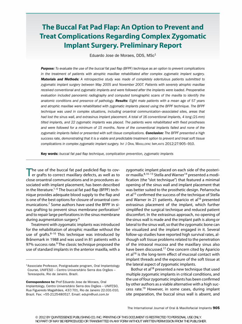
Purpose: To evaluate the use of the buccal fat pad flap (BFPF) technique as an option to prevent complications
in the treatment of patients with atrophic maxillae rehabilitated after complex zygomatic implant surgery.
Materials and Methods: A retrospective study was made of completely edentulous patients submitted to
zygomatic implant surgery between May 2005 and November 2007. Patients with severely atrophic maxillae
received conventional and zygomatic implants and were followed after the implants were loaded. Preoperative
evaluation included panoramic radiography and computed tomographic scans of the maxilla to identify the
anatomic conditions and presence of pathology. Results: Eight male patients with a mean age of 57 years
and atrophic maxillae were rehabilitated with zygomatic implants placed using the BFPF technique. The BFPF
technique was used in complex situations, including oroantral communication–associated sites, areas that
had lost the sinus wall, and extrasinus implant placement. A total of 16 conventional implants, 4 long (21-mm)
tilted implants, and 22 zygomatic implants was placed. The patients were rehabilitated with fixed prostheses
and were followed for a minimum of 15 months. None of the conventional implants failed and none of the
zygomatic implants failed or presented with soft tissue complications. Conclusion: The BFPF presented a high
success rate, demonstrating that it is a viable and predictable treatment option to prevent and treat soft tissue
complications in complex zygomatic implant surgery. Int J Oral MaxIllOfac IMplants 2012;27:905–910.
Key words: buccal fat pad flap technique, complication prevention, zygomatic implants
© 2012 BY QUINTESSENCE PUBLISHING CO, INC. PRINTING OF THIS DOCUMENT IS RESTRICTED TO PERSONAL USE ONLY. NO PART OF MAY BE REPRODUCED OR TRANSMITTED IN ANY FORM WITHOUT WRITTEN PERMISSION FROM THE PUBLISHER.

Moraes
906 Volume 27, Number 4, 2012
the extrasinus placement of implant promotes direct contact with the mucoperiosteal flap, which carries the risk of mucosal fenestration16 (Figs 1 to 3).
Petruson20 examined the maxillary sinuses of 14 patients with zygomatic implants using sinuscopy and found no signs of adverse reactions. However, Becktor et al21 observed sinusitis and oroantral communica-tions more frequently than exposed implant threads.
The aim of this article is to propose the use of the BFPF technique to prevent and treat soft tissue com-plications. This clinical study involved complex cases in zygomatic implant surgery with intraoperative compli-cations that increased the risk of postoperative oroan-tral communications and soft tissue problems.
Materials and Methods
PatientsBetween May 2005 and November 2007, patients of the author’s clinic with severely atrophic maxillae un-derwent therapy with conventional and zygomatic implants. Prior to surgery, an extraoral panoramic radiograph and computed tomographic scan of the maxilla to evaluate the patient’s condition were per-formed. All patients presented with inadequate bone volume for placement of conventional implants in the posterior maxilla.
The following information was recorded for all pa-tients: age, sex, general condition, number of zygo-matic implants placed, length of the implants, number
of standard implants placed, type of prosthesis, antag-onist dentition, duration of follow-up, and postopera-tive complications.
Included patients exhibited the following charac-teristics: (1) presence of a residual alveolar crest with less than 4 mm in width and height immediately distal to the canine pillar, (2) capacity to receive a minimum of two implants per quadrant of the alveolar crest, (3) an oroantral communication that appeared either before or after surgery, (4) substantial loss of the lateral sinus wall after placement of zygomatic implants, and (5) extrasinus placement of zygomatic implants that resulted in contact with mucosa and presented a risk of fenestration and implant exposure. Patients with general and local health conditions that prevented the use of general anesthesia and/or intraoral surgery were excluded from the study.
surgical Protocol and implant PlacementAll surgeries were performed under general anesthe-sia and infiltration with local anesthesia (lidocaine 2% with epinephrine 1:100,000 to reduce bleeding). A supracrestal incision was performed from one side of the maxillary tuberosity to the opposite side, with two bilateral posterior buccal releasing incisions and one vertical mesiobuccal releasing incision.16
Extensive dissection of the maxillary and zygomatic bone was performed with a periosteal elevator. Buccal exposure of the nasal cavity floor, the infraorbital rim, and the zygomatic bone buttress area was performed. The palatal mucosa was reflected only to expose the
Fig 1 Oroantral communication and im-plant with buccal approach.
Fig 2 Quad zygomatic with extrasinus approach.
Fig 3 Mucosal fenestration with implant exposure.
© 2012 BY QUINTESSENCE PUBLISHING CO, INC. PRINTING OF THIS DOCUMENT IS RESTRICTED TO PERSONAL USE ONLY. NO PART OF MAY BE REPRODUCED OR TRANSMITTED IN ANY FORM WITHOUT WRITTEN PERMISSION FROM THE PUBLISHER.

Moraes
The International Journal of Oral & Maxillofacial Implants 907
remaining alveolar crest. Conventional and zygomat-ic implants were placed (Sistema Conexão, Conexão Sistema de Prótese). Standard protocol and manufac-turer recommendations were followed for drilling, and implant placement varied by location and volume of the remaining bone.
The conventional implants were placed in the an-terior region of the maxilla with a palatal approach because of the bone atrophy; in some situations, long tilted implants were placed with nasal anchorage. The classic technique,8–10 the “sinus slot technique,”14 the extrasinus technique,16 and four zygomatic implants18 were used in the patients, depending on individual conditions. In patients who received four zygomatic implants, anterior implants were initially placed, fol-lowed by the posterior implants.
The implants were placed with 45 Ncm of torque and were submitted to immediate loading. For the implants that could not be placed with 45 Ncm, pros-thetic rehabilitation was performed after a period of 6 months.22–24 The flap was sutured with mononylon 5-0 (Johnson & Johnson/Ethicon), and sutures were re-moved after 1 week.
During the postoperative period, antibiotic therapy was prescribed (1 g cephalosporin four times a day for 7 days), along with anti-inflammatory medication (20 mg tenoxican one time per day for 3 days) and chemi-
cal control of bacterial plaque (0.12% chlorhexidine; 1-minute mouth rinses three times a day).10,18,22,23 Penicillin- or cephalosporin-sensitive patients received clindamycin.
Pedicled BFPF techniqueThe pedicled BFPF provides a large supply of tissue that helps to close oronasal communications. When associ-ated with implants, it allows the coverage of implant threads with the purpose of increasing tissue thickness and reducing the possibility of fenestration of the mu-cosa and subsequent exposure of the implant. An inci-sion of approximately 1 cm in length is made through the buccinator muscle behind the zygomatic buttress. A blunt dissection is then performed with Metzen-baum scissors to penetrate the BFP capsule. The fatty tissue must be stretched gently and gradually to avoid rupture and maintain its integrity. Then the pedicle must be positioned over the threads of zygomatic im-plants and sutured with resorbable material (4-0 Vicryl, Johnson & Johnson/Ethicon) over the oroantral com-munication (Figs 4 to 11).
Prosthetic rehabilitationMicrounit abutments (Conexão Sistema de Prótese) were placed on the zygomatic and standard implants. For implants submitted to immediate loading, the
Fig 4 Buccal approach with loss of sinus wall.
Fig 5 BFP flap technique. Fig 6 Clinical appearance at 1 week postoperative.
Fig 7 Panoramic radiograph after 1 week. Fig 8 Prosthetic rehabilitation after 1 week.
© 2012 BY QUINTESSENCE PUBLISHING CO, INC. PRINTING OF THIS DOCUMENT IS RESTRICTED TO PERSONAL USE ONLY. NO PART OF MAY BE REPRODUCED OR TRANSMITTED IN ANY FORM WITHOUT WRITTEN PERMISSION FROM THE PUBLISHER.

Moraes
908 Volume 27, Number 4, 2012
transfers were placed before the flap was sutured. Other- wise, the prosthetic components were inserted at stage-two surgery after 6 months.
Maxillomandibular records were made with an acrylic resin multifunctional guide based on the pa-tient’s conventional complete prosthesis. A cast was fabricated to manufacture the prosthesis and the me-tallic superstructure. Implants submitted to immedi-ate loading were rehabilitated with a fixed prosthesis, which was inserted between 48 hours and 1 week after implant placement. The patients were followed monthly, and the prostheses were removed after 6 months.25 At this point, osseointegration according to the success criteria of Albretksson et al26 and Buser et al27 was determined, associated with the removal torque test (10 Ncm of pressure applied), which is an indicator of clinical stability, and percussion over the abutment.13
results
Eight men with a mean age of 57 years and severely atrophied maxillae were rehabilitated with zygomatic implants placed using the BFPF technique between May 2005 and November 2007. The mean duration of follow-up was 24.6 months (range, 15 to 42 months).
Twenty-two zygomatic implants (Conexão Sistema de Prótese) were placed in the patients, with lengths ranging from 35 to 50 mm (Table 1). In five patients, two zygomatic implants (one on each side) were placed, and three patients each received four zygo-matic implants (two on each side). Sixteen conven-tional implants were placed in the following sizes: two 3.5 × 10 mm, two 3.5 × 11.5 mm, four 3.5 × 13 mm, two 3.75 × 13 mm, one 4.0 × 11.5 mm, and one 4.0 × 15 mm. In two patients, four long implants (3.75 × 21 mm) were placed with a 30-degree inclination and
Fig 9 Follow-up at 15 months. Fig 10 Oroantral communication and implant with buccal approach.
Fig 11 BFP technique with the closure of oroantral communication and covering implant threads.
© 2012 BY QUINTESSENCE PUBLISHING CO, INC. PRINTING OF THIS DOCUMENT IS RESTRICTED TO PERSONAL USE ONLY. NO PART OF MAY BE REPRODUCED OR TRANSMITTED IN ANY FORM WITHOUT WRITTEN PERMISSION FROM THE PUBLISHER.

Moraes
The International Journal of Oral & Maxillofacial Implants 909
anchored onto the cortical of the nasal fossae, one on each side, according to the “All on Four” technique 28 and were splinted to standard and zygomatic implants in the prosthetic rehabilitation. In six patients, the im-plants were anchored with 45 Ncm of torque and were submitted to immediate loading.
All patients were rehabilitated with fixed prostheses and were examined monthly for a minimum follow-up period of 15 months (Fig 9). There were no failures of any implants, resulting in a success rate of 100%. One patient presented soft tissue complications (peri-implantitis) in the region of two zygomatic implants because of their positions in the palatine region. In all cases, the sinus membrane was perforated, but no patients presented any symptoms of postoperative si-nusitis. In two patients, the BFPF technique was used to close an oroantral communication close to the site of a zygomatic implant. In the remaining patients, the technique was used to increase the amount of tissue over the implants to prevent fenestration of the buccal mucosa and implant exposure. All patients presented with loss of at least one sinus wall with buccal contact of zygomatic implants (Table 1).
disCussion
It is important to emphasize that the BFPF technique is frequently used with success in oral and maxillofacial surgery. The literature confirms its use as a pedicled graft for closure of oral defects1,6 and demonstrates that the BFPF offers sufficient blood supply for pro-tection of bone grafts in the maxilla and maxillary si-nuses.3 Some authors have stated that the procedure
can be accomplished with a single incision without generating changes in the patient’s appearance and function.2 Other authors have successfully used the BFPF technique for sinus augmentation3,4 and for the treatment of an oroantral communication associated with zygomatic implant surgery.5
Tissue engineering studies29–31 have concluded that adipose-derived adult stem cells are multipotent, dif-ferentiating along the adipocyte, chondrocyte, myo-cyte, neuronal, and osteoblast lineages, and can serve in other capacities, such as providing hematopoietic support and gene transfer. Adipose-derived adult stem cells have potential applications for the repair and re-generation of acute and chronically damaged tissues. In a recent study, Farré-Guasch et al30 affirmed that the adipose stem cells that are present in adipose tissue can differentiate into several lineages and express mul-tiple growth factors, which makes them suitable for clinical application. Studies have demonstrated that BFP, an adipose-encapsulated mass found in the oral cavity, could represent an easy source of adipose stem cells for dentists and oral surgeons.29,30
Although classical zygomatic implant surgery and the different approaches proposed by Stella and Warner and Aparicio et al are associated with high success rates,14,16,18 some aspects must be discussed. Oroantral communications and soft tissue complica-tions16,17,20 are cited as potential problems in the post-operative period. Becktor et al21 observed oroantral communications more frequently than exposed im-plant threads. Aparicio et al16 expressed concern re-garding the extrasinus technique in that the placement exposes the implant to the overlying buccal mucosa, which may be a potential risk for the development of
table 1 Clinical data of the Patients
Patient no. age (y)
Zygomatic implants
oaCloss of
sinus wall Follow-up
(mo) antagonist Complications no.
placed lengths (mm) il
1 46 2 35/40 0 1 1 42 FP-I
2 69 2 40/42 2 1 2 30 ND/RP
3 54 4 42/45/50/50 4 2 27 FP-I
4 54 4 40/42/45/50 4 2 25 ND
5 54 2 40/42 2 2 25 ND
6 65 2 42/45 2 1 18 FP-I
7 51 4 42/42/50/50 4 2 15 ND
8 65 2 35/40 0 2 15 ND Zygomatic implants: soft tissue (2)
Totals 22 18 2 13
IL = immediate loading; OAC = oroantral communication; FP-I = fixed implant-supported prosthesis, ND = natural dentition; RP = removable prostheses.
© 2012 BY QUINTESSENCE PUBLISHING CO, INC. PRINTING OF THIS DOCUMENT IS RESTRICTED TO PERSONAL USE ONLY. NO PART OF MAY BE REPRODUCED OR TRANSMITTED IN ANY FORM WITHOUT WRITTEN PERMISSION FROM THE PUBLISHER.
Moraes
910 Volume 27, Number 4, 2012
soft tissue problems. Lekholm and coworkers32 did not observe any increase in marginal bone loss or failure rates for machined implants with exposed threads at implant surgery compared with fully submerged im-plants. At present, zygomatic implants with a rough-ened oxidized surface are commercially available and the change from a machined surface to a roughened oxidized surface must be considered as a major modi-fication.16 Therefore, new studies on the long-term per-formance of implants with this surface are necessary to analyze the potential risks of soft tissue complications.16
ConClusion
In the present study, the buccal fat pad flap technique presented a high success rate and served as a valid surgical option to prevent complications following zy-gomatic implant placement. However, this technique is indicated only in complex cases (eg, loss of the lat-eral sinus wall, oroantral communications) and should not be employed as a routine approach to zygomatic implant surgery. Some authors consider that the adi-pose-derived adult stem cells present in the buccal fat pad have potential applications for the repair and re-generation of acute and chronically damaged tissues. However, more studies are necessary to confirm this proposal clinically.
reFerenCes
1. Egyedi P. Utilization of buccal fat pad for closure of oroantral com-munication. J Maxillofac Surg 1977;5:241–243.
2. Fan L, Chen G, Zhao S, Hu J. Experimental study of histologic changes of buccal fat pad used as a pedicled graft. Chin J Dent Res 2001;4:39–41.
3. Liversedge RL, Wong K. Use of the buccal fat pad in maxillary and si-nus grafting of the severely atrophic maxilla preparatory to implant reconstruction of the partially or completely edentulous patient: Technical note. Int J Oral Maxillofac Implants 2002;17:424–428.
4. Hassani A, Khojasteh A, Alikhasi M. Repair of the perforated sinus membrane with buccal fat pad during sinus augmentation. J Oral Implantol 2008;34:330–333.
5. Moraes EJ. Closure of oroantral communication with buccal fat pad flap in zygomatic implant surgery: A case report. Int J Oral Maxil-lofac Implants 2008;23:143–146.
6. Neder A. Use of buccal fat pad for grafts. J Oral Surg 1983;55:349–350. 7. Tideman H. Buccal fat pad as a pedicled graft. J Oral Maxillofac Surg
1986;44:435–438. 8. Ahlgren F, Størksen K, Tornes K. A study of 25 zygomatic dental
implants with 11 to 49 months’ follow-up after loading. Int J Oral Maxillofac Implants 2006;21:421–425.
9. Bedrossian E, Stumpel L, Beckeley M, Inderson T. The zygomatic im-plant: Preliminary data on treatment of severely resorbed maxillae. A clinical report. Int J Oral Maxillofac Implants 2002;17:861–865.
10. Malevez C, Abarca W, Durdu F, Daelemans P. Clinical outcome of 103 consecutive zygomatic implants. Clin Oral Implants Res 2003;15:18–22.
11. Boyes-Varley JG, Howes DG, Lownie JF, Blackbeard GA. Surgical modifications to the Brånemark zygomaticus protocol in the treat-ment of the severely resorbed maxilla: A clinical report. Int J Oral Maxillofac Implants 2003;18:232–237.
12. Farzad P, Andersson L, Gunnarsson S, Johansson B. Rehabilitation of severely resorbed maxillae with zygomatic implants: An evaluation of implant stability, tissue conditions and patients’ opinion before and after treatment. Int J Oral Maxillofac Implants 2006;21:399–404.
13. Zwahlen RA, Graetz KW, Oeschlin CK, Studer SP. Survival rate of zygomatic implants in atrophic or partially resected maxillae prior to functional loading: A retrospective clinical report. Int J Oral Maxillofac Implants 2006;21:413–420.
14. Stella JP, Warner MR. Sinus slot technique for simplification and improved orientation of zygomaticus dental implants: A technical note. Int J Oral Maxillofac Implants 2000;15:889–893.
15. Peñarrocha M, Garcia B, Marti E, Boronat A. Rehabilitation of severely atrophic maxillae with fixed implant-supported prostheses using zygomatic implants placed using sinus slot technique: Clini-cal report on a series of 21 patients. Int J Oral Maxillofac Implants 2007;22:645–650.
16. Aparicio C, Ouazzani W, Aparicio A, et al. Extrasinus zygomatic implants: A new surgical approach for patients with pronounced buccal concavities in the edentulous maxilla. Clin Implant Dent Relat Res 2010;12:55–61.
17. Al-Nawas B, Wegener J, Bender C, Wagner W. Critical soft tissue param-eters of the zygomatic implant. J Clin Periodontol 2004;31:497–500.
18. Bothur S, Jonsson G, Sandahl L. Modified technique using multiple zygomatic implants in reconstruction of the atrophic maxilla: A technical note. Int J Oral Maxillofac Implants 2003;18:902–904.
19. Moraes EJ, Moraes NB. Treatment of atrophic maxillae and complex cases with zygomatic implants. Clin Oral Implants Res 2007;185:62–63.
20. Petruson B. Sinuscopy in patients with titanium implants in the nose and sinuses. Scand J Plast Reconstr Surg Hand Surg 2004;38:86–93.
21. Becktor JP, Isaksson S, Abrahamsson P, Sennerby L. Evaluation of 31 zygomatic implants and 74 regular dental implants used in 16 patients for prosthetic reconstruction of the atrophic maxilla with cross-arch fixed bridges. Clin Implant Dent Relat Res 2005;7:159–165.
22. Bedrossian E, Rangert B, Stumpel L, Indresano T. Immediate func-tion with the zygomatic implant: A graftess solution for the patient with mild to advanced atrophy of the maxilla. Int J Oral Maxillofac Implants 2006;21:937–942.
23. Chow J, Hut E, Lee PKM, Li W. Zygomatic implants: Protocol for im-mediate occlusal loading: A preliminary report. Int J Oral Maxillofac Surg 2006;64:804–811.
24. Davó R, Malevez C, Rojas J, Rodríguez J, Regolf J. Clinical outcome of 42 patients treated with 81 immediately loaded zygomatic im-plants: A 12- to 42-month retrospective study. Eur J Oral Implantol 2008;1:141–150.
25. Mozzati M, Monfrin SB, Pedretti G, Schierano G, Bassi F. Immediate loading of maxillary fixed prostheses retained by zygomatic and conventional implants: 24-month preliminary data for a series of clinical case reports. Int J Oral Maxillofac Implants 2008;23:308–314.
26. Albrektsson T, Zarb GA, Worthington P, Eriksson AR. The long-term efficacy of currently used dental implants: A review and proposed criteria of success. Int J Oral Maxillofac Implants 1986;1:11–25.
27. Buser D, Weber HP, Lang NP. Tissue integration of nonsubmerged im-plants. 1 year results of a prospective study with 100 ITI hollow-cylin-der and hollow-screw implants. Clin Oral Implants Res 1990;1:33–40.
28. Maló P, Rangert B, Nobre M. All-on-4 immediate function concept with Brånemark system implants for completely edentulous maxil-lae: A 1-year retrospective clinical study. Clin Implant Dent Relat Res 2005;7(suppl 1):588–594.
29. Hicok KC, Du Laney TV, Zhou YS, et al. Human adipose-derived adult stem cells produce osteoid in vivo. Tissue Eng 2004;10:371–380.
30. Farré-Guasch E, Martí-Pagès C, Hernández-Alfaro F, Klein-Nulend J, Casals N. Buccal fat pad, an oral access source of human adipose stem cells with potential for osteochondral tissue engineering: An in vitro study. Tissue Eng Part C Methods 2010;16:1083–1094.
31. Gimble JM, Guilak F. Adipose-derived adult stem cells: Isolation, characterization, and differentiation potential. Tissue Engineering 2003;5:362–369.
32. Lekholm U, Sennerby L, Roos J, Becker W. Soft tissue and marginal bone conditions at osseointegrated implants that have exposed threads: A 5-year retrospective study. Int J Oral Maxillofac Implants 1996;11:599–604.
© 2012 BY QUINTESSENCE PUBLISHING CO, INC. PRINTING OF THIS DOCUMENT IS RESTRICTED TO PERSONAL USE ONLY. NO PART OF MAY BE REPRODUCED OR TRANSMITTED IN ANY FORM WITHOUT WRITTEN PERMISSION FROM THE PUBLISHER.