Embed Size (px)
Citation preview
End-of-term project
THEUTILITYOFIMPLANTABLELOOPRECORDERINTHE
CLINICALMANAGEMENTOFPEDIATRICPATIENTSWITHNON-HIGH-RISKBRUGADASYNDROME
Pediatric Arrhythmias Electrophysiology and Sudden Death Unit
Cardiology Department Hospital Sant Joan de Déu, Barcelona
AUTHOR: Judit Guix Camps
CLINICAL TUTOR: Dra. Sarquella Brugada
METHODOLOGICAL TUTOR: Dr. Ramos

A la meva família, per tots aquests anys de recolzament constant i per acompanyar-me pel camí que tot just acaba de començar. Donar gràcies als
meus germans, per ser un pilar fonamental. A tu, Georgia, un clar exemple d’esforç i superació. T’agraeixo infinitament que
m’hagis deixat formar part d’aquest magnífic projecte i poder aprendre de tu. Ets un referent a seguir.
A tota la Unitat d’Arítmies Pediàtriques de l’Hospital Sant Joan de Déu, per l’acollida i la fantàstica labor que realitzeu diàriament, en especial a l’Andrea,
Vic, Lola i en Sergi. Al Dr.Porto, qui sempre ha estat disposat a ajudar-me.
A Rafa Ramos, Marc Saez, per ajudar-me en la elaboració del projecte. A vosaltres, amics, per ser-hi sempre.
“Un viatge de mil milles, comença amb un pas” – Laozi

TABLE OF CONTENTS 1. LIST OF ABBREVIATIONS…………………………….......... 1 2. ABSTRACT……………………………………………………... 2 3. INTRODUCTION……………………………………………......
3.1 Definition of Brugada Syndrome
3.2 Brugada Syndrome history
3.3 Introduction to pediatric BrS
3.4 Epidemiology of BrS
3.5 Etiology and physiopathology of BrS
3.6 Genetic factors and tests in BrS
3.7 Clinical presentation
3.8 Diagnostic management
3.9 Sodium channel blockers challenge
3.10 Differential diagnosis and associated diseases
3.11 Risk assessment and prognosis
3.12 Clinical management
3.13 Implantable loop recorder
3.14 Therapeutic options
3 3
3
4
4
5
6
7
7
10
11
12
12
13
15
4. JUSTIFICATION OF THE PROJECT………………………... 16
5. HYPOTESIS AND OBJECTIVES…………………………….. 5.1 Hypothesis
5.2 Objectives
17 17
17
6. METHODS AND MATERIALS………………………………... 6.1 Study design
6.2 Study population 6.2.1 Inclusion criteria
6.2.2 Exclusion criteria
6.3 Sample 6.4 Study variables 6.5 Follow-up 6.6 Data acquisition 6.7 Statistical analysis
18 18
18
19
19
24
25
25

7. ETHICAL CONSIDERATIONS……………………………….. 26
8. RESULTS……………………………………………………….. 8.1 Population characteristics
8.2 Results regarding the main reason for evaluating BrS 8.3 Diagnostic tests performed and their results 8.4 ILR findings during follow-up 8.5 Estimated risk of arrhythmic events
27 27
27
28
30
32
9. DISCUSSION…………………………………………………… 35
10. IMPLANTABLE LOOP RECORDER LIMITATIONS…… 37 11. STUDY LIMITATIONS……………………………………… 37 12. CONCLUSIONS…………………………………………….. 39 13. REFERENCES………………………………………………. 40 14. ANNEXES…………………………………………………….
14.1 Annex I: Informed consent for invasive procedures (in Catalan
and in Spanish)
14.2 Annex II: CEIC’s authorization
14.3 Annex III: Implantable loop recorder protocol
14.4 Annex IV: Summary of patient characteristics
14.5 Annex V: ILR findings
14.6 Annex VI: ECG tracing of ILR
14.7 Annex VII: Levels of evidence in therapeutic studies
49 49
51
52
54
54
55
56
1
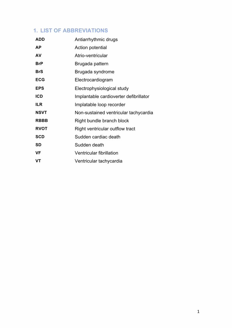
1. LIST OF ABBREVIATIONS ADD Antiarrhythmic drugs AP Action potential AV Atrio-ventricular BrP Brugada pattern BrS Brugada syndrome ECG Electrocardiogram
EPS Electrophysiological study ICD Implantable cardioverter defibrillator ILR Implatable loop recorder NSVT Non-sustained ventricular tachycardia RBBB Right bundle branch block RVOT Right ventricular outflow tract SCD Sudden cardiac death SD Sudden death VF Ventricular fibrillation VT Ventricular tachycardia
2
2. ABSTRACT BACKGROUND: Brugada syndrome (BrS) is an inherited arrhythmogenic
disorder characterized by a typical ECG pattern. The syndrome has incomplete
penetrance and variable expressivity, ranging from asymptomatic to lethal
ventricular arrhythmias and sudden death at a young age in individuals with
structurally normal hearts. So, all this requires an early diagnosis and an accurate
risk stratification of this population.
HYPOTHESIS AND OBJECTIVE: We suggest the use of remote monitoring
system using a subcutaneous loop recorder as a tool to detect arrhythmic events
that can help in the risk stratification of pediatric patients with non-high-risk BrS.
METHODS AND MATERIALS: A retrospective cohort study was performed to
describe data collected by implantable loop recorder (ILR) of 34 anonymized
pediatric patients with non-high-risk BrS. Their demographic and clinical
characteristics and the results obtained in all diagnostic tests performed, were
analyzed in comparison to the detection of arrhythmic events by ILR.
RESULTS: Within 34 patients, 24 were male (70,59%) and 10 were female
(29,41%). Among total arrhythmic events detected, 5 patients (45,45%) were
previously symptomatic and 6 (54,55%) were previously asymptomatic. During a
mean follow-up of 18 months, a total of 7 cases (63,64%), ILR was triggered by
symptoms, which in the majority of them (71,43%) were proved normal sinus
rhythm/sinus tachycardia or minimal rhythm disturbances. In the two remaining
symptomatic cases (28,57%), the ECG tracing identified episodes of NSVT. That
involved a change in those patients’ therapeutic management, requiring an early
implantation of ICD. In four asymptomatic patients (36,36%) the ILR recording
was auto-activated, showing in one of those cases (25%), various episodes of
asymptomatic NSVT. In six cases (17,65%), ILR recorded episodes of abrupt
change repolarization compatible with dynamic BrS.
CONCLUSIONS: The ILR was determinant to exclude ventricular arrhythmias as
a mechanism of symptoms in 71,43% of patients, delaying ICD implantation. In
contrast, it allowed to detect ventricular arrhythmias in two symptomatic patients,
leading to an early implantation of ICD. So, the ILR allows a long-term continue
monitoring of heart rhythm in patients with increased risk of suffering life-
threatening arrhythmias and should be considered as a tool for clinical
management of paediatric patients with non-high-risk BrS KEYWORDS: Brugada syndrome, children, arrhythmia, sudden cardiac death,
implantable loop recorder.

3
3. INTRODUCTION 3.1 Definition of Brugada Syndrome Sudden cardiac death (SCD) is defined as an unexpected cardiac function
cessation in apparently healthy individuals which accounts for nearly 85% of all
sudden death (SD). Most of cases of SCD in patients over 40 years old are the
result of coronary heart disease. In contrast, in the young-adult population (<35
years old) SCD is often caused by arrhythmic syndromes with or without
structural heart alterations called cardiomyopathies and channelopathies
respectively. Channelopathies are a group of familial arrhythmogenic syndromes
caused by pathogenic alteration in genes encoding ion channels or associated
proteins, which participate in cardiac cell action potential (AP), group from which
the Brugada syndrome (BrS) belongs. This syndrome can apparently predispose
healthy individuals to suffer from malignant cardiac arrhythmia or ultimately to
develop SCD.
Despite its life-threatening nature, most patients remain completely
asymptomatic and undiagnosed, but they have potential risk of SCD, leading to
an important medical challenge.
3.2 Brugada syndrome history 1-3,8,33
The syndrome of “Right Bundle Branch Block, Persistent ST Segment Elevation
and Sudden Cardiac Death”, better known nowadays as Brugada Syndrome was
first described in November 1992 by two cardiologists Pedro and Josep Brugada
as a new clinical-electrocardiographic syndrome causing ventricular arrhythmias
and SCD in patients with a structurally normal heart1. A year later, many countries
found out that this could be the same disease as what they called “Sudden
Unexplained Nocturnal Death Syndrome” (SUNDS) or with different colloquial
names such as Bangungut in Philippines, Pokkuri in Japan or Lai tai in Thailand,
places where BrS is more prevalent.
After its initial description, which included eight individuals, the documentation of
new isolated cases continued always within family nuclei so a genetic bases was
suspected. In 1998, Ramon Brugada’s research identified the first genetic
mutation related with BrS, confirming that it was a real new syndrome and it could
be genetically determined. From that moment, numerous studios were published

4
focused on trying to define better from its epidemiology, clinical and diagnostic
characteristics to its therapeutic managements, in all groups of age. After all
these years of scientific progress, there is still a long way to go, looking for new
improvements regarding the many facets of BrS that remain still unknown.
3.3 Introduction to pediatric BrS In the original description, three out of eight patients were children affected by
malignant arrhythmias1. Since then, several isolated cases have been reported,
but data of BrS in pediatric population remains limited.
The prevalence of BrS in pediatric population is lower than in adult population5,16
and lacks of male predominance, which is clearly defined in adult population,
despite equal genetic transmission of the defective gene, possibly because of the
low levels of testosterone found in children of both genders8,9,33 and its role on
phenotype expression. In consequence, the hormonal changes which take place
in puberty could explain why the risk of spontaneous arrhythmias increase after
puberty only in male15,54,55, when testosterone rises up, although females are not
spared from it8. Testosterone acts increasing the potassium currents, a key ion
involved in triggering ventricular arrhythmias. Surprisingly, most males develop
arrhythmias with a mean age of 41±15 years13,17,20 rather than shortly after
puberty, which means that additional proarrhythmic factors must come into play
during adulthood7,15.
3.4 Epidemiology of BrS Brugada syndrome is more common among adult men, where represents the
80% of all patients17,56-58, with an estimated prevalence of 0.14-0.7% in adult
population and much lower in children, around 0.0098%16,24, even though in
countries where this disease is endemic it could be increased24,33,38. BrS is
responsible for 4%-12% of all SD and up to 20% of SD in patients with structurally
normal hearts5,9,13. Geographical variability has been observed, being more
frequent in certain regions of Southeast Asian9.
5
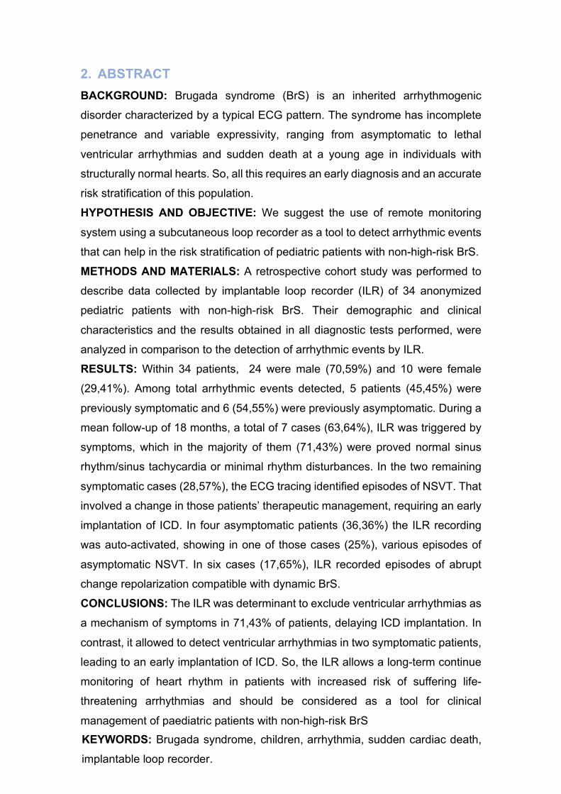
3.5 Etiology and physiopathology of BrS The BrS has a multifactorial etiology including genetics, hormones and
environmental components that modifies its phenotype7. In the healthy heart,
myocardial AP is generated by ionic changes across the membrane: inward
currents of Na+ and Ca2+ causing depolarization and outward currents of K+
enabling repolarization. Modifications in such AP is due to pathogenic variants in
genes encoding ion channels or associated proteins predispose to potentially
malignant arrhythmias. The first genetic alteration associated with BrS was
identified in the SCN5A gene5 which encodes the a-subunit of the cardiac sodium
channel, Nav 1.5 (a transmembrane protein that mediates the fast influx of Na+
ions generating the initial upstroke of the AP and enabling its propagation in the
excitable cardiac tissue)19. In BrS, SCN5A mutation leads to a loss-of-function
phenotype4 that manifests as a cardiac conduction disease. That mutation
reduces the inward sodium channel current disrupting the delicate ion balance of
the cardiac cell. Its impact it is larger in the epicardium compared to the
endocardium and myocardium due to the greater expression of the channel
carrying the transient outward potassium current (Ito), related with repolarization,
in epicardium. In consequence, it creates a transmural (epicardial-to-endocardial)
voltage gradient, more prominent at the base of the right ventricle, leading to
mark heterogeneity in repolarization, responsible for the ST-elevation in the ECG.
Finally, that heterogeneity allows a local re-excitation (referred to as “phase 2
reentry”), resulting in ventricular extrasystoles which may trigger episodes of
VT/VF18,19.
FIGURE 1: Schematic representation of right ventricular epicardial action potential changes
proposed to underline the ECG manifestation of the BrS. Modified from Antzelevitch18 with
permission. Endo, endocardium; M, myocardium; Epi, epicardium; ECG, electrocardiogram.
D

6
3.6 Genetic factors and tests in BrS Brugada syndrome is an inherited condition transmitted in an autosomal-
dominant way with incomplete penetrance17. Nevertheless, due to its variable
expressivity, it can be sporadic in a significant proportion of patients.
Consequently, an individual can be affected of BrS in absence of family history
related with this disease.
Since the identification of the first SCN5A gene related to BrS39, more than 450
pathogenic variants have been identified in 24 genes encoding sodium,
potassium, and calcium channels or associated proteins5,9. Some of them are:
GPDI-L gene (with the mutation A280V, that induces the sodium channel loss of
function), KCNJ8 gene (which encodes for a subunit related with the potassium
channels) and CACNA1c, CACNB2b and CACNA2D1 genes (encoding calcium
channels). However, the role for these genes remains unclear, it seems that all
of them could explain a BrS phenotype due to ion current imbalance. Other
genes, such as SCN10A, MOG1, MYH7 and HCN4 have also been recently
related19.
Despite all these genetic findings, only 20-30% of BrS patients are genetically
diagnosed, of which, approximately 14-25% have positive genetic test for SCN5A
mutation5, which means that the disease is genetically heterogeneous9,18,19.
Current guidelines only recommend performing a genetic test of the SCN5A gene
in family members of a successfully genotyped proband, giving the opportunity to
identify asymptomatic familiars at risk of being affected6,8.
FIGURE 2: Diagram showing genes associated with Brugada syndrome (BrS) and other
overlapping diseases. AF: atrial fibrillation; ARVC: arrhythmogenic right ventricular
cardiomyopathy; PCCD: progressive cardiac conduction disease; SSS: sick sinus syndrome;
LQT: long QT syndrome; SQT: short QT syndrome; ERS: early repolarization syndrome.
Reproduced with permission from Sarquella-Brugada et al.8.
7
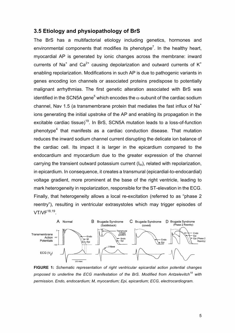
3.7 Clinical presentation The phenotypic expression of BrS varies from completely asymptomatic
individuals, who are the vast majority, to SCD at a young age9,52. The disease
typically manifests in the fourth decade of life, and despite being considered a
rare syndrome in children6,15, the most severe with the highest mortality rate have
been observed during childhood1,3,16,51,37.
Clinical manifestations of symptomatic BrS include unexplained syncope episode
and aborted SCD9 due to ventricular arrhythmias (VT or VF)33. The polymorphic
VT is more frequently related with BrS, although the monomorphic VT is the most
prevalent in children8,18, which often terminates spontaneously in them. That type
of arrhythmias typically occurs at night or at rest, bradycardia-related situations8,9.
Among other rhythm disturbances, a non-negligible proportion of Brugada
patients may suffer from sinus node dysfunction, which ranges from
asymptomatic sinus bradycardia or chronotropic incompetence to atrial standstill
(asystole), or supraventricular arrhythmias, mainly atrial flutter or fibrillation.
Atrioventricular block, complete RBBB or diffusely prolonged QRS complexes
have been also associated with BrS, being a reason for suspecting it.
In contrast, some affected patients can also be completely asymptomatic and
turn out to symptomatic later2, giving more importance to an early approach and
identification of people at risk.
FIGURE 3: Subcutaneous holter register of monomorphic ventricular tachycardia that terminates
spontaneously, in a patient from our series.
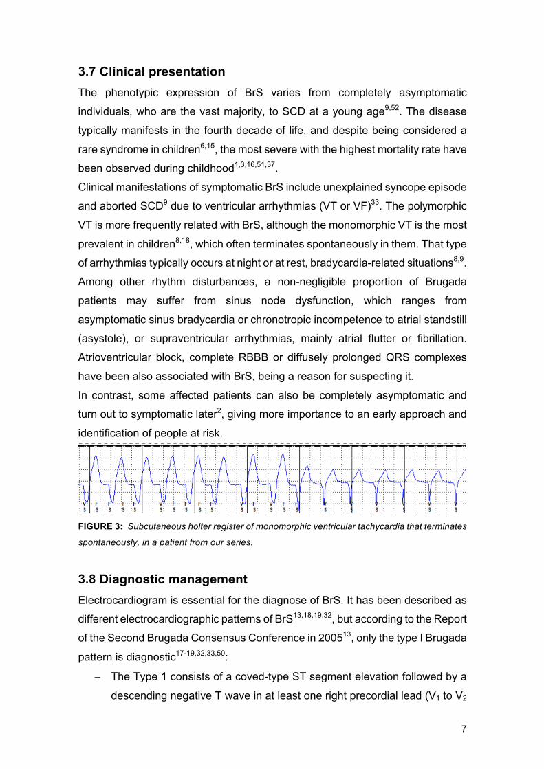
3.8 Diagnostic management Electrocardiogram is essential for the diagnose of BrS. It has been described as
different electrocardiographic patterns of BrS13,18,19,32, but according to the Report
of the Second Brugada Consensus Conference in 200513, only the type I Brugada
pattern is diagnostic17-19,32,33,50:
- The Type 1 consists of a coved-type ST segment elevation followed by a
descending negative T wave in at least one right precordial lead (V1 to V2
8
and, less frequently V3. The specific morphologic characteristics are: at
the end of QRS (which is longer than the QRS of a RBBB), an ascending
and quick slope with a high take-off ³ 2mm followed by a concave or
rectilinear downsloping ST (in comparison to the isoelectric baseline).
There is no clear r’ wave and the high take-off often does not correspond
with the J point. At 40ms of high take-off, the decrease in amplitude of ST
is £4mm (in RBBB and athletes could be much higher), and at the end, the
ST segment is followed by a negative symmetric T wave.
- The type 2 pattern is a saddleback pattern, characterized by a high take-
off of r’³ 2mm (that often does not coincide with J point). The descending
arm of r’ coincides with the beginning of ST, and its ST-segment presents
an ascent ³ 0.5mm. Then, the ST is followed by positive or biphasic T
wave in V2 (T peak > ST minimum > 0) and of variable morphology in V1.
Type II is more common in V1-V2 and less frequent in V3 as well.
- The type III pattern shows a right precordial ST-segment elevation £1mm
either with a coved-type or a saddleback morphology.
FIGURE 4: Features of ECG patterns associated with Brugada syndrome. Modified from
Mashar12 and Berne17 with their permission.
Brugada syndrome is diagnosed in patients with11,13,33:
1. ST-segment elevation with type 1 morphology ³2mm in ³1 lead among the
right precordial leads V1-V2, positioned in the 2nd, 3rd, or 4th intercostal
space occurring either spontaneously or after provocative drug test with
intravenous administration of Class I antiarrhythmic drugs (AAD).
2. Type 2 or type 3 ST-segment elevation in ³1 lead among the right
precordial leads V1-V2, positioned in the 2nd, 3rd, or 4th intercostal space
9
only when a provocative drug test with intravenous administration of Class
I antiarrhythmic drugs induces a type I ECG morphology.
It can present some clinical features that help to increment the consistency of the
diagnose, but they are not specifically necessary for getting it. These are:
- Previously documented ventricular fibrillation (VF) or polymorphic
ventricular tachycardia (VT), inductility of VT with programmed electrical
stimulation, unexplained syncope, febrile seizures, an episode of aborted
SCD and/or nocturnal agonal respiration.
- Family history of SCD younger than 45 years old and/or coved-type ECG
pattern in family members, unexplained syncope or nocturnal agonal
respiration.
Despite the three ECG patterns related with BrS can be observed in the same
patient at different times, type 2 and type 3 pattern are suggestive, but not
diagnostic of it9,17. Finally, drug induced conversion from normal ECG to a type 2
or type 3 pattern is considered inconclusive18.
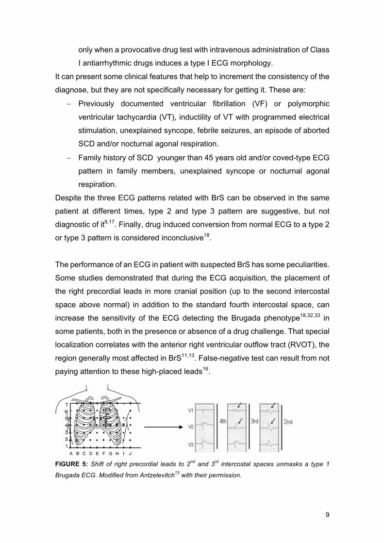
The performance of an ECG in patient with suspected BrS has some peculiarities.
Some studies demonstrated that during the ECG acquisition, the placement of
the right precordial leads in more cranial position (up to the second intercostal
space above normal) in addition to the standard fourth intercostal space, can
increase the sensitivity of the ECG detecting the Brugada phenotype18,32,33 in
some patients, both in the presence or absence of a drug challenge. That special
localization correlates with the anterior right ventricular outflow tract (RVOT), the
region generally most affected in BrS11,13. False-negative test can result from not
paying attention to these high-placed leads16.
FIGURE 5: Shift of right precordial leads to 2nd and 3rd intercostal spaces unmasks a type 1
Brugada ECG. Modified from Antzelevitch13 with their permission.
10
A diagnostic-type I Brugada pattern is not always easy to find. It’s true that, for
instance, Corcia et al.37 found out an abnormal baseline ECG in 75% of young
patients who presented with lethal events during follow-up. In contrast,
spontaneous diagnostic Brugada ECG pattern is only observed in a 25% of
tracings and most ECG will normalize at a follow-up2,6, due to its fluctuating
character1,18,19,23,32, fact that confers difficulties in BrS diagnosis. In consequence,
further tests are mandatory for uncertain diagnostics and risk assessment.
It has been identified modulators that play a major role in the dynamic nature of
the ECG and may also be responsible for ST-segment elevation unmasking the
Brugada ECG diagnostic-pattern32. Bradycardia and vagal tone may contribute
by decreasing calcium currents, fact that explains the greater ST-segment
elevation recorded in vagal settings26 and the higher incidence of ventricular
arrhythmias at night8. One of the most well-known precipitating factor, especially
among pediatric population, is fever14,21. The Nav1.5 sodium channel has shown
to be a temperature-dependent ionic channel21 and, at higher temperatures, the
premature inactivation of it is accentuated. So, it is crucial trying to record an ECG
during a febrile episode21 or few minutes after an atypical febrile convulsions,
relatively common occurrences in childhood. Other important modulators are
sodium channel blockers, anesthetics, some drugs and oral medication
(antidepressants or antiarrhythmics) and ionic imbalance (hyperkalemia and
hypercalcemia)8,10,13,23.
3.9 Sodium channel blockers challenge When there is clinical suspicion of BrS in absence of spontaneous type I ST-
segment elevation may be unmasked by administration of intravenous class IC
antiarrhythmic agents (ajmaline, flecainide, procainamide, pilsicainide) which act
as sodium channel blockers17-19,33,50. The test should be monitored with a
continuous ECG recording and be finished when the coved-type I ECG develops
(giving as a positive), premature ventricular beats or other arrhythmias appear,
or QRS widens ³130% of baseline13. Ajmaline seems to improve the diagnostic
efficiency better than flecainide27. However, it is not a risk-free diagnostic tool25,27,
especially in pediatric population, in which could cause life-threatening
arrhythmias (Conte et al. 25 registered an incidence of 1.8%) . For this reason, the
11
test should be done in a safe environment with a life-support equipment and two
external defibrillators available.
Because nearly 25% of drug-induced tests may result in a false-negative result,
particularly in children where exist an age-dependent response to ajmaline, a
repeat test after puberty (>15 years old) should be considered17,27.
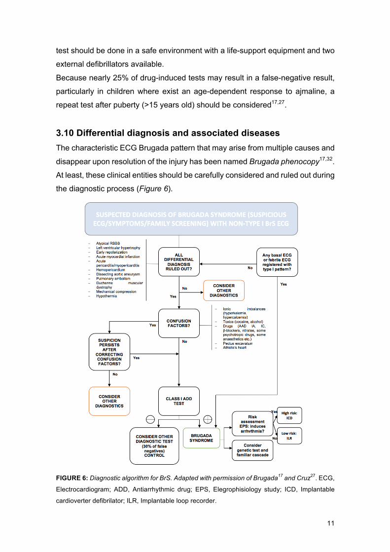
3.10 Differential diagnosis and associated diseases The characteristic ECG Brugada pattern that may arise from multiple causes and
disappear upon resolution of the injury has been named Brugada phenocopy17,32.
At least, these clinical entities should be carefully considered and ruled out during
the diagnostic process (Figure 6).
FIGURE 6: Diagnostic algorithm for BrS. Adapted with permission of Brugada17 and Cruz27. ECG,
Electrocardiogram; ADD, Antiarrhythmic drug; EPS, Elegrophisiology study; ICD, Implantable
cardioverter defibrilator; ILR, Implantable loop recorder.
12
The genetic heterogeneity of inherited conduction disorders often show
overlapping syndromes which include long QT syndrome type III, Brugada
syndrome, atrial fibrillation, progressive cardiac conduction disease or Lev-
Lenègre syndrome, early repolarization syndrome, first degree atrio-ventricular
block and sick sinus syndrome. The array of phenotypes exhibited in these
syndromes, is mainly due to pathogenic variants in the SCN5A gene8.
3.11 Risk assessment and prognosis An accurate arrhythmic risk stratification is mandatory to identify high-risk
patients of suffering cardiac arrhythmic events, who could benefit from an
implantable cardioverter defibrillator (ICD). However, nowadays, it still remains a
clinical challenge35, especially among pediatric population due to their
peculiarities and the lack of published data31.
A previous episode of aborted SCD and malignant syncope are the strongest
predictors of presenting future ventricular arrhythmias14,17,28,35,37,51, especially if
they appear in combination with a spontaneous type I ECG at baseline14,35. Other
markers of higher arrhythmic risk are the presence of fragmented QRS (f-
QRS)30,35, early repolarization abnormalities in lower or lateral leads, an effective
ventricular refractory period (VRP) <200ms, spontaneous atrial flutter or
fibrillation, prolonged QTc interval, PR interval or QRS complex, male gender and
elder population where exists a marked decrease of the conduction velocity that
may contribute to the arrhythmogenic substrate17. The value of inducibility of
sustained ventricular arrhythmias during an EPS as a tool to evaluate arrhythmic
risk in BrS is still controversial35. Brugada et al28 and other studies36,37 found that
inducibility during an EPS is an independent predictor for cardiac events, but
other registers have failed to demonstrate it37,52,56-58. Finally, neither family history
of SCD nor a SCN5A mutation15,28,35,37,51 have proven to be a risk marker in any
of the large studies. However, some specific types of mutations, for example,
those that result in a truncated protein might cause a worse BrS phenotype8.
3.12 Clinical management The management of a child with suspected BrS should be divided according to
his/her symptomatology16:

13
Symptomatic child: a child is not, usually, the first symptomatic member of a
family but, the presence of symptoms before diagnosis in combination with ECG
abnormalities at baseline is known that constitutes an important risk predictor2.
In the case of a rhythm abnormality or clinical event, the first diagnostic step
consist of performing a 12-lead ECG at baseline of the patient and first-degree
relatives. It is important to obtain new tracings in febrile episodes or convulsions
as well as treat it immediately21. Pharmacological test to unmask the Brugada
ECG pattern is the standard diagnostic method. Once diagnosed, genetic test
should be performed if it exists a family history related with BrS. Genotype-
positive individuals should be closely followed-up to identify possible clinical
manifestations8,14,16,17.
Asymptomatic child: asymptomatic pediatric relative with family members at
study or diagnosed of BrS correspond to the majority of cases seen in pediatric
arrhythmias units. The follow-up includes a 12-lead ECG every 6 month until
adolescence and yearly during childhood24. As in the previous case, at least one
ECG should be recorded during a febrile episode or convulsions during
childhood21. In selected cases with family history of SCD, a provocative test could
be considered, starting at the age of five years16,17. Genetic testing is
recommended in the index case and in first-degree family members after having
successfully genotyped the proband. All patients should be followed-up, including
those with negative tests. EPS should be performed in every diagnosed patient
to stratify his/her risk38,49. If ventricular arrhythmias are induced, an ICD is
recommended (Class IIb). On the contrary case, in our center is considered the
implantation of a loop recorder.
Finally, all patients with BrS, regardless of their estimated risk, should be advised
to avoid all drugs that may induce a type I ECG and/or trigger VT/VF. The
complete list can be consulted at: www.brugadadrugs.org60.
3.13 Implantable loop recorder The implantable loop recorder (ILR) is a small subcutaneous device (44.8mm x
7.2mm x 4.0mm) which is implanted in a minimally invasive way, just under the
skin of the chest for cardiac monitoring (Figure 8). The ILR monitors the electrical
activity of the heart, continuously storing information in its circular memory (the
“loop” of the name) as electrocardiograms. Abnormal activity such as arrhythmia
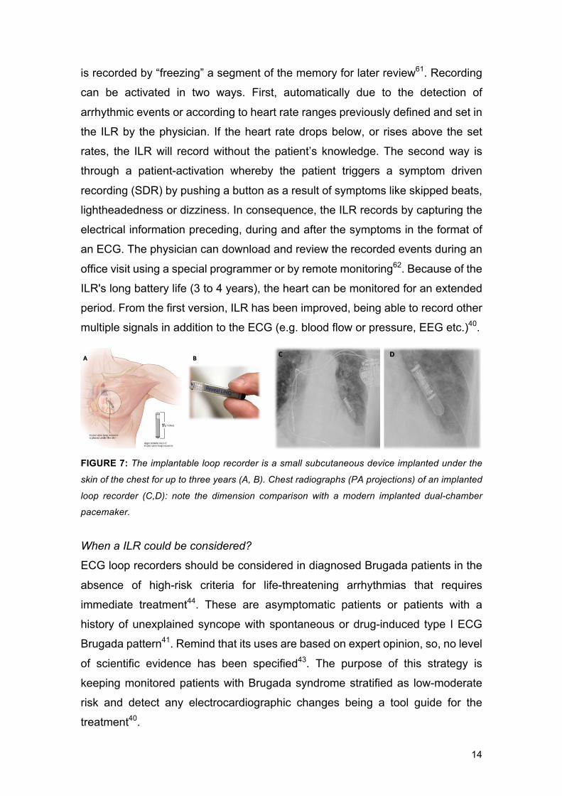
14
is recorded by “freezing” a segment of the memory for later review61. Recording
can be activated in two ways. First, automatically due to the detection of
arrhythmic events or according to heart rate ranges previously defined and set in
the ILR by the physician. If the heart rate drops below, or rises above the set
rates, the ILR will record without the patient’s knowledge. The second way is
through a patient-activation whereby the patient triggers a symptom driven
recording (SDR) by pushing a button as a result of symptoms like skipped beats,
lightheadedness or dizziness. In consequence, the ILR records by capturing the
electrical information preceding, during and after the symptoms in the format of
an ECG. The physician can download and review the recorded events during an
office visit using a special programmer or by remote monitoring62. Because of the
ILR's long battery life (3 to 4 years), the heart can be monitored for an extended
period. From the first version, ILR has been improved, being able to record other
multiple signals in addition to the ECG (e.g. blood flow or pressure, EEG etc.)40.
.
FIGURE 7: The implantable loop recorder is a small subcutaneous device implanted under the
skin of the chest for up to three years (A, B). Chest radiographs (PA projections) of an implanted
loop recorder (C,D): note the dimension comparison with a modern implanted dual-chamber
pacemaker.
When a ILR could be considered?
ECG loop recorders should be considered in diagnosed Brugada patients in the
absence of high-risk criteria for life-threatening arrhythmias that requires
immediate treatment44. These are asymptomatic patients or patients with a
history of unexplained syncope with spontaneous or drug-induced type I ECG
Brugada pattern41. Remind that its uses are based on expert opinion, so, no level
of scientific evidence has been specified43. The purpose of this strategy is
keeping monitored patients with Brugada syndrome stratified as low-moderate
risk and detect any electrocardiographic changes being a tool guide for the
treatment40.
15
Technical aspects40
The auto-activation of the ILR can be compromised by the detection of false
arrhythmias and the missed detection of true arrhythmias. Documented causes
of false arrhythmias storage include: undersensing related to sudden reductions
in R-wave signal amplitude during both normal sinus rhythm and arrhythmias and
undersensing by transient loss of ECG signal related to device amplifier
saturation, T-wave and myopotential. The prevalence of misdiagnosis is unknown
and it is clearly a priority of research to solve.
Like all implanted devices, ILRs also carry the risk of pocket infections, which
account for 1-3% of patients. It could be during either the periprocedural phase
or late during the follow-up, that resolve with device explantation.
MRI scanning of ILR patients can be performed without any harm to patient or
device, but artefacts that could be mistaken for a tachyarrhythmia are seen
frequently45. So, further clinical studies are needed to investigate whether
modified MRI techniques are helpful to eliminate these imaging artifacts46.
3.14 Therapeutic options To date, the implantable cardioverter defibrillator (ICD) is the only proven
effective therapeutic strategy for the prevention of SCD in BrS patients12,47-49. Its
implantation is a Class IA indication in patients with BrS and a history of either
ventricular arrhythmias or aborted SCD (Annex VII). Despite its effectiveness,
ICD placement is frequently associated with device-related complications, of
which, the most prevalent are led failure and inappropriate shocks12,47-49. In order
to avoid these problems, new subcutaneous ICDs has been proposed to use.
An alternative is pharmacological treatment with the objective of rebalancing the
ionic current. Isoproterenol, a L-type calcium channel current increaser, is only
useful when an arrhythmic event appears, during the acute phase. In contrast,
quinidine acts by stabilizing the transient outward ionic currents (Ito) and
converting them from polymorphic to monomorphic, improving clinical tolerance
of the arrhythmia. This drug can be useful as a chronic treatment, as a bridge
therapy to ICD, as an alternative to it or as a combination with the ICD, depending
on the patient’s individual risk17. Recently, epicardial radiofrequency catheter
ablation over RVOT has emerged as a potential treatment in patients with
recurring episodes, but this therapeutic option is not well-defined yet8,50.
16
4. JUSTIFICATION OF THE PROJECT To date, the implantable cardioverter defibrillator (ICD) remains the only tool that
has proven to be effective in reducing the mortality of patients with Brugada
syndrome, treating potentially lethal arrhythmias in more than 25% of cases, as
demonstrated by the recent publication of Gonzalez et al.16. Showing this high
effectiveness, it would be easy to think that it would be a very good strategy for
pediatric patients with BrS. However, these same studies stand out the high
incidence of complications and inappropriate shocks that children with ICD
receive52. Moreover, ICD are difficult to be adapted in children, as the body
grows, there is the need of electives revisions/re-implantations, and the long
patients’ life expectancy request a greater number of the generator substitutions.
In the pediatric population, when and to whom to implant an ICD not only
generates controversy, but it is an absolute unknown, since in order to stratify
their risk, it tends to extrapolate the protocol of adults to children assuming that
the pathology behaves the same in both populations. For this reason, it is
essential to improve stratification in children, helping to classify when and which
need ICDs and which do not.
The implantable loop recorder (ILR) is a small device that combines the
prolonged electrocardiographic record and a remote monitoring system with a
low degree of invasiveness. It allows a comprehensive follow-up of the ECG,
without the need for multiple hospital visits. There are several studies that
evaluate ILRs in patients with arrhythmic events in general population42,43 and
one in BrS patients41. Unfortunately, there is few data in pediatric patients with
suspected arrhythmia44 and no studies, to date, that have been dealt with
pediatric patients with BrS. This gap leaves them without an early detection of
arrhythmias (important for risk stratification) or a diagnosis facing a syncopal
event (to really know the syncope’s etiology: vasovagal, cardiac, neurological
etc.). In our study, we are going to intend to respond to this gap in the pediatric
population since we believe ILRs can provide useful supplementary information
as well as specify etiology of an unexplained syncope, helping in improving the
stratification of pediatric patients with BrS who, at first, do not have strict
indications of ICD and getting a more adjusted treatment that, in short and long
term, will significantly improve the quality of life of the patients with BrS.

17
5. HYPOTHESIS AND OBJECTIVES 5.1 Hypothesis The remote monitoring by implantable subcutaneous holter will allow the
detection of arrhythmic events in pediatric patients with non-high-risk Brugada
syndrome, helping in their risk stratification and guiding to therapy.
5.2 Objectives Primary objectives
- To quantify the incidence of arrhythmic events recorded by ILR in pediatric
patients with non-high-risk Brugada syndrome.
- To analyze the frequency of correlation between clinical symptoms and
rhythm abnormalities detected by ILR.
Secondary objectives
- To describe demographical data and clinical characteristics of anonymized
pediatric patients with BrS of Hospital Sant Joan de Déu.
- To early detect significant conduction abnormalities in pediatric patients
with BrS requiring further treatment.
- To determine the factors that influence the appearance of arrhythmias in
pediatric patients with BrS in order to improve and refine the clinical
management.

18
6. METHODS AND MATERIALS 6.1 Study design It has been designed a retrospective cohort study in order to analyse the data
recorded by subcutaneous holter implanted in pediatric patients with non-high-
risk Brugada syndrome, followed at San Joan de Déu Hospital, in Barcelona.
6.2 Study population The study population was based on pediatric patients with non-high-risk Brugada
syndrome (BrS) followed at the Pediatric Arrhythmia Unit of Sant Joan de Déu
Hospital, in Barcelona.
6.2.1 Inclusion criteria
- Pediatric patients under the age of 21 years old at the time of inclusion.
- Individuals with diagnostic ECG pattern of Brugada syndrome occurring
spontaneously, after provocative drug test or appeared during a febrile
episode (section 3.8).
o Symptomatic or asymptomatic.
o Positive genetic study (with or without identified casual mutation) or
not done.
- Patients who perform an electrophysiology study (EPS) and no sustained
ventricular arrhythmias or ventricular fibrillation were induced.
6.2.2 Exclusion criteria
- Patients with BrS and personal history of either ventricular arrhythmias (VT
or VF) or aborted SCD who have strong clinical indication for ICD.
- Type 2 or 3 Brugada pattern in basal ECG that does not change to type 1
pattern with pharmacological test or fever.
- Individuals who performed EPS and sustained ventricular tachycardia or
ventricular fibrillation were induced.
- Patients carriers of ICDs or pacemarkers previously to the study.
19
6.3. Sample A non-probabilistic consecutive sampling method was performed from our study
population. Finally, a total of 34 anonymous patients followed at the Pediatric
Arrhythmia Unit of Sant Joan de Déu Hospital from 2015 who accomplished all
the inclusion and none of the exclusion criteria, were included in the study in a
completely anonymous way.
In order to calculate our sample’s statistic power, it was used the Prof. Marc Saez’
software based on the library ‘pwr’ of the free statistical environment R (version
3.5.1). In a bilateral contrast, with an alfa level of 5% and a sample of 34 patients,
to detect an average difference (up to 30%), we had a statistical power of 54%.
6.4 Study variables Different variables were registered initially and during follow-up patients in their
medical records as part of the normal healthcare process. Before performing any
tests, nurses explained to each patient the medical procedure that was going to
be done, after the informed consent was obtained.
An exhaustive physical examination, a careful personal and family medical
history, a baseline 12-lead ECG or a provocative test with ajmaline in cases of
normal baseline ECG and an electrophysiological study was initially performed to
each patient. Once the non-induction of sustained ventricular arrhythmias during
EPS was assured, the option of placing an ILR was recommended. The possible
presence of underlying structural cardiac abnormalities was evaluated in all
patients by transthoracic echocardiography, despite not being an exclusion
criteria.
Dependent variable
Arrhythmic event An arrhythmic event is defined as an irregularity or loss of rhythm manifested in
the ECG. Some rhythm disturbances have more incidence in patients with BrS
(section 3.7). Those that more frequently lead to death and, therefore, the most
important ones to detect are ventricular arrhythmias. These are:

20
- Monomorphic ventricular tachycardia defined as an arrhythmia with
constant electrocardiographic configuration and a stable rate within a few
beats.
- Polymorphic ventricular tachycardia, arrhythmia that has a changing rate
and demonstrated varying configurations.
- Ventricular fibrillation defined as rapid and continuously varying
electrocardiograms with irregular cycle lengths.
When these last more than 30s, are called as sustained arrhythmia. Apart from
ventricular arrhythmias, other rhythm disturbances to consider were:
- Sinus node dysfunction manifested as inappropriate sinus bradycardia
according to age and activity level, sinus pause/arrest >2.5s, or
chronotropic incompetence (failure to achieve 85% of the age-predicted
maximum heart rate during the exercise test).
- AV blocks manifested as an extension of PR interval over 200ms.
- Supraventricular (SVEs) and/or ventricular extrasystoles (VEs) defined as
an extra beat originated in the atriums or ventricles respectively.
- Atrial arrhythmias defined as sustained atrial tachycardia, atrial fibrillation,
and atrial flutter.
- Spontaneous changes in the repolarization pattern compatible with
dynamic BrS.
The presence of arrhythmic events was recorded by implantable subcutaneous
holter (section 3.13). The procedure of its implantation was carried out in the
electrophysiology room in all pediatric patients with non-high-risk BrS. It
consisted of:
- The administration of subcutaneous anesthesia: 5% Lidocaine 4.5ml +
0.5% Bupivacaine 4.5ml + 1M/ml bicarbonate in the left submammary
midthoracic region. Massage the area.
- Insertion, for all patients, the same subcutaneous holter (RevelÒ LinQ,
Medtronic), using the kit insertion of the package and fitted with continuous
monitoring and symptom-driven activation functions.
21
- Performance of patient-specific programming of tachycardia and
bradycardia zones, taking into account patient age, medications, and
previous documented arrhythmia data (when available).
- Close by approximation with surgical glue (Dermabond) and placement of
porous dressing.
Once the device placement finished, it had to set up by application or interrogator
and activation of the remote monitoring system. Finally, a registration in the
remote monitoring website and the formation of the family for the use of the
domiciliary system was required (Annex III).
Co-variables
Demographic data
Age (years), sex (male or female), age at LINQ implant (years), months of follow-
up (months) and time to first ILR symptom (months).
Family history
Family tree up to three generations with a history of aborted or not sudden death
at younger than 45 years old, febrile seizures, epilepsy, rhythm disturbances,
carriers of pacemakers, repeat unexplained abortions, diagnosis of BrS
(circumstances and therapeutic management) and genetic studies performed.
Clinical data
- Personal medical history: it was asked specifically for cardiac and/or
neurological antecedents. The reason for considering these antecedent
remains in section 3.10, where possibly associated diseases have been
mentioned.
- Reason for suspecting BrS: it was classified in three groups:
o Familiar screening (family history related with BrS, with or without
genetic tests performed) or a personal genetic mutation related with
BrS founded in other circumstances (section 3.6).
o ECG rhythm disturbances at baseline or during a febrile episode.
o Clinical suspicion: when symptoms related with BrS are present
(unexplained syncope, pre-syncope, febrile seizures, dizziness,
chest pain during fever, nocturnal agonal awakenings etc.).

22
Unexplained syncope is defined as a non-traumatic and reversible
loss of consciousness caused by either ventricular arrhythmias or
vasovagal events (those occurring during abrupt postural changes,
exposure to heat and dehydration, emotional reactions to events
such as blood drawing etc.). Pre-syncope is defined as feeling of
imminent faintness, although without reaching a complete loss of it,
happening only a postural hypotonia.
- Asymptomatic or symptomatic: patients were considered symptomatic if
they presented any clinical manifestations related to BrS included
unexplained syncope and/or aborted SCD. Arrhythmic syncope was
suspected in the absence of prodrome or specific triggering
circumstances. Patients with clinical diagnosis of vasovagal syncope were
not considered as symptomatic.
- Usual medication: the active ingredient of the drug, dose and posology.
12-leads baseline ECG 12-leads baseline ECG was recorded to all patients using a CardioTechTM GT-
400 ECG Machine. It was considered diagnostic of BrS if a coved-type ST-
segment elevation of ³2mm was documented in ³1 lead from V1 to V3
spontaneously or after a sodium channel blocker (section 3.8).
Provocative test Provocative test with ajmaline was performed in cases of normal baseline
electrocardiogram. The test was carried out in the electrophysiology room with a
life-support equipment and two external defibrillators available in the room (Zoll
M Series Defibrillators). A peripheral intravenous access was placed and an
ajmaline intravenous infusion (maximum dose of 1mg/kg) was administered over
a 5-min period. In patients younger than 5 years old, the test was performed
under drug sedation by a single intravenous bolus of propofol. Ajmaline is
deactivated quickly and its effects wear off after a few minutes. Therefore, it was
monitored until the ECG normalized. During the test, it was continuously
monitored the patient’s cardiac rhythm, hemoglobin saturation and arterial
pressure. Ajmaline infusion was terminated before reaching the target dose if
type 1 ECG pattern in ³ 1 right precordial lead (V1-V3), QRS prolongations >30%
23
of baseline, frequent premature ventricular beats, high-degree atrioventricular
blocks or sustained ventricular arrhythmias (VT or VF) was documented. Once
the test was finished, a cardiac monitoring during 30 minutes was left.
All baseline and drug-induced 12-lead ECGs were recorded at a paper speed of
25mm/s and amplitude of 10mm/mV, with the right precordial leads positioned at
the sternal margin of the third and fourth intercostal space according to the
guidelines33 (Figure 5). Two independent experienced pediatric cardiologists of
the Unit of Pediatric Arrhythmias analyzed all the ECG. In case of disagreement,
a third physician was consulted. PR interval, QRS duration, and QTc interval
(determined using Bazett’s formula) were measured in milliseconds (ms) by
averaging hand measurement on three consecutive beats. Maximal ST-segment
elevations were measured at the J point in the right precordial leads (V1-V3), and
analyses of ST-segment elevation were performed in leads V1 and V2. The
presence of R waves in aVR and QRS fragmentations were identified and
noted30. Moreover, ECGs were reviewed to identify sinus node dysfunction (SND)
and atrial arrhythmias.
Genetic test Genetic testing with sequence analysis of SCN5A gene was recommended for
pediatric patients with first degree relatives diagnosed of BrS with a positive
genetic test and pediatric patient diagnosed with BrS. In this last case, if a
mutation-positive was obtained, a family cascade screening in first degree
relatives was proposed.
Sequence analyses of SCN5A were performed by extracting genomic DNA from
peripheral blood leucocytes or saliva samples: if the patient had a peripheral
intravenous access (for other procedure), a blood sample in an EDTA tube was
taken. If not, in order to avoid an invasive procedure of blood extraction, a saliva
sample was taken using a DNA Genotek Oragene 06-500 kit. Genomic DNA was
extracted from these samples, using Chemagic® (Chemagen Systems,
Germany). All genes studied are related with BrS (section 3.6): it was collected
the gene or genes analyzed, in addition to data on the variants identified in each
gene analyzed (location, population frequency, in silico prediction, type of variant
and information from international databases on the variant found).
24
Electrophysiological study
Electrophysiological study was performed in all pediatric patients with the
purpose of risk stratification. In individuals younger than 12 years old, study was
performed under propofol sedation. Intravenous accesses were gained using
right femoral venipuncture, and single catheters were used to determine baseline
intervals and to stimulate the heart.
Baseline intervals were measured including the AH (shows the conduction time
through the AV Node) and HV interval (displays the conduction time from the His
bundle to the first identifiable onset of ventricular activation). Evaluation of sinus
node disfunction (SND) was performed which included measurement of sinus
node recovery time (SNRT), corrected SNRT, and sinoatrial conduction time
estimated using the method described by Narula et al. The atrioventricular
conduction system was evaluated by measuring the Wenkeback cycle length and
the atrioventricular nodal effective refractory period in each case. Patients with
either history of palpitations or evidence by ECG monitor of supraventricular
tachycardia, an atrial stimulation protocol was also performed. The ventricular
stimulation protocol consisted of a maximum of three ventricular extrastimuli with
a minimal coupling interval of 200ms delivered from ³1 ventricular site. Results
were considered positive when sustained VT or VF were induced. In that case,
the patient was excluded from our study.
This test was performed with Philips Allura Xper FD20/10 biplane mixed with
cardiovascular X-ray guidance system, and X-ray protection was mandatory in all
personal present in the EPS room during the procedure.
6.5 Follow-up Clinical follow-up of patients consisted of physical examinations and ECG after
ILR implantation. Follow-up of the device was performed at 1 and 3 months after
implantation and thereafter every 6 months. The ILRs memory looking for
arrhythmic events was reviewed at least once a week by two expert pediatric
cardiologist independently, and each time that the activation of ILR was provoked
by symptoms or by the patient. Device memory was also interrogated in each
regular visit.
25
6.6 Data acquisition All the data used in the study, included the ECGs recorded by ILR, were extracted
from the medical history of pediatric Brugada patients followed at Arrhythmia Unit
of Sant Joan de Déu Hospital, in a completely anonymous way and numerically
codified. The process was made by the Department of Medical Informatics of the
centre, registering all data needed in an anonymous database platform hosted
on the servers of the hospital. Our Arrhythmia Unit has an internal regulation that
allow the double-anonymous review of the database collected of our patients. It
was used a general and previously approved CEIC’s authorization in order to be
able to carry it out. Finally, the database was analysed and exposed to this study.
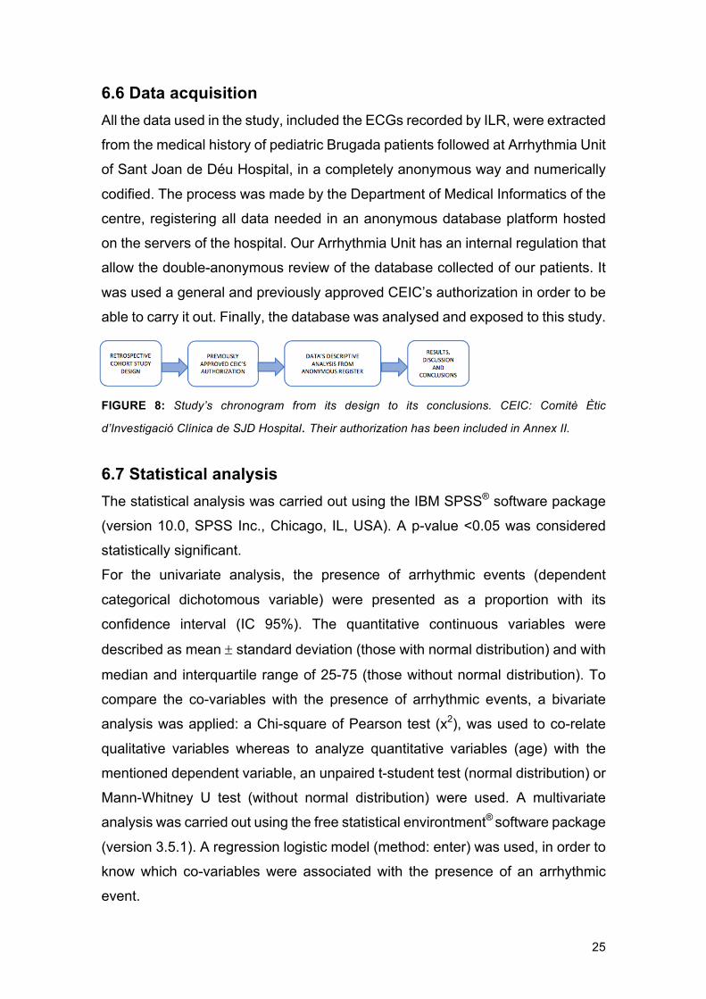
FIGURE 8: Study’s chronogram from its design to its conclusions. CEIC: Comitè Ètic
d’Investigació Clínica de SJD Hospital. Their authorization has been included in Annex II.
6.7 Statistical analysis The statistical analysis was carried out using the IBM SPSS® software package
(version 10.0, SPSS Inc., Chicago, IL, USA). A p-value <0.05 was considered
statistically significant.
For the univariate analysis, the presence of arrhythmic events (dependent
categorical dichotomous variable) were presented as a proportion with its
confidence interval (IC 95%). The quantitative continuous variables were
described as mean ± standard deviation (those with normal distribution) and with
median and interquartile range of 25-75 (those without normal distribution). To
compare the co-variables with the presence of arrhythmic events, a bivariate
analysis was applied: a Chi-square of Pearson test (x2), was used to co-relate
qualitative variables whereas to analyze quantitative variables (age) with the
mentioned dependent variable, an unpaired t-student test (normal distribution) or
Mann-Whitney U test (without normal distribution) were used. A multivariate
analysis was carried out using the free statistical environtment® software package
(version 3.5.1). A regression logistic model (method: enter) was used, in order to
know which co-variables were associated with the presence of an arrhythmic
event.

26
7. ETHICAL CONSIDERATIONS This project complies with the ethical principles of the Declaration of Helsinki
about researching involving human subjects established by the World Medical
Association. Before beginning our study, the correspond protocol was evaluated
by the Clinical Research Ethical Committee of Fundació Sant Joan de Déu
Hospital, in Barcelona.
According to the legal framework of human rights and data confidentiality
specified in Organic Law 15/1999 on the Protection of Personal Data (LOPD),
data was registered and analyzed anonymously and under non-identifying
numeric codes. The author did not have access to any confidential information of
the patients which was only used for the purpose of the research.
It was followed the Law 41/2002 of 14 November, that regulates the autonomy of
the patient and their right to information and clinical documentation and Royal
Decree 1720/2007 of December 21 that regulates the security of files containing
patient data. After receiving the appropriate information, the patients voluntary
signed an informed consent for implantable subcutaneous holter for being an
invasive procedure (Annex I), information contained in the Law 14/2007 for
invasive procedures. Legally, in the case of minors (in medical terms, below 16
years old), their parents or legal tutors were responsible for signing the informed
consent. However, the under-age patient was also properly informed and his/her
agreement was considered. From age 7 to 16, patient’s agreement was
fundamental, since it was considered that they could have reasoned decisions.
If a genetic test had to be performed, during all process the Law for investigation
on biologic samples was applied (Law 14/2007 and the Royal Decree
1716/2011). In case that a pediatric patient obtained a positive genetic test, a
screening process on first-degree relatives should be advised. It that situation,
when the doctors need to communicate the finding of a genetic rare disease in
the family, the language used must had to be extremely careful and always
following the legal and ethical premises.
This study has not any commercial bias nor interests.
27
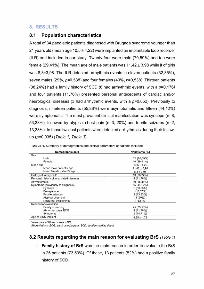
8. RESULTS 8.1 Population characteristics A total of 34 paediatric patients diagnosed with Brugada syndrome younger than
21 years old (mean age 10,5 ± 4,22) were implanted an implantable loop recorder
(ILR) and included in our study. Twenty-four were male (70,59%) and ten were
female (29,41%). The mean age of male patients was 11,42 ± 3.98 while it of girls
was 8,3±3,98. The ILR detected arrhythmic events in eleven patients (32,35%),
seven males (29%, p=0,538) and four females (40%, p=0,538). Thirteen patients
(38,24%) had a family history of SCD (6 had arrhythmic events, with a p=0,176)
and four patients (11,76%) presented personal antecedents of cardiac and/or
neurological diseases (3 had arrhythmic events, with a p=0,052). Previously to
diagnosis, nineteen patients (55,88%) were asymptomatic and fifteen (44,12%)
were symptomatic. The most prevalent clinical manifestation was syncope (n=8,
53,33%), followed by atypical chest pain (n=3, 20%) and febrile seizures (n=2,
13,33%). In those two last patients were detected arrhythmias during their follow-
up (p=0,035) (Table 1, Table 3).
TABLE 1. Summary of demographics and clinical parameters of patients included
Demographic data N=patients (%) Sex
Male Female
24 (70,59%) 10 (29,41%)
Mean age Mean male patient’s age Mean female patient’s age
10,5 ± 4,22 11,42 ± 3,98
8,3 ± 3,98 History of family SCD 13 (38,24%) Personal history of associated diseases 4 (11,76%) Asymptomatic Symptoms (previously to diagnosis)
Syncope Pre-syncope Febrile seizures Atypical chest pain Nocturnal awakenings
19 (55,88%) 15 (44,12%) 8 (53,33%) 1 (6,67%)
2 (13,33%) 3 (20%)
1 (6,67%) Reason for evaluation
Family screening Abnormal basal ECG Symptoms
25 (73,53%) 4 (11,76%) 5 (14,71%)
Age at LINQ implant 8,35 ± 4,73
Values are n(%) and mean ± DS. Abbreviations: ECG: electrocardiogram; SCD: sudden cardiac death
8.2 Results regarding the main reason for evaluating BrS (Table 1)
- Family history of BrS was the main reason in order to evaluate the BrS
in 25 patients (73,53%). Of these, 13 patients (52%) had a positive family
history of SCD.
28
- Abnormal basal ECG: four patients (11,76%) were evaluated due to the
discovery of electrocardiographic alterations related to BrS. - Symptoms: five patients (14,71%) were studied due to symptoms.
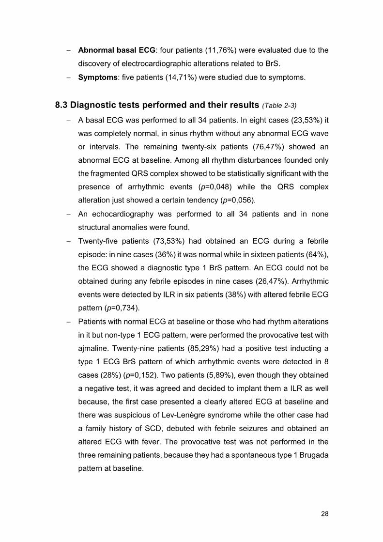
8.3 Diagnostic tests performed and their results (Table 2-3) - A basal ECG was performed to all 34 patients. In eight cases (23,53%) it
was completely normal, in sinus rhythm without any abnormal ECG wave
or intervals. The remaining twenty-six patients (76,47%) showed an
abnormal ECG at baseline. Among all rhythm disturbances founded only
the fragmented QRS complex showed to be statistically significant with the
presence of arrhythmic events (p=0,048) while the QRS complex
alteration just showed a certain tendency (p=0,056).
- An echocardiography was performed to all 34 patients and in none
structural anomalies were found.
- Twenty-five patients (73,53%) had obtained an ECG during a febrile
episode: in nine cases (36%) it was normal while in sixteen patients (64%),
the ECG showed a diagnostic type 1 BrS pattern. An ECG could not be
obtained during any febrile episodes in nine cases (26,47%). Arrhythmic
events were detected by ILR in six patients (38%) with altered febrile ECG
pattern (p=0,734).
- Patients with normal ECG at baseline or those who had rhythm alterations
in it but non-type 1 ECG pattern, were performed the provocative test with
ajmaline. Twenty-nine patients (85,29%) had a positive test inducting a
type 1 ECG BrS pattern of which arrhythmic events were detected in 8
cases (28%) (p=0,152). Two patients (5,89%), even though they obtained
a negative test, it was agreed and decided to implant them a ILR as well
because, the first case presented a clearly altered ECG at baseline and
there was suspicious of Lev-Lenègre syndrome while the other case had
a family history of SCD, debuted with febrile seizures and obtained an
altered ECG with fever. The provocative test was not performed in the
three remaining patients, because they had a spontaneous type 1 Brugada
pattern at baseline.
29
- A genetic test was performed to twenty-eight patients. Eight of them
(28,57%) were negative from mutations related to BrS. The remaining
twenty patients (61,76%) obtained a positive result: thirteen (65%) for
SCN5A mutation and seven (35%) expressed other mutations including
HCN4, ABCC9, PKP2, SCN1A, CACNA2D, MYH6/MYH7 and CSRP3. Six
patients (17,65%) had pending the results of genetic analysis. In none of
the genotype-positive cases was obtained statistically significant results
(p>0,05).
- All patients underwent an electrophysiological study (EPS): thirteen
(38,24%) obtained a completely normal test while the remaining twenty-
one patients (61,76%) showed altered parameters, none of them
statistically significant (p>0,05). No sustained ventricular arrhythmias were
induced during programmed ventricular stimulation in any case. However,
in three patients (14,29%) short runs of non-sustained ventricular
arrhythmias were induced and rhythm disturbances was detected by ILR
during their follow-up (p=0,009).
TABLE 2. Description of test performed and their results
Test performed Results (n=patients, %) Normal/negative/not done Abnormal/positive
Basal ECG 8 (23,53%)
Positive result Spontaneous type 1 pattern Non-type 1 pattern Complete/incomplete RBBB QRS complex alteration f-QRS PR interval alteration QT interval alteration
26 (76,47%) 5 (19,23%) 6 (23,08%) 15 (57,69%) 11 (42,31%) 6 (23,08%) 4 (15,38%) 1 (3,85%)
Ecocardiography 34 (100%) 0 (0%)
Febrile ECG abnormal 9 (26,47%)
ECG tracing obtained Normal (negative) pattern Abnormal (positive) pattern
25 (73,53%) 9 (36%) 16 (64%)
Ajmaline test 5 (14,71%) 29 (85,29%)
Genetic test
6 (17,65%)
Negative result Positive result
SCN5A positive Other mutations
8 (28,57%) 20 (71,43%)
13 (65%) 7(35%)
EPS 13 (38,24%)
Positive result VRP < 200ms AH > 120ms HV > 55ms NSTV
21 (61,76%) 11 (52,38%) 4 (19,05%) 5 (23,81%) 3 (14,29%)
Abbreviations: ECG: electrocardiogram; EPS: electrophysiology study; RBBB: right bundle branch block; f-QRS: fragmented QRS complex; VRP: ventricular refractory period; AH: atrio-His interval time; HV: His-ventricular interval time; NSVT: non-sustained ventricular tachycardia. In genetic test, other positive mutations include HCN4, ABCC9, PKP2, SCN1A, CACNA2D, MYH6, MYH7 and CSRP3 genes.
30
TABLE 3. Comparison of the presence of arrhythmic events between demographic, clinical and diagnostic parameters
PARAMETERS ARRYTHMIC
EVENTS Å
N=11 (%)
ARRHYTHMIC EVENTS
Ä N=23 (%)
p-value (<0,05)
chi-square(x2)
DEM
OG
RA
-PH
ICS
Agea 11 (9,82±5,4) 23 (10,83±3,7) 0,53
Sex
Male sex 7 (29) 17 (70)
0,538 Female sex 4 (40) 6 (60)
CLI
NIC
AL
PAR
AM
ETER
S
Family screening 8 (32) 17 (68) 0,942 Abnormal basal ECG 2 (50) 2 (50) 0,422 Symptoms 1 (20) 4 (80) 0,523 Family SCD 6 (46) 7 (54) 0,176 Associated diseases* 3 (75) 1 (25) 0,052* Asymptomatic 6 (32) 13 (68) 0,914
Symptomatic
Pre-syncope 1 (100) 0 (0) 0,142 Syncope 1 (13) 7 (88) 0,170 Febrile seizures** 2 (100) 0 (0) 0,035** Atypical chest pain 1 (33) 2 (67) 0,970 Nocturnal awakenings 0 (0) 1 (100) 0,483 Total 5 (33) 10 (67) 0.914
DIA
GN
OST
IC T
ESTS
Basal ECG abnormal
Spontaneous type 1 ECG pattern 2 (40) 3 (60) 0,692 Non-type 1 ECG pattern 2 (33) 4 (67) 0,955 Complete/incomplete RBBB 6 (40) 9 (60) 0,458 PR interval prolongation 2 (50) 2 (50) 0,422 QRS complex alteration* 6 (55) 5 (46) 0,056* QT interval alteration 0 (0) 1 (100) 0,483 f-QRS complex** 4 (67) 2 (33) 0,048**
Febrile ECG abnormal 6 (38) 10 (63) 0,734 Provocative test 8 (28) 21 (72) 0,152
EPS abnormal
VRP<200ms 3 (27) 8 (73) 0,661 AH>120ms 1 (25) 3 (75) 0,738 HV>55ms 2 (40) 3 (60) 0,692 NSVT** 3 (100) 0 (0) 0,009**
Genetic test positive
SCN5A 4 (31) 9 (69) 0,988 Other mutations 2 (29) 5 (71) 0,810
Abbreviations: P-value: probability value (according to chi-square analysis); ECG: electrocardiogram; SCD: sudden cardiac death; RBBB: right bundle branch block; f-QRS complex: fragmented QRS complex; EPS: electrophysiology study; VRP: ventricular refractory period; AH: atrio-His interval time; HV: His-ventricular interval time; NSVT: non-sustained ventricular tachycardia. In genetic test, other positive mutations include HCN4, ABCC9, PKP2, SCN1A, CACNA2D, MYH6, MYH7 and CSRP3 genes. a Mean (standard deviation). *p<<0,1; ** p<0,05
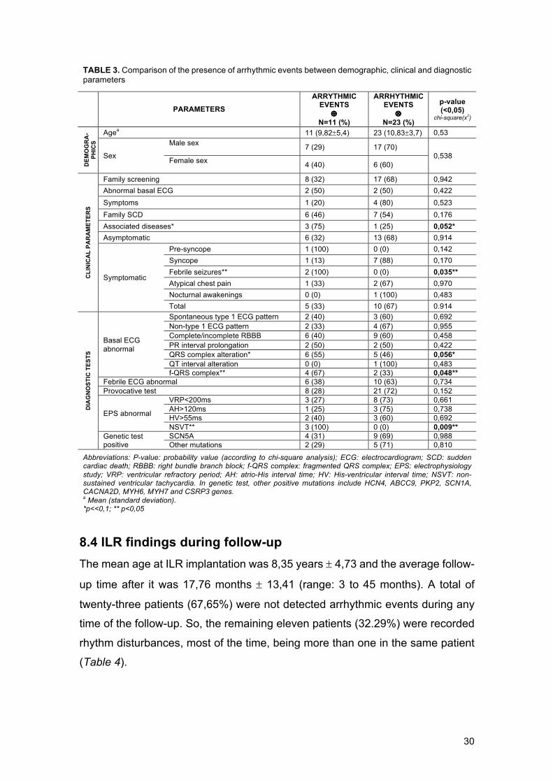
8.4 ILR findings during follow-up
The mean age at ILR implantation was 8,35 years ± 4,73 and the average follow-
up time after it was 17,76 months ± 13,41 (range: 3 to 45 months). A total of
twenty-three patients (67,65%) were not detected arrhythmic events during any
time of the follow-up. So, the remaining eleven patients (32.29%) were recorded
rhythm disturbances, most of the time, being more than one in the same patient
(Table 4).
31
Symptoms driven recordings (SDRs)
The mean time from ILR implantation to first symptom was 15,57 months ± 16,65.
A total of seven patients (63,64%) activated the LINQ’s recording function due to
symptom episodes including pre-syncope (the most repeated one among these
patients), palpitations, syncope, febrile seizures and atypical chest pain. Of 14
events transmitted by ILR in symptomatic cases, in two patients (28,57%) the
ECG tracing showed episodes of non-sustained monomorphic ventricular
tachycardia manifested as syncope. The remaining five patients (71,43%)
showed normal sinus rhythm, sinus tachycardia and minimal rhythm disturbances
such as isolated supra-ventricular extrasystoles.
Actionable ECG tracing in asymptomatic patients
In four asymptomatic patients (36,36%) the ILR recording was auto-activated. In
one of those cases (25%), ECG tracing showed various episodes of
asymptomatic NSVT lasting less than 20 seconds (Annex VI). In the same
patient, ILR also reported recurrent phases of asymptomatic sinus bradycardia
(down to 40 bpm) with frequent ventricular extrasytoles (Annex VI), and episodes
of abrupt repolarization changes compatible with a dynamic Brugada pattern
(Annex VI), fact that also triggered the ILR activation in the three (75%) remaining
patients.
Among total arrhythmic events detected, five patients (45,45%) were previously
(before the diagnosis of BrS) symptomatic while six patients (54,55%) were
previously asymptomatic. There was no statistical difference between them as
for the greater incidence of arrhythmic events in one group regarding the other
one (p=0,914).
During follow-up, ILR recordings showed abrupt and significant variations of
repolarization pattern compatible with dynamic Brugada pattern in six patients
(17,65%), and one of them (the same with asymptomatic NSVT) was without
previous evidence of altered ECG at baseline or during a febrile episode. No
episodes of advanced atrio-ventricular blocks or atrial fibrillation or flutter were
registered in our series.

32
TABLE 4. Description of ILR findings in our group during a mean follow-up of 17,76 months ± 13,41 SD
ILR findings N=patients (%) ILR symptoms driven recodrings (SDRs) 18 events Patients with ILR symptoms driven recordings (SDRs)
Non-clinically significant arrhythmias ST NSR SVEs
Clinically significant arrhythmias (ventricular ARRH)
7 (20,59%) 5 (71,43%) 4 (80%) 2 (40%) 1 (20%) 2 (28,57%)
Previously asymptomatic patient with ARRH 6 (31,58%) Previously symptomatic patient with ARRH 5 (33,33%) Implant to first SDR time 15,57 ± 16,65 Patients with dynamic Brugada pattern 6 (17,65%) Total arrhythmias detected
Symptomatic Asymptomatic
11 (32,35%) 7 (63,64%) 4 (36,36%)
Total arrhythmias detected Previously symptomatic patient Previously asymptomatic patient
11 (32,35%) 5 (45,45%) 6 (54,55%)
Patients without ARRH nor symptoms during FU 23 (67,65%) LINQ artefacts
FA (sinus arrhythmia) Pause (low QRS detection) VT (double counting QRS/T-wave)
12 (35,29%) 7 (58,83%) 3 (24,50%) 2 (16,67%)
Complications (end of ILR battery, peri-implantational hematoma, localized infection post-implant)
2 (5,88%)
Therapy (DAI implantation) 2 (5,88%)
Values are n(%),mean ± DS or n. Abbreviations: ILR: implantable loop recorder; SDR: symptoms driven recording; ST: sinus tachycardia; NSR: normal sinus rhythm; SVEs: supra-ventricular extrasystoles; ARRH: arrhythmias; FU: folloq-up; FA: atrial fibrillation.
8.5 Estimated risk of arrhythmic events In order to perform the multivariate analysis by logistic regression, we took into
account those variables that by literature and in the analysis of inference showed
to be statistically significant and those that in spite of not being so, did evidence
a certain tendency. However, we did not use the pre-syncope (p=0,142) and
febrile seizures (p=0,035) variables because upon representing a really small
portion of the sample, not converge in the multivariate analysis. The same
happened with for f-QRS complex (p=0,048) and provocative test (p=0,152)
variables. The induction of NSVT during EPS (p=0,009) was neither added for
analysing because all patients showed arrhythmic events during the follow-up.
With none of the variables used to perform the analysis, we could obtain a
significant p-value with a confidence interval of 95%. In contrast, the variables
that showed to be statistically significant with a confidence interval of 90%, to
present an incremented risk for arrhythmic events during follow-up were the
presence of associated diseases, family SCD and QRS complex alteration.
In order to calculate the risk of having an arrhythmic event was used (RR adjusted
– 1) x 100 (Table 5).

33
TABLE 5. Estimated risk1 - relative risk raw and adjusted (95% confidence intervals) and their p-values of arrhythmic events in relation to selected covariates.
VARIABLES RR raw (CI 95%) p-value RR adjusted (CI 95%) p-value Agea 0,944 (0,863-1,033) 0,258 0,934 (0,82-1,063) 0,292 Sex (female) 1,619 (0,727-3,606) 0,270 3,318 (0,871-12,643) 0,177 Abnormal basal ECG 2,333 (0,78-6,984) 0,216 6,310 (1,157-34,412) 0,131 Family SCD 2,743 (1,27-5,926) 0,041** 5,222 (1,429-19,093) 0,043** Associated diseases 8,25 (2,366-28,765) 0,043** 12,389 (2,604-58,94) 0,042** Symptomatic (syncope) 0,229 (0,071-0,732) 0,098* 0,49 (0,077-3,114) 0,345
Basal ECG abnormal
Complete/incomplete RBBB 1,733 (0,811-3,704) 0,230 0,427 (0,12-1,525) 0,245
PR interval prolongation 2,333 (0,779-6,984) 0,216 2,085 (0,473-9,192) 0,304 QRS complex alteration 4,320 (1,935-9,647) 0,032** 9,567 (2,073-44,173) 0,043**
1According to multivariate logistic regression analysis. Abbreviations: RR; Relative risk; ECG: electrocardiogram; SCD: sudden cardiac death; RBBB: right bundle branch block; f-QRS complex: fragmented QRS complex; EPS: electrophysiology study; NSVT: non-sustained ventricular tachycardia aMean (standard deviation). *p<<0,1; **p<0,05

34
9. DISCUSSION To our knowledge, this is the only study that evaluates ILRs, exclusively in
paediatric patients with BrS. There is one study performed with eleven adult BrS
patients41 and few data with pediatric patients with inherited arrhythmia
syndromes44. Our study presents a cohort of thirty-four young patients with BrS
without strict ICD implant indications in which a subcutaneous holter had
implanted. We are completely aware of the no statistical signification of the
obtained results due to the small sample size but there are some variables that
has showed a certain tendency event though not being significant.
As we see above, our sample size is insufficient, which implies a reduced
statistical power (54% in our case), that means, reduced probability of rejecting
the null hypothesis when it is false (i.e. the estimators are effectively statistically
significant). As we could not increase the size of the sample, we chose to
increase the level of significance (alpha), which would lead to reduce the
probability of committing type II error and, therefore, increasing the statistical
power. In particular, we chose a level of significance equal to 10%.
As for the multivariate analysis, the presence of associated diseases (p=0,042),
a family history of SCD (p=0,043) and the QRS complex alteration (p=0,043) were
variables statistically significant which could increment the risk of suffering
arrhythmic events, fact that coincide with current literature. However, a recent
study by Sarkozy59, among others, reported that a family history of SD in first-
degree relatives or at a young age is not predictive for future arrhythmic events.
But when it is associated with a spontaneous type 1 Brugada ECG pattern could
increment the risk. In our cohort of the thirteen patients with family history of SCD,
nine had a spontaneous ECG pattern (two of them type 1 pattern, three non-type
1 patterns, and four, Brugada pattern induced by fever). Therefore, only the four
remaining patients showed a completely normal ECG pattern at baseline or with
fever. There are studies that point out the risk associated with QRS complex
alteration, especially, when it is fragmented as pointed Morita et al.30.
In our study, the variables of age, presence of syncope and a
complete/incomplete RBBB, seems to be a protective factor that reduce the risk
of arrhythmic events (RRa<1), although we have not found evidence that none of
them has been statistically significant. Based on publications about BrS, we know
35
that is not like this, although not in our case. This lack of statistically significant
results could be explained due to the small sample size studied.
It seems that being female has a risk > 200% of having arrhythmic events. The
literature says the risk of having spontaneous arrhythmias is similar in children of
both genders during the pediatric age. However, that risk increases after puberty
only in male, due to their high levels of testosterone. For this reason, it is
extremely important to perform systematically re-evaluations, especially in male
during puberty, when the hormone starts to rise. Our results, possibly, are
because a few girls were included in the study, who more than 50% suffered of
arrhythmic events during the follow-up. Again, the results are not significant due
to the insufficient sample. Likewise, the sex variable, abnormal ECG at baseline
and PR interval prolongation variables showed a RR > 2 or > 100%, fact that
implied that was necessary to control some confounding factors, effect called
residual counfounding.
Finally, the variables that were excluded for the multivariate analysis, could be
related with an incremented risk of arrhythmias, but due to an insufficient sample,
in our study were not statistically significant. However, by bibliography, to have
suffered an arrhythmogenic syncope or the induction of NSVT during EPS has
been associated with an incremented risk (section 3.11).
As for ILR findings, during a mean follow-up of 17,76 months ± 13,41, a total of
seven patients (63,64%) activated the LINQ’s recording function due to symptom
episodes, which in the majority of the cases (71,43%) were proved normal sinus
rhythm/sinus tachycardia or minimal rhythm disturbances such as isolated supra-
ventricular extrasystoles. Likewise, Kubala et al.41 did not report any life-
threatening ventricular arrhythmia, based on ILR data, as a cause of syncope in
eight BrS patients after an average follow-up of 33 months or Avari Silvia JN et
al.44, when observing in their cohort of pediatric patients with inherited arrhythmia
syndromes carriers of ILRs that the 90% of symptomatic transmissions were not
associated with lethal arrhythmic events. Therefore, we could deduce that
symptoms do not correlate well with the presence of lethal arrhythmic events and
are not reliable markers for escalation of therapy or guidance around activity.

36
The use of the ILR to evaluate the aetiology of unexplained syncope in BrS
patients was reported in a Canadian study40 and in Giustetto et al.5.
Distinguishing vasovagal mechanism from the dreaded ventricular arrhythmias is
crucial in order to avoid ICD implant when it is not necessary (at least
temporarily). In our group, ILRs were substantially helpful in avoiding it in five
(71,43%) of our symptomatic patients. The same articles that point out the
therapeutic effectiveness of ICD, highlight also its high frequency of
complications and inappropriate shocks. For instance, Gonzalez et al.52, Probst
et al.58 in FINGER BrS Registry or more recently Sacher et al48, reported that rate
of appropriate ICD shocks in patients with BrS after 39 months mean follow-up
was lower in patients with history of syncope as compared with cardiac arrest
survivals (0 vs 45%). For these reasons, the indication of an ICD is a difficult
decision, especially in young patients that must be taken after a careful evaluation
and only to proceed to device implantation only in those patients who really need
it. When there is no clear indication of ICD, having a tool that allows continuous
monitoring of the child's heart and, at least, delaying its placement, is a very
useful strategy.
The ECG tracing identified episodes of non-sustained monomorphic ventricular
tachycardia in two symptomatic patients (28,57%) manifested as syncope
episodes. That involved a change in those patients’ therapeutic management,
requiring an early implantation of ICD. Especially in those cases, data provided
by ILR was critically important in guiding their management. In the same line,
Avari Silvia JN et al.44 the ILR helped to change therapies in 30% of subjects.
Finally, automatic transmissions of subclinical arrhythmias by ILR in
asymptomatic patients also had important consequences in their management.
In our cohort, ILR recorded various episodes of asymptomatic NSVT in one
patient who is being strictly followed with the aim of a prompt ICD implantation if
needed. Other cases were the detection of asymptomatic episodes of abrupt
repolarization changes, compatible with dynamic Brugada pattern, which could
be a signal of uncontrolled fever, the need to introduce some medication, or
insufficient quinidine titration. Those findings were in accordance with other
studies44 that found the 19% of automatically recorded transmission contained
data that altered the patients’ medical course.
37
10. IMPLANTABLE LOOP RECORDER LIMITATIONS Artefacts were reported in twelve (35,29%) patients during follow-up, often
related to misdetection of atrial fibrillation (due to significant sinus arrhythmia in
children), and to double counting or under-detection of QRS and T waves during
phases of signal alteration. However, those were easily recognized.
Two patients (5,88%) experienced device-related complications during follow-up.
One of those, suffered from device infection a week after the procedure. The
patient underwent re-implantation three months later without any further
complications. In the other case, a peri-implantational hematoma appeared
without infection. Finally, one patient ILR was replaced for a battery depletion
after 29th months (Table 4).
11. STUDY LIMITATIONS There are some potential limitations that should be considered:
- Brugada syndrome is considered a rare disease with a low prevalence
among paediatric population, fact that led to have a relatively reduced
number of patients. However, our study had performed in Arrhythmia
Pediatric Unit of Sant Joan de Déu Hospital, a centre designed as
coordinators of the European Reference Network in Rare Cardiac
Diseases – subnetwork paediatrics. So, possibly patients regardless of
their area of residence, came in order to be treated and followed there.
- A limitation from our study design was represented by its retrospective
fashion which could have led to some recordkeeping and information
biases.
- A non-probabilistic consecutive sampling method was used. Therefore,
we tried to avoid the selection bia by including in the database only well-
defined cases of patients who accomplished the inclusion and exclusion
criteria.
- A small sample size and a brevity time of follow-up have been another
limitation of our study, making difficult to draw statistically significant
conclusions. However, we were able to identify a certain trend in some
studied variables, which means, that a longer time of monitoring could
38
help to disclose further arrhythmic events and risk factors, especially
when patients come to puberty.
- It is difficult to control confusion variables in observational studies, but we
tried to minimize the possible bias ruling out all confusion factors known
by an exhaustive bibliography research.
- Measurement biases could both appear during the process of registering
some variables by the pertinent instrument and upon interpreting the
results. In order to minimize them, all instruments were calibrated
periodically and each result was analysed at least by two expert paediatric
cardiologists in the syndrome. In case of discrepancy, the results were
evaluated by a third expert.
- Due to the anonymization of the process, any significant rhythm
disturbance that could demand a treatment, we could not contact directly
to the anonymous patient. However, those were followed closely by their
reference paediatric cardiologist, responsible for reviewing the ILR’s
recorded data and took the appropriate measures based on its findings.
- Although we knew that using a multivariate COX model would have been
ideal, we did not get the different moments when arrhythmic events
appeared. For the same reason, we neither could calculate the incidence
rate of arrhythmic events using the Poisson model. So, we finally used a
regression logistic model and calculated the incidence as a proportion
with its IC 95%.
- ILR are compatible with total body nuclear magnetic resonance imaging
(MRI), although in cardiac MRI they could cause significant intensity
artefacts in diagnostic images as well as these could be mistaken for a
tachyarrhythmia in the ECG. Therefore, it is important to take this into
account upon interpreting an ECG. Also, further investigation about MRI
techniques is required in order to eliminate imaging artefacts.

39
12. CONCLUSIONS The purpose of our study was to prove the effectiveness of ILRs in order to detect
arrhythmic events in patients with non-high-risk BrS, considering it as a useful
tool for guiding their management.
This study summarizes a single-experience with 34 patients with Brugada
syndrome that received an ILR at an age of < 21 years. After a mean follow-up
of 18 months the ILR contributed to detect arrhythmic events in patients with
Brugada syndrome, confirming our main hypothesis. The ILR was determinant in
order to exclude ventricular arrhythmias as a cause symptoms in 71,43% of
symptomatic patients. In general, the device was crucial in decision making
regarding ICD implant in 81,82% of patients, both symptomatic and
asymptomatic, who ILR detected arrhythmias (8 avoided implants and one
potential implant in one asymptomatic patient). Especially important, the ILR
allowed to detect ventricular arrhythmias in two symptomatic patients, leading to
a change in their therapeutic management, requiring an early implantation of ICD.
As a conclusion, the implantable loop recorder allows a long-term continue
monitoring of heart rhythm in patients with increased risk of life-threatening
arrhythmias allowing to detect the best moment for ICD implantation. So, the
device should be considered as a tool for clinical management of paediatric
patients with non-high-risk BrS and other inherited arrhythmic diseases. Long-
term follow-up studies are needed to add more information in this field.
40
13. REFERENCES 1. Brugada P, Brugada J. Right bundle branch block, persistent st segment
elevation and sudden cardiac death: A distinct clinical and
electrocardiographic syndrome. A multicenter report. Journal of the American
College of Cardiology [Internet]. 1992 [cited 2018 Jul 02];20:1391-1396.
Available: https://www.ncbi.nlm.nih.gov/pubmed/1309182
2. Brugada J, Brugada P. Further characterization of the syndrome of right
bundle branch block, ST segment elevation, and sudden cardiac death. J
Cardiovasc Electrophysiol [Internet]. 1997 [cited 2018 Jul 02];8(3):325–31.
Available: https://www.ncbi.nlm.nih.gov/pubmed/9083883
3. Brugada P, Brugada J, Roy D. Brugada syndrome 1992-2012: 20 years of
scientific excitement, and more. Eur Heart J [Internet]. 2013 [cited 2018 Jul
02];34(47):3610–5.
Available: https://academic.oup.com/eurheartj/article/34/47/3610/619647
4. Grant AO, Carboni MP, Neplioueva V, Frank Starmer C, Memmi M,
Napolitano C, et al. Long QT syndrome, Brugada syndrome, and conduction
system disease are linked to a single sodium channel mutation. J Clin Invest
[Internet]. 2002 [cited 2018 Jul 02];110(8):1201–9. Available:
https://www.ncbi.nlm.nih.gov/pmc/articles/PMC150793
5. Hedley PL, Jorgensen P, Schlamowitz S, Moolman-Smook J, Kanters JK,
Corfield VA, et al. The genetic basis of Brugada syndrome: a mutation update.
Human mutation [Internet]. 2009 [cited 2018 Jul 02];30:1256-1266. Available:
https://onlinelibrary.wiley.com/doi/epdf/10.1002/humu.21066
6. Sarquella-Brugada G, Campuzano O, Iglesias A, Sanchez-Malagon J,
Guerra-Balic M, Brugada J, Brugada R. Genetics of sudden cardiac death in
children and young athletes. Cardiology in the Young [Internet]. 2013 [cited
2018 Jul 02];23:159-173.
Available: https://doi.org/10.1017/S1047951112001138
7. Probst V, Wilde AAM, Barc J, Sacher F, Babuty D, Mabo P, [et al.]. SCN5A
mutations and the role of genetic background in the pathophysiology of
Brugada syndrome. Circ Cardiovasc Genet [Internet]. 2009 [cited 2018 Jul
02];2(6):552–7.
Available:https://www.ahajournals.org/doi/10.1161/CIRCGENETICS.109.853
41
8. Sarquella-Brugada G, Campuzano O, Arbelo E, Brugada J, Brugada R.
Brugada syndrome: clinical and genetic findings. Genet Med [Internet]. 2016
[cited 2018 Jul 02];18(1):3–12.
Available: http://www.nature.com/doifinder/10.1038/gim.2015.35
9. Fernández-Falgueras A, Sarquella-Brugada G, Brugada J, Brugada R,
Campuzano O. Cardiac Channelopathies and Sudden Death: Recent Clinical
and Genetic Advances. Biology (Basel) [Internet]. 2017 [cited 2018 Jul
02];6(1):7. Available: http://www.mdpi.com/2079-7737/6/1/7
10. Sarquella-Brugada G. Channelopathies in Pediatric Sudden Death: Clinical
implications of the genetic diagnostic in Long QT Syndrome. Barcelona:
Universitat de Barcelona; 2015.
11. Antzelevitch C, Yan GX, J.Ackerman M, Borggrefe M, Corrado D, Guo J, et
al. J-Wave syndromes expert consensus conference report: emerging
concepts and gaps in knowledge. EP Europace [Internet]. 2017 [cited 2018
Jul 04];19(4):665-694.
Available: https://academic.oup.com/europace/article/19/4/665/2952412
12. Mashar M, Kwok AJ, Pinder R, Sabir I. The Brugada syndrome revisited.
Cardiovasc Med [Internet]. 2014 [cited 2018 Jul 04];24(5):191–6. Available:
http://dx.doi.org/10.1016/j.tcm.2013.11.001
13. Antzelevitch C, Brugada P, Borggrefe M, Brugada J, Brugada R, Corrado D,
et al. Brugada syndrome: Report of the second consensus conference.
Circulation [Internet]. 2005 [cited 2018 Jul 04];111:659-670. Available:
https://www.ahajournals.org/doi/pdf/10.1161/01.CIR.0000152479.54298.51
14. Probst V, Denjoy I, Meregalli PG, Amirault JC, Sacher F, Mansourati J, et al.
Clinical aspects and prognosis of Brugada syndrome in children. Circulation
[Internet]. 2007 [cited 2018 Jul 04];115:2042-2048. Available:
https://www.ncbi.nlm.nih.gov/pubmed/17404158
15. Viskin S. Brugada syndrome in children: Don't ask, don't tell? Circulation
[Internet]. 2007 [cited 2018 Jul 04];115:1970-1972. Available:
https://www.ahajournals.org/doi/10.1161/CIRCULATIONAHA.106.686758
16. Gonzalez Corcia MC, Asmundis C, Chierchia G-B, Brugada P. Brugada
syndrome in the paediatric population: a comprehensive approach to clinical
42
manifestations, diagnosis, and management. Cardiol Young [Internet]. 2016
[cited 2018 Jul 04];26(6):1044–55.
Available: http://www.journals.cambridge.org/abstract_S1047951116000548
17. Berne P, Brugada J. Brugada Syndrome 2012. Circulation J [Internet]. 2012
[cited 2018 Jul 04];76(7):1563–71.Available: https://doi.org/10.1253/circj.CJ-
12-0717
18. Antzelevitch C. Brugada Syndrome. Pacing Clin Electrophysiol [Internet].
2006 [cited 2018 Jul 04];29(10):1130–1159. Available:
http://doi.wiley.com/10.1111/j.1540-8159.2006.00507.x
19. Mizusawa Y, Wilde AAM. Brugada Syndrome. Methodist Debakey Cardiovasc
J [Internet]. 2014 [cited 2018 Jul 04];10(1):25–8. Available:
https://www.ncbi.nlm.nih.gov/medgen/411607
20. Mivelaz Y, Di Bernardo S, Pruvot E, Meijboom EJ, Sekarski N. Brugada
syndrome in childhood: A potential fatal arrhythmia not always recognized by
paediatricians. A case report and review of the literature. Eur J Pediatr
[Internet]. 2006 [cited 2018 Jul 04];165(8):507–11. Available:
https://link.springer.com/article/10.1007/s00431-006-0150-z
21. De Marco S, Giannini C, Chiavaroli V, De Leonibus C, Chiarelli F, Mohn A.
Brugada syndrome unmasked by febrile illness in an asymptomatic child. J of
Pediatrics [Internet]. 2012 [cited 2018 Jul 06];161(4):10–1. Available:
https://www.jpeds.com/article/S0022-3476(12)00432-5/pdf
22. Wang K, Asinger RW, Marriott HJL. ST-Segment Elevation in Conditions
Other Than Acute Myocardial Infarction. N Engl J Med [Internet]. 2003 [cited
2018 Jul 06];349(22):2128–35.
Available from: http://www.nejm.org/doi/abs/10.1056/NEJMra022580
23. Veltmann C, Schimpf R, Echternach C, Eckardt L, Kuschyk J, Streitner F, et
al. A prospective study on spontaneous fluctuations between diagnostic and
non-diagnostic ECGs in Brugada syndrome: Implications for correct
phenotyping and risk stratification. Eur Heart J [Internet]. 2006 [cited 2018 Jul
06];27(21):2544–52.
Available: https://academic.oup.com/eurheartj/article/27/21/2544/2887135
24. Hermida JS, Lemoine JL, Aoun FB, Jarry G, Rey JL, Quiret JC. Prevalence
of the Brugada syndrome in an apparently healthy population. AmJ Cardiol
43
[Internet]. 2000 [cited 2018 Jul 06];86(0002–9149 SB–A SB–M):91–4.
Available: https://europepmc.org/abstract/med/10867101
25. Conte G, Sieira J, Sarkozy A, De Asmundis C, Di Giovanni G, Chierchia GB,
et al. Life-threatening ventricular arrhythmias during ajmaline challenge in
patients with Brugada syndrome: Incidence, clinical features, and prognosis.
Hear Rhythm [Internet]. 2013 [cited 2018 Jul 06];10(12):1869–74. Available:
https://www.sciencedirect.com/science/article/pii/S1547527113010709
26. Miyazaki T, Mitamura H, Miyoshi S, Soejima K, Aizawa Y, Ogawa S.
Autonomic and antiarrhythmic drug modulation of ST segment elevation in
patients with Brugada syndrome. JACC [Internet]. 1996 [cited 2018 Jul
06];27(5):1061–70. Available: http://www.onlinejacc.org/content/27/5/1061
27. Cruz Puntí D. Flecainide challenge in pediatric Brugada syndrome: adding
further data to this diagnostic tool. [Internet]. Girona: Universitat de Girona;
2017 [cited 2018 Jul 06]. Available: https://dugi-
doc.udg.edu/handle/10256/15659
28. Brugada P, Brugada R, Brugada J, Priori SG, Napolitano C. Patients with an
asymptomatic Brugada electrocardiogram should undergo pharmacological
and electrophysiological testing. Circulation [Internet]. 2005 [cited 2018 Jul
06];112(2):279–92. Available: http://citeseerx.ist.psu.edu/viewdoc/summary?
doi=10.1.1.516.8401
29. Calvo D, Rubín JM, Pérez D, Gómez J, Flórez JP, Avanzas P, et al. Time-
dependent responses to provocative testing with flecainide in the diagnosis of
Brugada syndrome. Hear Rhythm [Internet]. 2015 [cited 2018 Jul
06];12(2):350–7. Available: http://dx.doi.org/10.1016/j.hrthm.2014.10.036
30. Morita H, Kusano KF, Miura D, Nagase S, Nakamura K, Morita ST, et al.
Fragmented QRS as a marker of conduction abnormality and a predictor of
prognosis of Brugada syndrome. Circulation [Internet]. 2008 [cited 2018 Jul
06];118:1697-1704. Available: https://www.ahajournals.org/doi/10.1161/
CIRCULATIONAHA.108.770917
31. Behere SP, Weindling SN. Brugada syndrome in children - stepping into
unchartered territory. Annals of pediatric cardiology [Internet]. 2017 [cited
2018 Jul 09];10:248-258.
Available: https://www.ncbi.nlm.nih.gov/pmc/articles/PMC5594936/
44
32. Bayés De Luna A, Brugada J, Baranchuk A, Borggrefe M, Breithardt G,
Goldwasser D, et al. Current electrocardiographic criteria for diagnosis of
Brugada pattern: A consensus report. J Electrocardiol [Internet]. 2012 [cited
2018 Jul 09];45(5):433–42.
Available: http://dx.doi.org/10.1016/j.jelectrocard.2012.06.004
33. Priori SG, Wilde AA, Horie M, Cho Y, Behr ER, Berul C, et al.
HRS/EHRA/APHRS expert consensus statement on the diagnosis and
management of patients with inherited primary arrhythmia syndromes. Heart
rhythm [Internet]. 2013 [cited 2018 Jul 09];10:1932-1963. Available:
https://www.heartrhythmjournal.com/article/S1547-5271(13)00552-3/abstract
34. Casado-Arroyo R, Berne P, Rao JY, Rodriguez-Manero M, Levinstein M,
Conte G, et al. Long-term trends in newly diagnosed Brugada syndrome:
Implications for risk stratification. JACC [Internet]. 2016 [cited 2018 Jul
09];68:614-623. Available: http://www.onlinejacc.org/content/68/6/614
35. Adler A, Rosso R, Chorin E, Havakuk O, Antzelevitch C, Viskin S. Risk
stratification in Brugada syndrome: Clinical characteristics,
electrocardiographic parameters, and auxiliary testing. Heart rhythm
[Internet]. 2016 [cited 2018 Jul 09];13:299-310. Available:
https://www.sciencedirect.com/science/article/pii/S1547527115011285
36. Sroubek J, Probst V, Mazzanti A, Delise P, Hevia JC, Ohkubo K, et al.
Programmed ventricular stimulation for risk stratification in the Brugada
syndrome: A pooled analysis. Circulation [Internet]. 2016 [cited 2018 Jul
09];133:622-630. Available:
https://www.ahajournals.org/doi/abs/10.1161/CIRCULATIONAHA.115.01788
37. Gonzalez Corcia MC, Sieira J, Pappaert G, de Asmundis C, Chierchia GB,
Sarkozy A, Brugada P. A clinical score model to predict lethal events in young
patients (£19 years) with the Brugada syndrome. The American journal of
cardiology [Internet]. 2017 [cited 2018 Jul 09];120:797-802. Available:
https://www.sciencedirect.com/science/article/pii/S000291491730958X
38. Gonzalez Corcia MC, Brugada P. The value of performing invasive risk
stratification in young patients with the Brugada syndrome. Cardiol Young
[Internet]. 2017 [cited 2018 Jul 09];27(7):1444–5. Available:
https://doi.org/10.1017/S1047951117001172
45
39. Chen Q, Kirsch GE, Zhang D, Brugada R, Brugada J, Brugada P, et al.
Genetic basis and molecular mechanism for idiopathic ventricular fibrillation.
Nature [Internet]. 1998 [cited 2018 Jul 09]; 392: 293-296. Available:
https://www.nature.com/articles/32675
40. Brignole M, Vardas P, Hoffman E, Huikuri H, Moya A, Ricci R, et al. Indications
for the use of diagnostic implantable and external ecg loop recorders. EP
Europace [Internet]. 2009 [cited 2018 Jul 09];11:671-687. Available:
https://academic.oup.com/europace/article/11/5/671/511160
41. Kubala M, Aissou L, Traulle S, Gugenheim AL, Hermida JS. Use of
implantable loop recorders in patients with Brugada syndrome and suspected
risk of ventricular arrhythmia. EP Europace [Internet]. 2012 [cited 2018 Jul
11];14:898-902.
Available: https://academic.oup.com/europace/article/14/6/898/545734
42. Sulke N, Sugihara C, Hong P, Patel N, Freemantle N. The benefit of a
remotely monitored implantable loop recorder as a first line investigation in
unexplained syncope: the EaSyAS II trial. EP Europace [Internet]. 2016 [cited
2018 Jul 11];18:912-918.
Available: https://academic.oup.com/europace/article/18/6/912/2467068
43. Todd D, Hernandez-Madrid A, Proclemer A, Bongiorni MG, Estner H,
Blomstrom-Lundqvist C. How are arrhythmias detected by implanted cardiac
devices managed in Europe? Results of the European Heart Rhythm
Association Survey. EP Europace [Internet]. 2015 [cited 2018 Jul
11];17:1449-1453.
Available: https://academic.oup.com/europace/article/17/9/1449/628171
44. Avari Silva JN, Bromberg BI, Emge FK, Bowman TM, Van Hare GF.
Implantable loop recorder monitoring for refining management of children with
inherited arrhythmia syndromes. J Am Heart Assoc [Internet]. 2016 [cited
2018 Jul 11];5(6) pii:e003632.
Available: https://www.ahajournals.org/doi/abs/10.1161/jaha.116.003632
45. Wong JA, Yee R, Gula LJ, Skanes AC, Ross IG, White JB, et al. Feasibility of
magnetic resonance imaging in patients with an implantable loop recorder.
PACE [Internet]. 2008 [cited 2018 Jul 11];31:333-337. Available:
https://onlinelibrary.wiley.com/doi/full/10.1111/j.1540-8159.2008.00994.x
46
46. Blaschke F, Lacour P, Walter T, Wutzler A, Huemer M, Parwani A, et al.
Cardiovascular magnetic resonance imaging in patients with an implantable
loop recorder. Annals of noninvasive electrocardiology [Internet]. 2016 [cited
2018 Jul 11];21(3):319-324.
Available: https://onlinelibrary.wiley.com/doi/full/10.1111/anec.12333
47. Pick JM, Batra AS. Implantable cardioverter-defibrillator implantation for
primary and secondary prevention: Indications and outcomes. Cardiology in
the Young [Internet]. 2017 [cited 2018 Jul 11];27:S126-S131. Available:
https://doi.org/10.1017/S1047951116002365
48. Sacher F, Probst V, Maury P, Babuty D, Mansourati J, Komatsu Y, et al.
Outcome after implantation of a cardioverter-defibrillator in patients with
Brugada syndrome: a multicenter study. Circulation [Internet]. 2013 [cited
2018 Jul 11];128:1739-1747. Available: https://hal.archives-ouvertes.fr/hal-
00750430
49. Conte G, Sieira J, Ciconte G, de Asmundis C, Chierchia GB, Baltogiannis G,
et al. Implantable cardioverter-defibrillator therapy in Brugada syndrome: a
20-year single-center experience. JACC [Internet]. 2015 [cited 2018 Jul
11];65:879-888. Available: http://www.onlinejacc.org/content/65/9/879
50. Priori SG, Blomströ m-Lundqvist C, Mazzanti A, Blom N, Borggrefe M, Camm
J, et al. 2015 ESC Guidelines for the management of patients with ventricular
arrhythmias and the prevention of sudden cardiac death. Eur Heart J
[Internet]. 2015 [cited 2018 Jul 11];36(41):2836–7. Available:
http://secardiologia.es/images/secciones/arritmias/2793-full.pdf
51. Gonzalez Corcia MC, Sieira J, Sarkozy A, Asmudis C, Chierchia GB,
Hernandez Ojeda J, et al. Brugada syndrome in the young: an assessment of
risk factors predicting future events. EP Europace [Internet]. 2017 [cited 2018
Jul 13];19(11):1864–1873.
Available: https://academic.oup.com/europace/article/19/11/1864/2194460
52. Gonzalez Corcia MC, Sieira J, Pappaert G, Asmundis C, Chierchia BG, La
Meir M, et al. Implantable cardioverter-defibrillators in children and
adolescents with Brugada syndrome. JACC [Internet]. 2018 [cited 2018 Jul
13];7:148-57. Available: https://www.sciencedirect.com/science/article/pii/
S0735109717415865
47
53. Tejman-Yarden S, Ben-Zeev B, Goldshmit Y, Sarquella-Brugada G, Cicurel
A, Katz U, et al. Utilization of an Insertable Cardiac Monitor in a Child with
Pallid Breath-Holding Spells. Pediatr Neurol [Internet]. 2016 [cited 2018 Jul
12];64:80-82. Available: https://www.sciencedirect.com/science/article/pii/
S0887899416302995
54. Shimizu W, Matsuo K, Kokubo Y, Satomi K, Kurita T, Noda T, et al. Sex
hormone and gender difference - Role of testosterone on male predominance
in Brugada syndrome. J Cardiovasc Electrophysiol [Internet]. 2007 [cited 2018
Jul 13];18(4):415–21. Available: https://onlinelibrary.wiley.com/doi/full/
10.1111/j.1540-8167.2006.00743.x
55. Eckardt L. Gender differences in Brugada syndrome. J Cardiovasc
electrophysiol [Internet]. 2007 [cited 2018 Jul 13];18(4):422–4. Available:
https://onlinelibrary.wiley.com/doi/full/10.1111/j.1540-8167.2006.00759.x
56. Priori SG, Napolitano C, Gasparini M, Pappone C, Della Bella P, Giordano U,
et al. Natural history of Brugada syndrome: Insights for risk strati cation and
management. Circulation [Internet]. 2002 [cited 2018 Jul 13]; 105: 1342–
1347.
Available: https://www.ahajournals.org/doi/abs/10.1161/hc1102.105288
57. Eckardt L, Probst V, Smits JPP, Bahr ES, Wolpert C, Schimpf R, et al. Long-
term prognosis of individuals with right precordial ST-segment-elevation
Brugada syndrome. Circulation [Internet]. 2005 [cited 2018 Jul 13]; 111: 257-
263. Available: https://www.ahajournals.org/doi/abs/10.1161/01.CIR.0000
153267.21278.8D
58. Probst V, Veltmann C, Eckardt L, Meregalli PG, Gaita F, Tan HL, et al. Long-
term prognosis of patients diagnosed with Brugada syndrome: Results from
the FINGER Brugada Syndrome Registry. Circulation [Internet]. 2010 [cited
2018 Jul 13];121: 635-643. Available: https://hal-hcl.archives-
ouvertes.fr/CARDIO/hal-00910144v1
59. Sarkozy A, Sorgente A, Boussy T, Casado R, Paparella G, Capulzini L et al.
The value of a family history of sudden death in patients with diagnostic type
I Brugada ECG pattern. Eur Heart J [Internet]. 2011 [cited 2018 Jul
13];32(17):2153–2160. Available: https://academic.oup.com/eurheartj
60. Brugadadrugs.org [Internet]. Amsterdam: University of Amsterdam Academic
Medical Center; 2018. Drug lists [consult].Available: www.brugadadrugs.org.

48
61. Seidl K, Rameken M, Breunung S, Senges J, Jung W, Andresen D, et al.
Diagnostic assessment of recurrent unexplained syncope with a new
subcutaneously implantable loop recorder. EP Europace [Internet]. 2000
[cited 2018 Jul 15];2(3):256–262. Available: https://doi.org/10.1053/eupc.
2000.0108
62. Brighamandwomens.org [Internet]. Brigham: Harvard Medical School
Teaching Hospital; 2015. Implantable loop recorder [consult]. Available:
https://www.brighamandwomens.org/heart-and-vascular-center/procedures/
implantable-loop-recorder

49
14. ANNEXES 14.1 Annex I: Informed consent for invasive procedures (in Catalan and in Spanish)

50

51
14.2 Annex II: CEIC’s authorization
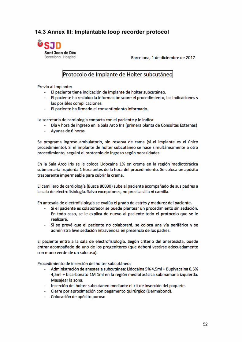
52
14.3 Annex III: Implantable loop recorder protocol
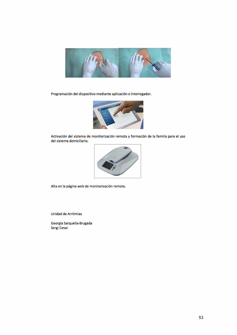
53
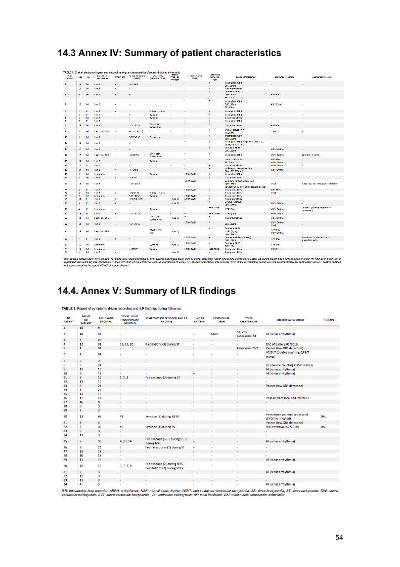
54
14.3 Annex IV: Summary of patient characteristics 14.4. Annex V: Summary of ILR findings
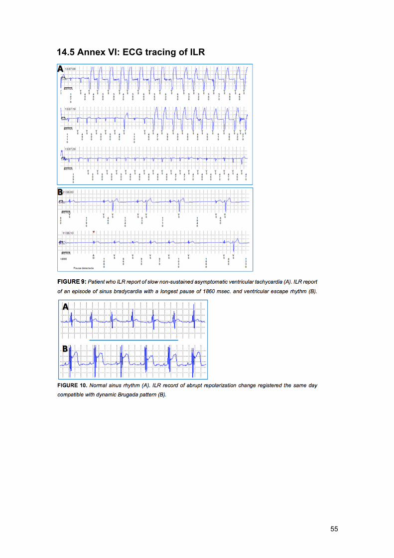
55
14.5 Annex VI: ECG tracing of ILR

56
14.6 Annex VII: Levels of evidence in therapeutic studies Levels of evidence for therapeutic studies, extracted from the Center for Evidence-Based Medicine