Embed Size (px)
Citation preview

The BRC repeats of human BRCA2 differentiallyregulate RAD51 binding on single- versusdouble-stranded DNA to stimulate strand exchangeMahmud K. K. Shivjia, Shreyas R. Mukunda,b, Eeson Rajendraa, Shaoxia Chenc, Judith M. Shortc, Jane Savilla,David Klenermanb, and Ashok R. Venkitaramana,1
aThe Medical Research Council Cancer Cell Unit, Hutchison/MRC Research Centre, Hills Road, Cambridge CB2 0XZ,United Kingdom; bDepartment ofChemistry, University of Cambridge, Lensfield Road, Cambridge CB2 1EW, United Kingdom; and cThe Medical Research Council Laboratory of MolecularBiology, Hills Road, Cambridge CB2 0QH, United Kingdom
Communicated by Stephen C. Kowalczykowski, University of California, Davis, CA, June 8, 2009 (received for review December 3, 2008)
The breast and ovarian cancer suppressor BRCA2 controls the enzymeRAD51 during homologous DNA recombination (HDR) to preservegenome stability. BRCA2 binds to RAD51 through 8 conserved BRCrepeat motifs dispersed in an 1127-residue region (BRCA2[BRC1–8]).Here, we show that BRCA2[BRC1–8] exerts opposing effects on thebinding of RAD51 to single-stranded (ss) versus double-stranded (ds)DNA substrates, enhancing strand exchange. BRCA2[BRC1–8] alters theelectrophoretic mobility of RAD51 bound to an ssDNA substrate,accompanied by an increase in ssDNA-bound protein assemblies,revealed by electron microscopy. Single-molecule fluorescence spec-troscopy shows that BRCA2[BRC1–8] promotes RAD51 loading ontossDNA. In contrast, BRCA2[BRC1–8] has a different effect on RAD51assembly on dsDNA; it suppresses and slows this process. Whenhomologous ssDNA and dsDNA are both present, BRCA2[BRC1–8] stim-ulates strand exchange, with delayed RAD51 loading onto dsDNAaccompanying the appearance of joint molecules representing re-combination products. Collectively, our findings suggest thatBRCA2[BRC1–8] targets RAD51 to ssDNA while inhibiting dsDNA bind-ing and that these contrasting activities together bolster one anotherto stimulate HDR. Our work provides fresh insight into the mechanismof HDR in humans, and its regulation by the BRCA2 tumor suppressor.
DNA recombination � electron microscopy �single-molecule fluorescence spectroscopy � tumor suppressor
Homologous DNA recombination (HDR) is essential in somaticcells not only for the error-free repair of DNA double-strand
breaks (DSBs), but also for the restoration of DNA replication forksthat stall at lesions in the template strand (1). The central event inHDR is the synapsis of a single-stranded (ss)DNA molecule withhomologous duplex DNA, which initiates the strand exchange thatleads to recombination. Recombinase molecules, conserved fromRecA in prokarya to RAD51 in eukaryal cells, mediate strandexchange via distinct reactions grouped into the presynaptic, syn-aptic, and postsynaptic phases (2).
The breast cancer suppressor protein, BRCA2, an �384-kDamolecule of 3418 residues, is essential in vivo for RAD51-mediatedHDR (3). Several regions of BRCA2 may contribute to this role (4).Human BRCA2 interacts with RAD51 through 8 copies of theBRC repeat motifs of �40 residues each, embedded in a 1127-residue fragment (BRCA2[BRC1–8]) within exon 11 (5), as well asthrough an unrelated carboxyl-terminal motif in exon 27 (6). TheBRC repeats appear to be the primary locus for the BRCA2-RAD51 interaction; unlike the C-terminal motif, their sequence isevolutionarily conserved in simple eukaryotes like fungi, as well asin plants (7). In addition, exon 17 of human BRCA2 encodes an�800 residue DNA-binding domain (BRCA2DBD) containing 3oligonucleotide-binding (OB) folds, ssDNA-binding modules alsofound in the abundant ssDNA-binding protein, replication proteinA (RPA) (8). In a fungal BRCA2 homolog, the OB-fold-containing, ssDNA-binding domain displaces RPA from DNAsubstrates in vitro to enable nucleation of the RAD51 filament by
the single BRC repeat found in this organism (9). Moreover,fragments containing 2 to 4 of the 8 human BRC repeats fused tothe ssDNA-binding BRCA2[DBD] domain can partially complementHDR in BRCA2-deficient cells in vivo (10) and promote RAD51-dependent strand exchange in vitro (11). Together, these observa-tions suggest that the BRCA2[DBD] cooperates with the BRCrepeats to target RAD51 to DNA substrates, acting as a recombi-nation mediator.
The precise role in HDR played by the multi-BRC repeat regionof human BRCA2 is not yet clear. Indeed, the BRC repeats arereported to have activities that could potentially inhibit (12–14) aswell as stimulate (11, 15, 16) RAD51 function, under differentexperimental conditions. We have previously purified the exon 11encoded 1,127-residue domain (BRCA2[BRC1–8]) from humanBRCA2 containing all 8 BRC repeats within their natural context,and shown that BRCA2[BRC1–8] suffices to promote RAD51-mediated strand exchange, independent of the ssDNA-bindingdomain BRCA2[DBD] (16). Here, we report results that suggest amechanism underlying the role of BRCA2[BRC1–8] in stimulatingHDR. We find that BRCA2[BRC1–8] promotes RAD51 loading onssDNA, while suppressing RAD51 assembly on dsDNA, and pro-pose that both these opposing effects cooperate to enhance theefficiency of RAD51-mediated HDR. Our findings provide addi-tional evidence in support of a recently proposed model (17)reported while this paper was under review.
Results and DiscussionElectrophoretic Mobility of Complexes Between BRCA2[BRC1–8], RAD51,and ssDNA. We have previously shown, using a well-established invitro assay for HDR, that BRCA2[BRC1–8] promotes strand ex-change between circular �X174 ssDNA and a homologous linearduplex DNA (16). To identify potential mechanisms underlying thisstimulatory activity, we first examined the effect of BRCA2[BRC1–8]on RAD51 binding to ssDNA. We generated a 2.5-kb resecteddouble-stranded (ds)DNA substrate from �X174 RF1 DNA by �exonuclease digestion according to the schematic shown in Fig.S1A. When radiolabeled at its 5� end, the great majority of theresected dsDNA preparation (Fig. S1B, lane 1) binds in an elec-trophoretic mobility shift assay (EMSA) to the ssDNA bindingprotein, RPA in a concentration-dependent manner (Fig. S1B,lanes 2–4), in contrast to radiolabeled dsDNA (Fig. S1C).
Author contributions: M.K.K.S. and A.R.V. designed research; M.K.K.S., S.R.M., S.C., andJ.M.S. performed research; J.S. contributed new reagents/analytic tools; M.K.K.S., S.R.M.,E.R., S.C., J.M.S., J.S., D.K., and A.R.V. analyzed data; and M.K.K.S., S.R.M., E.R., and A.R.V.wrote the paper.
Conflict of interest statement: The editor and A.V. recently (March 2009) co-authored apaper on a related topic.
Freely available online through the PNAS open access option.
1To whom correspondence should be addressed. E-mail: [email protected].
This article contains supporting information online at www.pnas.org/cgi/content/full/0906208106/DCSupplemental.
13254–13259 � PNAS � August 11, 2009 � vol. 106 � no. 32 www.pnas.org�cgi�doi�10.1073�pnas.0906208106
Dow
nloa
ded
by g
uest
on
Apr
il 10
, 202
0

We examined the effect of BRCA2[BRC1–8] on RAD51 bindingto the resected dsDNA substrate. The reaction conditions used herewere identical to those we have previously used for strand exchangestimulated by BRCA2[BRC1–8] (16), except that linear dsDNA wasomitted. Reactions included ATP and resected dsDNA preincu-bated with a low, subsaturating concentration of RPA, within arange previously reported to promote strand exchange (18, 19).RAD51 (3.3 �M) was incubated in a reaction mix either in theabsence or presence of a range of indicated BRCA2[BRC1–8] con-centrations. The RPA-bound, radiolabeled, resected dsDNA sub-strate (10 �M) was subsequently added to these preformed com-plexes. After incubation at 37 °C for 60 min, reaction products wereresolved by agarose gel electrophoresis (Fig. 1).
RAD51 binds to the resected dsDNA substrate, retarding itselectrophoretic mobility (Fig. 1B, compare lanes 1 and 2). Increas-ing concentrations of BRCA2[BRC1–8] affect this pattern (lanes3–6), indicative of complex formation between BRCA2[BRC1–8],RAD51, and a resected dsDNA substrate that is predominantlysingle-stranded, with a short ds/ssDNA junction (Fig. S1A). Similarchanges are observed when the nonhydrolyzable analog, AMP-PNP, is substituted for ATP (Fig. S2). Complex formation is evidentwith 0.4 �M BRCA2[BRC1–8] and plateaus between 1 to 2 �M.However, BRCA2[BRC1–8] alone at similar or higher concentrationsdoes not detectably alter the mobility of the resected dsDNAsubstrate (Fig. S1D), suggesting that RAD51 mediatesBRCA2[BRC1–8] binding to DNA in the complex.
BRCA2[BRC1–8] Increases ssDNA-Bound Protein Assemblies. We exam-ined by electron microscopy (EM) the complexes formed by ssDNA
with RAD51 in the presence or absence of BRCA2[BRC1–8] (Fig. 2)under conditions similar to those used in Fig. 1. As before, 5 �MRAD51 and 15 �M ssDNA (�X174 circle) were mixed at the 1:3ratio deemed optimal for assembly, and ATP was included. Theabsence of any ssDNA/dsDNA junction in the DNA substrate usedfor EM suggests such a region is not essential for the effects weobserve. In the absence of BRCA2[BRC1–8], RAD51 is visualizedpredominantly in the form of oligomeric rings (arrows) apparentlydevoid of DNA, although DNA-bound assemblies are observedoccasionally (Fig. 2A). The addition of 2 �M BRCA2[BRC1–8]markedly alters this pattern (Fig. 2B). There is an 11-fold reductionin the number of oligomeric RAD51 rings free of DNA, accom-panied by an increase in ssDNA-bound protein assemblies, whichbecome aggregated into large masses. Striations characteristic ofordered RAD51 filaments are not easily observed, due in part to theextensive aggregation of the assemblies, and/or the binding of theBRCA2 fragment to them, consistent with previous reports (20).Collectively, our EM findings suggest that BRCA2[BRC1–8] pro-motes the recruitment of RAD51 from DNA-free oligomeric ringsto ssDNA-bound protein assemblies; this effect is accompanied bythe changes in the electrophoretic mobility of the DNA-boundcomplexes shown in Fig. 1.
37°C
EMSA
±BRCA2[BRC1-8]
±BRCA2[BRC1-8]
A RAD51 RAD51RPA
resecteddsDNA
RPAresected dsDNAATP
B P –
UB
B
1.20.80.4 1.6
1 432 5 6
BRCA2[BRC1-8] (µM)
Fig. 1. The electrophoretic mobility of RAD51 complexes on a resecteddsDNA substrate is altered by BRCA2[BRC1–8]. (A) Schematic representation ofthe protocol and reagents used for EMSA to detect association betweenBRCA2[BRC1–8], RAD51, and resected dsDNA in the presence of RPA and ATP. (B)RAD51 forms complexes on a resected dsDNA substrate that are furtherretarded in an EMSA by increasing concentrations of BRCA2[BRC1–8], suggest-ing co-complex formation. Radiolabeled resected dsDNA (10 �M) was pre-mixed with 0.07 �M RPA before adding 3.3 �M RAD51 in the absence (lane 1)or presence of increasing concentrations of BRCA2[BRC1–8] (0.4–1.6 �M; lanes3–6) to reactions containing 1 mM ATP and incubated at 37 °C for 60 min.Protein-DNA complexes were resolved by gel electrophoresis and analyzed byphosphorimaging. In the presence of RAD51, the mobility of the resecteddsDNA is resolved as a diffuse band (lane 2). The addition of increasingconcentrations of BRCA2[BRC1–8] further retards this RAD51-resected dsDNAcomplex (lanes 3–6). The mobility of the radiolabeled resected dsDNA aloneis indicated in lane 1 as unbound (UB). Note that lanes 2–6 in this figure andin Fig. S2 are isolated from the same original gel, and lane 1 has beenduplicated.
RA
D51
- B
RC
A2 [B
RC
1-8]
B1B
B1
A
A1
A1
RA
D51
Fig. 2. EM visualization of RAD51-ssDNA assemblies in the presence or absenceof BRCA2[BRC1–8]. RAD51 (5 �M) was incubated with 15 �M �X174 circular ssDNAeither in the absence (A) or presence (B) of 2 �M BRCA2[BRC1–8] in a reaction mixcontaining ATP for 15 min at 37 °C. After staining with 1% uranyl acetate, thegrids were analyzed for protein-DNA complexes. In the absence of BRCA2[BRC1–8],visualization by EM reveals few DNA-bound assemblies of RAD51, but a largenumber of oligomeric RAD51 rings (black arrows) apparently free of DNA (com-pare A1 with B1). BRCA2[BRC1–8] markedly reduces the number of oligomericRAD51 rings. This is accompanied by an increase in ssDNA-bound protein assem-blies that appear as extensively aggregated masses. (Scale bar, 100 nm.) Quanti-tation (see SI Methods) to compare the ratios of areas occupied by rings, withareas enclosing filaments, revealed an 11-fold reduction.
Shivji et al. PNAS � August 11, 2009 � vol. 106 � no. 32 � 13255
BIO
CHEM
ISTR
Y
Dow
nloa
ded
by g
uest
on
Apr
il 10
, 202
0

Single Molecule Fluorescence Spectroscopy Reveals Increased RAD51-ssDNA Binding Promoted by BRCA2[BRC1–8]. We used single-moleculefluorescence spectroscopy to directly measure the effect ofBRCA2[BRC1–8] on ssDNA binding by RAD51. Fluorescentlylabeled RAD51 (RAD51-Atto647N; 3.3 �M) and a fluores-cently labeled, synthetic ssDNA oligomer (Alexa488-ssDNA; 10�M) without any ssDNA/dsDNA junctions were incubated withATP in the presence or absence of 1.2 �M BRCA2[BRC1–8]. Thesolution was then extensively diluted and analyzed by a single-molecule fluorescence spectroscopy technique, two-color coin-cidence detection (TCCD) (21) (Fig. 3A). In TCCD, fluorescentmolecules diffusing in solution are excited as they pass throughthe overlapping subfemtoliter focal volumes generated by focus-ing a red and a blue laser through an objective of high numericalaperture. Complexes of RAD51 with ssDNA give rise to coin-cident bursts of blue fluorescence from Alexa488-ssDNA andfar-red fluorescence from RAD51-Atto647N, allowing any com-plexes present in solution to be identified and analyzed.
We found that the frequency of coincident bursts fromRAD51-ssDNA complexes decreased significantly after the first6 min postdilution and became negligible after 30 min. Thisdissociation time agrees well with previous reports (22), and tooptimize sampling of coincident bursts, the experiment wasrepeated 8 times, and data collection was carried out over thefirst 6 min after dilution only. The frequency of these coincidentbursts was measured and compared with the overall frequencyof bursts originating from Alexa488-ssDNA to determine thefraction of DNA bound to RAD51-Atto647N (Fig. 3B).
In the presence of BRCA2[BRC1–8], the fraction of ssDNAbound to RAD51 was observed to be 15 � 5%, significantlyhigher than in its absence (3 � 2%). For coincident bursts from
the RAD51-ssDNA complex, the ratio of the fluorescenceintensity of RAD51-Atto647N and Alexa488-ssDNA in thepresence of BRCA2[BRC1–8] was found to be 1.7 � 0.4. This washigher than in the absence of BRCA2[BRC1–8], where the ratiowas 1.1 � 0.1 (Fig. 3C). This indicates that the number of RAD51molecules bound to ssDNA is on average higher in the presenceof BRCA2[BRC1–8], but absolute stoichiometries cannot be in-ferred due to nonuniform fluorescent labeling of RAD51.
Taken together, these observations indicate thatBRCA2[BRC1–8] promotes the binding of RAD51 to ssDNAsubstrates (Fig. 3), leading to the formation of protein-DNAcomplexes (Fig. 1) in which RAD51 is recruited from DNA-freeoligomeric rings into DNA-bound protein assemblies (Fig. 2).This prompted us to investigate the influence of BRCA2[BRC1–8]on the interaction between RAD51 and dsDNA (Fig. 4).
Inhibitory Effects of BRCA2[BRC1–8] on RAD51 Binding to dsDNA. Weused a radiolabeled linear dsDNA molecule that contains 2internal AflIII restriction sites (Fig. 4A) to elucidate the tem-poral pattern of RAD51 loading over time, combining analysisof ternary complex formation by EMSA with restriction enzymeprotection (23). The effect of a range of RAD51 concentrationson mobility shift was first compared with sensitivity to enzymaticcleavage (Fig. 4B). Radiolabeled dsDNA (7 �M) was incubatedeither in the absence (Fig. 4B, lane 1) or presence of variousconcentrations of RAD51 (Fig. 4B, lanes 2–7) for 60 min, beforeAflIII restriction digestion (Fig. 4B Upper) or EMSA (Fig. 4BLower). As expected, the extent of RAD51-dsDNA interactionrevealed by EMSA is inversely correlated with the sensitivity ofthe dsDNA substrate to enzymatic cleavage (Fig. 4B, lanes 2–7).RAD51 concentrations between 0.5–4 �M, which induce in-creasing EMSA shifts, also progressively render the dsDNAimmune to AflIII cleavage (Fig. 4B, lanes 2–5). These phenom-ena plateau at RAD51 concentrations between 2–4 �M, con-sistent with the reported optimum molar ratio for RAD51binding (16). Addition of BRCA2[BRC1–8] suppresses and slowsRAD51 loading onto radiolabeled dsDNA in a dose-dependentmanner over time, as monitored by protection from enzymedigestion (Fig. 4C). Whereas 3.3 �M RAD51 binds rapidly todsDNA in the absence of BRCA2[BRC1–8], conferring near-complete protection from digestion (0 min, lane 1), the additionof 1–1.9 �M BRCA2[BRC1–8] detectably inhibits this process(compare lanes 5–16). When 1.9 �M BRCA2[BRC1–8] is present(lanes 13–16), RAD51 binding to dsDNA is significantly sup-pressed and delayed, approaching completion by only �60 min.These findings are in marked contrast to the effect ofBRCA2[BRC1–8] on the binding of RAD51 to ssDNA substrates.
We used single-molecule fluorescence spectroscopy to furtherinvestigate the suppression of RAD51-dsDNA bindingby BRCA2[BRC1–8]. A fluorescently labeled dsDNA oligomer(Alexa488-dsDNA; 10 �M) was incubated with 3.3 �M RAD51-Atto647N and ATP in the presence or absence of 1.2 �MBRCA2[BRC1–8]; aliquots were analyzed by TCCD. After 7.5 minincubation, the fraction of dsDNA bound to RAD51-Atto647Nin the presence of BRCA2[BRC1–8] (Fig. 4D) was observed to be1.7 � 1.4%, significantly lower than in its absence (14 � 2%).Furthermore, for the coincident bursts observed from dsDNA-RAD51 complexes, the ratio of the fluorescence intensity ofRAD51-Atto647N and Alexa488-dsDNA (Fig. 4E) was found tobe lower in the presence of BRCA2[BRC1–8] (0.9 � 0.1) than inits absence (1.3 � 0.1). This suggests that the number of RAD51molecules bound to dsDNA is on average lower in the presenceof BRCA2[BRC1–8]. After 15 min incubation, both the fraction ofdsDNA bound and ratio of fluorescence intensities have in-creased in the presence and absence of BRCA2[BRC1–8], con-firming that the effect of BRCA2[BRC1–8] is to suppress and delaythe binding of Rad51 to dsDNA (Fig. 4 D and E).
A
1.2 2.00Fra
ctio
n o
f ss
DN
A b
ou
nd
(%
)
Ca2+(mM)BRCA2[BRC1-8] (µM)
B100
80
20
0
C3.0
2.0
1.0
Mea
n R
atio
of
Bu
rst
Inte
nsi
ties
(RA
D51
:ssD
NA
)
1.2 2.00BRCA2[BRC1-8] (µM) Ca2+(mM)
Fig. 3. Effect of BRCA2[BRC1–8] on RAD51-ssDNA association measured byTCCD. (A) Single-molecule bursts from RAD51-Atto647N (red) and Alexa488-ss/dsDNA (blue). Coincident bursts in both channels indicate the presence of aRAD51-DNA complex diffusing through the probe volume. RAD51-Atto647N(3.3 �M) alone or in the presence of either 1.2 �M BRCA2[BRC1–8] or 2 mM Ca2�
ions were incubated with 10 �M Alexa488-ssDNA to measure (B) the fractionof Alexa488-ssDNA fluorescent bursts that show a coincident RAD51-Atto647N burst (corrected for detector efficiency) and (C) the mean ratio ofintensities observed in coincident RAD51-Atto647N and Alexa488-ssDNAbursts. Calcium ions have been shown to stabilize RAD51 filaments on DNA(25) and were used as a positive control in the TCCD experiments.
13256 � www.pnas.org�cgi�doi�10.1073�pnas.0906208106 Shivji et al.
Dow
nloa
ded
by g
uest
on
Apr
il 10
, 202
0
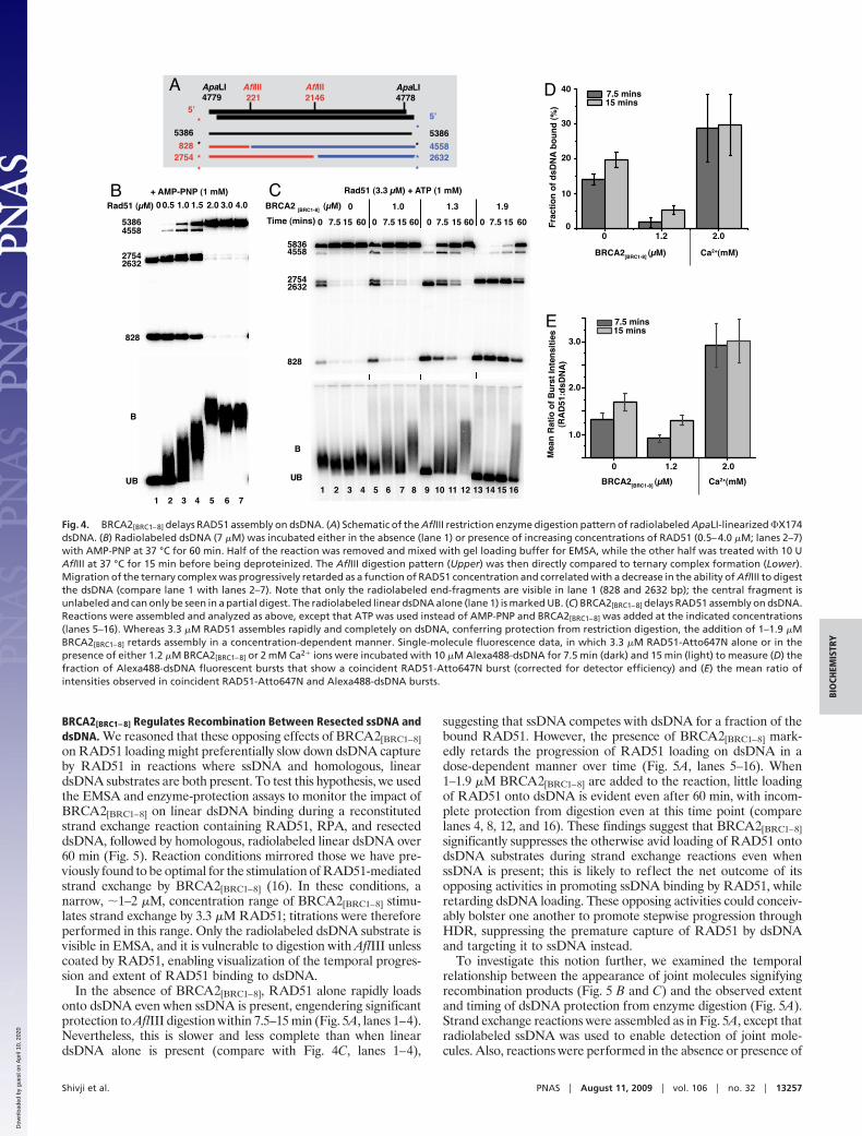
BRCA2[BRC1–8] Regulates Recombination Between Resected ssDNA anddsDNA. We reasoned that these opposing effects of BRCA2[BRC1–8]on RAD51 loading might preferentially slow down dsDNA captureby RAD51 in reactions where ssDNA and homologous, lineardsDNA substrates are both present. To test this hypothesis, we usedthe EMSA and enzyme-protection assays to monitor the impact ofBRCA2[BRC1–8] on linear dsDNA binding during a reconstitutedstrand exchange reaction containing RAD51, RPA, and resecteddsDNA, followed by homologous, radiolabeled linear dsDNA over60 min (Fig. 5). Reaction conditions mirrored those we have pre-viously found to be optimal for the stimulation of RAD51-mediatedstrand exchange by BRCA2[BRC1–8] (16). In these conditions, anarrow, �1–2 �M, concentration range of BRCA2[BRC1–8] stimu-lates strand exchange by 3.3 �M RAD51; titrations were thereforeperformed in this range. Only the radiolabeled dsDNA substrate isvisible in EMSA, and it is vulnerable to digestion with AflIII unlesscoated by RAD51, enabling visualization of the temporal progres-sion and extent of RAD51 binding to dsDNA.
In the absence of BRCA2[BRC1–8], RAD51 alone rapidly loadsonto dsDNA even when ssDNA is present, engendering significantprotection to AflIII digestion within 7.5–15 min (Fig. 5A, lanes 1–4).Nevertheless, this is slower and less complete than when lineardsDNA alone is present (compare with Fig. 4C, lanes 1–4),
suggesting that ssDNA competes with dsDNA for a fraction of thebound RAD51. However, the presence of BRCA2[BRC1–8] mark-edly retards the progression of RAD51 loading on dsDNA in adose-dependent manner over time (Fig. 5A, lanes 5–16). When1–1.9 �M BRCA2[BRC1–8] are added to the reaction, little loadingof RAD51 onto dsDNA is evident even after 60 min, with incom-plete protection from digestion even at this time point (comparelanes 4, 8, 12, and 16). These findings suggest that BRCA2[BRC1–8]significantly suppresses the otherwise avid loading of RAD51 ontodsDNA substrates during strand exchange reactions even whenssDNA is present; this is likely to reflect the net outcome of itsopposing activities in promoting ssDNA binding by RAD51, whileretarding dsDNA loading. These opposing activities could conceiv-ably bolster one another to promote stepwise progression throughHDR, suppressing the premature capture of RAD51 by dsDNAand targeting it to ssDNA instead.
To investigate this notion further, we examined the temporalrelationship between the appearance of joint molecules signifyingrecombination products (Fig. 5 B and C) and the observed extentand timing of dsDNA protection from enzyme digestion (Fig. 5A).Strand exchange reactions were assembled as in Fig. 5A, except thatradiolabeled ssDNA was used to enable detection of joint mole-cules. Also, reactions were performed in the absence or presence of
1 432 5 76
B
UB
+ AMP-PNP (1 mM)
Rad51 (µM) 1.00.50 1.5 4.03.02.0B
53864558
2632
828
2754
Rad51 (3.3 µM) + ATP (1 mM)
1.91.31.00
157.50 60 157.50 60 157.50 60 157.50 60
UB
B
Time (mins)
58364558
27542632
828
BRCA2 [BRC1-8] (µM)C
4321 8765 11109 1312 1514 16
40 7.5 mins15 mins
30
20
10
1.2 2.000F
ract
ion
of
dsD
NA
bo
un
d (
%)
BRCA2[BRC1-8] (µM) Ca2+(mM)
D
7.5 mins15 mins
3.0
2.0
1.0
1.2 2.00
Mea
n R
atio
of
Bu
rst
Inte
nsi
ties
(RA
D51
:dsD
NA
)
BRCA2[BRC1-8] (µM) Ca2+(mM)
E
**
5’5’
221 2146ApaLI4779
ApaLI4778
AflIII
*2754 *828 *
5386*
5386
*2632*4558
AflIIIA
Fig. 4. BRCA2[BRC1–8] delays RAD51 assembly on dsDNA. (A) Schematic of the AflIII restriction enzyme digestion pattern of radiolabeled ApaLI-linearized �X174dsDNA. (B) Radiolabeled dsDNA (7 �M) was incubated either in the absence (lane 1) or presence of increasing concentrations of RAD51 (0.5–4.0 �M; lanes 2–7)with AMP-PNP at 37 °C for 60 min. Half of the reaction was removed and mixed with gel loading buffer for EMSA, while the other half was treated with 10 UAflIII at 37 °C for 15 min before being deproteinized. The AflIII digestion pattern (Upper) was then directly compared to ternary complex formation (Lower).Migration of the ternary complex was progressively retarded as a function of RAD51 concentration and correlated with a decrease in the ability of AflIII to digestthe dsDNA (compare lane 1 with lanes 2–7). Note that only the radiolabeled end-fragments are visible in lane 1 (828 and 2632 bp); the central fragment isunlabeled and can only be seen in a partial digest. The radiolabeled linear dsDNA alone (lane 1) is marked UB. (C) BRCA2[BRC1–8] delays RAD51 assembly on dsDNA.Reactions were assembled and analyzed as above, except that ATP was used instead of AMP-PNP and BRCA2[BRC1–8] was added at the indicated concentrations(lanes 5–16). Whereas 3.3 �M RAD51 assembles rapidly and completely on dsDNA, conferring protection from restriction digestion, the addition of 1–1.9 �MBRCA2[BRC1–8] retards assembly in a concentration-dependent manner. Single-molecule fluorescence data, in which 3.3 �M RAD51-Atto647N alone or in thepresence of either 1.2 �M BRCA2[BRC1–8] or 2 mM Ca2� ions were incubated with 10 �M Alexa488-dsDNA for 7.5 min (dark) and 15 min (light) to measure (D) thefraction of Alexa488-dsDNA fluorescent bursts that show a coincident RAD51-Atto647N burst (corrected for detector efficiency) and (E) the mean ratio ofintensities observed in coincident RAD51-Atto647N and Alexa488-dsDNA bursts.
Shivji et al. PNAS � August 11, 2009 � vol. 106 � no. 32 � 13257
BIO
CHEM
ISTR
Y
Dow
nloa
ded
by g
uest
on
Apr
il 10
, 202
0

1.2 �M BRCA2[BRC1–8], a concentration effective in slowingdsDNA loading by RAD51 (Figs. 4C and 5A), as well as instimulating RAD51-mediated strand exchange under these condi-tions (16). In the absence of BRCA2[BRC1–8], the generation ofrecombination products even after 90 min is low (Fig. 5B, lanes1–5). It is accompanied by extensive and early RAD51 loading ontodsDNA marked by substantial protection from enzyme digestionsome 7.5 min after initiation of the reaction (compare with Fig. 5A,lanes 1–4). In contrast, when BRCA2[BRC1–8] is present, jointmolecule formation is visible some 20 min after initiation (Fig. 5B,lanes 7–10) with increasing product formation over time (Fig. 5C).The appearance of joint molecule products in the presence ofBRCA2[BRC1–8] correlates with the delayed onset and extent ofdsDNA binding by RAD51 marked by the observed changes inenzyme protection (Fig. 5A, lanes 9–12). These findings suggest amodel in which BRCA2 promotes sequential completion of thepresynaptic and synaptic steps of HDR (Fig. 6) by differentiallyregulating RAD51 loading onto ssDNA versus dsDNA, favoringssDNA binding, before capture of the incoming duplex followed bystrand exchange.
Regulation of RAD51-Mediated Strand Exchange by BRCA2. Theformation of a presynaptic filament of RAD51 on ssDNA is aprerequisite for HDR. However, several characteristics of RAD51binding to ssDNA affect both its kinetics and efficiency. Recent
ssDNA
dsDNA -
+
BRCA2[BRC1-8]
RAD51
Efficient strand exchangeEfficient strand exchangeX
Fig. 6. Opposing effects of BRCA2[BRC1–8] on RAD51 binding to ssDNA versusdsDNA together bolster one another to stimulate strand exchange. A hypothet-ical model for the regulation of RAD51-mediated HDR by BRCA2[BRC1–8] is de-picted. It refers to events that occur after the resection of a DSB to generate a3�-tailed ssDNA substrate, but does not show the putative role of the C-terminalssDNA-binding domain of BRCA2 in displacing RPA, and guiding RAD51 to itssubstrate near ssDNA/dsDNA junctions (9). When BRCA2[BRC1–8] is absent (left-hand side), ssDNA and dsDNA compete for RAD51 binding. Strand exchange isinefficient because premature, stable RAD51 assembly on dsDNA is futile.BRCA2[BRC1–8] favors the preferential assembly of RAD51 onto ssDNA because itpromotes RAD51-ssDNA binding while suppressing RAD51-dsDNA binding(right-handside).Theseopposingeffects reinforceoneanothertoguidestepwiseprogression from the presynaptic to the synaptic step of strand exchange.
BRCA2 [BRC1-8] (µM)A 1.0 1.3 1.90
Time (mins)
UB
58364558
27542632
828
157.50 60157.50 60 157.50 60 157.50 60
1 432 5 6 7 1098 11 141312 15 16
B
1 432 5 876 9 10
4 333 2 742 10 19
jm
resecteddsDNA
SE (%)
96 979797 96 909699 88 77resecteddsDNA (%)
32 313234 29 313129 32 27NS (arb. units)
ns
ldsDNA ldsDNA
Time (mins)
BRCA2 [BRC1-8] (µM) 0 1.2
252015 30 90252015 30 90
D
0 20 40 60 80 1000
5
10
15
20+ BRCA2[BRC1-8] ( 1.2 µM)
Time (mins)
% p
rod
uct
C
0 252015 30 605040 90
jm
ldsDNA
Time (mins)
1 432 5 876 9
Fig. 5. BRCA2[BRC1–8] stimulates RAD51-mediated strand exchange while delayingRAD51-dsDNAassembly.Strandexchangereactionsbetween10�MresecteddsDNAand 7 �M homologous dsDNA mediated by 3.3 �M RAD51 were assembled in thepresence or absence of BRCA2[BRC1–8]. (A) The dsDNA substrate was radiolabeled toallowRAD51assemblyondsDNAtobemonitoredviaprotectionfromAflIIIdigestionasdescribedinFig.4.(BandC)TheresecteddsDNAsubstratewaslabeledtoallowthedetection of joint molecule products of recombination. Without BRCA2[BRC1–8] (A,lanes 1–4), rapid protection of the dsDNA by RAD51 is evident between 0–7.5 min;and joint molecule (jm) formation is not detectable (B, lanes 1–5). By contrast, theadditionof1–1.9�MBRCA2[BRC1–8] tothestrandexchangereactionsmarkedlydelaysRAD51assemblyondsDNA(A, lanes5–16).DelayedRAD51assemblyondsDNAby1.3�M BRCA2[BRC1–8] between 15–60 min (A, lanes 9–12) is accompanied by the stimu-lation of jm formation (B, lanes 6–10). (B) A DNA species co-purifying with theresected dsDNA substrate (Fig. S1B) is marked ‘‘ns,’’ whose relative intensity is unal-tered between the lanes, indicating it does not interfere with the reaction. Thebottom panel compares the formation of jm products in the presence or absence ofBRCA2[BRC1–8] as a percentage of the resected dsDNA substrate, with the intensity ofthe ns band expressed in arbitrary units. (C) Shows an extended time-course analysisfor a strand-exchange reaction carried out as in B, in the presence of BRCA2[BRC1–8].(D) Shows the quantitation of jm formation from the data in B and C. Error barsindicate SEM.
13258 � www.pnas.org�cgi�doi�10.1073�pnas.0906208106 Shivji et al.
Dow
nloa
ded
by g
uest
on
Apr
il 10
, 202
0

studies suggest that when ATP is present, this process is driven pre-dominantly by the nucleation of RAD51 onto ssDNA at multiplesites, because binding is not highly cooperative, and dissociativeevents are prominent (24, 25). Thus, RAD51-ssDNA assembliesmay consist of multiple, short protein patches that incompletelycover the DNA and from which RAD51 dynamically dissociates.This is in contrast to the bacterial recombinase, RecA, for which lessfrequent nucleation events on ssDNA can nonetheless engenderextensive filament formation through rapid, cooperative growthfrom the nucleation sites (26). Moreover, unlike RecA, humanRAD51 can bind in vitro with similar affinity to both ssDNA anddsDNA (27–29). Therefore, in vitro strand exchange reactions haveusually been carried out sequentially, with the addition of dsDNAto preformed complexes between ssDNA and RAD51 (30, 31).
In the nuclear milieu, where both ssDNA and dsDNA substratesare generated together in the vicinity of a DSB, these features ofDNA binding by RAD51 could lead to the inefficient coupling ofpresynaptic filament formation on ssDNA, with subsequent reac-tions that involve the capture of dsDNA. Our results show thatBRCA2[BRC1–8] promotes RAD51 binding to ssDNA, while slowingdown its loading onto dsDNA, a competing process that would beinhibitory to strand exchange if performed prematurely. We reasonthat these opposing effects of BRCA2[BRC1–8] on RAD51-DNAbinding are therefore likely to reinforce one another, working to-gether to favor sequential progression through the steps involved instrand exchange (Fig. 6). Thus, the findings we report here identifypreviously unrecognized functions for the multi-BRC repeat regionof human BRCA2 in the regulation of RAD51, which providegeneral insight into the mechanism of HDR in mammalian cells.
Materials and MethodsProtein Expression and Purification. Human BRCA2[BRC1–8], RAD51, and RPAused in this study were prepared as previously described (16). See SI Methodsfor details.
EMSA, Strand Exchange, and Restriction Site Protection Assays. These assayswere assembled as previously described (16). Experimental details are de-scribed in the text, figure legends, and SI Methods.
Negative Staining EM. RAD51 either in the absence or presence of BRCA2[BRC1–8]
(2 �M) was assembled in a reaction mix containing 1 mM ATP. Preincubation for5 min at 37 °C was followed by addition of �X174 circular ssDNA and furtherincubation at 37 °C for 15 min. The samples were diluted 1:10 in Buffer A (seeEMSA Assay). Negatively stained EM grids were prepared according to standardprocedures (see SI Methods). Quantitation of oligomeric RAD51 rings per unitarea in images of RAD51-ssDNA and of BRCA2[BRC1–8]-RAD51-ssDNA is describedin SI Methods.
Two-Color Coincidence Detection. TCCD was carried out using Alexa488-labeled ssDNA or dsDNA substrates and Atto647N-labeled RAD51, whosepreparation is described in SI. Samples, in Buffer B (40 mM Tris, pH 7.8, 1 mMMgCl2, 1 mM ATP), consisted of 3.3 �M RAD51-Atto647N either in the absenceor presence of 1.2 �M BRCA2[BRC1–8], 10 �M Alexa488-ssDNA, or 10 �MAlexa488-dsDNA, were incubated at 37 °C. At indicated times, aliquots wereremoved and immediately flash-frozen to permit multiple samples to beanalyzed. A control experiment was carried out to confirm that there was nodifference in coincidence or relative burst intensities before and after flash-freezing. Samples were thawed individually and diluted in Buffer B to a finalfluorophore concentration of 150 pM onto a cover-glass precoated with BSAfor immediate analysis by TCCD (32). The procedure used for TCCD is furtherdescribed in SI. Positive controls were carried out similarly, in the absence ofBRCA2[BRC1–8] but with the addition of 2 mM calcium chloride to Buffer B.
ACKNOWLEDGMENTS. We thank Luca Pellegrini and Owen Davies (Universityof Cambridge, Cambridge, U.K.) for providing BRCA2[BRC1–8] protein, AngelOrte for assistance with TCCD, John Finch for assistance with EM, and RichardHenderson for comments on the manuscript. S.R.M. was supported by astudentship from the Engineering and Physical Sciences Research Council, andE.R. and J.S. were supported by studentships from the Medical ResearchCouncil, which also funded this work in A.R.V.’s laboratory.
1. Li X, Heyer WD (2008) Homologous recombination in DNA repair and DNA damagetolerance. Cell Res 18:99–113.
2. San Filippo J, Sung P, Klein H (2008) Mechanism of eukaryotic homologous recombi-nation. Annu Rev Biochem 77:229–257.
3. Moynahan ME, Pierce AJ, Jasin M (2001) BRCA2 is required for homology-directedrepair of chromosomal breaks. Mol Cell 7:263–272.
4. Thorslund T, West SC (2007) BRCA2: A universal recombinase regulator. Oncogene26:7720–7730.
5. Wong AK, Pero R, Ormonde PA, Tavtigian SV, Bartel PL (1997) RAD51 interacts with theevolutionarily conserved BRC motifs in the human breast cancer susceptibility genebrca2. J Biol Chem 272:31941–31944.
6. Sharan SK, et al. (1997) Embryonic lethality and radiation hypersensitivity mediated byRad51 in mice lacking Brca2. Nature 386:804–810.
7. Lo T, Pellegrini L, Venkitaraman AR, Blundell TL (2003) Sequence fingerprints in BRCA2and RAD51: Implications for DNA repair and cancer. DNA Repair (Amst) 2:1015–1028.
8. Yang H, et al. (2002) BRCA2 function in DNA binding and recombination from aBRCA2-DSS1-ssDNA structure. Science 297:1837–1848.
9. Yang H, Li Q, Fan J, Holloman WK, Pavletich NP (2005) The BRCA2 homologue Brh2nucleates RAD51 filament formation at a dsDNA-ssDNA junction. Nature 433:653–657.
10. Saeki H, et al. (2006) Suppression of the DNA repair defects of BRCA2-deficient cellswith heterologous protein fusions. Proc Natl Acad Sci USA 103:8768–8773.
11. San Filippo J, et al. (2006) Recombination mediator and Rad51 targeting activities ofa human BRCA2 polypeptide. J Biol Chem 281:11649–11657.
12. Pellegrini L, et al. (2002) Insights into DNA recombination from the structure of aRAD51-BRCA2 complex. Nature 420:287–293.
13. Davies AA, et al. (2001) Role of BRCA2 in control of the RAD51 recombination and DNArepair protein. Mol Cell 7:273–282.
14. Chen CF, Chen PL, Zhong Q, Sharp ZD, Lee WH (1999) Expression of BRC repeats inbreast cancer cells disrupts the BRCA2-Rad51 complex and leads to radiation hyper-sensitivity and loss of G(2)/M checkpoint control. J Biol Chem 274:32931–32935.
15. Galkin VE, et al. (2005) BRCA2 BRC motifs bind RAD51-DNA filaments. Proc Natl AcadSci USA 102:8537–8542.
16. Shivji MK, et al. (2006) A region of human BRCA2 containing multiple BRC repeatspromotes RAD51-mediated strand exchange. Nucleic Acids Res 34:4000–4011.
17. Carreira A, et al. (2009) The BRC repeats of BRCA2 modulate the DNA bindingselectivity of RAD51. Cell 136:1032–1043.
18. Sung P (1994) Catalysis of ATP-dependent homologous DNA pairing and strand ex-change by yeast RAD51 protein. Science 265:1241–1243.
19. Baumann P, Benson FE, West SC (1996) Human Rad51 protein promotes ATP-dependent homologous pairing and strand transfer reactions in vitro. Cell 87:757–766.
20. Petalcorin MI, Galkin VE, Yu X, Egelman EH, Boulton SJ (2007) Stabilization of RAD-51-DNA filaments via an interaction domain in Caenorhabditis elegans BRCA2. ProcNatl Acad Sci USA 104:8299–8304.
21. Li H, Ying L, Green JJ, Balasubramanian S, Klenerman D (2003) Ultrasensitive coinci-dence fluorescence detection of single DNA molecules. Anal Chem 75:1664–1670.
22. van der Heijden T, et al. (2007) Real-time assembly and disassembly of human RAD51filaments on individual DNA molecules. Nucleic Acids Res 35:5646–5657.
23. Chi P, Van Komen S, Sehorn MG, Sigurdsson S, Sung P (2006) Roles of ATP bindingand ATP hydrolysis in human Rad51 recombinase function. DNA Repair (Amst)5:381–391.
24. Ristic D, et al. (2005) Human Rad51 filaments on double- and single-stranded DNA:Correlating regular and irregular forms with recombination function. Nucleic Acids Res33:3292–3302.
25. Mine J, et al. (2007) Real-time measurements of the nucleation, growth and dissocia-tion of single Rad51-DNA nucleoprotein filaments. Nucleic Acids Res 35:7171–7187.
26. Galletto R, Amitani I, Baskin RJ, Kowalczykowski SC (2006) Direct observation ofindividual RecA filaments assembling on single DNA molecules. Nature 443:875–878.
27. Sung P, Robberson DL (1995) DNA strand exchange mediated by a RAD51-ssDNAnucleoprotein filament with polarity opposite to that of RecA. Cell 82:453–461.
28. Mazin AV, Zaitseva E, Sung P, Kowalczykowski SC (2000) Tailed duplex DNA is thepreferred substrate for Rad51 protein-mediated homologous pairing. EMBO J19:1148–1156.
29. Benson FE, Stasiak A, West SC (1994) Purification and characterization of the humanRad51 protein, an analogue of E. coli RecA. EMBO J 13:5764–5771.
30. Sigurdsson S, Trujillo K, Song B, Stratton S, Sung P (2001) Basis for avid homologousDNA strand exchange by human Rad51 and RPA. J Biol Chem 276:8798–8806.
31. Liu Y, et al. (2004) Conformational changes modulate the activity of human RAD51protein. J Mol Biol 337:817–827.
32. Orte A, et al. (2008) Direct characterization of amyloidogenic oligomers by single-molecule fluorescence. Proc Natl Acad Sci USA 105:14424–14429.
Shivji et al. PNAS � August 11, 2009 � vol. 106 � no. 32 � 13259
BIO
CHEM
ISTR
Y
Dow
nloa
ded
by g
uest
on
Apr
il 10
, 202
0