Embed Size (px)
Citation preview
Chapter 3
A map of the brain

Chapter 3 A map of the brain26
In this chapter:
Mini-atlas of the rat brain 26
>>
3 A map of the brain
Mini-atlas of the rat brainWhen the brain is cut into thin sections, these sections can be stained to show the nuclei and fiber
tracts. A series of sections forms an atlas, which shows us the position of the main structures inside
the brain. This section of the book is a set of simplified drawings of brain sections (a mini-atlas) to
assist those who are less familiar with the detailed anatomy of parts of the brain. The mini-atlas
contains twelve diagrams of coronal sections from one end of the rat brain to the other. The major
regions of the brain and some important nuclei and tracts are labeled. To assist your understanding
of these simplified sections, each drawing is accompanied by a short description of the main
features. Following the twelve coronal diagrams are diagrams of two sagittal sections of the brain.
These latter sections show some areas of the brain to advantage, and are particularly useful for
understanding the relationships between diencephalic, midbrain, and hindbrain regions.
The brain is made up of three main parts, the forebrain, the midbrain (also called mesencephalon),
and the hindbrain. The forebrain is divided into the cerebral cortex, the subpallium, the
hypothalamus, and the diencephalon. Each of these main subdivisions is labeled in Figure 3.1b.
The midbrain joins the hindbrain to the forebrain. The hindbrain has three major parts, the
isthmus, the rhombencephalon, and the cerebellum. The end of the rhombencephalon joins with
the spinal cord.
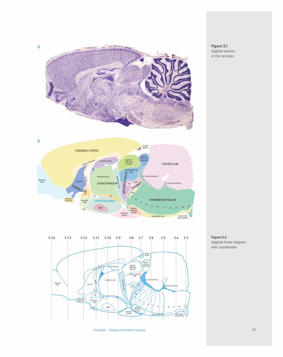
Figure 3.1
The upper image (a) is photograph of a Nissl-stained sagittal section of a rat brain. The section is
close to the midline. The dark folds of the cerebellum are seen on the right, and the dark blue
stripe on the far left is a part of the olfactory bulb. The dark diamond-shaped patch ventral to the
middle of the brain is the pituitary gland.
The lower image (b) is a color-coded diagram of the photograph seen in (a). The major parts of the
brain are labeled. Each of the structures shown here will be discussed in the remaining pages of
this chapter.
Figure 3.2
This diagram of a sagittal section of the rat brain shows the position of the coronal section
diagrams which form an atlas series in this chapter (Figures 3.3 to 3.14). Each vertical line shows
the position of the relevant coronal section. This diagram should be consulted when each of the
coronal diagrams is studied.
CEREBELLUM
DIENCEPHALON
hippocampus
inferiorcolliculus(auditory)superior
colliculus(visual)
periaqueductalgray
corpus callosum
thalamus (p2) fourth ventricle
fourth ventricle
CEREBRAL CORTEX
olfactorybulb
3N4N
scp
pyramidal tract pyramidaldecussation
pituitarygland
basilarpontinenuclei
RHOMBENCEPHALON
pinealgland
VMH
p3
HYPOTHALAMUS
septum
fornix
3V
PaST
ac
preopticarea
och
pretectum (p1) ISTHMUS
SUBPALLIUM MES
ENCE
PHAL
ON
olfactorytubercle
r1 r2 r3 r4 r5 r6 r7 r8 r9 r10 r11
The Brain Watson Kirkcaldie Paxinos 27
Figure 3.1Sagittal section
of the rat brain
hippocampus
inferiorcolliculus(auditory)superior
colliculus(visual)
periaqueductalgray
corpus callosum
thalamus (p2) fourth ventricle
fourth ventricle
olfactorybulb
3.14 3.13 3.12 3.33.43.53.63.73.83.93.103.11
3N4N
scp
pyramidal tract pyramidaldecussation
pituitarygland
basilarpontinenuclei
pinealgland
VMH
p3
septum
fornix
3V
PaST
ac
preopticarea
och
pretectum (p1)
olfactorytubercle
r1 r2 r3 r4 r5 r6 r7 r8 r9 r10 r11
r1
Figure 3.2Sagittal finder diagram
with coordinates
a
b
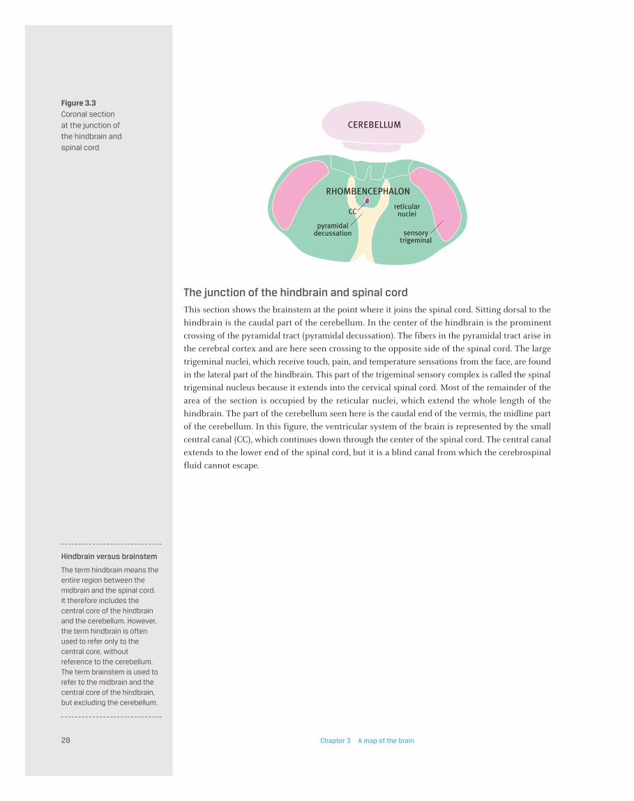
Figure 3.3Coronal section
at the junction of
the hindbrain and
spinal cord
The junction of the hindbrain and spinal cord
This section shows the brainstem at the point where it joins the spinal cord. Sitting dorsal to the
hindbrain is the caudal part of the cerebellum. In the center of the hindbrain is the prominent
crossing of the pyramidal tract (pyramidal decussation). The fibers in the pyramidal tract arise in
the cerebral cortex and are here seen crossing to the opposite side of the spinal cord. The large
trigeminal nuclei, which receive touch, pain, and temperature sensations from the face, are found
in the lateral part of the hindbrain. This part of the trigeminal sensory complex is called the spinal
trigeminal nucleus because it extends into the cervical spinal cord. Most of the remainder of the
area of the section is occupied by the reticular nuclei, which extend the whole length of the
hindbrain. The part of the cerebellum seen here is the caudal end of the vermis, the midline part
of the cerebellum. In this figure, the ventricular system of the brain is represented by the small
central canal (CC), which continues down through the center of the spinal cord. The central canal
extends to the lower end of the spinal cord, but it is a blind canal from which the cerebrospinal
fluid cannot escape.
Chapter 3 A map of the brain28
RHOMBENCEPHALON
reticularnuclei
CEREBELLUM
sensorytrigeminal
pyramidaldecussation
CC
Hindbrain versus brainstem
The term hindbrain means theentire region between themidbrain and the spinal cord.It therefore includes thecentral core of the hindbrainand the cerebellum. However,the term hindbrain is oftenused to refer only to thecentral core, withoutreference to the cerebellum.The term brainstem is used torefer to the midbrain and thecentral core of the hindbrain,but excluding the cerebellum.

Figure 3.4Coronal section of the
hindbrain at the level
of the inferior olive
The hindbrain at the level of the inferior olive
This section shows the brainstem below and the cerebellum above with the fourth ventricle in
between. This part of the hindbrain that lies under the cerebellum is the rhombencephalon.
The large trigeminal nuclei are found in the lateral part of the hindbrain. This part of the
trigeminal sensory complex is called the spinal trigeminal nucleus. The two hypoglossal nuclei
(which control the tongue) are next to the midline under the fourth ventricle. Lateral to the
hypoglossal nucleus is a cluster of cell groups called the solitary nucleus. This cluster receives taste
sensation and sensory information from internal organs such as the stomach and the lung. Two
large fiber bundles, the pyramids, lie on either side of the midline on the ventral margin of the
hindbrain. The fibers in the pyramids arise in the cerebral cortex and travel down to cross to the
opposite side of the spinal cord. Next to each pyramid is the inferior olive, which is functionally
connected with the cerebellum. Most of the remainder of the area of this section is occupied by
the reticular nuclei, which extend for the whole length of the hindbrain. They are involved in
basic sensory and motor functions.
The fourth ventricle contains cerebrospinal fluid (CSF), which has flowed down from its origin in
the lateral (cerebral) ventricles through the third ventricle and aqueduct to reach the hindbrain.
The CSF escapes from the roof of the fourth ventricle to fill the subarachnoid space.
The cerebellum is a large structure concerned with coordination of movement. It consists of an
outer layer of cerebellar cortex and a core of white matter (fibers).
The Brain Watson Kirkcaldie Paxinos 29
reticularnuclei
solitary nucleus
CEREBELLUM
inferiorolive
sensorytrigeminal
pyramidaltract
12N
4V
RHOMBENCEPHALON
Rhombencephalon
The rhombencephalon, asection of the neural tube, isthe embryonic precursor ofthe hindbrain.
Pyramids
The pyramids are largebundles of corticospinal axonson the ventral surface of thehindbrain next to the midline.

Figure 3.5Coronal section of the
hindbrain at the level
of the facial nucleus
The hindbrain at the level of the facial nucleus
This section shows the hindbrain below and the cerebellum above with the fourth ventricle in
between. The part of the hindbrain that lies under the cerebellum is called the rhombencephalon.
The large trigeminal nuclei are found in the lateral part of the hindbrain at this level. This part of
the trigeminal sensory complex is called the spinal trigeminal nucleus because it extends into the
cervical spinal cord. Two large fiber bundles, the pyramids, lie on either side of the midline on the
ventral margin of the hindbrain. The pyramids contain the pyramidal (corticospinal) tracts.
Between each pyramid and the trigeminal nucleus is a large group of motor neurons that supplies
the muscles of facial expression on that side, called the facial nucleus. The vestibular nuclei lie at
the junction of cerebellum and hindbrain at the side of the fourth ventricle. They receive
information from the position sense organs of the inner ear. At the lateral edge of the hindbrain
under the cerebellum are the cochlear nuclei, which receive auditory sensations from the inner
ear. Most of the remainder of the section is occupied by the reticular nuclei, which extend for the
whole length of the hindbrain.
The cerebellum consists of an outer layer of cerebellar cortex and a core of white matter (fibers).
At this level, some cell groups can be seen in the deep cerebellar white matter dorsal to the
ventricle and the vestibular nuclei. These cell groups are the cerebellar nuclei. They receive input
from the cerebellar cortex and send fibers to the brainstem and thalamus.
The fourth ventricle has lateral extensions at this level. They can be seen next to the cochlear
nuclei.
Chapter 3 A map of the brain30
reticularnuclei
CEREBELLUM
sensory trigeminal cochlearnuclei
pyramidaltract
facialnucleus
4V4V
icpvestibularnuclei
deep cerebellarnuclei
RHOMBENCEPHALON
The fourth ventricle
The fourth ventricle is a tent-shaped space locatedbetween the core of thehindbrain and the cerebellum.The roof of the fourth ventriclehas three holes that allowcerebrospinal fluid to escapeinto the subarachnoid space.If these holes are blocked, thefluid pressure in the ventriclesrises, causing expansion thatcan kill brain cells. This iscalled hydrocephalus.
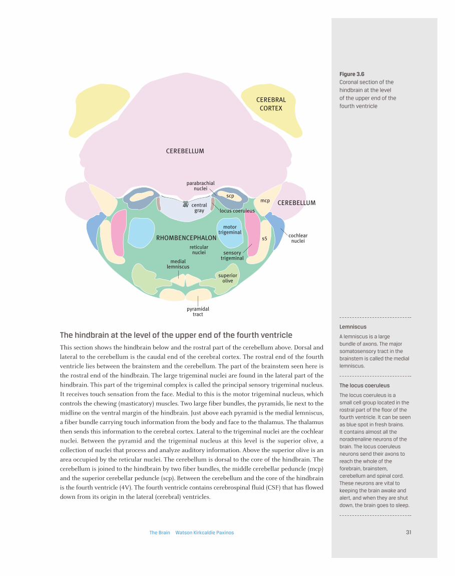
Figure 3.6Coronal section of the
hindbrain at the level
of the upper end of the
fourth ventricle
The hindbrain at the level of the upper end of the fourth ventricle
This section shows the hindbrain below and the rostral part of the cerebellum above. Dorsal and
lateral to the cerebellum is the caudal end of the cerebral cortex. The rostral end of the fourth
ventricle lies between the brainstem and the cerebellum. The part of the brainstem seen here is
the rostral end of the hindbrain. The large trigeminal nuclei are found in the lateral part of the
hindbrain. This part of the trigeminal complex is called the principal sensory trigeminal nucleus.
It receives touch sensation from the face. Medial to this is the motor trigeminal nucleus, which
controls the chewing (masticatory) muscles. Two large fiber bundles, the pyramids, lie next to the
midline on the ventral margin of the hindbrain. Just above each pyramid is the medial lemniscus,
a fiber bundle carrying touch information from the body and face to the thalamus. The thalamus
then sends this information to the cerebral cortex. Lateral to the trigeminal nuclei are the cochlear
nuclei. Between the pyramid and the trigeminal nucleus at this level is the superior olive, a
collection of nuclei that process and analyze auditory information. Above the superior olive is an
area occupied by the reticular nuclei. The cerebellum is dorsal to the core of the hindbrain. The
cerebellum is joined to the hindbrain by two fiber bundles, the middle cerebellar peduncle (mcp)
and the superior cerebellar peduncle (scp). Between the cerebellum and the core of the hindbrain
is the fourth ventricle (4V). The fourth ventricle contains cerebrospinal fluid (CSF) that has flowed
down from its origin in the lateral (cerebral) ventricles.
The Brain Watson Kirkcaldie Paxinos 31
mediallemniscus
centralgray
4V
s5
mcpscp
reticularnuclei
CEREBELLUM
CEREBELLUM
CEREBRALCORTEX
superiorolive
motortrigeminal
parabrachialnuclei
sensorytrigeminal
cochlearnuclei
pyramidaltract
locus coeruleus
RHOMBENCEPHALON
Lemniscus
A lemniscus is a large bundle of axons. The majorsomatosensory tract in thebrainstem is called the mediallemniscus.
The locus coeruleus
The locus coeruleus is a small cell group located in therostral part of the floor of thefourth ventricle. It can be seenas blue spot in fresh brains. It contains almost all thenoradrenaline neurons of thebrain. The locus coeruleusneurons send their axons toreach the whole of theforebrain, brainstem,cerebellum and spinal cord.These neurons are vital tokeeping the brain awake andalert, and when they are shutdown, the brain goes to sleep.
CEREBRAL CORTEXvisualcortex
ISTHMUS
medial lemniscus
subiculum
entorhinalcortexm5
s5mcp
scpreticular
nuclei
4N
pineal gland superiorcolliculus(visual)
inferiorcolliculus(auditory)
raphenuclei
aqueduct
basilarpontinenuclei
periaqueductalgray
RHOMBENCEPHALON
MESENCEPHALON
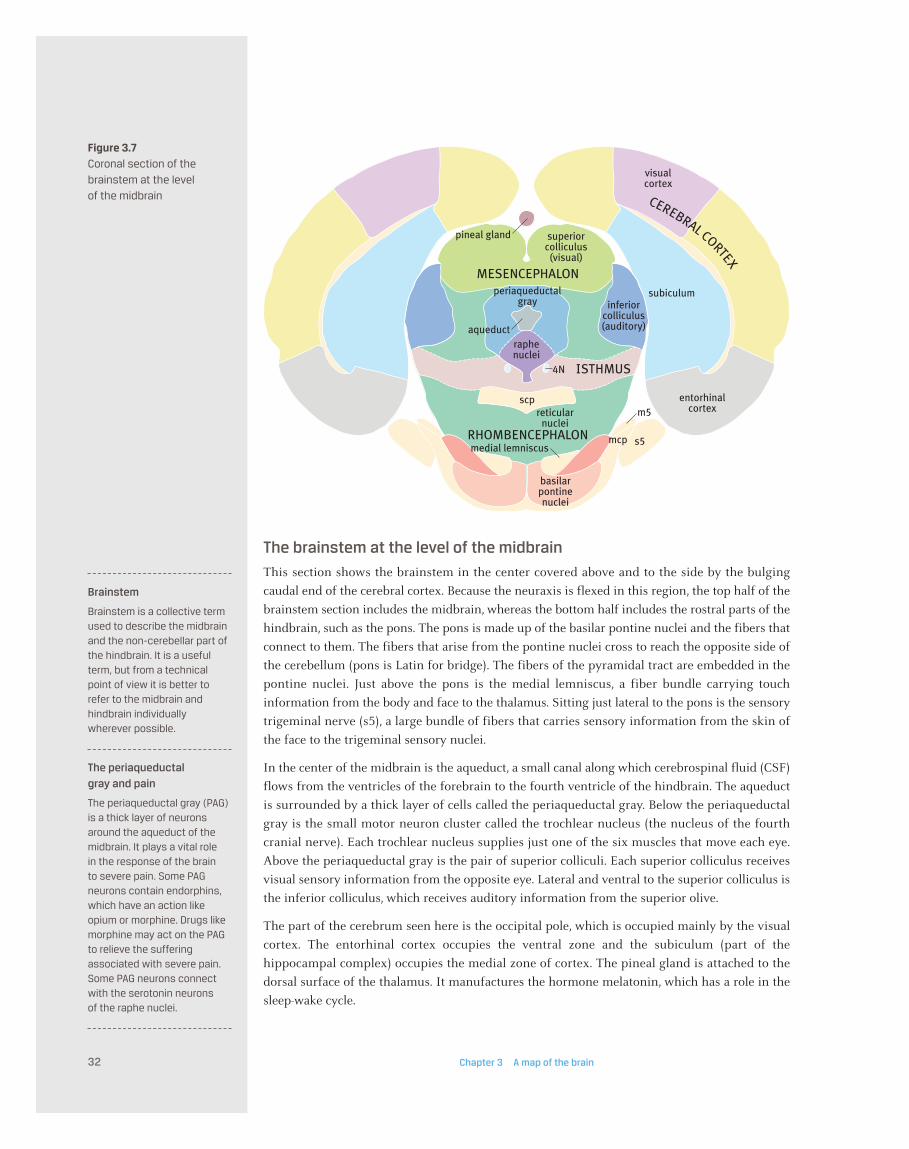
Figure 3.7Coronal section of the
brainstem at the level
of the midbrain
Chapter 3 A map of the brain32
The brainstem at the level of the midbrain
This section shows the brainstem in the center covered above and to the side by the bulging
caudal end of the cerebral cortex. Because the neuraxis is flexed in this region, the top half of the
brainstem section includes the midbrain, whereas the bottom half includes the rostral parts of the
hindbrain, such as the pons. The pons is made up of the basilar pontine nuclei and the fibers that
connect to them. The fibers that arise from the pontine nuclei cross to reach the opposite side of
the cerebellum (pons is Latin for bridge). The fibers of the pyramidal tract are embedded in the
pontine nuclei. Just above the pons is the medial lemniscus, a fiber bundle carrying touch
information from the body and face to the thalamus. Sitting just lateral to the pons is the sensory
trigeminal nerve (s5), a large bundle of fibers that carries sensory information from the skin of
the face to the trigeminal sensory nuclei.
In the center of the midbrain is the aqueduct, a small canal along which cerebrospinal fluid (CSF)
flows from the ventricles of the forebrain to the fourth ventricle of the hindbrain. The aqueduct
is surrounded by a thick layer of cells called the periaqueductal gray. Below the periaqueductal
gray is the small motor neuron cluster called the trochlear nucleus (the nucleus of the fourth
cranial nerve). Each trochlear nucleus supplies just one of the six muscles that move each eye.
Above the periaqueductal gray is the pair of superior colliculi. Each superior colliculus receives
visual sensory information from the opposite eye. Lateral and ventral to the superior colliculus is
the inferior colliculus, which receives auditory information from the superior olive.
The part of the cerebrum seen here is the occipital pole, which is occupied mainly by the visual
cortex. The entorhinal cortex occupies the ventral zone and the subiculum (part of the
hippocampal complex) occupies the medial zone of cortex. The pineal gland is attached to the
dorsal surface of the thalamus. It manufactures the hormone melatonin, which has a role in the
sleep-wake cycle.
Brainstem
Brainstem is a collective termused to describe the midbrainand the non-cerebellar part ofthe hindbrain. It is a usefulterm, but from a technicalpoint of view it is better torefer to the midbrain andhindbrain individuallywherever possible.
The periaqueductal
gray and pain
The periaqueductal gray (PAG)is a thick layer of neuronsaround the aqueduct of themidbrain. It plays a vital role in the response of the brain to severe pain. Some PAGneurons contain endorphins,which have an action likeopium or morphine. Drugs likemorphine may act on the PAGto relieve the sufferingassociated with severe pain.Some PAG neurons connectwith the serotonin neurons of the raphe nuclei.

CEREBRAL CORTEX
MESENCEPHALON
visualcortex
auditorycortex
subiculumsuperior
colliculus(visual) hippocam
pus
3N
VTA
medial lemniscus
reticularnuclei
red nucleus
periaqueductalgray dentate
gyrus
subiculum
entorhinalcortex
cerebralpeduncle
substantia n
igra
medialgeniculate
auditorythalamus
Figure 3.8Coronal section at
the rostral end of
the midbrain
The Brain Watson Kirkcaldie Paxinos 33
The rostral end of the midbrain
This section shows the midbrain in the center covered above and to the side by the bulging caudal
end of the cerebral cortex. The superior colliculus occupies the dorsal part of the midbrain, but
the lateral part contains the most caudal part of the thalamus–the medial geniculate nucleus,
which is the auditory thalamic nucleus.
At this level, the pyramidal (corticospinal) fibers and other descending fibers form a bundle called
the cerebral peduncle on the ventral aspect of the midbrain. Coating the inside (medial) border of
the fibers of the cerebral peduncle is the substantia nigra, a large cell group that is important for
motor control. Fibers from the substantia nigra project to the deep nuclei of the cerebrum,
especially the striatum. Just above the medial end of the substantia nigra is the medial lemniscus,
a fiber bundle carrying touch information from the body and face to the thalamus. On the medial
side of the medial lemniscus is another motor area, the ventral tegmental area (VTA).
Surrounding the aqueduct is a thick layer of neurons called the periaqueductal gray. Below the
periaqueductal gray is a prominent pair of motor nuclei called the oculomotor nuclei. Each
oculomotor nucleus (3N) supplies four of the six muscles that move each eye. In addition, the
oculomotor nerve supplies the main muscle of the upper eyelid and the muscles of the pupil and
lens. Between the periaqueductal gray and the medial lemniscus is the red nucleus. This large
group of cells gives rise to a fiber bundle that travels down the spinal cord, called the rubrospinal
tract. Between the two cerebral peduncles at the ventral margin is the interpeduncular nucleus.
The area between the red nucleus and the superior colliculus is filled with reticular nuclei.
Attached to the lateral edge of the midbrain is the medial geniculate nucleus of the thalamus.
The part of the cerebrum seen here contains the visual cortex above and the auditory cortex
laterally. The ventral part of the cortex is occupied by the entorhinal cortex, and the medial part
is occupied by the hippocampal structures (hippocampus, dentate gyrus, and subiculum).
Superior colliculus
The superior colliculus is a raised area of the dorsalsurface of the midbraininvolved in visual reflexes and attention.
Substantia nigra
The substantia nigra is a cellgroup in the ventral part of thebrainstem that is important inmotor control. Many cells inthe substantia nigra containdopamine, and give rise to adopaminergic pathway whichprojects to the striatum.
Entorhinal cortex
The entorhinal cortex is anarea of cortex in the temporallobe close to the hippo -campus that encodes aspatial map of the externalsurroundings for use by thehippocampus.

Figure 3.9Coronal section of the
forebrain at the level
of the caudal end of
the thalamus
The forebrain at the level of the caudal end of the thalamus
This section shows the thalamus and hypothalamus in the center covered above and to the side
by the cerebrum. In the midline is the third ventricle, which links the lateral ventricles to the
aqueduct.
The thalamus (prosomere 2 of the diencephalon) lies dorsal to the hypothalamus. The thalamus
receives input from touch, auditory, and visual pathways, and sends fibers to the cerebral cortex.
The part of the thalamus that receives sensory information from the eyes is called the dorsal
lateral geniculate nucleus (DLG). The dorsal lateral geniculate connects with the visual cortex of
the cerebrum. The somatosensory (touch sensation) nuclei of the thalamus (VPL and VPM) lie
ventral to the DLG. On the medial edge of thalamus is part of the pretectal area (prosomere 1 of
the diencephalon), and the lateral edge is the prethalamus (prosomere 3).
The hypothalamus is located ventral to the thalamus. At this level of the hypothalamus, there is a
pair of bulges called the mammillary bodies. At the lateral edge of the hypothalamus is the
cerebral peduncle. The cerebral peduncle contains the pyramidal (corticospinal) fibers as well as
the fibers traveling from the cortex to the pons.
The part of cerebral cortex seen here is about midway between occipital and frontal poles of the
cerebrum. The most obvious cortical areas at this level are the somatosensory cortex above, the
auditory cortex laterally, and the piriform (olfactory) cortex below. Above the piriform cortex is
the rostral end of the entorhinal cortex. Medial to the piriform cortex is a large group of nuclei
called the amygdala. The amygdala is associated with emotional responses and a number of
survival behaviors Medial to the lateral ventricle is the hippocampus, an easily recognizable part
of cerebral cortex involved with memory registration. The corpus callosum is a large sheet of
fibers connecting the left and right sides of the cerebrum.
Chapter 3 A map of the brain34
piriformcortex
CEREBRAL CORTEX
corpus callo
sum
amygdala
thalamus(p2)
hippocampus
hippocampus
3V
somatosensorycortex
HYPOTHALAMUS
entorhinalcortex
auditorycortex
DIENCEPHALON
cere
bal peduncle
mammillaryregion of
hypothalamus
pretectum(p1)
prethalamus (
p3)
visualthalamus
(DLG)
VPM &VPL
somatosensorythalamus
3V
lateralventricle
Prosomere
A prosomere is a segmentalcomponent of the developingdiencephalon. The threeprosomeres are the pretectum(prosomere 1), the thalamus(prosomere 2), and theprethalamus (prosomere 3).
Thalamus
The thalamus is the major part of the diencephalon; itreceives input from touch,auditory, and visual pathways(among others), and sendsfibers to the cerebral cortex.
Hypothalamus
The hypothalamus is a majorsubdivision of the forebrainlocated ventral to thethalamus. It is responsible for the initiation of survivalbehaviors and controls theautonomic andneuroendocrine systems.
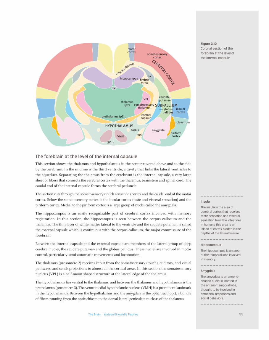
Figure 3.10Coronal section of the
forebrain at the level of
the internal capsule
The forebrain at the level of the internal capsule
This section shows the thalamus and hypothalamus in the center covered above and to the side
by the cerebrum. In the midline is the third ventricle, a cavity that links the lateral ventricles to
the aqueduct. Separating the thalamus from the cerebrum is the internal capsule, a very large
sheet of fibers that connects the cerebral cortex with the thalamus, brainstem and spinal cord. The
caudal end of the internal capsule forms the cerebral peduncle.
The section cuts through the somatosensory (touch sensation) cortex and the caudal end of the motor
cortex. Below the somatosensory cortex is the insular cortex (taste and visceral sensation) and the
piriform cortex. Medial to the piriform cortex is a large group of nuclei called the amygdala.
The hippocampus is an easily recognizable part of cerebral cortex involved with memory
registration. In this section, the hippocampus is seen between the corpus callosum and the
thalamus. The thin layer of white matter lateral to the ventricle and the caudate-putamen is called
the external capsule which is continuous with the corpus callosum, the major commissure of the
forebrain.
Between the internal capsule and the external capsule are members of the lateral group of deep
cerebral nuclei, the caudate-putamen and the globus pallidus. These nuclei are involved in motor
control, particularly semi-automatic movements and locomotion.
The thalamus (prosomere 2) receives input from the somatosensory (touch), auditory, and visual
pathways, and sends projections to almost all the cortical areas. In this section, the somatosensory
nucleus (VPL) is a half-moon shaped structure at the lateral edge of the thalamus.
The hypothalamus lies ventral to the thalamus, and between the thalamus and hypothalamus is the
prethalamus (prosomere 3). The ventromedial hypothalamic nucleus (VMH) is a prominent landmark
in the hypothalamus. Between the hypothalamus and the amygdala is the optic tract (opt), a bundle
of fibers running from the optic chiasm to the dorsal lateral geniculate nucleus of the thalamus.
The Brain Watson Kirkcaldie Paxinos 35
caudate-putamen
piriformcortex
insularcortex
motorcortex
CEREBRAL CORTEX
corpus callo
sum
globuspallidus
amygdala
thalamus(p2)
hippocampus fimbriafornix
LV
claustrum
optVMH
3V
3V
internalcapsule
VPL
somatosensorythalamus
somatosensorycortex
prethalamus (p3)
HYPOTHALAMUS
SUBPALLIUM
fornix
Insula
The insula is the area ofcerebral cortex that receivestaste sensation and visceralsensation from the intestines.In humans this area is anisland of cortex hidden in thedepths of the lateral fissure.
Hippocampus
The hippocampus is an area of the temporal lobe involvedin memory.
Amygdala
The amygdala is an almond-shaped nucleus located in the anterior temporal lobe,thought to be involved inemotional responses andsocial behaviors.

Figure 3.11Coronal section of
the forebrain at the
level of the anterior
commissure
The forebrain at the level of the anterior commissure
This section is taken immediately in front of the rostral pole of the thalamus. In the center of the
section is the anterior commissure, with the septum above, the preoptic area below, and the bed
nucleus of the stria terminalis around its lateral edge. In the midline of the preoptic area is the
third ventricle, which links the lateral ventricles to the cerebral aqueduct. Immediately below the
third ventricle is the optic chiasm, in which optic nerve fibers cross the midline.
The areas of cerebral cortex seen in this section are the somatosensory cortex and the motor
cortex. Below the somatosensory cortex is the insular cortex (taste and visceral sensation) and the
piriform cortex. On the surface of the piriform cortex is the olfactory tract, which connects the
olfactory bulb to the piriform cortex. Medial to the piriform cortex is the rostral part of the
amygdala. The cingulate cortex is seen on the medial side of the cerebrum above the corpus
callosum.
Medial to the external capsule (ec) lie representatives of the lateral group of deep cerebral nuclei,
the caudate-putamen and the globus pallidus. Along the medial border of the globus pallidus is
the rostral part of the internal capsule (ic). The internal capsule is a large sheet of fibers which
connects the cerebrum with the thalamus, brainstem, and spinal cord.
The thin layer of white matter lateral to the caudate-putamen is called the external capsule. The
lateral ventricle lies between the septum and the caudate-putamen in this section. The anterior
commissure (also labeled ac) is a large bundle of crossing fibers, which connects the olfactory bulb
and parts of the cerebrum to the same areas on the opposite side.
Chapter 3 A map of the brain36
caudate-putamen
SUBPALLIUM
lateralolfactory
tract
piriformcortex
cingulatecortex
insularcortex
motorcortex
CEREBRAL CORTEX
claus
trum
somatosensorycortex
anterior commissure
septum
olfactorytubercle
lateralventricle
ic
ec
ac
corpus callosum
preopticarea
optic chiasm
globuspallidus
ventralpallidum
amygdala
bed nucleus ofstria terminalis
Somatosensory cortex
The somatosensory cortex isa granular area of the cortexinvolved with processing oftouch and proprioceptiveinformation. In humans this is found in the post-centralgyrus.
Premotor cortex
The premotor cortex is thecortical area involved inplanning of movement. It islocated in the frontal lobe infront of the primary motorcortex.
Globus pallidus
The globus pallidus is themajor part of the pallidum. It receives input from thestriatum and projects mainlyto the thalamus.
Striatum and pallidum
The largest of the deepcerebral nuclei are thestriatum and pallidum. Theparts of the striatum are thecaudate, putamen, andaccumbens nucleus. Theparts of the pallidum are theglobus pallidus and the ventralpallidum. Both striatum andpallidum contain GABAneurons which influencemotor functions. The striatumreceives major dopaminepathways from the substantianigra and the ventraltegmental area.

Figure 3.12Coronal section of the
forebrain at the rostral end
of the corpus callosum
The forebrain at the rostral end of the corpus callosum
This section passes through the rostral end of the corpus callosum. The section cuts through the
rostral end of the somatosensory cortex and the middle of the motor cortex. Medial to the motor
cortex is the cingulate cortex. Below the somatosensory cortex are the insular cortex (taste and
visceral sensation) and the piriform cortex. On the surface of the piriform cortex is the lateral
olfactory tract which connects the olfactory bulb to the piriform cortex. Medial to the piriform
cortex is the olfactory tubercle, which also receives fibers from the olfactory bulb.
The layer of white matter lateral and dorsal to the caudate-putamen is called the external capsule.
Ventral to the corpus callosum is a large group of nuclei called the septum, which is the largest of
the medial group of deep cerebral nuclei. Medial to the external capsule lies the lateral ventricle
and the caudate-putamen. The caudate-putamen is the largest of the lateral group of deep cerebral
nuclei. The lateral ventricle lies between the septum and the forceps minor (fibers of the of the
corpus callosum that are traveling toward the frontal pole) medially, and the caudate-putamen
laterally. Lateral to the most ventral part of the external capsule is another deep nucleus of the
cerebrum called the claustrum.
The anterior limb of the anterior commissure (ac) is prominent bundle of fibers dorsal to the
olfactory tubercle. Surrounding the anterior limb of the anterior commissure is the accumbens
nucleus.
The Brain Watson Kirkcaldie Paxinos 37
SUBPALLIUM
corpus callosum
lateralolfactory
tract
piriformcortex
cingulatecortex
insularcortex
motorcortex
CEREBRAL CORTEX
cla
ustru
m
ac
somatosensorycortex
accumbensnucleus
septum
olfactorytubercle
lateralventricle
caudate-putamen
Corpus callosum
The corpus callosum is themajor cerebral commissure.The fibers of the corpuscallosum connect the twohemispheres of the cerebralcortex.
The accumbens nucleus
and addiction
The accumbens nucleus is a ventral part of the striatalcomplex, lying in the basalforebrain close to the midline.It receives an importantdopamine pathway from theventral tegmental area. Raiseddopamine levels in theaccumbens are associatedwith feelings of satisfactionand pleasure. Most addictivedrugs cause an increase indopamine levels in theaccumbens nucleus. Theaccumbens is thought to beconnected via the thalamus tothe orbitofrontal cortex, whichis also associated with theperception of reward.
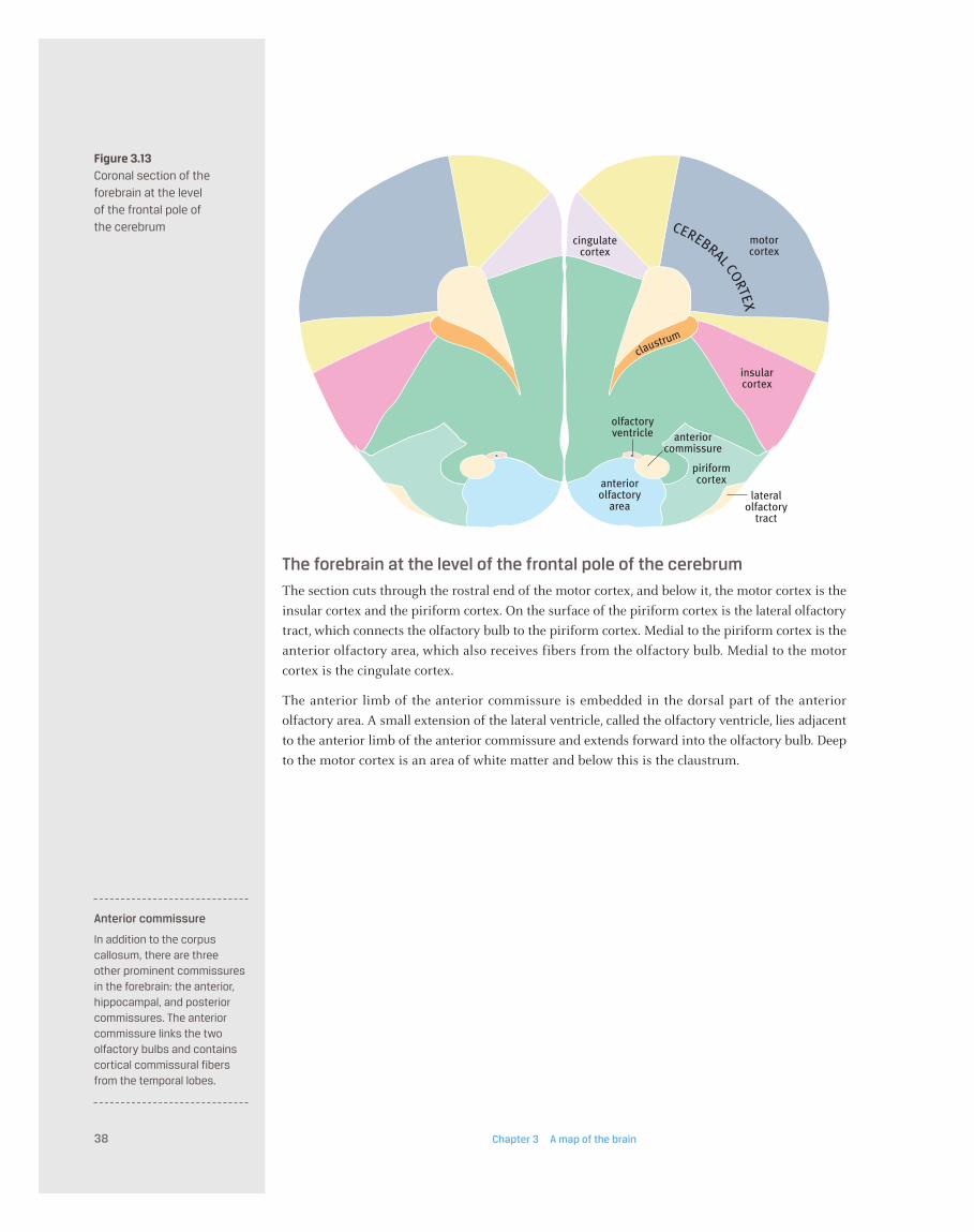
Figure 3.13Coronal section of the
forebrain at the level
of the frontal pole of
the cerebrum
The forebrain at the level of the frontal pole of the cerebrum
The section cuts through the rostral end of the motor cortex, and below it, the motor cortex is the
insular cortex and the piriform cortex. On the surface of the piriform cortex is the lateral olfactory
tract, which connects the olfactory bulb to the piriform cortex. Medial to the piriform cortex is the
anterior olfactory area, which also receives fibers from the olfactory bulb. Medial to the motor
cortex is the cingulate cortex.
The anterior limb of the anterior commissure is embedded in the dorsal part of the anterior
olfactory area. A small extension of the lateral ventricle, called the olfactory ventricle, lies adjacent
to the anterior limb of the anterior commissure and extends forward into the olfactory bulb. Deep
to the motor cortex is an area of white matter and below this is the claustrum.
Chapter 3 A map of the brain38
anteriorcommissure
lateralolfactory
tract
piriformcortexanterior
olfactoryarea
olfactoryventricle
cingulatecortex
insularcortex
motorcortex
CEREBRAL CORTEX
claustrum
Anterior commissure
In addition to the corpuscallosum, there are three other prominent commissuresin the forebrain: the anterior,hippocampal, and posteriorcommissures. The anteriorcommissure links the twoolfactory bulbs and containscortical commissural fibersfrom the temporal lobes.
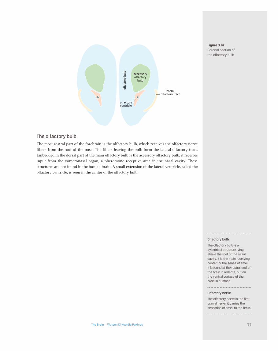
Figure 3.14Coronal section of
the olfactory bulb
The olfactory bulb
The most rostral part of the forebrain is the olfactory bulb, which receives the olfactory nerve
fibers from the roof of the nose. The fibers leaving the bulb form the lateral olfactory tract.
Embedded in the dorsal part of the main olfactory bulb is the accessory olfactory bulb; it receives
input from the vomeronasal organ, a pheromone receptive area in the nasal cavity. These
structures are not found in the human brain. A small extension of the lateral ventricle, called the
olfactory ventricle, is seen in the center of the olfactory bulb.
The Brain Watson Kirkcaldie Paxinos 39
olfa
ctor
y bu
lb
lateralolfactory tract
olfactoryventricle
accessoryolfactory
bulb
Olfactory bulb
The olfactory bulb is acylindrical structure lyingabove the roof of the nasalcavity. It is the main receivingcenter for the sense of smell.It is found at the rostral end ofthe brain in rodents, but onthe ventral surface of thebrain in humans.
Olfactory nerve
The olfactory nerve is the firstcranial nerve; it carries thesensation of smell to the brain.
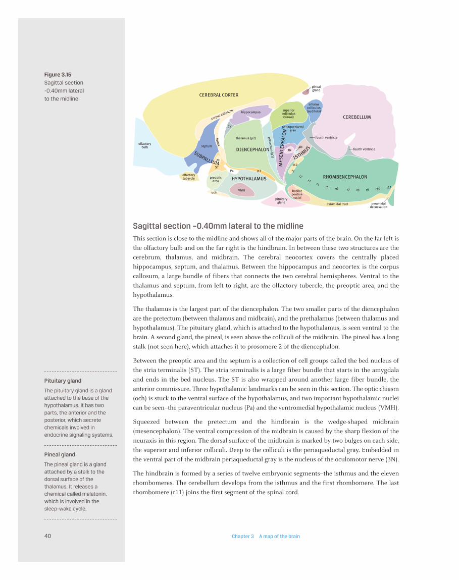
Figure 3.15Sagittal section
-0.40mm lateral
to the midline
Chapter 3 A map of the brain40
CEREBELLUM
DIENCEPHALON
hippocampus
inferiorcolliculus(auditory)superior
colliculus(visual)
periaqueductalgray
corpus callosum
thalamus (p2) fourth ventricle
fourth ventricle
CEREBRAL CORTEX
olfactorybulb
3N4N
scp
pyramidal tract pyramidaldecussation
pituitarygland
basilarpontinenuclei
RHOMBENCEPHALON
pinealgland
VMH
p3
HYPOTHALAMUS
septum
fornix
3V
PaST
ac
preopticarea
ochpretectum
(p1) ISTHMUS
SUBPALLIUM MES
ENCE
PHAL
ON
olfactorytubercle
r1 r2 r3 r4 r5 r6 r7 r8 r9 r10 r11
Sagittal section –0.40mm lateral to the midline
This section is close to the midline and shows all of the major parts of the brain. On the far left is
the olfactory bulb and on the far right is the hindbrain. In between these two structures are the
cerebrum, thalamus, and midbrain. The cerebral neocortex covers the centrally placed
hippocampus, septum, and thalamus. Between the hippocampus and neocortex is the corpus
callosum, a large bundle of fibers that connects the two cerebral hemispheres. Ventral to the
thalamus and septum, from left to right, are the olfactory tubercle, the preoptic area, and the
hypothalamus.
The thalamus is the largest part of the diencephalon. The two smaller parts of the diencephalon
are the pretectum (between thalamus and midbrain), and the prethalamus (between thalamus and
hypothalamus). The pituitary gland, which is attached to the hypothalamus, is seen ventral to the
brain. A second gland, the pineal, is seen above the colliculi of the midbrain. The pineal has a long
stalk (not seen here), which attaches it to prosomere 2 of the diencephalon.
Between the preoptic area and the septum is a collection of cell groups called the bed nucleus of
the stria terminalis (ST). The stria terminalis is a large fiber bundle that starts in the amygdala
and ends in the bed nucleus. The ST is also wrapped around another large fiber bundle, the
anterior commissure. Three hypothalamic landmarks can be seen in this section. The optic chiasm
(och) is stuck to the ventral surface of the hypothalamus, and two important hypothalamic nuclei
can be seen–the paraventricular nucleus (Pa) and the ventromedial hypothalamic nucleus (VMH).
Squeezed between the pretectum and the hindbrain is the wedge-shaped midbrain
(mesencephalon). The ventral compression of the midbrain is caused by the sharp flexion of the
neuraxis in this region. The dorsal surface of the midbrain is marked by two bulges on each side,
the superior and inferior colliculi. Deep to the colliculi is the periaqueductal gray. Embedded in
the ventral part of the midbrain periaqueductal gray is the nucleus of the oculomotor nerve (3N).
The hindbrain is formed by a series of twelve embryonic segments–the isthmus and the eleven
rhombomeres. The cerebellum develops from the isthmus and the first rhombomere. The last
rhombomere (r11) joins the first segment of the spinal cord.
Pituitary gland
The pituitary gland is a glandattached to the base of thehypothalamus. It has twoparts, the anterior and theposterior, which secretechemicals involved inendocrine signaling systems.
Pineal gland
The pineal gland is a glandattached by a stalk to thedorsal surface of thethalamus. It releases achemical called melatonin,which is involved in the sleep-wake cycle.

Figure 3.16Sagittal section
-2.62mm lateral
to the midline
Sagittal section –2.62mm lateral to the midline
This section is some distance from the midline, but the major features are similar to those seen in the
previous section. However, the olfactory bulb is no longer seen attached to the frontal pole of the
cerebrum; the piriform cortex is there in its place. The corpus callosum forms a roof over the caudate-
putamen and the hippocampus. Deep to the hippocampus is the diencephalon (pretectum, thalamus,
and prethalamus). The caudal part of the cerebral neocortex overhangs the colliculi of the midbrain.
In the ventral region of the cerebrum, from left to right, we can see the piriform cortex, the
olfactory tubercle, and the amygdala. The amygdala is a mass of gray matter in the temporal lobe
of the cerebrum. The other matter masses of gray matter seen in this lateral region of the cerebrum
are the caudate-putamen, the globus pallidus (GP), and the claustrum. The claustrum can be seen
in this section pressed against the rostral part of the corpus callosum. The caudate-putamen fills
the space between the corpus callosum and the olfactory tubercle. Embedded in the caudal border
of the caudate-putamen are the fibers of the anterior commissure. Separating the amygdala from
the diencephalon (prethalamus and thalamus) is a thick sheet of fibers, the internal capsule.
On the right of the diencephalon, caudal to the pretectum, is the midbrain (mesencephalon),
which is distinctly wedge-shaped. The large dorsal surface is occupied by the superior and inferior
colliculus, but the ventral surface is a small area between the pretectum and the isthmus.
The hindbrain is made up of the isthmus and the eleven rhombomeres. The cerebellum grows out
of the first rhombomere and the isthmus. In this lateral region of the hindbrain, the most obvious
feature is the trigeminal sensory nucleus. It extends from the second rhombomere down to the
third cervical segment of the spinal cord. The sensory root of the trigeminal nerve is seen here
entering the hindbrain at the level of the second rhombomere (r2). Ventral to the sensory
trigeminal nucleus in the region of the sixth rhombomere (r6) is the facial nucleus, which supplies
the muscles of facial expression. Although the facial nucleus is located in the sixth rhombomere
(r6), the facial nerve emerges from the hindbrain at the level of the fourth rhombomere (r4).
The cerebellum consists of a folded cortex and a central core of fibers. Within this central core are
the cerebellar nuclei. Between the cerebellum and the hindbrain is the fourth ventricle, one of the
reservoirs of cerebrospinal fluid in the central nervous system.
The Brain Watson Kirkcaldie Paxinos 41
opt
s57n
facialnucleus
CEREBELLUM
hippocampus
visualcortex
inferiorcolliculus(auditory)
superiorcolliculus(visual)
anteriorcommissure
GP
amygdala
corpus callosum
thalamus (p2)
LV
pretectum (p1)
CEREBRAL CORTEX
MESENCEPHALON
subiculum
prethalamus(p3)
trigeminal sensory
caudate-putamen
RHOM
BENCEPHALON
piriform cortex
olfactory tubercle
claustrum
ISTH
M
US
internal capsule
D I E N C E P H A L O N
SUBPALLIUM
r1 r2 r3 r4 r5 r6 r7
4V
8n
Old and new cerebral cortex
The more primitive areas ofcerebral cortex, such as theolfactory (piriform) cortex,have only three distinct layers.The ‘new’ areas of cortex(neocortex) that have evolvedin mammals have six layers.More than 95% of the humancerebral cortex is neocortex.
Piriform cortex
The piriform cortex is theprimary olfactory cortex. The human piriform cortex is located in the uncus.

a b
c d
e f
Figure 3.17 Brain sections
Photographs of coronal sections of rat brain which correspond to some of the diagrams in thisChapter. These images show that demarcating the clusters and layers of neurons that populate thebrain in Nissl stained sections is not a straightforward exercise. Finding boundaries between nucleiis a complex process, requiring a great deal of information on connections and histochemistry (seeChapter 11). Sections a, d, and e are stained for acetylcholinesterase, whereas sections b, c, and f areNissl stained. Refer to the corresponding figures for anatomical description–a:3.4; b:3.6; c:3.8; d:3.10;e:3.12; f:3.14. The images are taken from The Rat Brain in Stereotaxic Coordinates, 6th edition (Paxinosand Watson, 2007).
Chapter 3 A map of the brain42