Embed Size (px)
Citation preview
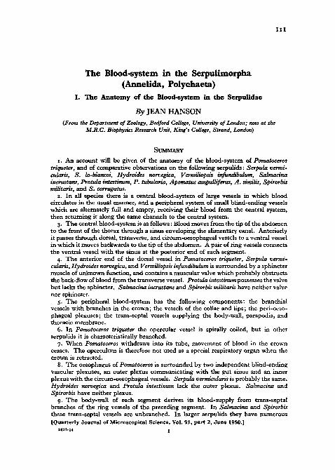
The Blood-system in the Serpulimorpha(Annelida, Polychaeta)
I. The Anatomy of the Blood-system in the Serpulidae
By JEAN HANSON
{From the Department of Zoology, Bedford College, University of London; now at theM.R.C. Biophysics Research Unit, King's College, Strand, London)
SUMMARY
I . An account will be given of the anatomy of the blood-system of Pomatocerostriqueter, and of comparative observations on the following serpulids: Serpula vermi-cularis, S. lo-biancoi, Hydroides norvegica, Vermiliopsis infundibulum, Salmacinaincrustans, Protula intestinum, P. tubularia, Apomatus ampulliferus, A. similis, Spirorbismilitaris, and S. corrugatus.
2. In all species there is a central blood-system of large vessels in which bloodcirculates in the usual manner, and a peripheral system of small blind-ending vesselswhich are alternately full and empty, receiving their blood from the central system,then returning it along the same channels to the central system.
3. The central blood-system is as follows: Blood moves from the tip of the abdomento the front of the thorax through a sinus enveloping the alimentary canal. Anteriorlyit passes through dorsal, transverse, and circum-oesophageal vessels to a ventral vesselin which it moves backwards to die tip of the abdomen. A pair of ring vessels connectsthe ventral vessel with the sinus at the posterior end of each segment.
4. The anterior end of the dorsal vessel in Pomatoceros triqueter, Serpula vermi-cularis, Hydroides norvegica, and Vermiliopsis infundibulum is surrounded by a sphinctermuscle of unknown function, and contains a muscular valve which probably obstructsthe back-flow of blood from the transverse vessel. Protula intestinum possesses the valvebut lacks the sphincter. Salmacina incrustans and Spirorbis militaris have neither valvenor sphincter.
5. The peripheral blood-system has the following components: the branchialvessels with branches in the crown; the vessels of the collar and lips; the peri-oeso-phageal plexuses; the trans-septal vessels supplying the body-wall, parapodia, andthoracic membrane.
6. In Pomatoceros triqueter the opercular vessel is spirally coiled, but in otherserpulids it is characteristically branched.
7. When Pomatoceros withdraws into its tube, movement of blood in the crownceases. The operculum is therefore not used as a special respiratory organ when thecrown is retracted.
8. The oesophagus of Pomatoceros is surrounded by two independent blind-endingvascular plexuses, an outer plexus communicating with the gut sinus and an innerplexus with the circum-oesophageal vessels. Serpula vermicularis is probably the same.Hydroides norvegica and Protula intestinum lack the outer plexus. Salmacina andSpirorbis have neither plexus.
9. The body-wall of each segment derives its blood-supply from trans-septalbranches of the ring vessels of the preceding segment. In Salmacina and Spirorbisthese trans-septal vessels are unbranched. In larger serpulids they have numerous[Quarterly Journal of Microscopical Science, Vol. 91, part 2, June 1950.]
2421-14 I

ii2 Jean Hanson—The Blood-system
branches under the epidermis, in the parapodia, and in some cases on the coelomicsurface of the body-wall. Branches of the thoracic trans-septal vessels supply thethoracic membrane. In all species except Salmacina, Spirorbis, and Protula intestinumthe thoracic trans-septal vessels end ventrally in two superficial ventro-lateral longi-tudinal vessels which communicate either with the circum-oesophageal vessels or withthe ventral vessel. In P. intestinum the thoracic trans-septal vessels enter the ventralvessel directly. The pattern of superficial vessels on the ventral surface of the thoraxis useful for identifying specimens.
10. Lateral vessels, such as are found in Sabella, are absent in all serpulids.
CONTENTS
INTRODUCTION .
PBBVIOUS WORK
MATERIAL AND METHODS
OBSERVATIONS .
1. Pomatoceros
2. Other serpulidsREFERENCES 28
INTRODUCTION
THIS paper is the first of four which will deal with the anatomy and histo-logy of the blood-system in the Serpulidae and Sabellidae. This research
was undertaken as part of a series of investigations by Professor H. Munro Foxand workers in his laboratory on serpulids and sabellids which, together withchlorhaemids, are the only animals definitely known to possess the blood pig-ment chlorocruorin. The chemical and physical properties of chlorocruorinhave been studied by Fox (1924, 1926, 1932, 1934, 1946) and Roche and Fox(1933), the occurrence of chlorocruorin in the Serpulimorpha by Fox (1949),and the respiration and blood circulation of sabellids by Fox (1933, 1938) andR. F. Ewer and Fox (1940). D. W. Ewer (1941) has described the anatomyof the blood-system of Sabella and has made preliminary observations on itshistology.
PREVIOUS WORK
A general account of the anatomy of atypical serpulid, Pomatoceros triqueter,has been published by Thomas (1940). The body consists of a prostomiumbearing the branchial crown, a thorax of several segments of which the first isthe peristomium, and an abdomen of many segments. The branchial crownconsists of filaments which bear pinnules; one of the filaments is modified asa peduncle carrying an operculum at its tip. At the anterior end of the thoraxis a collar; a thoracic membrane extends along each side of the thorax dorsalto the parapodia.
Thomas (1940) has given the most recent account of the blood-system of aserpulid, Pomatoceros triqueter. Earlier descriptions were published byHuxley (1855—Salmacina dysteri), Claparede (1868-70—Serpula vermicularis;1873—serpulids in general), Haswell (1885—Hydroides elegans and Pomato-ceros elaphus), Jaquet (1886—Protula intestinum), Meyer (1888—Protula

in the Serpulimorpha (Annelida, Polychaeta) 113
tubularia), Malaquin (1901—Salmacina dysteri), zur Loye (1908—Spirorbisborealis), Lee (1912—Serpula vermicularis, Hydroides norvegica, Vermiliopsisinfundibulum, Pomatoceros triqueter, Salmacina dysteri, and Protula intestinum),Woskressensky (1924—Spirorbis militaris), and Faulkner (1930—Filogranaimplexa and Salmacina dysteri). The most important of these papers are thoseof Meyer, Lee, Faulkner, and Thomas. The work of Jaquet and Meyer wasreviewed by Fuchs (1907), and that of Haswell by Mclntosh (1918).
The main features already known of the serpulid blood-system can be sum-marized as follows. A sinus envelops the alimentary canal from the tip of theabdomen to the junction of the stomach and oesophagus just behind theperistomium. Here the sinus leads into a short dorsal vessel connected bycircum-oesophageal vessels with a ventral vessel, which extends back alongthe whole length of the body and communicates with the gut sinus by seg-mentally arranged ring vessels. Meyer (1888) and Lee (1912) found that thefirst two pairs of ring vessels are branches of the circum-oesophageal vessels,the first pair leading to the dorsal vessel, and the second pair to the gut sinus;but according to all other observers the first ring vessels lead from the ventralvessel to the sinus. Meyer (1888), Lee (1912), and Thomas (1940) founda lateral vessel connecting the ring vessels on each side of the body. A peri-oesophageal vascular plexus, leading out of the sinus, was described byClaparede (1873), Haswell (1885), and Thomas (1940). Two branchial vesselsleave the circum-oesophageal vessels and branch into single blind-endingvessels in each filament and pinnule. The vessels in the collar and thoracicmembrane are also blind-ending. The collar vessels arise from the circum-oesophageal vessels. The vessels in the thoracic membrane are branches ofring vessels; according to Thomas (1940) the first pair of thoracic membranevessels are branches of the circum-oesophageal vessels. Little is known aboutthe blood-supply of the body-wall, parapodia, and intersegmental septa;branches of the ring vessels lead to these structures. According to Faulkner(1930) Filograna and Salmacina are in many ways different from otherserpulids. Her work will be discussed later (p. 125).
MATERIAL AND METHODS
The following species have been used: Serpula vermicularis L., S. lo-biancoiRioja, Hydroides norvegica (Gunnerus), Vermiliopsis infundibulum (Philippi),Pomatoceros triqueter L., Salmacina incrustans Claparede, Protula intestinum(Lamarck), P. tubularia (Montagu), Apomatus ampulliferus Philippi, A. similisMarion and Bobretzky, Spirorbis corrugatus (Montagu), and S. militaris(Claparede). Pomatoceros was obtained from Plymouth, and the other specieswere studied at Naples. Serpula lo-biancoi, previously recorded only from theAtlantic coast of Spain, was found at Naples for the first time in 1947 byProfessor H. Munro Fox, who lent me his specimens.
Observations were made on living worms, on benzidine preparations (seebelow), and on serially sectioned specimens. The whole of the blood-systemin Salmacina and Spirorbis and the superficial vessels of the larger species can

ii4 Jean Hanson—The Blood-system
be studied in whole living specimens. Serial sections are needed for tracingthe internal vessels in species of a medium size. Most parts of the blood-system of the largest species, Protula intestinum, can be studied in livingspecimens dissected under a binocular dissecting microscope.
Like haemoglobin, chlorocruorin is a peroxidase (Lankester, 1869). In thepresence of hydrogen peroxide and benzidine, a blue-black oxidation psoductof benzidine is deposited in the blood-vessels. This reaction has previouslybeen used by Prenant (1921), Faulkner (1930), and Ewer (1941) for tracingthe course of small vessels in polychaetes. I have employed, with equal suc-cess, the methods of Slonimsky (1927), Faulkner and Ziegler (1945). It isnecessary to use thin specimens. The vessels of the body-wall of medium-sized species, such as Pomatoceros, are well demonstrated in preparationsmade by bisecting the worm longitudinally and removing the alimentary canal.Stained specimens were fixed in 70 per cent, alcohol acidified with a trace ofacetic acid to control decolorization. They were then transferred to benzylalcohol, and subsequently to Canada balsam.
The following fixatives were found to be satisfactory for serially sectionedmaterial: 4 per cent, formaldehyde in sea-water, Duboscq-Brasil, Heiden-hain's 'Susa', Zenker-acetic, and Zenker-formol. Most of the sections werestained with 'Azan'.
OBSERVATIONS
The serpulid blood-system is unusual because all the smaller vessels endblindly and are alternately empty and full of blood. They receive blood fromthe larger vessels, then the direction of flow is reversed and all the bloodreturns to the larger vessels. The movement of blood in these small vesselscan be watched in the branchial crown, collar, and thoracic membrane. Theseblind-ending vessels constitute the peripheral blood-system, and the largervessels belong to the central blood-system, in which the blood circulates in theusual manner. As in most invertebrate animals it flows forwards dorsally andbackwards ventrally. Except at the front of the thorax, a sinus surroundingthe alimentary canal takes the place of a dorsal vessel. The blood is movedforwards along the gut sinus by antiperistaltic waves of contraction of the gutmuscle coat, which lies outside the sinus (Hanson, 1948a). I shall show ina later paper that all serpulid blood-vessels have muscle-fibres in their walls.
1. Pomatoceros
The central blood-system of Pomatoceros is diagrammatically representedin Text-figs. 1-4.
The gut sinus and the ventral vessel extend from the posterior end of thepenultimate abdominal segment (Text-fig. 1) to the anterior end of the secondthoracic segment (Text-fig. 2). In each segment the ventral vessel communi-cates with the ventro-lateral part of the sinus by a pair of ring vessel which lieon the anterior face of the posterior septum of the segment (Text-figs. 3 and 4).The thoracic ring vessels are longer than those of the abdomen and are looped;

in the SerpuUmorpha {Annelida, Polychaeta) 115
parts of them are suspended in the coelom by mesenteries attached to thesepta. The chloragogen tissue of Pomatoceros is situated on the thoracicringvessels and on the first few abdominal ring vessels. The gut sinus leadsanteriorly into a dorsal vessel lying above the oesophagus and terminating in
ring vessel
gut sinus
TEXT-FIC. I . Diagram of blood-system at posterior end of Pomatoceros triqueter,in ventral view.
trjnsvent
TEXT-FIG. 2. Diagram to show arrangement of vessels in peristomial and second and thirdthoracic segments of Pomatoceros triqueter. Lateral view, with half body-wall removed.
a transverse vessel situated just behind the cerebral ganglia. Each end of thetransverse vessel bifurcates into a branchial vessel and a circum-oesophagealvessel. The latter passes downwards through the peristomial cavity, entersthe connective tissue lying under the oesophagus (Text-fig. 7), and extendsbackwards through this tissue to the posterior end of the peristomium, whereit joins its fellow from the other side to form the ventral vessel.
The anterior part of the dorsal vessel (Text-fig. 5) is. surrounded by asphincter muscle, and contains a valve which probably ensures that bloodreturning from the branchial crown does not enter the dorsal vessel, but isdirected through the circum-oesophageal vessels to the ventral vessel. A

gut sinus \ f | i < f l t | \ \ / o r s a l .nwsclM**./ ^ : ! ' 5 o V " ~ : ' : i ' ^ y thoracic membrane
vessel
dorsal superficial
f * »ciiLj ai
(qj muscles
TEXT-FIG. 3. Diagrams to show arrangement of vessels in one thoracic segment of Pomatocerostriqueter, viewed from behind, (a) whole length, (b) half length of segment.
TEXT-FIG. 4. Diagrams to show arrangement of vessels in one abdominal segment ofPomatoceros triqueter, viewed from behind, (a) whole length, (6) half length of segment.

Jean Hanson—The Blood-system in the Serpulimorpha 117
transverse section through the middle of this region of the dorsal vessel showsthat the lumen is separated into two channels, dorsal and ventral, by ahorizontal septum. Posteriorly, the septum slopes downwards and fuses withthe floor of the vessel. Anteriorly, it slopes upwards and ends in a free edgewhere the dorsal vessel enters the transverse vessel. Clearly this septum canimpede the back-flow of blood from the transverse vessel into the dorsalvessel without hindering the flow of blood in the opposite direction. Thevalve consists of a thin sheet of connective tissue enclosing muscle-fibres and
muscle fibreof sphincter
muscle fibreof valve
TEXT-FIG. 5- 'Obliquely transverse section through anterior part of dorsal vessel of Pomatocerostriqueter, to show valve and sphincter.
covered on each surface by an endothelium. The muscle-fibres enter thevalve antero-laterally and pass obliquely backwards to converge towards themedian line. Their contraction will presumably flatten the valve and facilitatethe flow of blood out of the dorsal vessel.
The anterior end of the dorsal vessel is not visible in the living animal. Themovements of the valve have therefore not been observed, nor has it beenpossible to discover the circumstances in which the sphincter contracts.However, the following suggestion may be made. By watching animals livingin glass tubes, it has been found that movement of blood in the vessels of thecrown ceases soon after it has been withdrawn into the tube; but blood con-tinues to move forwards through the gut sinus. It is possible that the sphinctermuscle contracts at this time and prevents the flow of blood into the branchialvessels from the dorsal vessel. Its contraction would divert blood through thethoracic ring vessels from the gut sinus to the ventral vessel. Unfortunatelythe thorax of Pomatoceros and the other serpulids which possess the sphincter

n8 Jean Hanson—The Blood-system
is too opaque for this suggestion to be tested by direct observation. Thesphincter muscle was noticed by Thomas (1940). The valve has not previouslybeen described in Pomatoceros, but was found by Lee (1912) in Protulaintestinum.
The peripheral blood-system of Pomatoceros is diagrammatically representedin Text-figs. 2-4 and 8 and 11. The components of the peripheral blood-
system are as follows: the branchialvessels and their branches in thecrown; the vessels supplying the collar,lips, and anterior part of the alimentarycanal; the peri-oesophageal plexuses;the trans-septal vessels with branches inthe body-wall, parapodia, and thoracicmembrane.
The two branchial vessels leave thecentral blood-system where the circum-oesophageal vessels join the transversevessel (Text-fig. 2). They enter thecrown dorsally, then bend in a ventraldirection; they follow the curvatureof the base of each half of the crown,supply one vessel to each filament, andend in the vessels of the last filaments.Pomatoceros does not possess branchialvesicles, i.e. the vesicular proximal por-tions of the branchial vessels found insome sabellids. The filament vesselssend one branch to each pinnule. Thevessel of the most dorsal filament ofeach side gives off one branch to theadjacent 'palp'. Elsewhere (Hanson,1949) I have suggested that the palp'is a modified pinnule. The peduncleof the operculum receives a branch of
the left branchial vessel. In the operculum it ends in a wide, thin-walled,blind-ending vessel, which is coiled in a spiral round a core of connectivetissue and reaches nearly to the roof of the operculum (Text-fig. 6). A spiralopercular vessel has not previously been recorded in any serpulid, but thefigure published by Mclntosh (1926) of a longitudinal section through theoperculum of Mercierella enigmatica suggests that a spiral vessel also exists inthis species. In most serpulids the opercular vessel has branches ending inampullae.
The good blood-supply of the serpulid operculum has naturally suggestedto previous workers (Orley, 1884; zur Loye, 1908; Mclntosh, 1918, 1926)that it may have a respiratory function, particularly when the crown has been
peduncle
TEXT-FIG. 6. Benzidine preparationof operculum of Pomatoceros triqueter.Vessel distended with gas, and bloodadhering to walls. X 35.

in the Serpulimorpha {Annelida, Polychaeta) 119
withdrawn into the tube and the operculum blocks the mouth of the tube.However, observations made on Pomatoceros living in glass tubes have shownthat soon after the crown has been retracted the movement of blood ceases inall its vessels, including the peduncle vessel. Sabella behaves similarly (Fox,1933). Therefore neither the operculum nor the rest of the crown functions asa respiratory organ when the crown is inside the tube. The movement ofblood in the capillaries of the thoracic membrane and body-wall continues,and respiratory exchange is probably carried out between the blood in thesevessels and the surrounding water, which is kept moving through the tube byvigorous pumping movements of the abdomen (as in Sabella—Nicol, 1930;Fox, 1938) and also by the activity of the ciliary tracts described by Segrove(1938) and Thomas (1940).
The two collar vessels, ventral and lateral, leave each circum-oesophagealvessel near the place where it enters the connective tissue around the oeso-phagus (Text-figs. 2 and 11). The lateral collar vessel supplies the capillarieson the lateral surface of the peristomium and in the lateral part of the collar.The ventral collar vessel supplies the ventral part of the collar. Soon afterleaving the circum-oesophageal vessel it gives off a branch which supplies theconnective tissue around the cerebral ganglia, the anterior part of the oeso-phagus and the buccal cavity, and ends in the capillaries of the dorsal andventral lips.
A transverse section through the middle of the oesophagus (Text-fig. 7)shows the following layers in the oesqphageal wall: the epithelium lining thelumen; a thin layer of connective tissue with a few thin-walled capillaries;a layer of circular muscles; a thicker layer of connective tissue containingnumerous thin-walled capillaries; a sheath of connective tissue fibres; a layerof fatty connective tissue in which lie the circum-oesophageal vessels andseveral thick-walled vessels on either side of the oesophagus. These thick-walled vessels branch and anastomose with each other to form a two-dimen-sional plexus originating posteriorly from the gut sinus and ending blindlyanteriorly (Text-fig. 2). The numerous thin-walled capillaries lying deeper inthe wall of the oesophagus form a three-dimensional plexus ending blindlyposteriorly and communicating anteriorly with the circum-oesophageal vesselsby a small vessel on each side of the body (Text-fig. 2). There is no com-munication between the inner and outer peri-oesophageal plexuses. Thedifference in thickness of the walls of the two systems of vessels is very con-spicuous. It is probable that blood-pressure is higher in the outer plexus,which is a blind-ending prolongation of the gut sinus, than it is in theinner plexus, which is supplied by one of several branches of each circum-oesophageal vessel. It may be suggested that the inner plexus consti-tutes the blood-supply of the oesophageal wall, whilst the outer plexusis more important as a place where blood can be received from the sinuswhenever its exit from the anterior end of the dorsal vessel is prevented,for example during contraction of the sphincter muscle around the dorsalvessel.

12O
A anson

in the Serpulimorpha (Annelida, Polychaeta) 121
Branches from these vessels supply the ventral surface of the thorax, includingthe peristomium.
A thoracic trans-septal vessel leaves the ring vessel at the upper edge of thelateral longitudinal muscle block (Text-fig. 3). It passes through the septumand branches in the next segment. The main branch passes ventrally throughthe connective tissue around the muscles of the notopodial chaetae. It givesoff branches to these muscles, and short, wide, blind-ending capillaries lyingunder the neuropodial uncini. It ends in the ventro-lateral thoracic vessel.
•superficial \ of dorsalbranch J trans-septaldeep branch ) vessel
ampulla onsuperficial brancrof ventraltrans-septal vessel
-notopodialuncini
_neuropodialchaetae
ringvessel
ventralvessel
TEXT-PIG. 8. Diagram to show blood-system of abdominal body-wall of Pomatocerostriqueter.Wall cut mid-dorsally and spread flat. Right half of two segments seen from inside. Musclesof one segment depicted as transparent.
Soon after passing through the septum it gives off one large branch to thethoracic membrane and two or three smaller branches which end in capillariessituated just under the basement membrane of the epidermis on the dorsalsurface (Text-figs. 2 and 3). Thomas (1940) has stated that the first thoracicmembrane vessel is a branch of the circum-oesophageal vessel. I have foundthat it is a branch of the first trans-septal vessel, which originates in the circum-oesophageal vessel.
An abdominal ventral trans-septal vessel leaves the ring vessel at the lateraledge of the ventral longitudinal muscle block (Text-figs. 4 and 8), passesthrough the septum, and branches in the next segment into superficialcapillaries in the ventral, lateral, and latero-dorsal regions of the body-wall.It also gives off short, wide, blind-ending capillaries situated under the noto-podial uncini and projecting into the cavity of the segmental organ. (Thesegmental organ is a lateral pouch of the coelom extending dorsally into theparapodium and ventrally under the body-wall as a gonoduct, which opens tothe exterior near the mid-ventral line.)
An abdominal dorsal trans-septal vessel leaves the ring vessel at the lateral

122 Jean Hanson—The Blood-system
edge of the dorsal longitudinal muscle block (Text-figs. 4 and 8). There aresometimes two, rarely three, dorsal trans-septal vessels originating in one ringvessel. After passing into the next segment the vessel branches over thecoelomic surface of the dorsal muscle block. Each of these branches bends atright angles and, without branching, penetrates through the muscle block byway of the connective tissue separating the muscle compartments, and thenemerges under the epidermis of the dorsal surface of the body. Here it givesoff a few blind-ending branches which do not anastomose with the capillariesof the ventral trans-septal vessel. These superficial capillaries of the dorsaltrans-septal vessels are easily recognized (Text-fig. 8). They lie either parallelto or at right angles to the longitudinal muscle-fibres, and the longitudinalcapillaries become sinuous when the muscles contract. The ventral trans-septal vessels, on the other hand, branch at acute angles.
A few capillaries lie just underneath the epithelium of the excretory sacs ofthe thoracic nephridia, but I have been unable to find their source.
Lateral vessels, such as are found in Sabella, are absent in Pomatoceros andall the other serpulids I have examined. It is probable that Meyer (1888), Lee(1912), and Thomas (1940) mistook trans-septal vessels for lateral vessels.
2. Other serpulidsThe central blood-system of all the serpulids examined is like that of Poma-
toceros, except for differences in the valve and muscular sphincter of the dorsalvessel. Hydroides, Serpula vermicularis, and Vermiliopsis possess both valveand sphincter. Protula intestinum possesses the valve but lacks the sphincter.Spirorbis militaris and Salmacina lack both valve and sphincter. Suitableserial sections of the dorsal vessels of other serpulids have not been prepared.
Whereas the central blood-system appears to be uniform throughout thefamily, variations have been found in the peripheral blood-system. Some ofthese variations are attributable to differences in body size, and others are atpresent inexplicable from a functional point of view.
Pomatoceros is unusual in possessing a spiral opercular blood-vessel. Inother genera the vessel bears branches ending in ampullae. These vesselshave been described in Hydroides norvegica by Okada (1932-3), H. lunuliferaby Claparede (1868-70), H. uncinata and Serpula vermicularis by Orley(1884), Apomatus similis by de St. Joseph (1894), and A ampulliferus byZeleny (1905). The opercular vessel of Serpula lo-biancoi is like that of S.vermicularis. In Vermiliopsis infundibulum the vessel branches just under thechitinoid cap of the operculum into a number of short vessels ending inampullae.
Serpula vermicularis possesses a double peri-oesophageal plexus like that ofPomatoceros, but I have been unable to discover if the inner plexus is connectedwith the circum-oesophageal vessels. The outer plexus is absent in Hydroides,Vermiliopsis, and Protula intestinum. I have been unable to trace the origin ofthe small vessels found in the oesophageal wall of these serpulids. Salmacinaand Spirorbis militaris lack peri-oesophageal plexuses.
in the SerpuUmorpha (Annelida, Polychaeta) 123
The blood-supply to the body-wall in Salmacina and Spirorbis is greatlyreduced, presumably because of the small size of these serpulids. Salmacinais 2-3 mm. long, Spirorbis even smaller. There is a single pair of unbranchedblind-ending trans-septal vessels in each segment, extending from the ringvessels of one segment to the parapodia of the next posterior segment. Ventro-lateral thoracic vessels are absent.
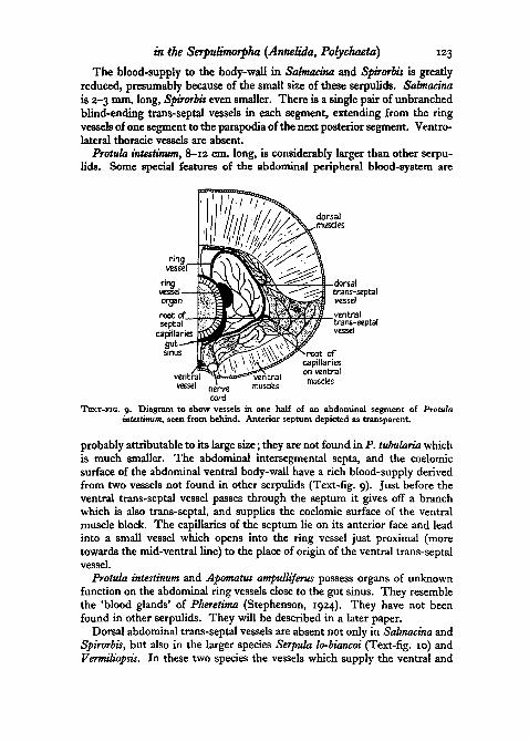
Protula intestinum, 8-12 cm. long, is considerably larger than other serpu-lids. Some special features of the abdominal peripheral blood-system are
ofcapillarieson ventralmusdes
TEXT-FIG. 9. Diagram to show vessels in one half of an abdominal segment of Protulaintestinum, seen from behind. Anterior septum depicted as transparent.
probably attributable to its large size; they are not found in P. tubularia whichis much smaller. The abdominal intersegmental septa, and the coelomicsurface of the abdominal ventral body-wall have a rich blood-supply derivedfrom two vessels not found in other serpulids (Text-fig. 9). Just before theventral trans-septal vessel passes through the septum it gives off a branchwhich is also trans-septal, and supplies the coelomic surface of the ventralmuscle block. The capillaries of the septum lie on its anterior face and leadinto a small vessel which opens into the ring vessel just proximal (moretowards the mid-ventral line) to the place of origin of the ventral trans-septalvessel.
Protula intestinum and Apomatus ampulliferus possess organs of unknownfunction on the abdominal ring vessels close to the gut sinus. They resemblethe 'blood glands' of Pheretima (Stephenson, 1924). They have not beenfound in other serpulids. They will be described in a later paper.
Dorsal abdominal trans-septal vessels are absent not only in Salmacina andSpirorbis, but also in the larger species Serpula lo-biancoi (Text-fig. 10) andVermiliopsis. In these two species the vessels which supply the ventral and

J24 Jean Hanson—The Blood-system
lateral surfaces of the abdominal body-wall also supply the dorsal surface.Serpula vermicularis, unlike S. lo-biancoi, possesses dorsal trans-septal vessels.It also differs from S. lo-biancoi and from all other serpulids in that the super-ficial branches of the dorsal trans-septal vessels are connected with each other,on each side of the body, by a longitudinal vessel, which also receives the maindorsal branches of the ventral trans-septal vessels.
On the ventral surface of the thorax of all serpulids except Salmacina andSpirorbis there are numerous blood-vessels situated just under the epidermis
branches ofvenbral trans-septai vessel
neuropodialchaebae postenor
TEXT-FIG, IO. Diagrams of abdominal superficial blood-system of Serpula lo-biancoi,(a) in dorsal, (6) in lateral view.
and visible in living animals. This system of vessels is useful in the identifica-tion of specimens, because the pattern varies from species to species, and isinvariable in its main features within any one species. Diagrams of thesevessels are given in Text-figs. 11-18.
In Pomatoceros triqueter (Text-fig. 11) three longitudinal vessels are visible,the ventral vessel and the two ventro-lateral thoracic vessels. These threevessels are not directly connected with each other. The ventro-lateral vesselsoriginate in the circum-oesophageal vessels and end blindly in the ventro-posterior part of the thoracic membrane. Two pairs of collar vessels, ventraland lateral, leave the circum-oesophageal vessels. In Hydroides norvegica(Text-fig. 12), Serpula vermicularis, and S. lo-biancoi (Text-fig. 13) eachventro-lateral vessel is directly connected with the ventral vessel by a super-ficial transverse vessel at the posterior end of each segment. The ventro-lateral vessels begin anteriorly in the first transverse vessel and end posteriorlyin the last transverse vessel. In H. norvegica and S. vermicularis the ventro-posterior part of the thoracic membrane is supplied by two branches of thelast transverse vessel. S. lo-biancoi, like Vermiliopsis infundibulum (Text-fig.14), lacks the ventral part of the thoracic membrane. In V. infundibulum the

in the Serpulimorpha {Annelida, Polychaeta) 125
ventro-lateral vessels are situated farther away from the uncinal ridges thanin other serpulids. The main collar vessel of H. norvegica is an anteriorprolongation of the ventral vessel. S. vermicularis resembles H. norvegicaexcept for the absence of this conspicuous collar vessel. In S. lo-biancoi asimilar vessel supplies the surface of the peristomial segment which, in thisspecies, is longer than usual.
Protula (Text-figs. 15 and 16) and Apomatus (Text-figs. 17 and 18) differfrom other serpulids in the absence of uncinal ridges. P. intestinum (Text-fig.15) is conspicuously different in that it lacks ventro-lateral thoracic vessels; thetrans-septal vessels extend across the ventral surface of the thorax and enterthe ventral vessel. The ventro-posterior part of the thoracic membrane issupplied by two branches of the ventral vessel. A. ampulliferus (Text-fig. 18)is unusual in that the first two pairs of trans-septal vessels join the circum-oesophageal vessels instead of the ventro-lateral vessels. P. intestinum is inmany ways different from P. tubularia, and P. tubularia in many ways closelyresembles Apomatus. Elsewhere (Hanson, 19486) I have discussed the rela-tionship of the two genera and suggested that they need to be revised.
Faulkner (1930) has published an account of the blood-system of Filogranaimplexa and Salmacina dysteri in which she had described a system of sinuseswhich only in the anterior part of the body become restricted to form vessels.The sinus around the alimentary canal spreads into sinuses in the septa andmesenteries. These extend into sub-epidermal sinuses. The ventral mesen-teric sinus takes the place of a ventral vessel. The ring vessels are wide partsof the septal sinuses, and they connect the sinuses in the dorsal and ventralmesenteries. The ring vessels sometimes have backward-pointing caeca.Anteriorly there are distinct dorsal, circum-oesophageal, and branchial vesselsarranged in the usual manner. I have been unable to find mesenteric, septal,or sub-epidermal sinuses in Salmacina incrustans. In living specimens onecan see a well-defined ventral vessel connected by discrete ring vessels withthe gut sinus; each ring vessel gives off an unbranched trans-septal vesselwhich ends blindly near the parapodium of the next posterior segment. Theseobservations have been confirmed in benzidine preparations and serialsections.
In benzidine preparations, Faulkner observed superficial blue-colouredpatches, and in untreated animals the epidermal cells appeared to be outlinedin pale green, as though blood containing chlorocruorin were present betweenthe basal parts of the cells. From these observations she concluded thatextensive sub-epidermal blood sinuses are present. I have confirmed herobservations, but prefer an alternative explanation. The cell outlines are palegreen in colour not only in surface view, but also in profile view; and in thelatter case the outer margin of the cell also has the pale green colour. Thecolour therefore seems to be a property of the cell surface and is not necessarilydue to the presence of a sub-epidermal blood sinus. The benzidine reactionis not specific for blood pigments, but is a reaction for all peroxidases (Prenant,1924).

(
c i r cum-oesophageal —
vessel
thoracicmembrane-
ventralvessel
trans-septalvessel
•ollar
i<- .z
s—*=£3I
1 i
TEXT-FIG.
ventral collarvessel
'_y\ lateralrTA)/ \ collar
^ )neuropodial
~ uncini
ventro-lateralthoracicvessel
. ^notopodialchaetae
ostero-ventralpart of thoracic
membraneI I
collar
brans-septa!vessel ~~
postero-ventral .
part of thoracicmembrane
TEXT-FIG. 12
bransversevessel
TEXT-FIGS. 11-12. Diagrams of ventral thoracic superficial blood-systems of: 11, Pomatocerostriqueter; 12, Hydroides norvegtca.

collar collar vessel
collar vessel collar
thoracic _membrane
TEXT-FIG. 14
TEXT-FIGS. 13-14. Diagrams of ventral thoracic superficial blood-systems of: 13, Serpulalo-biancoi; 14, Vermiiopsis infundibulum.

128 Jean Hanson—The Blood-system
In benzidine preparations the vessels often have irregular outlines, and theblue-black granules are not uniformly distributed; uncoloured patches areoften found. Faulkner has concluded from this appearance that the blood isnot confined in well-defined vessels, but is in sinuses; for example, there isa ventral mesenteric sinus instead of a ventral vessel. However, the benzidine
-/TVIS T—^-—i—' 16"
: membrane vessel
TEXT-FIGS. 15-18Diagrams of ventral thoracic superficial blood-systems of: 15, Protula intestinum; 16, P.
tubularia; 17, Apomatus similis; 18, A.'ampulliferus.reaction is violent, and numerous bubbles of oxygen are liberated (see Text-fig. 6). The irregular appearance of vessels after the reaction is an artifact,although the general anatomy of the blood-system is usefully demonstratedby this method. Careful examination of living Sahnacina incrustans leaves nodoubt that its vessels have well-defined walls and are not sinuses.
I acknowledge with gratitude the help of Professor H. Munro Fox whosuggested, encouraged, and usefully criticized the research reported in thispaper. I am also indebted to Dr. A. Stock for many discussions of problemsand results, to the British Association for the Advancement of Science for theuse of its Table at the Zoological Station of Naples, to the staff of the Stationfor the facilities they provided, and to ijie University of London for a grantwhich enabled me to go to Naples.
REFERENCESCLAPAREDE, E., 1868-70. 'Les Annelides Chetopodes du Golfe de Naples.' Mem. Soc. Phys.
Geneve, 19, 313; 20, 1 and 365.1873. 'Recherches sur la structure des Annelides sedentaires.' Ibid., 22, 1.
EWER, D. W., 1941. 'The Blood-systems of Sabella and Spirographis.' Quart. J. micr. Sci.,82, 587.
EWEK, R. F., and Fox, H. M., 1940. 'On the Function of Chlorocruorin.' Proc. Roy. Soc. B,129, '37-
FAULKNER, G. H., 1930. 'The Anatomy and the Histology of Bud-formation in the SerpulidFilograna implexa, together with some cytological observations on the nuclei of the neo-blasts.' J. Linn. Soc. (Zool.), 37, 109.
Fox, H. M., 1924. 'On Chlorocruorin, I Proc. Camb. phil. Soc. biol. Sci., 1, 204.1926. 'Chlorocruorin: a Pigment allied to Haemoglobin.' Proc. Roy. Soc. B, 99, 199.1932. 'The Oxygen Affinity of Chlorocruorin.' Ibid., i n , 356.1933- 'The Blood Circulation of Animals possessing Chlorocruorin.' Ibid., 112, 479.1934- 'Oxygen to Iron ratio of Oxychlorocruorin and the Total Quantity of Oxygen
carried by the Pigment in Spirographis.' Ibid., 115, 368.

in the Serpulimorpha (Annelida, Polychaeta) 129
Fox, H. M., 1938. 'On the Blood Circulation and Metabolism of Sabellids.' Ibid., 1*5, 554.1946. 'Chemical Taxonomy.' Nature, Lond., 157, 511.1949- 'On Chlorocruorin and Haemoglobin.' Proc. Roy. Soc. B, 136, 378.
FUCHS, K., 1907. 'DieTopographiedesBlutgefSBsystemsder Chatopoden.' Jena. Z. Naturw.,4*. 375-
HANSON, J., 1948a. 'Transport of Food through the Alimentary Canals of Aquatic Annelids.'Quart. J. micr. Sci., 89, 47.19486. 'The Genera Apomatus and Protula (Polychaeta, Serpulidae).' J. mar. biol.
Ass. U.K., 27, 581.1949. 'Observations on the Branchial Crown of the Serpulidae (Annelida, Polychaeta).'
Quart. J. micr. Sci., 90, 221.HASWELL, W. A., 1885. 'The Marine Annelids of the Order Serpulea. Some observations on
their anatomy, with the characteristics of the Australian species.' Proc. Linn. Soc.N.S.W., 9, 649.
HUXLEY, T. H., 1885. 'On a Hermaphrodite and Fissiparous Species of Tubicolar Annelid.*Edinb. new philos. Journ. (N.S.), I, 113.
JAQUET, M., 1886. 'Recherches sur le systeme vasculaire des Annelides.' Mitt. zool. Sta.Neapel, 6, 297.
LANKESTER, E. R., 1869. 'Note on a New Method of examining Blood under the Microscope,and on the Blood-Fluids of Invertebrates, and on a Natural Standard for registeringAbsorption Spectra.9 Quart. J. micr. Sci., 9, 296.
LEE, E., 1912. 'Beitrage zur Kenntnis der Serpuliden, speziell ihres Blutefafisystems.'Jena. Z. Naturw., 48, 433.
LOYE, J. F. ZUR, 1908. 'Die Anatomie von Spirorbis borealis mit besonderer Beriicksichtigungder Unregelmassigkeiten des Korperbaues und deren Ursachen.' Zool. Jb., Abt. 2,26, 305.
MCINTOSH, W. C , 1918. 'Notes from the Gatty Marine Laboratory, St. Andrews. No. XLI.'Ann. Mag. nat. Hist. (9), a, 1.
1926. 'Notes from the Gatty Marine Laboratory, St. Andrews. No. XLIX.' Ibid.,18, 402.
MALAQXJIN, A., 1901. 'Le Parasitisme evolutif des monstrillides (Crustaces Copepodes).'Arch. Zool. exp. gen. (3), 9, 81.
MEYER, E., 1888. 'Studien iiber den Korperbau der Anneliden. IV. Die Korperform derSerpulaceen und Hermellen.' Mitt. zool. Sta. Neapel, 8, 462.
NICOL, E. A. T., 1930. 'The Feeding Mechanism, Formation of the Tube and Physiology ofDigestion in Sabella pavonina.' Trans. Roy. Soc. Edinb., 56, 537.
OKADA, Y. K., 1932-3. 'Remarks on the Reversible Asymmetry in the Opercula of thePolychaete Hydroides.' J. mar. biol. Ass. U.K., 18, 655.
ORLEY, L., 1884. 'Die Kiemen der Serpulaceen und ihre morphologische Bedeutung.' Mitt.zool. Sta. Neapel, 5, 197.
PRENANT, M., 1921. 'Sur une technique de coloration des vaisseaux.' Bull. Soc. zool. Fr.,46, 140.1924. 'Etudes histologiques sur les peroxidases animales.' Arch. Morph. gen. exp. 1924,
Fasc. 21, p. 1.ROCHE, J., and Fox, H. M., 1933. 'Crystalline chlorocruorin.' Proc. Roy. Soc. B, 114, 161.SAINT-JOSEPH, LE BARON DE, 1894., 'Les Annelides Polychetes des Cdtes de Dinard.' Ann.
Sci. nat. (7), 17, 1.SEGROVE, F., 1938. 'An Account of Surface Ciliation in some Polychaete worms.' Proc. zool.
Soc. Lond. B, 108, 85.SLONIMSKY, P., 1927. 'Sur une modification de r"Ultra-micro-methode" de Wu-Hsien et son
application a la recherche de l'hemoglobine dans les discs germinatifs des oiseaux.'C. R. Soc. Biol. Paris, 96, 1496.
STEPHENSON, J., 1924. 'On the Blood-glands of Earthworms of the Genus Pheretima.' Proc.Roy. Soc. B, 97, 177.
THOMAS, J., 1940. Pomatoceros, Sabella and Amphitrite. Liverpool (University Press).WOSKRESSENSKY, N., 1924. 'Sur Panatomie de Polychaeta Sedentaria (Pileolaria militaris)
Clprd.). Russk. zool. Zh. 4, 302. In Russian with French summary.ZELENY, C, 1905. 'Compensatory Regulation.' J. exp. Zool., 2, 1.ZIEGLER, J. A., 1945. 'Use of Benzidine Staining Method for the Study of Capillaries in the
Cornea.' Canad. J. Res. E, 23, 115.