Embed Size (px)
Citation preview

The Australian Capital Territory and South East New South Wales Region
Breast cancer treatment groupQuality Assurance Project Fifteen-Year Report July 1997–June 2012

Accessibility
The ACT Government is committed to making its information, services, events and venues as accessible as possible.
If you have difficulty reading a standard printed document and would like to receive this publication in an alternative format such as large print, please phone 13 22 81 or email [email protected]
If English is not your first language and you require the Translating and Interpreting Service (TIS), please call 13 14 50.
If you are Deaf, or have a speech or hearing impairment and need the teletypewriter service, please phone 13 36 77 and ask for 13 22 81.
For speak and listen users, please phone 1300 555 727 and ask for 13 22 81. For more information on these services visit http://www.relayservice.com.au
© Australian Capital Territory, Canberra, June 2015
This work is copyright. Apart from any use as permitted under the Copyright Act 1968, no part may be reproduced by any process without written permission from the Territory Records Office, Community and Infrastructure Services, Territory and Municipal Services, ACT Government, GPO Box 158, Canberra City ACT 2601.
Enquiries about this publication should be directed to ACT Health Communications and Marketing Unit, GPO Box 825 Canberra City ACT 2601 or email: [email protected]
www.health.act.gov.au | www.act.gov.au
Enquiries: Canberra 13ACT1 or 132281 | Publication No XXXXX
Quality Assurance Project Fifteen-Year Report
July 1997–June 2012
The ACT and SE NSW Breast Cancer
Treatment Group

Photo of the Breast Cancer Treatment Group at one of the 2013 meetings
The Quality Assurance Project Team accepting the 2015 ACT Health Australia Day Achievement Awards at the National Museum of Australia on 22 January, 2015
Quality Assurance Project Fifteen-Year Report July 1997–June 2012 1
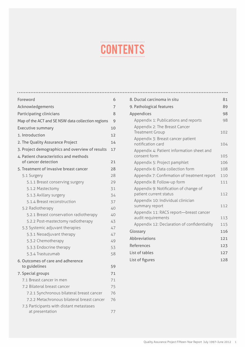
Foreword 6
Acknowledgements 7
Participating clinicians 8
Map of the ACT and SE NSW data collection regions 9
Executive summary 10
1. Introduction 12
2. The Quality Assurance Project 14
3. Project demographics and overview of results 17
4. Patient characteristics and methods of cancer detection 21
5. Treatment of invasive breast cancer 28
5.1 Surgery 28
5.1.1 Breast conserving surgery 29
5.1.2 Mastectomy 31
5.1.3 Axillary surgery 34
5.1.4 Breast reconstruction 37
5.2 Radiotherapy 40
5.2.1 Breast conservation radiotherapy 40
5.2.2 Post-mastectomy radiotherapy 43
5.3 Systemic adjuvant therapies 47
5.3.1 Neoadjuvant therapy 47
5.3.2 Chemotherapy 49
5.3.3 Endocrine therapy 53
5.3.4 Trastuzumab 58
6. Outcomes of care and adherence to guidelines 59
7. Special groups 71
7.1 Breast cancer in men 71
7.2 Bilateral breast cancer 75
7.2.1 Synchronous bilateral breast cancer 76
7.2.2 Metachronous bilateral breast cancer 76
7.3 Participants with distant metastases at presentation 77
8. Ductal carcinoma in situ 81
9. Pathological features 89
Appendices 98
Appendix 1: Publications and reports 98
Appendix 2: The Breast Cancer Treatment Group 102
Appendix 3: Breast cancer patient notification card 104
Appendix 4: Patient information sheet and consent form 105
Appendix 5: Project pamphlet 106
Appendix 6: Data collection form 108
Appendix 7: Confirmation of treatment report 110
Appendix 8: Follow-up form 111
Appendix 9: Notification of change of patient current status 112
Appendix 10: Individual clinician summary report 112
Appendix 11: RACS report—breast cancer audit requirements 113
Appendix 12: Declaration of confidentiality 115
Glossary 116
Abbreviations 121
References 123
List of tables 127
List of figures 128
Contents

2 Breast Cancer Treatment Group
The ACT & SE NSW Breast Cancer Quality Assurance Project is conducted by the ACT and SE NSW Breast Cancer Treatment Group (BCTG) which was created shortly after the release of the National Health and Medical Research Council (NHMRC) Clinical Practice Guidelines for the Management of Early Breast Cancer. One of its aims is to show that the ACT and SE NSW treatment practices demonstrate a close compliance with Australian and international guidelines on breast cancer treatment.
The BCTG comprises clinicians and other practitioners committed to improving the quality of breast cancer treatment delivery and outcomes in the region. Since 1997, ACT Health has funded a small team of quality assurance project officers to support the Group in achieving its goals.
Over the years, the project office team has worked collaboratively with numerous health professionals on collection of patient data. The emphasis on high quality for this data underscores the value of the project. With its high participation rate, the aggregation of individual case data over a prolonged timeframe, and the embedded strong focus on quality assurance, the project is unique.
This report confirms that breast cancer care delivered by the ACT and SE NSW multi-disciplinary teams is of high quality and that breast cancer survival in our region is excellent. The 15-year report is also a testament to the 4,709 women and men who consented to have their information included in the Quality Assurance Project.
FOREWORD

Quality Assurance Project Fifteen-Year Report July 1997–June 2012 3
This report was made possible through the contribution and support of numerous individuals. The main authors of this report were: Paul Craft (Medical Oncologist), Angela Rezo (Radiation Oncologist), Jeremy Price (Radiologist), Carolyn Cho (Breast Surgeon), Robin Stuart-Harris (Medical Oncologist), Jane Dahlstrom (Pathologist), Yanping Zhang (Project Coordinator and Data Manager), Jenny Green (Project Officer), and Kerri Beckmann (Epidemiologist). Members of the ACT & SE NSW Breast Cancer Treatment and Data Management Subcommittee are acknowledged for their active involvement and ongoing support.
The authors acknowledge Yvonne Epping (Director of BreastScreen ACT) and Denise Lamb (Executive Director of Cancer, Ambulatory & Community Health Support) for their management and administrative support. Financial support provided by ACT Health and the John James Memorial Foundation is greatly appreciated.
The authors would like to recognise the current BCTG Quality Assurance Project team: Yanping Zhang, Thet Khin, Jenny Green, and Mary-Claire Tryon for their work on data collection and preliminary data analysis.
Special thanks are given to David Roder (Professor of Cancer Epidemiology), Ian Clark, Jane Twin (Capital Pathology), Peter Green, Kun Zhao, and Katherine Green for their valuable comments and contributions to this report.
The authors would like to acknowledge the assistance of breast surgeons, general surgeons, general practitioners, medical and radiation oncologists, pathologists, radiologists, nurses, and support and administration staff in the ACT and SE NSW regions for their voluntary participation in the collection of data. A large number of pathologists have contributed to the project by providing crucial information. In particular, the authors would like to acknowledge the contribution of pathologists from ACT Pathology and Capital Pathology.
The collaboration of McGrath Nurses, Bosom Buddies, Breast Cancer Network Australia and the ACT and NSW Registries of Births, Deaths and Marriages is gratefully acknowledged.
The Group acknowledges the contribution of the late Associate Professor John Buckingham, who passed away in 2011. Associate Professor Buckingham was a strong supporter of the Group since its inception in 1997 and was a longstanding and valued member of the Data Management Subcommittee. Throughout his career, John was committed to improving the care of women with breast cancer.
Finally, special thanks go to the thousands of participants who consented to have their data collected. This project would not have been possible without their voluntary contribution.
Acknowledgements
Associate Professor John Buckingham AM
4 Breast Cancer Treatment Group
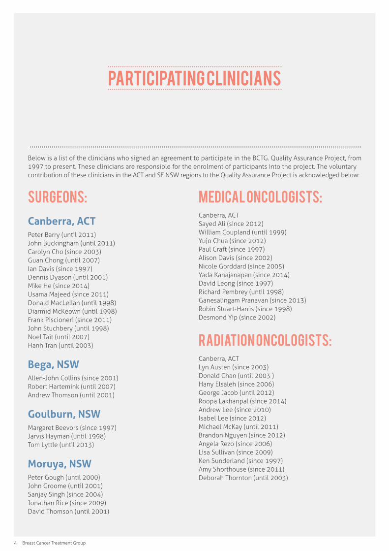
Below is a list of the clinicians who signed an agreement to participate in the BCTG. Quality Assurance Project, from 1997 to present. These clinicians are responsible for the enrolment of participants into the project. The voluntary contribution of these clinicians in the ACT and SE NSW regions to the Quality Assurance Project is acknowledged below:
Participating Clinicians
Surgeons:
Canberra, ACTPeter Barry (until 2011) John Buckingham (until 2011) Carolyn Cho (since 2003) Guan Chong (until 2007) Ian Davis (since 1997) Dennis Dyason (until 2001) Mike He (since 2014) Usama Majeed (since 2011) Donald MacLellan (until 1998) Diarmid McKeown (until 1998) Frank Piscioneri (since 2011) John Stuchbery (until 1998) Noel Tait (until 2007) Hanh Tran (until 2003)
Bega, NSWAllen-John Collins (since 2001) Robert Hartemink (until 2007) Andrew Thomson (until 2001)
Goulburn, NSWMargaret Beevors (since 1997) Jarvis Hayman (until 1998) Tom Lyttle (until 2013)
Moruya, NSWPeter Gough (until 2000) John Groome (until 2001) Sanjay Singh (since 2004) Jonathan Rice (since 2009) David Thomson (until 2001)
Medical Oncologists:Canberra, ACT Sayed Ali (since 2012) William Coupland (until 1999) Yujo Chua (since 2012) Paul Craft (since 1997) Alison Davis (since 2002) Nicole Gorddard (since 2005) Yada Kanajanapan (since 2014) David Leong (since 1997) Richard Pembrey (until 1998) Ganesalingam Pranavan (since 2013) Robin Stuart-Harris (since 1998) Desmond Yip (since 2002)
Radiation Oncologists:Canberra, ACT Lyn Austen (since 2003) Donald Chan (until 2003 ) Hany Elsaleh (since 2006) George Jacob (until 2012) Roopa Lakhanpal (since 2014) Andrew Lee (since 2010) Isabel Lee (since 2012) Michael McKay (until 2011) Brandon Nguyen (since 2012) Angela Rezo (since 2006) Lisa Sullivan (since 2009) Ken Sunderland (since 1997) Amy Shorthouse (since 2011) Deborah Thornton (until 2003)
Quality Assurance Project Fifteen-Year Report July 1997–June 2012 5
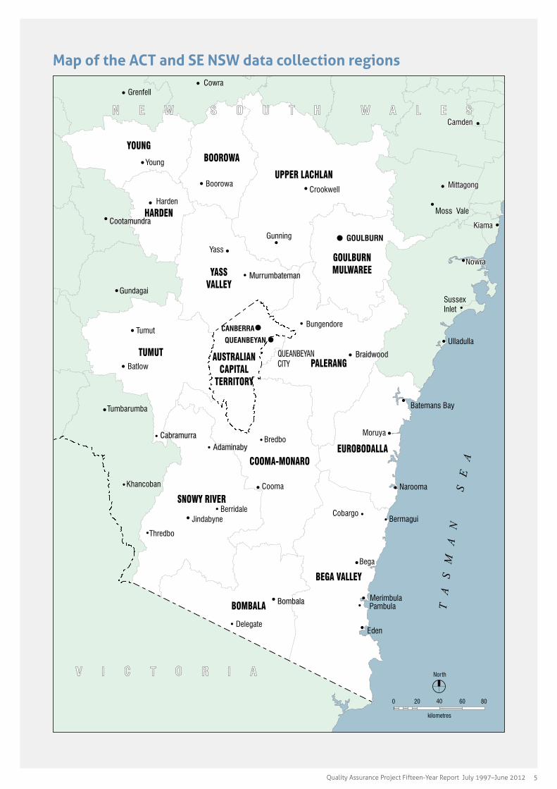
Map of the ACT and SE NSW data collection regions
6 Breast Cancer Treatment Group
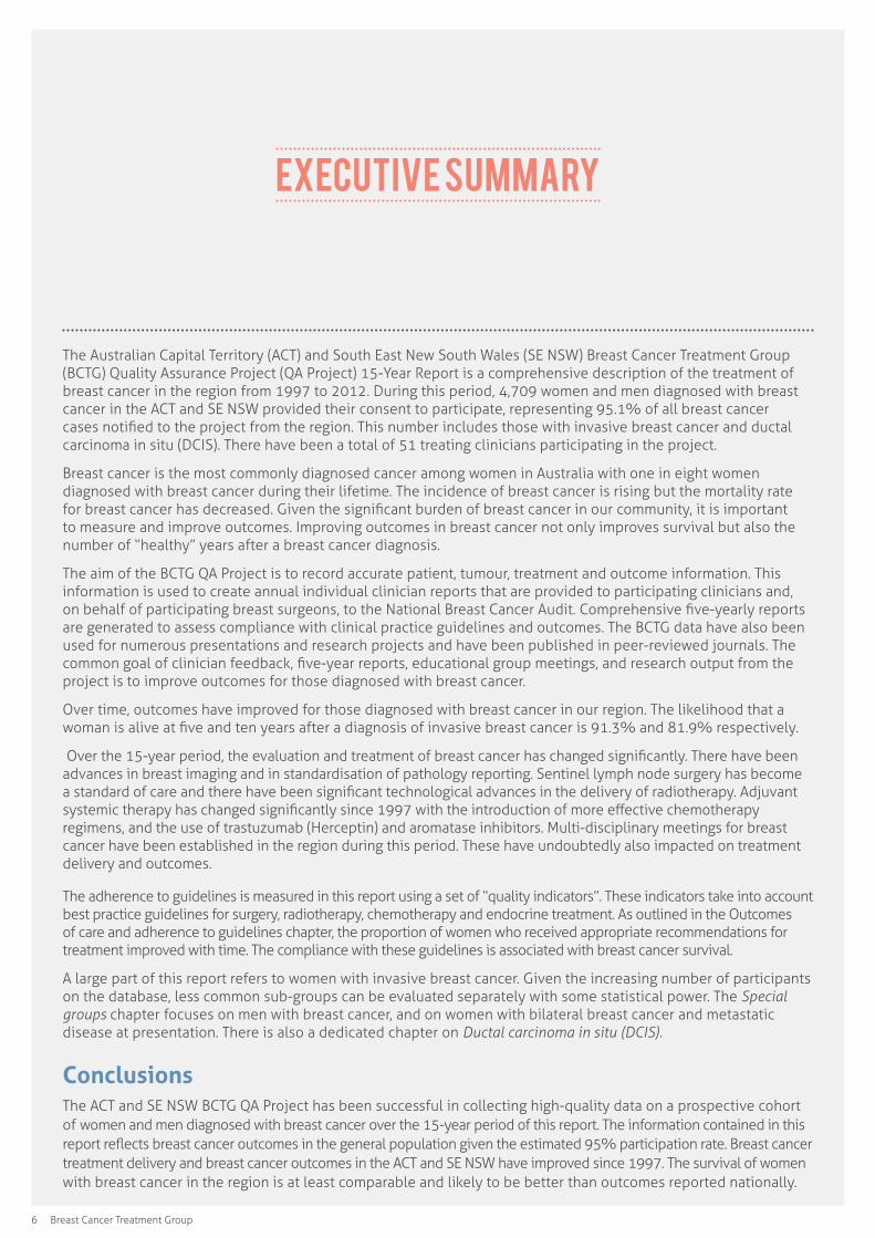
The Australian Capital Territory (ACT) and South East New South Wales (SE NSW) Breast Cancer Treatment Group (BCTG) Quality Assurance Project (QA Project) 15-Year Report is a comprehensive description of the treatment of breast cancer in the region from 1997 to 2012. During this period, 4,709 women and men diagnosed with breast cancer in the ACT and SE NSW provided their consent to participate, representing 95.1% of all breast cancer cases notified to the project from the region. This number includes those with invasive breast cancer and ductal carcinoma in situ (DCIS). There have been a total of 51 treating clinicians participating in the project.
Breast cancer is the most commonly diagnosed cancer among women in Australia with one in eight women diagnosed with breast cancer during their lifetime. The incidence of breast cancer is rising but the mortality rate for breast cancer has decreased. Given the significant burden of breast cancer in our community, it is important to measure and improve outcomes. Improving outcomes in breast cancer not only improves survival but also the number of “healthy” years after a breast cancer diagnosis.
The aim of the BCTG QA Project is to record accurate patient, tumour, treatment and outcome information. This information is used to create annual individual clinician reports that are provided to participating clinicians and, on behalf of participating breast surgeons, to the National Breast Cancer Audit. Comprehensive five-yearly reports are generated to assess compliance with clinical practice guidelines and outcomes. The BCTG data have also been used for numerous presentations and research projects and have been published in peer-reviewed journals. The common goal of clinician feedback, five-year reports, educational group meetings, and research output from the project is to improve outcomes for those diagnosed with breast cancer.
Over time, outcomes have improved for those diagnosed with breast cancer in our region. The likelihood that a woman is alive at five and ten years after a diagnosis of invasive breast cancer is 91.3% and 81.9% respectively.
Over the 15-year period, the evaluation and treatment of breast cancer has changed significantly. There have been advances in breast imaging and in standardisation of pathology reporting. Sentinel lymph node surgery has become a standard of care and there have been significant technological advances in the delivery of radiotherapy. Adjuvant systemic therapy has changed significantly since 1997 with the introduction of more effective chemotherapy regimens, and the use of trastuzumab (Herceptin) and aromatase inhibitors. Multi-disciplinary meetings for breast cancer have been established in the region during this period. These have undoubtedly also impacted on treatment delivery and outcomes.
The adherence to guidelines is measured in this report using a set of “quality indicators”. These indicators take into account best practice guidelines for surgery, radiotherapy, chemotherapy and endocrine treatment. As outlined in the Outcomes of care and adherence to guidelines chapter, the proportion of women who received appropriate recommendations for treatment improved with time. The compliance with these guidelines is associated with breast cancer survival.
A large part of this report refers to women with invasive breast cancer. Given the increasing number of participants on the database, less common sub-groups can be evaluated separately with some statistical power. The Special groups chapter focuses on men with breast cancer, and on women with bilateral breast cancer and metastatic disease at presentation. There is also a dedicated chapter on Ductal carcinoma in situ (DCIS).
ConclusionsThe ACT and SE NSW BCTG QA Project has been successful in collecting high-quality data on a prospective cohort of women and men diagnosed with breast cancer over the 15-year period of this report. The information contained in this report reflects breast cancer outcomes in the general population given the estimated 95% participation rate. Breast cancer treatment delivery and breast cancer outcomes in the ACT and SE NSW have improved since 1997. The survival of women with breast cancer in the region is at least comparable and likely to be better than outcomes reported nationally.
Executive Summary
Quality Assurance Project Fifteen-Year Report July 1997–June 2012 7
Introduction
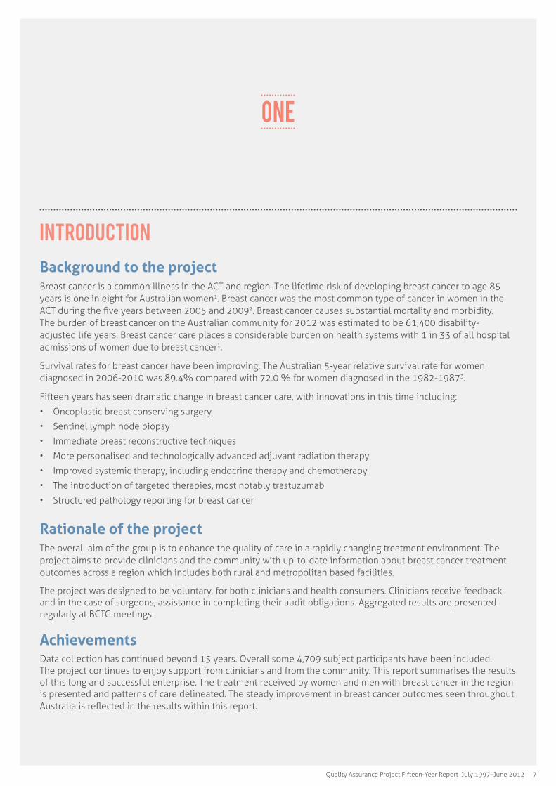
Background to the projectBreast cancer is a common illness in the ACT and region. The lifetime risk of developing breast cancer to age 85 years is one in eight for Australian women1. Breast cancer was the most common type of cancer in women in the ACT during the five years between 2005 and 20092. Breast cancer causes substantial mortality and morbidity. The burden of breast cancer on the Australian community for 2012 was estimated to be 61,400 disability-adjusted life years. Breast cancer care places a considerable burden on health systems with 1 in 33 of all hospital admissions of women due to breast cancer1.
Survival rates for breast cancer have been improving. The Australian 5-year relative survival rate for women diagnosed in 2006-2010 was 89.4% compared with 72.0 % for women diagnosed in the 1982-19873.
Fifteen years has seen dramatic change in breast cancer care, with innovations in this time including:
• Oncoplastic breast conserving surgery
• Sentinel lymph node biopsy
• Immediate breast reconstructive techniques
• More personalised and technologically advanced adjuvant radiation therapy
• Improved systemic therapy, including endocrine therapy and chemotherapy
• The introduction of targeted therapies, most notably trastuzumab
• Structured pathology reporting for breast cancer
Rationale of the projectThe overall aim of the group is to enhance the quality of care in a rapidly changing treatment environment. The project aims to provide clinicians and the community with up-to-date information about breast cancer treatment outcomes across a region which includes both rural and metropolitan based facilities.
The project was designed to be voluntary, for both clinicians and health consumers. Clinicians receive feedback, and in the case of surgeons, assistance in completing their audit obligations. Aggregated results are presented regularly at BCTG meetings.
AchievementsData collection has continued beyond 15 years. Overall some 4,709 subject participants have been included. The project continues to enjoy support from clinicians and from the community. This report summarises the results of this long and successful enterprise. The treatment received by women and men with breast cancer in the region is presented and patterns of care delineated. The steady improvement in breast cancer outcomes seen throughout Australia is reflected in the results within this report.
ONE

8 Breast Cancer Treatment Group
Publications in peer reviewed journalsAlthough not the primary aim of the project, data collection has enabled a number of research focussed activities, including important publications in the peer-reviewed scientific literature (Appendix 1).
Future directionsBreast cancer treatment is continuing to evolve and develop. Improvement has been driven by spectacularly enhanced scientific knowledge of the biology of human breast cancer. Knowledge of treatment effectiveness has been slowly and incrementally gained from carefully conducted clinical trials, leading to enhanced and more specific treatments. With the development of more specifically targeted therapies, and a more personalised approach to treatment, steady improvement in treatment outcome is expected and eagerly anticipated.
There remains a role for treatment-focussed and community-based quality enhancing projects such as the ACT and SE NSW BCTG Project. The discovery of new treatments alone does not ensure accurate and timely implementation into clinical practice. The stimulation and feedback provided by the QA Project contributes to the overall goal of breast cancer control into the future.

Quality Assurance Project Fifteen-Year Report July 1997–June 2012 9
The Quality Assurance Project
Breast Cancer Treatment GroupAll clinicians involved or interested in the control of breast cancer in the ACT and SE NSW were invited to participate in the multidisciplinary Breast Cancer Treatment Group (Appendix 2). In addition, consumer representatives were invited to join the group. Over the 15 years of the project, 51 clinicians have participated at various times. The Breast Cancer Treatment Group adopted treatment guidelines based initially on the National Health & Medical Research Council (NHMRC) Clinical Practice Guidelines4 and developed a set of indicators agreed on by the group, against which treatment decisions could be compared in a community-based audit.
Practice indicatorsThe group initially adopted four indicators for which unanimous local agreement about the relevant guideline was available:
1. Women with completely resected invasive breast cancer greater than two centimetres in diameter or who have axillary lymph node involvement should be recommended adjuvant systemic therapy (either chemotherapy, endocrine therapy, or both).
2. Pre-menopausal women or women under the age of 50 years with completely resected, axillary lymph node positive breast cancer should be recommended adjuvant chemotherapy.
3. Women undergoing less than total mastectomy for operable invasive breast cancer should be recommended post-operative adjuvant radiotherapy.
4. Women with operable invasive breast cancer should be recommended to undergo some form of axillary surgery sufficient to develop an axillary lymph node based prognosis.
In 2015, a fifth indicator was added:
5. Women with invasive breast cancer undergoing mastectomy with 5 or more axillary lymph nodes involved should be recommended to undergo post-mastectomy radiotherapy (PMRT). Ideally women with 4 or more involved nodes should be recommended to have PMRT. The nodal categories in the study that were defined in 1997 (0, 1-4, >4 nodes involved) and were not according to current TNM staging.
TWO
10 Breast Cancer Treatment Group
AuditA community-based prospective study of breast cancer treatment was commenced in May 1997. The study was designed as a Quality Assurance Project and notified as such under Section 7 of the Health Act (1993) in May 1997. The study was approved by the ACT Health Human Research Ethics, Department of Health and Community Care.
All clinicians involved specifically with the care of people with breast cancer were invited to participate in the study. An inclusive culture was generated within the group. Participating clinicians agreed to approach all of their patients presenting with newly-diagnosed breast cancer. Eligible patients were women or men with newly-diagnosed invasive or in situ breast cancer. For each eligible patient a notification form (Appendix 3) was completed and submitted by mail to the BCTG office located within the BreastScreen ACT Program or collected by the Project Officer.
Written informed consent (Appendix 4 and 5) was obtained from the patient by the notifying clinician and, subsequently, a detailed data form completed outlining details of presentation, clinical and pathological staging, and treatment (Appendix 6). The data set was based on a prior survey conducted by the Rural Surgeons Association. Additional data relating to enrolled patients were obtained from treatment units located in the region. Arrangements are in place to link existing client data to the National Death Index to check for the death of any enrolled patients who have not been notified by participating clinicians. Regular checks are also conducted with the ACT and NSW Registries of Births, Deaths and Marriages.
Aggregated data across the whole clinician group is presented at regular meetings of the treatment group. First follow up is at 12 months at which time, the treatment details are confirmed (Appendix 7). Afterwards, a biennial follow-up form requesting details of disease relapse or death is forwarded to each notifying clinician and to the patient’s general practitioner (Appendix 8). The current status of a patient can be reported to the project at any time (Appendix 9). Data collected during the first 15 years of this continuing project forms the basis of this report.
Information from the audit forms, supplemented where necessary from pathology reports and treatment facility records, is entered into a secure relational database.
The database was created by the Project Coordinator specifically for the BCTG Quality Assurance Project using a Microsoft Access platform. The system and Users’ Manual was written by the Project Coordinator with assistance from the Project Officer. The major functions of the database are data entry, secure storage and management of data, generation of data query letters and provision of summarised results. On behalf of participating breast surgeons, the database provides an individualised annual report to the Royal Australasian College of Surgeons—National Breast Cancer Audit (Appendix 11). As well as assisting in managing the project, the database incorporates facilities to produce individualised, confidential reports for each participating clinician (Appendix 10). These reports provide detailed individual feedback to each clinician about their own practice, with comparisons across the whole group and against the agreed criteria. Any data collected from clinicians remains confidential and identifying information is only available to the individual clinician. Data collected remain secure within the BCTG Project office. Only aggregated non-identifying information is released in project reports or research. Collection of data is governed by the Privacy Act 1988 (C’wlth) and the ACT Information Privacy Act, and all staff are required to sign a declaration of confidentiality (Appendix 12).
Data analysis was performed using IBM SPSS Version 22, except where specified.
Quality Assurance Project Fifteen-Year Report July 1997–June 2012 11
Project demographics and overview of resultsThe results for the 15-year report have been collected from patients treated from 1 July 1997 to 30 June 2012 with follow-up censored on 3 March 2015. During the 15-year period, 4,709 patients gave their consent to participate in the project, representing 95% of all patients notified by participating clinicians in the region. In the ACT, 2,479 participants were registered over the 15 years – an average of 165 per year. Although not directly comparable, an average of 207 cases per year were registered to the ACT Cancer Registry during the period 2004 to 2008. The age-standardised incidence of breast cancer in the ACT is 123.9 cases per 100,000 female population5.
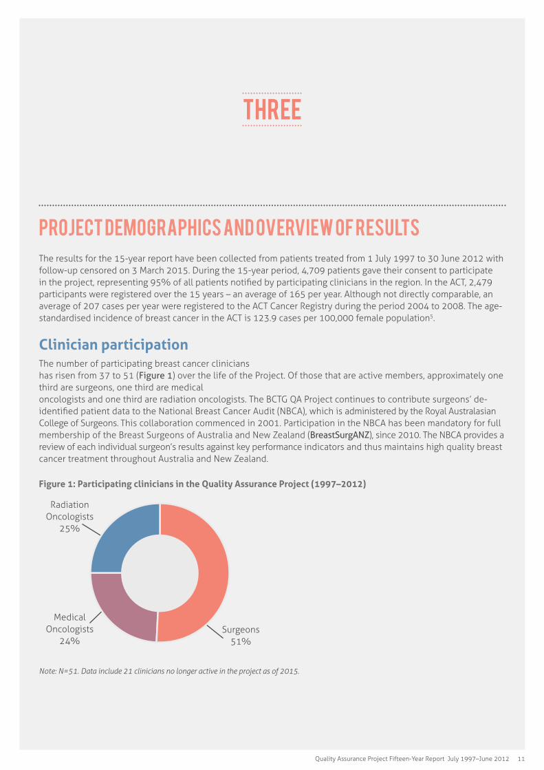
Clinician participation The number of participating breast cancer clinicians has risen from 37 to 51 (Figure 1) over the life of the Project. Of those that are active members, approximately one third are surgeons, one third are medical oncologists and one third are radiation oncologists. The BCTG QA Project continues to contribute surgeons’ de-identified patient data to the National Breast Cancer Audit (NBCA), which is administered by the Royal Australasian College of Surgeons. This collaboration commenced in 2001. Participation in the NBCA has been mandatory for full membership of the Breast Surgeons of Australia and New Zealand (BreastSurgANZ), since 2010. The NBCA provides a review of each individual surgeon’s results against key performance indicators and thus maintains high quality breast cancer treatment throughout Australia and New Zealand.
Figure 1: Participating clinicians in the Quality Assurance Project (1997–2012)
Surgeons 51%
Radiation Oncologists
25%
Medical Oncologists
24%
Note: N=51. Data include 21 clinicians no longer active in the project as of 2015.
Three
12 Breast Cancer Treatment Group
Patient participationThe total number of breast cancer notifications to the BCTG office was 4,949. Of these, 4,709 patients provided consent to participate. Reasons for non-participation included inability to contact patients (184 cases), consent declined by patients (33 cases), and ineligibility (23 cases). The mean age at diagnosis of breast cancer in this cohort of participants was 58.05 years for women and 62.4 years for men. The majority of participants were diagnosed between the ages of 50-69 (56.9%). Participants who were less than 40 years old comprised 6.4%, and those greater than 80 years old comprised 4.7% of the cohort. During this 15-year period, there were 28 male participants.
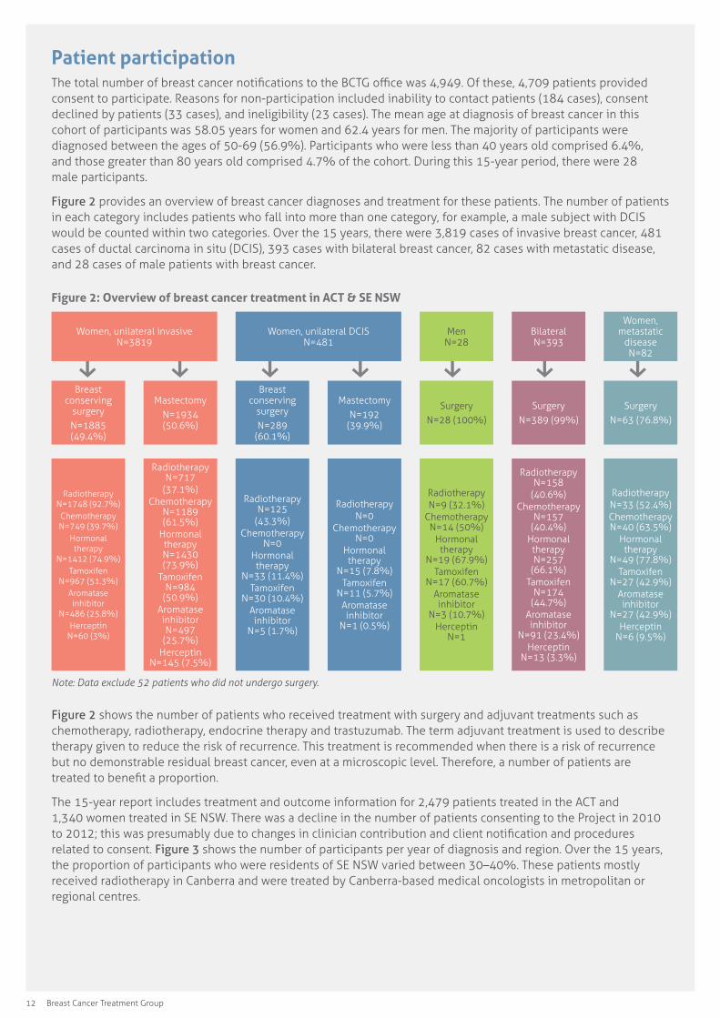
Figure 2 provides an overview of breast cancer diagnoses and treatment for these patients. The number of patients in each category includes patients who fall into more than one category, for example, a male subject with DCIS would be counted within two categories. Over the 15 years, there were 3,819 cases of invasive breast cancer, 481 cases of ductal carcinoma in situ (DCIS), 393 cases with bilateral breast cancer, 82 cases with metastatic disease, and 28 cases of male patients with breast cancer.
Figure 2: Overview of breast cancer treatment in ACT & SE NSW
Women, unilateral invasive N=3819
Women, unilateral DCIS N=481
Men N=28
Bilateral N=393
Women, metastatic
disease N=82
Breast conserving
surgeryN=1885 (49.4%)
MastectomyN=1934 (50.6%)
Breast conserving
surgeryN=289
(60.1%)
MastectomyN=192
(39.9%)
SurgeryN=28 (100%)
SurgeryN=389 (99%)
SurgeryN=63 (76.8%)
Radiotherapy N=1748 (92.7%)Chemotherapy N=749 (39.7%)
Hormonal therapy
N=1412 (74.9%)Tamoxifen
N=967 (51.3%)Aromatase inhibitor
N=486 (25.8%)Herceptin
N=60 (3%)
Radiotherapy N=717
(37.1%)Chemotherapy
N=1189 (61.5%)
Hormonal therapy N=1430 (73.9%)
Tamoxifen N=984
(50.9%)Aromatase inhibitor N=497
(25.7%)Herceptin
N=145 (7.5%)
Radiotherapy N=125
(43.3%)Chemotherapy
N=0Hormonal therapy
N=33 (11.4%)Tamoxifen
N=30 (10.4%)Aromatase inhibitor
N=5 (1.7%)
RadiotherapyN=0
Chemotherapy N=0
Hormonal therapy
N=15 (7.8%)Tamoxifen
N=11 (5.7%)Aromatase inhibitor
N=1 (0.5%)
RadiotherapyN=9 (32.1%)
Chemotherapy N=14 (50%)
Hormonal therapy
N=19 (67.9%)Tamoxifen
N=17 (60.7%)Aromatase inhibitor
N=3 (10.7%)Herceptin
N=1
Radiotherapy N=158
(40.6%)Chemotherapy
N=157 (40.4%)
Hormonal therapy N=257
(66.1%)Tamoxifen
N=174 (44.7%)
Aromatase inhibitor
N=91 (23.4%)Herceptin
N=13 (3.3%)
RadiotherapyN=33 (52.4%)Chemotherapy N=40 (63.5%)
Hormonal therapy
N=49 (77.8%)Tamoxifen
N=27 (42.9%)Aromatase inhibitor
N=27 (42.9%)Herceptin
N=6 (9.5%)
Note: Data exclude 52 patients who did not undergo surgery.
Figure 2 shows the number of patients who received treatment with surgery and adjuvant treatments such as chemotherapy, radiotherapy, endocrine therapy and trastuzumab. The term adjuvant treatment is used to describe therapy given to reduce the risk of recurrence. This treatment is recommended when there is a risk of recurrence but no demonstrable residual breast cancer, even at a microscopic level. Therefore, a number of patients are treated to benefit a proportion.
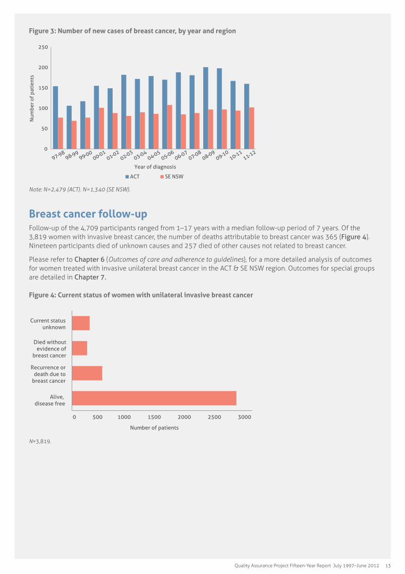
The 15-year report includes treatment and outcome information for 2,479 patients treated in the ACT and 1,340 women treated in SE NSW. There was a decline in the number of patients consenting to the Project in 2010 to 2012; this was presumably due to changes in clinician contribution and client notification and procedures related to consent. Figure 3 shows the number of participants per year of diagnosis and region. Over the 15 years, the proportion of participants who were residents of SE NSW varied between 30–40%. These patients mostly received radiotherapy in Canberra and were treated by Canberra-based medical oncologists in metropolitan or regional centres.
Quality Assurance Project Fifteen-Year Report July 1997–June 2012 13
Figure 3: Number of new cases of breast cancer, by year and region
0
50
100
150
200
250
97-98 98-99 99-00
00-01 01-02
02-03 03-04
04-05 05-06
06-07 07-08
08-09 09-10
10-11 11-12
Num
ber o
f pa
tien
ts
Year of diagnosis
ACT SE NSW
Note: N=2,479 (ACT). N=1,340 (SE NSW).
Breast cancer follow-upFollow-up of the 4,709 participants ranged from 1–17 years with a median follow-up period of 7 years. Of the 3,819 women with invasive breast cancer, the number of deaths attributable to breast cancer was 365 (Figure 4). Nineteen participants died of unknown causes and 257 died of other causes not related to breast cancer.
Please refer to Chapter 6 (Outcomes of care and adherence to guidelines), for a more detailed analysis of outcomes for women treated with invasive unilateral breast cancer in the ACT & SE NSW region. Outcomes for special groups are detailed in Chapter 7.
Figure 4: Current status of women with unilateral invasive breast cancer
0 500 1000 1500 2000 2500 3000
Alive, disease free
Recurrence or death due to
breast cancer
Died without evidence of
breast cancer
Current status unknown
Number of patients
N=3,819.
14 Breast Cancer Treatment Group
Patient characteristics and methods of cancer detectionThere are a number of risk factors associated with breast cancer including increasing age, living in an affluent country such as Australia, family history of breast or ovarian cancer, personal history of breast cancer or pre-cancerous breast condition, high breast density and, for post-menopausal women, high levels of circulating oestrogens6.
The national screening program, BreastScreen Australia, was established in 1991 and provides 2-yearly screening mammograms for women aged 40 and over and actively invites women aged 50–74 years to participate. Since the inception of the BreastScreen program, there has been a steady decline in breast cancer mortality7. In Australia, the National Breast and Ovarian Cancer Centre (NBOCC) categorises women into three groups based on family history: NBOCC Category 1 is average or slightly above average risk of developing breast cancer, NBOCC Category 2 is moderately increased risk and NBOCC Category 3 is potentially high risk. About 5% of all breast cancers can be attributed to inheritance of a genetic mutation such as BRCA1 or BRCA2. Since February 2009, a rebate for annual MRI has been available (on the Medicare Benefits Schedule) for women in NBOCC Category 3 who are under the age of 50 years and have been referred by a specialist.
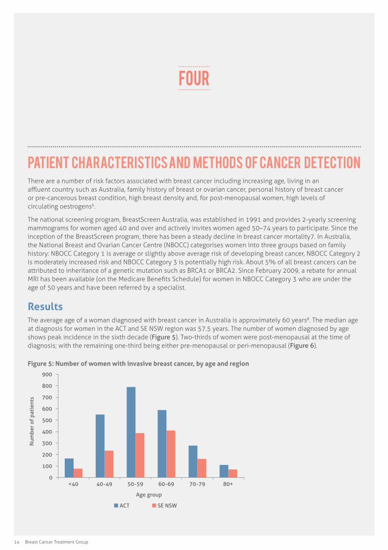
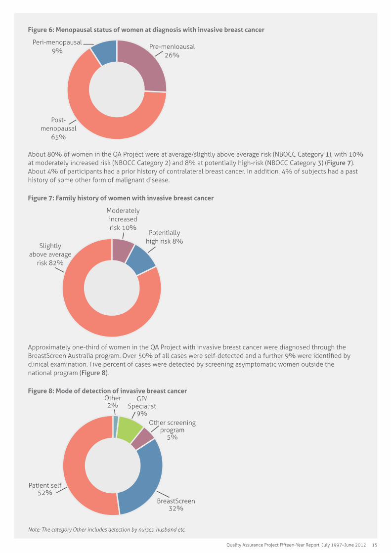
ResultsThe average age of a woman diagnosed with breast cancer in Australia is approximately 60 years8. The median age at diagnosis for women in the ACT and SE NSW region was 57.5 years. The number of women diagnosed by age shows peak incidence in the sixth decade (Figure 5). Two-thirds of women were post-menopausal at the time of diagnosis; with the remaining one-third being either pre-menopausal or peri-menopausal (Figure 6).
Figure 5: Number of women with invasive breast cancer, by age and region
0
100
200
300
400
500
600
700
800
900
<40 40-49 50-59 60-69 70-79 80+
Num
ber
of p
atie
nts
Age group
ACT SE NSW
FOUR
Quality Assurance Project Fifteen-Year Report July 1997–June 2012 15
Figure 6: Menopausal status of women at diagnosis with invasive breast cancer
Post- menopausal
65%
Pre-menioausal 26%
Peri-menopausal 9%
About 80% of women in the QA Project were at average/slightly above average risk (NBOCC Category 1), with 10% at moderately increased risk (NBOCC Category 2) and 8% at potentially high-risk (NBOCC Category 3) (Figure 7). About 4% of participants had a prior history of contralateral breast cancer. In addition, 4% of subjects had a past history of some other form of malignant disease.
Figure 7: Family history of women with invasive breast cancer
Slightly above average
risk 82%
Potentially high risk 8%
Moderately increased risk 10%
Approximately one-third of women in the QA Project with invasive breast cancer were diagnosed through the BreastScreen Australia program. Over 50% of all cases were self-detected and a further 9% were identified by clinical examination. Five percent of cases were detected by screening asymptomatic women outside the national program (Figure 8).
Figure 8: Mode of detection of invasive breast cancer
Patient self 52%
Other 2%
GP/Specialist
9%Other screening
program 5%
BreastScreen 32%
Note: The category Other includes detection by nurses, husband etc.
16 Breast Cancer Treatment Group
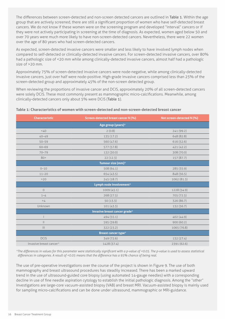
The differences between screen-detected and non-screen detected cancers are outlined in Table 1. Within the age group that are actively screened, there are still a significant proportion of women who have self-detected breast cancers. We do not know if these women were on the screening program and developed “interval” cancers or if they were not actively participating in screening at the time of diagnosis. As expected, women aged below 50 and over 70 years were much more likely to have non-screen-detected cancers. Nevertheless, there were 22 women over the age of 80 years who had screen-detected cancers.
As expected, screen-detected invasive cancers were smaller and less likely to have involved lymph nodes when compared to self-detected or clinically-detected invasive cancers. For screen-detected invasive cancers, over 80% had a pathologic size of <20 mm while among clinically-detected invasive cancers, almost half had a pathologic size of >20 mm.
Approximately 75% of screen-detected invasive cancers were node-negative, while among clinically-detected invasive cancers, just over half were node-positive. High-grade invasive cancers comprised less than 25% of the screen-detected group and approximately 45% of the non-screen detected group.
When reviewing the proportions of invasive cancer and DCIS, approximately 20% of all screen-detected cancers were solely DCIS. These most commonly present as mammographic micro-calcifications. Meanwhile, among clinically-detected cancers only about 5% were DCIS (Table 1).
Table 1: Characteristics of women with screen-detected and non-screen-detected breast cancer
Characteristic Screen-detected breast cancer N (%) Not screen-detected N (%)
Age group (years)*
<40 2 (0.8) 241 (99.2)
40–49 135 (17.2) 648 (82.8)
50–59 560 (47.6) 616 (52.6)
60–69 577 (57.8) 421 (42.2)
70–79 132 (30.0) 308 (70.0)
80+ 22 (12.3) 157 (87.7)
Tumour size (mm)*
0–10 508 (64.1) 285 (35.9)
11–20 654 (43.5) 848 (56.5)
>20 245 (18.7) 1062 (81.3)
Lymph node involvement*
0 1009 (45.1) 1228 (54.9)
1–4 268 (27.5) 705 (72.5)
>4 50 (13.3) 326 (86.7)
Unknown 101 (43.3) 132 (56.7)
Invasive breast cancer grade*
I 494 (55.1) 402 (44.9)
II 595 (39.8) 900 (60.2)
III 322 (23.2) 1065 (76.8)
Breast cancer type*
DCIS 349 (72.6) 132 (27.4)
Invasive breast cancer* 1428 (37.4) 2391 (62.6)
*�The�differences�in�values�for�this�parameter�were�statistically�significant�with�a�p-value�of�<0.05.�The�p-value�is�used�to�assess�statistical�differences�in�categories.�A�result�of�<0.05�means�that�the�difference�has�a�95%�chance�of�being�real.
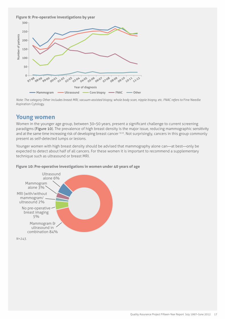
The use of pre-operative investigations over the course of the project is shown in Figure 9. The use of both mammography and breast ultrasound procedures has steadily increased. There has been a marked upward trend in the use of ultrasound-guided core biopsy (using automated 14-gauge needles) with a corresponding decline in use of fine needle aspiration cytology to establish the initial pathologic diagnosis. Among the “other” investigations are large-core vacuum-assisted biopsy (VAB) and breast MRI. Vacuum-assisted biopsy is mainly used for sampling micro-calcifications and can be done under ultrasound, mammographic or MRI-guidance.
Quality Assurance Project Fifteen-Year Report July 1997–June 2012 17
Figure 9: Pre-operative investigations by year
0
50
100
150
200
250
300
Num
ber
of p
atie
nts
Year of diagnosis
Mammogram Ultrasound Core biopsy FNAC Other
97-98 98-99 99-00
00-01 01-02
02-03 03-04
04-05 05-06
06-07 07-08
08-09 09-10
10-11 11-12
Note:�The�category�Other�includes�breast�MRI,�vacuum-assisted�biopsy,�whole�body�scan,�nipple�biopsy,�etc.�FNAC�refers�to�Fine Needle Aspiration Cytology.
Young womenWomen in the younger age group, between 30–50 years, present a significant challenge to current screening paradigms (Figure 10). The prevalence of high breast density is the major issue, reducing mammographic sensitivity and at the same time increasing risk of developing breast cancer 9,10. Not surprisingly, cancers in this group commonly present as self-detected lumps or lesions.
Younger women with high breast density should be advised that mammography alone can—at best—only be expected to detect about half of all cancers. For these women it is important to recommend a supplementary technique such as ultrasound or breast MRI.
Figure 10: Pre-operative investigations in women under 40 years of age
Mammogram & ultrasound in
combination 84%
No pre-operative breast imaging
5%
MRI (with/without mammogram/ultrasound 2%
Mammogram alone 3%
Ultrasound alone 6%
N=243.

18 Breast Cancer Treatment Group
Future directionsIncreased imaging surveillance with either supplementary ultrasound or MRI is increasingly advocated for women with increased risk of breast cancer. Breast MRI is the preferred imaging method as the sensitivity is approximately double that of mammography and ultrasound combined11. In 2012, a total of 140 Medicare-rebated breast MRI scans were performed at The Canberra Hospital alone. Women who have had a previous diagnosis of breast cancer have a two-to-three-fold increased risk of a contralateral primary breast cancer being diagnosed (when compared to the risk of an unaffected woman receiving a first breast cancer diagnosis) and this risk increases further if there is also a significant family history12. Emerging data suggest that these women should also be considered as “high-risk” with similar yields of otherwise occult cancers detected by supplementary screening with MRI in a number of recent studies13,14,15.
In the continuing data collection, it will be important to clearly define the imaging technique which made the primary cancer diagnosis, be it mammography, tomosynthesis, ultrasound or breast MRI. Currently, this crucial information is not resolved in the category of pre-operative investigations. Anecdotal evidence suggests a significant number of instances in which the initial diagnosis was made by a screening breast MRI study. Unfortunately this information is not adequately captured in the current data collection.

Quality Assurance Project Fifteen-Year Report July 1997–June 2012 19
Five
Treatment of invasive breast cancer
5.1 SurgeryThe QA Project has collected data on the surgical management of breast cancer for over 15 years. Over this time there have been many changes in both surgical techniques as well as the philosophy of surgical treatment. This is due to advances in surgical techniques, changes in practice as a result of clinical trials, the influence of clinical practice guidelines, and increasing patient awareness and demand for modern techniques.
The primary aim of surgery for breast cancer is complete resection of the primary tumour as well as assessment of lymph node status. It remains the most common initial treatment for patients with early breast cancer.
Results
Over the study period, the majority of surgical procedures were performed within the ACT. In the last five years, there has been an increase in procedures performed in rural SE NSW locations, as seen in Figure 11. This may be explained by an increase in breast surgeons working in those areas.
Figure 11: Proportion of women with invasive breast cancer, by place of surgery and year
0
10
20
30
40
50
60
70
80
90
100
Prop
orti
on o
f pa
tien
ts
Year of diagnosis
Urban Rural Outside regions
Note: The category Outside regions refers to clinicians not located in ACT or SE NSW.
20 Breast Cancer Treatment Group
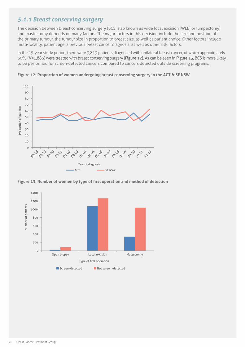
5.1.1 Breast conserving surgeryThe decision between breast conserving surgery (BCS, also known as wide local excision [WLE] or lumpectomy) and mastectomy depends on many factors. The major factors in this decision include the size and position of the primary tumour, the tumour size in proportion to breast size, as well as patient choice. Other factors include multi-focality, patient age, a previous breast cancer diagnosis, as well as other risk factors.
In the 15-year study period, there were 3,819 patients diagnosed with unilateral breast cancer, of which approximately 50% (N=1,885) were treated with breast conserving surgery (Figure 12). As can be seen in Figure 13, BCS is more likely to be performed for screen-detected cancers compared to cancers detected outside screening programs.
Figure 12: Proportion of women undergoing breast conserving surgery in the ACT & SE NSW
0
10
20
30
40
50
60
70
80
90
100
Prop
orti
on o
f pa
tien
ts
Year of diagnosis
ACT SE NSW
Figure 13: Number of women by type of first operation and method of detection
0
200
400
600
800
1000
1200
1400
Open biopsy Local excision Mastectomy
Num
ber
of p
atie
nts
Type of first operation
Screen-detected Not screen -detected
Quality Assurance Project Fifteen-Year Report July 1997–June 2012 21
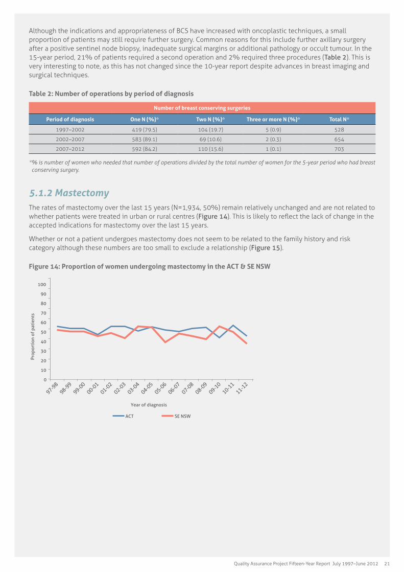
Although the indications and appropriateness of BCS have increased with oncoplastic techniques, a small proportion of patients may still require further surgery. Common reasons for this include further axillary surgery after a positive sentinel node biopsy, inadequate surgical margins or additional pathology or occult tumour. In the 15-year period, 21% of patients required a second operation and 2% required three procedures (Table 2). This is very interesting to note, as this has not changed since the 10-year report despite advances in breast imaging and surgical techniques.
Table 2: Number of operations by period of diagnosis
Number of breast conserving surgeries
Period of diagnosis One N (%)* Two N (%)* Three or more N (%)* Total N*
1997–2002 419 (79.5) 104 (19.7) 5 (0.9) 528
2002–2007 583 (89.1) 69 (10.6) 2 (0.3) 654
2007–2012 592 (84.2) 110 (15.6) 1 (0.1) 703
*�%�is�number�of�women�who�needed�that�number�of�operations�divided�by�the�total�number�of�women�for�the�5-year�period�who�had�breast�conserving surgery.
5.1.2 MastectomyThe rates of mastectomy over the last 15 years (N=1,934, 50%) remain relatively unchanged and are not related to whether patients were treated in urban or rural centres (Figure 14). This is likely to reflect the lack of change in the accepted indications for mastectomy over the last 15 years.
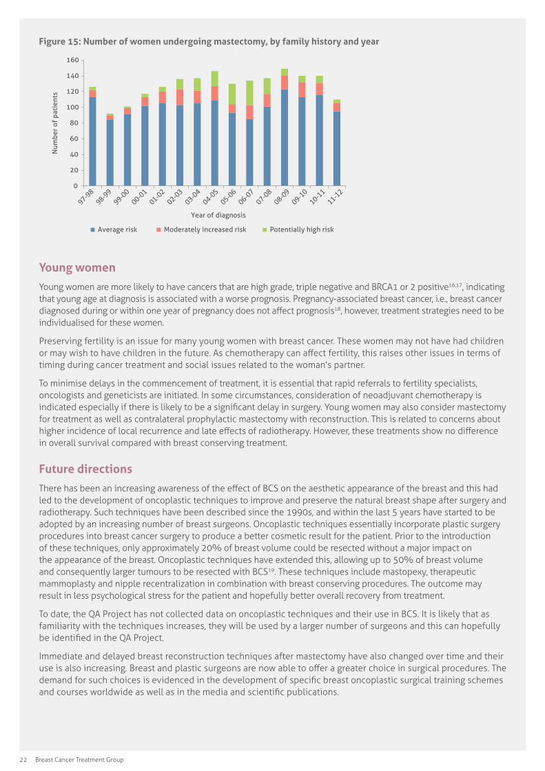
Whether or not a patient undergoes mastectomy does not seem to be related to the family history and risk category although these numbers are too small to exclude a relationship (Figure 15).
Figure 14: Proportion of women undergoing mastectomy in the ACT & SE NSW
0
10
20
30
40
50
60
70
80
90
100
Prop
orti
on o
f pa
tien
ts
Year of diagnosis
ACT SE NSW
22 Breast Cancer Treatment Group
Figure 15: Number of women undergoing mastectomy, by family history and year
0
20
40
60
80
100
120
140
160 N
umbe
r of
pat
ient
s
Year of diagnosis
Average risk Moderately increased risk Potentially high risk
Young women
Young women are more likely to have cancers that are high grade, triple negative and BRCA1 or 2 positive16,17, indicating that young age at diagnosis is associated with a worse prognosis. Pregnancy-associated breast cancer, i.e., breast cancer diagnosed during or within one year of pregnancy does not affect prognosis18, however, treatment strategies need to be individualised for these women.
Preserving fertility is an issue for many young women with breast cancer. These women may not have had children or may wish to have children in the future. As chemotherapy can affect fertility, this raises other issues in terms of timing during cancer treatment and social issues related to the woman’s partner.
To minimise delays in the commencement of treatment, it is essential that rapid referrals to fertility specialists, oncologists and geneticists are initiated. In some circumstances, consideration of neoadjuvant chemotherapy is indicated especially if there is likely to be a significant delay in surgery. Young women may also consider mastectomy for treatment as well as contralateral prophylactic mastectomy with reconstruction. This is related to concerns about higher incidence of local recurrence and late effects of radiotherapy. However, these treatments show no difference in overall survival compared with breast conserving treatment.
Future directions
There has been an increasing awareness of the effect of BCS on the aesthetic appearance of the breast and this had led to the development of oncoplastic techniques to improve and preserve the natural breast shape after surgery and radiotherapy. Such techniques have been described since the 1990s, and within the last 5 years have started to be adopted by an increasing number of breast surgeons. Oncoplastic techniques essentially incorporate plastic surgery procedures into breast cancer surgery to produce a better cosmetic result for the patient. Prior to the introduction of these techniques, only approximately 20% of breast volume could be resected without a major impact on the appearance of the breast. Oncoplastic techniques have extended this, allowing up to 50% of breast volume and consequently larger tumours to be resected with BCS19. These techniques include mastopexy, therapeutic mammoplasty and nipple recentralization in combination with breast conserving procedures. The outcome may result in less psychological stress for the patient and hopefully better overall recovery from treatment.
To date, the QA Project has not collected data on oncoplastic techniques and their use in BCS. It is likely that as familiarity with the techniques increases, they will be used by a larger number of surgeons and this can hopefully be identified in the QA Project.
Immediate and delayed breast reconstruction techniques after mastectomy have also changed over time and their use is also increasing. Breast and plastic surgeons are now able to offer a greater choice in surgical procedures. The demand for such choices is evidenced in the development of specific breast oncoplastic surgical training schemes and courses worldwide as well as in the media and scientific publications.

Quality Assurance Project Fifteen-Year Report July 1997–June 2012 23
The most recent Clinical Practice Guidelines on the Management of Early Breast Cancer in patients with BRCA1 or BRCA2 mutations20 state that BCS or mastectomy are both acceptable surgical treatments for women with BRCA1 or BRCA2 mutations, however, there is an increased risk of ipsilateral recurrence in these women following BCS. Rapid genetic testing can also influence a patient’s decision between BCS and mastectomy. Public awareness of mastectomy and reconstruction has also been raised following reports of the use of these procedures in high-profile patients. The most notable was Angelina Jolie, who underwent risk-reducing mastectomy and reconstruction in 201321. Such reports have also made these procedures more socially acceptable as part of breast cancer management.
5.1.3 Axillary surgeryThe nodal status in women with newly diagnosed breast cancer remains an important prognostic factor that guides surgical management and adjuvant therapy. Pre-operative assessment of axillary and other lymph node regions is performed mainly by clinical assessment and axillary ultrasound and less commonly by other imaging modalities. Ultrasound is a more accurate method of assessing lymph nodes compared to clinical examination and it also allows biopsy of any suspicious lymph nodes. The use of pre-operative ultrasound reflects a change in clinical practice since the publication of the Clinical Practice Guidelines in 2011 and 200822,23.
Patients who have no evidence of lymph node metastases (negative lymph nodes) are recommended sentinel lymph node biopsy (SNB) initially. SNB enables identification of the first draining lymph nodes of the breast, using a combination of radioactive dye (technetium 99m-labelled antimony colloid) and blue dye (Patent V Blue). These lymph nodes are removed at surgery and can be tested intraoperatively as well as by formal histology. If the sentinel nodes are negative, then axillary clearance and potential significant morbidity can be avoided. Recent results from the SNAC1 trial have shown a persistent benefit in the reduction of side effects such as arm swelling, pain and dysfunction24.
The Clinical Practice Guidelines of 2011 also recommended that SNB be performed in unifocal tumours <3 cm in size22.
Results
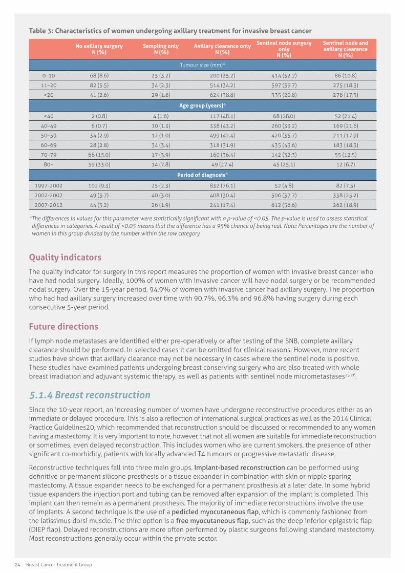
Table 3 shows the change in trends from axillary clearance or sampling towards SNB during the 5-year treatment periods of the QA Project. In the most recent 5-year period, sentinel node surgery with or without axillary dissection was performed in 77.5% of women compared to 12.3% of women in the earliest 5-year period. SNB is associated with a reduction in surgical morbidity such as lymphoedema, shoulder stiffness and dysaesthesia. The avoidance of axillary clearance results in a faster recovery from surgery and return to normal function.
The use of SNB also correlates with tumour size. Larger tumours greater than 20 mm are more likely to have lymph node metastases and therefore require axillary clearance. Conversely, smaller tumours are more likely to be lymph node negative and only require SNB. A large proportion of these smaller tumours are detected through breast screening.
Another notable trend is that women older than 70 and especially those older than 80 are the group who have the highest rate of no axillary surgery. This may be a decision made with the intent to reduce morbidity from surgery or possibly made as knowledge of nodal status would not change management.
24 Breast Cancer Treatment Group
Table 3: Characteristics of women undergoing axillary treatment for invasive breast cancer
No axillary surgery N (%)
Sampling only N (%)
Axillary clearance only N (%)
Sentinel node surgery only
N (%)
Sentinel node and axillary clearance
N (%)
Tumour size (mm)*
0–10 68 (8.6) 25 (3.2) 200 (25.2) 414 (52.2) 86 (10.8)
11–20 82 (5.5) 34 (2.3) 514 (34.2) 597 (39.7) 275 (18.3)
>20 41 (2.6) 29 (1.8) 624 (38.8) 335 (20.8) 278 (17.3)
Age group (years)*
<40 2 (0.8) 4 (1.6) 117 (48.1) 68 (28.0) 52 (21.4)
40–49 6 (0.7) 10 (1.3) 338 (43.2) 260 (33.2) 169 (21.6)
50–59 34 (2.9) 12 (1.0) 499 (42.4) 420 (35.7) 211 (17.9)
60–69 28 (2.8) 34 (3.4) 318 (31.9) 435 (43.6) 183 (18.3)
70–79 66 (15.0) 17 (3.9) 160 (36.4) 142 (32.3) 55 (12.5)
80+ 59 (33.0) 14 (7.8) 49 (27.4) 45 (25.1) 12 (6.7)
Period of diagnosis*
1997-2002 102 (9.3) 25 (2.3) 832 (76.1) 52 (4.8) 82 (7.5)
2002-2007 49 (3.7) 40 (3.0) 408 (30.4) 506 (37.7) 338 (25.2)
2007-2012 44 (3.2) 26 (1.9) 241 (17.4) 812 (58.6) 262 (18.9)
*�The�differences�in�values�for�this�parameter�were�statistically�significant�with�a�p-value�of�<0.05.�The�p-value�is�used�to�assess�statistical�differences�in�categories.�A�result�of�<0.05�means�that�the�difference�has�a�95%�chance�of�being�real.�Note:�Percentages�are�the�number�of�women in this group divided by the number within the row category.
Quality indicators
The quality indicator for surgery in this report measures the proportion of women with invasive breast cancer who have had nodal surgery. Ideally, 100% of women with invasive cancer will have nodal surgery or be recommended nodal surgery. Over the 15-year period, 94.9% of women with invasive cancer had axillary surgery. The proportion who had had axillary surgery increased over time with 90.7%, 96.3% and 96.8% having surgery during each consecutive 5-year period.
Future directions
If lymph node metastases are identified either pre-operatively or after testing of the SNB, complete axillary clearance should be performed. In selected cases it can be omitted for clinical reasons. However, more recent studies have shown that axillary clearance may not be necessary in cases where the sentinel node is positive. These studies have examined patients undergoing breast conserving surgery who are also treated with whole breast irradiation and adjuvant systemic therapy, as well as patients with sentinel node micrometastases25,26.
5.1.4 Breast reconstructionSince the 10-year report, an increasing number of women have undergone reconstructive procedures either as an immediate or delayed procedure. This is also a reflection of international surgical practices as well as the 2014 Clinical Practice Guidelines20, which recommended that reconstruction should be discussed or recommended to any woman having a mastectomy. It is very important to note, however, that not all women are suitable for immediate reconstruction or sometimes, even delayed reconstruction. This includes women who are current smokers, the presence of other significant co-morbidity, patients with locally advanced T4 tumours or progressive metastatic disease.
Reconstructive techniques fall into three main groups. Implant-based reconstruction can be performed using definitive or permanent silicone prosthesis or a tissue expander in combination with skin or nipple sparing mastectomy. A tissue expander needs to be exchanged for a permanent prosthesis at a later date. In some hybrid tissue expanders the injection port and tubing can be removed after expansion of the implant is completed. This implant can then remain as a permanent prosthesis. The majority of immediate reconstructions involve the use of implants. A second technique is the use of a pedicled myocutaneous flap, which is commonly fashioned from the latissimus dorsi muscle. The third option is a free myocutaneous flap, such as the deep inferior epigastric flap (DIEP flap). Delayed reconstructions are more often performed by plastic surgeons following standard mastectomy. Most reconstructions generally occur within the private sector.
Quality Assurance Project Fifteen-Year Report July 1997–June 2012 25
The decision between immediate and delayed reconstruction can be complex27 and there is much debate within the surgical literature as to the most appropriate approach.
Approximately 50% of patients undergoing mastectomy and reconstruction also require radiotherapy as the risk of local recurrence is high enough to warrant this adjuvant treatment. Radiation related fibrosis affects the skin microcirculation and the quality of the tissue in general. It can increase the risk of infection, wound healing problems and capsular contracture. Thus, the aesthetic results of both immediate and delayed reconstructive techniques can be affected by radiotherapy and the timing of this surgery requires close collaboration between breast and plastic surgeons, as well as medical and radiation oncologists.
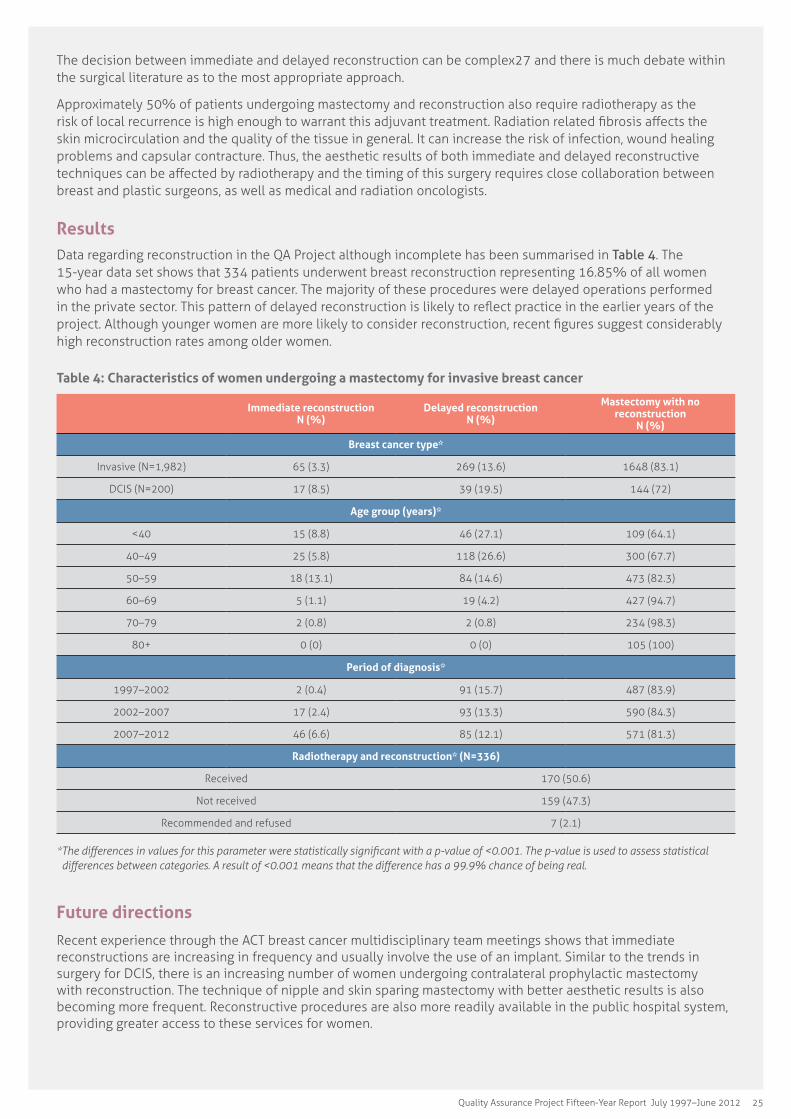
ResultsData regarding reconstruction in the QA Project although incomplete has been summarised in Table 4. The 15-year data set shows that 334 patients underwent breast reconstruction representing 16.85% of all women who had a mastectomy for breast cancer. The majority of these procedures were delayed operations performed in the private sector. This pattern of delayed reconstruction is likely to reflect practice in the earlier years of the project. Although younger women are more likely to consider reconstruction, recent figures suggest considerably high reconstruction rates among older women.
Table 4: Characteristics of women undergoing a mastectomy for invasive breast cancer
Immediate reconstruction N (%)
Delayed reconstruction N (%)
Mastectomy with no reconstruction
N (%)
Breast cancer type*
Invasive (N=1,982) 65 (3.3) 269 (13.6) 1648 (83.1)
DCIS (N=200) 17 (8.5) 39 (19.5) 144 (72)
Age group (years)*
<40 15 (8.8) 46 (27.1) 109 (64.1)
40–49 25 (5.8) 118 (26.6) 300 (67.7)
50–59 18 (13.1) 84 (14.6) 473 (82.3)
60–69 5 (1.1) 19 (4.2) 427 (94.7)
70–79 2 (0.8) 2 (0.8) 234 (98.3)
80+ 0 (0) 0 (0) 105 (100)
Period of diagnosis*
1997–2002 2 (0.4) 91 (15.7) 487 (83.9)
2002–2007 17 (2.4) 93 (13.3) 590 (84.3)
2007–2012 46 (6.6) 85 (12.1) 571 (81.3)
Radiotherapy and reconstruction* (N=336)
Received 170 (50.6)
Not received 159 (47.3)
Recommended and refused 7 (2.1)
*�The�differences�in�values�for�this�parameter�were�statistically�significant�with�a�p-value�of�<0.001.�The�p-value�is�used�to�assess�statistical�differences�between�categories.�A�result�of�<0.001�means�that�the�difference�has�a�99.9%�chance�of�being�real.
Future directions
Recent experience through the ACT breast cancer multidisciplinary team meetings shows that immediate reconstructions are increasing in frequency and usually involve the use of an implant. Similar to the trends in surgery for DCIS, there is an increasing number of women undergoing contralateral prophylactic mastectomy with reconstruction. The technique of nipple and skin sparing mastectomy with better aesthetic results is also becoming more frequent. Reconstructive procedures are also more readily available in the public hospital system, providing greater access to these services for women.
26 Breast Cancer Treatment Group
In the last five years there has been an increasing number of reconstructive procedures performed both in the public and private hospitals. These are more commonly immediate reconstructions. Although this is not yet seen in this 15-year report (until June 2012), international experience shows that skin or nipple sparing mastectomy in combination with immediate reconstruction is increasing in younger women, patients with high grade or extensive disease, and where treatment is performed at an academic facility. Post-mastectomy radiotherapy is rarely recommended for DCIS after mastectomy. Margin status is not affected by the use of skin sparing mastectomy or reconstruction16, 17, 28.
5.2 Radiotherapy
5.2.1 Breast conservation radiotherapyAdjuvant radiotherapy following breast conserving surgery is associated with improved local control and survival29. There is no cohort of women who undergo breast conservation surgery for invasive breast cancer in which radiotherapy can be omitted without a detriment to local control. The greatest benefit is in young women with large node positive breast cancer and the least benefit is found in older women with small, node negative, and ER positive breast cancers.
Results
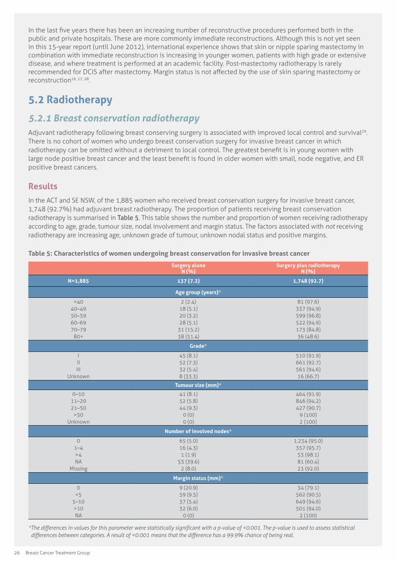
In the ACT and SE NSW, of the 1,885 women who received breast conservation surgery for invasive breast cancer, 1,748 (92.7%) had adjuvant breast radiotherapy. The proportion of patients receiving breast conservation radiotherapy is summarised in Table 5. This table shows the number and proportion of women receiving radiotherapy according to age, grade, tumour size, nodal involvement and margin status. The factors associated with not receiving radiotherapy are increasing age, unknown grade of tumour, unknown nodal status and positive margins.
Table 5: Characteristics of women undergoing breast conservation for invasive breast cancer
Surgery alone N (%)
Surgery plus radiotherapy N (%)
N=1,885 137 (7.2) 1,748 (92.7)
Age group (years)*
<4040–4950–5960–6970–79
80+
2 (2.4)18 (5.1)20 (3.2)28 (5.1)
31 (15.2)38 (51.4)
81 (97.6)337 (94.9)599 (96.8)522 (94.9)173 (84.8)36 (48.6)
Grade*
IIIIII
Unknown
45 (8.1)52 (7.3)32 (5.4)8 (33.3)
510 (91.9)661 (92.7)561 (94.6)16 (66.7)
Tumour size (mm)*
0–1011–2021–50
>50Unknown
41 (8.1)52 (5.8)44 (9.3)
0 (0)0 (0)
464 (91.9)846 (94.2)427 (90.7)
9 (100)2 (100)
Number of involved nodes*
01–4>4NA
Missing
65 (5.0)16 (4.3)1 (1.9)
53 (39.6)2 (8.0)
1,234 (95.0)357 (95.7)53 (98.1)81 (60.4)23 (92.0)
Margin status (mm)*
0<5
5–10>10NA
9 (20.9)59 (9.5)37 (5.4)32 (6.0)
0 (0)
34 (79.1)562 (90.5)649 (94.6)501 (94.0)
2 (100)
*�The�differences�in�values�for�this�parameter�were�statistically�significant�with�a�p-value�of�<0.001.�The�p-value�is�used�to�assess�statistical�differences�between�categories.�A�result�of�<0.001�means�that�the�difference�has�a�99.9%�chance�of�being�real.
Quality Assurance Project Fifteen-Year Report July 1997–June 2012 27
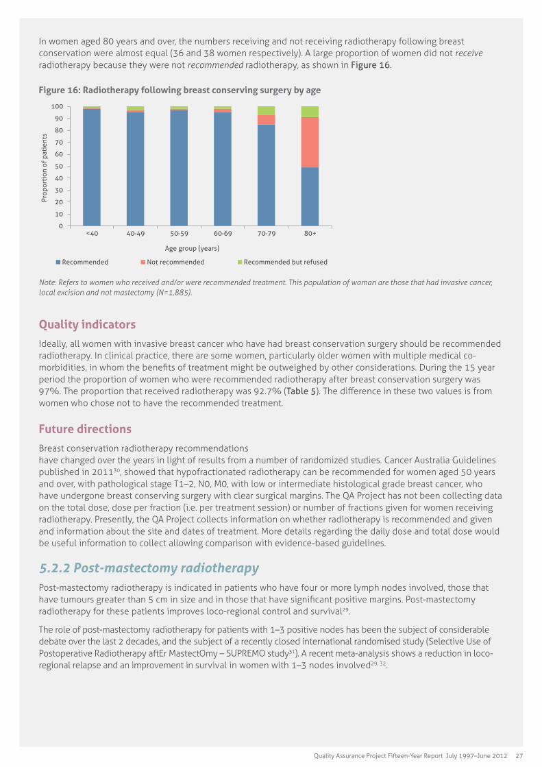
In women aged 80 years and over, the numbers receiving and not receiving radiotherapy following breast conservation were almost equal (36 and 38 women respectively). A large proportion of women did not receive radiotherapy because they were not recommended radiotherapy, as shown in Figure 16.
Figure 16: Radiotherapy following breast conserving surgery by age
0
10
20
30
40
50
60
70
80
90
100
<40 40-49 50-59 60-69 70-79 80+
Prop
orti
on o
f pa
tien
ts
Age group (years)
Recommended Not recommended Recommended but refused
Note: Refers to women who received and/or were recommended treatment. This population of woman are those that had invasive cancer, local excision and not mastectomy (N=1,885).
Quality indicators
Ideally, all women with invasive breast cancer who have had breast conservation surgery should be recommended radiotherapy. In clinical practice, there are some women, particularly older women with multiple medical co-morbidities, in whom the benefits of treatment might be outweighed by other considerations. During the 15 year period the proportion of women who were recommended radiotherapy after breast conservation surgery was 97%. The proportion that received radiotherapy was 92.7% (Table 5). The difference in these two values is from women who chose not to have the recommended treatment.
Future directions
Breast conservation radiotherapy recommendations have changed over the years in light of results from a number of randomized studies. Cancer Australia Guidelines published in 201130, showed that hypofractionated radiotherapy can be recommended for women aged 50 years and over, with pathological stage T1–2, N0, M0, with low or intermediate histological grade breast cancer, who have undergone breast conserving surgery with clear surgical margins. The QA Project has not been collecting data on the total dose, dose per fraction (i.e. per treatment session) or number of fractions given for women receiving radiotherapy. Presently, the QA Project collects information on whether radiotherapy is recommended and given and information about the site and dates of treatment. More details regarding the daily dose and total dose would be useful information to collect allowing comparison with evidence-based guidelines.
5.2.2 Post-mastectomy radiotherapyPost-mastectomy radiotherapy is indicated in patients who have four or more lymph nodes involved, those that have tumours greater than 5 cm in size and in those that have significant positive margins. Post-mastectomy radiotherapy for these patients improves loco-regional control and survival29.
The role of post-mastectomy radiotherapy for patients with 1–3 positive nodes has been the subject of considerable debate over the last 2 decades, and the subject of a recently closed international randomised study (Selective Use of Postoperative Radiotherapy aftEr MastectOmy – SUPREMO study31). A recent meta-analysis shows a reduction in loco-regional relapse and an improvement in survival in women with 1–3 nodes involved29, 32.
28 Breast Cancer Treatment Group
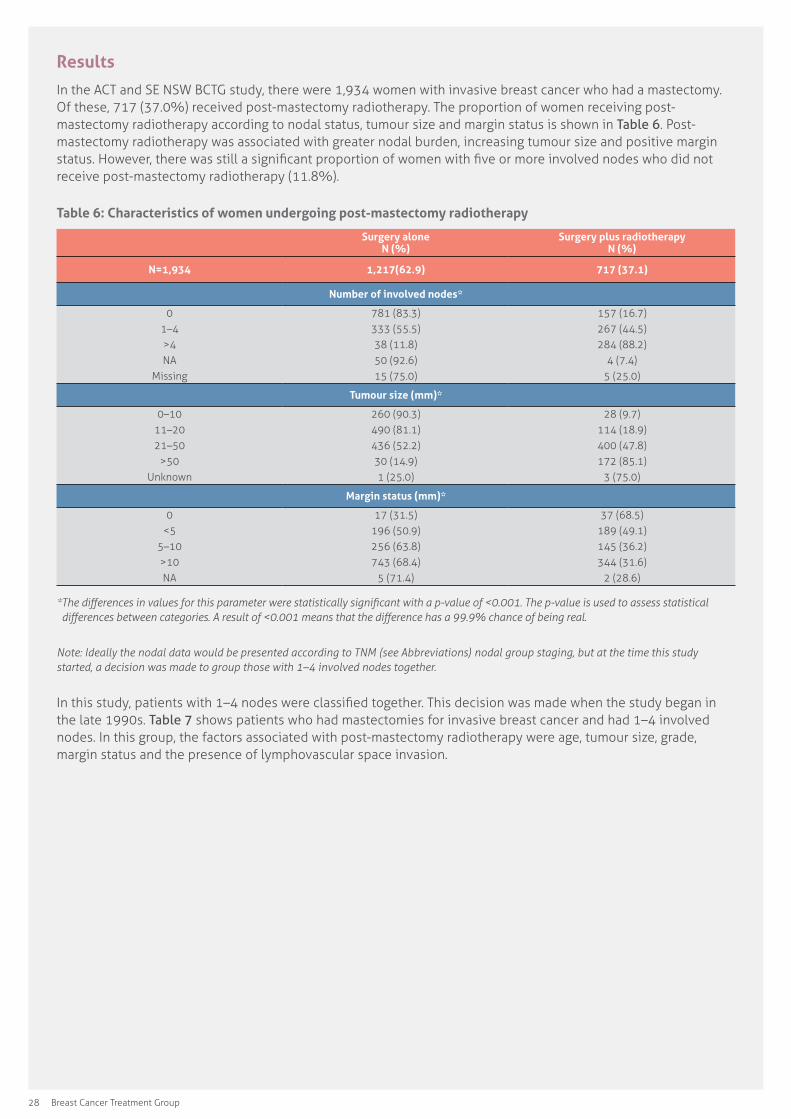
Results
In the ACT and SE NSW BCTG study, there were 1,934 women with invasive breast cancer who had a mastectomy. Of these, 717 (37.0%) received post-mastectomy radiotherapy. The proportion of women receiving post-mastectomy radiotherapy according to nodal status, tumour size and margin status is shown in Table 6. Post-mastectomy radiotherapy was associated with greater nodal burden, increasing tumour size and positive margin status. However, there was still a significant proportion of women with five or more involved nodes who did not receive post-mastectomy radiotherapy (11.8%).
Table 6: Characteristics of women undergoing post-mastectomy radiotherapy
Surgery alone N (%)
Surgery plus radiotherapy N (%)
N=1,934 1,217(62.9) 717 (37.1)
Number of involved nodes*
01–4>4NA
Missing
781 (83.3)333 (55.5)38 (11.8)50 (92.6)15 (75.0)
157 (16.7)267 (44.5)284 (88.2)
4 (7.4)5 (25.0)
Tumour size (mm)*
0–1011–2021–50
>50Unknown
260 (90.3)490 (81.1)436 (52.2)30 (14.9)1 (25.0)
28 (9.7)114 (18.9)400 (47.8)172 (85.1)
3 (75.0)
Margin status (mm)*
0<5
5–10>10NA
17 (31.5)196 (50.9)256 (63.8)743 (68.4)
5 (71.4)
37 (68.5)189 (49.1)145 (36.2)344 (31.6)
2 (28.6)
*�The�differences�in�values�for�this�parameter�were�statistically�significant�with�a�p-value�of�<0.001.�The�p-value�is�used�to�assess�statistical�differences�between�categories.�A�result�of�<0.001�means�that�the�difference�has�a�99.9%�chance�of�being�real.
Note: Ideally the nodal data would be presented according to TNM (see Abbreviations) nodal group staging, but at the time this study started, a decision was made to group those with 1–4 involved nodes together.
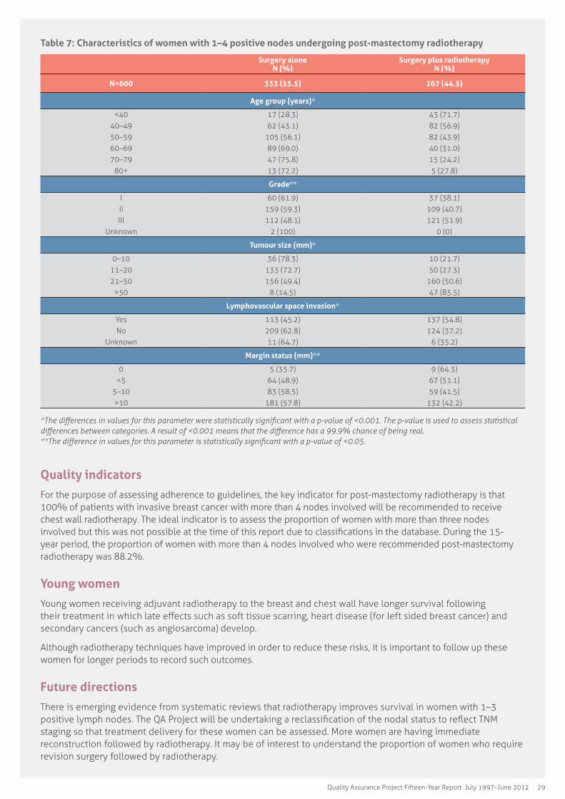
In this study, patients with 1–4 nodes were classified together. This decision was made when the study began in the late 1990s. Table 7 shows patients who had mastectomies for invasive breast cancer and had 1–4 involved nodes. In this group, the factors associated with post-mastectomy radiotherapy were age, tumour size, grade, margin status and the presence of lymphovascular space invasion.
Quality Assurance Project Fifteen-Year Report July 1997–June 2012 29
Table 7: Characteristics of women with 1–4 positive nodes undergoing post-mastectomy radiotherapy
Surgery alone N (%)
Surgery plus radiotherapy N (%)
N=600 333 (55.5) 267 (44.5)
Age group (years)*
<4040–4950–5960–6970–79
80+
17 (28.3)62 (43.1)
105 (56.1)89 (69.0)47 (75.8)13 (72.2)
43 (71.7)82 (56.9)82 (43.9)40 (31.0)15 (24.2)5 (27.8)
Grade**
IIIIII
Unknown
60 (61.9)159 (59.3)112 (48.1)
2 (100)
37 (38.1)109 (40.7)121 (51.9)
0 (0)
Tumour size (mm)*
0–1011–2021–50
>50
36 (78.3)133 (72.7)156 (49.4)
8 (14.5)
10 (21.7)50 (27.3)
160 (50.6)47 (85.5)
Lymphovascular space invasion*
YesNo
Unknown
113 (45.2)209 (62.8)11 (64.7)
137 (54.8)124 (37.2)
6 (35.2)
Margin status (mm)**
0<5
5–10>10
5 (35.7)64 (48.9)83 (58.5)
181 (57.8)
9 (64.3)67 (51.1)59 (41.5)
132 (42.2)
*The�differences�in�values�for�this�parameter�were�statistically�significant�with�a�p-value�of�<0.001.�The�p-value�is�used�to�assess�statistical�differences�between�categories.�A�result�of�<0.001�means�that�the�difference�has�a�99.9%�chance�of�being�real. **The�difference�in�values�for�this�parameter�is�statistically�significant�with�a�p-value�of�<0.05.
Quality indicators
For the purpose of assessing adherence to guidelines, the key indicator for post-mastectomy radiotherapy is that 100% of patients with invasive breast cancer with more than 4 nodes involved will be recommended to receive chest wall radiotherapy. The ideal indicator is to assess the proportion of women with more than three nodes involved but this was not possible at the time of this report due to classifications in the database. During the 15-year period, the proportion of women with more than 4 nodes involved who were recommended post-mastectomy radiotherapy was 88.2%.
Young women
Young women receiving adjuvant radiotherapy to the breast and chest wall have longer survival following their treatment in which late effects such as soft tissue scarring, heart disease (for left sided breast cancer) and secondary cancers (such as angiosarcoma) develop.
Although radiotherapy techniques have improved in order to reduce these risks, it is important to follow up these women for longer periods to record such outcomes.
Future directions
There is emerging evidence from systematic reviews that radiotherapy improves survival in women with 1–3 positive lymph nodes. The QA Project will be undertaking a reclassification of the nodal status to reflect TNM staging so that treatment delivery for these women can be assessed. More women are having immediate reconstruction followed by radiotherapy. It may be of interest to understand the proportion of women who require revision surgery followed by radiotherapy.
30 Breast Cancer Treatment Group
5.3 Systemic adjuvant therapies
Background
Since 1990, there has been a substantial fall in mortality from breast cancer in many Western countries. In Australia, between 1990 and 2000, mortality from breast cancer fell by approximately 25%33. This is due to a number of factors, including the introduction of breast screening for asymptomatic women and more effective systemic adjuvant therapies.
The systemic adjuvant therapies in use today are chemotherapy, trastuzumab (Herceptin) and endocrine therapy.
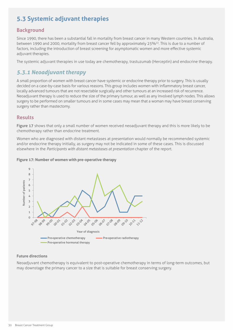
5.3.1 Neoadjuvant therapyA small proportion of women with breast cancer have systemic or endocrine therapy prior to surgery. This is usually decided on a case-by-case basis for various reasons. This group includes women with inflammatory breast cancer, locally advanced tumours that are not resectable surgically and other tumours at an increased risk of recurrence. Neoadjuvant therapy is used to reduce the size of the primary tumour, as well as any involved lymph nodes. This allows surgery to be performed on smaller tumours and in some cases may mean that a woman may have breast conserving surgery rather than mastectomy.
Results
Figure 17 shows that only a small number of women received neoadjuvant therapy and this is more likely to be chemotherapy rather than endocrine treatment.
Women who are diagnosed with distant metastases at presentation would normally be recommended systemic and/or endocrine therapy initially, as surgery may not be indicated in some of these cases. This is discussed elsewhere in the Participants with distant metastases at presentation chapter of the report.
Figure 17: Number of women with pre-operative therapy
0
1
2
3
4
5
6
7
8
9
Num
ber
of p
atie
nts
Year of diagnosis
Pre-operative chemotherapy Pre-operative radiotherapy Pre-operative hormonal therapy
Future directions
Neoadjuvant chemotherapy is equivalent to post-operative chemotherapy in terms of long-term outcomes, but may downstage the primary cancer to a size that is suitable for breast conserving surgery.
Quality Assurance Project Fifteen-Year Report July 1997–June 2012 31
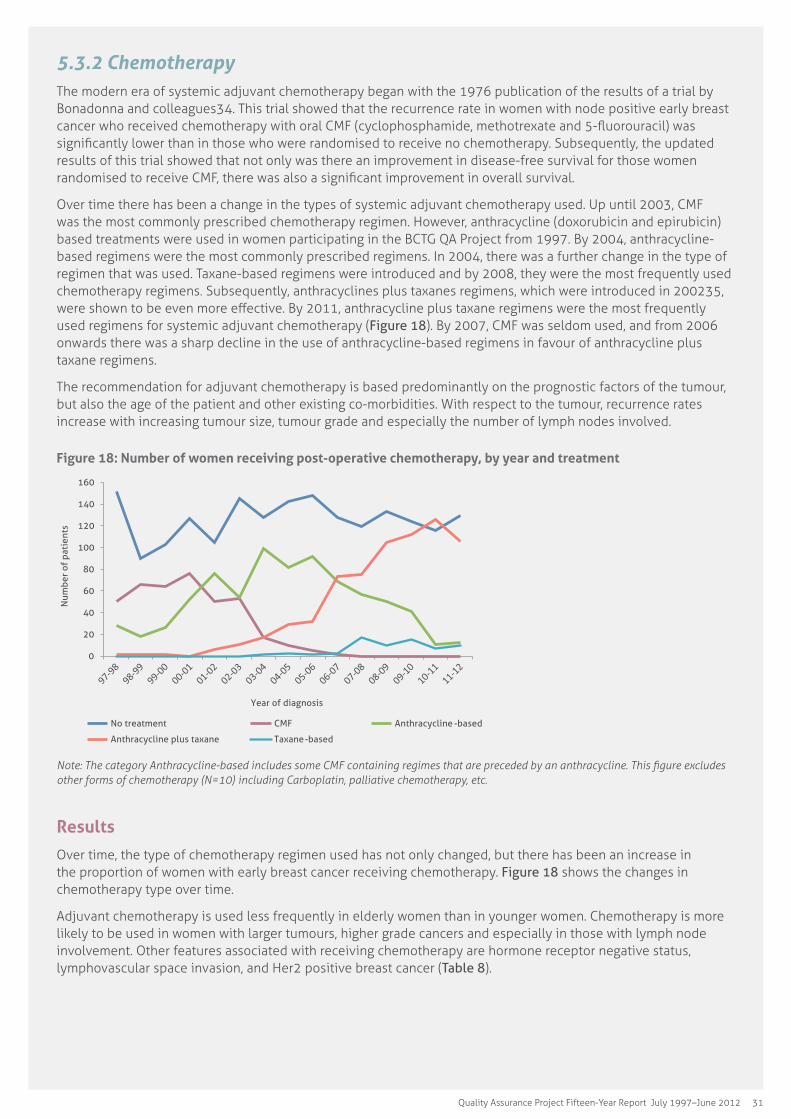
5.3.2 ChemotherapyThe modern era of systemic adjuvant chemotherapy began with the 1976 publication of the results of a trial by Bonadonna and colleagues34. This trial showed that the recurrence rate in women with node positive early breast cancer who received chemotherapy with oral CMF (cyclophosphamide, methotrexate and 5-fluorouracil) was significantly lower than in those who were randomised to receive no chemotherapy. Subsequently, the updated results of this trial showed that not only was there an improvement in disease-free survival for those women randomised to receive CMF, there was also a significant improvement in overall survival.
Over time there has been a change in the types of systemic adjuvant chemotherapy used. Up until 2003, CMF was the most commonly prescribed chemotherapy regimen. However, anthracycline (doxorubicin and epirubicin) based treatments were used in women participating in the BCTG QA Project from 1997. By 2004, anthracycline-based regimens were the most commonly prescribed regimens. In 2004, there was a further change in the type of regimen that was used. Taxane-based regimens were introduced and by 2008, they were the most frequently used chemotherapy regimens. Subsequently, anthracyclines plus taxanes regimens, which were introduced in 200235, were shown to be even more effective. By 2011, anthracycline plus taxane regimens were the most frequently used regimens for systemic adjuvant chemotherapy (Figure 18). By 2007, CMF was seldom used, and from 2006 onwards there was a sharp decline in the use of anthracycline-based regimens in favour of anthracycline plus taxane regimens.
The recommendation for adjuvant chemotherapy is based predominantly on the prognostic factors of the tumour, but also the age of the patient and other existing co-morbidities. With respect to the tumour, recurrence rates increase with increasing tumour size, tumour grade and especially the number of lymph nodes involved.
Figure 18: Number of women receiving post-operative chemotherapy, by year and treatment
0
20
40
60
80
100
120
140
160
Num
ber
of p
atie
nts
Year of diagnosis
No treatment CMF Anthracycline -based
Anthracycline plus taxane Taxane-based
Note:�The�category�Anthracycline-based�includes�some�CMF�containing�regimes�that�are�preceded�by�an�anthracycline.�This�figure�excludes�other forms of chemotherapy (N=10) including Carboplatin, palliative chemotherapy, etc.
Results
Over time, the type of chemotherapy regimen used has not only changed, but there has been an increase in the proportion of women with early breast cancer receiving chemotherapy. Figure 18 shows the changes in chemotherapy type over time.
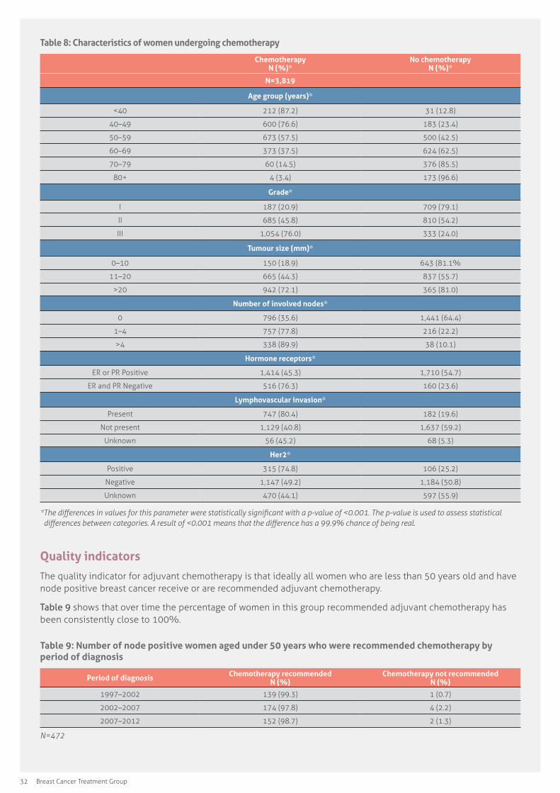
Adjuvant chemotherapy is used less frequently in elderly women than in younger women. Chemotherapy is more likely to be used in women with larger tumours, higher grade cancers and especially in those with lymph node involvement. Other features associated with receiving chemotherapy are hormone receptor negative status, lymphovascular space invasion, and Her2 positive breast cancer (Table 8).
32 Breast Cancer Treatment Group
Table 8: Characteristics of women undergoing chemotherapy
Chemotherapy N (%)*
No chemotherapy N (%)*
N=3,819
Age group (years)*
<40 212 (87.2) 31 (12.8)
40–49 600 (76.6) 183 (23.4)
50–59 673 (57.5) 500 (42.5)
60–69 373 (37.5) 624 (62.5)
70–79 60 (14.5) 376 (85.5)
80+ 4 (3.4) 173 (96.6)
Grade*
I 187 (20.9) 709 (79.1)
II 685 (45.8) 810 (54.2)
III 1,054 (76.0) 333 (24.0)
Tumour size (mm)*
0–10 150 (18.9) 643 (81.1%
11–20 665 (44.3) 837 (55.7)
>20 942 (72.1) 365 (81.0)
Number of involved nodes*
0 796 (35.6) 1,441 (64.4)
1–4 757 (77.8) 216 (22.2)
>4 338 (89.9) 38 (10.1)
Hormone receptors*
ER or PR Positive 1,414 (45.3) 1,710 (54.7)
ER and PR Negative 516 (76.3) 160 (23.6)
Lymphovascular invasion*
Present 747 (80.4) 182 (19.6)
Not present 1,129 (40.8) 1,637 (59.2)
Unknown 56 (45.2) 68 (5.3)
Her2*
Positive 315 (74.8) 106 (25.2)
Negative 1,147 (49.2) 1,184 (50.8)
Unknown 470 (44.1) 597 (55.9)
*�The�differences�in�values�for�this�parameter�were�statistically�significant�with�a�p-value�of�<0.001.�The�p-value�is�used�to�assess�statistical�differences�between�categories.�A�result�of�<0.001�means�that�the�difference�has�a�99.9%�chance�of�being�real.
Quality indicators
The quality indicator for adjuvant chemotherapy is that ideally all women who are less than 50 years old and have node positive breast cancer receive or are recommended adjuvant chemotherapy.
Table 9 shows that over time the percentage of women in this group recommended adjuvant chemotherapy has been consistently close to 100%.
Table 9: Number of node positive women aged under 50 years who were recommended chemotherapy by period of diagnosis
Period of diagnosis Chemotherapy recommended N (%)
Chemotherapy not recommended N (%)
1997–2002 139 (99.3) 1 (0.7)
2002–2007 174 (97.8) 4 (2.2)
2007–2012 152 (98.7) 2 (1.3)
N=472
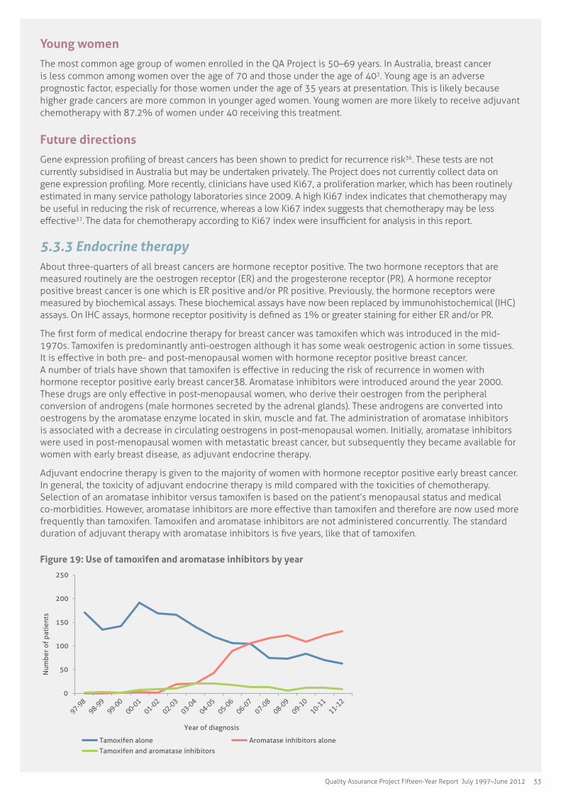
Quality Assurance Project Fifteen-Year Report July 1997–June 2012 33
Young women
The most common age group of women enrolled in the QA Project is 50–69 years. In Australia, breast cancer is less common among women over the age of 70 and those under the age of 403. Young age is an adverse prognostic factor, especially for those women under the age of 35 years at presentation. This is likely because higher grade cancers are more common in younger aged women. Young women are more likely to receive adjuvant chemotherapy with 87.2% of women under 40 receiving this treatment.
Future directions
Gene expression profiling of breast cancers has been shown to predict for recurrence risk36. These tests are not currently subsidised in Australia but may be undertaken privately. The Project does not currently collect data on gene expression profiling. More recently, clinicians have used Ki67, a proliferation marker, which has been routinely estimated in many service pathology laboratories since 2009. A high Ki67 index indicates that chemotherapy may be useful in reducing the risk of recurrence, whereas a low Ki67 index suggests that chemotherapy may be less effective37. The data for chemotherapy according to Ki67 index were insufficient for analysis in this report.
5.3.3 Endocrine therapy About three-quarters of all breast cancers are hormone receptor positive. The two hormone receptors that are measured routinely are the oestrogen receptor (ER) and the progesterone receptor (PR). A hormone receptor positive breast cancer is one which is ER positive and/or PR positive. Previously, the hormone receptors were measured by biochemical assays. These biochemical assays have now been replaced by immunohistochemical (IHC) assays. On IHC assays, hormone receptor positivity is defined as 1% or greater staining for either ER and/or PR.
The first form of medical endocrine therapy for breast cancer was tamoxifen which was introduced in the mid-1970s. Tamoxifen is predominantly anti-oestrogen although it has some weak oestrogenic action in some tissues. It is effective in both pre- and post-menopausal women with hormone receptor positive breast cancer. A number of trials have shown that tamoxifen is effective in reducing the risk of recurrence in women with hormone receptor positive early breast cancer38. Aromatase inhibitors were introduced around the year 2000. These drugs are only effective in post-menopausal women, who derive their oestrogen from the peripheral conversion of androgens (male hormones secreted by the adrenal glands). These androgens are converted into oestrogens by the aromatase enzyme located in skin, muscle and fat. The administration of aromatase inhibitors is associated with a decrease in circulating oestrogens in post-menopausal women. Initially, aromatase inhibitors were used in post-menopausal women with metastatic breast cancer, but subsequently they became available for women with early breast disease, as adjuvant endocrine therapy.
Adjuvant endocrine therapy is given to the majority of women with hormone receptor positive early breast cancer. In general, the toxicity of adjuvant endocrine therapy is mild compared with the toxicities of chemotherapy. Selection of an aromatase inhibitor versus tamoxifen is based on the patient’s menopausal status and medical co-morbidities. However, aromatase inhibitors are more effective than tamoxifen and therefore are now used more frequently than tamoxifen. Tamoxifen and aromatase inhibitors are not administered concurrently. The standard duration of adjuvant therapy with aromatase inhibitors is five years, like that of tamoxifen.
Figure 19: Use of tamoxifen and aromatase inhibitors by year
0
50
100
150
200
250
Num
ber
of p
atie
nts
Year of diagnosis
Tamoxifen alone Aromatase inhibitors alone
Tamoxifen and aromatase inhibitors
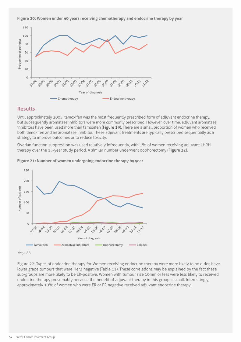
34 Breast Cancer Treatment Group
Figure 20: Women under 40 years receiving chemotherapy and endocrine therapy by year
0
20
40
60
80
100
120
Prop
orti
on o
f pa
tien
ts
Year of diagnosis
Chemotherapy Endocrine therapy
ResultsUntil approximately 2005, tamoxifen was the most frequently prescribed form of adjuvant endocrine therapy, but subsequently aromatase inhibitors were more commonly prescribed. However, over time, adjuvant aromatase inhibitors have been used more than tamoxifen (Figure 19). There are a small proportion of women who received both tamoxifen and an aromatase inhibitor. These adjuvant treatments are typically prescribed sequentially as a strategy to improve outcomes or to reduce toxicity.
Ovarian function suppression was used relatively infrequently, with 1% of women receiving adjuvant LHRH therapy over the 15-year study period. A similar number underwent oophorectomy (Figure 22).
Figure 21: Number of women undergoing endocrine therapy by year
0
50
100
150
200
250
Num
ber
of p
atie
nts
Year of diagnosis
Tamoxifen Aromatase inhibitors Oophorectomy Zoladex
N=3,088
Figure 22: Types of endocrine therapy for Women receiving endocrine therapy were more likely to be older, have lower grade tumours that were Her2 negative (Table 11). These correlations may be explained by the fact these sub-groups are more likely to be ER-positive. Women with tumour size 10mm or less were less likely to received endocrine therapy presumably because the benefit of adjuvant therapy in this group is small. Interestingly, approximately 10% of women who were ER or PR negative received adjuvant endocrine therapy.
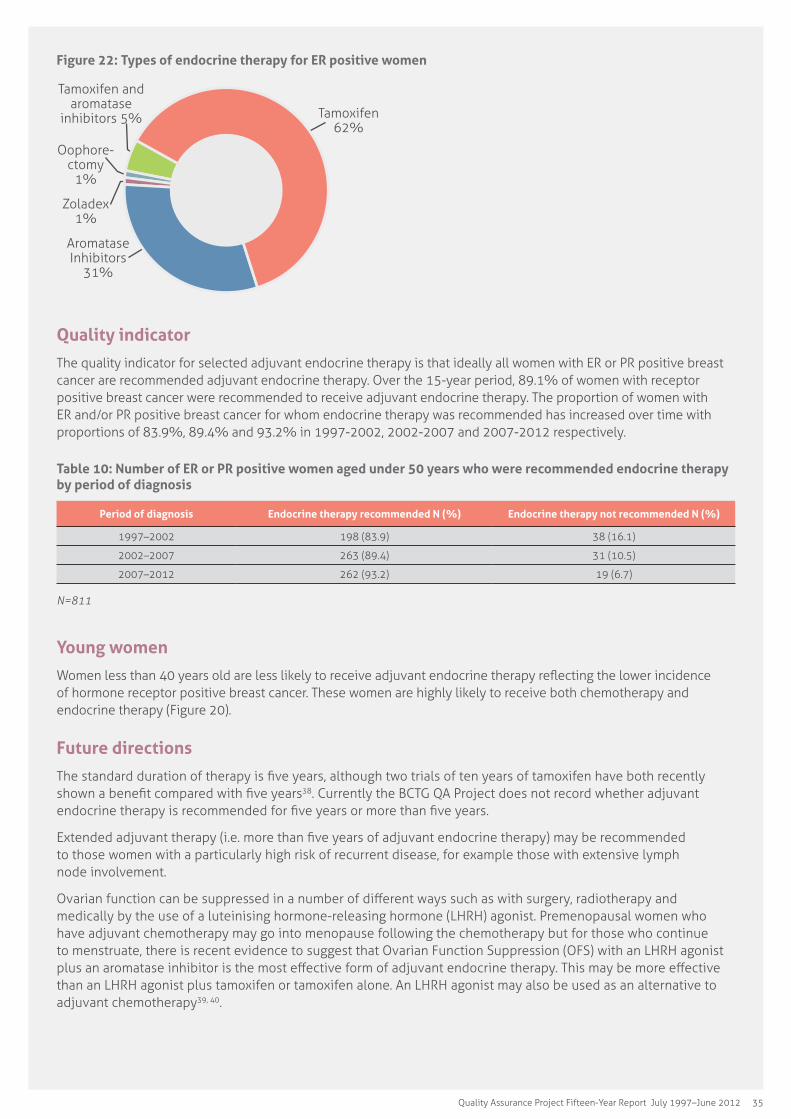
Quality Assurance Project Fifteen-Year Report July 1997–June 2012 35
Figure 22: Types of endocrine therapy for ER positive women
Tamoxifen 62%
Aromatase Inhibitors
31%
Zoladex 1%
Oophore-ctomy
1%
Tamoxifen and aromatase
inhibitors 5%
Quality indicator
The quality indicator for selected adjuvant endocrine therapy is that ideally all women with ER or PR positive breast cancer are recommended adjuvant endocrine therapy. Over the 15-year period, 89.1% of women with receptor positive breast cancer were recommended to receive adjuvant endocrine therapy. The proportion of women with ER and/or PR positive breast cancer for whom endocrine therapy was recommended has increased over time with proportions of 83.9%, 89.4% and 93.2% in 1997-2002, 2002-2007 and 2007-2012 respectively.
Table 10: Number of ER or PR positive women aged under 50 years who were recommended endocrine therapy by period of diagnosis
Period of diagnosis Endocrine therapy recommended N (%) Endocrine therapy not recommended N (%)
1997–2002 198 (83.9) 38 (16.1)
2002–2007 263 (89.4) 31 (10.5)
2007–2012 262 (93.2) 19 (6.7)
N=811
Young women
Women less than 40 years old are less likely to receive adjuvant endocrine therapy reflecting the lower incidence of hormone receptor positive breast cancer. These women are highly likely to receive both chemotherapy and endocrine therapy (Figure 20).
Future directions
The standard duration of therapy is five years, although two trials of ten years of tamoxifen have both recently shown a benefit compared with five years38. Currently the BCTG QA Project does not record whether adjuvant endocrine therapy is recommended for five years or more than five years.
Extended adjuvant therapy (i.e. more than five years of adjuvant endocrine therapy) may be recommended to those women with a particularly high risk of recurrent disease, for example those with extensive lymph node involvement.
Ovarian function can be suppressed in a number of different ways such as with surgery, radiotherapy and medically by the use of a luteinising hormone-releasing hormone (LHRH) agonist. Premenopausal women who have adjuvant chemotherapy may go into menopause following the chemotherapy but for those who continue to menstruate, there is recent evidence to suggest that Ovarian Function Suppression (OFS) with an LHRH agonist plus an aromatase inhibitor is the most effective form of adjuvant endocrine therapy. This may be more effective than an LHRH agonist plus tamoxifen or tamoxifen alone. An LHRH agonist may also be used as an alternative to adjuvant chemotherapy39, 40.
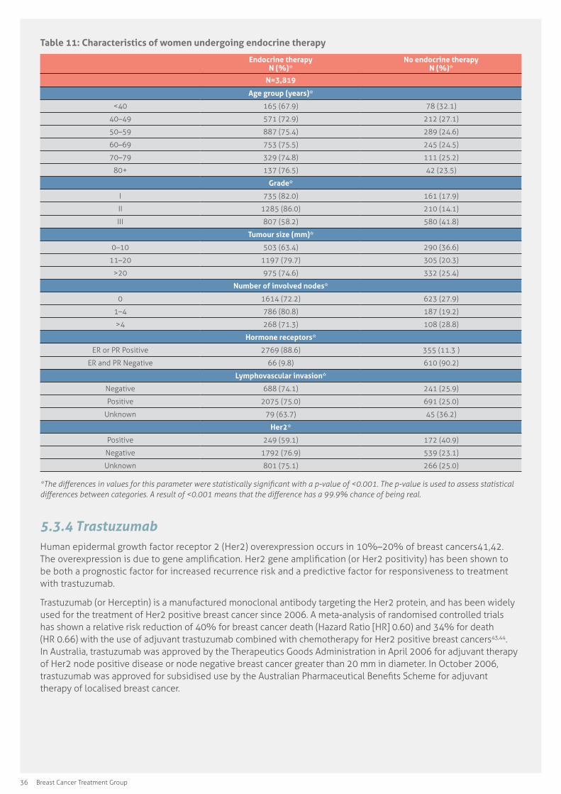
36 Breast Cancer Treatment Group
Table 11: Characteristics of women undergoing endocrine therapy
Endocrine therapy N (%)*
No endocrine therapy N (%)*
N=3,819
Age group (years)*
<40 165 (67.9) 78 (32.1)
40–49 571 (72.9) 212 (27.1)
50–59 887 (75.4) 289 (24.6)
60–69 753 (75.5) 245 (24.5)
70–79 329 (74.8) 111 (25.2)
80+ 137 (76.5) 42 (23.5)
Grade*
I 735 (82.0) 161 (17.9)
II 1285 (86.0) 210 (14.1)
III 807 (58.2) 580 (41.8)
Tumour size (mm)*
0–10 503 (63.4) 290 (36.6)
11–20 1197 (79.7) 305 (20.3)
>20 975 (74.6) 332 (25.4)
Number of involved nodes*
0 1614 (72.2) 623 (27.9)
1–4 786 (80.8) 187 (19.2)
>4 268 (71.3) 108 (28.8)
Hormone receptors*
ER or PR Positive 2769 (88.6) 355 (11.3 )
ER and PR Negative 66 (9.8) 610 (90.2)
Lymphovascular invasion*
Negative 688 (74.1) 241 (25.9)
Positive 2075 (75.0) 691 (25.0)
Unknown 79 (63.7) 45 (36.2)
Her2*
Positive 249 (59.1) 172 (40.9)
Negative 1792 (76.9) 539 (23.1)
Unknown 801 (75.1) 266 (25.0)
*The�differences�in�values�for�this�parameter�were�statistically�significant�with�a�p-value�of�<0.001.�The�p-value�is�used�to�assess�statistical�differences�between�categories.�A�result�of�<0.001�means�that�the�difference�has�a�99.9%�chance�of�being�real.
5.3.4 TrastuzumabHuman epidermal growth factor receptor 2 (Her2) overexpression occurs in 10%–20% of breast cancers41,42. The overexpression is due to gene amplification. Her2 gene amplification (or Her2 positivity) has been shown to be both a prognostic factor for increased recurrence risk and a predictive factor for responsiveness to treatment with trastuzumab.
Trastuzumab (or Herceptin) is a manufactured monoclonal antibody targeting the Her2 protein, and has been widely used for the treatment of Her2 positive breast cancer since 2006. A meta-analysis of randomised controlled trials has shown a relative risk reduction of 40% for breast cancer death (Hazard Ratio [HR] 0.60) and 34% for death (HR 0.66) with the use of adjuvant trastuzumab combined with chemotherapy for Her2 positive breast cancers43,44. In Australia, trastuzumab was approved by the Therapeutics Goods Administration in April 2006 for adjuvant therapy of Her2 node positive disease or node negative breast cancer greater than 20 mm in diameter. In October 2006, trastuzumab was approved for subsidised use by the Australian Pharmaceutical Benefits Scheme for adjuvant therapy of localised breast cancer.

Quality Assurance Project Fifteen-Year Report July 1997–June 2012 37
Her2 positive tumours
Routine Her2 testing began in 2003 when trastuzumab became potentially available for adjuvant therapy in Australia. Prior to this, testing was only performed on an ad hoc basis, usually for women with metastatic disease. For this reason, data are missing for 801 subjects. Of 421 Her2 positive cases since October 2006, 201 women received trastuzumab combined with adjuvant chemotherapy.
Future directions
Although randomised clinic trials provide the best evidence of effectiveness of a novel therapy, trial populations are inevitably selected. More complete understanding of the effectiveness and safety of novel therapies requires the systematic study of outcomes in a general community treatment setting. Systemic therapy for early breast cancer is continuing to evolve with a move towards personalised cancer therapy based on increasing knowledge of molecular factors which predict benefit from specific agents. As predictive molecular markers enter clinical practice, they will be incorporated into the data collection. In the case of trastuzumab, further data regarding outcomes will be available over the next five years.
38 Breast Cancer Treatment Group
Outcomes of care and adherence to guidelines
IntroductionBreast cancer treatment for early operable primary cancer is primarily focussed on local treatments to the breast such as surgery and radiation therapy, often combined with systemic therapies (chemotherapy and endocrine therapy). The systemic therapy is aimed at reducing the risk of recurrence after successful local treatment. All treatments for early breast cancer have been tested with rigorous, large clinical trials and the evidence for effectiveness is strong. Despite these effective therapies, there remains a risk of recurrence, even some years after treatment has been completed.
As part of the ongoing QA Project, information was collected outlining breast cancer recurrence and overall survival over the 15-year period.
Treatment recommendations have evolved over the 15-year period of the project. At the initiation of the project, four clinical practice indicators were adopted by the group describing clinical circumstances where there would be little controversy that a particular type of care was warranted. These have been modified slightly for this report based on consensus that the indicators represent an appropriate minimum standard across the timeline of the project. In addition, a further indicator (Indicator 5) was added for this report.
Practice indicators1. Women with completely resected invasive breast cancer greater than two centimetres in diameter or who have
axillary lymph node involvement should be recommended adjuvant systemic therapy (either chemotherapy, endocrine therapy, or both).
2. Pre-menopausal women or women under the age of 50 years with completely resected, axillary lymph node positive breast cancer should be recommended adjuvant chemotherapy.
3. Women undergoing less than total mastectomy for operable invasive breast cancer should be recommended post-operative adjuvant radiotherapy.
4. Women with operable invasive breast cancer should be recommended to undergo some form of axillary surgery sufficient to develop an axillary lymph node based prognosis.
5. Women with invasive breast cancer undergoing mastectomy with 5 or more axillary lymph nodes involved should be recommended to undergo post-mastectomy radiotherapy (PMRT).
Method of analysisFollow-up information on the status of participant was sought, the first following 12 months after the date of diagnosis and then every two years by mailing a short data request form to the treating clinician and to the participant’s general practitioner. Public sources of death notification were monitored to check for events not notified by clinicians. The cause of death was ascertained from the notifying clinicians and/or death certificate information.
Place of surgery was defined as urban when surgery was performed by surgeons operating in hospitals located within Canberra, or rural when performed by surgeons operating in hospitals within the SE NSW region. Cases with specific missing data were excluded on a case-by-case basis during the analysis.
SIX
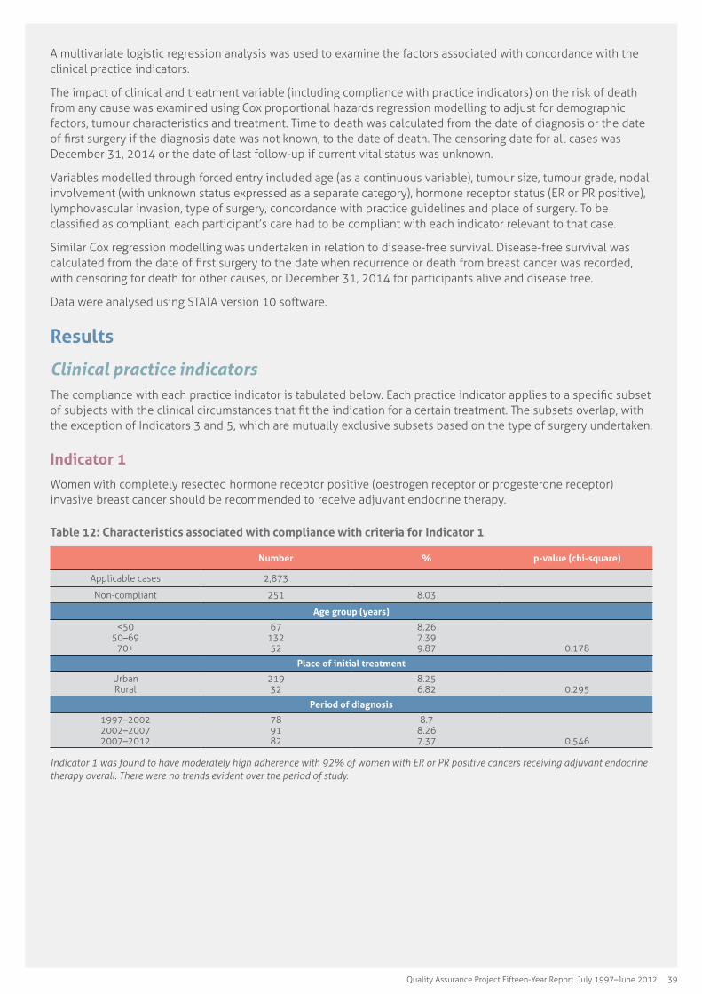
Quality Assurance Project Fifteen-Year Report July 1997–June 2012 39
A multivariate logistic regression analysis was used to examine the factors associated with concordance with the clinical practice indicators.
The impact of clinical and treatment variable (including compliance with practice indicators) on the risk of death from any cause was examined using Cox proportional hazards regression modelling to adjust for demographic factors, tumour characteristics and treatment. Time to death was calculated from the date of diagnosis or the date of first surgery if the diagnosis date was not known, to the date of death. The censoring date for all cases was December 31, 2014 or the date of last follow-up if current vital status was unknown.
Variables modelled through forced entry included age (as a continuous variable), tumour size, tumour grade, nodal involvement (with unknown status expressed as a separate category), hormone receptor status (ER or PR positive), lymphovascular invasion, type of surgery, concordance with practice guidelines and place of surgery. To be classified as compliant, each participant’s care had to be compliant with each indicator relevant to that case.
Similar Cox regression modelling was undertaken in relation to disease-free survival. Disease-free survival was calculated from the date of first surgery to the date when recurrence or death from breast cancer was recorded, with censoring for death for other causes, or December 31, 2014 for participants alive and disease free.
Data were analysed using STATA version 10 software.
Results
Clinical practice indicatorsThe compliance with each practice indicator is tabulated below. Each practice indicator applies to a specific subset of subjects with the clinical circumstances that fit the indication for a certain treatment. The subsets overlap, with the exception of Indicators 3 and 5, which are mutually exclusive subsets based on the type of surgery undertaken.
Indicator 1
Women with completely resected hormone receptor positive (oestrogen receptor or progesterone receptor) invasive breast cancer should be recommended to receive adjuvant endocrine therapy.
Table 12: Characteristics associated with compliance with criteria for Indicator 1
Number % p-value (chi-square)
Applicable cases 2,873
Non-compliant 251 8.03
Age group (years)
<50 50–69
70+
67 132 52
8.26 7.39 9.87
0.178
Place of initial treatment
Urban Rural
219 32
8.25 6.82
0.295
Period of diagnosis
1997–2002 2002–2007 2007–2012
78 91 82
8.7 8.26 7.37
0.546
Indicator�1�was�found�to�have�moderately�high�adherence�with�92%�of�women�with�ER�or�PR�positive�cancers�receiving�adjuvant�endocrine�therapy overall. There were no trends evident over the period of study.

40 Breast Cancer Treatment Group
Indicator 2
Women aged 50 years or less, with completely resected axillary lymph node positive breast cancer should be recommended to receive adjuvant chemotherapy.
Table 13: Characteristics associated with compliance with criteria for Indicator 2
Number % p-value (chi-square)
Applicable cases 472
Non-compliant 7 1.48
Place of initial treatment
Urban Rural
6 1
1.45 1.72
0.87
Period of diagnosis
1997–2002 2002–2007 2007–2012
1 4 2
0.71 2.25 1.30
0.519
Indicator�2�assessment�revealed�very�high�compliance�over�the�whole�study�period�with�98.5%�compliance�and�no�change�over�the�different�five-year�diagnosis�cohorts.
Indicator 3
Women undergoing less than total mastectomy for operable invasive breast cancer should be recommended to receive post-operative adjuvant radiotherapy.
Table 14: Characteristics associated with compliance with criteria for Indicator 3
Number % p-value (chi-square)
Applicable cases 1,885
Non-compliant 80 4.24
Age group (years)
<50 50–69
70+
9 23 48
2.05 1.97
17.27
<0.001
Place of initial treatment
Urban Rural
49 31
3.14 9.60
<0.001
Period of diagnosis
1997–2002 2002–2007 2007–2012
20 37 23
3.79 5.66 3.27
0.077
Women aged 70 years or more had lower rates of compliance with this practice indicator, as did women from rural locations. The social burden of undergoing adjuvant radiation therapy is significant for rural and elderly women. Variation in care based around these factors has been demonstrated elsewhere45. Although not statistically significant, compliance with Indicator 3 may have decreased in the period 2002–2007. Following this time, results from the 10-year data set were presented to the group and published, which may have contributed to the subsequent improved compliance observed in 2007–201246.
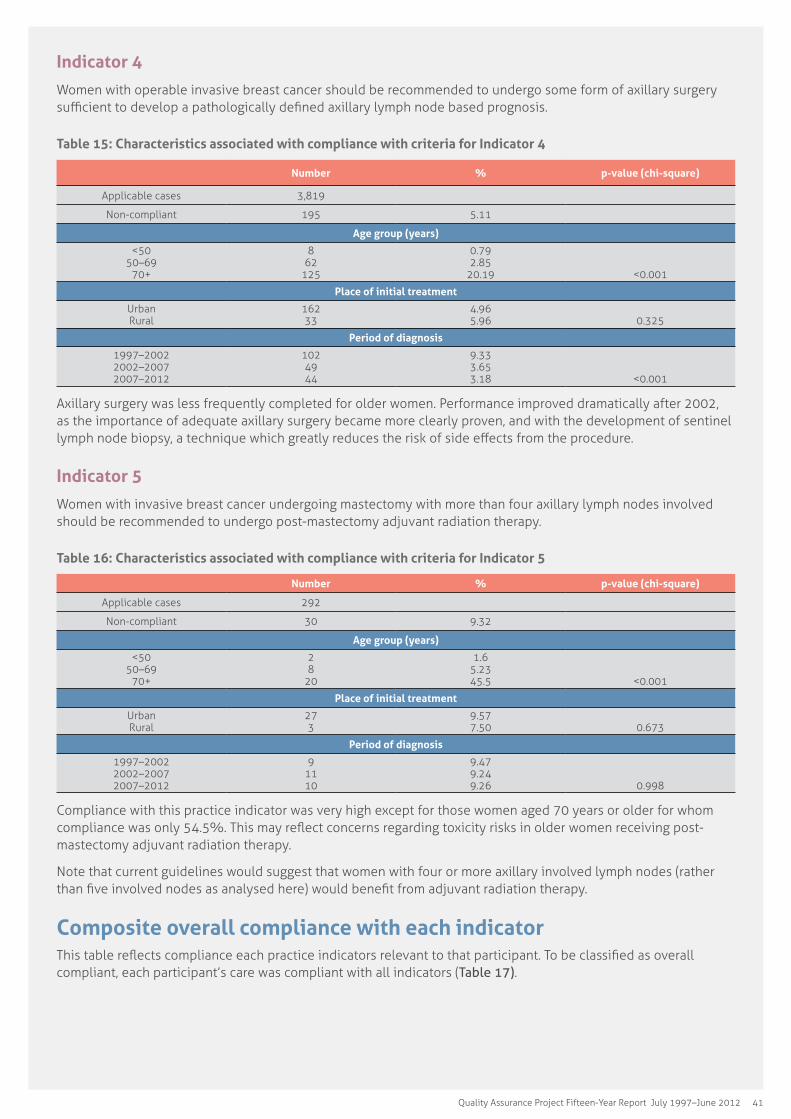
Quality Assurance Project Fifteen-Year Report July 1997–June 2012 41
Indicator 4
Women with operable invasive breast cancer should be recommended to undergo some form of axillary surgery sufficient to develop a pathologically defined axillary lymph node based prognosis.
Table 15: Characteristics associated with compliance with criteria for Indicator 4
Number % p-value (chi-square)
Applicable cases 3,819
Non-compliant 195 5.11
Age group (years)
<50 50–69
70+
8 62
125
0.79 2.85
20.19
<0.001
Place of initial treatment
Urban Rural
162 33
4.96 5.96
0.325
Period of diagnosis
1997–2002 2002–2007 2007–2012
102 49 44
9.33 3.65 3.18
<0.001
Axillary surgery was less frequently completed for older women. Performance improved dramatically after 2002, as the importance of adequate axillary surgery became more clearly proven, and with the development of sentinel lymph node biopsy, a technique which greatly reduces the risk of side effects from the procedure.
Indicator 5
Women with invasive breast cancer undergoing mastectomy with more than four axillary lymph nodes involved should be recommended to undergo post-mastectomy adjuvant radiation therapy.
Table 16: Characteristics associated with compliance with criteria for Indicator 5
Number % p-value (chi-square)
Applicable cases 292
Non-compliant 30 9.32
Age group (years)
<50 50–69
70+
2 8
20
1.6 5.23 45.5
<0.001
Place of initial treatment
Urban Rural
27 3
9.57 7.50
0.673
Period of diagnosis
1997–2002 2002–2007 2007–2012
9 11 10
9.47 9.24 9.26
0.998
Compliance with this practice indicator was very high except for those women aged 70 years or older for whom compliance was only 54.5%. This may reflect concerns regarding toxicity risks in older women receiving post-mastectomy adjuvant radiation therapy.
Note that current guidelines would suggest that women with four or more axillary involved lymph nodes (rather than five involved nodes as analysed here) would benefit from adjuvant radiation therapy.
Composite overall compliance with each indicatorThis table reflects compliance each practice indicators relevant to that participant. To be classified as overall compliant, each participant’s care was compliant with all indicators (Table 17).
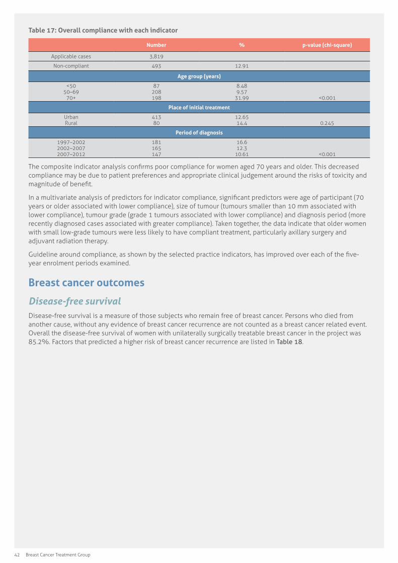
42 Breast Cancer Treatment Group
Table 17: Overall compliance with each indicator
Number % p-value (chi-square)
Applicable cases 3,819
Non-compliant 493 12.91
Age group (years)
<50 50–69
70+
87 208 198
8.48 9.57
31.99
<0.001
Place of initial treatment
Urban Rural
413 80
12.65 14.4
0.245
Period of diagnosis
1997–2002 2002–2007 2007–2012
181 165 147
16.6 12.3
10.61
<0.001
The composite indicator analysis confirms poor compliance for women aged 70 years and older. This decreased compliance may be due to patient preferences and appropriate clinical judgement around the risks of toxicity and magnitude of benefit.
In a multivariate analysis of predictors for indicator compliance, significant predictors were age of participant (70 years or older associated with lower compliance), size of tumour (tumours smaller than 10 mm associated with lower compliance), tumour grade (grade 1 tumours associated with lower compliance) and diagnosis period (more recently diagnosed cases associated with greater compliance). Taken together, the data indicate that older women with small low-grade tumours were less likely to have compliant treatment, particularly axillary surgery and adjuvant radiation therapy.
Guideline around compliance, as shown by the selected practice indicators, has improved over each of the five-year enrolment periods examined.
Breast cancer outcomes
Disease-free survivalDisease-free survival is a measure of those subjects who remain free of breast cancer. Persons who died from another cause, without any evidence of breast cancer recurrence are not counted as a breast cancer related event. Overall the disease-free survival of women with unilaterally surgically treatable breast cancer in the project was 85.2%. Factors that predicted a higher risk of breast cancer recurrence are listed in Table 18.
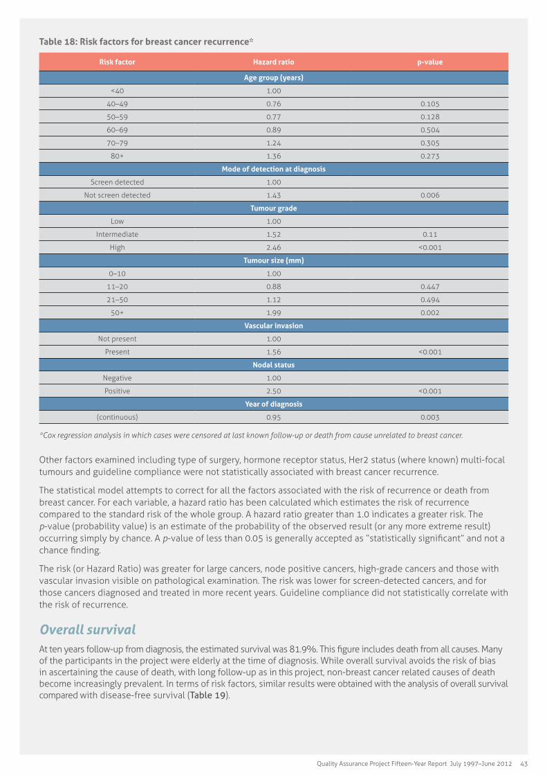
Quality Assurance Project Fifteen-Year Report July 1997–June 2012 43
Table 18: Risk factors for breast cancer recurrence*
Risk factor Hazard ratio p-value
Age group (years)
<40 1.00
40–49 0.76 0.105
50–59 0.77 0.128
60–69 0.89 0.504
70–79 1.24 0.305
80+ 1.36 0.273
Mode of detection at diagnosis
Screen detected 1.00
Not screen detected 1.43 0.006
Tumour grade
Low 1.00
Intermediate 1.52 0.11
High 2.46 <0.001
Tumour size (mm)
0–10 1.00
11–20 0.88 0.447
21–50 1.12 0.494
50+ 1.99 0.002
Vascular invasion
Not present 1.00
Present 1.56 <0.001
Nodal status
Negative 1.00
Positive 2.50 <0.001
Year of diagnosis
(continuous) 0.95 0.003
*Cox�regression�analysis�in�which�cases�were�censored�at�last�known�follow-up�or�death�from�cause�unrelated�to�breast�cancer.
Other factors examined including type of surgery, hormone receptor status, Her2 status (where known) multi-focal tumours and guideline compliance were not statistically associated with breast cancer recurrence.
The statistical model attempts to correct for all the factors associated with the risk of recurrence or death from breast cancer. For each variable, a hazard ratio has been calculated which estimates the risk of recurrence compared to the standard risk of the whole group. A hazard ratio greater than 1.0 indicates a greater risk. The p-value (probability value) is an estimate of the probability of the observed result (or any more extreme result) occurring simply by chance. A p-value of less than 0.05 is generally accepted as “statistically significant” and not a chance finding.
The risk (or Hazard Ratio) was greater for large cancers, node positive cancers, high-grade cancers and those with vascular invasion visible on pathological examination. The risk was lower for screen-detected cancers, and for those cancers diagnosed and treated in more recent years. Guideline compliance did not statistically correlate with the risk of recurrence.
Overall survivalAt ten years follow-up from diagnosis, the estimated survival was 81.9%. This figure includes death from all causes. Many of the participants in the project were elderly at the time of diagnosis. While overall survival avoids the risk of bias in ascertaining the cause of death, with long follow-up as in this project, non-breast cancer related causes of death become increasingly prevalent. In terms of risk factors, similar results were obtained with the analysis of overall survival compared with disease-free survival (Table 19).

44 Breast Cancer Treatment Group
Table 19: Risk factors for overall survival
Risk factor Hazard ratio p-value
Age group (years)
<40 0.86 0.414
40–49 0.79 0.086
50–59 (reference) 1.00 -
60–69 1.38 0.010
70–79 3.30 <0.001
80+ 5.41 <0.001
Mode of detection at diagnosis
Screen-detected 1.00
Not screen-detected 0.74 0.006
Tumour grade
Low 1.00
Intermediate 1.25 0.066
High 1.69 <0.001
Tumour size (mm)
0–10 1.00
11–20 1.08 0.579
21–50 1.12 0.032
50+ 1.91 <0.001
Place of initial surgery
Urban 1.00
Rural 1.54 <0.001
Vascular invasion
Not present 1.00
Present 1.26 0.021
Nodal status
Negative 1.00
Positive 1.91 <0.001
Period of diagnosis
1997–2002 (reference) 1.00
2002–2007 0.67 0.010
2007–2012 0.58 0.002
Guideline compliant (all indicators)
No 1.00 -
Yes 0.73 0.045
For overall survival, compliance with treatment guidelines predicted the risk of death, indicating that non-adherence to the indicator may have been due to considerations of other illness (co-morbid conditions) which might influence overall survival significantly. As expected, age correlated with overall survival, but age was not associated with increased risk of relapse.
Young womenAlthough there was a trend evident in Table 18, women under the age of 40 years in the ACT and SE NSW region undergoing modern breast cancer care, did not have a statistically significant lower risk of recurrence of breast cancer. Unadjusted estimated 10-year overall survival in this group was 81.7%, which was not different from the mean survival of the whole cohort. As might be expected, compliance with practice indicators was higher in young women.
Future directionsLong-term direct follow-up of treatment outcomes becomes increasingly resource intensive. However the results provide insights into treatment effects on recurrence rates. With extended adjuvant endocrine therapy treatment, effects are seen 15 years after the initial diagnosis, making long-term studies very useful.
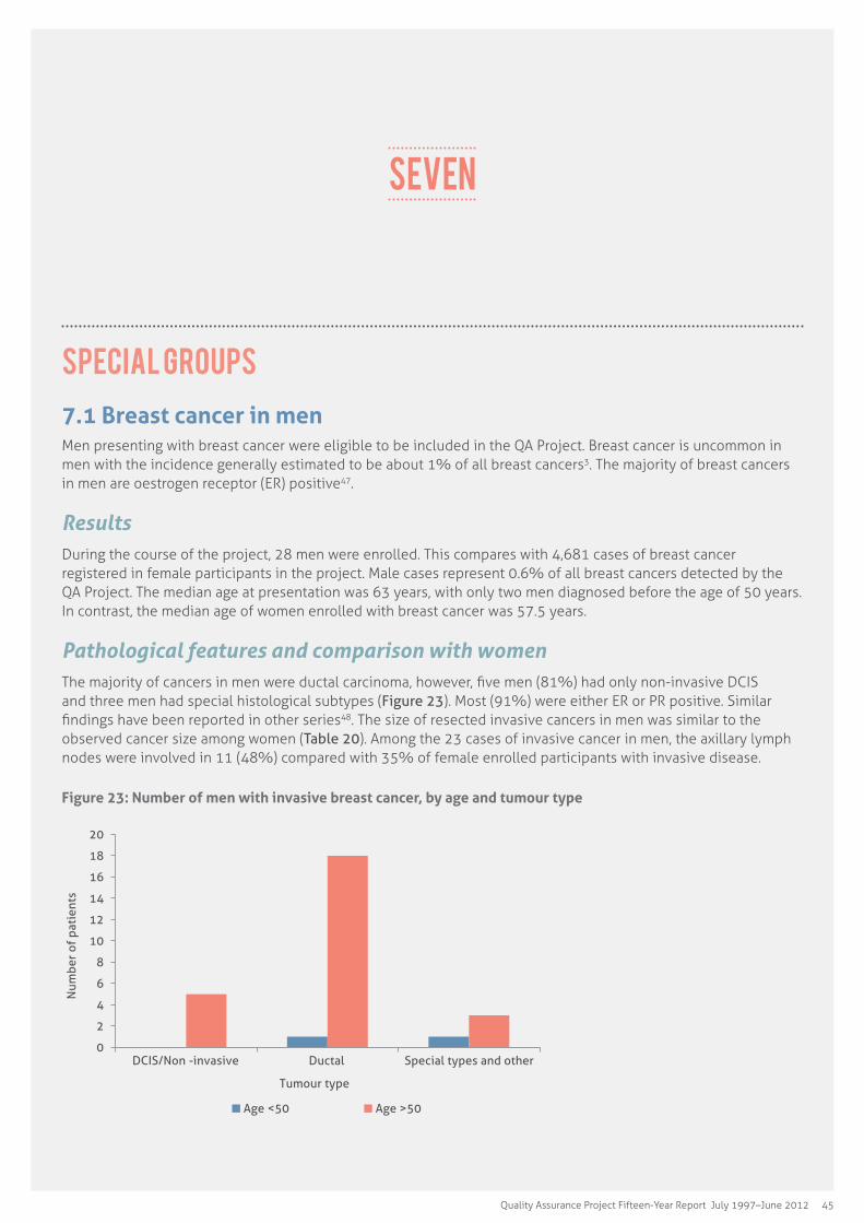
Quality Assurance Project Fifteen-Year Report July 1997–June 2012 45
Seven
Special groups
7.1 Breast cancer in menMen presenting with breast cancer were eligible to be included in the QA Project. Breast cancer is uncommon in men with the incidence generally estimated to be about 1% of all breast cancers3. The majority of breast cancers in men are oestrogen receptor (ER) positive47.
ResultsDuring the course of the project, 28 men were enrolled. This compares with 4,681 cases of breast cancer registered in female participants in the project. Male cases represent 0.6% of all breast cancers detected by the QA Project. The median age at presentation was 63 years, with only two men diagnosed before the age of 50 years. In contrast, the median age of women enrolled with breast cancer was 57.5 years.
Pathological features and comparison with womenThe majority of cancers in men were ductal carcinoma, however, five men (81%) had only non-invasive DCIS and three men had special histological subtypes (Figure 23). Most (91%) were either ER or PR positive. Similar findings have been reported in other series48. The size of resected invasive cancers in men was similar to the observed cancer size among women (Table 20). Among the 23 cases of invasive cancer in men, the axillary lymph nodes were involved in 11 (48%) compared with 35% of female enrolled participants with invasive disease.
Figure 23: Number of men with invasive breast cancer, by age and tumour type
0 2 4 6 8
10 12 14 16 18 20
DCIS/Non -invasive Ductal Special types and other
Num
ber
of p
atie
nts
Tumour type Age <50 Age >50
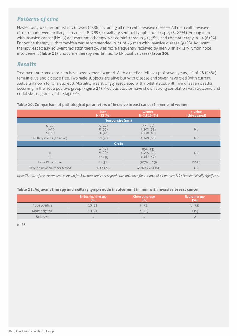
46 Breast Cancer Treatment Group
Patterns of careMastectomy was performed in 26 cases (93%) including all men with invasive disease. All men with invasive disease underwent axillary clearance (18; 78%) or axillary sentinel lymph node biopsy (5; 22%). Among men with invasive cancer (N=23) adjuvant radiotherapy was administered in 9 (39%), and chemotherapy in 14 (61%). Endocrine therapy with tamoxifen was recommended in 21 of 23 men with invasive disease (91%). Adjuvant therapy, especially adjuvant radiation therapy, was more frequently received by men with axillary lymph node involvement (Table 21). Endocrine therapy was limited to ER positive cases (Table 20).
ResultsTreatment outcomes for men have been generally good. With a median follow-up of seven years, 15 of 28 (54%) remain alive and disease free. Two male subjects are alive but with disease and seven have died (with current status unknown for one subject). Mortality was strongly associated with nodal status, with five of seven deaths occurring in the node positive group (Figure 24). Previous studies have shown strong correlation with outcome and nodal status, grade, and T stage49, 50.
Table 20: Comparison of pathological parameters of invasive breast cancer in men and women
Men N=23 (%)
Women N=3,819 (%)
p-value (chi-squared)
Tumour size (mm)
0–10 11–20 21–50
5 (22) 8 (35)
10 (43)
793 (22) 1,502 (39) 1,518 (40)
NS
Axillary nodes (positive) 11 (48) 1,349 (35) NS
Grade
I II III
4 (17) 6 (26)11 ( 9)
896 (23) 1,495 (39) 1,387 (36)
NS
ER or PR positive 21 (91) 3076 (80.5) 0.024
Her2 positive /number tested 1/13 (7.6) 418/2,726 (15) NS
Note:�The�size�of�the�cancer�was�unknown�for�6�women�and�cancer�grade�was�unknown�for�1�man�and�41�women.�NS�=Not�statistically�significant.
Table 21: Adjuvant therapy and axillary lymph node involvement in men with invasive breast cancer
Endocrine therapy (%)
Chemotherapy (%)
Radiotherapy (%)
Node positive 10 (91) 8 (73) 8 (73)
Node negative 10 (91) 5 (45) 1 (9)
Unknown 1 1 0
N=23

Quality Assurance Project Fifteen-Year Report July 1997–June 2012 47
Figure 24: Survival of men with invasive breast cancer by nodal status
7.2 Bilateral breast cancerOver the 15-year period, 174 women presented with synchronous bilateral breast cancer, defined as a primary breast cancer within each breast diagnosed within a 90-day interval50. In addition, there were 102 women who developed metachronous bilateral breast cancer, defined as a second primary cancer in the contralateral breast with an interval between episodes of greater than 90 days.
Table 22: Characteristics of women diagnosed with unilateral and bilateral breast cancer
Synchronous
Metachronous (treatment relates to
second presentation)
Unilateral
Median age at diagnosis (years) 59.3 61.9 57.5
Family history N (%) N (%) N (%)
Average risk 116 (67.4) 49 (48.5) 3,094 ( 81.5)
Moderately increased risk 36 (20.9) 13 (12.9) 395 (10.4)
Potentially high risk 20 (11.6) 39 38.6) 306 (8.1)
Lobular cancer 27 (15.5) 12 (11.8) 382 (10.0)
Surgery
Mastectomy 133 (77.8) 74 (73.3) 1,934 (50.6)
Breast conserving surgery 38 (22.2) 27 (26.7) 1,885 (49.4)
Adjuvant endocrine therapy
Received 136 (78.2) 61 (59.8) 2,842 (74.4)
Not recommended 30 (17.2) 39 (38.2) 869 (22.8)
Recommended and refused 8 (4.6) 2 (2.0) 108 (2.8)
Adjuvant chemotherapy
Received 87 (50.0) 31 (30.4) 1,932 (50.6)
Not recommended 74 (42.5) 65 (63.7) 1,680 (44.0)
Recommended and refused 13 (7.5) 6 (5.9) 207 (5.4)

48 Breast Cancer Treatment Group
7.2.1 Synchronous bilateral breast cancer The rate of synchronous bilateral breast cancer has been reported to be in the range 0.3% to 3% of all breast cancer in women. Bilateral breast cancer is associated with the lobular subtype and a family history of breast cancer.
Results
The QA Project observed a 3.7% rate of synchronous bilateral breast cancer, somewhat higher than reported historically65. The rate of bilateral breast cancer may be increasing over time, possibly due to more sensitive diagnostic techniques51.
The median age of women with synchronous, metachronous and unilateral breast cancer was 59.3, 61.9 and 57.5 years respectively.
Lobular carcinoma was significantly more frequent in women with synchronous bilateral cancer compared with unilateral cancers in the 15-year period.
Women presenting with bilateral breast cancer were more likely to undergo mastectomy, receive adjuvant chemotherapy and adjuvant endocrine than women with unilateral breast cancer (Table 22).
Future directions
A detailed study using data from the QA Project has previously shown that synchronous bilateral breast cancer has a poorer outcome when known prognostic features are controlled in a multivariate analysis52.
7.2.2 Metachronous bilateral breast cancerOf the 102 women developing metachronous bilateral disease, the majority were treated with mastectomy (73.3%) rather than breast conservation therapy, compared to 50.6% with unilateral breast cancer. Adjuvant systemic therapy was more likely to be endocrine therapy alone, rather than including chemotherapy, when compared with unilateral cases.
Survival of women with a second primary metachronous bilateral breast cancer was reduced compared with unilateral participants (Table 22).
7.3 Participants with distant metastases at presentation Only a small proportion of women with invasive breast cancer present with detectable distant metastatic disease. The management of metastatic (Stage 4) breast cancer involves a multidisciplinary approach to maximise the health and quality of life of the patient and to prolong survival, even when curing the disease may not be possible.
Depending on the circumstances, surgery may be helpful, as well as radiation therapy and systemic treatment. Endocrine therapy is often used first in preference to chemotherapy in ER positive cancers. Targeted therapy with trastuzumab may be appropriate for Her2 positive tumours.
Results
Distant metastatic disease was present at the time of presentation in 82 participants (1.8%). The median age at diagnosis was 58.9 years.
Breast cancers in women presenting with metastatic disease were less likely to be ER positive (21%) compared with early stage breast cancer. Proportionately very few Stage 4 cancers were histological Grade 1 (Figure 25). Overall, of 82 patients with Stage 4 metastatic breast cancer, 64 (78%) patients underwent some form of breast surgery. Of these patients, 54 underwent mastectomy. Surgery was performed less commonly in women aged 70 years or older (Figures 26, 27 and 28).
The median survival of subjects presenting with metastatic breast cancer was 48.8 months (Figure 29). After therapy, 9 out of 83 subjects presenting with Stage 4 breast cancer remained progression free.
Future directions
Only a very small proportion of cases notified to the project had Stage 4 disease. This may represent under reporting. As these women are often cared for by oncologists, more active enrolment of patients by clinicians other than surgeons may overcome this difficulty in the future and allow more complete assessment of this important group.
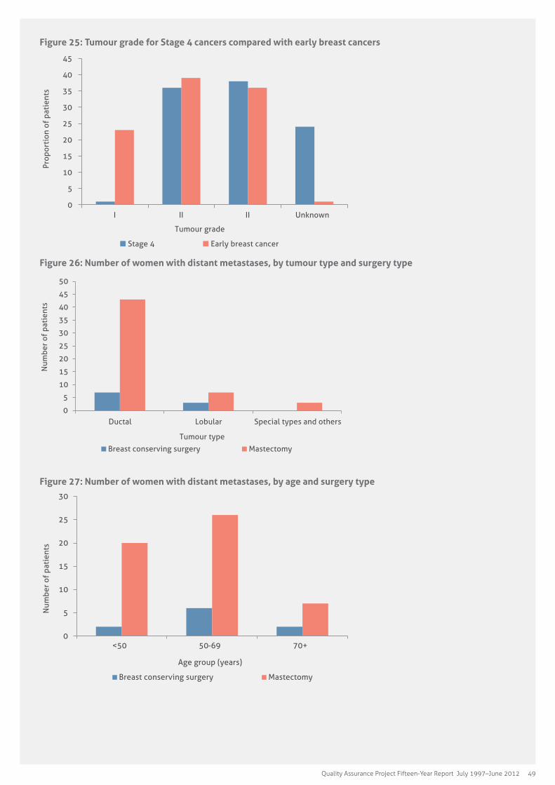
Quality Assurance Project Fifteen-Year Report July 1997–June 2012 49
Figure 25: Tumour grade for Stage 4 cancers compared with early breast cancers
0
5
10
15
20
25
30
35
40
45
I II II Unknown
Prop
orti
on o
f pa
tien
ts
Tumour grade
Stage 4 Early breast cancer
Figure 26: Number of women with distant metastases, by tumour type and surgery type
0
5
10
15
20
25
30
35
40
45
50
Ductal Lobular Special types and others
Num
ber
of p
atie
nts
Tumour type Breast conserving surgery Mastectomy
Figure 27: Number of women with distant metastases, by age and surgery type
0
5
10
15
20
25
30
<50 50-69 70+
Num
ber
of p
atie
nts
Age group (years)
Breast conserving surgery Mastectomy
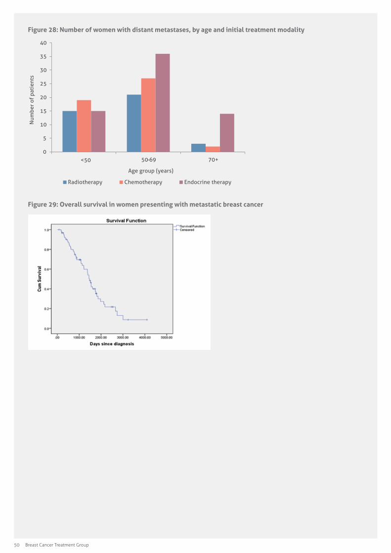
50 Breast Cancer Treatment Group
Figure 28: Number of women with distant metastases, by age and initial treatment modality
0
5
10
15
20
25
30
35
40
<50 50-69 70+
Num
ber
of p
atie
nts
Age group (years)
Radiotherapy Chemotherapy Endocrine therapy
Figure 29: Overall survival in women presenting with metastatic breast cancer
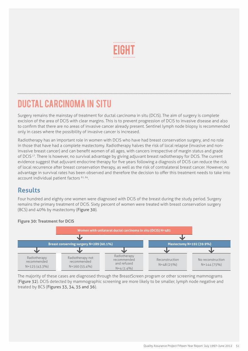
Quality Assurance Project Fifteen-Year Report July 1997–June 2012 51
Ductal carcinoma in situ Surgery remains the mainstay of treatment for ductal carcinoma in situ (DCIS). The aim of surgery is complete excision of the area of DCIS with clear margins. This is to prevent progression of DCIS to invasive disease and also to confirm that there are no areas of invasive cancer already present. Sentinel lymph node biopsy is recommended only in cases where the possibility of invasive cancer is increased.
Radiotherapy has an important role in women with DCIS who have had breast conservation surgery, and no role in those that have had a complete mastectomy. Radiotherapy halves the risk of local relapse (invasive and non-invasive breast cancer) and can benefit women of all ages, with cancers irrespective of margin status and grade of DCIS 17. There is however, no survival advantage by giving adjuvant breast radiotherapy for DCIS. The current evidence suggest that adjuvant endocrine therapy for five years following a diagnosis of DCIS can reduce the risk of local recurrence after breast conservation therapy, as well as the risk of contralateral breast cancer. However, no advantage in survival rates has been observed and therefore the decision to offer this treatment needs to take into account individual patient factors 61, 64.
ResultsFour hundred and eighty one women were diagnosed with DCIS of the breast during the study period. Surgery remains the primary treatment of DCIS. Sixty percent of women were treated with breast conservation surgery (BCS) and 40% by mastectomy (Figure 30).
Figure 30: Treatment for DCIS
Women with unilateral ductal carcinoma in situ (DCIS) N=481
Breast conserving surgery N=289 (60.1%) Mastectomy N=192 (39.9%)
Radiotherapy recommended
N=125 (43.3%)
Radiotherapy not recommended
N=160 (55.4%)
Radiotherapy recommended
and refusedN=4 (1.4%)
ReconstructionN=48 (25%)
No reconstructionN=144 (75%)
The majority of these cases are diagnosed through the BreastScreen program or other screening mammograms (Figure 32). DCIS detected by mammographic screening are more likely to be smaller, lymph node negative and treated by BCS (Figures 33, 34, 35 and 36).
EIGHT
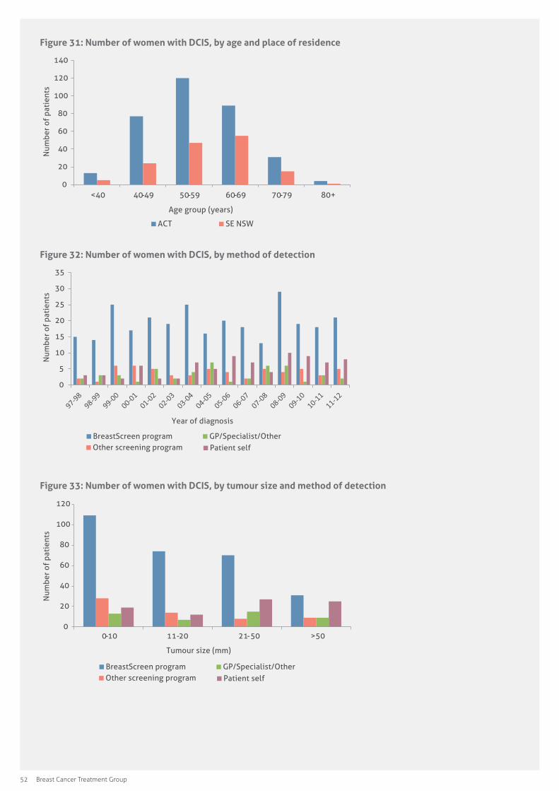
52 Breast Cancer Treatment Group
Figure 31: Number of women with DCIS, by age and place of residence
0
20
40
60
80
100
120
140
<40 40-49 50-59 60-69 70-79 80+
Num
ber
of p
atie
nts
Age group (years)
ACT SE NSW
Figure 32: Number of women with DCIS, by method of detection
0
5
10
15
20
25
30
35
Num
ber
of p
atie
nts
Year of diagnosis
BreastScreen program Other screening program
GP/Specialist/Other Patient self
Figure 33: Number of women with DCIS, by tumour size and method of detection
0
20
40
60
80
100
120
0-10 11-20 21-50 >50
Num
ber
of p
atie
nts
Tumour size (mm)
BreastScreen program Other screening program
GP/Specialist/Other Patient self
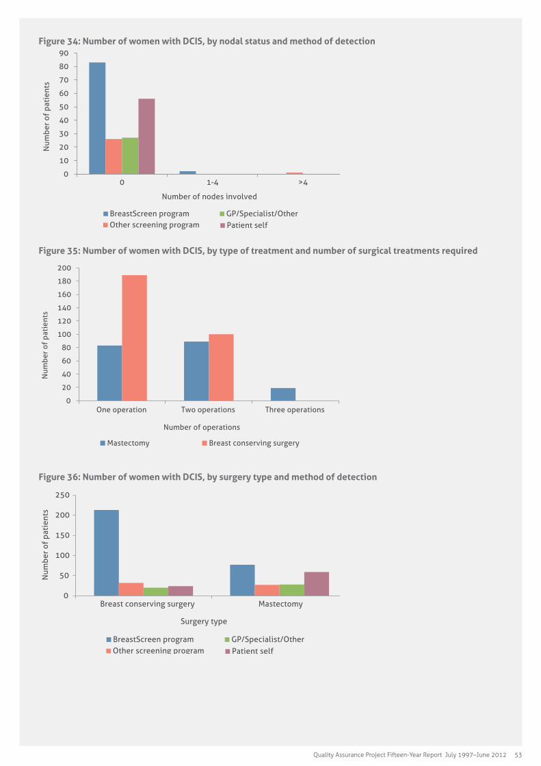
Quality Assurance Project Fifteen-Year Report July 1997–June 2012 53
Figure 34: Number of women with DCIS, by nodal status and method of detection
0
10
20
30
40
50
60
70
80
90
0 1-4 >4
Num
ber
of p
atie
nts
Number of nodes involved
BreastScreen program Other screening program
GP/Specialist/Other Patient self
Figure 35: Number of women with DCIS, by type of treatment and number of surgical treatments required
0
20
40
60
80
100
120
140
160
180
200
One operation Two operations Three operations
Num
ber
of p
atie
nts
Number of operations
Mastectomy Breast conserving surgery
Figure 36: Number of women with DCIS, by surgery type and method of detection
0
50
100
150
200
250
Breast conserving surgery Mastectomy
Num
ber
of p
atie
nts
Surgery type
BreastScreen program Other screening program
GP/Specialist/Other Patient self
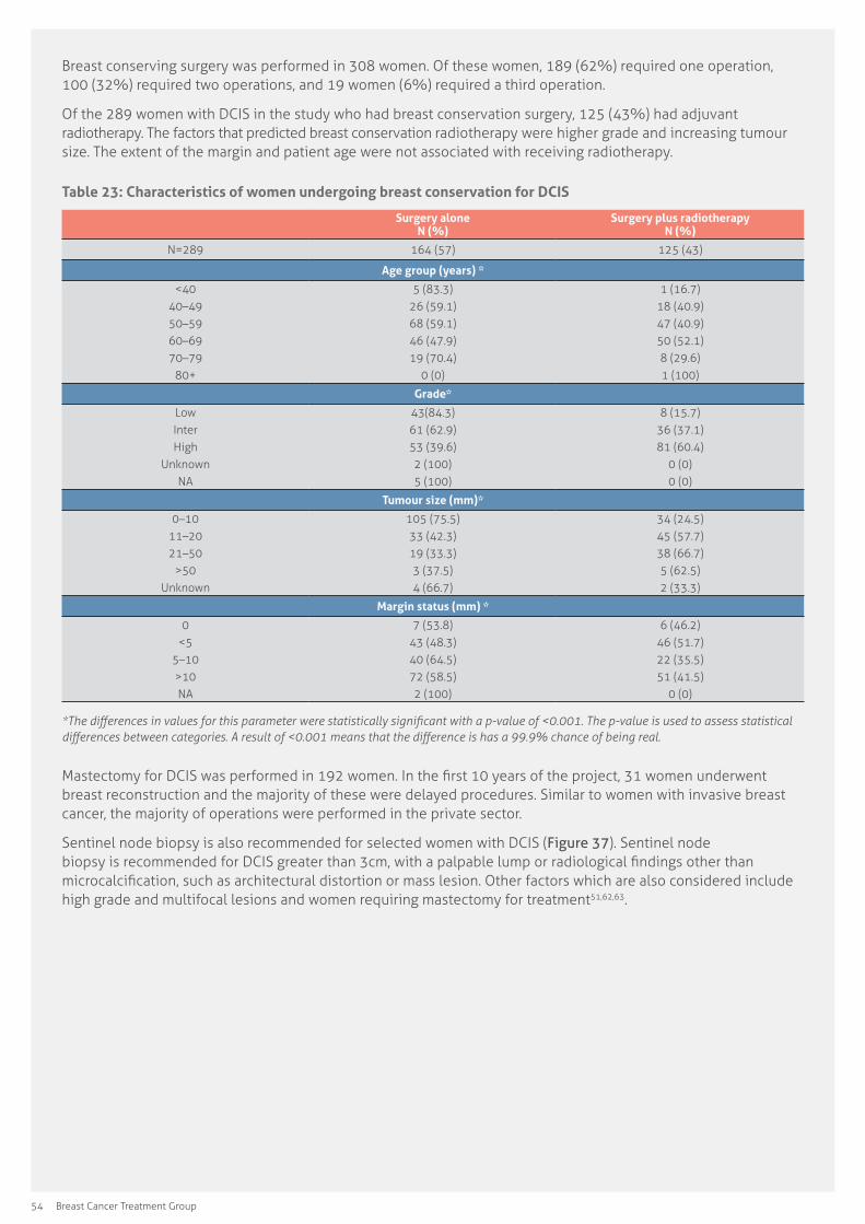
54 Breast Cancer Treatment Group
Breast conserving surgery was performed in 308 women. Of these women, 189 (62%) required one operation, 100 (32%) required two operations, and 19 women (6%) required a third operation.
Of the 289 women with DCIS in the study who had breast conservation surgery, 125 (43%) had adjuvant radiotherapy. The factors that predicted breast conservation radiotherapy were higher grade and increasing tumour size. The extent of the margin and patient age were not associated with receiving radiotherapy.
Table 23: Characteristics of women undergoing breast conservation for DCIS
Surgery alone N (%)
Surgery plus radiotherapy N (%)
N=289 164 (57) 125 (43)
Age group (years) *
<4040–4950–5960–6970–79
80+
5 (83.3)26 (59.1)68 (59.1)46 (47.9)19 (70.4)
0 (0)
1 (16.7)18 (40.9)47 (40.9)50 (52.1)8 (29.6)1 (100)
Grade*
LowInterHigh
UnknownNA
43(84.3)61 (62.9)53 (39.6)2 (100)5 (100)
8 (15.7)36 (37.1)81 (60.4)
0 (0)0 (0)
Tumour size (mm)*
0–1011–2021–50
>50Unknown
105 (75.5)33 (42.3)19 (33.3)3 (37.5)4 (66.7)
34 (24.5)45 (57.7)38 (66.7)5 (62.5)2 (33.3)
Margin status (mm) *
0<5
5–10>10NA
7 (53.8)43 (48.3)40 (64.5)72 (58.5)2 (100)
6 (46.2)46 (51.7)22 (35.5)51 (41.5)
0 (0)
*The�differences�in�values�for�this�parameter�were�statistically�significant�with�a�p-value�of�<0.001.�The�p-value�is�used�to�assess�statistical�differences�between�categories.�A�result�of�<0.001�means�that�the�difference�is�has�a�99.9%�chance�of�being�real.
Mastectomy for DCIS was performed in 192 women. In the first 10 years of the project, 31 women underwent breast reconstruction and the majority of these were delayed procedures. Similar to women with invasive breast cancer, the majority of operations were performed in the private sector.
Sentinel node biopsy is also recommended for selected women with DCIS (Figure 37). Sentinel node biopsy is recommended for DCIS greater than 3cm, with a palpable lump or radiological findings other than microcalcification, such as architectural distortion or mass lesion. Other factors which are also considered include high grade and multifocal lesions and women requiring mastectomy for treatment51,62,63.

Quality Assurance Project Fifteen-Year Report July 1997–June 2012 55
Figure 37: Number of women with DCIS, undergoing sentinel node biopsy
0
5
10
15
20
25
99-00 00-01
01-02 02-03
03-04 04-05
05-06 06-07
07-08 08-09
09-10 10-11
11-12
Num
ber
of p
atie
nts
Year of diagnosis
The use of adjuvant endocrine therapy for DCIS in hormone receptor positive tumours is decreasing (Figure 38).
Initial survival data for women with DCIS (as at March 2015) showed that 391 of 481 women (81.3%) were alive and disease free, 23 women (4.8%) were alive with recurrent disease, 27 (5.6%) were deceased, of which 4 cases (0.8%) were due to breast cancer. Forty women (8.3%) were recorded as having an unknown current status.
Figure 38: Number of women with DCIS, receiving tamoxifen and aromatase inhibitors
0
1
2
3
4
5
6
7
8
Num
ber
of p
atie
nts
Year of diagnosis
Aromatase inhibitors Tamoxifen
Figure 39: Number of women with DCIS, receiving tamoxifen by hormone receptor status
0
5
10
15
20
25
ER positive PR positive ER and PR Positive ER and PR Neg
Num
ber
of p
atie
nts
Receptor status
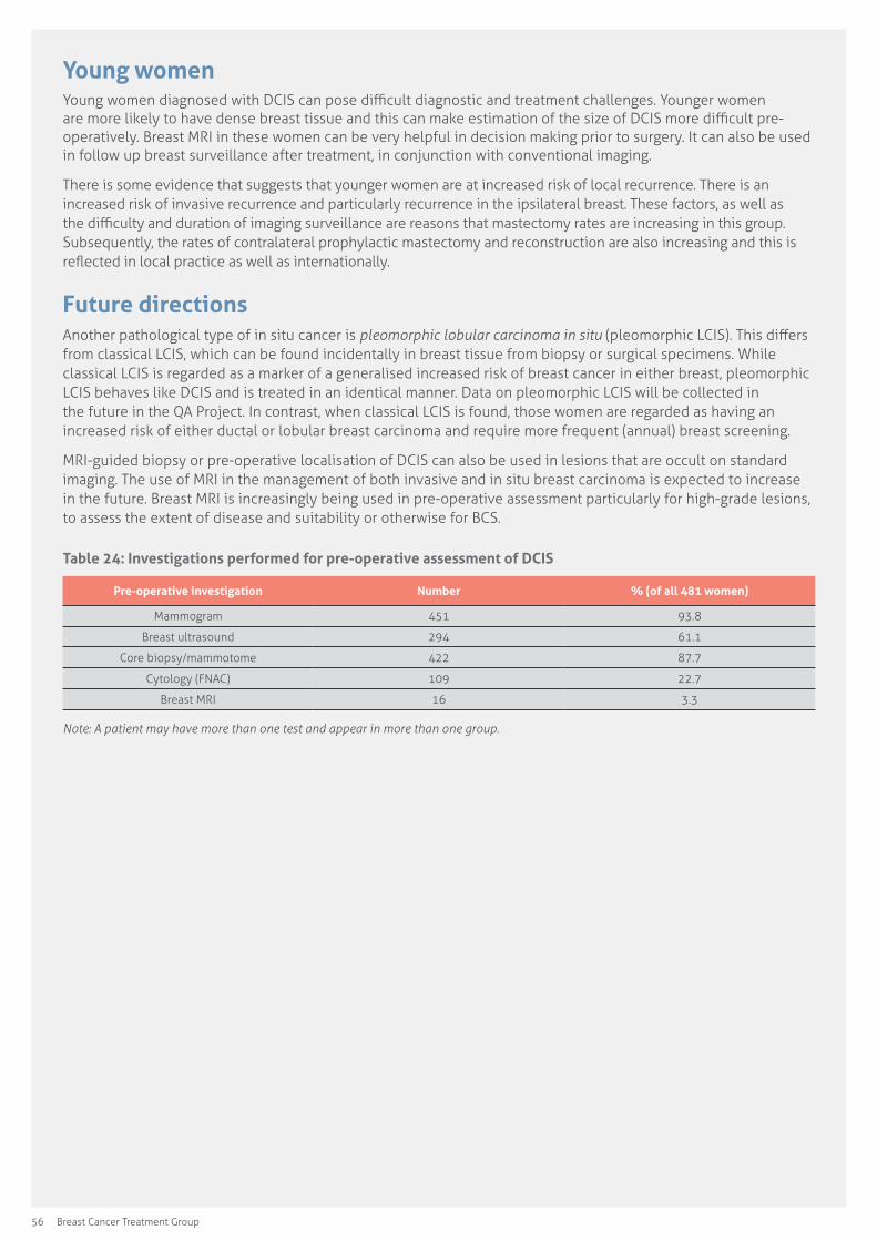
56 Breast Cancer Treatment Group
Young womenYoung women diagnosed with DCIS can pose difficult diagnostic and treatment challenges. Younger women are more likely to have dense breast tissue and this can make estimation of the size of DCIS more difficult pre-operatively. Breast MRI in these women can be very helpful in decision making prior to surgery. It can also be used in follow up breast surveillance after treatment, in conjunction with conventional imaging.
There is some evidence that suggests that younger women are at increased risk of local recurrence. There is an increased risk of invasive recurrence and particularly recurrence in the ipsilateral breast. These factors, as well as the difficulty and duration of imaging surveillance are reasons that mastectomy rates are increasing in this group. Subsequently, the rates of contralateral prophylactic mastectomy and reconstruction are also increasing and this is reflected in local practice as well as internationally.
Future directionsAnother pathological type of in situ cancer is pleomorphic lobular carcinoma in situ (pleomorphic LCIS). This differs from classical LCIS, which can be found incidentally in breast tissue from biopsy or surgical specimens. While classical LCIS is regarded as a marker of a generalised increased risk of breast cancer in either breast, pleomorphic LCIS behaves like DCIS and is treated in an identical manner. Data on pleomorphic LCIS will be collected in the future in the QA Project. In contrast, when classical LCIS is found, those women are regarded as having an increased risk of either ductal or lobular breast carcinoma and require more frequent (annual) breast screening.
MRI-guided biopsy or pre-operative localisation of DCIS can also be used in lesions that are occult on standard imaging. The use of MRI in the management of both invasive and in situ breast carcinoma is expected to increase in the future. Breast MRI is increasingly being used in pre-operative assessment particularly for high-grade lesions, to assess the extent of disease and suitability or otherwise for BCS.
Table 24: Investigations performed for pre-operative assessment of DCIS
Pre-operative investigation Number % (of all 481 women)
Mammogram 451 93.8
Breast ultrasound 294 61.1
Core biopsy/mammotome 422 87.7
Cytology (FNAC) 109 22.7
Breast MRI 16 3.3
Note: A patient may have more than one test and appear in more than one group.
Quality Assurance Project Fifteen-Year Report July 1997–June 2012 57
Pathological features
Introduction Histopathology of the breast with or without examination of other tissues such as sentinel node(s) is essential for determining the diagnosis, prognosis and further management of patients with breast carcinoma. Structured reporting of breast pathology is now considered standard of care in Australia and is followed by all laboratories in our region. The Royal College of Pathologists of Australasia (RCPA) has developed protocols to assist pathologists in reporting of relevant information for specific cancers. Each protocol includes “standards” and “guidelines” which are indicators of ‘minimum requirements’ and ‘recommendations’, which reflect the opinion of the relevant expert authoring groups56. The RCPA breast cancer protocol was first published in 2010. In addition to the widespread implementation of standardised reporting, the high quality of pathology data in the BCTG QA Project has been assisted by regular audit of the data entered that includes feedback from researchers utilising the data. Since 2005 the pathology laboratories in the region have participated in an RCPA external quality assurance program that audits aspects of the pathology report such as the rates of positivity for hormone receptor status.
Ancillary pathology investigations such as assessment of hormone receptor status (oestrogen receptor [ER] and progesterone receptor [PR]), proliferation index (Ki67) and Her2/neu status are now routinely performed on breast carcinoma samples. These tests help guide adjuvant treatments and also provide additional prognostic information. Over the past 15 years there has been a change in the quantification of these markers. In 1997 biochemical analysis (ligand binding assay and enzyme immunoassay) and immunohistochemistry were used to assess oestrogen and progesterone receptor status. No standardized scoring system or universal cut-off for determining a positive result was available.
In 2001 the Australian Cancer Network Working Party recommended a cut-off point for a positive assay for ER and PR was greater than 1% of positive stained nuclei with intermediate – or high-intensity staining, or greater than 10% of nuclei staining with low intensity57. Since 2005 a cut off of ≥1% of positive stained nuclei, irrespective of the intensity of staining, has been used according to the American Society of Clinical Oncology (ASCO) guidelines58. Her2 status was initially assessed by immunohistochemistry with those cases assessed as being 2+ (equivocal) being sent for testing by fluorescent in situ hybridisation (FISH). In October 2006 Herceptin (trastuzumab) was made available for the treatment of Her2 positive breast carcinomas through the pharmaceutical benefits scheme (PBS) if in situ hybridisation (ISH) demonstrated Her2 positivity. In 2006 chromogenic in situ hybridisation (CISH) testing commenced in Canberra with 2+ cases being referred for FISH testing. In 2007 silver in situ hybridisation (SISH) was introduced at ACT Pathology and replaced CISH testing. With SISH testing (with or without Chromosome 17 testing) referral for FISH testing ceased. In 2012 dual in situ hybridisation (DISH) was introduced at Capital Pathology. The proliferative index of carcinomas (Ki67) has been routinely assessed since 2010 and recorded on the BCTG form since 2013. Genomic testing, while available, is yet to become part of routine pathology report.
NINE
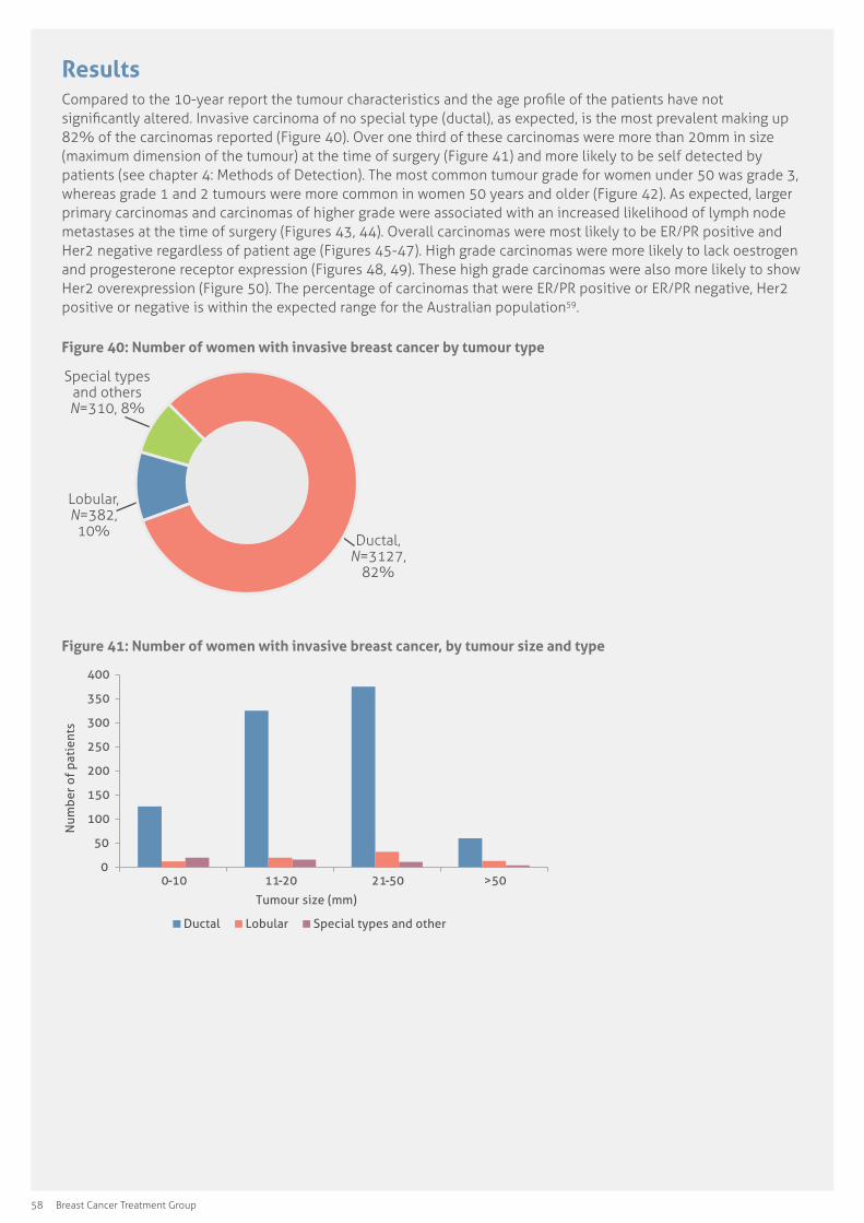
58 Breast Cancer Treatment Group
ResultsCompared to the 10-year report the tumour characteristics and the age profile of the patients have not significantly altered. Invasive carcinoma of no special type (ductal), as expected, is the most prevalent making up 82% of the carcinomas reported (Figure 40). Over one third of these carcinomas were more than 20mm in size (maximum dimension of the tumour) at the time of surgery (Figure 41) and more likely to be self detected by patients (see chapter 4: Methods of Detection). The most common tumour grade for women under 50 was grade 3, whereas grade 1 and 2 tumours were more common in women 50 years and older (Figure 42). As expected, larger primary carcinomas and carcinomas of higher grade were associated with an increased likelihood of lymph node metastases at the time of surgery (Figures 43, 44). Overall carcinomas were most likely to be ER/PR positive and Her2 negative regardless of patient age (Figures 45-47). High grade carcinomas were more likely to lack oestrogen and progesterone receptor expression (Figures 48, 49). These high grade carcinomas were also more likely to show Her2 overexpression (Figure 50). The percentage of carcinomas that were ER/PR positive or ER/PR negative, Her2 positive or negative is within the expected range for the Australian population59.
Figure 40: Number of women with invasive breast cancer by tumour type
Ductal, N=3127,
82%
Lobular, N=382,
10%
Special types and others N=310, 8%
Figure 41: Number of women with invasive breast cancer, by tumour size and type
0
50
100
150
200
250
300
350
400
0-10 11-20 21-50 >50
Num
ber
of p
atie
nts
Tumour size (mm)
Ductal Lobular Special types and other

Quality Assurance Project Fifteen-Year Report July 1997–June 2012 59
igure 42: Number of women with invasive breast cancer, by age and tumour grade
0 50
100 150 200 250 300 350 400 450 500
<40 40-49 50-59 60-69 70-79 80+
Num
ber
of p
atie
nts
Age group (years)
Grade I Grade II Grade III
Figure 43: Number of women with invasive breast cancer, by size and lymph node status
0
200
400
600
800
1000
1200
0-10 11-20 21-50 50+
Num
ber
of p
atie
nts
Tumour size (mm)
Node negative Node positive
Figure 44: Number of women with invasive breast cancer, by grade and lymph node status
0 100 200 300 400 500 600 700 800 900
1000
I II III
Num
ber
of p
atie
nts
Tumour grade
Node negative Node positive
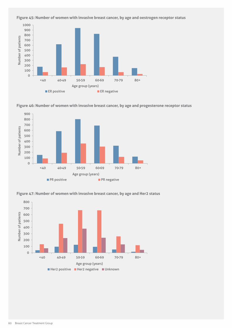
60 Breast Cancer Treatment Group
Figure 45: Number of women with invasive breast cancer, by age and oestrogen receptor status
0
100
200
300
400
500
600
700
800
900
1000
<40 40-49 50-59 60-69 70-79 80+
Num
ber
of p
atie
nts
Age group (years)
ER positive ER negative
Figure 46: Number of women with invasive breast cancer, by age and progesterone receptor status
0
100
200
300
400
500
600
700
800
900
<40 40-49 50-59 60-69 70-79 80+
Num
ber
of p
atie
nts
Age group (years)
PR positive PR negative
Figure 47: Number of women with invasive breast cancer, by age and Her2 status
0
100
200
300
400
500
600
700
800
<40 40-49 50-59 60-69 70-79 80+
Num
ber
of p
atie
nts
Age group (years)
Her2 positive Her2 negative Unknown

Quality Assurance Project Fifteen-Year Report July 1997–June 2012 61
Figure 48: Number of women with invasive breast cancer, by grade and oestrogen receptor status
0
200
400
600
800
1000
1200
1400
1600
I II III
Num
ber
of p
atie
nts
Tumour grade
ER positive ER negative
Figure 49: Number of women with invasive breast cancer, by grade and progesterone receptor status
0
200
400
600
800
1000
1200
1400
I II III
Num
ber
of p
atie
nts
Tumour grade
PR positive PR negative
Figure 50: Number of women with invasive breast cancer, by grade and Her2 status
0
200
400
600
800
1000
1200
I II III
Num
ber
of p
atie
nts
Tumour grade
Her2 positive Her2 negative

62 Breast Cancer Treatment Group
Young womenWhile invasive carcinoma of no special type (ductal) was the most common carcinoma in all age groups it was relatively more common in women younger than 40 years than other carcinoma types (Figure 51). In women younger than 40 years the carcinoma was more likely to be high grade (Figure 42) with lymphovascular invasion (Figure 52) and have an increased likelihood of lymph node metastases at the time of surgery (Figure 53).
Figure 51: Number of women with invasive breast cancer, by age and tumour type
0
100
200
300
400
500
600
700
800
900
1000
<40 40-49 50-59 60-69 70-79 80+
Num
ber
of p
atie
nts
Age group (years)
Ductal Lobular Special types and other
Figure 52: Number of women with invasive breast cancer, by lymphovascular invasion and age
0
100
200
300
400
500
600
700
800
900
1000
<40 40-49 50-59 60-69 70-79 80+
Num
ber
of p
atie
nts
Age group (years)
LVI present LVI absent LVI unknown
Figure 53: Number of women with invasive breast cancer, by age and lymph node status
0
100
200
300
400
500
600
700
800
<40 40-49 50-59 60-69 70-79 80+
Num
ber
of p
atie
nts
Age group (years)
Node negative Node positive
Quality Assurance Project Fifteen-Year Report July 1997–June 2012 63
Future directions Features such as multifocality, neoadjuvant treatment effect, involvement of skin or muscle, presence of co-existing in situ carcinoma and its distance from surgical margins are also now routinely recorded in pathology reports. Further analysis of these parameters could be assessed.
More detailed information on nodal status is being collected prospectively. The Group is considering retrospective data collection of the number of nodes removed and the number of lymph nodes involved by carcinoma to allow further analysis on patient outcomes.
Technology may improve interfaces between the pathology report and databases. This has the potential to improve accuracy by reducing human error with data entry and enable more detailed analysis of tumour characteristics.
Personalised treatment means clinicians are interested in sub-classification of tumours according to novel molecular characteristics. The BCTG QA Project database fields need to reflect these changes in the future.
64 Breast Cancer Treatment Group
appendices
Appendix 1: Publications and reports
1 Papers published in academic journalsImplementing clinical practice guidelines: a community-based audit of breast cancer treatment. Craft P, Zhang Y, Brogan J, Tait N, Buckingham J and the ACT & SENSW BCTG. Medical Journal of Australia, March 2000, 172(5): 213-216.
Breast cancer guidelines in action: editorial comment, Redman S and Reeve TS. Medical Journal of Australia, March 2000, 172(5): 196-197.
Surgical Management of Invasive Breast Cancer: A 5–year Prospective Study of Treatment in the Australian Capital Territory and South-Eastern New South Wales. Koshy A, Buckingham J, Zhang Y, Craft P, Dahlstrom J, Tait N and members of the ACT & SE NSW Breast Cancer Treatment Group. Australian & New Zealand Journal of Surgery, September 2005, 75 (9): 757-761.
Synchronous multiple ipsilateral breast cancers: implications for patient management. Jain S, Rezo A, Shadbolt B and Dahlstrom J. Pathology: Journal of the Royal College of Pathologists of Australasia, January 2009, 41:57-67.
Variation in the management of early breast cancer in rural and metropolitan centres: Implications for the organisation of rural cancer services. Craft PS, Buckingham JM, Dahlstrom JE, Beckmann KR, Zhang Y, Stuart-Harris R, Jacob G, Roder D, Tait N. The Breast, October 2010, 19(5): 396-401.
Clinical characteristics and outcomes of bilateral breast cancer in an Australian cohort. Beckmann KR, Buckingham J, Craft P, Dahlstrom JE, Zhang Y, Roder D, Stuart-Harris R. The Breast, April 2011, 20(2): 158-164.
Tumor size and survival in multicentric and multifocal breast cancer. Rezo A, Dahlstrom J, Shadbolt B, Rodins K, Zhang Y, Davis AJ, in collaboration with ACT & SENSW BCTG. The Breast, June 2011, 20(3): 258-263.
Prognostic value of LINE – 1 retrotransposon expression and its subcellular localization in breast cancer. Chen L, Dahlstrom JE, Chandra A, Board P, Rangasamy D. Breast Cancer Research and Treatment, November 2012, 136(1): 129-142.
Surgical margins and risk of locoregional recurrence in invasive breast cancer: an analysis of 10-year data from the Breast Cancer Treatment Quality Assurance Project. Behm EC, Beckmann KR, Dahlstrom JE, Zhang Y, Cho C, Stuart-Harris R, Craft P, Rezo A, Buckingham JM. The Breast, 2013 Oct, 22(5): 839-844. Doi: 10.1016/j.breast.2013.02.018. Epub 2013 Mar 22.
2 Published abstractsA regional approach in Australia to quality and safety in breast cancer management. Buckingham J, Zhang Y, Dahlstrom J, Craft P, Stuart-Harris R, Tait N. The Australian and New Zealand Journal of Surgery 2008; 78:5.
Variation in treatment outcomes and care provided for operable breast cancer in rural and metropolitan centers. Craft P, Roder D, Buckingham J, Zhang Y, Beckmann K, Dahlstrom J, Jacob G, Stuart-Harris R, Tait N, in conjunction with the ACT & SE NSW Breast Cancer Treatment Group. American Society of Clinical Oncology Annual Meeting Proceedings (Supplement), 2009: e11517.
Synchronous multiple ipsilateral breast cancers: implications for patient management. Jain S, Rezo A, Shadbolt B, Dahlstrom JE. Journal of the Royal College of Pathologists of Australasia, January 2009, 41(1):57-67.

Quality Assurance Project Fifteen-Year Report July 1997–June 2012 65
3 Presentations/posters at national and international conferencesTreatment guideline implementation for early breast cancer: a community based project. Craft P, Zhang Y, for the ACT & SE NSW Breast Cancer Treatment Group. The Clinical Oncological Society of Australia 25th Annual Scientific Conference, November 1998, Sydney, Australia.
Treatment guideline implementation strategies for early breast cancer: a community based project. Brogan J, Zhang Y and ACT & SE NSW Breast Cancer Treatment Group. The National Centre for Epidemiology and Population Health 10th Anniversary Conference, November 1998, Canberra, Australia.
A study for the treatment of newly diagnosed women with early breast cancer in the Australian Capital Territory and South Eastern New South Wales: A model for guideline implementation. Craft P, Zhang Y, Brogan J, Tait N, Buckingham J, on behalf of ACT & SE NSW Breast Cancer Treatment Group. The China National Cancer Conference, 5th Scientific Meeting, April 2000, Xiamen, China.
Clinical predictors and histopathological associates of primary failure of breast conserving therapy. Fergusson J, Zhang Y, Dahlstrom J, Buckingham J, Tait N, for the ACT & SE NSW Breast Cancer Treatment Group. The Royal Australasian College of Surgeons Annual Scientific Congress, May 2001, Canberra, Australia.
Histopathological predictors of primary failure of breast conserving therapy. Fergusson J, Zhang Y, Dahlstrom J, Buckingham J, Tait N, for the ACT & SE NSW Breast Cancer Treatment Group. The Clinical Oncological Society of Australia 28th Annual Scientific Conference, November 2001, Brisbane, Australia.
Surgical management of breast cancer in the ACT & SE NSW. Koshy A, Buckingham J, Zhang Y, Craft P, Dahlstrom J, Tait N, for the ACT & SE NSW Breast Cancer Treatment Group. The Royal Australasian College of Surgeons Annual Scientific Conference, May 2004, Perth, Australia.
Surgical management of invasive breast cancer in the ACT & SE NSW. Koshy A, Buckingham J, Zhang Y, Craft P, Dahlstrom J, Tait N and the ACT & SE NSW Breast Cancer Treatment Group. The Leura 5th International Breast Cancer Conference, November 2004, Sydney, Australia.
Surgical management of ductal carcinoma in situ in the ACT & SE NSW. Koshy A, Buckingham J, Zhang Y, Craft P, Dahlstrom J, Tait N and the ACT & SE NSW Breast Cancer Treatment Group. The Leura 5th International Breast Cancer Conference, November 2004, Sydney, Australia.
Breast cancer and treatment in the ACT & SE NSW Quality Assurance Project: 1997–2002. Dahlstrom J, Craft P, Zhang Y, Buckingham J, Jacob G, Tait N and the ACT & SE NSW Breast Cancer Treatment Group. The Clinical Oncological Society of Australia 31st Annual Scientific Meeting, November 2004, Canberra, Australia.
Surgical management of invasive breast cancer in the ACT & SE NSW. Buckingham J, Zhang Y, Craft P, Dahlstrom J, Tait N and the ACT & SE NSW Breast Cancer Treatment Group. The Australian and Chinese Surgeons’ Conference, November 2004, Guangzhou, China.
Measuring up multifocal breast cancer—a retrospective analysis of tumour size and lymph node status in multifocal/multicentric breast cancer in the ACT & SE NSW. Rezo A, Rodins K, Dahlstrom J, Davis A. Grand Rounds, November 2005, The Canberra Hospital, Canberra, Australia.
A retrospective analysis of tumour size and lymph node status in multifocal breast cancer in the ACT & SE NSW. Pathology Update 2006. Rodins K, Rezo A, Davis A, Dahlstrom J, 10–12 March, Sydney Convention Centre, Sydney, Australia [This presentation won the Board of Education poster prize].
Assessment of tumour size and its relationship to nodal involvement in multifocal and multicentric breast cancer. Rezo A, Rodins K, Davis A, Shadbolt B, Zhang Y, Huynh F, Dahlstrom J and in conjunction with the ACT & SE NSW Breast Cancer Treatment Group. Am J Clin Oncol 2007. American Society of Clinical Oncology Annual Meeting Proceedings Part I. 25:18S (June 20 Supplement), 2007: 10602.
A regional approach in Australia to quality and safety in breast cancer management: 10 years on. Buckingham J, Zhang Y, Dahlstrom J, Craft P, Stuart-Harris R, Jacob G, Tait N, for the ACT & SE NSW Breast Cancer Treatment Group. The Royal Australasian College of Surgeons and The College of Surgeons of Hong Kong Conjoint Annual Scientific Congress, May 2008, Hong Kong, China.
A regional approach in Australia to quality and safety in breast cancer management: first report of treatment outcomes. Craft P for the ACT & SE NSW Breast Cancer Treatment Group. Grand Rounds, May 2008, The Canberra Hospital, Canberra, Australia.
66 Breast Cancer Treatment Group
Breast cancer treatment and outcome in the ACT and surrounding regions. Zhang Y, Craft P, Buckingham J, Dahlstrom J, Cho C and in conjunction with ACT & SE NSW Breast Cancer Treatment Group. The Australasian Health & Research Data Managers’ Association 18th Annual Scientific Meeting, March 2009, Perth, Australia.
Ten years of a breast cancer database in the ACT and SE NSW. Cho C, Craft P, Price J, Rezo A, Zhang Y, Stuart-Harris R for the ACT & SE NSW Breast Cancer Treatment Group. The Australasian Society for Breast Disease 7th Scientific Meeting. October 2009, Gold Coast, Australia.
Benefit of adjuvant trastuzumab with chemotherapy (ATWC) in T1N0 Her2 positive breast cancer—interim results. Kanjanapan Y, Yip D, Dahlstrom J, & Craft P at Canberra Health Annual Research Meeting (CHARM) and the Medical Oncology Group of Australia, Sydney, August 2014.
Combined Immunohistochemical4 (IHC4) score and local recurrence in breast cancer. Lakhanpal R, et al. The American Society of Clinical Oncology. May 2014, Chicago, Illinois.
Predictive value of Combined Immunohistochemical-4 (IHC4) plus clinical treatment score in determining risk of locoregional recurrence in early breast cancer. Lakhanpal R, Rezo A at Canberra Health Annual Research Meeting (CHARM). August 2014, Canberra & Sydney Australia.
A 15-year longitudinal study of breast cancer treatment outcomes in a region. Craft P, Epping Y, and Zhang Y at Clinical Oncology Society of Australia (COSA), 41st Annual Scientific Meeting, December 2014, Melbourne.
4 Research reportsAustralian Capital Territory and South East New South Wales Breast Cancer Treatment Group. Breast cancer and treatment in ACT and surrounding regions: Quality Assurance Project five-year report. Craft P, Dahlstrom J, Zhang Y, Buckingham J, Jacob G, Tait N, Shadbolt B, for the ACT & SE NSW Breast Cancer Treatment Group, ACT Health, October 2004, Canberra, Australia.
Australian Capital Territory and South East New South Wales Breast Cancer Treatment Group. Breast cancer and treatment in ACT and surrounding regions: Quality Assurance Project ten-year report. Craft P, Dahlstrom J, Zhang Y, Cho C, for the ACT & SE NSW Breast Cancer Treatment Group, ACT Health, September 2010, Canberra, Australia.
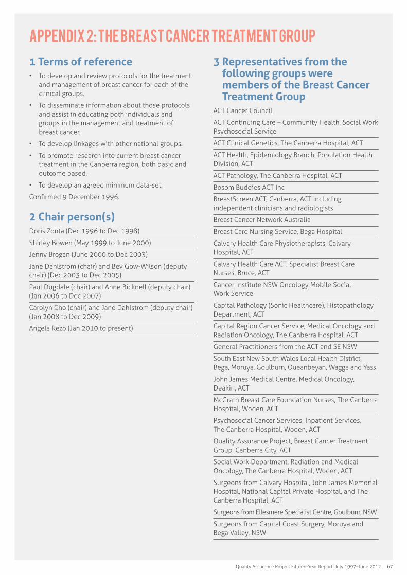
Quality Assurance Project Fifteen-Year Report July 1997–June 2012 67
Appendix 2: The Breast Cancer Treatment Group
1 Terms of reference• To develop and review protocols for the treatment
and management of breast cancer for each of the clinical groups.
• To disseminate information about those protocols and assist in educating both individuals and groups in the management and treatment of breast cancer.
• To develop linkages with other national groups.
• To promote research into current breast cancer treatment in the Canberra region, both basic and outcome based.
• To develop an agreed minimum data-set.
Confirmed 9 December 1996.
2 Chair person(s)Doris Zonta (Dec 1996 to Dec 1998)
Shirley Bowen (May 1999 to June 2000)
Jenny Brogan (June 2000 to Dec 2003)
Jane Dahlstrom (chair) and Bev Gow-Wilson (deputy chair) (Dec 2003 to Dec 2005)
Paul Dugdale (chair) and Anne Bicknell (deputy chair) (Jan 2006 to Dec 2007)
Carolyn Cho (chair) and Jane Dahlstrom (deputy chair) (Jan 2008 to Dec 2009)
Angela Rezo (Jan 2010 to present)
3 Representatives from the following groups were members of the Breast Cancer Treatment Group
ACT Cancer Council
ACT Continuing Care – Community Health, Social Work Psychosocial Service
ACT Clinical Genetics, The Canberra Hospital, ACT
ACT Health, Epidemiology Branch, Population Health Division, ACT
ACT Pathology, The Canberra Hospital, ACT
Bosom Buddies ACT Inc
BreastScreen ACT, Canberra, ACT including independent clinicians and radiologists
Breast Cancer Network Australia
Breast Care Nursing Service, Bega Hospital
Calvary Health Care Physiotherapists, Calvary Hospital, ACT
Calvary Health Care ACT, Specialist Breast Care Nurses, Bruce, ACT
Cancer Institute NSW Oncology Mobile Social Work Service
Capital Pathology (Sonic Healthcare), Histopathology Department, ACT
Capital Region Cancer Service, Medical Oncology and Radiation Oncology, The Canberra Hospital, ACT
General Practitioners from the ACT and SE NSW
South East New South Wales Local Health District, Bega, Moruya, Goulburn, Queanbeyan, Wagga and Yass
John James Medical Centre, Medical Oncology, Deakin, ACT
McGrath Breast Care Foundation Nurses, The Canberra Hospital, Woden, ACT
Psychosocial Cancer Services, Inpatient Services, The Canberra Hospital, Woden, ACT
Quality Assurance Project, Breast Cancer Treatment Group, Canberra City, ACT
Social Work Department, Radiation and Medical Oncology, The Canberra Hospital, Woden, ACT
Surgeons from Calvary Hospital, John James Memorial Hospital, National Capital Private Hospital, and The Canberra Hospital, ACT
Surgeons from Ellesmere Specialist Centre, Goulburn, NSW
Surgeons from Capital Coast Surgery, Moruya and Bega Valley, NSW
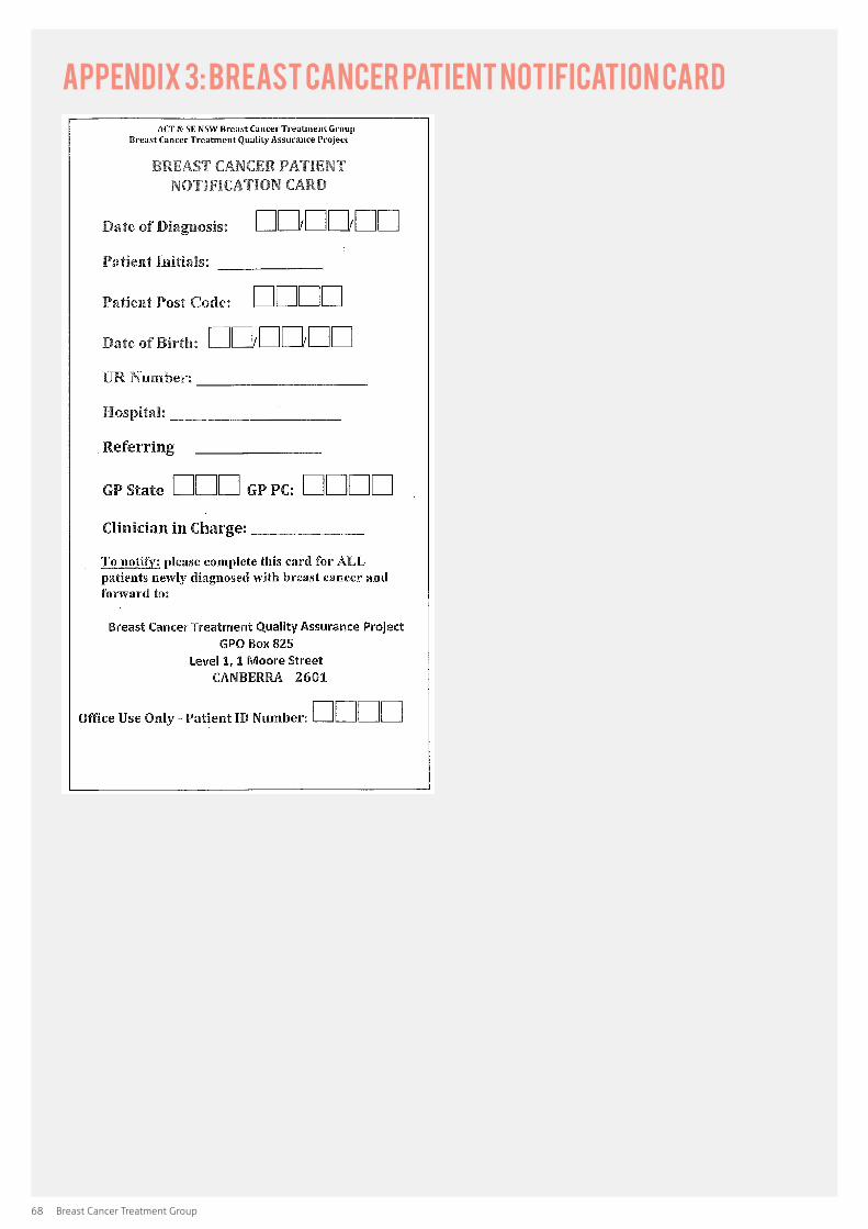
68 Breast Cancer Treatment Group
Appendix 3: Breast cancer patient notification card

Quality Assurance Project Fifteen-Year Report July 1997–June 2012 69
Appendix 4: Patient information sheet and consent form
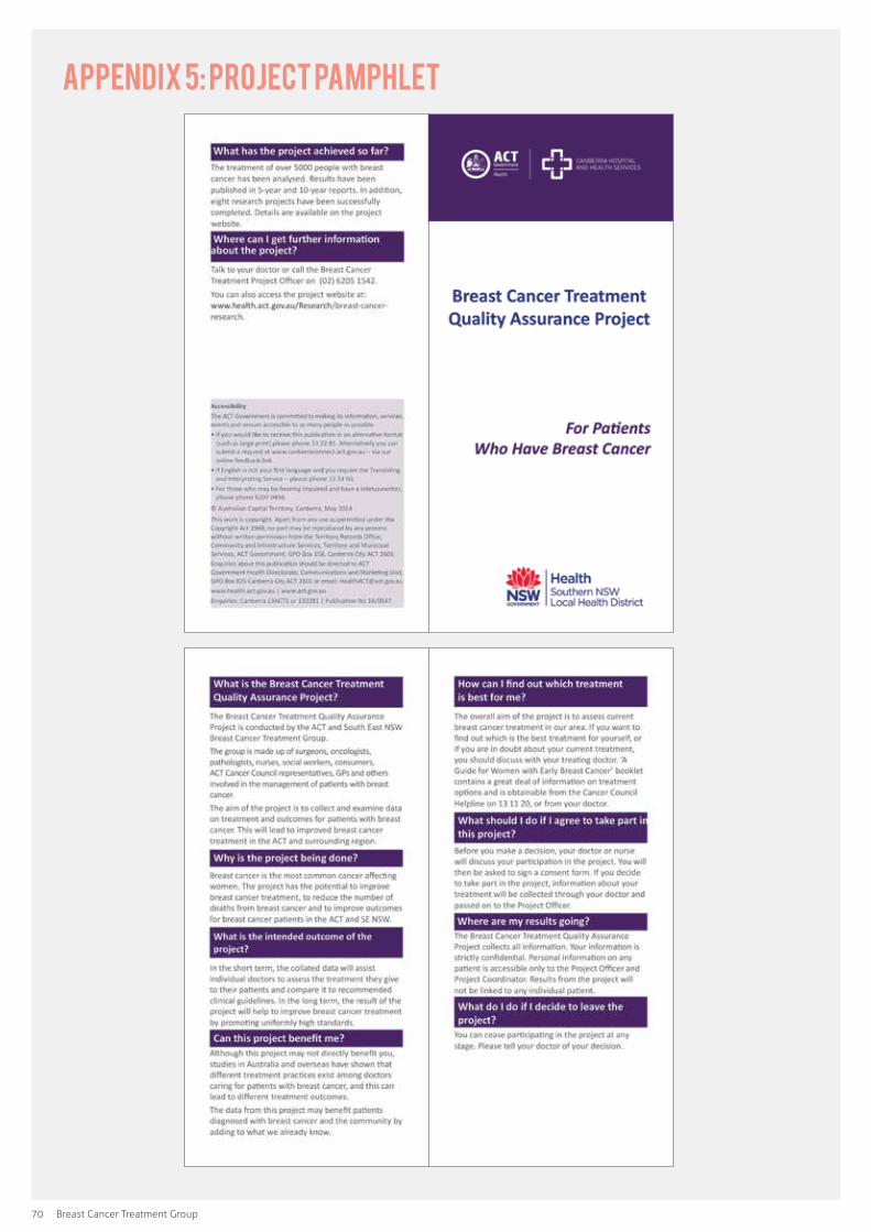
70 Breast Cancer Treatment Group
Appendix 5: Project pamphlet
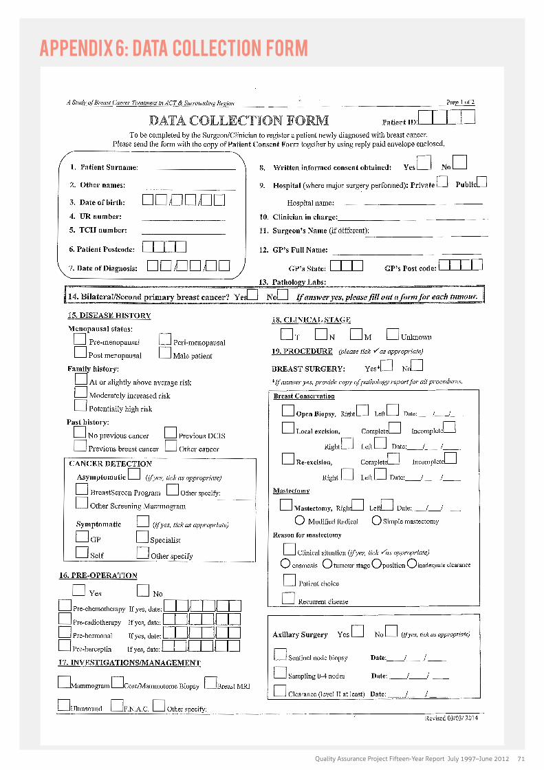
Quality Assurance Project Fifteen-Year Report July 1997–June 2012 71
Appendix 6: Data collection form

72 Breast Cancer Treatment Group
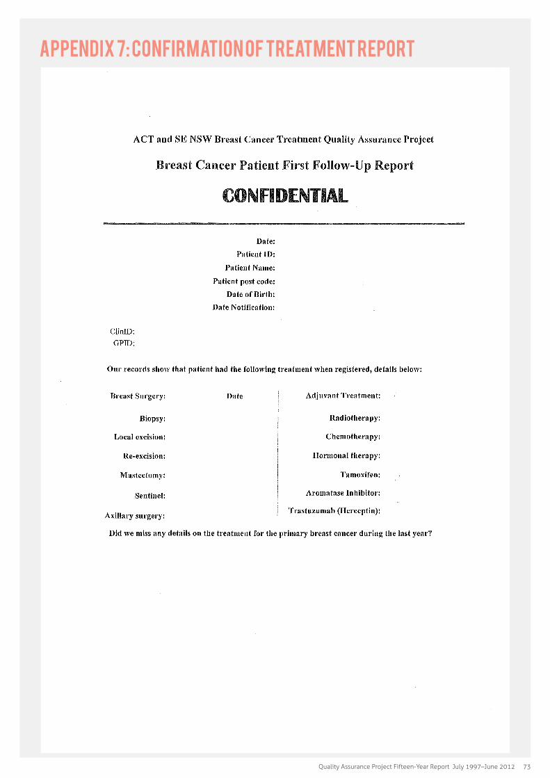
Quality Assurance Project Fifteen-Year Report July 1997–June 2012 73
Appendix 7: Confirmation of treatment report
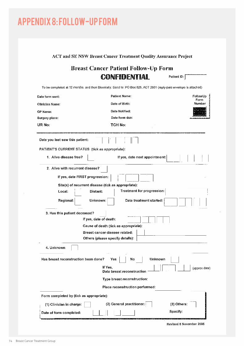
74 Breast Cancer Treatment Group
Appendix 8: Follow-up form
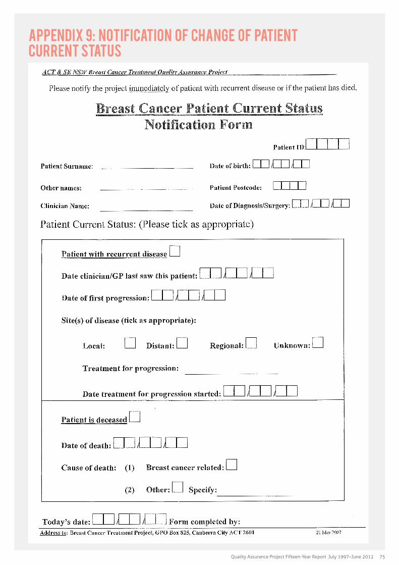
Quality Assurance Project Fifteen-Year Report July 1997–June 2012 75
Appendix 9: Notification of change of patient current status
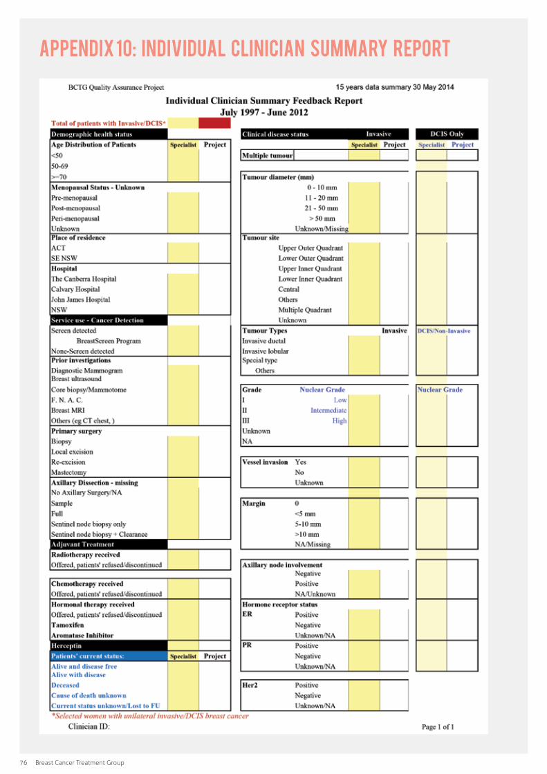
76 Breast Cancer Treatment Group
Appendix 10: Individual clinician summary report
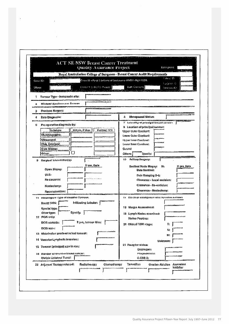
Quality Assurance Project Fifteen-Year Report July 1997–June 2012 77
Appendix 11: RACS report- breast cancer audit requirements

78 Breast Cancer Treatment Group
Appendix 12: Declaration of confidentiality
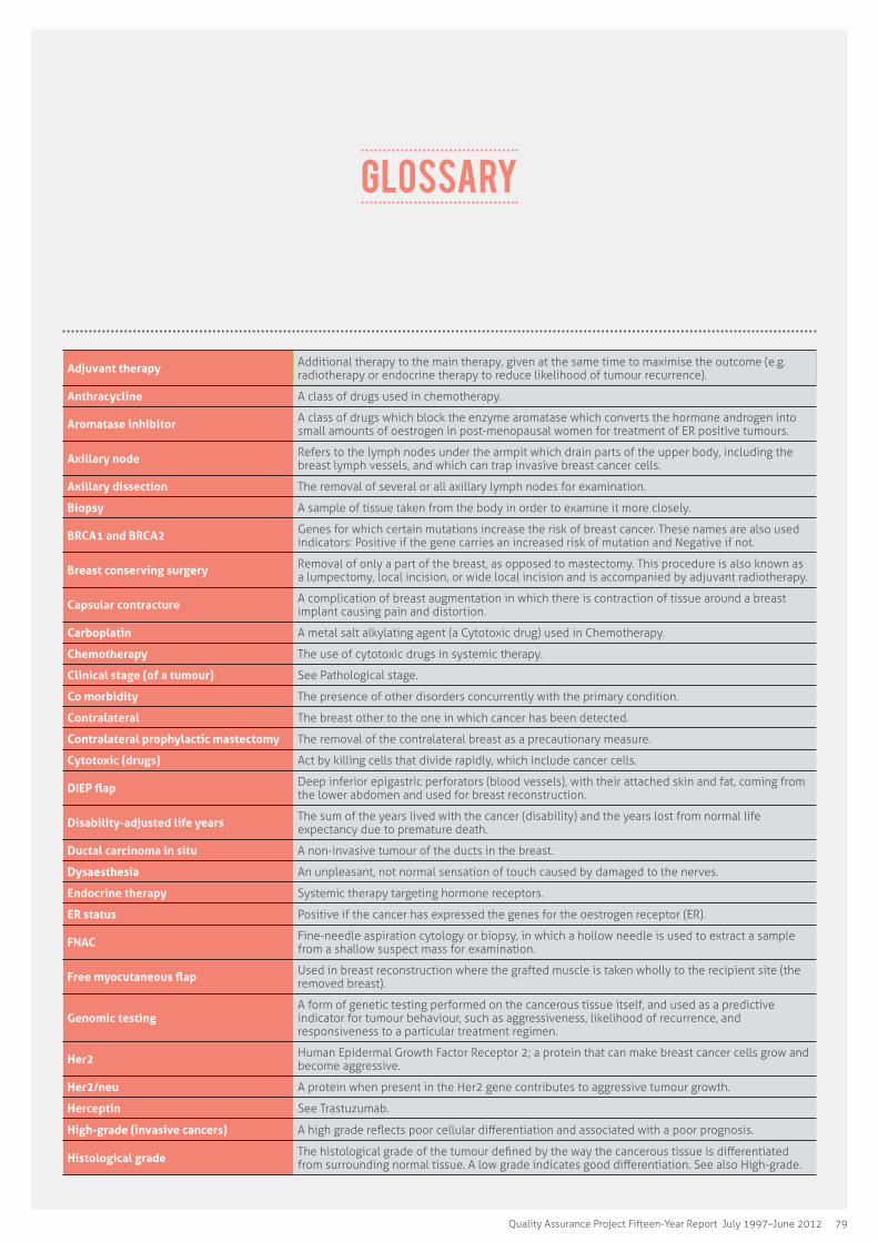
Quality Assurance Project Fifteen-Year Report July 1997–June 2012 79
Adjuvant therapy Additional therapy to the main therapy, given at the same time to maximise the outcome (e.g. radiotherapy or endocrine therapy to reduce likelihood of tumour recurrence).
Anthracycline A class of drugs used in chemotherapy.
Aromatase inhibitor A class of drugs which block the enzyme aromatase which converts the hormone androgen into small amounts of oestrogen in post-menopausal women for treatment of ER positive tumours.
Axillary node Refers to the lymph nodes under the armpit which drain parts of the upper body, including the breast lymph vessels, and which can trap invasive breast cancer cells.
Axillary dissection The removal of several or all axillary lymph nodes for examination.
Biopsy A sample of tissue taken from the body in order to examine it more closely.
BRCA1 and BRCA2 Genes for which certain mutations increase the risk of breast cancer. These names are also used indicators: Positive if the gene carries an increased risk of mutation and Negative if not.
Breast conserving surgery Removal of only a part of the breast, as opposed to mastectomy. This procedure is also known as a lumpectomy, local incision, or wide local incision and is accompanied by adjuvant radiotherapy.
Capsular contracture A complication of breast augmentation in which there is contraction of tissue around a breast implant causing pain and distortion.
Carboplatin A metal salt alkylating agent (a Cytotoxic drug) used in Chemotherapy.
Chemotherapy The use of cytotoxic drugs in systemic therapy.
Clinical stage (of a tumour) See Pathological stage.
Co morbidity The presence of other disorders concurrently with the primary condition.
Contralateral The breast other to the one in which cancer has been detected.
Contralateral prophylactic mastectomy The removal of the contralateral breast as a precautionary measure.
Cytotoxic (drugs) Act by killing cells that divide rapidly, which include cancer cells.
DIEP flap Deep inferior epigastric perforators (blood vessels), with their attached skin and fat, coming from the lower abdomen and used for breast reconstruction.
Disability-adjusted life years The sum of the years lived with the cancer (disability) and the years lost from normal life expectancy due to premature death.
Ductal carcinoma in situ A non-invasive tumour of the ducts in the breast.
Dysaesthesia An unpleasant, not normal sensation of touch caused by damaged to the nerves.
Endocrine therapy Systemic therapy targeting hormone receptors.
ER status Positive if the cancer has expressed the genes for the oestrogen receptor (ER).
FNAC Fine-needle aspiration cytology or biopsy, in which a hollow needle is used to extract a sample from a shallow suspect mass for examination.
Free myocutaneous flap Used in breast reconstruction where the grafted muscle is taken wholly to the recipient site (the removed breast).
Genomic testingA form of genetic testing performed on the cancerous tissue itself, and used as a predictive indicator for tumour behaviour, such as aggressiveness, likelihood of recurrence, and responsiveness to a particular treatment regimen.
Her2 Human Epidermal Growth Factor Receptor 2; a protein that can make breast cancer cells grow and become aggressive.
Her2/neu A protein when present in the Her2 gene contributes to aggressive tumour growth.
Herceptin See Trastuzumab.
High-grade (invasive cancers) A high grade reflects poor cellular differentiation and associated with a poor prognosis.
Histological grade The histological grade of the tumour defined by the way the cancerous tissue is differentiated from surrounding normal tissue. A low grade indicates good differentiation. See also High-grade.
GLOSSARY
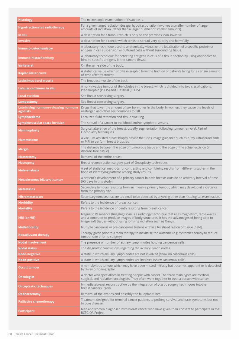
80 Breast Cancer Treatment Group
Histology The microscopic examination of tissue cells.
Hypofractionated radiotherapy For a given target radiation dosage, hypofractionation involves a smaller number of larger amounts of radiation (rather than a larger number of smaller amounts).
In situ A description for a tumour which is only on the premises; non-invasive.
Invasive A description for a cancer which tends to spread very quickly and harmfully.
Immuno-cytochemistry A laboratory technique used to anatomically visualize the localization of a specific protein or antigen in cell suspension or cultured cells without surrounding tissue.
Immuno-histochemistry A laboratory technique for detecting antigens in cells of a tissue section by using antibodies to bind to specific antigens in the sample tissue.
Ipsilateral On the same side of the body.
Kaplan Meier curve A statistical value which shows in graphic form the fraction of patients living for a certain amount of time after treatment.
Latissimus dorsi muscle The broadest muscle of the back.
Lobular carcinoma in situ A non-invasive tumour of the lobules in the breast, which is divided into two classifications: Pleomorphic (PLCIS) and Classical (CLCIS).
Local excision See Breast conserving surgery.
Lumpectomy See Breast conserving surgery.
Luteinising hormone-releasing hormone agonists
Drugs that lower the amount of sex hormones in the body. In women, they cause the levels of oestrogen and other sex hormones to fall.
Lymphoedema Localized fluid retention and tissue swelling.
Lymphovascular space invasion The spread of a cancer to the blood and/or lymphatic vessels.
Mammoplasty Surgical alteration of the breast, usually augmentation following tumour removal. Part of Oncoplasty techniques.
Mammotome A vacuum-assisted breast biopsy device that uses image guidance such as X-ray, ultrasound and/or MRI to perform breast biopsies.
Margin The distance between the edge of tumourous tissue and the edge of the actual excision (in disease-free tissue).
Mastectomy Removal of the entire breast.
Mastopexy Breast reconstruction surgery, part of Oncoplasty techniques.
Meta-analysis A set of statistical methods for contrasting and combining results from different studies in the hope of identifying patterns among study results.
Metachronous bilateral cancer A patient’s development of a primary cancer in both breasts outside an arbitrary interval of time (90 days in this study).
Metastases Secondary tumours resulting from an invasive primary tumour, which may develop at a distance from the primary site.
Micrometastases Secondary tumours that are too small to be detected by anything other than histological examination.
Morbidity Refers to the incidence of breast cancer.
Mortality Refers to the incidence of death resulting from breast cancer.
MRI (or MR)Magnetic Resonance (Imaging) scan is a radiology technique that uses magnetism, radio waves, and a computer to produce images of body structures. It has the advantages of being able to image soft tissues without using ionising radiation such as X-rays.
Multi-focality Multiple cancerous or pre-cancerous lesions within a localised region of tissue (field).
Neoadjuvant therapy Therapy given prior to a main therapy to maximise the outcome (e.g. systemic therapy to reduce tumour size prior to surgery).
Nodal involvement The presence or number of axillary lymph nodes holding cancerous cells.
Nodal status The diagnostic conclusions regarding the axillary lymph nodes.
Node-negative A state in which axillary lymph nodes are not involved (show no cancerous cells).
Node-positive A state in which axillary lymph nodes are involved (show cancerous cells).
Occult tumour A non-obvious tumour which may have been missed initially but becomes apparent or is detected by X-ray or tomography.
Oncologist A doctor who specialises in treating people with cancer. The three main types are medical, surgical, and radiation oncologists. They often work together to treat a person with cancer.
Oncoplastic techniques Immediate breast reconstruction by the integration of plastic surgery techniques into the breast cancer surgery.
Oophorectomy Removal of the ovaries and possibly the fallopian tubes.
Palliative chemotherapy Treatment designed for terminal cancer patients to prolong survival and ease symptoms but not to cure disease.
Participant Men and women diagnosed with breast cancer who have given their consent to participate in the BCTG QA Project

Quality Assurance Project Fifteen-Year Report July 1997–June 2012 81
Pathological stageClassifications which reflect categories of tumour size, nodal involvement, and spread, e.g., Stage T2 N0 and M0 means a tumour up to 50 mm in diameter (T2), no nodal involvement (N0), and no metastases (M0). Also known as the TNM staging system.
Pedicled myocutaneous flap Used in breast reconstruction where the grafted muscle is left partially attached to its original site until the graft has taken, after which the transfer is completed.
Pleomorphic Cells which have variability in their size or shape, and/or their nuclei.
PR status Positive if the cancer has expressed the genes for the progesterone receptor (PR).
Primary (tumour) The initial tumour formed.
Prognosis Determination of the likely outcome of one’s current standing using available evidence.
Proliferation index Measurement of the fraction of KI67-positive tumour cells, which is used as a prognostic aid for survival and tumour recurrence.
Radiotherapy Use of ionizing radiation to kill malignant cells.
Resection All or part of an internal organ or part of the body is removed.
Secondary (tumour) See Metastases.
Sentinel node The lymph nodes considered to be the first to drain a cancer of the breast.
Survival function See Kaplan-Meier curve.
Survival rates The percentage of people who are alive for a specified time after diagnosis.
SNAC1 Name of a trial where sentinel node only biopsies were performed in the first instance for women with confirmed breast cancer, rather than axillary dissection.
Synchronous bilateral cancer Refers to a patient’s development of a primary cancer in both breasts within an arbitrary interval of time (90 days in this study).
Systemic therapy Therapy affecting the whole body, for example, hormone therapy and chemotherapy.
Tamoxifen An anti-oestrogen therapy for ER-positive breast cancer.
Targeted therapies A chemical therapy which targets specific molecules of tumour cells required for their propagation.
Taxane A class of chemotherapy agent.
Trastuzumab Also known as Herceptin, a targeted therapy used for breast cancer.
Triple negative An indication that the breast cancer has the status of ER, PR, and Her2 all negative.
Unifocal Cancer confined to a single instance.
Unilateral Primary tumour confined to a single breast.
Wide local excision See Breast conserving surgery. The significance of ‘Wide’ in the name is the implication of large margins. It may be used on lesions up to 4 cm in diameter.
Zoladex An LHRH agonist used to suppress production of oestrogen in the treatment of ER+ breast cancers.
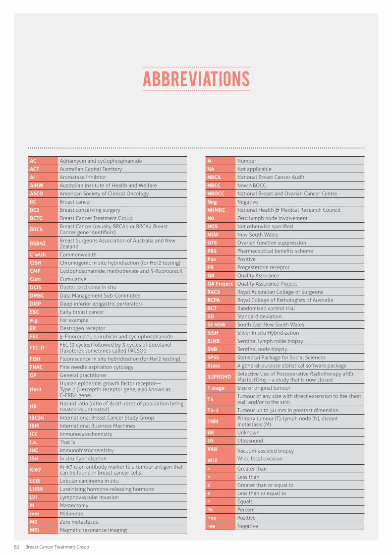
82 Breast Cancer Treatment Group
AC Adriamycin and cyclophosphamide
ACT Australian Capital Territory
AI Aromatase inhibitor
AIHW Australian Institute of Health and Welfare
ASCO American Society of Clinical Oncology
BC Breast cancer
BCS Breast conserving surgery
BCTG Breast Cancer Treatment Group
BRCA Breast Cancer (usually BRCA1 or BRCA2 Breast Cancer gene identifiers)
BSANZ Breast Surgeons Association of Australia and New Zealand
C’wlth Commonwealth
CISH Chromogenic in situ hybridization (for Her2 testing)
CMF Cyclophosphamide, methotrexate and 5-fluorouracil
Cum Cumulative
DCIS Ductal carcinoma in situ
DMSC Data Management Sub-Committee
DIEP Deep inferior epigastric perforators
EBC Early breast cancer
e.g. For example
ER Oestrogen receptor
FEC 5-Fluorouacil, epirubicin and cyclophosphamide
FEC-D FEC (3 cycles) followed by 3 cycles of docetaxel (Taxotere); sometimes called PACSO1
FISH Fluorescence in situ hybridization (for Her2 testing)
FNAC Fine needle aspiration cytology
GP General practitioner
Her2Human epidermal growth factor receptor— Type 2 (Herceptin receptor gene, also known as C-ERB2 gene)
HR Hazard ratio (ratio of death rates of population being treated vs untreated)
IBCSG International Breast Cancer Study Group
IBM International Business Machines
ICC Immunocytochemistry
i.e. That is
IHC Immunohistochemistry
ISH In situ hybridization
Ki67 Ki-67 is an antibody marker to a tumour antigen that can be found in breast cancer cells.
LCIS Lobular carcinoma in situ
LHRH Luteinising hormone-releasing hormone
LVI Lymphovascular invasion
M Mastectomy
mm Millimetre
M0 Zero metastases.
MRI Magnetic resonance imaging
N Number
NA Not applicable
NBCA National Breast Cancer Audit
NBCC Now NBOCC.
NBOCC National Breast and Ovarian Cancer Centre
Neg Negative
NHMRC National Health & Medical Research Council
N0 Zero lymph node involvement
NOS Not otherwise specified
NSW New South Wales
OFS Ovarian function suppression
PBS Pharmaceutical benefits scheme
Pos Positive
PR Progesterone receptor
QA Quality Assurance
QA Project Quality Assurance Project
RACS Royal Australian College of Surgeons
RCPA Royal College of Pathologists of Australia
RCT Randomised control trial
SD Standard deviation
SE NSW South East New South Wales
SISH Silver In situ Hybridization
SLNB Sentinel lymph node biopsy
SNB Sentinel node biopsy.
SPSS Statistical Package for Social Sciences
Stata A general-purpose statistical software package
SUPREMO Selective Use of Postoperative Radiotherapy aftEr MastectOmy – a study that is now closed.
T stage Size of original tumour
T4 Tumour of any size with direct extension to the chest wall and/or to the skin.
T1-2 Tumour up to 50 mm in greatest dimension.
TNM Primary tumour (T), lymph node (N), distant metastasis (M)
UK Unknown
US Ultrasound
VAB
WLE
Vacuum-assisted biopsyWide local excision
> Greater than
< Less than
≥ Greater than or equal to
≤ Less than or equal to
= Equals
% Percent
+ve Positive
-ve Negative
Abbreviations
Quality Assurance Project Fifteen-Year Report July 1997–June 2012 83
References
1. Report to the Nation – Breast Cancer 2012 Cancer Australia ISBN 978-1-74127-198-0. http://canceraustralia.gov.au/sites/default/files/publications/ca_report_to_the_nation_breast_cancer_2012_d15_508de9ff41cae.pdf
2. Australian Capital Territory Chief Health Officer’s Report 2014.
3. Breast cancer in Australia: an over view 2012. Australian Institute of Health and Welfare. AIHW 2012 Cancer Series No. 71 CAN 67 Canberra.
4. Clinical practice guidelines for the management of early breast cancer: (1st Ed 1995). Publication of the National Health and Medical Research Council. (2nd Ed 2001).
5. Cancer in the ACT: 2004-2008. ACT Government, ACT Health 2011, Canberra.
6. Breast cancer risk factors: a review of the evidence. National Breast and Ovarian Cancer Centre, Surry Hills, NSW, 2009.
7. Breast cancer for Australia: Australian Institute of Health and Welfare. Australian Cancer Incidence and Mortality (ACIM) Books – (ICD10 C50). www.aihw.gov.au/acim-books/ [Accessed Mar 2015].
8. Cancer in Australia: an overview, 2012. Australian Institute of Health and Welfare & Australasian Association of Cancer Registries. AIHW 2012 Cancer Series No. 74 CAN 70. Canberra.
9. Dense Breasts: a review of reporting legislation and
available supplemental screening options. Ho JM, Jafferjee N, Covarrubias GM, Ghesani GM, Handler B. American Journal of Roentgenology. 2014 Aug; 203(2):449-56.
10. Alternative Screening for Dense Breasts: MRI. O’Flynn EAM, Ledger AEW, M.deSouza N. American Journal of Roentgenology. 2015 Feb; 204(2): W141-W149. DOI:10.2214/AJR.14.13636.
11. Prospective multicenter cohort study to refine management recommendations for women at elevated familial risk of breast cancer: the EVA trial. Kuhl C, Weigel S, Schrading S, et al. Journal of Clinical Oncology: official journal of the American Society of Clinical Oncology. 2010; 28(9):1450-57.
12. Risk of second primary cancer in the contralateral breast in women treated for early-stage breast cancer: a population-based study. Gao X, Fisher SG, Emami B. International Journal of Radiation Oncology, Biology, Physics. 2003 Jul;56(4):1038-45.
13. Breast MRI screening of women with a personal history of breast cancer. Brennan S, Liberman L, Dershaw DD, Morris E. American Journal of Roentgenology. 2010 Aug; 195(2):510-16.
14. Importance of a personal history of breast cancer as a risk factor for the development of subsequent breast cancer: results from screening breast MRI. Schacht DV, Yamaguchi K, Lai J, Kulkarni K, Sennett CA, Abe H. American Journal of Roentgenology. 2014 Feb; 202(2):289-92.
15. Breast MR imaging screening in women with a history of breast conservation therapy. Gweon HM, Cho N, Han W, Yi A, Moon H-G, Noh D-Y, et al. Radiology. 2014 Aug; 272(2):366-73.
16. Breast cancer under age 40: a different approach. Ribnikar D1, Ribeiro JM, Pinto D, Sousa B, Pinto AC, Gomes E, Moser EC, Cardoso MJ, Cardoso F. Current Treat Options in Oncology. 2015 Apr; 16(4):334. DOI: 10.1007/S11864-015-0334-8.
17. Growing use of mastectomy for Ductal Carcinoma-In situ of the breast among young women in the United States. Rutter CE, Park HS, Killelea BK, Evans SB. Annals Surgical Oncology. 2015 Jan 7. [Epub ahead of print].
18. Prognosis of women with primary breast cancer diagnosed during pregnancy: results from an international collaborative study. Amant F1, von Minckwitz G, Han SN, et al. Journal of Clinical Oncology: official journal of the American Society of Clinical Oncology. 2013 Jul; 31(20):2532-39. DOI: 10.1200/JCO.2012.45.6335. [Epub 2013 Apr 22].
19. Improving Breast Cancer Surgery: A Classification and Quadrant per Quadrant Atlas for Oncoplastic Surgery. Clough KB, Kaufman GJ, Nos C, Buccimazza I, Sarfati IM. Annals Surgical Oncology. 2010 May; 17(5):1375-91.
20. Recommendations for the management of early breast cancer in women with an identified BRCA1 or BRCA2 gene mutation or at high risk of a gene mutation. 2014 Feb. www.canceraustralia.gov.au /publications-and-resources.
21. The Angelina Effect. Kluger J, Park A. Time Magazine. 2013 May.
22. Recommendations for staging and managing the axilla in early (operable) breast cancer. 2011 Sep. www.canceraustralia.gov.au.
23. Recommendations for use of sentinel node biopsy in early (operable) breast cancer. National Breast and Ovarian Cancer Centre. 2008 Jun. www.canceraustralia.gov.au.
24. Sentinel Lymph Node Based Management or Routine Axillary Clearance? Three-year Outcomes of the RACS Sentinel Node Biopsy versus Axillary Clearance (SNAC) 1 Trial. Wetzig N, Gill PG, Zannino D, Stockler MR, et al. Annals of Surgical Oncology. 2015 Jan; 22(1):17-23.
25. Locoregional Recurrence after Sentinel Lymph Node Dissection With or Without Axillary Dissection in Patients With Sentinel Lymph Node Metastases. The American College of Surgeons Oncology Group Z0011 Randomized Trial. Giuliano AE, McCall L, Beitsch P, et al. Annals of Surgery. 2010 Sep; 252(3):426-33.
26. Axillary dissection versus no axillary dissection in patients with sentinel-node micrometastases (IBCSG 23-01): a phase 3 randomised controlled trial. Galimberti V, Cole BF, Zurrida S, et al. International Breast Cancer Study Group Trial 23-01 investigators. Lancet Oncology. 2013 Apr; 14(4):297-305. DOI: 10.1016/S1470-2045(13)70035-4.
27. Factors affecting decisions about breast reconstruction. http://canceraustralia.gov.au/affected-cancer/cancer-types/breast-cancer/treatment/breast-reconstruction/deciding/factors-affecting-decisions-about-breast-reconstruction. [Accessed Mar 2015]. www.canceraustralia.gov.au.
84 Breast Cancer Treatment Group
28. Should women younger than 40 years of age with invasive breast cancer have a mastectomy? 15-year outcomes in a population-based cohort. Cao JQ, Truong PT, Olivotto IA, Olson R, et al. International Journal of Radiation Oncology, Biology, Physics. 2014 Nov; 90(3):509-17. DOI: 10.1016/j.ijrobp.2014.06.041. [Epub 2014 Sep 3].
29. Effect of radiotherapy after mastectomy and axillary surgery on 10-year recurrence and 20-year breast cancer mortality: meta-analysis of individual patient data for 8135 women in 22 randomised trials. Early Breast Cancer Trialists’ Collaborative Group (EBCTCG), McGale P, Taylor C, et al. The Lancet. 2014 Jun; 383(9935):2127-35.
30. Recommendations for the use of hypofractionated radiotherapy in early (operable) breast cancer. ISBN 13 978-1-74127-175-1. http://canceraustralia.gov.au/publications-and-resources/cancer-australia-publications/recommendations-use-hypofractionated-radiotherapy-early-operable-breast-cancer-0. [Accessed Mar 2015]. www.canceraustralia.gov.au.
31. Elucidating the role of chest wall irradiation in ‘intermediate-risk’ breast cancer: the MRC/EORTC SUPREMO trial. Kunkler IH, Canney P, van Tienhoven G, Russell NS. Clinical Oncology (Royal College of Radiologists). 2008 Feb; 20(1):31-4.
32. Effect of radiotherapy after breast-conserving surgery on 10-year recurrence and 15-year breast cancer death: meta-analysis of individual patient data for 10,801 women in 17 randomised trials. Early Breast Cancer Trialists’ Collaborative Group (EBCTCG), Darby S, McGale P, Correa C, Taylor C, et al. The Lancet. 2011 Nov; 378(9804):1707-16.
33. Breast cancer for Australia: Australian Institute of Health and Welfare. Australian Cancer Incidence and Mortality (ACIM) Books – (ICD10 C50). www.aihw.gov.au/acim-books/[Accessed 23 Apr 2015].
34. Combination chemotherapy as an adjuvant treatment in operable breast cancer. Bonadonna G, Brusamolino E, Rossi A, et al. The New England Journal of Medicine. 1976 Feb; 294(8):405-10.
35. Recommendations for the use of Taxane containing chemotherapy regimens for the treatment of early (operable) breast cancer. National Breast and Ovarian Cancer Centre Cancer Australia. ISBN:978-1-74127-116-4. http://guidelines.canceraustralia.gov.au/guidelines/guideline_4.pdf. 2008 Jun. www.canceraustralia.gov.au .
36. The impact of genomic assay (Oncotype DX) on adjuvant treatment recommendations in early breast cancer. de Boer RH, Baker C, Speakman D, et al. The Medical Journal of Australia. 2013 Aug; 199(3): 205-08.
37. Proliferation Marker Ki-67 in Early Breast Cancer. Urruticoechea A, Smith IE, Dowsett M. Journal of Clinical Oncology: official journal of the American Society of Clinical Oncology. 2005 Oct; 23(28): 7212-20. DOI: 10.1200/JCO.2005.07.501.
38. Long-term effects of continuing adjuvant tamoxifen to 10 years versus stopping at 5 years after diagnosis of oestrogen receptor-positive breast cancer: ATLAS, a randomised trial. Davies C, Pan H, Godwin J, Gray R, Arriagada R, Raina V, et al. The Lancet. 2013 Mar; 381(9869):805–16.
39. Survival analyses from the ZEBRA study. goserelin (Zoladex) versus CMF in premenopausal women with node-positive breast cancer. Kaufmann M, Jonat W, Blamey R, Cuzick J,et al. Zoladex Early Breast Cancer Research Association (ZEBRA) Trialists’ Group. European Journal of Cancer. 2003 Aug; 39(12):1711-17.
40. Adjuvant Ovarian Suppression in Premenopausal Breast Cancer. Francis PA, Regan MM, Fleming GF, et al. The SOFT Investigators and the International Breast Cancer Study Group. New England Journal of Medicine. 2015 Jan; 372(5):436-46. DOI: 10.1056/NEJMoa1412379.
41. Trastuzumab—mechanism of action and use in clinical practice. Hudis CA. New England Journal of Medicine. 2007 Jul; 357(1): 39–55.
42. Human epidermal growth factor receptor 2 overexpression as a prognostic factor in a large tissue microarray series of node-
negative breast cancers. Chia S, Norris B, Speers C, et al. Journal of Clinical Oncology: official journal of the American Society of Clinical Oncology. 2008 Dec; 26(35):5697-704.
43. Trastuzumab containing regimens for early breast cancer. Moja, Lorenzo, et al. The Cochrane Database of systemic Reviews. 2012 Apr; 4.
44. Rational use of trastuzumab in metastatic and locally advanced breast cancer: Implications of recent research. Goel S, Chirgwin J, Francis P, Stuart-Harris R, et al. Breast. 2011 Apr; 20(2):101-10. DOI: 10.1016/j.breast.2010.11.008. [ Epub 2010 Dec 22].
45. Variation in receipt of radiation therapy after breast conserving surgery: assessing the impact of physicians and geographic regions. Feinstein AJ, Soulos PR, Long JB, Herrin J, et al. Medical care. 2013 Apr; 51(4):330-38.
46. Clinical characteristics and outcomes of bilateral breast cancer in an Australian cohort. Buckingham J, Craft P, Dahlstrom JE, Zhang Y, et al. Breast. 2011 Apr; 20(2):158-64.
47. Male breast cancer: A single centre experience and current evidence. Kaushik M, Olivera-Cunha M, Shokuhi S. Breast. 2014 Nov-Dec; 20(6):674-5.
48. Male breast carcinoma: A clinicopathological and immunohistochemical characterization study. Zhou R, Zhou S, Bi R, Yu B, et al. International Journal of Clinical and Experimental Pathology. 2014 Sep; 7(10):6852-61.
49. The impact of race in male breast cancer treatment and outcome in the United States: a population-based analysis of 4279 patients. Shin JY, Kachnic LA, Hirsch AE. International Journal of Breast Cancer, 2014; article ID 685842. [Epub 2014 Oct 2].
50. Breast cancer in Australia: an overview 2006. Australian Institute of Health and Welfare. AIHW 2006 Cancer Series No. 34 CAN 29 Canberra.
51. Incidence & Prognosis Of Synchronous & Metachronous Bilateral Breast Cancer. Hartman M, Czene K, Reilly M, Adolfsson J, et al. Journal of Clinical Oncology : official journal of the American Society of Clinical Oncology. 2007 Sep; 25(27):4210-16.
52. Surgical margins and risk of locoregional recurrence in invasive breast cancer: an analysis of 10-year data from the Breast Cancer Treatment Quality Assurance Project. Behm EC, Beckmann KR, Dahlstrom JE, Zhang Y, Cho C,et al. Breast. 2013 Oct; 22(5):839-44.
53. Post-operative radiotherapy for ductal carcinoma in situ of the breast--a systematic review of the randomised trials. Goodwin A, Parker S, Ghersi D, Wilcken N. Breast. 2009 Jun; 18(3):143-49.
54. Sentinel lymph node biopsy for patients with early-stage breast cancer: American Society of Clinical Oncology clinical practice guideline update. Lyman GH, Temin S, Edge SB, Newman LA, et al. Journal of Clinical Oncology: official journal of the American Society of Clinical Oncology. 2014 May; 32(13):1365-83.
55. Sentinel node procedure is warranted in ductal carcinoma in situ with high risk of occult invasive carcinoma and microinvasive carcinoma treated by mastectomy. Tunon-de-Lara C, Giard S, Buttarelli M, Blanchot J, et al. Breast . 2008 Mar-Apr; 14(2):135-40.
56. Invasive breast cancer structured reporting protocol (2nd Ed 2012). 2012 Nov. www.rcpa.edu.au/Library (accessed February 2015).
57. The Pathology Reporting of Breast Cancer: A Guide for Pathologists, Surgeons, Radiologists and Oncologists (2nd Ed 2001). www.cancer.org.au/health-professionals . Australian Cancer Network 2001; p24. ISBN 1-877054-00-3.
58. American Society of Clinical Oncology/College of American Pathologists guideline recommendations for immunohistochemical testing of estrogen and progesterone receptors in breast cancer. Hammond ME, Hayes DF, Dowsett M, Allred DC, et al. Journal of Clinical Oncology: official journal of the American Society of Clinical Oncology. 2010 Jun; 28(16):2784-95.

Quality Assurance Project Fifteen-Year Report July 1997–June 2012 85
59. Frequency and reliability of oestrogen receptor, progesterone receptor and Her2 in breast carcinoma determined by immunohistochemistry in Australasia: results of the RCPA Quality Assurance Program. Francis GD, Dimech M, Giles L, Hopkins A. Journal of clinical pathology. 2007 Nov; 60(11): 1277–83.
60. RTOG 9804: A Prospective Randomized Trial for Good-Risk Ductal Carcinoma In situ Comparing Radiotherapy With Observation. McCormick B, Winter K, Hudis C, Kuerer HM, et al. Journal of Clinical Oncology: official journal of the American Society of Clinical Oncology. 2015 Jan. [Epub 2015 Jan 20].
61. NCCN Clinical Practice Guidelines in Oncology: Breast Cancer, Version 1.2015. Ductal Carcinoma in situ. 2015 Feb; DCIS-1-DCISA. National Comprehensive Cancer Network. www.cjcpt.org/file.
62. Incidence and consequence of close margins in patients with ductal carcinoma-in situ treated with mastectomy: is further therapy warranted? Fitzsullivan E, Lari SA, Smith B, Caudle AS, et al. Annals Surgical Oncology. 2013 Dec; 20(13):4103-12.
63. Changing surgical trends in young patients with early stage breast cancer, 2003 to 2010: a report from the National Cancer Data Base. Pesce CE, Liederbach E, Czechura T, Winchester DJ, Yao K. Journal of the American College Surgeons. 2014 Jul;219(1):19-28.
64. Long-term impact of young age at diagnosis on treatment outcome and patterns of failure in patients with ductal carcinoma in situ treated with breast-conserving therapy. Vicini FA, Shaitelman S, Wilkinson JB, Shah C,et al. Breast. 2013 Jul-Aug; 19(4):365-73.
65. Evolving perspectives in contralateral breast cancer. Dawson LA, Chow E, Goss PE.. Eur J Cancer. 1998 Dec; 34(13):2000-9.
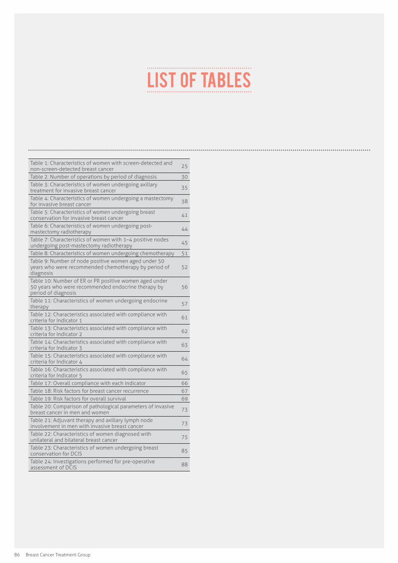
86 Breast Cancer Treatment Group
Table 1: Characteristics of women with screen-detected and non-screen-detected breast cancer 25
Table 2: Number of operations by period of diagnosis 30Table 3: Characteristics of women undergoing axillary treatment for invasive breast cancer 35
Table 4: Characteristics of women undergoing a mastectomy for invasive breast cancer 38
Table 5: Characteristics of women undergoing breast conservation for invasive breast cancer 41
Table 6: Characteristics of women undergoing post-mastectomy radiotherapy 44
Table 7: Characteristics of women with 1–4 positive nodes undergoing post-mastectomy radiotherapy 45
Table 8: Characteristics of women undergoing chemotherapy 51Table 9: Number of node positive women aged under 50 years who were recommended chemotherapy by period of diagnosis
52
Table 10: Number of ER or PR positive women aged under 50 years who were recommended endocrine therapy by period of diagnosis
56
Table 11: Characteristics of women undergoing endocrine therapy 57
Table 12: Characteristics associated with compliance with criteria for Indicator 1 61
Table 13: Characteristics associated with compliance with criteria for Indicator 2 62
Table 14: Characteristics associated with compliance with criteria for Indicator 3 63
Table 15: Characteristics associated with compliance with criteria for Indicator 4 64
Table 16: Characteristics associated with compliance with criteria for Indicator 5 65
Table 17: Overall compliance with each indicator 66Table 18: Risk factors for breast cancer recurrence 67Table 19: Risk factors for overall survival 69Table 20: Comparison of pathological parameters of invasive breast cancer in men and women 73
Table 21: Adjuvant therapy and axillary lymph node involvement in men with invasive breast cancer 73
Table 22: Characteristics of women diagnosed with unilateral and bilateral breast cancer 75
Table 23: Characteristics of women undergoing breast conservation for DCIS 85
Table 24: Investigations performed for pre-operative assessment of DCIS 88
List of tables
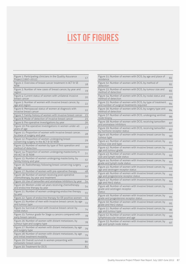
Quality Assurance Project Fifteen-Year Report July 1997–June 2012 87
Figure 1: Participating clinicians in the Quality Assurance Project (1997–2012) 17
Figure 2: Overview of breast cancer treatment in ACT & SE NSW 18
Figure 3: Number of new cases of breast cancer, by year and region 19
Figure 4: Current status of women with unilateral invasive breast cancer 20
Figure 5: Number of women with invasive breast cancer, by age and region 22
Figure 6: Menopausal status of women at diagnosis with invasive breast cancer 22
Figure 7: Family history of women with invasive breast cancer 23Figure 8: Mode of detection of invasive breast cancer 23Figure 9: Pre-operative investigations by year 26Figure 10: Pre-operative investigations in women under 40 years of age 27
Figure 11: Proportion of women with invasive breast cancer, by place of surgery and year 28
Figure 12: Proportion of women undergoing breast conserving surgery in the ACT & SE NSW 29
Figure 13: Number of women by type of first operation and method of detection 30
Figure 14: Proportion of women undergoing mastectomy in the ACT & SE NSW 31
Figure 15: Number of women undergoing mastectomy, by family history and year 32
Figure 16: Radiotherapy following breast conserving surgery by age 42
Figure 17: Number of women with pre-operative therapy 48Figure 18: Number of women receiving post-operative chemotherapy, by year and treatment 50
Figure 19: Use of tamoxifen and aromatase inhibitors by year 54Figure 20: Women under 40 years receiving chemotherapy and endocrine therapy by year 54
Figure 21: Number of women undergoing endocrine therapy by year 55
Figure 22: Types of endocrine therapy for ER positive women 55Figure 23: Number of men with invasive breast cancer, by age and tumour type 72
Figure 24: Survival of men with invasive breast cancer by nodal status 74
Figure 25: Tumour grade for Stage 4 cancers compared with early breast cancers 78
Figure 26: Number of women with distant metastases, by tumour type and surgery type 78
Figure 27: Number of women with distant metastases, by age and surgery type 79
Figure 28: Number of women with distant metastases, by age and initial treatment modality 79
Figure 29: Overall survival in women presenting with metastatic breast cancer 80
Figure 30: Treatment for DCIS 81
Figure 31: Number of women with DCIS, by age and place of residence 82
Figure 32: Number of women with DCIS, by method of detection 82
Figure 33: Number of women with DCIS, by tumour size and method of detection 83
Figure 34: Number of women with DCIS, by nodal status and method of detection 83
Figure 35: Number of women with DCIS, by type of treatment and number of surgical treatments required 84
Figure 36: Number of women with DCIS, by surgery type and method of detection 84
Figure 37: Number of women with DCIS, undergoing sentinel node biopsy 86
Figure 38: Number of women with DCIS, receiving tamoxifen and aromatase inhibitors 87
Figure 39: Number of women with DCIS, receiving tamoxifen by hormone receptor status 87
Figure 40: Number of women with invasive breast cancer by tumour type 90
Figure 41: Number of women with invasive breast cancer, by tumour size and type 91
Figure 42: Number of women with invasive breast cancer, by age and tumour grade 91
Figure 43: Number of women with invasive breast cancer, by size and lymph node status 92
Figure 44: Number of women with invasive breast cancer, by grade and lymph node status 92
Figure 45: Number of women with invasive breast cancer, by age and oestrogen receptor status 93
Figure 46: Number of women with invasive breast cancer, by age and progesterone receptor status 93
Figure 47: Number of women with invasive breast cancer, by age and Her2 status 94
Figure 48: Number of women with invasive breast cancer, by grade and oestrogen receptor status
94
Figure 49: Number of women with invasive breast cancer, by grade and progesterone receptor status 95
Figure 50: Number of women with invasive breast cancer, by grade and Her2 status 95
Figure 51: Number of women with invasive breast cancer, by age and tumour type 96
Figure 52: Number of women with invasive breast cancer, by lymphovascular invasion and age 96
Figure 53: Number of women with invasive breast cancer, by age and lymph node status 97
list of figures

The Australian Capital Territory and South East New South Wales Region
Breast cancer treatment groupQuality Assurance Project Fifteen-Year Report July 1997–June 2012