Embed Size (px)
Citation preview

The Arabidopsis thaliana Checkpoint Kinase WEE1Protects against Premature Vascular Differentiation duringReplication Stress W
Toon Cools,a,b Anelia Iantcheva,a,b,1 Annika K. Weimer,c Shannah Boens,a,b Naoki Takahashi,a,b,2 Sara Maes,a,b
Hilde Van den Daele,a,b Gert Van Isterdael,a,b Arp Schnittger,c and Lieven De Veyldera,b,3
a Department of Plant Systems Biology, VIB, 9052 Ghent, BelgiumbDepartment of Plant Biotechnology and Genetics, Ghent University, B-9052 Ghent, BelgiumcDepartment of Molecular Mechanisms of Phenotypic Plasticity, Institut de Biologie Moleculaire des Plantes–Centre National de
Recherche Scientifique, Unite Propre de Recherche 2357, Universite de Strasbourg, 67084 Strasbourg Cedex, France
A sessile lifestyle forces plants to respond promptly to factors that affect their genomic integrity. Therefore, plants have
developed checkpoint mechanisms to arrest cell cycle progression upon the occurrence of DNA stress, allowing the DNA to
be repaired before onset of division. Previously, the WEE1 kinase had been demonstrated to be essential for delaying
progression through the cell cycle in the presence of replication-inhibitory drugs, such as hydroxyurea. To understand the
severe growth arrest of WEE1-deficient plants treated with hydroxyurea, a transcriptomics analysis was performed,
indicating prolonged S-phase duration. A role for WEE1 during S phase was substantiated by its specific accumulation in
replicating nuclei that suffered from DNA stress. Besides an extended replication phase, WEE1 knockout plants accumu-
lated dead cells that were associated with premature vascular differentiation. Correspondingly, plants without functional
WEE1 ectopically expressed the vascular differentiation marker VND7, and their vascular development was aberrant. We
conclude that the growth arrest of WEE1-deficient plants is due to an extended cell cycle duration in combination with a
premature onset of vascular cell differentiation. The latter implies that the plant WEE1 kinase acquired an indirect
developmental function that is important for meristem maintenance upon replication stress.
INTRODUCTION
Plant growth depends on meristem activity that provides,
through continuous cell division, the cells required for tissue
expansion and organogenesis (Harashima and Schnittger,
2010). During development, however, interior and exterior
agents attack the meristems, endangering their size, organiza-
tion, and function. During cell cycle progression, the presence of
these agents can lead to faulty cell divisions, causing cells either
to lose their meristem identity or to be pushed into programmed
cell death (PCD). Massive loss of mitotic cells by aberrant cell
divisions results in reduced cell production and decreased
growth. Therefore, it is essential that the meristematic cells are
preserved by activating cell cycle checkpoints that arrest the cell
cycle as long as the stress endures. By contrast, to produce
offspring, plants need to sustain growth in a highly competitive
environment, even under unfavorable conditions. Hence, they
are in need of stress adaptation mechanisms that maintain
meristem productivity during stress without compromising mer-
istem function. Discovery and understanding of the molecular
basis of these pathways are one of the first steps in the devel-
opment of stress-resistant crops.
One of the stresses threatening meristem cells is DNA dam-
age. DNA damage can originate from exogenous (such as UV
irradiation and heavy metals) or endogenous sources (such as
reactive oxygen species and metabolic byproducts) that poten-
tially arrest DNA duplication and cause genomic abnormalities. A
progressive accumulation of mutations might initiate uncon-
trolled growth, provoking cancer in mammals. Although plants
are unlikely to develop cancer (Doonan and Sablowski, 2010),
they still use the same basic framework to sense and repair
damaged DNA. ATAXIA TELANGIECTASIA MUTATED (ATM)
and ATM AND RAD3-RELATED (ATR) proteins are the main
regulators of the DNA damage pathway and perform functions in
plants very similar to those of their orthologs in mammals (Garcia
et al., 2003; Culligan et al., 2004, 2006). ATMand ATRboth sense
DNA damage and induce the coordinated expression of DNA
repair and cell cycle–arresting genes. ATM reacts to double-
strand breaks (DSBs), whereas ATR primarily responds to single-
strand breaks and stalled replication forks.
Many plant DNA repair genes controlled through ATM or ATR
have been discovered, but only two genes participating in cell
cycle checkpoint activation havebeen identified,SOG1andWEE1
1 Current address: AgroBioInstitute, Buld. Dragan Tzankov 8, 1164Sofia, Bulgaria.2 Current address: Graduate School of Biological Sciences, NaraInstitute of Science and Technology, 8916-5 Takayama, Ikoma, Nara630-0192, Japan.3 Address correspondence to [email protected] author responsible for distribution of materials integral to thefindings presented in this article in accordance with the policy describedin the Instructions for Authors (www.plantcell.org) is: Lieven De Veylder([email protected]).WOnline version contains Web-only data.www.plantcell.org/cgi/doi/10.1105/tpc.110.082768
The Plant Cell, Vol. 23: 1435–1448, April 2011, www.plantcell.org ã 2011 American Society of Plant Biologists
Dow
nloaded from https://academ
ic.oup.com/plcell/article/23/4/1435/6097613 by guest on 16 N
ovember 2021
(Preuss and Britt, 2003; De Schutter et al., 2007; Yoshiyama
et al., 2009). SOG1 is themain transcription factor responsible for
the induction of the different ATM/ATR targets upon DNA dam-
age (Yoshiyama et al., 2009). The WEE1 kinase gene is induced
quickly upon DNA stress and interferes directly with cell cycle
progression through a mechanism that probably involves inhib-
itory phosphorylation of the main drivers of the cell cycle, the
cyclin-dependent kinases (CDKs) (De Schutter et al., 2007). In
yeasts and mammals, WEE1 activity is counteracted by the
CDC25 phosphatase, and both are important to time the G2-
to-M transition (Gould et al., 1990; Perry and Kornbluth, 2007).
Not surprisingly, considering their importance in cell cycle timing,
WEE1 and CDC25 are targets of the DNA damage checkpoints
that attenuate or halt the cell cycle progression upon genome
damage (Harper and Elledge, 2007). Remarkably, in plants, no
functional homolog of CDC25 exists, and it has been proposed
that its function as cell cycle timer at theG2-to-M transitionmight
have been replaced by plant-specific cell cycle control mecha-
nisms (Boudolf et al., 2006; Dissmeyer et al., 2009, 2010).
Nevertheless, despite the absence of a functional CDC25, treat-
ment of Arabidopsis thaliana root tips with a replication stress-
inducing drug is associated with the phosphorylation of CDKs.
This phosphorylation depends on WEE1 because it cannot be
observed inWEE1 knockout (WEE1KO) plants (De Schutter et al.,
2007). Plants lacking a functional WEE1 are indistinguishable
from wild-type plants when grown under nonstress conditions
but are extremely sensitive to replication-inhibiting chemicals,
showing a root growth inhibition phenotype (De Schutter et al.,
2007). Thus, although WEE1 might lack a function as cell cycle
regulator under nonstress conditions, its kinase activity seems to
be essential upon replication stress.
Organisms generally suffer from replication stress when sub-
stances or conditions interfere with the progression of the
replication fork. A typical substance triggering DNA replication
stress is hydroxyurea (HU), which targets and inhibits the small
subunit of ribonucleotide reductase. Treatment of cells with HU
reduces deoxynucleotide triphosphate (dNTP) levels, conse-
quently affecting replication fork progression (Wang and Liu,
2006; Saban and Bujak, 2009). Under these circumstances, it is
important that cells monitor replication progression to prevent
replication fork stalling with possibly concomitant fork reversal
and the occurrence of long stretches of single-stranded DNA
(ssDNA) (Lopes et al., 2001, 2003; Postow et al., 2001; Sogo
et al., 2002). This mechanism is controlled by the replication
checkpoint that handles DNA stress by stabilizing replication
forks, inhibiting origin firing, and reducing the replication speed.
The latter two events ensure that only reduced levels of dNTPs
are needed, thus indirectly averting the occurrence of new stalled
replication forks (Alvino et al., 2007; Segurado and Tercero,
2009; Zegerman and Diffley, 2009). In budding yeast and mam-
mals, the replication checkpoint is controlled byMEC1andRAD53
and the orthologous ATR and CHK1, respectively (Segurado and
Tercero, 2009; Zegerman and Diffley, 2009; Branzei and Foiani,
2010). The onset of the S-phase replication checkpoint corre-
lates with an increased phosphorylation of CDKs on Tyr-15
through degradation of the CDC25 phosphatase by the ATR/
CHK1 pathway (Zhao et al., 2002; Sørensen et al., 2003; Cook,
2009; Zegerman and Diffley, 2009).
Here, we aimed at understanding why WEE1KO plants fail to
sustain root growth upon DNA replication stress. In contrast with
its anticipated role as a G2/M cell cycle timer, WEE1 is shown to
play an essential role during the DNA replication phase in the
presence of DNA stress. Absence ofWEE1 was found to result in
prolonged S-phase duration upon HU treatment, corresponding
with the specific accumulation of the kinase in replicating nuclei.
In addition, we demonstrate that WEE1KO plants suffer from cell
death in the vascular meristem because of premature cell differ-
entiation that triggers meristem loss and irregular xylem forma-
tion, illustrating that WEE1 safeguards root meristem activity
under replication stress.
RESULTS
Cell Cycle, but NotDNARepair, Is Affected byHUTreatment
inWEE1KO
Upon treatment with HU, WEE1KO Arabidopsis plants, with a
T-DNA insertion corresponding to a null allele in theWEE1 gene,
show a root growth inhibition phenotypewithin 24 h, pointing to a
role for WEE1 during the DNA replication checkpoint. It had been
postulated that the observed growth arrest might result from the
inability to arrest mitosis in response to replication defects (De
Schutter et al., 2007). To analyze whether WEE1 also plays a role
in the G2 DNA damage checkpoint, root growth of WEE1KO
plantswasmeasured after germination onmedium supplemented
with 0.6mg/mL bleomycin (BLM) (Figure 1). AsWEE1 is known as
a G2/M checkpoint regulator in other organisms (Perry and
Kornbluth, 2007), we expected WEE1KO plants to be hypersen-
sitive to this DSB-inducing treatment. Surprisingly, WEE1KO
plants did not display any root growth inhibition. By contrast,
ku70 plants, which lack an important DSB repair protein, showed
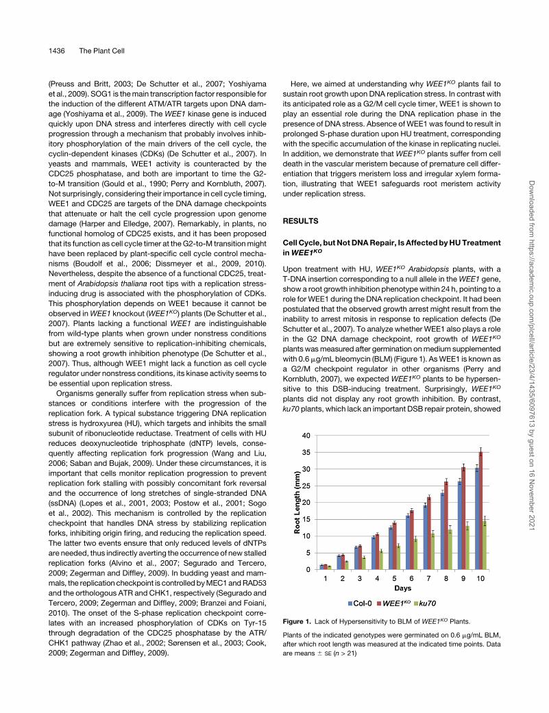
Figure 1. Lack of Hypersensitivity to BLM of WEE1KO Plants.
Plants of the indicated genotypes were germinated on 0.6 mg/mL BLM,
after which root length was measured at the indicated time points. Data
are means 6 SE (n > 21)
1436 The Plant Cell
Dow
nloaded from https://academ
ic.oup.com/plcell/article/23/4/1435/6097613 by guest on 16 N
ovember 2021

a strong growth phenotype when germinated on BLM, when
compared with wild-type seedlings (ecotype Columbia-0 [Col-0]).
Sensitivity of WEE1KO plants to HU but not BLM implies that
WEE1 is dispensable during the G2 DNA damage response but
indispensable during the S-phase checkpoint. To better under-
stand the molecular basis of the root growth inhibitory response
phenotype of WEE1KO plants upon HU treatment, a microarray
experiment was set up to compare the transcriptomes of Col-0
andWEE1KOplants grown in the presence or absence of HU. The
transcript levels were monitored at two defined time points: 5
and 24 h after treatment with 2 mM HU for the short-term and
long-term responses, respectively. These time points were
preselected by cDNA-amplified fragment length polymorphism
analysis to screen for time points with significant transcriptional
changes. As WEE1 gene transcription is concentrated mainly at
the root meristem upon HU treatment (De Schutter et al., 2007),
only root tips (<2 to 3 mm) were harvested for RNA extraction.
The samples treated with and without HU were separately
analyzed by two-way analysis of variance (ANOVA) for genotype
(Col-0 and WEE1KO) and time (0, 5, and 24 h). Under control
conditions, no genes showed significantly altered expression
levels, neither between Col-0 andWEE1KO, nor over time, nor for
an interaction between time and genotype. The lack of strong
transcriptional differences indicates that WEE1KO and Col-0
behave similarly in the absence of DNA damage stress, confirm-
ing the lack of a root phenotype under control growth conditions.
By contrast, 251 genes were differentially regulated upon HU
treatment, showing significantly altered gene expression over
time. To graphically visualize these transcriptional differences,
the significantly altered genes were clustered into seven groups
based on their expression levels (seeMethods for details) (Figure
2; see Supplemental Data Set 1 online). Gene ontology (GO)
overrepresentation analysis of these clusters revealed a signif-
icant enrichment for biological processes in four clusters (Figure
2, Table 1). Genes within cluster A displayed a strong (>2.5- to
10-fold change in expression in Col-0 and WEE1KO after 24 h)
and similar upregulation in both genotypes at both time points
after transfer to HU-containing medium (Figure 2A; see Supple-
mental Data Set 1 online). This cluster was enriched for genes
associated with DNA metabolism, DNA repair, and reaction to
genotoxic radiation, such as BRCA1, PARP2, and XRI1. Also,
TSO2 and a putative thymidine kinase, both involved in nucle-
otide metabolism, reacted to the reduction in the dNTP pool
caused by HU treatment. All the 18 genes from this cluster had
been identified previously as being strongly induced in g-irradi-
ated seedlings (see Supplemental Table 1 online; Culligan et al.,
2006), indicating that upon addition of HU the DNA damage
pathway was activated and that a functional copy of WEE1 was
not necessary for sensing DNA stress and transducing the signal
to induce DNA repair genes. Similar to cluster A, genes in cluster
B were upregulated at both time points upon HU treatment but
were less induced (<4-fold induction) (Figure 2B). Like in cluster A,
these genes were associated with DNA metabolism and more
specificallyDNA replication. AsHU interfereswithbothprocesses,
the induced expression of these genes probably resulted from
adjustment of the replication process to reduced dNTP levels.
Cluster C consisted of genes that were induced transcriptionally
only after 24 h in both Col-0 and WEE1KO (Figure 2C). The GO
Figure 2. Clustering of Microarray Data of HU-Treated Col-0 and WEE1KO.
Genes with significantly altered transcription levels upon HU treatment were clustered into seven different groups based on their expression levels at
different time points in mutant and wild-type root tips. Each panel shows the mean normalized expression values of all the genes within the cluster for
Col-0 (blue) and WEE1KO (red) after 0, 5, and 24 h on HU. The number of genes that each cluster contains is given on the right.
WEE1 Maintains Meristem Integrity 1437
Dow
nloaded from https://academ
ic.oup.com/plcell/article/23/4/1435/6097613 by guest on 16 N
ovember 2021

terms enriched in this gene set were all associated with light
stimuli (Table 1), probably linked to the photoinactivating effects
of HU on photosystem II in plants (Kawamoto et al., 1994).
Cluster D contained 47 array elements (corresponding to 49
genes) that were downregulated in both genotypes after 5 h on
HU. However, cluster D genes reacted differentially at the 24 h
time point, showing a further decrease in Col-0, but returning to
control levels in the mutant (Figure 2D; see Supplemental Data
Set 1 online). Interestingly, within this cluster, 37 out of the 45
array elements that corresponded to a single gene had been
previously found to be cell cycle phase-dependently expressed,
as supported by GO enrichment analysis (Table 1), with 35 of
them peaking during the M phase (see Supplemental Table 2
online; Menges et al., 2003). As described previously, down-
regulation of M-phase genes in the root meristem of control
plants results from cell cycle synchrony imposed by the HU
treatment (Cools et al., 2010). Synchrony is achieved through
HU-induced depletion of the dNTP pool, resulting in a transient
accumulation of cells at the G1-to-S transition. An increase in
intracellular dNTPs through salvage pathways probably in-
creases the level of nucleotides above a threshold level, after
which cells synchronously resume cell division. In this synchro-
nization system, Col-0 root tip cells progress into mitosis 16 h
after HU treatment, and enter S phase around 6 and 22 h. At
these two time points, cells are depleted for G2/M cells, explain-
ing the reduction in M-phase gene expression at the 5- and 24-h
time points in the microarray experiment (Cools et al., 2010).
Although the statistical analysis of the microarray data did not
allow us to call the altered kinetics of G2/M gene transcription in
WEE1KO after HU treatment significantly different from that seen
in Col-0, the data suggested an altered cell cycle regulation at the
24-h time point in WEE1KO plants in the presence of HU.
WEE1KO Plants Display Altered S-Phase Kinetics
The HU-imposed cell cycle synchronization allowed us to inves-
tigate in more detail the possibility of altered cell cycle regulation
in WEE1KO plants upon replication stress. Root tips were col-
lected for transcript analysis at 2-h time intervals from 0 to 22 h
after transfer to HU. RNA levels were measured with the
nCounter analysis system (NanoString Technologies) that allows
a direct multiplexed analysis of selected transcripts (Geiss et al.,
Table 1. GO Analysis of the Different Clusters after Microarray Analysis
Cluster GO Description P Value
A Nucleobase, nucleoside, nucleotide, and nucleic acid metabolic process 4.01E-03
Nucleic acid metabolic process 1.43E-03
DNA metabolic process 2.28E-05
DNA repair 1.84E-06
DSB repair 1.24E-03
Cellular response to stimulus 8.15E-04
Cellular response to stress 3.96E-05
Response to DNA damage stimulus 1.84E-06
DNA repair 1.84E-06
DSB repair 1.24E-03
Response to ionizing radiation 3.62E-06
Response to g-radiation 2.71E-04
Cell cycle phasea 8.16E-03
M phasea 6.09E-03
B Nucleobase, nucleoside, nucleotide, and nucleic acid metabolic process 4.96E-03
Nucleic acid metabolic process 1.06E-03
DNA metabolic process 2.54E-07
DNA replication 2.05E-04
DNA methylation on cytosine 3.67E-03
C Photosynthesis 1.90E-09
Photosynthesis, light reaction 1.77E-03
Response to radiation 2.69E-03
Response to light stimulus 2.69E-03
Nonphotochemical quenching 1.16E-03
D Microtubule-based process 2.56E-06
Microtubule cytoskeleton organization 2.73E-03
Microtubule based movement 2.93E-03
Regulation of cell cycle 2.56E-06
E None
F None
G None
After microarray analysis of Col-0 and WEE1KO plants treated with HU, the selected genes were divided into seven different clusters that were
analyzed for GO enrichment (P value < 0.01). Indentations represent child terms of the above GO terms.aThe detection of the M-phase and cell cycle phase GO groups is the result of the presence in this cluster of XRI1 and SYN2 that are needed both for
DNA repair and M-phase progression.
1438 The Plant Cell
Dow
nloaded from https://academ
ic.oup.com/plcell/article/23/4/1435/6097613 by guest on 16 N
ovember 2021

2008). When compared with Col-0, WEE1KO plants initially
displayed identical cell cycle progression, as exemplified by
the transcription profile of the S-phase marker gene CYCA3;1
(Figure 3A), showing thatWEE1KO is susceptible to HU synchro-
nization. As indicated by the expression profiles of CYCA3;1 and
histone H4, DNA replication initiated around 4 to 6 h after transfer
to HU (Figures 3A and 3B). Also, the mid-S-phase marker gene
histone H2Bwas induced at the same time in Col-0 andWEE1KO
(Figure 3C). However, altered S-phase kinetics became apparent
when the transcription of the late-S-phase marker gene histone
H1 was examined. Like histone H4 and H2B, histone H1 was
induced simultaneously in both backgrounds but had a pro-
longed window of expression and superinduction (10 to 22 h) in
WEE1KO (Figure 3D). Together, the histone gene expression
kinetics imply thatWEE1-deficient plants progress normally into
S phase but encounter stress during the replication process.
Accordingly, downstream cell cycle events are clearly affected in
theWEE1KO plants, as illustrated by the attenuated and delayed
expression profiles of theG2/Mmarker genesCYCA2;1 (peaks at
18 h versus 14 h in Col-0) andCYCB1;2 (peaks at 20 h versus 16 h
inCol-0) (Figures 3E and 3F). Genes present in the cluster D of the
microarray analysis (CYCA1;1,CYCB2;1, andCYCB2;4) showed
similar delayed expression kinetics (see Supplemental Figure
1 online), confirming the earlier observations of the microarray
analysis and pointing toward a delayed progression through
mitosis in WEE1KO plants. Despite the prolonged S phase, the
peaks of early G2 (CYCA2;1) and late G2/M (CYCB1;2) genes
were separated by 2 h in both Col-0 and mutant, indicating that
mainly the S-phase progression is sensitive to the HU treatment
in WEE1KO.
Among all G2/M genes tested, CYCB1;1 displayed a unique
transcriptional profile (Figure 3G). Previously, CYCB1;1 expres-
sion had been connected with a role in the DNA damage
response because its expression was found to be strongly
induced and stabilized during g-irradiation (Culligan et al.,
2006). In contrast with the related CYCB1;2, CYCB1;1 levels
were not downregulated during S phase in HU-synchronized
Col-0 root tips (Cools et al., 2010). However, in WEE1KO,
CYCB1;1 was rather strongly transcriptionally activated at the
start of replication (Figure 3G), as observed for DNA damage
genes, such as BRCA1 (Figure 3H), and had lost the typical
induction kinetics of G2/M genes.
WEE1 Controls S-Phase Progression during
Replication Stress
In contrast with its anticipated role as timer of the G2-to-M
transition upon the occurrence of DNA stress (Perry and Kornbluth,
2007; Geiss et al., 2008), our data indicated that Arabidopsis
WEE1 rather plays an important part during DNA replication.
Correspondingly, within the root synchronization system, WEE1
transcripts accumulated after 4 to 6 h, the time of S-phase onset,
with kinetics comparable to those of the S-phase marker
CYCA3;1 (Figure 4A). Furthermore, similar to DNA repair genes,
WEE1 levels remained induced during thewhole period of theHU
treatment.
To confirm thatWEE1 specifically accumulates during S phase
upon replication stress,WEE1KO plants were transformed with a
PWEE1:GFP-WEE1 complementation construct. Transgenic plants
harboring the construct partially rescued the HU hypersensitivity
of theWEE1-deficient plants (Figures 4B and 4C). In the absence
of DNA stress, only background fluorescence was detected in
the root. By contrast, a nuclear-localized GFP-WEE1 signal
could be observed in the vascular meristem cells after 24 h
treatment with HU, corresponding with its previously reported
expression pattern (Figure 4C; De Schutter et al., 2007). To
pinpoint the cell cycle phase at which the GFP-WEE1–positive
cells accumulate, fluorescent cells were separated fromnegative
ones by fluorescence-activated cell sorting, either from control
root tips (not treated with HU) or root tips of plants treated with
HU for 48 h. Subsequently, nuclei were extracted from the sorted
cells, and their DNA content wasmeasured by flow cytometry. In
the total population of nuclei within the root tip of untreated
plants, two distinct populations were observed with a 2C and 4C
DNA content, corresponding to G1 and G2 nuclei, respectively
(Figure 4D). Because of the lack of a GFP signal in the absence of
DNA stress, no GFP-positive nuclei could be detected. When
transferred to HU, the relative abundance of nuclei with a DNA
content in between 2C and 4C, corresponding to S-phase nuclei,
increased (Figure 4E). When the DNA content of GFP-WEE1–
positive cells was measured, a single population of nuclei was
observed, localized precisely between the 2C and 4C peaks
(Figure 4E). These data substantiate the hypothesis that WEE1
operates specifically in replicating cells that undergo replication
defects.
HUTriggers Vascular Cell Death inWEE1KORootMeristems
Treatment of plants with DSB-inducing drugs commonly results
in death of the root stem cells (Fulcher and Sablowski, 2009;
Furukawa et al., 2010). As WEE1KO plants show strong growth
retardation in the presence of HU, they were tested for the
occurrence of dead cells. Plants were transferred to HU-con-
taining medium and stained at different time points with propi-
dium iodide (PI) that stains the walls of living cells but penetrates
dead cells. As HU can be used to invoke root synchronization,
the appearance of cell death could be correlated with cell cycle
progression. At the first time points analyzed (3 to 8 h), no PI-
stained cells were detected in both Col-0 and WEE1KO root
meristems (Figure 5A). By contrast, at the moment that cells
started accumulating in mid-S-phase (9 to 10 h), the first dead
cells were visible in the mutant, in contrast with Col-0, where no
PI-positive cells were observed (Figure 5A). The timing of cell
death appearance suggested that defective S-phase progres-
sion might lie at the basis of the phenotype.
To investigate the spatial occurrence of cell death, plants were
transferred for 24 h to medium supplemented with HU and again
stained with PI. A large amount of dead cells was seen through-
out the WEE1KO meristem but not in Col-0. Interestingly, this
spatial cell death pattern differed clearly from that provoked by
DSBs, such as after treatment with BLM (Figure 5B), which
induced cell death specifically in the stem and progenitor (StPr)
zone (Fulcher and Sablowski, 2009; Furukawa et al., 2010). By
contrast, dead cells in the meristem of WEE1KO plants did not
occur in the stem cells but were located in the transiently
amplifying (TA) region of the vascular tissue (Figure 5A). To
WEE1 Maintains Meristem Integrity 1439
Dow
nloaded from https://academ
ic.oup.com/plcell/article/23/4/1435/6097613 by guest on 16 N
ovember 2021

Figure 3. Time-Course Analysis of Cell Cycle Genes in HU-Synchronized Col-0 and WEE1KO Root Tips.
Transcript levels were measured by nCounter analysis. Fold induction levels of the different transcripts are presented for Col-0 (blue) andWEE1KO (red).
EMB2386, PAC1, and RPS26C were used as reference genes. Transcript levels were rescaled to the level in Col-0 at 0 h (=1). Time after treatment with
HU is given on the horizontal axis. Data are means 6 SE.
(A) CYCA3;1.
(B) Histone H4.
(C) Histone H2B.
(D) Histone H1.
(E) CYCA2;1.
(F) CYCB1;2.
(G) CYCB1;1.
(H) BRCA1.
1440 The Plant Cell
Dow
nloaded from https://academ
ic.oup.com/plcell/article/23/4/1435/6097613 by guest on 16 N
ovember 2021

investigate whether this specific cell death localization was
caused only by replication stress or also depended on WEE1
deficiency, we increased the HU concentrations to a level that
induces cell death in Col-0 plants (5 mM HU). If cell death
localization depended solely on HU, the cell death pattern in
Col-0 would be expected to be the same as that of the WEE1KO
plants. Surprisingly, the observed pattern resembled that of
plants suffering from DSBs, with cell death restricted to the StPr
cells (Figure 5C). Also,WEE1KO plants now displayed dead cells
around the quiescent center, in addition to those in the vascular
TA region. Remarkably, cell death in the StPr zone was not
limited to vascular tissue. These data indicate that increased
levels of HU indirectly cause cell death in Col-0, probably by the
secondary occurrence of DSBs with dead StPr cells as a con-
sequence. Moreover, together with the lack of a root growth
phenotype in WEE1KO plants growing on BLM (Figure 1), it
implies thatWEE1-independent pathways cope with DSBs in the
StPr cells and that WEE1 preferentially protects the vascular TA
zone upon replication stress.
Previously, WEE1 had been shown to operate downstream of
ATM and ATR (De Schutter et al., 2007). Both ATM and ATR have
been demonstrated to be required for the onset of cell death in
the StPr cells upon DNA damage (Fulcher and Sablowski, 2009;
Furukawa et al., 2010). To investigate the occurrence and pattern
of cell death upon replication stress in atm-1 and atr-2 mutant
plants, they were treated for 24 h with 1 mMHU. As atm-1 plants
are not hypersensitive to replication stress, they evidently did not
suffer from cell death in the root meristem (Figure 5D). By
contrast, atr-2 plants, which are highly sensitive to HU (Culligan
et al., 2004), exhibited severe cell death (Figure 5D). The cell
death phenotype in atr-2 plants was more severe than that in
WEE1KO plants and also occurred in the stem cell zone. This
observation can be attributed to the fact that besides its inability
to induce WEE1 expression, atr-2 also fails to induce the DNA
repair machinery to repair the afflicted DNA. Nevertheless, the
common cell death phenotype in the vascular TA zone in
WEE1KO and atr-2 plants indicates that the ability of WEE1 to
inhibit cell death in the distal vascular meristem upon replication
stress depends on ATR.
WEE1 Is Required to Prevent Premature Tracheary Element
Differentiation upon Replication Stress
Because stem cells are the first to react to DNA damage, the lack
of dead stem cells in WEE1KO plants indicated that the root
growth arrest might be unrelated to the occurrence of severe
DNA damage. Indeed, upon HU treatment, no substantial differ-
ences were observed between Col-0 andWEE1-deficient plants
when examining DNA damage hallmarks, such as the occur-
rence of DNA fragmentation or increased recombination (see
Supplemental Figure 2 online). Because dead cells were specif-
ically observed in the provascular zone and PCD is an intrinsic
process of vascular maturation, the cell death in WEE1KO plants
could reflect the onset of premature differentiation in the vascular
tissue, rather than DNA damage–induced PCD, as suggested
previously (Ricaud et al., 2007; Fulcher and Sablowski, 2009).
Therefore, we took a closer look at genes that function in
tracheary element (TE) differentiation (Turner et al., 2007).
Figure 4. Stabilization of WEE1 during S Phase upon Replication Stress.
(A) Transcript analysis of CYCA3;1 (blue) and WEE1 (red) in HU-syn-
chronized Col-0 root tips. Data are means 6 SE.
(B) Daily growth of Col-0 (blue), WEE1KO (red), and PWEE1:GFP-WEE1
(green) roots after transfer to 1 mM HU. Growth was followed until 4 d
after transfer. Data are means 6 SE (n > 37).
(C) Confocal microscopy of the PWEE1:GFP-WEE1 line transferred for 24
h to 0 mM HU (top) and 1 mM HU (bottom). Epidermis-localized green
signal at 0 mM HU is due to background signal. Nuclear GFP-WEE1
signal in the vascular cells was seen after treatment with 1 mM HU. Bar =
0.1 mm.
(D) Flow cytometry profile of unsorted and untreated PWEE1:GFP-WEE1
root tips. 2C, 4C, and S indicate G1, G2, or S-phase nuclei, respectively.
(E) Flow cytometry profile of unsorted (red) and sorted GFP-WEE1 (blue)
root tips treated for 24 h with 10 mM HU.
WEE1 Maintains Meristem Integrity 1441
Dow
nloaded from https://academ
ic.oup.com/plcell/article/23/4/1435/6097613 by guest on 16 N
ovember 2021

Transcripts of five of these genes (CESA4, IRX1, IRX3,XCP1, and
XCP2) were quantitatively measured by real-time PCR in both
Col-0 and WEE1KO 0, 24, and 48 h after treatment with HU.
Whereas transcript levels of the TE differentiation genes de-
creased slightly upon HU treatment in Col-0, they increased in
WEE1KO (Figure 6A; see Supplemental Figure 3 online). The latter
suggested that WEE1KO plants undergo premature TE differen-
tiation upon HU treatment. To further test this hypothesis, a
VND7 reporter construct (VND7pro:GUS) was introgressed into a
WEE1KO background. VND7 is a transcription factor implicated
in xylemdifferentiation (Kubo et al., 2005; Yamaguchi et al., 2008,
2010). Under normal growth conditions, the rootmeristem shows
no VND7 expression in Col-0 and WEE1KO plants (Kubo et al.,
2005) (Figure 6B). After treatment of VND7pro:GUS lines with
1 mM HU for 24 h, b-glucuronidase (GUS) staining could be
observed specifically within the root meristem of the WEE1KO
plants (Figure 6B). At higher HU concentration (2.5 mM HU),
Col-0 root tips occasionally showed an individual cell with weak
VND7 expression. However, expression was much more pro-
nounced in WEE1KO, showing many cells with a strong VND7
induction, demonstrating the molecular onset of vascular differ-
entiation.
Protoxylem formation is inhibited by cytokinin treatment
(Mahonen et al., 2006). If premature protoxylem differentiation
were at the basis of the observed cell death after HU treatment in
WEE1KO, N6-benzyladenine (BA) treatment would reduce the
vascular cell death phenotype upon replication stress. To test
this hypothesis, WEE1KO plants were transferred to medium
containing no drugs, 1 mM HU, 100 nM BA, or a combination of
HU and BA. After 24 h, a reduction in the number of PI-stained
cells was visible in plants treatedwithHU in combinationwith BA,
compared with plants treated with 1 mM HU only (Figure 7; see
Supplemental Figure 4 online). This effect was even more obvi-
ous at the 48-h timepoint, when almost no dead cells were visible
Figure 5. Vascular Cell Death Phenotype of WEE1KO roots upon Replication Stress.
(A) Time course of Col-0 and WEE1KO root tips transferred to HU and stained with confocal microscopy. Dead cells are stained with PI. Time after
transfer to HU medium is indicated above the root tips.
(B) PI staining of Col-0 and WEE1KO after treatment with 0.6 mg/mL BLM for 24 h.
(C) PI staining of Col-0 and WEE1KO grown for 24 h on 5 mM HU.
(D) PI staining of atm-1 and atr-2 transferred either for 24 h to control medium or 1 mM HU.
1442 The Plant Cell
Dow
nloaded from https://academ
ic.oup.com/plcell/article/23/4/1435/6097613 by guest on 16 N
ovember 2021

after BA and HU treatment. To connect the ectopic expression of
VND7, and consequent vascular differentiation, with the ob-
served cell death in WEE1KO, we analyzed whether the addition
of BA attenuated the VND7 induction in WEE1KO after HU
treatment. Indeed, 24 h after transfer to HU medium supple-
mented with BA, VND7 expression was strongly reduced in the
WEE1KO root tip (Figure 8A). Together, these data strongly
indicate that replication stress-induced cell death and xylem
differentiation, due to the lack of WEE1, are coupled events.
Absence of WEE1 Results in Aberrant Vascular
Differentiation upon Long-Term Replication Stress
Because of the observed link between replication stress and TE
differentiation, the vascular tissue was examined in WEE1KO
plants after a prolonged and strong HU treatment. Wild-type
plants displayed normally organized protoxylem and metaxylem
development after 4 d of growth on HU. By contrast, inWEE1KO
plants, the xylem was disorganized with interrupted and addi-
tional xylem rows (Figures 8B and 8C). The presence of multiple
and extra xylem rows immediately above the root meristem
indicates that vascular meristem cells in WEE1KO plants trigger
their maturation program prematurely. These data clearly dem-
onstrate a function for WEE1 as inhibitor of premature vascular
differentiation under replication stress conditions.
DISCUSSION
WEE1 Controls S-Phase Progression under
Replication Stress
DNA damage checkpoint research is far more advanced in
nonplant organisms, illustrated by the large number of known
regulators contributing to the cell cycle arrest and DNA repair
mechanisms activated upon DSBs or replication defects in
yeasts and mammals. Although the presence of a functional
ATM and ATR suggests that the main mechanisms controlling
the DNA damage response in plants might be similar, homologs
of several key signaling components have not been identified in
the Arabidopsis genome (Cools and De Veylder, 2009). The
mechanisms by which yeast and mammalian cells arrest their
proliferation in response to DNA stress depend on the cell cycle
phase at which the cell resides at the moment the damage is
inflicted. Independently of the followed pathway, the common
goal is inhibition of the CDK activity to prevent cell cycle pro-
gression in the presence of damaged or incompletely replicated
DNA. When damage occurs during replication, the intra-S
checkpoint is activated, correlating with the accumulation of
P-loop–phosphorylated inactive CDKs, probably through inhibi-
tion of CDC25 (Zegerman and Diffley, 2009). The CHK1-CDC25
pathway dominates S-phase regulation in mammals and bud-
ding yeast during replication stress, but no direct role has been
demonstrated for its counterpart WEE1. By contrast, we
Figure 6. Premature Vascular Differentiation Induced by Replication
Stress in WEE1KO Plants.
(A) Tracheary element differentiation genes are induced in WEE1KO root
tips upon replication stress. qRT-PCR transcript analysis of IRX1 and
CESA4 in Col-0 and WEE1KO 0, 24, and 48 h after HU treatment.
EMB2386, PAC1, and RPS26C were used as reference genes. The
transcript levels were rescaled to the level in Col-0 at 0 h (=1). Asterisks
indicate significantly altered transcription levels compared with the 0-h
time point (*P value < 0.05; **P value <0.001). Data are means 6 SE
(n = 4).
(B) Confocal microscopy of Col-0 andWEE1KO plants after PI staining to
detect cell death. Col-0 and WEE1KO harboring a VND7pro:GUS con-
struct were stained with GUS. Plants were treated either with 0 mM (left),
1 mM HU (center), or 2.5 mM HU (right) for 24 h. Bars = 0.1 mm.
WEE1 Maintains Meristem Integrity 1443
Dow
nloaded from https://academ
ic.oup.com/plcell/article/23/4/1435/6097613 by guest on 16 N
ovember 2021
identified WEE1 as an essential intra-S-phase checkpoint gene
in Arabidopsis, as demonstrated by the delayed progression
through S phase in synchronized WEE1KO root tips and the
specific accumulation of its gene product in replicating nuclei
upon HU treatment. As there is no functional CDC25 or CHK1
homolog in Arabidopsis, the upregulation of WEE1 during S
phase and the hypersensitivity of the WEE1KO mutant to repli-
cation stress put WEE1 forward as the main regulator of the
S-phase checkpoint in plants.
Plants without a functionalWEE1 gene show a superinduction
and prolonged transcription of different histone genes after
treatment with HU. This result implies a problematic S-phase
progression with a delayed entry into G2 as a consequence. By
contrast, the timing between G2 and G2/M progression (2 h) was
not extended in WEE1KO compared with Col-0 plants. Upon
treatment with HU, cells need to adjust replication with the
depleted dNTPpools. Generally, dNTPdepletion is counteracted
by slowing down replication and delaying the initiation of repli-
cation origins (Willis and Rhind, 2009), which is accomplished by
reducing the CDK activity during S phase through the ATR-CHK1
pathway that inactivates CDC25 (Cook, 2009; Willis and Rhind,
2009; Zegerman and Diffley, 2009). Because of the absence of
functional CDC25 and CHK1 genes in plants, we hypothesize a
role for WEE1 during DNA stress as coordinator of CDK activity
with replication fork progression and/or origin firing. Disruption of
the CDK activity–controlling pathway in budding yeast during HU
treatment by means of a mutated Rad53 (the ortholog of CHK1)
causes the checkpoint mutants to produce long stretches of
ssDNA and fork reversal, forming structures similar to Holliday
junctions (Lopes et al., 2001; Sogo et al., 2002; Branzei and
Foiani, 2010). Similarly, the observed replication difficulties in
WEE1KO plants might arise from the inability to attenuate suffi-
ciently DNA replication to the limited dNTP levels. The lack of
checkpoint activity and CDK control probably prevents inhibition
of origin firing and/or deceleration of replication. This will even-
tually increase the number of stalled replication forks inWEE1KO,
with potentially concomitantly extended ssDNA regions or fork
reversal as a result. Evidently, the aggravation of replication
problems takes more time to repair, with a cell cycle delay as a
consequence.
Whereas theWEE1protein is an essential checkpoint regulator
under replication stress, its presence appears to be dispensable
for the response to other types of DNA damage, such as DSBs. If
WEE1 is still an element of the DSB checkpoint, its role will be, at
most, redundant with other mechanisms. Currently, thesemech-
anisms are still unknown, but likely candidates might be found
among the class of cell cycle inhibitors (CKIs). Plants have two
classes of CKIs (KIP-RELATED PROTEINS and SIAMESE/
SIAMESE-RELATED) with a limited sequence similarity to the
mammalian Cip/Kip CDK inhibitors (De Veylder et al., 2001;
Churchman et al., 2006) of which some members are strongly
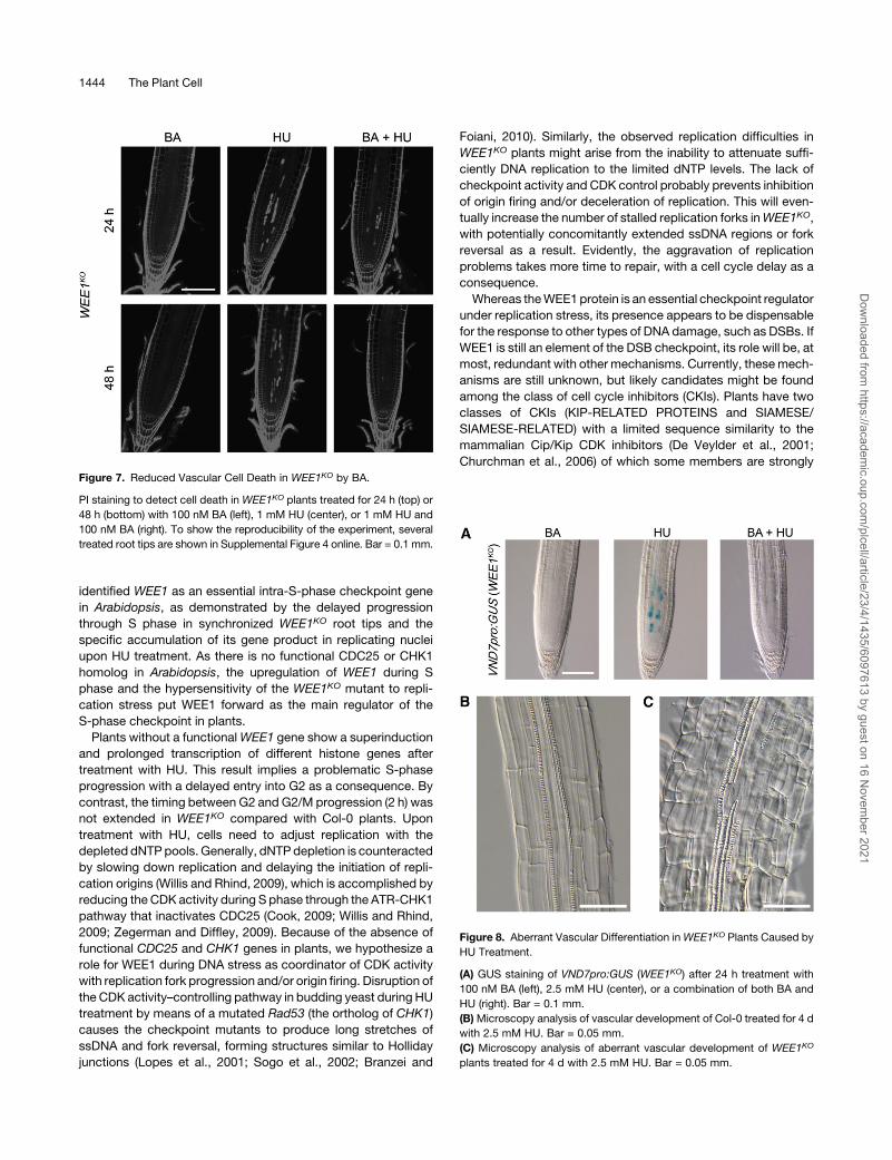
Figure 7. Reduced Vascular Cell Death in WEE1KO by BA.
PI staining to detect cell death in WEE1KO plants treated for 24 h (top) or
48 h (bottom) with 100 nM BA (left), 1 mM HU (center), or 1 mM HU and
100 nM BA (right). To show the reproducibility of the experiment, several
treated root tips are shown in Supplemental Figure 4 online. Bar = 0.1 mm.
Figure 8. Aberrant Vascular Differentiation inWEE1KO Plants Caused by
HU Treatment.
(A) GUS staining of VND7pro:GUS (WEE1KO) after 24 h treatment with
100 nM BA (left), 2.5 mM HU (center), or a combination of both BA and
HU (right). Bar = 0.1 mm.
(B)Microscopy analysis of vascular development of Col-0 treated for 4 d
with 2.5 mM HU. Bar = 0.05 mm.
(C) Microscopy analysis of aberrant vascular development of WEE1KO
plants treated for 4 d with 2.5 mM HU. Bar = 0.05 mm.
1444 The Plant Cell
Dow
nloaded from https://academ
ic.oup.com/plcell/article/23/4/1435/6097613 by guest on 16 N
ovember 2021

transcriptionally induced by genotoxic stress treatments (Peres
et al., 2007).
WEE1 and Vascular Differentiation
Besides halting S-phase progression, the absence of a functional
WEE1 protein in the presence of replication stress causes clear
developmental defects. Upon addition of HU, dead cells accu-
mulate in the root tip of WEE1KO plants. These cells are located
predominantly in the vascular meristem, in contrast with DSB-
inducing treatments that mainly target the stem cells of all root
cell types (Fulcher and Sablowski, 2009; Furukawa et al., 2010).
PCD is an intrinsic part of vascular development (Turner et al.,
2007); indeed, through VND7 promoter activity measurements,
cell death in theWEE1KO plants was associated with the onset of
the vascular differentiation program. Moreover, cell death could
be reduced by cytokinin treatment that is known to inhibit
protoxylem differentiation in Arabidopsis roots (Mahonen et al.,
2006). PI-stained WEE1KO cells could be observed within 10 h
after HU treatment and were positioned close to the stem cells,
indicating that their position in the meristem arose by a prema-
ture onset of vascular differentiation, rather than because of
shrinkage of the meristem as a result of the described effect on
cell cycle duration. Likewise, ectopic expression in the meristem
of VND7 and other vascular differentiation genes, normally only
observed in the immature xylem cells outside the meristem
(Kubo et al., 2005; Yamaguchi et al., 2008, 2010), hints at a
premature onset of vascular differentiation and likely explains the
irregular xylem organization inWEE1KO roots upon long-termHU
treatment. Interestingly, in contrast with the WEE1KO plants, the
Col-0 roots showed a decrease in expression of vascular differ-
entiation genes within the root meristem upon replication stress,
which might be correlated with the transcriptional induction of
WEE1 in the vascular meristem (De Schutter et al., 2007). This
observation is corroborated on the protein level by the WEE1
stabilization after HU treatment in the vascular meristem cells.
Altogether, these data indicate an indirect developmental role for
WEE1 upon replication stress as an inhibitor of premature xylem
formation. Strikingly, even high HU concentrations could not
elicit the vascular cell death response in control plants. Rather,
cell death was limited to the root initial cells, very similarly to that
observed upon exposure to DSB-inducing drugs (Curtis and
Hays, 2007; Fulcher andSablowski, 2009; Furukawa et al., 2010).
Hence, WEE1 is a very potent safeguard of the mitotic status of
vascular cells, specifically toward replication stress.
The specific prevascular cell death upon HU application
suggests that replication stress might be an intrinsic part of the
vascular differentiation process. In agreement, the treatment of
cells of zinnia (Zinnia elegans) with aphidicolin, a compound that
induces replication stress through the inhibition of DNA poly-
merases, promoted their differentiation into tracheary elements
(Mourelatou et al., 2004). In addition, the gene EFFECTOR OF
TRANSCRIPTION2, implicated in xylem differentiation, has an
endonuclease domain responsible for making single-strand cuts
within the DNA and is essential for the protein’s role in vascular
development (Ivanov et al., 2008), suggesting that the occur-
rence of ssDNA might start xylem differentiation. Thus, WEE1
and replication stress apparently play an antagonistic role in the
development of vascular tissue, in which WEE1 prevents loss of
vascular meristem identity upon replication stress to sustain
meristem activity.
Plant WEE1 and CDC25 during Evolution
In contrast with other organisms, inArabidopsis,WEE1 and other
DNA damage checkpoint proteins are not essential during un-
perturbed cell cycle conditions (Cools and De Veylder, 2009), as
exemplified by the lack of any phenotype of WEE1KO plants
under nonstress growth conditions (De Schutter et al., 2007).
Nevertheless, the amino acid residues of CDKs targeted for
phosphorylation by WEE1 seem to be conserved, and their
substitution into phosphorylation-mimicking sites results in a
severe growth phenotype due to a cell cycle arrest (Dissmeyer
et al., 2009), implying that the inhibitory function of the P-loop is
conserved. However, whereas in other model species control of
CDKactivity through P-loop phosphorylation/dephosphorylation
is used for timing of the G2-to-M transition (Gould et al., 1990;
Perry and Kornbluth, 2007), plants use apparently other mech-
anisms to time their M phase onset, likely with the plant-specific
B-type CDKs, because their activity is required for progression
through the G2-to-M transition (Porceddu et al., 2001; Boudolf
et al., 2004; Cools et al., 2010). Considering the number of
striking parallels between the plant B1-type CDKs and mamma-
lian CDC25, it has been suggested previously that during evo-
lution, CDC25 control of the onset of mitosis might have been
replaced in the plant kingdom by B1-type CDKs (Boudolf et al.,
2006). Our work strengthens the case for a lack of G2-to-M
control through P-loop phosphorylation because WEE1 tran-
script abundance peaks during S-phase progression, whereas
its associated kinase activity is only required in times of replica-
tion stress. Moreover, its importance in maintaining meristem
structure through the inhibition of premature vascular cell differ-
entiation suggests an indirect role for WEE1 as a developmental
regulator. It indicates that during evolution, WEE1 and CDC25
have lost their function as antagonistic checkpoint controllers in
plants, combined with the acquisition of a developmental role for
WEE1. Once such a developmental role had been established,
there might have been no reason to retain a functional CDC25 in
the genome. Consequently, WEE1 controls two important pro-
cesses influencing the meristem integrity under HU conditions: it
controls meristem size by affecting cell cycle duration, and it
ensures meristem structure by preventing premature differenti-
ation. Hence, we conclude that WEE1 is an essential element to
ensure meristem maintenance during replication stress.
METHODS
Plant Materials and Growth Conditions
Arabidopsis thaliana plants (ecotype Col-0) were grown in vitro vertically
under long-day conditions (16 h light/8 h darkness) at 218C on half-
strengthMurashige and Skoog (2.151 g/L) (Duchefa), 10 g/L sucrose, and
0.5 g/L MES, pH 5.7, adjusted with 1 M KOH and 10 g/L agar. For HU
treatments, plants were grown for 7 d (2 d of germination and 5 d of
growth) on control medium and then transferred either to fresh control
medium or HU-containing media (Sigma-Aldrich). The HU concentration
used was 1 mM unless stated otherwise and that of BLM (Duchefa) was
WEE1 Maintains Meristem Integrity 1445
Dow
nloaded from https://academ
ic.oup.com/plcell/article/23/4/1435/6097613 by guest on 16 N
ovember 2021

0.6 mg/mL, whereas BA (Duchefa) was brought to a final concentration of
100 nM. Whenever synchronization conditions (Cools et al., 2010) were
used, plants were grown vertically under continuous light conditions at
218C with 13Murashige and Skoog medium (4.302 g/L), 10 g/L sucrose,
0.1 g/L myo-inositol, 0.5 g/L MES, 100 mL thiamine hydrochloride (10 mg/
mL), 100 mL pyridoxine (5 mg/mL), and 100 mL nicotinic acid (5 mg/mL),
pH 5.7, adjusted with 1 M KOH and 10 g/L agar. Seeds were placed on a
nylon mesh (20 mm pore size) (Prosep) and transferred after 1 week to
plates with 2 mM HU. The root growth response of Col-0 and WEE1KO
plants grown in the presence of 2mMHU on nylon was identical to that of
plants growth directly on medium supplemented with 1 mM HU (data not
shown).
The PWEE1:GFP-WEE1 construct was created with Gateway technol-
ogy (Invitrogen). An N-terminal fusion of GFP with the WEE1 protein
under the control of the endogenous WEE1 promoter (2605 to 21) was
constructed with the MultiSite Gateway three-fragment vector construc-
tion kit (Invitrogen). Three different vectors were used (WEE1 promoter
in pDONRP4P1R [Invitrogen], GFP in pDONR221 [Invitrogen], and
WEE1_ORF in pDONRP2RP3 [Invitrogen]) and were cloned in the
pK7m34GW destination vector (Karimi et al., 2005) by a MultiSite LR
reaction. After transformation of the sequenced constructs to Agro-
bacterium tumefaciens, WEE1KO plants were transformed by floral dip
(Clough and Bent, 1998). Kanamycin-resistant plants were selected and
analyzed for their ability to complement the WEE1KO phenotype on HU.
GU-US (uidA recombination), ku70, atm-1, atr-2, VND7pro:GUS, and
WEE1KO (wee1-1 and wee1-2) lines had been described previously
(Swoboda et al., 1994; Riha et al., 2002; Garcia et al., 2003; Culligan
et al., 2004; Kubo et al., 2005; DeSchutter et al., 2007).wee1-1 andwee1-2
plants were pooled in the microarray experiment. Nanostring and pheno-
typic analysis were done with wee1-1.
Microarray Analysis
Plant lines and growth conditions were as described above. Col-0 and
WEE1KO (wee1-1 andwee1-2) seedswere germinated on controlmedium
on a nylon mesh and transferred 5 d after germination to control medium
or medium supplemented with 2 mM HU. All sampling points were
collected over four independent biological repeats for Col-0, two inde-
pendent repeats for wee1-1, and two for wee1-2. For each independent
time point of each experiment 650 root tips (<2 to 3 mm) were collected
and frozen in liquid nitrogen. RNA was extracted from root tissue with
TriZol reagent (Invitrogen) and purified with the RNeasy plant mini kit
(Qiagen). The RNA of two independent experiments for Col-0 and
WEE1KO were subsequently pooled and used for microarray analysis. A
total of 24 samples were individually hybridized to microarrays, repre-
senting two genotypes (Col-0 and WEE1KO) grown under two conditions
(2HU and +HU), harvested at three time points (0, 5, and 24 h), in
duplicate. Out of 5mgof total RNA, biotinylated-copyRNAwasproduced,
fragmented, and hybridized to ATH1 arrays (Affymetrix). Washing, de-
tection, and scanning were done as described previously (Hennig et al.,
2003). Array data were made available as Affymetrix.CEL files, and the
quality was assessed before inclusion for analysis. The gcRMA-normal-
ized data were subjected to two-factor ANOVA for genotype (wild type
and WEE1KO) and time (0, 5, and 24 h) with The Institute for Genomic
ResearchMultiExperiment Viewer 4.5 (TMeV 4.5; http://www.tm4.org) (df
genotype = 1; df time = 2; df interaction = 2; and df error = 6) (Saeed et al.,
2003). P values were based on permutations (10,000), and a multiple
comparison correction was applied by calculating the false discovery rate
usingMixtureModel ofGenStat (http://www.vsni.co.uk/software/genstat/).
Genes were selected for analysis when they were 1.8-fold upregulated
($1.8) or downregulated (#0.56) in Col-0 orWEE1KO, either 5 or 24 h after
treatment, and if one of the three P values obtained after ANOVA analysis
was <0.01 and false discovery rate < 0.1. Clustering was based on the six
expression values per gene obtained by microarray analysis (Col-0 and
WEE1KO after 0, 5, or 24 h under stress conditions). After mean normal-
ization, significant genes were clustered into seven clusters via K-means
clustering; distance metric is Euclidean distance (Soukas et al., 2000).
This method divides the genes in a specified number of clusters, deter-
mined in advance by calculating a figure of merit that estimates the
predictive power of a certain algorithm, such as K-means clustering
(Yeung et al., 2001). Finally, gene clusters were analyzed for an enrich-
ment in genes with a certain GO annotation with BiNGO (http://www.
psb.ugent.be/cbd/papers/BiNGO/Home.html) with significance level at
P value < 0.01 (Maere et al., 2005).
RNA Extraction, Nanostring nCounter Assay, and
Quantitative RT-PCR
RNA extraction, nCounter assay (Nanostring Technologies), and syn-
chronization were done as described (Cools et al., 2010). Briefly, syn-
chronized root tips were harvested and RNA was extracted from these
tissues with RNeasy plant mini kit (Qiagen). RNA levels were measured
with the nCounter analysis system by the VIB MicroArrays Facility (www.
microarrays.be) as described (Geiss et al., 2008) or with quantitative real-
time PCR (qRT-PCR) and normalized with three reference genes
(EMB2386, RPS26C, and PAC1). RNA levels were rescaled to the levels
of wild-type plants growing for 0 h on 2mMHU. For qRT-PCR, cDNAwas
prepared from 1 mg of total RNA with the iScript cDNA synthesis kit (Bio-
Rad) according to the manufacturer’s instructions. qRT-PCR was per-
formed with LightCycler 480 SYBR Green I Master (Roche) in a final
volume of 5 mL and 0.2 mM primer concentration and analyzed with a
LightCycler 480 (Roche). For each reaction, three technical repeats and four
biological repeats were done. The primer sequences were 59-TCCGG-
TGGAGTGGTGTAAGCAT-39 and 59-GGCTTCGTCACTGGCTCCTTTT-39
for CESA4 (AT5G44030), 59-GCTCGCTGGTCTCGACACAAAT-39 and
59-GAAGTGACGTCGGAGGGATCAA-39 for IRX1 (AT4G18780), 59-GGAAC-
GTCGAGCCATGAAGAGA-39 and 59-GGCCGAGGAAGACTTGGATCAT-39
for IRX3 (AT5G17420), 59-GATACACGCCGGAGCATTTGAC-39 and 59-GCA-
CCTTCTCCTCCACGCTTTT-39 for XCP1 (AT4G35350), 59-GTTTGCGG-
ATTTGAGCCATGAGG-39 and 59-AACTTCCGCCACAGCTCCTTTC-39
for XCP2 (AT1G20850), 59-CTCTCGTTCCAGAGCTCGCAAAA-39 and
59-AAGAACACGCATCCTACGCATCC-39 for EMB2386 (AT1G02780),
59-TCTCTTTGCAGGATGGGACAAGC-39 and 59-AGACTGAGCCGC-
CTGATTGTTTG-39 for PAC1 (AT3G22110), and 59-GACTTTCAAGCG-
CAGGAATGGTG-39 and 59-CCTTGTCCTTGGGGCAACACTTT-39 for
RPS26C (AT3G56340).
Microscopy Analysis
Plants used for confocal microscopy were analyzed with either LSM 510
or LSM 5 exciter confocal microscope (Zeiss). Plants were stained for 2
min in a 10 mM PI solution (Sigma-Aldrich). For GUS staining, whole
seedlings were stained in a 6-well multiwell plate (Falcon 3046; Becton
Dickinson) as described (Beeckman and Engler, 1994). All light micros-
copy samples, irrespective of whether they were stained with GUS, were
cleared with lactic acid and visualized by differential interference contrast
microscopes DM LB (Leica) and BX51 (Olympus).
Sorting of GFP-Positive Nuclei
PWEE1:GFP-WEE1 plants (in a wee1-1) background were treated for 24 h
on 10 mM HU. Root tips (400) were harvested and used for protoplasting
and sorting as described (Birnbaum et al., 2003). Afterward, 200 mL of
Cystain UV Precise P nuclei extraction buffer (Partec) and 800 mL of
Cystain UV Precise P staining buffer (Partec) were added to the untreated
and treated root tip cells (sorted and unsorted). Flow cytometry was
executed with a Cyflow flow cytometer (Partec).
1446 The Plant Cell
Dow
nloaded from https://academ
ic.oup.com/plcell/article/23/4/1435/6097613 by guest on 16 N
ovember 2021

Comet Assay
DNA damage was detected with a CometAssay kit (Trevigen). Whole
roots, harvested from plants transferred to 1 mM HU for 24 h, were
prepared as described (Wang and Liu, 2006).
Accession Numbers
Microarray results have been submitted to Miamexpress (www.ebi.ac.
uk/miamexpress) with accession numbers E-MEXP-3048 (2HU) and
E-MEXP-3053 (+HU). Sequence data from this article can be found in the
Arabidopsis Genome Initiative or GenBank/EMBL databases under the
following accession numbers: WEE1, At1g02970; ATR, At5g40820; ATM,
At3g48190; VND7, At1g71930; EMB2386, At1g02780; PAC1, At3g22110;
and RPS26C, At3g56340.
Supplemental Data
The following materials are available in the online version of this article.
Supplemental Figure 1. Delayed Progression through Cell Cycle and
G2/M in WEE1KO.
Supplemental Figure 2. Lack of Significant DNA Damage Pheno-
types after Treatment with HU in WEE1KO Plants.
Supplemental Figure 3. Induction of Tracheary Element Differentia-
tion Genes in WEE1KO Root Tips upon Replication Stress.
Supplemental Figure 4. Reduced Vascular Cell Death Phenotype by
Benzyladenine in WEE1KO.
Supplemental Table 1. Transcriptional Induction in g-Irradiated
Seedlings of Genes in Cluster A (DNA Damage Genes).
Supplemental Table 2. Cell Cycle Regulation of Genes Grouped into
Cluster D after Microarray Analysis.
Supplemental Data Set 1. Overview of the Seven Different Clusters
with Significantly Altered Genes after Microarray Analysis.
ACKNOWLEDGMENTS
We thank the members of the cell cycle group (VIB) for fruitful discus-
sions and useful suggestions, Jacob Pollier for help with cDNA-ampli-
fied fragment length polymorphism analysis, Frederik Coppens and
Marnik Vuylsteke for help with statistical analysis, Taku Demura for
VND7pro:GUS plants, and Martine De Cock for help in preparing the
manuscript. This work was supported by grants of the Interuniversity
Poles of Attraction (IUAP VI/.33) initiated by the Belgian State, Science
Policy Office. T.C. is grateful to the Agency for Innovation by Science
and Technology in Flanders for a predoctoral fellowship.
Received December 24, 2010; revised March 22, 2011; accepted March
30, 2011; published April 15, 2011.
REFERENCES
Alvino, G.M., Collingwood, D., Murphy, J.M., Delrow, J., Brewer,
B.J., and Raghuraman, M.K. (2007). Replication in hydroxyurea:
It’s a matter of time. Mol. Cell. Biol. 27: 6396–6406.
Beeckman, T., and Engler, G. (1994). An easy technique for the
clearing of histochemically stained plant tissue. Plant Mol. Biol. Rep.
12: 37–42.
Birnbaum, K., Shasha, D.E., Wang, J.Y., Jung, J.W., Lambert, G.M.,
Galbraith, D.W., and Benfey, P.N. (2003). A gene expression map of
the Arabidopsis root. Science 302: 1956–1960.
Boudolf, V., Barroco, R., de Almeida Engler, J., Verkest, A., Beeckman,
T., Naudts, M., Inze, D., and De Veylder, L. (2004). B1-type cyclin-
dependent kinases are essential for the formation of stomatal com-
plexes in Arabidopsis thaliana. Plant Cell 16: 945–955.
Boudolf, V., Inze, D., and De Veylder, L. (2006). What if higher plants
lack a CDC25 phosphatase? Trends Plant Sci. 11: 474–479.
Branzei, D., and Foiani, M. (2010). Maintaining genome stability at the
replication fork. Nat. Rev. Mol. Cell Biol. 11: 208–219.
Churchman, M.L., et al. (2006). SIAMESE, a plant-specific cell cycle
regulator, controls endoreplication onset in Arabidopsis thaliana. Plant
Cell 18: 3145–3157.
Clough, S.J., and Bent, A.F. (1998). Floral dip: A simplified method for
Agrobacterium-mediated transformation of Arabidopsis thaliana. Plant
J. 16: 735–743.
Cook, J.G. (2009). Replication licensing and the DNA damage check-
point. Front. Biosci. 14: 5013–5030.
Cools, T., and De Veylder, L. (2009). DNA stress checkpoint control
and plant development. Curr. Opin. Plant Biol. 12: 23–28.
Cools, T., Iantcheva, A., Maes, S., Van Den Daele, H., and De
Veylder, L. (2010). A replication stress-induced synchronization
method for Arabidopsis thaliana root meristems. Plant J. 64: 705–714.
Culligan, K., Tissier, A., and Britt, A. (2004). ATR regulates a G2-phase
cell-cycle checkpoint in Arabidopsis thaliana. Plant Cell 16: 1091–1104.
Culligan, K.M., Robertson, C.E., Foreman, J., Doerner, P., and Britt,
A.B. (2006). ATR and ATM play both distinct and additive roles in
response to ionizing radiation. Plant J. 48: 947–961.
Curtis, M.J., and Hays, J.B. (2007). Tolerance of dividing cells to repli-
cation stress in UVB-irradiated Arabidopsis roots: Requirements for DNA
translesion polymerases h and z. DNA Repair (Amst.) 6: 1341–1358.
De Schutter, K., Joubes, J., Cools, T., Verkest, A., Corellou, F.,
Babiychuk, E., Van Der Schueren, E., Beeckman, T., Kushnir, S.,
Inze, D., and De Veylder, L. (2007). Arabidopsis WEE1 kinase
controls cell cycle arrest in response to activation of the DNA integrity
checkpoint. Plant Cell 19: 211–225.
De Veylder, L., Beeckman, T., Beemster, G.T.S., Krols, L., Terras, F.,
Landrieu, I., Van Der Schueren, E., Maes, S., Naudts, M., and Inze,
D. (2001). Functional analysis of cyclin-dependent kinase inhibitors of
Arabidopsis. Plant Cell 13: 1653–1667.
Dissmeyer, N., Weimer, A.K., De Veylder, L., Novak, B., and
Schnittger, A. (2010). The regulatory network of cell cycle progres-
sion is fundamentally different in plants versus yeast or metazoans.
Plant Signal. Behav. 5: 1613–1618.
Dissmeyer, N., Weimer, A.K., Pusch, S., De Schutter, K., Lessa
Alvim Kamei, C., Nowack, M.K., Novak, B., Duan, G.-L., Zhu, Y.-G.,
De Veylder, L., and Schnittger, A. (2009). Control of cell proliferation,
organ growth, and DNA damage response operate independently of
dephosphorylation of the Arabidopsis Cdk1 homolog CDKA;1. Plant
Cell 21: 3641–3654.
Doonan, J.H., and Sablowski, R. (2010). Walls around tumours–Why
plants do not develop cancer. Nat. Rev. Cancer 10: 794–802.
Fulcher, N., and Sablowski, R. (2009). Hypersensitivity to DNA damage
in plant stem cell niches. Proc. Natl. Acad. Sci. USA 106: 20984–
20988.
Furukawa, T., Curtis, M.J., Tominey, C.M., Duong, Y.H., Wilcox, B.W.
L., Aggoune, D., Hays, J.B., and Britt, A.B. (2010). A shared DNA-
damage-response pathway for induction of stem-cell death by UVB
and by gamma irradiation. DNA Repair (Amst.) 9: 940–948.
Garcia, V., Bruchet, H., Camescasse, D., Granier, F., Bouchez, D.,
and Tissier, A. (2003). AtATM is essential for meiosis and the somatic
response to DNA damage in plants. Plant Cell 15: 119–132.
Geiss, G.K., et al. (2008). Direct multiplexed measurement of gene
expression with color-coded probe pairs. Nat. Biotechnol. 26: 317–
325. Erratum. Nat. Biotechnol. 26, 709.
WEE1 Maintains Meristem Integrity 1447
Dow
nloaded from https://academ
ic.oup.com/plcell/article/23/4/1435/6097613 by guest on 16 N
ovember 2021

Gould, K.L., Moreno, S., Tonks, N.K., and Nurse, P. (1990). Comple-
mentation of the mitotic activator p80cdc25, by a human protein-
tyrosine phosphatase. Science 250: 1573–1576.
Harashima, H., and Schnittger, A. (2010). The integration of cell
division, growth and differentiation. Curr. Opin. Plant Biol. 13: 66–74.
Harper, J.W., and Elledge, S.J. (2007). The DNA damage response:
Ten years after. Mol. Cell 28: 739–745.
Hennig, L., Menges, M., Murray, J.A.H., and Gruissem, W. (2003).
Arabidopsis transcript profiling on Affymetrix GeneChip arrays. Plant
Mol. Biol. 53: 457–465.
Ivanov, R., Tiedemann, J., Czihal, A., Schallau, A., Diep le, H., Mock,
H.P., Claus, B., Tewes, A., and Baumlein, H. (2008). EFFECTOR OF
TRANSCRIPTION2 is involved in xylem differentiation and includes a
functional DNA single strand cutting domain. Dev. Biol. 313: 93–106.
Karimi, M., De Meyer, B., and Hilson, P. (2005). Modular cloning in
plant cells. Trends Plant Sci. 10: 103–105.
Kawamoto, K., Chen, G.-X., Mano, J., and Asada, K. (1994). Photo-
inactivation of photosystem II by in situ-photoproduced hydroxyurea
radicals. Biochemistry 33: 10487–10493.
Kubo, M., Udagawa, M., Nishikubo, N., Horiguchi, G., Yamaguchi,
M., Ito, J., Mimura, T., Fukuda, H., and Demura, T. (2005). Tran-
scription switches for protoxylem and metaxylem vessel formation.
Genes Dev. 19: 1855–1860.
Lopes, M., Cotta-Ramusino, C., Liberi, G., and Foiani, M. (2003). Branch
migrating sister chromatid junctions form at replication origins through
Rad51/Rad52-independent mechanisms. Mol. Cell 12: 1499–1510.
Lopes, M., Cotta-Ramusino, C., Pellicioli, A., Liberi, G., Plevani, P.,
Muzi-Falconi, M., Newlon, C.S., and Foiani, M. (2001). The DNA
replication checkpoint response stabilizes stalled replication forks.
Nature 412: 557–561.
Maere, S., Heymans, K., and Kuiper, M. (2005). BiNGO: A Cytoscape
plugin to assess overrepresentation of Gene Ontology categories in
biological networks. Bioinformatics 21: 3448–3449.
Mahonen, A.P., Bishopp, A., Higuchi, M., Nieminen, K.M., Kinoshita,
K., Tormakangas, K., Ikeda, Y., Oka, A., Kakimoto, T., and
Helariutta, Y. (2006). Cytokinin signaling and its inhibitor AHP6
regulate cell fate during vascular development. Science 311: 94–98.
Menges, M., Hennig, L., Gruissem, W., and Murray, J.A.H. (2003).
Genome-wide gene expression in an Arabidopsis cell suspension.
Plant Mol. Biol. 53: 423–442.
Mourelatou, M., Doonan, J.H., and McCann, M.C. (2004). Transition of
G1 to early S phase may be required for zinnia mesophyll cells to
trans-differentiate to tracheary elements. Planta 220: 172–176.
Peres, A., et al. (2007). Novel plant-specific cyclin-dependent kinase
inhibitors induced by biotic and abiotic stresses. J. Biol. Chem. 282:
25588–25596.
Perry, J.A., and Kornbluth, S. (2007). Cdc25 and Wee1: Analogous
opposites? Cell Div. 2: 12.
Porceddu, A., Stals, H., Reichheld, J.-P., Segers, G., De Veylder, L.,
De Pinho Barroco, R., Casteels, P., Van Montagu, M., Inze, D., and
Mironov, V. (2001). A plant-specific cyclin-dependent kinase is in-
volved in the control of G2/M progression in plants. J. Biol. Chem. 276:
36354–36360.
Postow, L., Ullsperger, C., Keller, R.W., Bustamante, C., Vologodskii,
A.V., and Cozzarelli, N.R. (2001). Positive torsional strain causes the
formation of a four-way junction at replication forks. J. Biol. Chem.
276: 2790–2796.
Preuss, S.B., and Britt, A.B. (2003). A DNA-damage-induced cell cycle
checkpoint in Arabidopsis. Genetics 164: 323–334.
Ricaud, L., Proux, C., Renou, J.P., Pichon, O., Fochesato, S., Ortet,
P., and Montane, M.H. (2007). ATM-mediated transcriptional and
developmental responses to gamma-rays in Arabidopsis. PLoS ONE
2: e430.
Riha, K., Watson, J.M., Parkey, J., and Shippen, D.E. (2002). Telo-
mere length deregulation and enhanced sensitivity to genotoxic stress
in Arabidopsis mutants deficient in Ku70. EMBO J. 21: 2819–2826.
Saban, N., and Bujak, M. (2009). Hydroxyurea and hydroxamic acid
derivatives as antitumor drugs. Cancer Chemother. Pharmacol. 64:
213–221.
Saeed, A.I., et al. (2003). TM4: A free, open-source system for micro-
array data management and analysis. Biotechniques 34: 374–378.
Segurado, M., and Tercero, J.A. (2009). The S-phase checkpoint:
Targeting the replication fork. Biol. Cell 101: 617–627.
Sogo, J.M., Lopes, M., and Foiani, M. (2002). Fork reversal and ssDNA
accumulation at stalled replication forks owing to checkpoint defects.
Science 297: 599–602.
Sørensen, C.S., Syljuasen, R.G., Falck, J., Schroeder, T., Ronnstrand,
L., Khanna, K.K., Zhou, B.-B., Bartek, J., and Lukas, J. (2003). Chk1
regulates the S phase checkpoint by coupling the physiological
turnover and ionizing radiation-induced accelerated proteolysis of
Cdc25A. Cancer Cell 3: 247–258.
Soukas, A., Cohen, P., Socci, N.D., and Friedman, J.M. (2000).
Leptin-specific patterns of gene expression in white adipose tissue.
Genes Dev. 14: 963–980.
Swoboda, P., Gal, S., Hohn, B., and Puchta, H. (1994). Intrachromo-
somal homologous recombination in whole plants. EMBO J. 13:
484–489.
Turner, S., Gallois, P., and Brown, D. (2007). Tracheary element
differentiation. Annu. Rev. Plant Biol. 58: 407–433.
Wang, C., and Liu, Z. (2006). Arabidopsis ribonucleotide reductases are
critical for cell cycle progression, DNA damage repair, and plant
development. Plant Cell 18: 350–365.
Willis, N., and Rhind, N. (2009). Regulation of DNA replication by the
S-phase DNA damage checkpoint. Cell Div. 4: 13.
Yamaguchi, M., Goue, N., Igarashi, H., Ohtani, M., Nakano, Y.,
Mortimer, J.C., Nishikubo, N., Kubo, M., Katayama, Y., Kakegawa,
K., Dupree, P., and Demura, T. (2010). VASCULAR-RELATED NAC-
DOMAIN6 and VASCULAR-RELATED NAC-DOMAIN7 effectively in-
duce transdifferentiation into xylem vessel elements under control of
an induction system. Plant Physiol. 153: 906–914.
Yamaguchi, M., Kubo, M., Fukuda, H., and Demura, T. (2008).
VASCULAR-RELATED NAC-DOMAIN7 is involved in the differentia-
tion of all types of xylem vessels in Arabidopsis roots and shoots.
Plant J. 55: 652–664.
Yeung, K.Y., Haynor, D.R., and Ruzzo, W.L. (2001). Validating clus-
tering for gene expression data. Bioinformatics 17: 309–318.
Yoshiyama, K., Conklin, P.A., Huefner, N.D., and Britt, A.B. (2009).
Suppressor of gamma response 1 (SOG1) encodes a putative tran-
scription factor governing multiple responses to DNA damage. Proc.
Natl. Acad. Sci. USA 106: 12843–12848.
Zegerman, P., and Diffley, J.F.X. (2009). DNA replication as a target of
the DNA damage checkpoint. DNA Repair (Amst.) 8: 1077–1088.
Zhao, H., Watkins, J.L., and Piwnica-Worms, H. (2002). Disruption of
the checkpoint kinase 1/cell division cycle 25A pathway abrogates
ionizing radiation-induced S and G2 checkpoints. Proc. Natl. Acad.
Sci. USA 99: 14795–14800.
1448 The Plant Cell
Dow
nloaded from https://academ
ic.oup.com/plcell/article/23/4/1435/6097613 by guest on 16 N
ovember 2021