Embed Size (px)
Citation preview

LUND UNIVERSITY
PO Box 117221 00 Lund+46 46-222 00 00
The Antimicrobial Peptide LL-37 Alters Human Osteoblast Ca Handling and InducesCa(2+)-Independent Apoptosis.
Sernevi Säll, Johanna; Carlsson, Martin; Gidlöf, Olof; Holm, Anders; Humlén, Johan; Öhman,Jenny; Svensson, Daniel; Nilsson, Bengt-Olof; Jönsson, DanielPublished in:Journal of Innate Immunity
DOI:10.1159/000346587
Published: 2013-01-01
Link to publication
Citation for published version (APA):Sernevi Säll, J., Carlsson, M., Gidlöf, O., Holm, A., Humlén, J., Öhman, J., ... Jönsson, D. (2013). TheAntimicrobial Peptide LL-37 Alters Human Osteoblast Ca Handling and Induces Ca(2+)-Independent Apoptosis.Journal of Innate Immunity, 5(3), 290-300. DOI: 10.1159/000346587
General rightsCopyright and moral rights for the publications made accessible in the public portal are retained by the authorsand/or other copyright owners and it is a condition of accessing publications that users recognise and abide by thelegal requirements associated with these rights.
• Users may download and print one copy of any publication from the public portal for the purpose of privatestudy or research. • You may not further distribute the material or use it for any profit-making activity or commercial gain • You may freely distribute the URL identifying the publication in the public portal

Take down policyIf you believe that this document breaches copyright please contact us providing details, and we will removeaccess to the work immediately and investigate your claim.
Download date: 13. Jul. 2018

Ms. No. 201210007 Revised version
The antimicrobial peptide LL-37 alters human osteoblast Ca2+
handling and induces Ca2+
-independent apoptosis
Johanna Sälla, Martin Carlsson
a,c, Olof Gidlöf
b, Anders Holm
a, Johan Humlén
a,c,
Jenny Öhmanb, Daniel Svensson
a, Bengt-Olof Nilsson
a,*, Daniel Jönsson
a,c
aDepartment of Experimental Medical Science, Lund University, Lund, Sweden,
bDepartment
of Clinical Science, Lund University, Skåne University Hospital, Lund, Sweden, cDepartment
of Periodontology, Faculty of Odontology, Malmö University, Malmö, Sweden
Short title: LL-37-induced apoptosis and Ca2+
signaling
*Correspondence: Prof. Bengt-Olof Nilsson
Department of Experimental Medical Science
Lund University
BMC D12
SE-221 84 Lund
Sweden
Phone:+46-46-2227767
Fax:+46-46-2224546
E-mail: [email protected]
2
Abstract
The human antimicrobial peptide cathelicidin LL-37 has, besides its antimicrobial properties,
also been shown to regulate apoptosis in a cell type specific manner. Mechanisms involved in
LL-37-regulated apoptotic signaling are not identified. Here, we show that LL-37 reduces
human osteoblast-like MG63 cell number and cell viability in the µM concentration-range
with an IC50 value of about 5 µM. Treatment with 4 µM LL-37 increased the number of
Annexin V positive cells and stimulated activation of caspase 3 showing that LL-37 promotes
apoptosis. Treatment with 4 µM LL-37 caused an acute and sustained rise in intracellular Ca2+
concentration assessed by laser-scanning confocal microscopy of Fluo 4-AM loaded MG63
cells. LL-37 increased Ca2+
also in the presence of the respective L and T-type voltage-
sensitive Ca2+
channel blockers nifedipine and NiCl2. LL-37 had no effect on Ca2+
in cells
incubated with Ca2+
-free solution. LL-37 (4 and 8 µM) reduced MG63 cell number both in the
presence and absence of Ca2+
in the medium. In conclusion, LL-37 reduces osteoblast cell
number by promoting apoptosis, and furthermore, LL-37 stimulates Ca2+
inflow via a
mechanism independent of voltage-sensitive Ca2+
channels. Interestingly, LL-37-induced
lowering of cell number seems to be mediated via a mechanism independent of Ca2+
.
Key words: Annexin V; Apoptosis; Calcium; Caspase 3; LL-37; Osteoblast

3
Introduction
The human antimicrobial peptide cathelicidin LL-37 is stored as the proprotein form hCAP-
18 in neutrophils and epithelial cells until activated by the serine protease proteinase 3 and
subsequently released [1, 2]. LL-37 is an amphiphilic α-helical cationic peptide possessing
hydrophobic as well as hydrophilic properties [3]. LL-37 exerts a direct antimicrobial activity
through disrupting the cell wall of both gram-negative and gram-positive bacteria causing
bacterial cell lysis and by neutralizing lipopolysaccharide [4-6]. The cationic LL-37 molecule
binds to negatively charged microbial membrane lipids thereby showing membrane selectivity
[3]. Furthermore, LL-37 potentiates chemokine production by microbial stimuli in
keratinocytes and other epithelial cells, suggesting that it enhances the immune defense of the
skin [7].
LL-37 has, besides its antimicrobial and immune modulator activities, also been shown to
affect various cellular functions, such as phagocytosis [8], cell differentiation [9] and
apoptosis [10-16]. Importantly, the effects of LL-37 on apoptosis seem to be cell type
specific; LL-37 promotes apoptosis in vascular smooth muscle cells [10], periodontal
ligament cells [11], neutrophils [12], T cells [13] and airway epithelium [14] but suppresses
apoptosis in keratinocytes [15] and dermal fibroblasts [16]. For neutrophils, LL-37 has been
reported to exert both pro- and anti-apoptotic effects [12, 17].
In periodontitis, which is a progressive inflammatory disease, the end-stage is characterized
by destruction of the alveolar bone leading to loss of teeth. Interestingly, the LL-37

4
concentrations are high (µM) in the gingival crevicular fluid from patients suffering from
chronic periodontitis [18], suggesting that this peptide may influence the disease process. The
bone forming osteoblasts represent a very important cell type in both periodontal health and
disease responsible for maintaining the alveolar bone mass [19].
The signaling pathways and mechanisms involved in LL-37 regulated apoptosis are not
completely understood [3]. The Ca2+
ion is thought to play a role in cell killing and apoptosis
and dysfunctional regulation of Ca2+
homeostasis can result in apoptosis [20, 21], suggesting
that Ca2+
may be involved in LL-37-induced pro-apoptotic signaling. LL-37 has been reported
to interact with different cellular proteins such as the purinergic P2X7 receptor [17, 22] and
the epidermal growth factor receptor [23-25], but LL-37 interacts also with DNA [26]. In the
present study, we investigate effects of LL-37 on human osteoblast-like MG63 cell viability
and Ca2+
signaling demonstrating that LL-37 induces apoptosis also in this cell type.
Additionally, we demonstrate that LL-37 causes a rapid and sustained rise in the intracellular
Ca2+
concentration through inflow of Ca2+
from the extracellular space, suggesting that LL-37
alters Ca2+
handling. Interestingly, the LL-37-induced attenuation of osteoblast cell number
and cell viability seems to be mediated through a mechanism independent of Ca2+
.

5
Materials and methods
Cell culture and experimental procedure
The human osteoblast-like MG63 cell line and the primary human osteoblast cell line hFOB
1.19 from American Type Tissue Culture Collection (ATCC, Manassas, VA, USA) were
cultured in DMEM/Ham’s F12 (1:1) cell culture medium (Life Technologies, Invitrogen,
Carlsbad, CA, USA) supplemented with antibiotics (penicillin 50 U/ml, streptomycin 50
µg/ml) and 10% fetal calf serum in accordance with instructions from ATCC. The cells were
kept in a water-jacketed cell incubator at 37 °C under 5% CO2 in air. The MG63 cells express
markers for osteogenic differentiation such as alkaline phosphatase (ALP) enzyme activity
and form mineralized nodules showing that they are representative for native osteoblasts [27,
28]. The cells were trypsinized (0.25% trypsin/EDTA) and reseeded upon reaching
confluence. Medium was exchanged every second day. Experiments were performed on sub-
confluent cells (80% confluence). The cells were used for experiments in passages 3-10.
Incubation with LL-37 caused similar effects irrespective of passage number. Before
experiments fetal calf serum was omitted in order to standardize the experimental conditions.
For experiments assessing the importance of Ca2+
for LL-37-induced effects on cell number,
MG63 cells were incubated in either DMEM cell culture medium containing 1.8 mM Ca2+
or
Ca2+
-free DMEM medium, both from Life Technologies. The Ca2+
-free DMEM medium was
further supplemented with 1.8 mM MgCl2 to achieve iso-osmolar conditions and to
supplement with divalent cations to compensate for those lost by the omission of Ca2+
. In
order to achieve culture conditions with low Ca2+
concentration, the Ca2+
containing DMEM
medium was supplemented with 1.8 mM of the Ca2+
chelator ethylene glycol-bis(β-

6
aminoethyl ether)-N,N,N’,N’-tetraacetic acid (EGTA, Sigma Chemicals, St Louis, MO,
USA).
Assessment of cell number and cell viability
Number of cells was calculated in a Bürker chamber as appropriate. Cell viability was
assessed using trypan blue exclusion test. The cells were incubated for 2 min with 0.4%
trypan blue (Sigma) and then washed three times in 0.9% NaCl. Cells containing trypan blue
was counted as a measure of dead/dying cells. Cell morphology was assessed in digital
photographs from a Nikon TMS microscope equipped with digital camera (Pixelink, Nikon,
Nikon Nordic AB, Solna, Sweden).
Determination of active caspase 3
After washing in phosphate buffered saline (PBS), cellular proteins were extracted using a
protein extraction buffer provided in the active caspase 3 (aCasp-3) ELISA kit from R&D
(R&D Systems, Minneapolis, MN, USA). The amount of active caspase 3 was determined by
ELISA according to manufacturer’s instructions, and normalized to total protein in each
sample measured by a Bio-Rad protein assay kit (BioRad, Hercules, CA, USA). Each sample
was analyzed in duplicate.
Annexin V flow cytometry
Apoptosis was assessed by flow cytometry using Annexin V staining. Cells were washed with
PBS and then incubated with FITC-labeled Annexin V and the fluorescent viability dye 7-

7
aminoactinomycin D (7-AAD) using FITC Annexin V apoptosis detection kit 1 (BD, Franklin
Lakes, NJ, USA) according to manufacturer’s instructions. The proportion of Annexin V
positive/negative and 7-AAD positive/negative cells was determined by flow cytometry using
an Accuri C6 flow cytometer (BD). Cells in early stages of apoptosis are Annexin V positive
and 7-AAD negative, whereas cells in later stages of apoptosis and necrosis stain positive for
both Annexin V and 7-AAD. Gates for Annexin V and 7-AAD positive cells were set using
fluorescence minus one control. 20 000 events were recorded in each sample.
Measurement of intracellular Ca2+
concentration
For determination of intracellular Ca2+
concentration, the cells were cultured on glass bottom
cell culture Petri dishes (MatTek, Ashland, MA, USA). The cells were washed with PBS,
incubated with the Ca2+
sensitive fluorescent dye Fluo 4-AM (3 µM, Invitrogen) for 40 min at
room temperature, and then washed carefully. During the Ca2+
measurements the cells were
incubated in a HEPES buffered salt solution containing 2.5 mM Ca2+
. This Ca2+
concentration
is somewhat higher than the plasma concentration of Ca2+
in healthy adults. We used HEPES
buffered salt solution with 2.5 mM Ca2+
only in these acute experiments. The composition of
the HEPES buffered salt solution was (mM): NaCl 135.5, KCl 5.9, CaCl2 2.5, MgCl2 1.2, N-
2-hydroxyethylpiperazine-N’-2-ethanesulfonic acid (HEPES) 11.6 and glucose 11.5. In some
experiments measurement of Ca2+
was performed under Ca2+
-free conditions, i.e. in HEPES
buffered salt solution without CaCl2 and with addition of 2 mM EGTA (Sigma). The
experiments were performed at room temperature. Fluorescence was recorded using a laser-
scanning confocal microscope (LSM 510 PASCAL, Carl Zeiss AG, Göttingen, Germany).
The excitation and emission wavelengths were 488 and 505 nm, respectively. The Ca2+
measurements were performed on sub-confluent (80% confluence) cells as an integrated

8
signal from all cells (about 300 cells) within the visual field at x100 magnification. The
confocal pinhole setting was kept identical for all experiments.
Drugs
LL-37 was purchased from Bachem AG (Bubendorf, Switzerland) and dissolved in dimethyl
sulfoxide (DMSO) according to manufacturer’s instructions. The L-type Ca2+
channel blocker
nifedipine (Sigma), the thromboxane A2 analogue U46619 (Tocris Bioscience, Bristol, United
Kingdom) and the P2X7 receptor antagonist AZ 11645373 (Tocris) were dissolved in DMSO.
The controls received vehicle, DMSO, as appropriate. The final concentration of DMSO was
0.1%.
Statistics
Values are presented as means ± S.E.M. Statistical significance was calculated using ANOVA
and Student’s two-tailed t-test for unpaired comparisons with Bonferroni correction for post
hoc analysis as appropriate. P values less than 0.05 were regarded to denote statistical
significance.
9
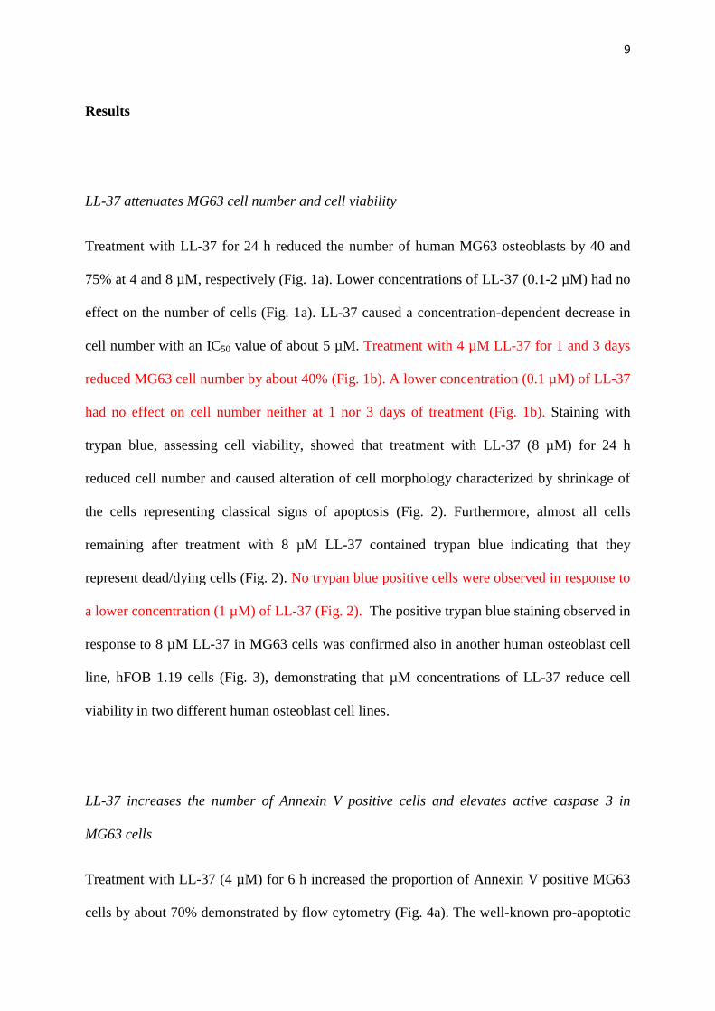
Results
LL-37 attenuates MG63 cell number and cell viability
Treatment with LL-37 for 24 h reduced the number of human MG63 osteoblasts by 40 and
75% at 4 and 8 µM, respectively (Fig. 1a). Lower concentrations of LL-37 (0.1-2 µM) had no
effect on the number of cells (Fig. 1a). LL-37 caused a concentration-dependent decrease in
cell number with an IC50 value of about 5 µM. Treatment with 4 µM LL-37 for 1 and 3 days
reduced MG63 cell number by about 40% (Fig. 1b). A lower concentration (0.1 µM) of LL-37
had no effect on cell number neither at 1 nor 3 days of treatment (Fig. 1b). Staining with
trypan blue, assessing cell viability, showed that treatment with LL-37 (8 µM) for 24 h
reduced cell number and caused alteration of cell morphology characterized by shrinkage of
the cells representing classical signs of apoptosis (Fig. 2). Furthermore, almost all cells
remaining after treatment with 8 µM LL-37 contained trypan blue indicating that they
represent dead/dying cells (Fig. 2). No trypan blue positive cells were observed in response to
a lower concentration (1 µM) of LL-37 (Fig. 2). The positive trypan blue staining observed in
response to 8 µM LL-37 in MG63 cells was confirmed also in another human osteoblast cell
line, hFOB 1.19 cells (Fig. 3), demonstrating that µM concentrations of LL-37 reduce cell
viability in two different human osteoblast cell lines.
LL-37 increases the number of Annexin V positive cells and elevates active caspase 3 in
MG63 cells
Treatment with LL-37 (4 µM) for 6 h increased the proportion of Annexin V positive MG63
cells by about 70% demonstrated by flow cytometry (Fig. 4a). The well-known pro-apoptotic

10
agent staurosporine (0.5 µM) was included as a positive control [29]. Treatment with 0.5 µM
staurosporine for 6 h increased the number of Annexin V positive cells by about 9 times (Fig.
4a). Staurosporine (0.5 µM) but not LL-37 (4 µM) increased the proportion of Annexin V
positive cells also at a shorter time-point, i.e. 2 h (Fig. 4a). Stimulation with LL-37 (4 µM) for
24 h increased the amount of active caspase 3 between 2 and 3 times in MG63 cells (Fig. 4b).
The positive control staurosporine (0.5 µM) increased active caspase 3 by about 2-fold (Fig.
4b). Taken together these data show that LL-37 is pro-apoptotic for osteoblasts.
LL-37 increases MG63 intracellular Ca2+
concentration
Defective cellular Ca2+
handling is thought to be involved in pro-apoptotic signaling [21], and
therefore we assessed the effects of LL-37 on the intracellular Ca2+
concentration. Treatment
with LL-37 (4 µM) caused an acute (within about 60 s) and sustained rise in intracellular Ca2+
concentration demonstrated by laser-scanning confocal microscopy of Fluo 4-AM labeled
MG63 cells incubated in Ca2+
containing (2.5 mM) HEPES-buffered salt solution (Figs. 5a
and b). A lower concentration of LL-37 (0.4 µM) also elevated Ca2+
but the Ca2+
response to
0.4 µM LL-37 was small compared to that of 4 µM (Fig. 5b). Addition of vehicle control
(0.1% DMSO) had no effect on Ca2+
(Fig. 5a). LL-37 (0.4 and 4 µM) had no effect on
intracellular Ca2+
concentration in Ca2+
-free solution (Fig. 5c), suggesting that the LL-37-
induced increase in Ca2+
depends on inflow of extracellular Ca2+
. Treatment with 100 nM and
1 µM of the L-type voltage-sensitive Ca2+
channel blocker nifedipine had no effect on the LL-
37-induced Ca2+
response (4 µM LL-37), suggesting that LL-37 acts via another mechanism
than by stimulation of Ca2+
inflow through L-type Ca2+
channels (Figs. 6 and 7a). The T-type
Ca2+
channel blocker NiCl2 (100 µM) had no effect on the LL-37-induced (4 µM) Ca2+
response (Fig. 7b), suggesting that inflow of Ca2+
by LL-37 is mediated through another

11
pathway than via T-type Ca2+
channels. Furthermore, inclusion of the P2X7 receptor
antagonist AZ 11645373 (10 µM) had no impact on the rise in Ca2+
evoked by 4 µM LL-37
(Fig. 7c). The thromboxane A2 analogue U46619 was included as positive control causing a
rapid and powerful rise in intracellular Ca2+
concentration (Fig. 7d).
LL-37 reduces MG63 cell number both in the presence and absence of extracellular Ca2+
Treatment with LL-37 (8 µM) for 24 h reduced the number of MG63 cells by about 55% for
cells cultured in DMEM culture medium containing 1.8 mM Ca2+
and by about 85% for cells
cultured in Ca2+
-free DMEM culture medium (Fig. 8a). In fact, the LL-37-induced reduction
of cell number was more powerful (P<0.01) in the absence than in the presence of
extracellular Ca2+
(Fig. 8a). A lower concentration of LL-37 (4 µM) reduced cell cumber by
about 15% in the presence (86 ± 2% for LL-37-treated cells vs. 100 ± 5% for control cells,
P<0.05, n=3 in each group) and by about 35% in the absence (66 ± 7% for LL-37-treated cells
vs. 100 ± 16% for control cells, P<0.05, n=3 in each group) of extracellular Ca2+
. Omitting
Ca2+
for 24 h reduced slightly, but not significantly, the number of cells as demonstrated when
cell-counts are plotted as absolute data (Fig. 8b). The MG63 cells showed similar morphology
in the presence and absence of Ca2+
(Fig. 8c). Treatment with LL-37 (4 µM) for 24 h
attenuated cell number by about 60% when the Ca2+
chelator agent EGTA (1.8 mM) was
included in the Ca2+
-containing DMEM culture medium in order to lower Ca2+
(Fig. 9). LL-37
(4 µM) reduced cell number by about 60% also when extra Ca2+
(final Ca2+
concentration 3.6
mM) was included in the EGTA containing medium (Fig. 9). For these experiments EGTA
was administered in an equimolar concentration to extracellular Ca2+
(1.8 mM) in order to
bind most of the Ca2+
ions. Thus, LL-37-induced lowering of osteoblast cell number is

12
observed both when the cells are cultured under Ca2+
-free conditions and when Ca2+
ions are
absorbed with EGTA.

13
Discussion
In the present study, we demonstrate that the human antimicrobial peptide LL-37 induces
apoptosis in human osteoblast-like MG63 cells and that this effect is associated with elevated
intracellular Ca2+
concentration. LL-37 causes, in the µM concentration range, an acute and
sustained rise in intracellular Ca2+
in Ca2+
-containing but not in Ca2+
-free solution showing
that the LL-37-induced increase in intracellular Ca2+
is due to an inflow of Ca2+
along its
gradient from the extracellular space to the cytosol. The increase in Ca2+
by LL-37 was much
stronger at 4 than at 0.4 µM, suggesting a concentration-dependent effect, although it is
difficult to conclude firmly the concentration-dependence since we have investigated only two
concentrations of LL-37. Membrane depolarization results in an inflow of Ca2+
from the
extracellular space causing a sustained and long-lasting increase in intracellular Ca2+
[30]
similar to that observed in response to LL-37 in the present study. The pattern of LL-37-
induced rise in intracellular Ca2+
concentration is thus compatible with inflow of Ca2+
from
the extracellular space. Osteoblasts (MC3T3-E1 cells and MG63 cells) have been reported to
express voltage-sensitive Ca2+
channels [31, 32]. Our data show that neither the L-type Ca2+
channel blocker nifedipine nor the T-type Ca2+
channel blocker NiCl2 have impact on the LL-
37 evoked rise in Ca2+
in MG63 cells, suggesting that the inflow of Ca2+
triggered by LL-37 is
through another mechanism than via voltage-sensitive L-type and T-type Ca2+
channels. We
used relevant concentrations of nifedipine (0.1 and 1 µM), which fully inhibit inflow of Ca2+
via L-type Ca2+
channels in cultured vascular smooth muscle cells [30], and NiCl2 (100 µM)
inhibiting T-type but not L-type Ca2+
channels [33]. Furthermore, inclusion of the selective
and highly potent P2X7 receptor antagonist AZ 11645373 [34] had no effect on the rise in
Ca2+
evoked by LL-37, suggesting an alternative mechanism. Based on our findings presented

14
here, we suggest that the LL-37-induced inflow of extracellular Ca2+
represents a novel
signaling pathway for LL-37 that may involve LL-37-induced permeabilization of the cell-
membrane causing formation of trans-membrane pores and/or that LL-37 acts as a detergent.
These data implicate that LL-37 may work through a similar mechanism in human osteoblasts
as in LL-37-induced permeabilization of the bacterial cell wall [3].
Here, we show for the first time that the antimicrobial peptide LL-37 reduces human MG63
osteoblast cell number by promoting apoptosis. LL-37-induced apoptosis was demonstrated
by both enhanced proportion of Annexin V positive cells and by elevated active caspase 3
level in response to LL-37-treatment. Furthermore, LL-37-treated cells showed altered
morphology such as cell shrinkage representing a classical sign of apoptosis. Previously, pro-
apoptotic effects of LL-37 have been demonstrated in vascular smooth muscle cells,
periodontal ligament cells, neutrophils, T cells and airway epithelial cells [10-14]. We
demonstrate reduction of osteoblast cell number by LL-37 in the µM concentration-range,
while no effect on cell number is observed at lower concentrations of LL-37. We have
previously shown that LL-37 reduces lipopolysaccharide-induced MCP-1 and IL-6 expression
at low concentrations (0.1 and 1 µM) and induces apoptosis only at high (>5 µM)
concentrations in human periodontal ligament cells [11]. LL-37-induced pro-apoptosis is thus
observed in different cell systems but only in the µM concentration-range. Very high levels of
LL-37 have been demonstrated in lesional tissue from patients suffering from autoimmune
diseases such as psoriasis, rosacea and ulcerative colitis [35-37]. For example, the median
concentration of LL-37 is 304 µM in psoriatic skin lesions [35]. The LL-37 levels are elevated
locally in chronic periodontitis, in fact they are well within the µM concentration-range [18],
suggesting that LL-37-induced apoptosis of osteoblasts may have impact on alveolar bone

15
homeostasis in patients suffering from this disease. Thus, we may conclude that the LL-37-
induced apoptosis of osteoblasts, observed in the µM concentration-range in the present study,
is relevant for the in-vivo situation considering the very high levels of LL-37 observed in
various autoimmune and inflammatory diseases.
Ca2+
governs many important cellular processes and is thought to be involved in apoptosis
[21, 38]. A rise in intracellular Ca2+
may originate from intracellular stores such as the
endoplasmic reticulum, but also from the extracellular space [21]. Here, we show that LL-37
elevates intracellular Ca2+
concentration in human MG63 osteoblasts through stimulation of
Ca2+
inflow. The LL-37 evoked inflow of Ca2+
seems not to be critically important for the LL-
37-induced attenuation of MG63 cell number, since LL-37 lowers cell number both in the
presence and absence of Ca2+
in the cell culture medium and furthermore, LL-37 causes a rise
in intracellular Ca2+
but no apoptosis at a low concentration (0.4 µM). Ca2+
-independent
apoptosis has been described in different experimental systems [39, 40]. Interestingly, the LL-
37 evoked inward flow of Ca2+
, occurring independent of LL-37-induced apoptosis, may
represent an important mechanism regulating Ca2+
-dependent cellular processes governed by
LL-37. Thus, we demonstrate here that LL-37 alters human osteoblast Ca2+
handling and
induces Ca2+
-independent apoptosis.
In summary, we show that LL-37 alters cellular Ca2+
homeostasis by causing stimulation of
Ca2+
inflow through a voltage-sensitive L- and T-type Ca2+
channel independent mechanism
in human osteoblasts. Furthermore, we demonstrate that LL-37 induces apoptosis of
osteoblasts via a mechanism that is independent of Ca2+
.

16
Acknowledgements
This study was supported by grants from the Swedish Research Council, the Faculty of
Odontology at Malmö University, the Medical Faculty at Lund University, the Swedish
Dental Society, the Patent Revenue Fund and the foundations of Crafoord, Magnus Bergvall,
Lars Hierta, Sven and Lilly Thuréus and Greta and Johan Kock.

17
References
1 Gudmundsson GH, Agerberth B, Odeberg J, Bergman T, Olsson B, Salcedo R: The human
gene FALL39 and processing of the cathelin precursor to the antibacterial peptide LL-37 in
granulocytes. Eur J Biochem 1996;238:325-332.
2 Sorensen OE, Follin P, Johnsen AH, Calafat J, Tjabringa GS, Hiemstra PS, Borregaard N:
Human cathelicidin, hCAP-18, is processed to the antimicrobial peptide LL-37 by
extracellular cleavage with proteinase 3. Blood 2001;97:3951-3959.
3 Burton MF, Steel PG: The chemistry and biology of LL-37. Nat Prod Rep 2009;26:1572-
1584.
4 Turner J, Cho Y, Dinh NN, Waring AJ, Lehrer RI: Activities of LL-37, a cathelin-
associated antimicrobial peptide of human neutrophils. Antimicrob Agents Chemother
1998;42:2206-2214.
5 Nizet V, Ohtake T, Lauth X, Trowbridge J, Rudisill J, Dorschner RA, Pestonjamasp V,
Piraino J, Huttner K, Gallo RL: Innate antimicrobial peptide protects the skin from invasive
bacterial infection. Nature 2001;414:454-457.

18
6 Larrick JW, Hirata M, Balint RF, Lee J, Zhong J, Wright SC. Human CAP18: a novel
antimicrobial lipopolysaccharide-binding protein. Infect Immun 1995;63:1291-1297.
7 Nijnik A, Pistolic J, Filewod NCJ, Hancock REW: Signaling pathways mediating
chemokine induction in keratinocytes by cathelicidin LL-37 and flagellin. J Innate Immun
2012;4:377-386.
8 Liu PT, Stenger S, Li H, Wenzel L, Tan BH, Krutzik SR, Ochoa MT, Schauber J, Wu K,
Meinken C, Kamen DL, Wagner M, Bals R, Steinmeyer A, Zügel U, Gallo RL, Eisenberg D,
Hewison M, Hollis BW, Adams JS, Bloom BR, Modlin RL: Toll-like receptor triggering of a
vitamin D-mediated human antimicrobial response. Science 2006;311:1770-1773.
9 Zhang Z, Shively JE: Generation of novel bone forming cells (monoosteophils) from the
cathelicidin-derived peptide LL-37 treated monocytes. PLOS ONE 2010;5:e13985.
10 Ciornei CD, Tapper H, Bjartell A, Sternby NH, Bodelsson M: Human antimicrobial
peptide LL-37 is present in atherosclerotic plaques and induces death of vascular smooth
muscle cells: a laboratory study. BMC Cardiovasc Disord 2006;6:49.
11 Jönsson D, Nilsson BO: The antimicrobial peptide LL-37 is anti-inflammatory and
proapoptotic in human periodontal ligament cells. J Periodont Res 2012;47:330-335.

19
12 Zhang Z, Cherryholmes G, Shively JE: Neutrophil secondary necrosis is induced by LL-37
derived from cathelicidin. J Leukoc Biol 2008;84:780-788.
13 Mader JS, Ewen C, Hancock RE, Bleackley RC: The human cathelicidin, LL-37, induces
granzyme-mediated apoptosis in regulatory T cells. J Immunother 2011;34:229-235.
14 Barlow PG, Beaumont PE, Cosseau C, Mackellar A, Wilkinson TS, Hancock RE, Haslett
C, Govan JR, Simpson AJ, Davidson DJ: The human cathelicidin LL-37 preferentially
promotes apoptosis of infected airway epithelium. Am J Respir Cell Mol Biol 2010;43:692-
702.
15 Chamorro CI, Weber G, Gronberg A, Pivarcsi A, Stahle M. The human antimicrobial
peptide LL-37 suppresses apoptosis in keratinocytes. J Invest Dermatol 2009;129:937-944.
16 Kim HJ, Cho DH, Lee KJ, Cho CS, Bang SI, Cho BK, Park HJ: LL-37 suppresses sodium
nitroprusside-induced apoptosis of systemic sclerosis dermal fibroblasts. Exp Dermatol
2011;20:843-845.

20
17 Nagaoka I, Tamura H, Hirata M: An antimicrobial cathelicidin peptide, human CAP18/LL-
37, suppresses neutrophil apoptosis via the activation of formyl-peptide receptor-like 1 and
P2X7. J Immunol 2006;176:3044-3052.
18 Turkoglu O, Emingil G, Kutukculer N, Atilla G: Gingival crevicular fluid levels of
cathelicidin LL-37 and interleukin-18 in patients with chronic periodontitis. J Periodontol
2009;80:969-976.
19 Lerner UH: Inflammation-induced bone remodeling in periodontal disease and the
influence of post-menopausal osteoporosis. J Dent Res 2006;85:596-607.
20 Orrenius S, Nicotera P: The calcium ion and cell death. J Neural Transm Suppl 1994;43:1-
11.
21 Berridge MJ, Lipp P, Bootman MD: The versatility and universality of calcium signaling.
Nat Rev Cell Biol 2000;1:11-21.
22 Elssner A, Duncan M, Gavrilin M, Wewers MD: A novel P2X7 receptor activator, the
human cathelicidin-derived peptide LL37, induces IL-1 beta processing and release. J
Immunol 2004;172:4987-4994.

21
23 Tjabringa GS, Aarbiou J, Ninaber DK, Drijfhout JW, Sorensen OE, Borregaard N, Rabe
KF, Hiemstra PS: The antimicrobial peptide LL-37 activates innate immunity at the airway
epithelial surface by transactivation of the epidermal growth factor receptor. J Immunol
2003;171:6690-6696.
24 von Haussen J, Koczulla R, Shaykhiev R, Herr C, Pinkenburg O, Reimer D, Wiewrodt R,
Biesterfeld S, Aigner A, Czubayko F, Bals R: The host defence peptide LL-37/hCAP-18 is a
growth factor for lung cancer cells. Lung Cancer 2008;59:12-23.
25 Coffelt SB, Waterman RS, Florez L, Bentrup KH, Zwezdaryk KJ, Tomchuck SL, LaMarca
HL, Danka ES, Morris CA, Scandurro AB: Ovarian cancers overexpress the antimicrobial
protein hCAP-18 and its derivative LL-37 increases ovarian cancer cell proliferation and
invasion. Int J Cancer 2008;122:1030-1039.
26 Nygren PA: Alternative binding proteins: affibody binding proteins developed from a
small three-helix bundle scaffold. FEBS J 2008;275:2668-2676.
27 Kraus D, Deschner J, Jäger A, Wenghoefer M, Bayer S, Jepsen S, Allam JP, Novak N,
Meyer R, Winter J: Human β-defensins differently affect proliferation, differentiation, and
mineralization of osteoblast-like MG63 cells. J Cell Physiol 2012;227:994-1003.
22
28 Kawano M, Ariyoshi W, Iwanaga K, Okinaga T, Habu M, Yoshioka I, Tominaga K,
Nishihara T: Mechanism involved in enhancement of osteoblast differentiation by hyaluronic
acid. Biochem Biophys Res Commun 2011;405:575-580.
29 Dezaki K, Maeno E, Sato K, Akita T, Okada Y: Early-phase occurrence of K+ and Cl-
efflux in addition to Ca 2+ mobilization is a prerequisite to apoptosis in HeLa cells. Apoptosis
2012;17:821-831.
30 Byron KL, Taylor CW: Spontaneous Ca2+
spiking in a vascular smooth muscle cell line is
independent of the release of intracellular Ca2+
stores. J Biol Chem 1993;268:6945-6952.
31 Bergh JJ, Shao Y, Puente E, Duncan RL, Farach-Carson MC: Osteoblast Ca2+
permeability
and voltage-sensitive Ca2+
channel expression is temporally regulated by 1,25-
dihydroxyvitamin D3. Am J Physiol Cell Physiol 2006;290:C822-C831.
32 Burns DM, Stehno-Bittel L, Kawase T: Calcitonin gene related peptide elevates calcium
and polarizes membrane potential in MG-63 cells by both cAMP-independent and -dependent
mechanisms. Am J Physiol Cell Physiol 2004;287:C457-C467.

23
33 Bhattacharjee A, Whitehurst RM, Zhang M, Wang L, Li M: T-type calcium channels
facilitate insulin secretion by enhancing general excitability in the insulin-secreting β-cell
line, INS-1. Endocrinology 1997;138:3735-3740.
34 Michel AD, Ng SW, Roman S, Clay WC, Dean DK, Walter DS: Mechanism of action of
species-selective P2X7 receptor antagonists. Br J Pharmacol 2009;156:1312-1325.
35 Ong PY, Ohtake T, Brandt C, Strickland I, Boguniewicz M, Ganz T, Gallo RL, Leung DY:
Endogenous antimicrobial peptides and skin infections in atopic dermatitis. N Engl J Med
2002;347:1151-1160.
36 Yamasaki K, Di Nardo A, Bardan A, Murakami M, Ohtake T, Coda A, Dorschner RA,
Bonnart C, Descargues P, Hovnanian A, Morhenn VB, Gallo RL: Increased serine protease
activity and cathelicidin promotes skin inflammation in rosacea. Nat Med 2007;13:975-980.
37 Schauber J, Rieger D, Weiler F, Wehkamp J, Eck M, Fellermann K, Scheppach W, Gallo
RL, Stange EF: Heterogeneous expression of human cathelicidin hCAP18/LL-37 in
inflammatory bowel diseases. Eur J Gastroenterol Hepatol 2006;18:615-621.

24
38 Giorgi C, Baldassari F, Bononi A, Bonora M, De Marchi E, Marchi S, Missiroli S,
Patergnani S, Rimessi A, Suski JM, Wieckowski MR, Pinton P: Mitochondrial Ca2+
and
apoptosis. Cell Calcium 2012;52:36-43.
39 Shabbir M, Ryten M, Thompson C, Mikhailidis D, Burnstock G: Characterization of
calcium-independent purinergic receptor-mediated apoptosis in hormone-refractory prostate
cancer. BJU Int 2008;101:352-359.
40 Liu SI, Huang CC, Huang CJ, Wang BW, Chang PM, Fang YC, Chen WC, Wang JL, Lu
YC, Chu ST, Chou CT, Jan CR: Thimerosal-induced apoptosis in human SCM1 gastric
cancer cells: activation of p38 MAP kinase and caspase-3 pathways without involvement of
[Ca2+]i elevation. Toxicol Sci 2007;100:109-117.

25
Figure legends
Fig. 1. (a) Treatment with LL-37 for 24 h reduces MG63 osteoblast cell number in a
concentration-dependent manner. LL-37 at 4 and 8 µM reduces osteoblast cell number by 40
and 75%, respectively, while lower concentrations of LL-37 lack effect. (b) Treatment with 4
µM LL-37 for 1 and 3 days reduces MG63 cell number by about 40%. A lower concentration
(0.1 µM) of LL-37 has no effect on cell number neither at 1 nor 3 days of treatment.
Values are means ± SEM of 4-15 observations in each group. *, ** and *** represent P<0.05,
P<0.01 and P<0.001, respectively, compared to controls (ctrl).
Fig. 2. Staining with trypan blue, assessing cell viability, shows that treatment with 8 µM LL-
37 for 24 h reduces MG63 cell number and causes alteration of cell morphology characterized
by shrinkage of the cells. Furthermore, nearly all LL-37-treated cells contain trypan blue
indicating that these cells represent dead/dying cells. No trypan blue positive cells were
observed in response to a lower concentration (1 µM) of LL-37. Control cells (ctrl) show
normal morphology with no trypan blue positive cells. Bars represent 25 µm.
Fig. 3. Treatment with LL-37 (8 µM) for 24 h causes accumulation of trypan blue in primary
human osteoblast hFOB 1.19 cells. Control cells (ctrl) show no trypan blue positive cells.
Trypan blue positive cells represent dead/dying cells. Bars represent 20 µm.

26
Fig. 4. (a) Treatment with 4 µM LL-37 for 6 h increases the proportion of apoptotic MG63
cells. Apoptosis was determined by flow cytometric analysis of Annexin V positive cells.
Staurosporine (0.5 µM) was included as positive control. (b) Treatment with 4 µM LL-37 for
24 h increases MG63 cellular active caspase 3 (aCasp-3) level 2 to 3 times. The amount of
aCasp-3 was determined by ELISA and normalized to total protein in each sample. Values are
means ± SEM of 3-4 observations in each group. *, ** and *** represent P<0.05, P<0.01 and
P<0.001, respectively, compared to controls (ctrl). N.S. = not significant.
Fig. 5. (a) Treatment with LL-37 (4 µM) causes an acute and sustained rise in intracellular
Ca2+
concentration assessed by laser-scanning confocal microscopy of Fluo 4-AM loaded
MG63 cells incubated in Ca2+
containing (2.5 mM) HEPES-buffered salt solution. No
treatment (left panel) represents the Ca2+
signal in the presence of vehicle control (0.1%
DMSO). Addition of 0.1% DMSO has no effect on Ca2+
. The Ca2+
indicator Fluo 4-AM
fluorescence is shown in red. (b) Line trace showing that both 0.4 and 4 µM LL-37 elevates
Ca2+
. (c) LL-37 (0.4 and 4 µM) has no effect on intracellular Ca2+
concentration in Ca2+
-free
solution. Ca2+
-free conditions were achieved by removing CaCl2 from the HEPES-buffered
salt solution and by inclusion of 2 mM EGTA. Each experiment was repeated at least twice.
Fig. 6. LL-37 (4 µM) increases MG63 intracellular Ca2+
concentration monitored by laser-
scanning confocal microscopy of Fluo 4-AM loaded cells in the presence of L-type Ca2+
channel blocker nifedipine (100 nM). Nifedipine was included at the arrow and present
throughout the experiment. This trace shows one representative experiment out of two.

27
Fig. 7. Treatment with (a) 1 µM nifedipine, (b) 100 µM NiCl2 and (c) 10 µM AZ11645373
has no effect on the LL-37-induced (4 µM) Ca2+
response in MG63 cells. (d) The
thromboxane A2 analogue U46619 (10 µM) was included as positive control causing a rapid
and powerful rise in intracellular Ca2+
concentration. The intracellular Ca2+
concentration was
monitored by laser-scanning confocal microscopy of Fluo 4-AM loaded MG63 cells.
Nifedipine, NiCl2 and AZ11645373 were included at the arrow and present throughout the
experiment. Each experiment was repeated at least twice.
Fig. 8. (a) Treatment with 8 µM LL-37 for 24 h reduces MG63 cell number by about 55% for
cells cultured in DMEM culture medium containing 1.8 mM Ca2+
and by 85% for cells
cultured in Ca2+
-free DMEM culture medium. (b) Omitting Ca2+
for 24 h reduces slightly, but
not significantly, the number of cells as demonstrated when cell-count data are plotted as
absolute data. (c) The MG63 cells show similar morphology in the presence and absence of
Ca2+
. Bars in panel c represent 20 µm. Values are means ± SEM of 4-5 observations in each
group. * and *** represent P<0.05 and P<0.001 compared to controls (ctrl) and ** represents
P<0.01 for LL-37-treated groups as indicated.
Fig. 9. Treatment with 4 µM LL-37 for 24 h reduces cell number by about 60% for cells
grown in culture medium (DMEM + 1.8 mM Ca2+
) containing either 1.8 mM EGTA alone or
1.8 mM EGTA in combination with Ca2+
in excess (3.6 mM Ca2+
). Values are means ± SEM
of 3 observations in each group. ** represents P<0.01 compared to controls (ctrl).

28
Fig. 1

29
Fig. 2

30
Fig. 3

31
Fig. 4

32
Fig. 5

33
Fig. 6

34
Fig. 7

35
Fig. 8

36
Fig. 9