Embed Size (px)
Citation preview

RESEARCH ARTICLE Open Access
The anti-inflammatory effect ofAndrographis paniculata (Burm. f.) Nees onpelvic inflammatory disease in rats throughdown-regulation of the NF-κB pathwayWei Zou1,2†, Zuoqi Xiao1†, Xiaoke Wen1*, Jieying Luo3, Shuqiong Chen1, Zeneng Cheng2, Daxiong Xiang4,Jian Hu1 and Jingyu He5*
Abstract
Background: Andrographis paniculata (Burm. f.) Nees (APN), a principal constituent of a famous traditional Chinesemedicine Fukeqianjin tablet which is used for the treatment of pelvic inflammatory disease (PID), has been reportedto have anti-inflammatory effect in vitro. However, whether it has pharmacological effect on PID in vivo is unclear.Therefore, the aim of this study is to test the anti-inflammatory effect of APN and illuminate a potential mechanism.
Methods: Thirty-six female specific pathogen-free SD rats were randomly divided into control group, PID group,APN1 group, APN2 group, APN3 group and prednisone group. Pathogen-induced PID rats were constructed. TheAPN1, APN2 and APN3 group rats were orally administrated with APN extract at different levels. The prednisonegroup rats were administrated with prednisone. Eight days after the first infection, the histological examination ofupper genital tract was carried out, and enzyme-linked immunosorbent assay (ELISA) was carried out using homogenateof the uterus and fallopian tube. Furthermore, immunohistochemical evaluations of NF-κB p65 and IκB-α in uterus wasconducted.
Results: APN obviously suppressed the infiltrations of neutrophils and lymphocytes, and it could significantly reduce theexcessive production of cytokines and chemokines including IL-1β, IL-6, CXCL-1, MCP-1 and RANTES in a dose-dependentmanner. Furthermore, APN could block the pathogen-induced activation of NF-κB pathway.Conclusion: APN showed potent anti-inflammatory effect on pathogen-induced PID in rats, with a potential mechanismof inhibiting the NF-κB signal pathway.
Keywords: Andrographis paniculata (Burm. f.) Nees, Anti-inflammatory, Pelvic inflammatory disease, NF-κB, Rats
BackgroundPelvic inflammatory disease (PID) is a common gynecologicaldisease that usually causes ectopic pregnancy, tubalfactor infertility and chronic pelvic pain, which hasbeen deemed as a great threat for life quality of woman.It includes endometritis, salpingitis, peritonitis, etc., whose
etiopathogenisis is the infection of pathogenic microorgan-isms in upper genital tract [1]. Infiltrations of neutrophilsand lymphocytes in the upper genital tract could beemployed as a criteria to diagnose PID in clinic, whichcould be observed on hematoxylin and eosin (H & E)-stained biopsy sections [2]. With the inflammatory cellrecruitment, amount of proinflammatory cytokines,such as interleukin (IL)-1β, IL-6, etc., are released ex-cessively in local tissue and play an important role inthe pathogenesis of PID [3]. The important reason forthe excessive production and release of proinflamma-tory cytokines is the activation of nuclear factor-kappa
* Correspondence: [email protected]; [email protected]†Equal contributors1Key Laboratory of Hunan Province for Traditional Chinese Medicine inObstetrics and Gynecology Research, Hunan Provincial Maternal and ChildHealth Care Hospital, No. 53 XiangChun Road, Changsha 410008, China5Guangzhou Institute of Advanced Technology, Chinese Academy ofSciences, No. 1121 Haibin Road, Guangzhou 511458, Guangdong, ChinaFull list of author information is available at the end of the article
© The Author(s). 2016 Open Access This article is distributed under the terms of the Creative Commons Attribution 4.0International License (http://creativecommons.org/licenses/by/4.0/), which permits unrestricted use, distribution, andreproduction in any medium, provided you give appropriate credit to the original author(s) and the source, provide a link tothe Creative Commons license, and indicate if changes were made. The Creative Commons Public Domain Dedication waiver(http://creativecommons.org/publicdomain/zero/1.0/) applies to the data made available in this article, unless otherwise stated.
Zou et al. BMC Complementary and Alternative Medicine (2016) 16:483 DOI 10.1186/s12906-016-1466-5

B (NF-κB) signaling pathway when pathogens are rec-ognized by their receptors [4].According to the guidelines from the Center for Disease
Control and Prevention (CDC) in US, antibiotics is the firstchoice for the treatment of PID [5], but the bacterial drugresistance and drug side effects are shortages for clinicaluse of antibiotics. Therefore, new complementary medi-cines used for PID are needed for further improvement inclinical outcomes. Andrographis paniculata (Burm. f.) Nees(APN) is a famous traditional medicine, widely used to treatsore throat, flu, and upper respiratory tract infections inmany Asian countries [6]. Phytochemical studies on APNhave found the principal bioactive compound andro-grapholide [7] and many other constituents, includingditerpenoids, flavonoids, quinic acids, xanthones [8, 9], andnoriridoids [10, 11]. Based on the abundant bioactiveconstituents, APN showed many salutary effects, suchas anticancer [12], hepatoprotective [13], antiviral [14, 15],antipyretic and analgesic [16] effects, etc. Interestingly,the immunomodulatory [17], antioxidant [18], anti-inflammatory [19] and antimicrobial [20, 21] activitiesof APN were also reported, which may suggest theclinical use of APN in treatment for PID. Besides, APN isthe principal constituent in a famous traditional Chinesemedicine Fukeqianjin tablet which is used to treat PID.However, whether APN has pharmacological effect onPID has not been eliminated yet. In this study, we test theanti-inflammatory effect of APN on PID rats, and illus-trate a potential mechanism of this activity.
MethodsReagents and materialsPentobarbital was from Xiya Reagent (Chengdu, China).Progesterone injection was obtained from Xianju Pharma(Taizhou, China). Distilled water was used in all of thisexperiment. Absorbable gelatin sponge was from JinlingPharmaceutical Co. (Nanjing, China). The dried APN waspurchased from Tianxiang Co. (Yueyang, China), whichwas identified by Prof. Zhuxin Wang (Hunan university ofChinese medicine, Changsha, China), with the total amountof andrographolide and dehydroandrographolide about 1%.A voucher specimen (No. CXL20150610) is depositedin the Key Laboratory of Hunan Province for TraditionalChinese Medicine in Obstetrics & Gynecology Research(Changsha, China). Prednisone acetate tablets were ob-tained from Guangdong Huanan Pharma (Guangdong,China). According to the previous method [22], a 1000 gof dried APN was extracted with 80% ethanol at roomtemperature and filtrated. The solution was freeze-driedto yield 62.5 g of the APN extract.
Rat PID model construction and sample collectionThe animal experimental procedure was approved bythe Animal Care and Use Committee of Central South
University. Thirty-six female specific pathogen-free SDrats, 9-week aged and weighing 220–240 g, were ran-domly divided into 6 groups, including control group,PID group, APN1 group, APN2 group, APN3 group andprednisone group. Rats were acclimated for 7 days andthen injected subcutaneously with 10 mg progesterone.One week later, the PID model construction was carriedout referring to our previous method with some revisions[23]. Absorbable gelatin sponge, a volume of 0.125 ml, wasimmersed in microbe-mixing solution with U. urealyticum(t-strain mycoplasma) concentration of 1 × 10⁸ ccu/ml andpathogenic E. coli concentration of 1 × 10⁸ cfu/ml. Eachupper genital tract of all rats except control group rats wasinserted with a microbe-containing gelatin sponge, andthen the rat was forced to be down for 3 min. Themicrobe-free gelatin sponges were implanted into the cer-vixes of control group rats. Four times infections were con-ducted with a 2-day interval. From the first infection, theAPN1, APN2 and APN3 group rats were orally adminis-trated with APN extract at a dose of 167, 334 (clinical dose)and 668 mg/kg/day, respectively. The prednisone grouprats were administrated with prednisone at a dose of1.7 mg/kg. Eight days after the first infection, rats wereinjected subcutaneously with pentobarbital at a dose of30 mg/kg. The right uterus and fallopian tube were col-lected and restored at −80 °C, and the left uterus and fallo-pian tube was immersed in neutral-buffered formalin(10%). At last, rats were sacrificed by cervical dislocation.
Histological evaluationAfter paraffin embedding, the left uterus and fallopiantube was cut into 2 μm sections, followed by staining withH & E. The semi-quantification was carried out accordingto the previous method [24]. Three parts of each slide(tissue) were checked under microscopy (×100) by ablinded observer. The inflammation of each uterus andfallopian tube was semi-scored by evaluation on the extentof inflammatory cells infiltration (graded from 0 to 3).
Enzyme-linked immunosorbent assay (ELISA)Each right uterus and fallopian tube was weighted,immersed in physiologic saline at the ratio of 5:1 (v/w), andhomogenized. The amounts of IL-1β, IL-6, CXCL-1, MCP-1 and RANTES in homogenate were determined by usingELISA kits (Neobioscience, Beijing, China). The totalprotein in tissue homogenate was measured with bicinch-onininc acid (BCA) protein assay kit (Beyotime, Shanghai,China), and concentrations of these cytokines and chemo-kines were expressed as μg/g protein of homogenate.
Immunohistochemical evaluationThe paraffin embedded uterus was cut in to 2 μm sections,and then the paraffin was removed through xylene. Theslide was boiled in 10 mM sodium citrate (pH 6.0) for
Zou et al. BMC Complementary and Alternative Medicine (2016) 16:483 Page 2 of 7

30 min for antigen retrieval. Non-specific binding sites wereblocked by bovine serum. The specimens were incubatedwith NF-κB p65 or IκB-α primary antibody (Abclonal,Maryland, US) at 37 °C followed by secondary antibodiesincubation. After that, the slices were treated with diamino-benzidine (DAB) reagent for color development, followedby hematoxylin counterstaining. For negative control,phosphate-buffered saline (PBS) was used instead of pri-mary antibodies. The slices were observed under lightmicroscopy at a magnification of × 400. Brown stainingin uterus was considered as NF-κB p65 or IκB-α. Thenuclear with translocation of NF-κB p65 was consid-ered as positive. The semi-quantification of IκB-α wasconducted according to a previous method with somerevisions [25]. The integrated optical density (IOD)values of each slide (tissue) was obtained from its threefields with the software Image-Pro Plus 6.0. IOD indexof each sample was calculated as its IOD value dividedby the average IOD value of control group.
Statistic analysisAll data are expressed as means ± standard deviation (SD).One-way analysis of variance (ANOVA) followed byDunnett’s t test was employed for statistical analysis.Statistical analysis was conducted by using SPSS 16.0.Difference was considered as statistically significantwhen p < 0.05.
ResultsAPN attenuated the infiltration of inflammatory cellsThe results of histological evaluation are showed inFig. 1. Mass inflammatory cells, including neutrophilsand lymphocytes, infiltrated into the uterus and fallo-pian tube of the PID group rat, indicating the occur-rence of inflammation in upper genital tract. Afteradministration of APN, the infected rats showed de-creased infiltrations of neutrophils and lymphocytes,compared to PID group rats. Therefore, APN couldsuppress the infiltration of inflammatory cells in theupper genital tract of PID rats.
APN reduced the excessive production of cytokines andchemokinesTo observe the inflammatory response and investigatethe reason of inflammatory cells infiltration in the uterusand fallopian tube, the IL-1β, IL-6, CXCL-1, MCP-1, andRANTES in tissue homogenate were determined byusing ELISA kits. As showed in Fig. 2 all these cytokinesor chemokines were excessively produced in PID groupcompared to control group, and APN significantly re-duced this pathogens-induced excessive production in adose-dependent manner.
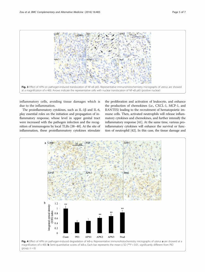
APN exerted its anti-inflammatory effect via down-regulatingthe NF-κB pathwayTo illustrate a potential mechanism of anti-inflammatoryeffect of APN, NF-κB p65 and IκB-α, two important sig-naling molecules in NF-κB signaling pathway, were deter-mined by immunohistochemical method (presented inFigs. 3 and 4, respectively). In PID group, NF-κB p65 wastranslocated to cell nucleus, and IκB-α showed a lowercontent than that in the other groups. In control, APNand prednisone groups, more NF-κB p65 was distributedin cytoplasm. These results indicated that the NF-κB path-way was up-regulated in PID group, and that APN couldsuppress the up-regulation of signaling pathway when theupper genital tract was infected with pathogen.
DiscussionIn clinic, the common pathogens, whose infections inupper genital tract can lead to PID, include Chlamydiatrachomatis, Neisseria gonorrhoeae, genital mycoplasmas,some gram-negative and gram-positive bacteria, etc.[26–29]. Each pathogen could be recognized by one ormore members of Toll-like receptor (TLR) family andthen initiate inflammation. TLR2 and TLR4 are two im-portant members of TLR family in upper genital tract[30], and U. urealyticum and E. coli can be recognizedby TLR2 and TLR4, respectively [31, 32]. Therefore, weattempted to use U. urealyticum and E. coli mixed solu-tion to provoke an augmented inflammation in uppergenital tract.Neutrophils in blood circulation could firmly adhere
to the endothelium cell barrier, cross it, and are recruitedinto sites of inflammation in different pathogen-infectedtissues, which is the first line of innate immune defenseagainst pathogens [33]. Recently, study also showed theinfiltration of neutrophils into the endometrium of micewith lipopolysaccharides (LPS)-induced endometritis [34].When mass neutrophils reach the tissue at the site ofinfection, they will release abundant inflammatory fac-tors, oxygen free radical and proteolytic enzyme to killpathogens. However, the excessive products will alsocause tissue damage and lead to structural disease inupper genital tract. Infiltration of lymphocyte in genitaltract of mice were observed after chronic pathogengenital infection, and this infiltration play a significantrole in controlling the infection [35]. Additionally, re-searchers found more T lymphocyte and fewer plasmacells throughout the stroma and within the epithelium[36]. Unfortunately, Patton et al. reported that the tis-sue damage including epithelial cell degeneration oc-curred close approximation to lymphocytes [37]. In thepresent study, after chronic pathogen infections, largenumber of neutrophils and lymphocytes infiltrated intothe epithelium of upper genital tract, and APN showeda good activity in attenuating the infiltration of these
Zou et al. BMC Complementary and Alternative Medicine (2016) 16:483 Page 3 of 7
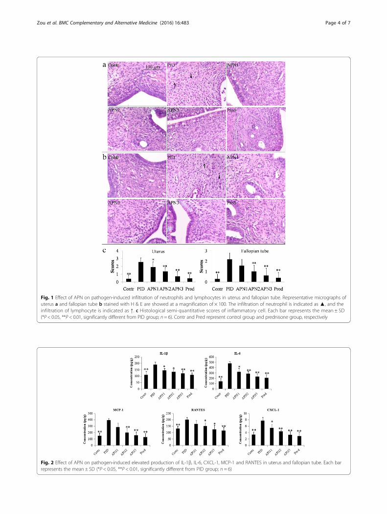
Fig. 2 Effect of APN on pathogen-induced elevated production of IL-1β, IL-6, CXCL-1, MCP-1 and RANTES in uterus and fallopian tube. Each barrepresents the mean ± SD (*P < 0.05, **P < 0.01, significantly different from PID group; n = 6)
Fig. 1 Effect of APN on pathogen-induced infiltration of neutrophils and lymphocytes in uterus and fallopian tube. Representative micrographs ofuterus a and fallopian tube b stained with H & E are showed at a magnification of × 100. The infiltration of neutrophil is indicated as ▲, and theinfiltration of lymphocyte is indicated as ↑. c Histological semi-quantitative scores of inflammatory cell. Each bar represents the mean ± SD(*P < 0.05, **P < 0.01, significantly different from PID group; n = 6). Contr and Pred represent control group and prednisone group, respectively
Zou et al. BMC Complementary and Alternative Medicine (2016) 16:483 Page 4 of 7
inflammatory cells, avoiding tissue damages which isdue to the inflammation.The proinflammatory cytokines, such as IL-1β and IL-6,
play essential roles on the initiation and propagation of in-flammatory response, whose level in upper genital tractwere increased with the pathogen infection and the recog-nition of immunogens by local TLRs [38–40]. At the site ofinflammation, these proinflammatory cytokines stimulate
the proliferation and activation of leukocyte, and enhancethe production of chemokines (i.e., CXCL-1, MCP-1, andRANTES) leading to the recruitment of hematopoietic im-mune cells. Then, activated neutrophils will release inflam-matory cytokines and chemokines, and further intensify theinflammatory response [41]. At the same time, various pro-inflammatory cytokines will enhance the survival or func-tion of neutrophil [42]. In this case, the tissue damage and
Fig. 3 Effect of APN on pathogen-induced translocation of NF-κB p65. Representative immunohistochemistry micrographs of uterus are showedat a magnification of × 400. Arrows indicate the representative cells with nuclear translocation of NF-κB p65 (positive nuclear)
Fig. 4 Effect of APN on pathogen-induced degradation of IκB-α. Representative immunohistochemistry micrographs of uterus a are showed at amagnification of × 400. b Semi-quantitative scores of IκB-α. Each bar represents the mean ± SD (**P < 0.01, significantly different from PIDgroup; n = 6)
Zou et al. BMC Complementary and Alternative Medicine (2016) 16:483 Page 5 of 7

structural disease in upper genital tract may occur due tothe intensified inflammatory response. In the present study,elevated productions of cytokines and chemokines in-cluding IL-1β, IL-6, CXCL-1, MCP-1 and RANTESwere observed in upper genital tract of PID rats, indi-cating an obvious local inflammatory response. Besides,APN could significantly lower the levels of these cytokinesand chemokines in a dose-dependent manner, suggestinga potent anti-inflammatory effect of APN for PID. Asandrographolide is the major bioactive substance and hasa critical effect on inhibiting the release of an importantproinflammatory factor TNF-α in APN [43], it may alsocontribute importantly to suppressing the inflammatoryresponse in this study.NF-κB is a pivotal factor in promoting the transcription
of genes involved in inflammatory and immune responses[44]. In most resting cells, NF-κB family members arecovalently bound to IκB family members and located inthe cytoplasm with no activity [45]. When the TLRs onsurface of these cells recognize pathogens, resulting in thephosphorylation and degradation of IκB members, theNF-κB members will translocate to the nucleus and bindto the cis-acting NF-κB enhancer element of genes, pro-moting the expression of inflammatory mediators, such asIL-1β, IL-6, etc. [31]. These produced proinflammatorycytokines act as a positive autocrine feedback so as to thefurther activation of NF-κB, and subsequently more proin-flammatory mediators are produced [46]. NF-κB p65 andIκB-α are representative members of NF-κB and IκBfamily in uterus and fallopian tube, respectively [47], andtherefore were chosen as indexes to test whether theNF-κB pathway was activated in the present study. Ourresults demonstrated the activation of NF-κB signal path-way after multi-infection of pathogens in uterus. In the ratPID model, APN exerted its anti-inflammatory with apotential mechanism of blocking the activation of NF-κBpathway. Previous studies reporting that both of APN andits main substance andrographolide showed the effect oninhibiting NF-κB pathway in vitro support this result[48, 49]. On the other hand, andrographolide also showedthe other anti-inflammatory effects, such as inhibitingJAK/STAT signaling [49], inhibiting p38 MAPK pathway[50], suppressing TLRs family expressions [49], etc., andwhether APN have the same or more effects in uppergenital tract should be verified in the further studies tofacilitate its clinical use.
ConclusionsIn this study, oral administration of APN showed significantanti-inflammatory activity in pathogen-induced PID rats,including suppressing the infiltration of neutrophils andlymphocytes and reducing excessive production of cyto-kines or chemokines. A potential mechanism of this effectwas involved in inhibiting the activation of NF-κB pathway.
AbbreviationsAPN: Andrographis paniculata (Burm. f.) Nees; BCA: Bicinchonininc acid;CDC: Center for disease control and prevention; DAB: Diaminobenzidine;ELISA: Enzyme-linked immunosorbent assay; H & E: Hematoxylin and eosin;IL: Interleukin; IOD: Integrated optical density; LPS: Lipopolysaccharides; NF-κB: Nuclear factor-kappa B; PBS: Phosphate-buffered saline; PID: Pelvicinflammatory disease; SD: Standard deviation; TLR: Toll-like receptor
AcknowledgementsWe sincerely thank Dr. Mimi Tang, Miss Yi Zheng and Miss Hui Li for assistancesin animal experiment, and thank Prof. Zhuxin Wang for botanical identification.
FundingThis research was supported by National Natural Science Foundation(No.81501218), China Postdoctoral Science Foundation (No. 2015 M582330),and Science and Technology Project of Hunan Province (No. 2015RS4054).
Availability of data and materialsData are included in paper.
Authors’ contributionsJL, SC and XW carried out the design of the study. WZ, ZX, XW and JHcarried out the experiments. ZC and DX performed the statistical analysis.XW and WZ wrote the manuscript and made revisions. All authors read andapproved the final manuscript.
Competing interestsThe authors declare that they have no conflict of interest.
Consent for publicationNot applicable.
Ethics approval and consent to participateThe animal experimental procedure was approved by the Animal Care andUse Committee of Central South University.
Author details1Key Laboratory of Hunan Province for Traditional Chinese Medicine inObstetrics and Gynecology Research, Hunan Provincial Maternal and ChildHealth Care Hospital, No. 53 XiangChun Road, Changsha 410008, China.2School of Pharmaceutical Sciences, Central South University, Changsha410011, China. 3College of Pharmacy, Hunan University of Chinese Medicine,Changsha 410007, China. 4Clinic Pharmacy Research Laboratory, the SecondXiangya Hospital of Central South University, Changsha 410011, China.5Guangzhou Institute of Advanced Technology, Chinese Academy ofSciences, No. 1121 Haibin Road, Guangzhou 511458, Guangdong, China.
Received: 30 March 2016 Accepted: 17 November 2016
References1. Soper DE. Pelvic inflammatory disease. Obstet gynecol. 2010;116(2 Pt 1):
419–28.2. Vicetti Miguel RD, Chivukula M, Krishnamurti U, Amortegui AJ, Kant JA,
Sweet RL, Wiesenfeld HC, Phillips JM, Cherpes TL. Limitations of the criteriaused to diagnose histologic endometritis in epidemiologic pelvicinflammatory disease research. Pathol res pract. 2011;207(11):680–5.
3. Lee SA, Tsai HT, Ou HC, Han CP, Tee YT, Chen YC, Wu MT, Chou MC, WangPH, Yang SF. Plasma interleukin-1beta, −6, −8 and tumor necrosis factor-alpha as highly informative markers of pelvic inflammatory disease. Clinchem lab med. 2008;46(7):997–1003.
4. Epstein FH, Barnes PJ, Karin M. Nuclear factor-κB-a pivotal transcriptionfactor in chronic inflammatory diseases. N engl j med. 1997;336(15):1066–71.
5. Control CFD. Prevention: sexually transmitted diseases treatment guidelines,2010. Ann emerg med. 2011;58(1):67–8.
6. Jayakumar T, Hsieh CY, Lee JJ, Sheu JR. Experimental and clinical pharmacologyof andrographis paniculata and its major bioactive phytoconstituentandrographolide. Evid based complement alternat med. 2013;2013:846740.
7. SHARMA M, SHARMA R. Identification, purification and quantification ofandrographolide from andrographis paniculata (burm. F.) nees by HPTLC atdifferent stages of life cycle of crop. J curr chem pharm sci. 2013;3(1):23–32.
Zou et al. BMC Complementary and Alternative Medicine (2016) 16:483 Page 6 of 7

8. Subramanian R, Asmawi MZ, Sadikun A. A bitter plant with a sweet future?a comprehensive review of an oriental medicinal plant: andrographispaniculata. Phytochem rev. 2012;11(1):39–75.
9. Zhou K-L, Chen L-X, Zhuang Y-L, Wang N-L, Yao X-S, Qiu F. Two new ent-labdane diterpenoid glycosides from the aerial parts of andrographispaniculata. J asian nat prod res. 2008;10(10):939–43.
10. Sareer O, Ahad A, Umar S. Prophylactic and lenitive effects of Andrographispaniculata against common human ailments: an exhaustive andcomprehensive reappraisal. J Pharm Res Opin. 2014;2(10):138–62.
11. Xu C, Chou G-X, Wang C-H, Wang Z-T. Rare noriridoids from the roots ofandrographis paniculata. Phytochemistry. 2012;77:275–9.
12. Sheeja K, Kuttan G. Activation of cytotoxic T lymphocyte responses andattenuation of tumor growth in vivo by andrographis paniculata extract andandrographolide. Immunopharmacol immunotoxicol. 2007;29(1):81–93.
13. Rana A, Avadhoot Y. Hepatoprotective effects ofAndrographis paniculata againstcarbon tetrachloride-induced liver damage. Arch pharm res. 1991;14(1):93–5.
14. Chang RS, Ding L, Gai-Qing C, Qi-Choa P, Ze-Lin Z, Smith KM.Dehydroandrographolide succinic acid monoester as an inhibitor againstthe human immunodeficiency virus. Exp biol med. 1991;197(1):59–66.
15. Lin T-P, Chen S-Y, Duh P-D, Chang L-K, Liu Y-N. Inhibition of the Epstein-Barrvirus lytic cycle by andrographolide. Biol pharm bull. 2008;31(11):2018–23.
16. Madav S, Tripathi H, Mishra S. Analgesic, antipyretic and antiulcerogeniceffects of andrographolide. Indian j pharm sci. 1995;57(3):121.
17. Kumar RA, Sridevi K, Kumar NV, Nanduri S, Rajagopal S. Anticancerand immunostimulatory compounds from andrographis paniculata.J ethnopharmacol. 2004;92(2):291–5.
18. Verma N, Vinayak M. Antioxidant action of andrographis paniculata onlymphoma. Mol biol rep. 2008;35(4):535–40.
19. Liu J, Wang Z-T, Ji L-L, Ge B-X. Inhibitory effects of neoandrographolide onnitric oxide and prostaglandin E2 production in LPS-stimulated murinemacrophage. Mol cell biochem. 2007;298(1–2):49–57.
20. Voravuthikunchai SP, Limsuwan S. Medicinal plant extracts as anti-Escherichiacoli O157: H7 agents and their effects on bacterial cell aggregation. J foodprotect. 2006;69(10):2336–41.
21. Xu Y, Marshall RL, Mukkur TK. An investigation on the antimicrobial activityof andrographis paniculata extracts and andrographolide in vitro. Asian jplant sci. 2006;5:527–30.
22. Zhang XF, Tan BKH. Antihyperglycaemic and anti‐oxidant properties ofandrographis paniculata in normal and diabetic rats. Clin exp pharmacolphysiol. 2000;27(5–6):358–63.
23. Zou W, Wen X, Zheng Y, Xiao Z, Luo J, Chen S, Wang Y, Cheng Z, Xiang D,Nie Y. Metabolomic study on the preventive effect of patrinia scabiosaefoliafisch on multipathogen induced pelvic inflammatory disease in rats. Evidbased complement alternat med. 2015;501:170792.
24. Luo YL, Zhang CC, Li PB, Nie YC, Wu H, Shen JG, Su WW. Naringinattenuates enhanced cough, airway hyperresponsiveness and airwayinflammation in a guinea pig model of chronic bronchitis induced bycigarette smoke. Int immunopharmacol. 2012;13(3):301–7.
25. Jiang R, Wang H, Deng L, Hou J, Shi R, Yao M, Gao Y, Yao A, Wang X, Yu L,et al. IL-22 is related to development of human colon cancer by activationof STAT3. BMC cancer. 2013;13:59.
26. World Health Organization. Global incidence and prevalence of selectedcurable sexually transmitted infections: 2008. vol. 20. In: World HealthOrganization, editor. Geneva: Reproductive Health Matters; 2012. p. 207–9.
27. Quentin R, Verdon R. Microbiologic basis of diagnosis and treatment ofpelvic inflammatory disease. J gynecol obstet biol reprod.2012;41(8):850–63.
28. Saini S, Gupta N, Batra G, Arora DR. Role of anaerobes in acute pelvicinflammatory disease. Indian j med microbiol. 2003;21(3):189–92.
29. Zhang D, Wen J, Zhou W, Wu X. Pathogenic bacteria distribution and drugresistance isolated from women with pelvic inflammatory disease. Chinese jnosocomiolog. 2009;19(13):1747–50.
30. Pioli PA, Amiel E, Schaefer TM, Connolly JE, Wira CR, Guyre PM. Differentialexpression of toll-like receptors 2 and 4 in tissues of the human femalereproductive tract. Infect immun. 2004;72(10):5799–806.
31. He J, You X, Zeng Y, Yu M, Zuo L, Wu Y. Mycoplasma genitalium-derivedlipid-associated membrane proteins activate NF-κB through toll-likereceptors 1, 2, and 6 and CD14 in a MyD88-dependent pathway. Clinvaccine immunol. 2009;16(12):1750–7.
32. Sheldon IM, Rycroft AN, Dogan B, Craven M, Bromfield JJ, Chandler A,Roberts MH, Price SB, Gilbert RO, Simpson KW. Specific strains of Escherichia
coli are pathogenic for the endometrium of cattle and cause pelvicinflammatory disease in cattle and mice. Plos one. 2010;5(2):e9192.
33. Rebordão M, Carneiro C, Alexandre-Pires G, Brito P, Pereira C, Nunes T,Galvão A, Leitão A, Vilela C, Ferreira-Dias G. Neutrophil extracellular trapsformation by bacteria causing endometritis in the mare. J reprod immunol.2014;106:41–9.
34. Lv X, Fu K, Li W, Wang Y, Wang J, Li H, Tian W, Cao R. TIIA attenuates LPS-Induced mouse endometritis by suppressing the NF-κB signaling pathway.Can J Physiol Pharmacol. 2015;93(11):967–71.
35. Rank R, Soderberg L, Barron A. Chronic chlamydial genital infection incongenitally athymic nude mice. Infect immun. 1985;48(3):847–9.
36. Kiviat NB, Wølner-Hanssen P, Eschenbach DA, Wasserheit JN, Paavonen JA,Bell TA, Critchlow CW, Stamm WE, Moore DE, Holmes KK. Endometrialhistopathology in patients with culture-proved upper genital tract infectionand laparoscopically diagnosed acute salpingitis. Am j surg pathol. 1990;14(2):167–75.
37. Patton DL, Askienazy-Elbhar M, Henry-Suchet J, Campbell LA, Cappuccio A,Tannous W, Wang SP, Kuo CC. Detection of Chlamydia trachomatis infallopian tube tissue in women with postinfectious tubal infertility. Am jobstet gynecol. 1994;171(1):95–101.
38. Tortorella C, Piazzolla G, Matteo M, Pinto V, Tinelli R, Sabbà C, Fanelli M,Cicinelli E. Interleukin-6, interleukin-1β, and tumor necrosis factor α inmenstrual effluents as biomarkers of chronic endometritis. Fertil steril. 2014;101(1):242–7.
39. Cronin JG, Turner ML, Goetze L, Bryant CE, Sheldon IM. Toll-like receptor 4and MYD88-dependent signaling mechanisms of the innate immunesystem are essential for the response to lipopolysaccharide by epithelial andstromal cells of the bovine endometrium. Biol reprod. 2012;86(2):51.
40. Turner ML, Cronin JG, Healey GD, Sheldon IM. Epithelial and stromal cells ofbovine endometrium have roles in innate immunity and initiateinflammatory responses to bacterial lipopeptides in vitro via toll-likereceptors TLR2, TLR1, and TLR6. Endocrinology. 2014;155(4):1453–65.
41. Gasperini S, Zambello R, Agostini C, Trentin L, Tassinari C, Cadrobbi P,Semenzato G, Cassatella MA. Impaired cytokine production by neutrophilsisolated from patients with AIDS. Aids. 1998;12(4):373–9.
42. Milot E, Filep JG. Regulation of neutrophil survival/apoptosis by Mcl-1.Scientific world journal. 2011;11:1948–62.
43. Low M, Khoo CS, Munch G, Govindaraghavan S, Sucher NJ. An in vitro study ofanti-inflammatory activity of standardised andrographis paniculata extracts andpure andrographolide. BMC complement altern med. 2015;15:18.
44. Helenius M, Hänninen M, Lehtinen SK, Salminen A. Aging-induced up-regulationof nuclear binding activities of oxidative stress responsive NF-kB transcriptionfactor in mouse cardiac muscle. J mol cell cardiol. 1996;28(3):487–98.
45. Ghosh S, May MJ, Kopp EB. NF-κB and Rel proteins: evolutionarily conservedmediators of immune responses. Annu rev immunol. 1998;16(1):225–60.
46. Bours V, Bonizzi G, Bentires-Alj M, Bureau F, Piette J, Lekeux P, Merville M-P.NF-κB activation in response to toxical and therapeutical agents: role ininflammation and cancer treatment. Toxicology. 2000;153(1):27–38.
47. Dai L, Gu L, Di W. MiR-199a attenuates endometrial stromal cell invasivenessthrough suppression of the IKKβ/NF-κB pathway and reduced interleukin-8expression. Mol hum reprod. 2012;18(3):136–45.
48. Chao WW, Kuo YH, Hsieh SL, Lin BF. Inhibitory effects of ethyl acetateextract of andrographis paniculata on NF-kappaB trans-activation activityand LPS-induced acute inflammation in mice. Evid based complementalternat med. 2011;2011:254531.
49. Parichatikanond W, Suthisisang C, Dhepakson P, Herunsalee A. Study ofanti-inflammatory activities of the pure compounds from andrographispaniculata (burm.f.) nees and their effects on gene expression. Intimmunopharmacol. 2010;10(11):1361–73.
50. Shao ZJ, Zheng XW, Feng T, Huang J, Chen J, Wu YY, Zhou LM, Tu WW, Li H.Andrographolide exerted its antimicrobial effects by upregulation of humanbeta-defensin-2 induced through p38 MAPK and NF-kappaB pathway inhuman lung epithelial cells. Can j physiol pharmacol. 2012;90(5):647–53.
Zou et al. BMC Complementary and Alternative Medicine (2016) 16:483 Page 7 of 7


