Embed Size (px)
Citation preview

The 9th International Workshop
on Surface Modification
for Chemical and Biochemical Sensing
Programme &
Book of Abstracts
Żelechów (near Warsaw), Poland 8 - 12 November 2019


Table of contents
Preface ….................................................................................................................... 10 Organizers.................................................................................................................. 11 Programme & Book of Abstracts ..................................................................... 13 Friday, 8 Nov., 2019 ......................................................................................... 14 Saturday, 9 Nov., 2019..................................................................................... 28 Sunday, 10 Nov., 2019 .................................................................................... 72 Monday, 11 Nov., 2019 .................................................................................. 106 Tuesday, 12 Nov., 2019 ................................................................................. 150 Posters ........................................................................................................................ 164 Index ............................................................................................................................ 264
ISBN: 978-83-939295-2-8
Acknowledgements
The International Society of Electrochemistry Rue de Sébeillon 9b, CH-1004 Lausanne Switzerland Fax: +41 (0)21 648 39
[email protected] www.ise-online.org
The Bioelectrochemical Society
www.bioelectrochemical-soc.org
LAMBDA SYSTEM Kreft Barszczewski Spółka Jawna Kickiego 4a, 50 04-369 Warszawa, Poland
[email protected] www.lambdasystem.pl
Spectro-Lab Warszawska 100/102 05-092 Łomianki, Poland
tel. +48 22 675 25 67 fax. +48 22 811 98 18 [email protected] www.spectro-lab.pl
Linegal Chemicals Sp. z o.o. Kasprzaka 44/52 01-224 Warszawa, Poland
tel. +48 22 631 72 81 [email protected] www.linegal.pl

Palmsens BV Randhoeve 221 3995 GA Houten
tel. +31 (0)30 2459211 [email protected] www.palmsens.com Polskie Towarzystwo Chemiczne Freta 16 00-227 Warszawa, Poland
tel. +48 22 831 13 04 [email protected] www.ptchem.pl Milo Solutions Sp. z o.o. Sp. Komandytowa Aleja Bohaterów Września 3/75 02-389 Warszawa, Poland
[email protected] www.milosolutions.com
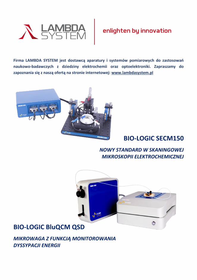
Firma LAMBDA SYSTEM jest dostawcą aparatury i systemów pomiarowych do zastosowań
naukowo-badawczych z dziedziny elektrochemii oraz optoelektroniki. Zapraszamy do
zapoznania się z naszą ofertą na stronie internetowej: www.lambdasystem.pl
BIO-LOGIC SECM150
NOWY STANDARD W SKANINGOWEJ MIKROSKOPII ELEKTROCHEMICZNEJ
BIO-LOGIC BluQCM QSD
MIKROWAGA Z FUNKCJĄ MONITOROWANIA DYSSYPACJI ENERGII
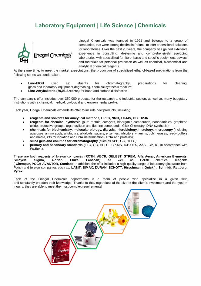
Laboratory Equipment | Life Science | Chemicals
Linegal Chemicals was founded in 1991 and belongs to a group of
companies, that were among the first in Poland, to offer professional solutions
for laboratories. Over the past 28 years, the company has gained extensive
experience in consulting, designing and comprehensively equipping
laboratories with specialized furniture, basic and specific equipment, devices
and materials for personal protection as well as chemical, biochemical and
analytical chemical reagents.
At the same time, to meet the market expectations, the production of specialized ethanol-based preparations from the
following series was undertaken:
Line-EtOH used as: eluents for chromatography, preparations for cleaning, glass and laboratory equipment degreasing, chemical synthesis medium;
Line-Antybakteria (70,96 Srebrna) for hand and surface disinfection
The company's offer includes over 350,000 products for the research and industrial sectors as well as many budgetary institutions with a chemical, medical, biological and environmental profile.
Each year, Linegal Chemicals expands its offer to include new products, including:
reagents and solvents for analytical methods, HPLC, NMR, LC-MS, GC, UV-IR
reagents for chemical synthesis (pure metals, catalysts, bioorganic compounds, nanoparticles, graphene oxide, protective groups, organosilicon and fluorine compounds, Click Chemistry, DNA synthesis);
chemicals for biochemistry, molecular biology, dialysis, microbiology, histology, microscopy (including agaroses, amino acids, antibiotics, alkaloids, sugars, enzymes, inhibitors, vitamins, polymerases, ready buffers and media, kits for isolation and DNA determination / RNA and proteins);
silica gels and columns for chromatography (such as SPE, GC, HPLC);
primary and secondary standards (TLC, GC, HPLC, ICP-MS, ICP-OES, AAS, ICP, IC, in accordance with Ph.Eur .).
These are both reagents of foreign companies (ROTH, ABCR, GELEST, STREM, Alfa Aesar, American Elements, Silicycle, Sigma, Aldrich, Fluka, Labscan), as well as Polish chemical reagents ( Chempur, POCH-AVANTOR, Stanlab). In addition, the offer includes a high-quality range of laboratory glassware from Polish and foreign companies such as: LABIT, SIMAX, DURAN, SCHOTT, Hirschmann, Quickfit, Schmidt, Rettberg, Pyrex.
Each of the Linegal Chemicals departments is a team of people who specialize in a given field and constantly broaden their knowledge. Thanks to this, regardless of the size of the client's investment and the type of inquiry, they are able to meet the most complex requirements!

ul. Warszawska 100/102
05-092 Łomianki
tel. +48 22 675 25 67
fax. +48 22 811 98 18
www.spectro-lab.pl

9
10
Preface With a great pleasure and honor we present you the Program and Abstracts of the 9th International
Workshop on Surface Modification for Chemical and Biochemical Sensing, SMCBS'2019. The
Workshop is organized by the Institute of Physical Chemistry, Polish Academy of Science, Warsaw
(Poland) together with the Faculty of Mathematics and Natural Sciences. School of Sciences,
Cardinal Stefan Wyszynski University in Warsaw (Poland).
By continuing tradition of previous workshops of the SMCBS series, organized in Białowieża
(2003), Kazimierz Dolny (2005), Włodowice (2007), Przegorzały near Cracow (2009), Łochów
(2011 & 2013), Pułtusk (2015), and Żelechów (2017), the present workshop hosts all the
participants in a single location to give them a unique opportunity to get acquainted, to meet for ad
hock discussions, and exchange ideas that might lead to new research concepts and, even more
importantly, to launch new collaborations.
In the spirit of previous workshops of the SMCBS series, we are especially happy to welcome
many contributions of young scientists who present their results as short oral communications or
posters. We are thankful to top specialists in their fields for sharing their latest break-through
results presented as keynote lectures. Moreover, we are pleased that half a dozen of scientists of
international renown have accepted our invitation to deliver tutorial lectures that can be considered
as inspirations for further discussions.
Similarly as formerly, the present interdisciplinary Workshop involves the science of both
chemical and non-chemical modification of solid surfaces. Several research works aim at
improvement of recognition ability of the resulting chemosensors with respect to target analytes.
Main themes of the Workshop include different aspects of surface chemistry, mostly related to
chemo- and biosensing in solutions or gases, not being limited to:
chemo- and biosensing
chemical and biochemical surface modification
polymer film coating
inorganic, organic, and biomaterials for catalysis as well as for electric energy generation
and storage
charge transport in surface films
novel techniques and instrumentation for surfaces examination
signal transduction and processing, detection techniques and protocols, system
miniaturization and nanotechnology use.
Progress in modern sophisticated chemo- and biosensing requires broad collaboration of
specialists not only from the fields of chemistry and biology but also from physics, materials
science, electronics, and other. Although, predominately, the SMCBS workshops involve
electrochemical aspects of sensing, we hope that the broad spectrum of participants can enjoy the
interdisciplinary meetings that give rise to new important sensing ideas.
The Organizing and Program Committee cordially thanks all those who helped making the
9th SMCBS'2019 Workshop a great event. We are particularly thankful to contributing Authors, to
the sessions chairing persons, and the members of the International Scientific Advisory Board for
their outstanding job done in preparing and evaluating the scientific profile of this event.
On behalf of the Organizing and Program Committee we welcome all the participants wishing
them to have a splendid time at the Workshop, both scientifically and socially.
Włodzimierz Kutner
Warsaw, October 2019
11
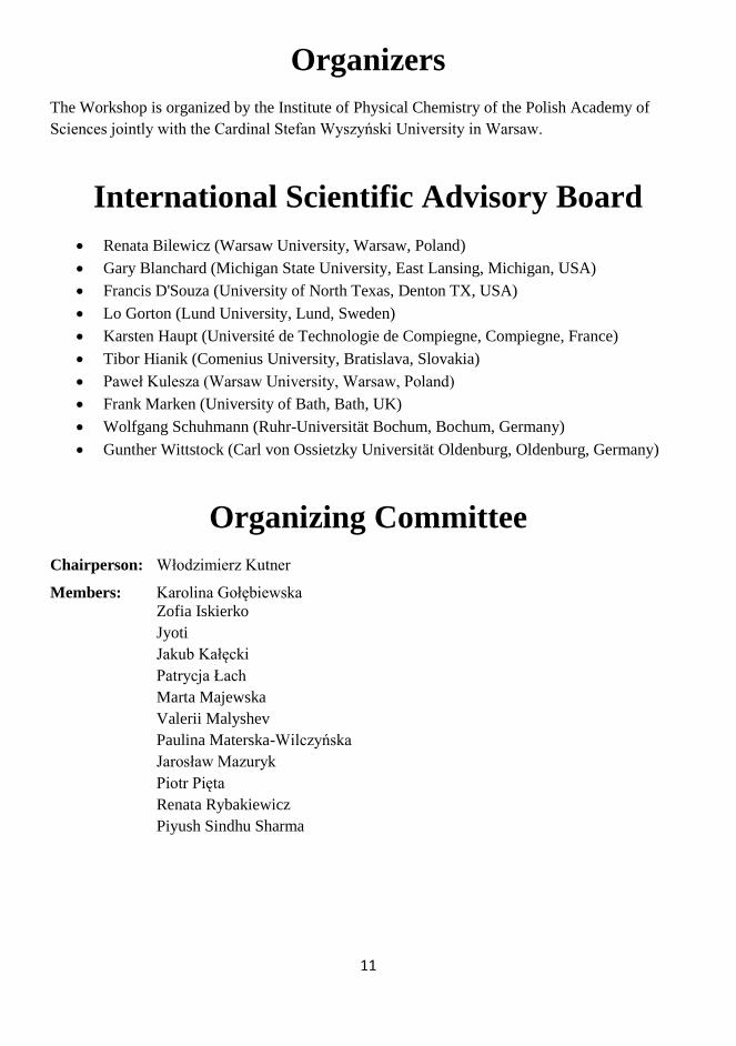
Organizers
The Workshop is organized by the Institute of Physical Chemistry of the Polish Academy of
Sciences jointly with the Cardinal Stefan Wyszyński University in Warsaw.
International Scientific Advisory Board
Renata Bilewicz (Warsaw University, Warsaw, Poland)
Gary Blanchard (Michigan State University, East Lansing, Michigan, USA)
Francis D'Souza (University of North Texas, Denton TX, USA)
Lo Gorton (Lund University, Lund, Sweden)
Karsten Haupt (Université de Technologie de Compiegne, Compiegne, France)
Tibor Hianik (Comenius University, Bratislava, Slovakia)
Paweł Kulesza (Warsaw University, Warsaw, Poland)
Frank Marken (University of Bath, Bath, UK)
Wolfgang Schuhmann (Ruhr-Universität Bochum, Bochum, Germany)
Gunther Wittstock (Carl von Ossietzky Universität Oldenburg, Oldenburg, Germany)
Organizing Committee
Chairperson: Włodzimierz Kutner
Members: Karolina Gołębiewska
Zofia Iskierko
Jyoti
Jakub Kałęcki
Patrycja Łach
Marta Majewska
Valerii Malyshev
Paulina Materska-Wilczyńska
Jarosław Mazuryk
Piotr Pięta
Renata Rybakiewicz
Piyush Sindhu Sharma

12

13
SMCBS’2019
The 9th International Workshop
on Surface Modification
for Chemical and Biochemical Sensing
Programme &
Book of Abstracts
Organized by the Institute of Physical Chemistry, Polish Academy of Sciences
jointly with the Cardinal Stefan Wyszyński University in Warsaw
Żelechów, Poland
November 8-12, 2019

14
SMCBS’2019 Programme
Friday, November 8
09:00-16:00 Registration at IPC PAS
11:45-15:30 Lunch at IPC PAS
13:30 / 16:00 1st / 2nd bus departure to Żelechów
16:00-19:00 Setting up posters in the Żelechów hotel
19:00-20:00 Dinner
20:00-
21:20
Evening session
Chairs: F. Marken / Y. Efremenko
20:00-
20:40
T01 Róbert E. Gyurcsányi
Design of SPR Imaging Chips for Multiplexed Affinity Assays
20:40-
21:00
K01 Nicolas Plumeré
Thin Films for Protection of O2-Sensitive Electrocatalysts
21:00-
21:20
K02 Andreas Lesch
Inkjet Printing of Nanostructured Electrodes for Biosensing
Saturday, November 9
08:00-09:00 Breakfast
09:00-
10:35
Morning session 1
Chairs: A. Lesch / E. Suprun
09:00-
09:40
T02 Julea Butt
All Will Be Revealed: Mapping Protein Redox Activity with Protein Film
Voltammetry
09:40-
10:00
K03 Lars J.C. Jeuken
Multilayered Lipid Membrane Stacks for Biocatalysis Using Membrane
Enzymes
10:00-
10:20
K04 Elisabeth Lojou
Enzymatic O2 Reduction on Gold Surfaces: Probing Functional Enzyme
Immobilization by Coupling Electrochemistry to SPR, Elllipsometry and
PMIRRAS
10:20-
10:35
SC01 Ievgen Mazurenko
Orientation-dependent Direct Electrochemistry of Laccase from Thermus
Thermophilus Reveals Cuprous Oxidase Activity

15
10:35-11:00 Coffee break
11:00-
12:50
Morning session 2
Chairs: N. Plumere / I. Mazurenko
11:00-
11:20
K05 Fred Lisdat
Cytochrome C as Valuable Building Block in Multilayered Architectures
of Biocatalysts on Electrodes
11:20-
11:40
K06 Johan Bobacka
Ion Sensors with Coulometric Transduction – Limitations and Possibilities
11:40-
12:00
K07 Frédéric Kanoufi
Probing the Interaction of Single Nanoparticle with Interfaces by in Situ
Optical Microscopies
12:00-
12:20
K08 Patrizia Romana Mussini
Enantiodiscrimination at Electrochemical Interphases through
Implementation of Inherently Chiral Selectors: New Insights and
Perspectives
12:20-
12:35
SC02 Vanousheh Rahemi
Hydrogen Peroxide-less Horseradish Peroxidase Based Biosensor for the
Detection of Phenols
12:35-
12:50
SC03 Julian Szczesny
Polymer Modified Gas Diffusion Electrodes Containing Hydrogenases or
Their Artificial Mimics as Active H2 Oxidation Catalysts
13:00-14:30 Lunch
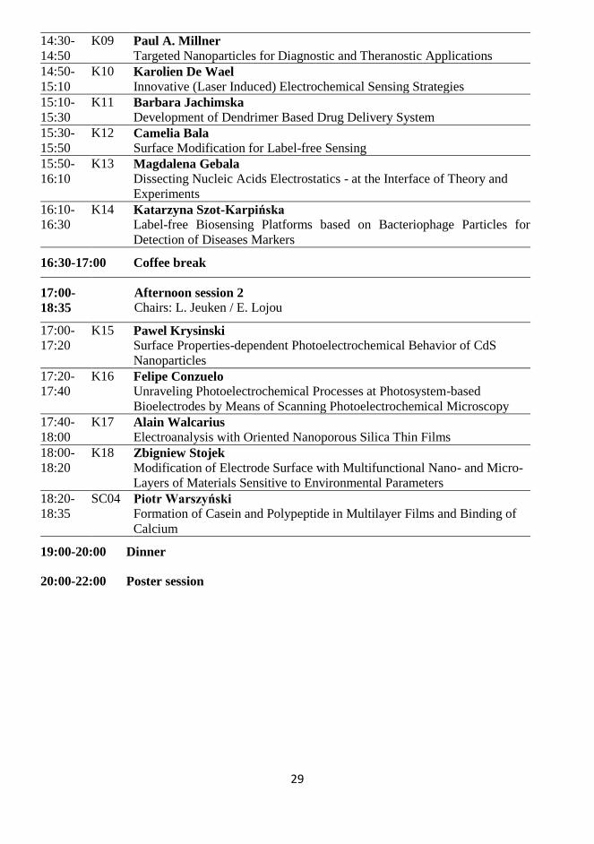
14:30-
16:30
Afternoon session 1
Chairs: F. Lisdat / J. Szczęsny
14:30-
14:50
K09 Paul A. Millner
Targeted Nanoparticles for Diagnostic and Theranostic Applications
14:50-
15:10
K10 Karolien De Wael
Innovative (Laser Induced) Electrochemical Sensing Strategies
15:10-
15:30
K11 Barbara Jachimska
Development of Dendrimer Based Drug Delivery System
15:30-
15:50
K12 Camelia Bala
Surface Modification for Label-free Sensing
15:50-
16:10
K13 Magdalena Gebala
Dissecting Nucleic Acids Electrostatics - at the Interface of Theory and
Experiments
16:10-
16:30
K14 Katarzyna Szot-Karpińska
Label-free Biosensing Platforms based on Bacteriophage Particles for
Detection of Diseases Markers
16:30-17:00 Coffee break

16
17:00-
18:35
Afternoon session 2
Chairs: L. Jeuken / E. Lojou
17:00-
17:20
K15 Pawel Krysinski
Surface Properties-dependent Photoelectrochemical Behavior of CdS
Nanoparticles
17:20-
17:40
K16 Felipe Conzuelo
Unraveling Photoelectrochemical Processes at Photosystem-based
Bioelectrodes by Means of Scanning Photoelectrochemical Microscopy
17:40-
18:00
K17 Alain Walcarius
Electroanalysis with Oriented Nanoporous Silica Thin Films
18:00-
18:20
K18 Zbigniew Stojek
Modification of Electrode Surface with Multifunctional Nano- and Micro-
Layers of Materials Sensitive to Environmental Parameters
18:20-
18:35
SC04 Piotr Warszyński
Formation of Casein and Polypeptide in Multilayer Films and Binding of
Calcium
19:00-20:00 Dinner
20:00-22:00 Poster session
Sunday, November 10
08:00-09:00 Breakfast
09:00-
10:35
Morning session 1
Chairs: P. Krysiński / J. Bobacka
09:00-
09:40
T03 Francis D’Souza
Interfacial Electron Transfer of Multimolecular Assemblies on Metal
Oxide Semiconductors
09:40-
10:00
K19 Frank Marken
Polymers of Intrinsic Microporosity (PIMs) in Electrochemistry
10:00-
10:20
K20 Stéphane Arbault
Using Cold Plasmas to Improve Electrochemical Sensors and vice versa
10:12-
10:35
SC05 Krzysztof R. Noworyta
Oxidation and Coupling of the Selected Carbazole Derivatives – Why It
Does Not Always Lead to Polymer Formation
10:35-11:00 Coffee break
11:00-
12:50
Morning session 2
Chairs: K. Noworyta / A. Yarman
17
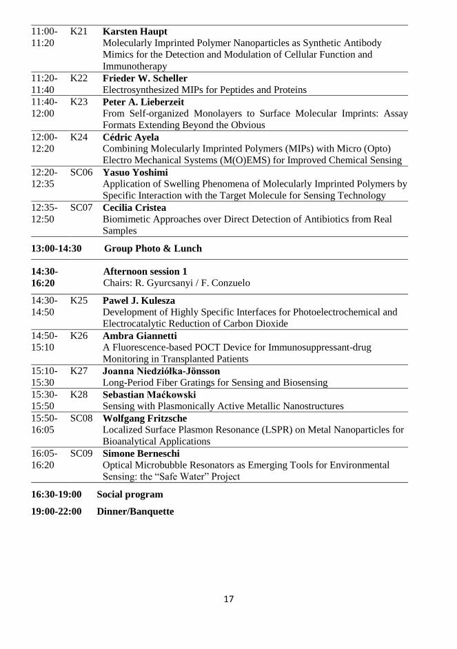
11:00-
11:20
K21 Karsten Haupt
Molecularly Imprinted Polymer Nanoparticles as Synthetic Antibody
Mimics for the Detection and Modulation of Cellular Function and
Immunotherapy
11:20-
11:40
K22 Frieder W. Scheller
Electrosynthesized MIPs for Peptides and Proteins
11:40-
12:00
K23 Peter A. Lieberzeit
From Self-organized Monolayers to Surface Molecular Imprints: Assay
Formats Extending Beyond the Obvious
12:00-
12:20
K24 Cédric Ayela
Combining Molecularly Imprinted Polymers (MIPs) with Micro (Opto)
Electro Mechanical Systems (M(O)EMS) for Improved Chemical Sensing
12:20-
12:35
SC06 Yasuo Yoshimi
Application of Swelling Phenomena of Molecularly Imprinted Polymers by
Specific Interaction with the Target Molecule for Sensing Technology
12:35-
12:50
SC07 Cecilia Cristea
Biomimetic Approaches over Direct Detection of Antibiotics from Real
Samples
13:00-14:30 Group Photo & Lunch
14:30-
16:20
Afternoon session 1
Chairs: R. Gyurcsanyi / F. Conzuelo
14:30-
14:50
K25 Pawel J. Kulesza
Development of Highly Specific Interfaces for Photoelectrochemical and
Electrocatalytic Reduction of Carbon Dioxide
14:50-
15:10
K26 Ambra Giannetti
A Fluorescence-based POCT Device for Immunosuppressant-drug
Monitoring in Transplanted Patients
15:10-
15:30
K27 Joanna Niedziółka-Jönsson
Long-Period Fiber Gratings for Sensing and Biosensing
15:30-
15:50
K28 Sebastian Maćkowski
Sensing with Plasmonically Active Metallic Nanostructures
15:50-
16:05
SC08 Wolfgang Fritzsche
Localized Surface Plasmon Resonance (LSPR) on Metal Nanoparticles for
Bioanalytical Applications
16:05-
16:20
SC09 Simone Berneschi
Optical Microbubble Resonators as Emerging Tools for Environmental
Sensing: the “Safe Water” Project
16:30-19:00 Social program
19:00-22:00 Dinner/Banquette
18
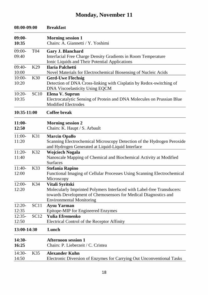
Monday, November 11
08:00-09:00 Breakfast
09:00-
10:35
Morning session 1
Chairs: A. Giannetti / Y. Yoshimi
09:00-
09:40
T04 Gary J. Blanchard
Interfacial Free Charge Density Gradients in Room Temperature
Ionic Liquids and Their Potential Applications
09:40-
10:00
K29 Ilaria Palchetti
Novel Materials for Electrochemical Biosensing of Nucleic Acids
10:00-
10:20
K30 Gerd-Uwe Flechsig
Detection of DNA Cross-linking with Cisplatin by Redox-switching of
DNA Viscoelasticity Using EQCM
10:20-
10:35
SC10 Elena V. Suprun
Electrocatalytic Sensing of Protein and DNA Molecules on Prussian Blue
Modified Electrodes
10:35-11:00 Coffee break
11:00-
12:50
Morning session 2
Chairs: K. Haupt / S. Arbault
11:00-
11:20
K31 Marcin Opałło
Scanning Electrochemical Microscopy Detection of the Hydrogen Peroxide
and Hydrogen Generated at Liquid-Liquid Interface
11:20-
11:40
K32 Wojciech Nogala
Nanoscale Mapping of Chemical and Biochemical Activity at Modified
Surfaces
11:40-
12:00
K33 Stefania Rapino
Functional Imaging of Cellular Processes Using Scanning Electrochemical
Microscopy
12:00-
12:20
K34 Vitali Syritski
Molecularly Imprinted Polymers Interfaced with Label-free Transducers:
towards Development of Chemosensors for Medical Diagnostics and
Environmental Monitoring
12:20-
12:35
SC11 Aysu Yarman
Epitope-MIP for Engineered Enzymes
12:35-
12:50
SC12 Yulia Efremenko
Electrical Control of the Receptor Affinity
13:00-14:30 Lunch
14:30-
16:25
Afternoon session 1
Chairs: P. Lieberzeit / C. Cristea
14:30-
14:50
K35 Alexander Kuhn
Electronic Diversion of Enzymes for Carrying Out Unconventional Tasks
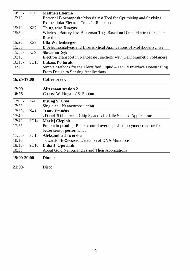
19
14:50-
15:10
K36 Mathieu Etienne
Bacterial Biocomposite Materials: a Tool for Optimizing and Studying
Extracellular Electron Transfer Reactions
15:10-
15:30
K37 Tautgirdas Ruzgas
Wireless, Battery-less Biosensor Tags Based on Direct Electron Transfer
Reactions
15:30-
15:50
K38 Ulla Wollenberger
Bioelectrocatalysis and Bioanalytical Applications of Molybdoenzymes
15:50-
16:10
K39 Sławomir Sęk
Electron Transport in Nanoscale Junctions with Helicomimetic Foldamers
16:10-
16:25
SC13 Łukasz Półtorak
Simple Methods for the Electrified Liquid – Liquid Interface Downscaling.
From Design to Sensing Applications
16:25-17:00 Coffee break
17:00-
18:25
Afternoon session 2
Chairs: W. Nogala / S. Rapino
17:00-
17:20
K40 Insung S. Choi
Single-cell Nanoencapsulation
17:20-
17:40
K41 Jenny Emnéus
2D and 3D Lab-on-a-Chip Systems for Life Science Applications
17:40-
17:55
SC14 Maciej Cieplak
Protein imprinting. Better control over deposited polymer structure for
better sensor performance.
17:55-
18:10
SC15 Aleksandra Jaworska
Towards SERS-based Detection of DNA Mutations
18:10-
18:25
SC16 Lidia J. Opuchlik
About Gold Nanotriangles and Their Applications
19:00-20:00 Dinner
21:00- Disco

20
Tuesday, November 12
08:00-09:00 Breakfast
09:00-
10:30
Morning session 1
Chairs: A. Kuhn / Ł. Półtorak
09:00-
09:40
T05 Sergey Shleev
Non-invasive Electrochemical (Bio)sensors Operating in Human
Physiological Fluids
09:40-
10:00
K42 Hanna Radecka
Ultrasensitive Electrochemical Sensors for Exploring of Anion
Recognition Processes at Aqueous/Solid Interface
10:00-
10:15
SC17 Nabila Yasmeen
Electropolymerized Molecularly Imprinted Polymer towards
Chemosensing of an Autism Biomarker
10:15-
10:30
SC18 Luís C. Almeida
Electrosynthesized Poly(catecholamine) Films Modified with
Ethanolamine for Immunosensing
10:30-11:00 Coffee break
11:00-
12:00
Morning session 2
Chairs: S. Shleev / K. Szot-Karpińska
11:00-
11:20
K43 Lo Gorton
Connecting Biological Membranes and Bacterial Cells to Electrodes
through Redox Polymers
11:20-
11:35
SC19 Kamila Łępicka
The poly[NBI-(DTP)2] as an electrode material for the inherently
asymmetric supercapacitor
11:35-11:50 Closing
12:00-13:00 Lunch
13:30- Departures

21
22
T01. Design of SPR Imaging Chips for Multiplexed Affinity Assays
László Simon, Zsófia Bognár, Róbert E. Gyurcsányi
BME “Lendület” Chemical Nanosensors Research Group, Department of Inorganic and
Analytical Chemistry, Budapest University of Technology and Economics,
Szent Gellért tér 4, H-1111, Budapest, Hungary
In terms of affinity assays surface plasmon resonance imaging (SPRi) offers high throughput, label-free and
real-time monitoring of the binding kinetics. However, it offers also in a broader sense an extremely
sophisticated platform for understanding the immobilization of synthetic and natural receptors on gold that
can provide the basis of rational chemical modification of nanoconfinements. Such modifications require
extreme control over the surface concentration and distribution of immobilized receptors that we will show
that is feasible by appropriate methodology. Indeed, the difficulties we encountered in the chemical
modification of gold nanopores with nucleic acids probes, especially peptide nucleic acids (PNA),1 were
solved after gaining a deeper insight on the relevant immobilization and binding processes through a surface
plasmon resonance imaging (SPRi) study.2 Within this study we developed methods for quantitative
determination of the surface concentration of DNA/ PNA probes and their reliable self-regulation within a
single-step process.3
The general requirement for the efficient use of SPRi is the localized immobilization of the studied
receptors while strictly controlling (determining) their surface concentration. The localized immobilization
is done by an off-line microspotting (or microelectrospotting)4 procedure which hinders the direct
determination of the surface concentration of the immobilized receptors. We are going to show how these
can be determined for PNA and DNA and aptamer probes. Most importantly we found that for all DNA
probes the small molecular weight ruthenium(III) hexamine complex (RuHex) introduced earlier for
electrochemical quantitation of DNA coverage on gold electrodes can be used also in SPRi to assess the
surface density of DNA probes in DNA microarrays.5 A single injection of RuHex solution allows the
simultaneous visualization and quantification of the surface density of DNA probes (ranging in this study
from 4×10¹¹ to 1.7×10¹³ molecules cm-2) on all spots of a DNA microarray made by microspotting thiol
labeled short DNA probes both in prehybridized and single-stranded form on a gold SPRi chip. The excellent
controll over the surface concentration of nucleic acid probes could be used for increased efficiency
hybridization and protein assays.
Acknowledgements: The support of ERA-Chemistry (OTKA NN117637) and BME-Nanotechnology FIKP grant of EMMI (BME FIKP-NAT) is gratefully
acknowledged.
References:
1 G. Jágerszki, R. E. Gyurcsányi, L. Höfler, E. Pretsch, Nano Lett., 2007, 7, 1609-1612.
2 I. Makra, A. Brajnovits, G. Jágerszki, P. Fürjes, R. E. Gyurcsányi, Nanoscale, 2017, 9, 739-747. 3 L. Simon, G. Lautner, R. E. Gyurcsanyi, Anal. Methods, 2015, 7, 6077-6082.
4 M. Bosserdt, J. Erdőssy, G. Lautner, J. Witt, K. Köhler, N. Gajovic-Eichelmann, A. Yarman, G. Wittstock, F. W. Scheller, R. E.
Gyurcsányi, Biosens. Bioelectron., 2015, 73, 123-129. 5 L. Simon, R. E. Gyurcsányi, Anal. Chim. Acta, 2019, 1047, 131-138.

23
24
K01. Thin Films for Protection of O2-Sensitive Electrocatalysts
Huaiguang Li,a Darren Buesen,a Sébastien Dementin,b Christophe Léger,b Vincent Fourmond,b
Nicolas Plumeréa
a Center for Electrochemical Sciences (CES), Faculty of Chemistry and Biochemistry, Ruhr
University Bochum, Universitätsstr. 150, D-44780 Bochum, Germany. b Laboratoire de Bioénergétique et Ingénierie des Protéines.
CNRS, Aix Marseille Université, Marseille, France
Energy conversion schemes involving dihydrogen hold great potential for meeting sustainable energy needs,
but widespread implementation cannot proceed without solutions that mitigate the cost of rare metal catalysts
and the intrinsic O2-instability of bio-inspired replacements. Recently, thick films (>100 µm) of redox
polymers were shown to prevent O2 catalyst damage,1,2 but also resulted in unnecessary catalyst load and
mass transport limitations.3 Here, we apply novel homogeneous thin films down to 3 µm4 that provide O2-
immunity while achieving highly efficient catalyst utilization. Our empirical data is explained by modeling
demonstrating that resistance to O2 inactivation can be obtained for non-limiting periods of time when the
optimal thickness for catalyst utilization and current generation is achieved even when using highly fragile
catalysts such as the enzyme hydrogenase. We show that different protection mechanisms operate depending
on matrix dimensions and intrinsic catalyst properties, and can be integrated together synergistically to
achieve large and stable H2 oxidation currents in the presence of O2, potentially enabling a plethora of
practical applications for bio-inspired catalysts in harsh oxidative conditions.
Acknowledgements:
Financial support by the Cluster of Excellence RESOLV (EXC 2033), by the ERC starting grant 715900 and by the ANR-DFG project
SHIELDS (PL 746/2-1) is gratefully acknowledged.
References:
1 N. Plumeré, O. Rüdiger, A. Alsheikh Oughli, R. Williams, J. Vivekananthan, S. Pöller, W. Schuhmann, W. Lubitz, Nature Chemistry, 2014, 6, 822–827.
2 A. Alsheikh Oughli, F. Conzuelo, M. Winkler, T. Happe, W. Lubitz, W. Schuhmann, O. Rüdiger, N. Plumeré, Angew. Chem. Int.
Ed., 2015, 54, 12329 –12333. 3 V. Fourmond, S. Stapf, H. Li, D. Buesen, J. Birrell, O. Rüdiger, W. Lubitz, W. Schuhmann, N. Plumeré, C. Léger, J. Am. Chem.
Soc., 2015, 137, 5494-5505.
4 H. Li, D. Buesen, R Williams, J. Henig, S. Stapf, K. Mukherjee, E. Freier, W. Lubitz, M. Winkler, T. Happe, N. Plumeré, Chemical Science, 2018, 9, 7596-7605.

25
26
K02. Inkjet Printing of Nanostructured Electrodes for Biosensing
Andreas Lesch,a Milica Jović,b Victor Costa Bassetto,b Yingdi Zhu,b Horst Pick,c
Bhawna Nagar,b Domenica Tonelli,a Hubert H. Giraultb
a Department of Industrial Chemistry "Toso Montanari", University of Bologna, Bologna, Italy.
b Laboratory of Physical and Analytical Electrochemistry, Ecole Polytechnique Fédérale de
Lausanne (EPFL) Valais Wallis, Sion, Switzerland. c Institute of Chemical Sciences and Engineering (ISIC), Ecole Polytechnique Fédérale de
Lausanne (EPFL), Lausanne, Switzerland.
Inkjet printing (IJP) gains continuously in importance in the field of sensor development. As a digital material
deposition technique, IJP is suitable to print with micrometer resolution thin layers of a broad range of
material containing inks. Compared to screen printing, major advantages include contact-less and mask-less
fabrication with up-scalability from prototype to industrial production level. Furthermore, only few hundred
microliters of ink, thus extremely low amounts of electrode materials, are required in drop-on-demand,
piezoelectric inkjet printers to fabricate hundreds of sensors in reasonable time, making IJP very attractive
for developing, prototyping and manufacturing. Major bottlenecks are the ink formulation to achieve stable
jetting of picoliter droplets, high dispersibility of nanoparticles to avoid nozzle clogging and well-adhered,
defect-free films on substrate surfaces of interest.
Herein, we shall demonstrate the successful development and application of inkjet printed amperometric
electrodes based on carbon nanotubes (CNT) and graphene nanosheets. Flexible and transparent CNT
electrodes have been used on a large-scale as voltammetric sensors to measure erythrocyte concentrates in
blood transfusion medicine1 and as electrochemiluminescence platforms for cancer diagnostics.2 In
combination with antibodies on magnetic beads, inkjet-printed microtiter plate electrodes have been used to
detect rapidly bacterial species and to identify the antimicrobial resistance of such in infected blood samples
in a point-of-care (POC) electrochemical reader.3
We shall further demonstrate how inkjet printing can be applied for the synthesis of nanostructured
electrode coatings by combining IJP of molecules and salts with light processing. For instance, we have
combined IJP with UV photopolymerization to coat CNT electrodes with nanometer-thin polyacrylamide
hydrogels that were for instance applied for the detection of molecular bacteria markers in wound fluids.4
Finally, metal and mixed metal nanoparticles can be accurately and rapidly synthesized in situ directly on
the electrodes by combining inkjet printing of metal precursor salts with pulsed light irradiation. In this
process, named Print-Light-Synthesis, thin liquid films containing precursor salts are printed and
immediately exposed to microsecond light flashes from a Xe flash lamp. The result is a light-induced
reduction of the precursors under ambient conditions without the need of using nanoparticle capping agents
or surfactants. All side products of the reactions are gaseous. Examples will include the fabrication of Pt-
nanostructured ITO-coated glass slides5 and Ni/CNT as well as NixFe(1-x)/CNT electrodes.6
References:
1 A. Lesch, F. Cortés-Salazar, M. Prudent, J. Delobel, S. Rastgar, N. Lion, J. D. Tissot, P. Tacchini, Hubert H. Girault, J. Electroanal. Chem., 2014, 717, 61-68.
2 G. Valenti, S. Scarabino, B. Goudeau, A. Lesch, M. Jović, E. Villani, M. Sentic, S. Rapino, S. Arbault, F. Paolucci, N. Sojic, J.
Am. Chem. Soc., 2017, 139, 16830-16837. 3 Y. Zhu, M. Jović, A. Lesch, L. Tissières Lovey, M. Prudent, H. Pick, H. H. Girault, Angew.Chem. Int. Ed., 2018, 57, 14942-14946.
4 R. Jarošová, S. E. Mcclure, M. Gajda, M. Jović, H. H. Girault, A. Lesch, M. Maiden, C. Waters, G. M. Swain, Anal. Chem., 2019,
91, 8835-8844. 5 A. Lesch, Adv. Mater. Technol., 2018, 3, 1700201.
6 V. Costa Bassetto, M. Mensi, E. Oveisi, H. H. Girault, A. Lesch, ACS Appl. Energy Mat., accepted, DOI: 10.1021/acsaem.9b00957.

27
28
Saturday, November 9
08:00-09:00 Breakfast
09:00-
10:35
Morning session 1
Chairs: A. Lesch / E. Suprun
09:00-
09:40
T02 Julea Butt
All Will Be Revealed: Mapping Protein Redox Activity with Protein Film
Voltammetry
09:40-
10:00
K03 Lars J.C. Jeuken
Multilayered Lipid Membrane Stacks for Biocatalysis Using Membrane
Enzymes
10:00-
10:20
K04 Elisabeth Lojou
Enzymatic O2 Reduction on Gold Surfaces: Probing Functional Enzyme
Immobilization by Coupling Electrochemistry to SPR, Elllipsometry and
PMIRRAS
10:20-
10:35
SC01 Ievgen Mazurenko
Orientation-dependent Direct Electrochemistry of Laccase from Thermus
Thermophilus Reveals Cuprous Oxidase Activity
10:35-11:00 Coffee break
11:00-
12:50
Morning session 2
Chairs: N. Plumere / I. Mazurenko
11:00-
11:20
K05 Fred Lisdat
Cytochrome C as Valuable Building Block in Multilayered Architectures
of Biocatalysts on Electrodes
11:20-
11:40
K06 Johan Bobacka
Ion Sensors with Coulometric Transduction – Limitations and Possibilities
11:40-
12:00
K07 Frédéric Kanoufi
Probing the Interaction of Single Nanoparticle with Interfaces by in Situ
Optical Microscopies
12:00-
12:20
K08 Patrizia Romana Mussini
Enantiodiscrimination at Electrochemical Interphases through
Implementation of Inherently Chiral Selectors: New Insights and
Perspectives
12:20-
12:35
SC02 Vanousheh Rahemi
Hydrogen Peroxide-less Horseradish Peroxidase Based Biosensor for the
Detection of Phenols
12:35-
12:50
SC03 Julian Szczesny
Polymer Modified Gas Diffusion Electrodes Containing Hydrogenases or
Their Artificial Mimics as Active H2 Oxidation Catalysts
13:00-14:30 Lunch
14:30-
16:30
Afternoon session 1
Chairs: F. Lisdat / J. Szczęsny
29
14:30-
14:50
K09 Paul A. Millner
Targeted Nanoparticles for Diagnostic and Theranostic Applications
14:50-
15:10
K10 Karolien De Wael
Innovative (Laser Induced) Electrochemical Sensing Strategies
15:10-
15:30
K11 Barbara Jachimska
Development of Dendrimer Based Drug Delivery System
15:30-
15:50
K12 Camelia Bala
Surface Modification for Label-free Sensing
15:50-
16:10
K13 Magdalena Gebala
Dissecting Nucleic Acids Electrostatics - at the Interface of Theory and
Experiments
16:10-
16:30
K14 Katarzyna Szot-Karpińska
Label-free Biosensing Platforms based on Bacteriophage Particles for
Detection of Diseases Markers
16:30-17:00 Coffee break
17:00-
18:35
Afternoon session 2
Chairs: L. Jeuken / E. Lojou
17:00-
17:20
K15 Pawel Krysinski
Surface Properties-dependent Photoelectrochemical Behavior of CdS
Nanoparticles
17:20-
17:40
K16 Felipe Conzuelo
Unraveling Photoelectrochemical Processes at Photosystem-based
Bioelectrodes by Means of Scanning Photoelectrochemical Microscopy
17:40-
18:00
K17 Alain Walcarius
Electroanalysis with Oriented Nanoporous Silica Thin Films
18:00-
18:20
K18 Zbigniew Stojek
Modification of Electrode Surface with Multifunctional Nano- and Micro-
Layers of Materials Sensitive to Environmental Parameters
18:20-
18:35
SC04 Piotr Warszyński
Formation of Casein and Polypeptide in Multilayer Films and Binding of
Calcium
19:00-20:00 Dinner
20:00-22:00 Poster session

30
T02. All Will Be Revealed: Mapping Protein Redox Activity
with Protein Film Voltammetry
Julea Butt
School of Chemistry, School of Biological Sciences, University of East Anglia, Norwich, UK.
Redox active proteins are ubiquitous in biology. They underpin respiration and photosynthesis, contribute to
the synthesis of amino acids and assist in the removal of toxins. Some of these proteins are drug targets, some
allow for selective detection of chemicals, others provide inspiration for developing sustainable routes to
clean energy and chemicals. As a consequence there is huge interest in understanding protein redox chemistry
and exciting opportunities for contributions from dynamic electrochemistry. When proteins exchange
electrons directly with electrodes techniques such as cyclic voltammetry can quantify not only reduction
potentials, but ligand binding and catalysis. This information is available with particularly high resolution
when the protein of interest is adsorbed as an electroactive (sub-)monolayer film on the electrode surface. In
such a configuration there is minimal, if any contribution to the voltammetry from protein diffusion, and rate
limiting events intrinsic to the protein dominate the response. These aspects of protein film electrochemistry
will be illustrated in this contribution. A series of case studies, drawn from experiments on a recently
discovered class of protein, will be presented. The benefits of using rotating and stationary electrodes, and
experiments with different electrode materials will be explained as a high-resolution map of the protein’s
redox properties is revealed.
References:
1 J. Kurth, C. Dahl and J. Butt, J. Am. Chem. Soc., 2015, 137, 13232-13235.
2 J. Kurth, et al. J. Biol. Chem., 2016, 291, 24804-24818. 3 L. Jenner et al. J. Biol. Chem., 2019, in press.

31
32
K03. Multilayered Lipid Membrane Stacks for Biocatalysis
Using Membrane Enzymes
George R. Heath,a Mengqiu Li,a Valentin Radu,a Stefan Frielingsdorf,b Oliver Lenz,b
Lars J.C. Jeukenb
a School of Biomedical Sciences , University of Leeds, Leeds, LS2 9JT, United Kingdom b Institut für Chemie, Technische Universität Berlin, Straße des 17. Juni 135, 10623 Berlin,
Germany
Multilayered or stacked lipid membranes are a common principle in biology and have various functional
advantages compared to single-lipid membranes, such as their ability to spatially organize processes,
compartmentalize molecules, and greatly increase surface area and hence membrane protein concentration.
Here, a supramolecular assembly of a multilayered lipid membrane system is reported in which poly-l-lysine
electrostatically links negatively charged lipid membranes. When suitable membrane enzymes are
incorporated, either an ubiquinol oxidase (cytochrome bo3 from Escherichia coli) or an oxygen-tolerant
hydrogenase (the membrane-bound hydrogenase from Ralstonia eutropha), cyclic voltammetry (CV) reveals
a linear increase in biocatalytic activity with each additional membrane layer. Electron transfer between the
enzymes and the electrode is mediated by the quinone pool that is present in the lipid phase. Using atomic
force microscopy, CV, and fluorescence microscopy it is deduced that quinones are able to diffuse between
the stacked lipid membrane layers via defect sites where the lipid membranes are interconnected. This
assembly is akin to that of interconnected thylakoid membranes or the folded lamella of mitochondria and
has significant potential for mimicry in biotechnology applications such as energy production of biosensing.
References: 1 G. R. Heath, M. Li, I. L. Polignano, J. L. Richens, G. Catucci, P. O’Shea, S. J. Sadeghi, G. Gilardi, J. N. Butt and L. J. C. Jeuken,
Biomacromolecules, 2015, 17, 324-335.
2 G. R. Heath, M. Li, H. Rong, V. Radu, S. Frielingsdorf, O. Lenz, J. N. Butt and L. J. C. Jeuken, Adv. Funct. Mat., 2017, 27,
1606265.

33
34
K04. Enzymatic O2 Reduction on Gold Surfaces: Probing Functional
Enzyme Immobilization by Coupling Electrochemistry to SPR,
Elllipsometry and PMIRRAS
V. Hitaishi,a S. Lecomte,b A. De Poulpiquet,a D. Duché,c I. Mazurenko,a E. Lojoua
a Bioénergétique et Ingénierie des Protéines, CNRS-AMU, 31 Chemin Aiguier, 13009 Marseille,
France b Centre Biochimie des Membranes et Nanoobjets, CNRS, Bordeaux, France
c Aix Marseille Université, CNRS, IM2NP UMR 7334, 13397 Marseille, France
Identification and production to purity of redox enzymes from various microorganisms living in very
different environments have allowed to envision many of these biomolecules as catalysts in biosensors,
bioreactors or bioenergy devices. Among these, one new generation of enzymatic fuel cells is based on very
specific enzymes for H2 oxidation and O2 reduction, hydrogenase and bilirubin oxidase (BOD) respectively,
in replacement of rare and expensive platinum metal, providing a fully sustainable fuel cell. Great
improvement has been made during the recent years in the performance of H2/O2 enzymatic fuel cells.1-3
Despite these progresses, two remaining limitations severely limit the large scale development of the
biodevices. The first one is the electrical wiring of the enzyme. It was in particular recently shown that less
than 10% of the enzymes are in electrical contact with the carbon structure.4 This issue implies the control
of the oriented immobilization of the enzyme to enhance the electron transfer rate and optimize the loading
of enzyme at the electrode. The second one is the stability of bioelectrodes. Beyond the research in the
biodiversity of more stable enzymes, there is a need to decipher between the various origins for such
instability, protein leaching, reorientation, reconformation, denaturation…, then to be able to propose a
remediation process.
In this work we present how we have addressed the issues of enzyme wiring and bioelectrode stability
by adsorbing BOD and laccase from different origins on thiol-based Self-Assembled-Monolayers (SAM) on
gold electrodes.5-7 SAMs carrying different surface charge and hydrophobicity are used to tune the interaction
with the enzymes. pH during enzyme adsorption or during catalysis, and applied potentials are especially
studied as factors affecting both the global charge of the enzymes, the charge around the entry point of
electrons, and that of the electrode, hence the electrocatalytic process. Coupling electrochemistry to SPR,
ellipsometry and PMIRRAS, we establish the correlation between the electrocatalytic current and leaching
of the protein or switch in the protein orientation. We discuss the effect of enzyme coverage on the efficiency
of the electrocatalysis. We especially highlight the dynamic of enzyme orientation depending on the force of
electrostatic interactions. We finally discuss whether the molecular basis obtained on SAMs can be extended
to conductive materials such as carbon nanotubes which are mostly required to enhance the biofuel cell
performance.8,9
References:
1 I. Mazurenko et al., Sust. Energ. & Fuels, 2017, 1, 1475-1501. 2 I. Mazurenko et al., Curr. Op. Electrochem., 2017, 5, 74-84.
3 X. Xiao et al., Chem. Rev., 2019, 119, 9509-9558.
4 I. Mazurenko et al., Energy Environ. Sci., 2017, 10, 1966-1982. 5 C. Guttierrez-Sanchez et al., ACS Catal., 2016, 6, 5482-5492.
6 V. Hitaishi et al., ACS Catal., 2018, 8, 12004-12014.
7 V. Hitaishi et al., Catalysts, 2018, 8(5), 192. 8 I. Mazurenko et al., ACS Appl. Mater. Interfaces, 2016, 8, 23074-23085.
9 K. Monsalve et al., ChemElectroChem, 2016, 3, 2179-2188.

35
36
SC01. Orientation-dependent Direct Electrochemistry of Laccase
from Thermus Thermophilus Reveals Cuprous Oxidase Activity
Vivek P. Hitaishi, Romain Clement, Ludovica Quattrocchi, Marianne Ilbert, Elisabeth Lojou,
Ievgen Mazurenko
Aix Marseille Univ, CNRS, BIP, UMR7281, 31 chemin Joseph Aiguier, 13402, Marseille, France
Multicopper oxidases (MCO) are a group of oxidoreductases containing a couple of Cu-centers: mononuclear
T1 and trinuclear T2/T3. These enzymes are able of four-electron oxygen reduction at high potentials
reaching 0.78 V (NHE) which is beneficial for different enzymatic and biohybrid fuel cells.1 A subgroup of
MCO called laccases can perform low-specificity oxidation of aromatic compounds which can also be
exploited for bioremediation purposes.
In this work we present the electrochemical characterization of the laccase from a hyperthermophilic bacteria
Thermus thermophilus2 on CNT-modified electrodes. The as-purified laccase demonstrates direct electron
transfer in 4-electron oxygen reduction reaction with current densities that vary significantly depending on
the charge and chemical functionalization of CNTs as a consequence of different preferred enzyme
orientation.
In addition, we discovered an additional catalytic wave upon the addition of Cu(II) into the
electrochemical cell appearing only in the presence of active immobilized laccase. The onset potential and
the magnitude of this wave depends on Cu(II) concentration and on the type of CNTs. In homogeneous
assays, some MCOs can be activated by addition of Cu(II). Such activation was notably reported for copper
efflux oxidase from E.coli (CueO), an enzyme responsible for Cu(I) detoxication of the periplasm.3 Although
the exact mechanism and the physiological role of this activation is elusive, it was suggested that it involves
Cu(II) binding to the methionine-rich domain near the T1 centre.4,5 Although the laccase from Thermus
thermophilus shares only 31% of sequence identity with CueO, a similar methionine-rich domain can be
identified suggesting possible Cu(II) binding. We propose that the observed Cu-dependent wave is related
to cation binding and cuprous oxidase activity of the laccase. We use electrochemistry to investigate the
mechanism of laccase-Cu(II) interaction suggesting that the complexation of additional copper ions creates
a new electron pathway within the enzyme molecule.
References:
1 N. Mano, A. De Poulpiquet, Chem. Rev., 2018, 118, 2392–2468. 2 P. Agbo, J. R. Heath, H. B. Gray, J. Phys. Chem. B, 2013, 117, 527–534.
3 S. A. Roberts, A. Weichsel, G. Grass, K. Thakali, J. T. Hazzard, G. Tollin, C. Rensing, W. R. Montfort, Proc. Natl. Acad. Sci., 2002, 99, 2766–2771.
4 K. Y. Djoko, L. X. Chong, A. G. Wedd, Z. Xiao, J. Am. Chem. Soc., 2010, 132, 2005–2015.
5 S. K. Singh, S. A. Roberts, S. F. McDevitt, A. Weichsel, G. F. Wildner, G. B. Grass, C. Rensing, W. R. Montfort, J. Biol. Chem., 2011, 286, 37849–37857.

37
38
K05. Cytochrome C as Valuable Building Block in Multilayered
Architectures of Biocatalysts on Electrodes
Fred Lisdat
Biosystems Technology, Institute of Life Sciences and Biomedical Technologies,
Technical University Wildau, Hochschulring 1, 15745 Wildau, Germany,
The creation of artificial electron transfer (ET) chains based on the defined arrangement of enzymes and
redox proteins on electrode surfaces represents an emerging field in bioelectronics.1 Precondition for a
functional systems exploiting defined electron pathways among the different biocomponents is first of all, a
fast heterogeneous electron transfer of the chosen redox protein with the electrode. Furthermore it is
beneficial, when interprotein electron transfer is feasible, which is often called self-exchange. Finally, a
defined reaction of the biocatalytic units with the chosen redox protein is essential to establish a signal chain
from the enzyme substrate via the enzyme and the redox proteins towards the electrode.
For this purpose we have been applying the small redox protein cytochrome c and combining it with
several enzymes such as bilirubin oxidase, sulfite oxidase, cellobiose dehydrogenase or fructose
dehydrogenase2-4 or more recently with photocatalytic proteins such as photosystem I.5
In case of FDH we have studied the ET reaction of the flavin-dependent enzyme and cyt c first in solution.
Here two different pH optima are found for the reaction. When one reaction partner - cyt c - is immobilized
on a modified electrode, ET proceeds efficiently at neutral pH. In addition, a defined dependence on the
substrate concentration has been observed. In acidic media the reaction can also be verified but appears to
be less efficient.
It can be demonstrated that both partners can be assembled in a stable multilayer architecture, using the
biopolymer DNA as a negatively charged polyelectrolyte. This can be verified by SPR measurements.
Prepared on electrodes, substantial catalytic currents are recorded upon addition of fructose. The response
can be enhanced by the number of layers deposited on the surface. This shows that also in this case a signal
chain can be constructed through multiple protein layers4. Here the interaction of cyt c with DNA as basis of
the multilayer construction has been studied in more detail by NMR spectroscopy.6 Furthermore, the analogy
of protein multilayers with cyt c crystals have been evaluedted.7
In order to investigate effects influencing the self exchange between different cyt c molecules a
mutational study has been performed.8 Five alanine variants of the wild type protein (LysAla) have been
prepared to change the chemical properties of the surface area near the heme edge. The structural integrity
of the mutants can be verified by NMR and UV/Vis measurements. It is shown that electro-active
protein/silica nanoparticle multilayers can be constructed with all forms of human cyt c prepared. The scan
rate dependent voltammetric behavior for the mutant proteins in comparison to the wild type is altered in
some multilayer arrangements. A higher self-exchange rate has been found for e.g. K79A. The results
demonstrate that the position of the introduced change in the charge situation has a profound influence on
the exchange behavior. In addition, the behavior of the cyt c proteins in assembled multilayers is found rather
similar to the situation of cyt c self exchange in solution verified by NMR. Based on a model for self
exchange also the self exchange rate constants have been estimated, demonstrating that effective electron
transfer through defined molecular arrangements of cyt c is feasible.
References: 1 S. Feifel et al. Biosensors Based on Aptamers and Enzymes, Springer 2014, 140, 253-298.
2 F. Lisdat et al. ChemComm., 2009, 3, 274-283.
3 S. Feifel et al. Angew. Chem., 2014, 126, 5782-5786. 4 C. Wettstein et al. Anal. Chem., 2016, 88, 6382-6389.
5 K. R. Stieger et al. Nanoscale, 2016, 8, 10695-10705. 6 D. Ciornii et al. J. Am. Chem. Soc., 2017, 139, 16478-16481.
7 C. Wettstein et al. Nanoscale, 2014, 6, 13779-13786.
8 R. E. McGovern et al. Angew. Chem., 2015, 21, 6356-6359.
9 S. Feifel et al. ACS Omega, 2016, 1, 1058-1066.

39
40
K06. Ion Sensors with Coulometric Transduction –
Limitations and Possibilities
Tingting Han, Ulriika Mattinen, Zekra Mousavi, Johan Bobacka
Åbo Akademi University, Faculty of Science and Engineering, Johan Gadolin Process Chemistry
Centre, Laboratory of Molecular Science and Engineering, Turku/Åbo, Finland
Coulometric signal transduction was developed and evaluated for solid-contact ion-selective electrodes (SC-
ISEs).1-5 The SC-ISEs were based on poly(3,4-ethylenedioxythiophene) (PEDOT) as the solid contact and
plasticized PVC-based ion-selective membranes. The main goal was to improve the sensitivity of SC-ISEs
by using coulometry instead of potentiometry.
For a given change in ion activity and thus a given potential change of the SC-ISE, the magnitude of the
measured coulometric signal is proportional to the (redox)capacitance of the solid-contact layer. Therefore,
the analytical signal could be amplified by increasing the capacitance of the solid contact.2 Under optimized
conditions, a 0.1 % change in ion activity could be detected when utilizing the coulometric transduction
method.5
The coulometric method involves charging/discharging of the solid-contact layer of the SC-ISE.
Simultaneously, charge-compensating ions must transfer to/from the solid-contact layer via the ion-selective
membrane. These processes tend to increase the response time of the SC-ISE. In order to shorten the response
time, the electrode resistance was minimized by varying the electrode geometry and by using thin-layer (spin-
coated) ion-selective membranes.3-5 A capacitive model was used to describe the coulometric readout of SC-
ISEs.4
Limitations and possibilities of the coulometric transduction method for SC-ISEs will be presented and
discussed.
References:
1 E. Hupa, U. Vanamo, J. Bobacka, Electroanalysis, 2015, 27, 591-594. 2 U. Vanamo, E. Hupa, V. Yrjänä, J. Bobacka, Anal. Chem., 2016, 88. 4369-4374.
3 T. Han, U. Vanamo, J. Bobacka, ChemElectroChem, 2016, 3. 2071-2077.
4 Z. Jarolímová, T. Han, U. Mattinen, J. Bobacka, E. Bakker, Anal. Chem., 2018, 90, 8700-8707. 5 T. Han, U. Mattinen, J. Bobacka, ACS Sens., 2019, 4, 900-906.

41
42
K07. Probing the Interaction of Single Nanoparticle
with Interfaces by in Situ Optical Microscopies
Jean-François Lemineur, T. Jane Stockmann, Jean-Marc Noël, Catherine Combellas,
Frédéric Kanoufi
Université de Paris, ITODYS laboratory, CNRS UMR 70786, Paris, France
The control of the attachment of nanoparticles to surface is ubiquitous in various research and applications
fields, from the conception of sensing platform to the building electrode materials for the optimization of
charge transport processes in electrochemical energy storage/conversion devices.
Recent analytical and electroanalytical developments propose to apprehend the behavior of nanoparticle
systems at the single entity level. In this respect optical microscopies provide an interesting instrumental
platform to visualize various nanoobjects, metallic or dielectric, down to 10nm, in a chemical or
electrochemical environment. These methods have been used, by us and others, to quantify in situ and real
time the electrochemical growth1 or dissolution of nanoparticles on electrode at the single nanoparticle level.
In this talk we will describe how such optical microscopies can be used to depict the interaction between
individual nanoparticles and a surface. This will be presented in the context of microfluidic-based sensors
relying on the capture of nanoparticles on a sensing surface2 or of the electrochemical conversion of
nanoparticles.
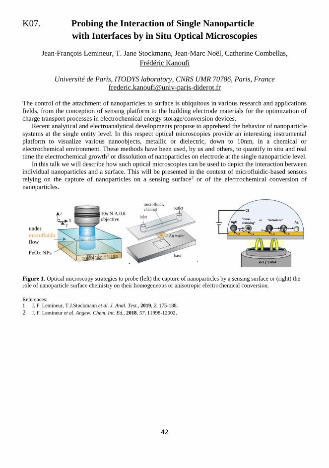
Figure 1. Optical microscopy strategies to probe (left) the capture of nanoparticles by a sensing surface or (right) the
role of nanoparticle surface chemistry on their homogeneous or anisotropic electrochemical conversion.
References:
1 J. F. Lemineur, T.J.Stockmann et al. J. Anal. Test., 2019, 2, 175-188.
2 J. F. Lemineur et al. Angew. Chem. Int. Ed., 2018, 57, 11998-12002.
water dipping
40x N.A.0.8 immersion
objective
Ag or PS NPs
Reflectivity and scattering
l = 490nm
FeOx NPs
under
microfluidic
flow
10x N.A.0.8 objective
Eref
Escat
Eref

43
44
K08. Enantiodiscrimination at Electrochemical Interphases
through Implementation of Inherently Chiral Selectors:
New Insights and Perspectives
Patrizia Romana Mussini,a Serena Arnaboldi,a Sara Grecchi,a Mirko Magni,a Mariangela Longhi,a Vittoria
Guglielmi,a Francesco Orsini,a Emanuela Licandro,a Silvia Cauteruccio,a Giovanna Longhi,b Lorenzo
Guazzelli,c Roberto Cirilli,d Fabiana Arduini,e Laura Micheli,e Heinrich Lang,f Claudio Fontanesi,g Simona
Rizzo,h Tiziana Benincori,i Francesco Sannicolòa
a Università degli Studi di Milano, Milano, Italy b Università degli Studi di Brescia, Brescia, Italy
c Università degli Studi di Pisa, Pisa, Italy; d Istituto Superiore di Sanità, Roma, Italy e Università degli Studi di Roma Tor Vergata, Roma, Italy
f Technische Universität Chemnitz, Germany g Università degli Studi di Modena e Reggio Emilia, Modena, Italy
h CNR ISTM, Milano, Italy i Università degli Studi dell'Insubria, Como, Italy
To achieve enantioselective electrochemistry and electroanalysis, electron transfer processes at the electrochemical
interphase require the presence of a suitable enantiopure chiral selector, resulting in energetically different
diastereoisomeric conditions for the two probe enantiomers.1 A groundbreaking strategy was recently proposed,
based on the use of molecular selectors endowed with "inherently chirality", i.e. with chirality and key functional
properties originating from the same structural element, which in our case identifies with the main molecular
backbone, featuring a tailored torsion with a racemization energy barrier too high to be overcome at room
temperature. In such conditions, large peak potential differences have been observed in voltammetry for the
enantiomers of even very different chiral probes either (i) working in achiral media, on electrode surfaces modified
with thin films of inherently chiral electroactive oligomers1,2 or (ii) working on achiral electrodes, implementing
inherent chirality in the medium, particularly in ionic liquids ILs, either chiral themselves, or modified by a chiral
additive,1,3 exploiting the peculiar IL high order at the interphase with a charged electrode.
Both strategies are being now extended and refined, particularly aiming to collect clues for the elucidation of
the recognition mechanism, as well as to highlight attractive applications.
In the film case, advanced techniques are finely highlighting morphology, chemical composition as well as
functional properties of inherently chiral electroactive oligomer films, both as electrode surfaces and as self
standing membranes. In the media case, chiral and inherently chiral molecules are being studied both as bulk media
and/or as media additives, with impressive results. A wide palette of selectors (films or media) and/or probes are
being investigated, encompassing four classes of stereogenic elements, i.e. corresponding to stereocentre-based
chirality, axial chirality, helical chirality and planar chirality.
Finally, the outstanding enantiodiscrimination ability of the new selectors is being considered beyond
molecular chiral probes, i.e. towards polarized light components (in terms of circular dichroism and circularly
polarized luminescence) and electron spins (in magnetoelectrochemistry experiments). Not only impressive effects
have been already observed, but fascinating correlations and connections are emerging among the three areas,
worthy to be explored in detail, possibly providing further interpretative clues, as well as for possible exploitation
in photonics and spintronics.
Support by Regione Lombardia and Fondazione Cariplo (Project 2016-0923) as well as from Università degli Studi di Milano is gratefully acknowledged.
References: 1 S. Arnaboldi, M. Magni, P. R. Mussini, Curr. Opin. Electrochem., 2018, 8, 60-72.
2 F. Sannicolò, S. Arnaboldi, T. Benincori, V. Bonometti, R. Cirilli, L. Dunsch, W. Kutner, G. Longhi, P. R. Mussini, M. Panigati, M. Pierini, S. Rizzo, Angew. Chem. Int. Ed., 2014, 53, 2623-2627.
3 S. Rizzo, S. Arnaboldi, V. Mihali, R. Cirilli, A. Forni, A. Gennaro, A. A. Isse, M. Pierini, P. R. Mussini, F. Sannicolò, Angew.
Chem. Int. Ed., 2017, 56, 2079-2082.

45
46
SC02. Hydrogen Peroxide-less Horseradish Peroxidase Based
Biosensor for the Detection of Phenols
V. Rahemi,a S. Trashin,a Z. Hafideddine,b S. Van Doorslaer,b V. Meynen,c L. Gorton,d
K. De Waela
a AXES research group, Department of Chemistry, University of Antwerp, Groenenborgerlaan
171, 2020 Antwerp, Belgium b Laboratory of Adsorption and Catalysis (LADCA), Department of Chemistry, University of
Antwerp, Universiteitsplein 1, B-2610 Wilrijk, Belgium c Department of Physics, University of Antwerp, Universiteitsplein 1, B-2610 Wilrijk, Belgium
d Department of Analytical Chemistry/Biochemistry and Structural Biology, Lund University, PO
Box 124, SE-22100 Lund, Sweden
Titanium dioxide (TiO2)-based enzymatic sensors for the determination of phenolic compounds usually
comprise tyrosinase,1 peroxidase2 or laccase enzymes.3 The working principle of these biosensors is based
on the redox cycling of a biocatalytic oxidation product of an analyte and the following electrochemical
reduction. Hydrogen peroxide (in case of peroxidases) or oxygen (in case of laccase or tyrosinase) plays the
role of an ultimate electron acceptor that continuously regenerates the reactive form of the enzyme.4
Horseradish peroxidase (HRP) is advantageous for developing phenolic biosensors due its high catalytic
activity towards a broad range of phenols, but the need of the presence of H2O2 in the solution complicates
the analysis and increases background noise.5 Therefore, it is required to develop a hydrogen peroxide-less
HRP based biosensor for the detection of phenols.
For the first time we show that TiO2 can accumulate reactive oxygen species (ROS) under daylight
irradiation and can support the catalytic cycle of horseradish peroxidase (HRP) without the need of H2O2 to
be present in the solution. The ROS act as the sacrificial oxidant or at least produces some amount of reactive
species such as H2O2, locally and near the site of the enzyme location. Phenolic compounds, such as
hydroquinone (HQ) and 4-aminophenol (4-AP), were detected amperometrically in flow-injection analysis
mode via the use of an electrode modified with TiO2 impregnated with HRP. In contrast to the conventional
detection scheme, no H2O2 was added to the analyte solution. Basically, the inherited ability of TiO2 to
generate ROS is used as a strategy to avoid adding H2O2 in the solution during the detection of phenolic
compounds. Electron paramagnetic resonance (EPR) spectroscopy indicates the presence of ROS on titania
which, in interaction with HRP, initiate the electrocatalysis towards phenolic compounds. The amperometric
response to 4-AP was linear in the concentration range between 0.05 and 2 µM. The sensitivity was 0.51 A
M-1 cm-2 and the limit of detection (LOD) 26 nM. The proposed sensor design opens new opportunities for
the detection of phenolic traces by HRP-based electrochemical biosensors, yet in more straightforward and
sensitive way.
References:
1 J. Yu, S. Liu and H. Ju, Biosens. Bioelectron., 2003, 19, 509-514.
2 J. Yu, Anal. Chem., 2002, 74, 3579-3583.
3 J. Kochana, P. Nowak, A. Jarosz-Wilkołazka and M. Bieroń, Microchem. J., 2008, 89, 171-174.
4 S. Yang, Y. Li, X. Jiang, Z.Chen and X. Lin, Sens. Actuators, B, 2006, 114, 774-780.
5 S. Trashin, V. Rahemi, K. Ramji, L.Neven, S. M. Gorun and K. De Wael, Nat. Commun., 2017, 8, 16108.

47
48
SC03. Polymer Modified Gas Diffusion Electrodes
Containing Hydrogenases or Their Artificial Mimics
as Active H2 Oxidation Catalysts
Julian Szczesny,a Felipe Conzuelo,a Sónia Zacarias,b Inês A. C. Pereira,b Wolfgang Lubitz,c
Adrian Ruff,a Wolfgang Schuhmanna
a Analytical Chemistry – Center for Electrochemical Sciences (CES), Faculty of Chemistry and
Biochemistry, Ruhr-University Bochum, D-44870 Bochum, Germany. b Instituto de Tecnologia Química e Biológica António Xavier, Universidade Nova de Lisboa,
2780-157 Oeiras, Portugal. c Max-Planck-Institut für Chemische Energiekonversion, Stiftstrasse 34-36, D-45470, Mülheim an der
Ruhr, Germany.
The use of highly active but highly sensitive non-noble metal catalysts for energy conversion, i.e.
hydrogenases or DuBois-type catalysts for H2 oxidation is limited by their fragility and the difficulties to
connect such catalysts to electrode surfaces. The incorporation of these catalysts into redox polymers
overcomes these limitations and the redox polymers simultaneously provide a hydrophilic immobilization
matrix and an electron relay matrix that shuttles electrons between the active catalyst and the electrode
surface. Moreover, by using low potential redox polymers, air-sensitive catalysts can be protected by the in-
situ reduction of incoming O2 at the polymer electrolyte interface.1
However, in (bio)electrochemical devices based on flat and non-porous electrode systems the diffusional
mass transport of the gaseous substrates typically limits the catalytic current due to their low solubility in
aqueous media. Hence, fabrication of high current density (bio)electrodes is still a major challenge. The use
of gas diffusion electrodes is a promising approach to overcome this limitation. Within these electrodes a
three-phase boundary at the electrolyte-catalyst-gas interphase is established to ensure a high local substrate
flux and thus high substrate concentrations at the catalytically active sites.
The combination of the benefits of redox polymers, i.e. protection of sensitive catalysts and high catalyst
loading with the concept of a gas diffusion electrode (enhanced mass transport) is supposed to ensure high
current densities which are desired for the fabrication of high performance (bio-)fuel cells.
In this contribution, we present a dual gas-breathing H2/air biofuel cell that is equipped with a H2
oxidizing bioanode consisting of a hydrogenase embedded in specifically designed redox polymers, coupled
to a conventional O2 reducing, bilirubin oxidase-based biocathode operating in a direct electron transfer
regime. The biofuel cell exhibits an open circuit voltage of 1.13 V and delivers an outstanding power output
of 3.6 mW cm-² at 0.7 V, setting a benchmark for redox polymer/hydrogenase based biofuel cells.2
Furthermore, we transposed the concept for polymer based gas diffusion electrodes including the protection
and wiring ability to an artificial and highly active but also O2-sensitive DuBois-type catalyst for successful
H2 oxidation.3
References: 1 A. Ruff, J. Szczesny, S. Zacarias, I. A. C. Pereira, N. Plumere, W. Schuhmann, ACS Energy Lett., 2017, 2, 964−968.
2 J. Szczesny, N. Marković, F. Conzuelo, S. Zacarias, I. A. C. Pereira, W. Lubitz, N. Plumere, W. Schuhmann, A. Ruff, Nat.
Commun., 2018, 9, 4715. 3 A. Ruff, S. Janke, J. Szczesny, S. Alsaoub, I. Ruff, W. Lubitz, W. Schuhmann, ACS Appl. Energy Mater., 2019, 2, 2921–2929.

49

50
K09. Targeted Nanoparticles for Diagnostic
and Theranostic Applications
Paul A. Millner, Shazana H. Shamsuddin , Arindam Pramanik, Arwen I. Tyler, Yazan Khaled,
Darren C. Tomlinson, P. Louise Colletta, David G. Jayne
University of Leeds, UK
Targeted nanoparticles provide an exciting opportunity for delivery of drugs to specific tissue locations but
also for imaging and sensing applications. Many drugs, and in particular chemotherapy agents, are too toxic
to deliver systemically, but when packaged into nanoparticles and directed to a specific target off-target
effects are avoided and a much higher doses are delivered. The same nanoparticles can carry fluors and other
imaging agents and in some case can act both to image solid tumours and also to deliver therapy. Work will
be described using antibodies1,2 and synthetic binding proteins, Affimers3,4 against the biomaker protein
CEA1 to target nanoparticles to colorectal cancer cells, both in 2D culture and as 3D spheroids and in mouse
xenograft models. Targeting of silica nanoparticles, bearing fluors for imaging or photosensitizers for
photodymanic therapy, and of lipidic cubosomes5 loaded with hydrophobic organo-copper cytotoxins results
in specific and efficient imaging and cell killing of cancer eells but not control (non-cancer) cells. The
immobilisation chemistries involved to tag the bioreceptor (targeting agent) to the nanoparticle surface will
be discussed.
References:
1 J. P. Tiernan, S. L. Perry, E. T. Verghese, N. P. West, S. Yeluri, D. G. Jayne, T. A. Hughes, Br. J. Cancer, 2013, 108, 662–667. 2 J. P. Tiernan, N. Ingram, G. Marston, S. L. Perry, J. V. Rushworth, P. L. Coletta, P. A. Millner, D. G. Jayne, T. A. Hughes,
Nanomedicine, 2015, 10, 1223-1231.
3 C. Tiede, A. A. S. Tang, S. E. Deacon et al., Protein Eng. Des. Sel., 2014, 27, 145-155. 4 C. Tiede, R. Bedford, S. J. Heseltine et al., eLife, 2017, 6, e24903.
5 M. Kluzek, A. I. Tyler, S. Wang, R. Chen, C. M. Marques, F. Thalmann, J. M. Seddon, M. Schmutz, Soft Matter, 2017, 13, 7571-
7577.

51
52
K10. Innovative (Laser Induced) Electrochemical Sensing Strategies
Karolien De Wael
Antwerp University, AXES research group, 2020 Antwerp, Belgium
Today the demand for ultra-sensitive and selective on-site detection systems resounds from the health, food
and environmental sector. These systems must be able to detect and quantify target molecules, important in
point-of-care testing and for assessing the level of contamination in food and environmental samples for
example. Electrochemical sensors are very attractive for monitoring the presence and concentration of
pollutants as these devices are fast, portable and extremely sensitive and selective towards electro-active
species.
After a short introduction on general activities, innovative concepts in electrochemical sensing will be
discussed by focusing on topics with high societal and industrial relevance (e.g. drugs of abuse, antibiotics,
antigens).
References: 1 S. Trashin, V. Rahemi, K. Ramji, L. Neven, S. Gorun, K. De Wael, Nat. Commun., 2017, 8, 16108.
2 L. Neven, S. Thiruvottriyur Shanmugam, V. Rahemi, S. Trashin, N. Sleegers, E. N. Carrión, S. M. Gorun, K. De Wael, Anal.
Chem., 2019, 91, 9962-9969. 3 N. Sleegers, A. L. N. van Nuijs, M. van den Berg, K. De Wael, Anal. Chem., 2019, 91, 2035-2041.
4 M. De Jong, A. Florea, J. Eliaerts, F. Van Durme, N. Samyn, K. De Wael, Anal. Chem., 2018, 90, 6811-6819.

53
54
K11. Development of Dendrimer Based Drug Delivery System
Barbara Jachimska
Jerzy Haber Institute of Catalysis and Surface Chemistry
Polish Academy of Sciences, Kraków, Poland,
Previous experimental studies confirm that dendrimers, due to their unique physical and chemical properties
resulting mainly from their structure, have high application potential.1 Particularly interesting is the use of
these systems as drug carriers in molecular targeted therapy. The dendrimer structure potentially allows two
types of therapeutic agent immobilization: in a multilayer dendrimer shell or inside the molecular structure.
Since the biggest problem of many drugs is their low water solubility, dendrimers were designed with water-
soluble terminal groups and hydrophobic interiors, which leads to the encapsulation of hydrophobic drugs.
Understanding the nature of dendrimer-drug interaction is essential for understanding the molecular
mechanism of system optimization and control. The study of dendrimer-drug complex formation in
physiological conditions using a wide range of analytical methods allows optimizing the physicochemical
properties of nanosystems in terms of the efficiency of drug accumulation and release.2,3
Nanoparticles entering biological systems are almost always covered with biofluids. Thus, to develop an
effective system of selective drug delivery, it is necessary to understand the mechanism associated with the
change of conformation and competition of proteins to the surface of the carrier. The interaction of functional
materials with various types of proteins present in plasma, along with the analysis of conformational changes
and reorganization of protein structures have a high cognitive value.4
Acknowledgment: This work was supported by Grant NCN OPUS 2016/23/B/ST5/02788
References: 1 B. Jachimska, M. Łapczyńska, S. Zapotoczny, J. Phys. Chem. C, 2013, 117, 1136–1145.
2 B. Jachimska, K. Tokarczyk, J. Phys. Chem. C, 2016, 120, 19678-19685.
3 B. Jachimska, Nanoparticles in Pharmacotherapy, Elsevier ISBN: 9780128165041, 2019, 251-274.
4 K. Tokarczyk, B. Jachimska, Colloids Surf. A, 2019, 56, 357-363.

55

56
K12. Surface Modification for Label-free Sensing
Camelia Bala
University of Bucharest, Department of Analytical Chemistry, Bucharest, Romania
This lecture intends to be an overview on the research activity developed by our group in the sensing field,
especially the design of the interface between the physical transducer and the biological recognition elements.
The immobilization of the active biological component on the transducer surface represents a critical stage
in the biosensors development and it has as a goal the settlement of the bioactive part on the surface of the
physical transducer. The chosen immobilization procedure has to keep the biological component in the native
conformation. On the other hand the physical transducers should have an adequate sensitivity toward the
species to be detected.
The lecture will illustrate the practical applications of surface modification of electrochemical and SAW
sensors, focusing on layer-by-layer assembly and thiol-driven self-assembly and sol-gel chemistry, grafting
chemistries including click chemistry and practical applications from environmental and biomedical analysis
areas.1-4
Acknowledgement:
This work was supported by a grant of Ministry of Research and Innovation, CNCS - UEFISCDI, project number PN-III-P4-ID-PCE-
2016-0288.
References:
1 M. Puiu, C. Bala, Current Opinion in Electrochemistry, 2018, 12, 13-20. 2 M Puiu, C Bala, Bioelectrochemistry, 2018, 120, 66-75.
3 M. Puiu, L. G. Zamfir, V. Buiculescu, A. Baracu, C. Mitrea, C. Bala, Sensors, 2018, 18, 3541.
4 L. Rotariu, F. Lagarde, N. Jaffrezic-Renault, C. Bala, Trends Anal. Chem., 2016, 79, 80-87.

57

58
K13. Dissecting Nucleic Acids Electrostatics -
at the Interface of Theory and Experiments
Magdalena Gebala
Department of Biochemistry, Stanford University, Stanford, CA 94305 USA
Nucleic acids are one of the most charged polymers in nature, carrying two negative charges per base pair.
Hence, the molecules generate a strong negative electrostatic field that influences their mechanical properties
and their interactions with other molecules. This field also plays a critical role in modulating the orientation
of tethered nucleic acids at electrified interfaces, as well as other processes such as hybridization,
dehybridization and recognition of small molecules, which are key components in designing biosensors and
other engineering applications.1-3
Despite, the fundamental important of electrostatic interactions in structure and function of nucleic acids,
they are poorly understood. The primary barrier to progress is the lack of experimental approaches to
quantitatively study interactions between macromolecules and the ions required to mitigate their charge and
to correlate these effects with their energetic consequences. Importantly, the clear majority of ions that play
this role are not site-specifically bound, but rather are part of a highly mobile and intrinsically disordered
cloud of ions, which is referred to as the ion atmosphere.
I will present an experimental approach, buffer equilibration–mass spectroscopy (BE-MS), that ‘counts’
the number of ions thermodynamically associated with a macromolecule or complex, allows dissection of
energetic properties of the ion atmosphere, and provides direct comparison to theoretical results.4 I will show
an additional new experimental approach that provides a local electrostatic meter, determining the
electrostatics surface potential of DNA molecule.5 I will also discuss electrochemical studies on effects of
electrostatic field on thermodynamics and kinetics of DNA hybridization at electrified interfaces.3
References:
1 A. Vainrub, B. M. Pettitt, Biopolymers, 2003, 68, 265-270.
2 M. Gebala, W. Schuhmann, Phys. Chem. Chem. Phys., 2012, 14, 14933-14942.
3 J. Tymoczko, W. Schuhmann, M. Gebala, ACS App. Mater. Interfaces, 2014, 6, 21851-21858.
4 M. Gebala, G. Giambasu, J. Lipfert, N. Basari, S. Bonilla, G. C. Li, D. M. York, D. Herschlag, J. Am. Chem. Soc., 2015, 137,
14705-14715.
5 B. E. Allred, M. Gebala, D. Herschlag, J. Am. Chem. Soc., 2017, 139, 7540-7548.

59
60
K14. Label-free Biosensing Platforms based on
Bacteriophage Particles for Detection of Diseases Markers
Katarzyna Szot-Karpińska
Institute of Physical Chemistry Polish Academy of Sciences, Kasprzaka 44/52, 01-224
Warsaw, Poland
A constant search for new applicable materials in electrochemical sensors and electronic devices encourages
scientists turn towards interdisciplinary research. Particularly interesting in this context is the combination
of biology with chemistry and material sciences. Recently, bacteriophages (phages - viruses that infect
bacteria) are increasingly used in electrochemical applications, e.g. as recognition elements in biosensors to
develop new sensing platforms for medical applications or as templates to create new electrode materials for
batteries and biofuel cells.1-3
In our studies we are applying phage particles, antibodies, carbon nanomaterials and the phage display
technique for the detection of various disease markers e.g. the thiol biomarker
L-cysteine4 and C-reactive protein (CRP). This is investigated using electrochemical, optical and biological
methods. Also microscopic techniques (AFM, SEM, and TEM), and spectroscopic analysis (UV-Vis, IR, and
XPS). The phage display technique is used to isolate new receptor (bacteriophages/peptides) that specifically
recognize CRP – a marker of inflammatory processes in the human body. The new binding receptor obtained
within our studies could become a long sought-after superior alternative to traditional used antibodies and
can be utilized as artificial antibodies for the differentiation between viral and bacterial infections.
References: 1 M. Janczuk, J. Niedziółka-Jönsson, K. Szot-Karpińska, J. Electroanal. Chem., 2016, 779, 207-219.
2 K. Szot-Karpińska, P. Golec, A. Leśniewski, B. Pałys, F. Marken, J. Niedziółka-Jönsson, G. Węgrzyn, M. Łoś, Bioconjugate
Chem., 2016, 27, 2900–2910. 3 M. Janczuk-Richter, I. Marinović, J. Niedziółka-Jönsson, K. Szot-Karpińska, Electrochem. Commun., 2019, 99, 11-15.
4 K. Szot-Karpińska, A. Leśniewski, M. Jönsson-Niedziółka, F. Marken, J. Niedziółka-Jönsson, Sensor Actuat. B-Chem., 2019, 287,
78-85.

61
62
K15. Surface Properties-dependent Photoelectrochemical Behavior
of CdS Nanoparticles
Momoka Nagamine,a Magdalena Osial,b Justyna Widera-Kalinowska,b Krystyna Jackowska,b
Pawel Krysinskib
a Department of Chemistry, Adelphi University, One South Avenue, Garden City, NY 11530, USA b Faculty of Chemistry, University of Warsaw, Pasteur 1, 02-093 Warsaw, Poland
CdS nanoparticles with either hydrophilic or hydrophobic properties were synthesized via hydrothermal
method. Oleylamine (OA) in hexane was used as a capping agent resulting in hydrophobic nanoparticles.
Morphology of synthesized nanoparticles was characterized by SEM, their hydrodynamic size and zeta
potential were determined by DLS dynamic light scattering. UV-vis spectroscopy was used to determine
their band gap energy.
Comparative photoelectrochemical studies of hydrophilic, drop-casted CdS nanoparticles and 10, 20 and
50 Langmuir-Blodgett layers of hydrophobic CdS nanoparticles deposited on ITO were performed after
immersing in SO42-/SO3
2- redox couple solution. The electron lifetime in both hydrophilic and hydrophobic
nanocrystalline CdS was determined. The observed longer carrier lifetime for oleylamine-capped CdS
nanoparticles can be assigned to the interface between the organic shell and nanoparticle core and by the
resistance of this shell against the flux of charges. Photocurrent of CdS L-B electrodes depends on the number
of deposited layers and increases with the number of layers. Substantial hindrance of photopotential and
photocurrent was observed for those films as compared to the hydrophilic, uncoated nanoparticles drop-
casted directly on ITO and we assign this result to the impaired flux of charges due to the presence of organic
shell.

63
64
K16. Unraveling Photoelectrochemical Processes
at Photosystem-based Bioelectrodes by Means of Scanning
Photoelectrochemical Microscopy
Fangyuan Zhao, Adrian Ruff, Wolfgang Schuhmann, Felipe Conzuelo
Analytical Chemistry – Center for Electrochemical Sciences, Faculty of Chemistry and
Biochemistry, Ruhr University Bochum, Universitätsstr. 150, D-44780, Bochum, Germany
Owing to their remarkable abundance and high quantum efficiency, the biological photosystems are
particularly interesting building blocks for the fabrication of biophotovoltaic devices. Coupling of
photosynthetic protein complexes with electrodes enables the development of semi-artificial devices aiming
for an efficient and cost-effective solar-to-chemical energy conversion. For an optimized electron transfer
and operation of the assembly, an adequate integration of the isolated redox proteins into implemented
photoelectrochemical devices is required. To date, several reports have shown the design of advanced
architectures and engineered electron transfer chains enabling the coupling of isolated photosynthetic
proteins with various electrode surfaces. However, different factors inherent to the complex nature of such
biomolecules make the design of photosystem-based devices particularly challenging. For instance, the
possibility for short-circuiting processes as a result of the large voltage difference between opposite redox
sites generated upon light-induced charge separation compromises the effective extraction of photocurrents.
Moreover, light-induced damage under certain experimental conditions may cause degradation of the
biological components with a consequent drop in activity over time, substantially limiting the applicability
of biohybrid devices. Thus, a better understanding of charge recombination pathways and electron transfer
processes is essential to enable the development of state-of-the-art devices with an improved and highly
efficient performance. In this context, scanning photoelectrochemical microscopy (SPECM) has proven to
be a powerful tool for the desired extensive evaluation of photosystem-based semi-artificial devices and
diverse photoactive electrode architectures.1-4 In this technique, an accurately positioned microelectrode is
coupled to a light source enabling local illumination of the analyzed sample surface. As a result, it is possible
to monitor local photoelectrocatalytic reactions with the microelectrode tip acting simultaneously as the
electrochemical probe in a sample generator/tip collector arrangement. As it will be presented, this technique
is a versatile tool for the local evaluation of photoelectrochemical processes at the micro scale. Examples
including the analysis of biological photosystems integrated in suitably designed redox polymers will be
shown, providing a better understanding of charge recombination pathways and electron transfer processes
towards the development of more efficient biophotovoltaic devices. Acknowledgments:
The authors are grateful to the Cluster of Excellence RESOLV (EXC 2033) and the DIP project “Nanoengineered optobioelectronics with biomaterials and bioinspired assemblies” funded by the DFG for financial support. This work was performed in cooperation with
the groups of Prof. Nicolas Plumeré (Center of Electrochemical Sciences – Molecular Nanostructures, Ruhr University Bochum), Prof.
Matthias Rögner & Dr. Marc M. Nowaczyk (Plant Biochemistry, Ruhr University Bochum), and Prof. Wolfgang Lubitz (Max Planck Institute for Chemical Energy Conversion, Mülheim an der Ruhr).
References: 1 F. Zhao, N. Plumeré, M. M. Nowaczyk, A. Ruff, W. Schuhmann, F. Conzuelo, Small, 2017, 13, 1604093.
2 F. Zhao, F. Conzuelo, V. Hartmann, H. Li, S. Stapf, M. M. Nowaczyk, M. Rögner, N. Plumeré, W. Lubitz, W. Schuhmann, Biosens.
Bioelectron. 2017, 94, 433–437. 3 F. Zhao, V. Hartmann, A. Ruff, M. M. Nowaczyk, M. Rögner, W. Schuhmann, F. Conzuelo, Electrochim. Acta 2018, 290, 451–
456.
4 F. Zhao, S. Hardt, V. Hartmann, H. Zhang, M. M. Nowaczyk, M. Rögner, N. Plumeré, W. Schuhmann, F. Conzuelo, Nat. Commun., 2018, 9, 1973.

65
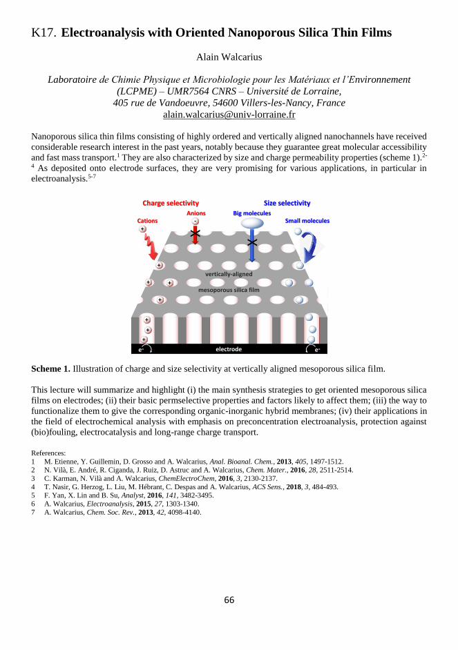
66
K17. Electroanalysis with Oriented Nanoporous Silica Thin Films
Alain Walcarius
Laboratoire de Chimie Physique et Microbiologie pour les Matériaux et l’Environnement
(LCPME) – UMR7564 CNRS – Université de Lorraine,
405 rue de Vandoeuvre, 54600 Villers-les-Nancy, France
Nanoporous silica thin films consisting of highly ordered and vertically aligned nanochannels have received
considerable research interest in the past years, notably because they guarantee great molecular accessibility
and fast mass transport.1 They are also characterized by size and charge permeability properties (scheme 1).2-
4 As deposited onto electrode surfaces, they are very promising for various applications, in particular in
electroanalysis.5-7
Scheme 1. Illustration of charge and size selectivity at vertically aligned mesoporous silica film.
This lecture will summarize and highlight (i) the main synthesis strategies to get oriented mesoporous silica
films on electrodes; (ii) their basic permselective properties and factors likely to affect them; (iii) the way to
functionalize them to give the corresponding organic-inorganic hybrid membranes; (iv) their applications in
the field of electrochemical analysis with emphasis on preconcentration electroanalysis, protection against
(bio)fouling, electrocatalysis and long-range charge transport.
References:
1 M. Etienne, Y. Guillemin, D. Grosso and A. Walcarius, Anal. Bioanal. Chem., 2013, 405, 1497-1512. 2 N. Vilà, E. André, R. Ciganda, J. Ruiz, D. Astruc and A. Walcarius, Chem. Mater., 2016, 28, 2511-2514.
3 C. Karman, N. Vilà and A. Walcarius, ChemElectroChem, 2016, 3, 2130-2137.
4 T. Nasir, G. Herzog, L. Liu, M. Hébrant, C. Despas and A. Walcarius, ACS Sens., 2018, 3, 484-493.
5 F. Yan, X. Lin and B. Su, Analyst, 2016, 141, 3482-3495.
6 A. Walcarius, Electroanalysis, 2015, 27, 1303-1340.
7 A. Walcarius, Chem. Soc. Rev., 2013, 42, 4098-4140.
CationsBig molecules
Small molecules+
-
X
X
+
+
+
vertically-aligned
mesoporous silica film
electrodee- e-
+
+
+ +
Anions
Charge selectivity Size selectivity

67
68
K18. Modification of Electrode Surface with Multifunctional
Nano- and Micro-Layers of Materials
Sensitive to Environmental Parameters
Kamil Marcisz, Klaudia Kaniewska, Marcin Mackiewicz, Marcin Karbarz, Zbigniew Stojek
Faculty of Chemistry, University of Warsaw, ul. Pasteura 1, 02-093 Warsaw, Poland
Polymeric hydrogels became attractive mainly because of their ability to perform a reversible phase
transformation that is associated with a large change in their volume and triggered by a change in
environmental conditions. A big research effort was directed to extension of the list of such environmental
parameters and to modification of the phase transformations. In this respect the changes in temperature, ionic
strength and pH, substitution of the crosslinker, use of additional monomers in the synthesis, and the presence
of some ions can be mentioned. A clear trend is emerging: it is the synthesis of hydrogels of micro and nano
sizes.1,2 It is mainly associated with a significant reduction of phase-transformation time and facilitation of
reaching, by ions and molecules, the deep inside and outside of the gel. Progress in hydrogel deposition on
conductive surfaces can be useful in a number of issues; it can lead to an improvement in the construction of
artificial muscles and it can also improve the performance of biosensors and the ON-OFF systems in
microfluidic systems and labs on chips.
This presentation focuses on discussion of various ways of modification of electrodes/conductive-
materials with nano- and micro-hydrogels. The main emphasis will be given to electrodeposition of very thin
layers and chemisorption of spherical nanoparticles. The electrochemical properties of the electrodes
modified with the hydrogels in various ways differ a lot. They particularly differ in their response to volume
phase transition. The conditions have been found for the electrochemical induction of the volume phase
transformation at the surface of an electrode.3 After the introduction of either amino ferrocene or hexaamino
ruthenium(III)/(II) system into the hydrogel, it was possible to trigger the phase transition by applying an
appropriate potential. In addition, the change in microgel size / volume using the electrochemically induced
volume phase transition was significant and reversible. It also turned out that thin hydrogel films are very
convenient matrices for enzymes.
References: 1 M. Karbarz, M. Mackiewicz, K. Kaniewska, K. Marcisz and Z. Stojek, Appl. Mater. Today, 2017, 9, 516–532.
2 E. Katz, Electroanalysis, 2016, 28, 1916-1929.
3 K. Marcisz, A. Gawronska, Z. Stojek and M. Karbarz, ACS Appl. Mater. Interfaces, 2019, 11, 12114−12120.

69
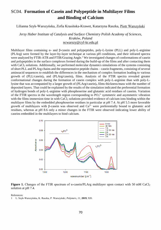
70
SC04. Formation of Casein and Polypeptide in Multilayer Films
and Binding of Calcium
Lilianna Szyk-Warszyńska, Zofia Krasińska-Krawet, Katarzyna Raszka, Piotr Warszyński
Jerzy Haber Institute of Catalysis and Surface Chemistry Polish Academy of Sciences,
Kraków, Poland
Multilayer films containing α- and β-casein and polypeptides, poly-L-lysine (PLL) and poly-L-arginine
(PLArg) were formed by the layer-by-layer technique at various pH conditions, and their infrared spectra
were analyzed by FTIR-ATR and FTIR/Grazing Angle.1 We investigated changes of conformations of casein
and polypeptides in the surface complexes formed during the build-up of the films and after contacting them
with CaCl2 solutions. Additionally, we performed molecular dynamics simulations of the systems consisting
of short PLL and PLArg chains and the representative peptide chains – casein fragments, consisting of several
aminoacid sequences to establish the differences in the mechanism of complex formation leading to various
growth of (PLL/casein)n and (PLArg/casein)n films. Analysis of the FTIR spectra revealed greater
conformational changes during the formation of casein complex with poly-L-arginine than with poly-L-
lysine that was accompanied by a larger growth of (PLArg/casein)n films thickness/mass with the number of
deposited layers. That could be explained by the results of the simulation indicated the preferential formation
of hydrogen bonds of poly-L-arginine with phosphoserine and glutamic acid residues of caseins. Variation
of the FTIR spectra in the wavelength region corresponding to PO32- symmetric and asymmetric vibration
with the films immersion time in with CaCl2 solutions provided evidence of calcium ions binding within the
multilayer films by the embedded phosphoserine residues in particular at pH 7.4. At pH 5.5 more favorable
growth of multilayers with β-casein was observed and Ca2+ were preferentially bound to glutamic acid
residues, whereas at pH 8.6 only a minor changes in the FTIR were observed indicating lower ability of
caseins embedded in the multilayers to bind calcium.
Figure 1. Changes of the FTIR spectrum of α-casein/PLArg multilayer upon contact with 50 mM CaCl2
solution at pH 7.4.
References:
1 L. Szyk-Warszyńska, K. Raszka, P. Warszyński, Polymers, 11, 2019, 920.

71
72
Sunday, November 10
08:00-09:00 Breakfast
09:00-
10:35
Morning session 1
Chairs: P. Krysiński / J. Bobacka
09:00-
09:40
T03 Francis D’Souza
Interfacial Electron Transfer of Multimolecular Assemblies on Metal
Oxide Semiconductors
09:40-
10:00
K19 Frank Marken
Polymers of Intrinsic Microporosity (PIMs) in Electrochemistry
10:00-
10:20
K20 Stéphane Arbault
Using Cold Plasmas to Improve Electrochemical Sensors and vice versa
10:12-
10:35
SC05 Krzysztof R. Noworyta
Oxidation and Coupling of the Selected Carbazole Derivatives – Why It
Does Not Always Lead to Polymer Formation
10:35-11:00 Coffee break
11:00-
12:50
Morning session 2
Chairs: K. Noworyta / A. Yarman
11:00-
11:20
K21 Karsten Haupt
Molecularly Imprinted Polymer Nanoparticles as Synthetic Antibody
Mimics for the Detection and Modulation of Cellular Function and
Immunotherapy
11:20-
11:40
K22 Frieder W. Scheller
Electrosynthesized MIPs for Peptides and Proteins
11:40-
12:00
K23 Peter A. Lieberzeit
From Self-organized Monolayers to Surface Molecular Imprints: Assay
Formats Extending Beyond the Obvious
12:00-
12:20
K24 Cédric Ayela
Combining Molecularly Imprinted Polymers (MIPs) with Micro (Opto)
Electro Mechanical Systems (M(O)EMS) for Improved Chemical Sensing
12:20-
12:35
SC06 Yasuo Yoshimi
Application of Swelling Phenomena of Molecularly Imprinted Polymers by
Specific Interaction with the Target Molecule for Sensing Technology
12:35-
12:50
SC07 Cecilia Cristea
Biomimetic Approaches over Direct Detection of Antibiotics from Real
Samples
13:00-14:30 Group Photo & Lunch
14:30-
16:20
Afternoon session 1
Chairs: R. Gyurcsanyi / F. Conzuelo
14:30-
14:50
K25 Pawel J. Kulesza
Development of Highly Specific Interfaces for Photoelectrochemical and
Electrocatalytic Reduction of Carbon Dioxide
73
14:50-
15:10
K26 Ambra Giannetti
A Fluorescence-based POCT Device for Immunosuppressant-drug
Monitoring in Transplanted Patients
15:10-
15:30
K27 Joanna Niedziółka-Jönsson
Long-Period Fiber Gratings for Sensing and Biosensing
15:30-
15:50
K28 Sebastian Maćkowski
Sensing with Plasmonically Active Metallic Nanostructures
15:50-
16:05
SC08 Wolfgang Fritzsche
Localized Surface Plasmon Resonance (LSPR) on Metal Nanoparticles for
Bioanalytical Applications
16:05-
16:20
SC09 Simone Berneschi
Optical Microbubble Resonators as Emerging Tools for Environmental
Sensing: the “Safe Water” Project
16:30-19:00 Social program
19:00-22:00 Dinner/Banquette
74
T03. Interfacial Electron Transfer of Multimolecular Assemblies
on Metal Oxide Semiconductors
Francis D’Souza
Department of Chemistry, University of North Texas, Denton, TX 76203, USA
Semiconducting metal oxide nanoparticles offer a unique substrate for the assembly of multiple, photoactive,
molecular components at the interface. The choice of photoactive molecules, nature of semiconducting metal
oxide (p-type or n-type), and the type of the assembly method (covalent versus self-assembly), and nature of
spacer connecting the photoactive molecules to the surface, can have a profound influence on the mechanism,
rate, and efficiency of photoinduced energy and electron transfer events at the interface. The variety and high
level of control, these interfacial assemblies are of interest for many applications including solar energy
harvesting, optoelectronics, photo-electrosynthesis, photo-sensing, photo-writable memory, and more. These
assemblies, although, are generated with different end goals, they rely on similar surface binding motifs and
molecular structure-property relationships. The goal of this tutorial talk is to summarize the various strategies
(i.e. covalent binding, axial coordination, ion-ion and ion-dipole electrostatics, host-guest interactions, etc.),
developed mainly from our laboratories, for assembling chromophores, hosts, and electron donors/acceptors
on mesoporous metal oxide substrates and conducting electrode surfaces. The assembly, characterization,
and subsequent photoinduced events (i.e. cross-surface electron transfer, interchromophore energy transfer,
electron injection and recombination, and others), relevant to dye sensitized solar cell applications, will be
discussed for the various assembly approaches.

75
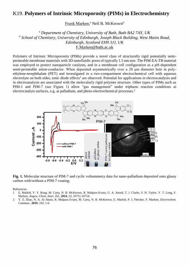
76
K19. Polymers of Intrinsic Microporosity (PIMs) in Electrochemistry
Frank Marken,a Neil B. McKeownb
a Department of Chemistry, University of Bath, Bath BA2 7AY, UK b School of Chemistry, University of Edinburgh, Joseph Black Building, West Mains Road,
Edinburgh, Scotland EH9 3JJ, UK
Polymers of Intrinsic Microporosity (PIMs) provide a novel class of structurally rigid potentially semi-
permeable membrane materials with 3D nanofluidic pores of typically 1.5 nm size. The PIM-EA-TB material
was employed to protect nanoparticle catalysts, and in a membrane cell configuration as a pH-dependent
semi-permeable anion-conductor. When deposited asymmetrically over a 20 μm diameter hole in poly-
ethylene-terephthalate (PET) and investigated in a two-compartment electrochemical cell with aqueous
electrolyte on both sides, ionic diode effects1 are observed. Potential for applications in electrocatalysis and
in electroanalysis are associated with the molecularly rigid polymer structure. Other types of PIMs such as
PIM-1 and PIM-7 (see Figure 1) allow “gas management” under triphasic reaction conditions at
electrocatalyst surfaces, e.g. at palladium, and photo-electrochemical processes.2
Fig. 1. Molecular structure of PIM-7 and cyclic voltammetry data for nano-palladium deposited onto glassy
carbon with/without a PIM-7 coating.
References:
1 E. Madrid, Y. Y. Rong, M. Carta, N. B. McKeown, R. Malpass-Evans, G. A. Attard, T. J. Clarke, S. H. Taylor, Y. T. Long, F. Marken, Angew. Chem.-Inter. Ed., 2014, 53, 10751-10754.
2 Y. Z. Zhao, N. A. Al Abass, R. Malpass-Evans, M. Carta, N. B. McKeown, E. Madrid, P. J. Fletcher, F. Marken, Electrochem.
Commun., 2019, 103, 1-6.

77

78
K20. Using Cold Plasmas to Improve Electrochemical Sensors
and vice versa
Colin Camille,a Badets Vasilica,a Girard Fanny,a,b Lefrançois Pauline,a Botsos-Margerit Ugo,a
Zigah Dodzi,a Blanc Sylvie,b Sojic Neso,a Clément Franck,b Arbault Stéphanea
a Univ. BORDEAUX, ISM, Bordeaux INP, CNRS UMR 5255, NSysA Group, Pessac, France b UPPA, IPREM, CNRS UMR 5254, Pau, 64000, France
Cold plasmas are weakly ionized gases, generated by applying High-Voltage electrical discharges in a
gaseous environment. They can be produced by sinusoidal or pulse power supplies, at either low pressures
(ex. mTorr) or in ambient conditions depending on the electric field intensity as well as on the used gas
(mixtures of N2, O2, Ar, He, methane etc.). Cold plasmas are complex mixtures of neutral species, electrons,
ions, excited species (radiative and metastable states, radicals…) and emitted photons (UV, Vis, IR). In
particular, when generated in oxygen-nitrogen mixtures (air), multiple reactive oxygen and nitrogen species
(ROS, RNS) are present in the gas phase. Because of their diverse and intense reactivities, and relative ease
for production, cold plasmas reveal themselves as excellent approaches to treat surfaces in order to modify
their physical (cleaning, roughening) and chemical (functional groups) states.
In this context, we applied cold plasmas to modify the reactivity of metal and carbon electrode surfaces
and studied the resulting effects on the detection of compounds involved by biosensors.1,2 We first treated
platinum and black platinum modified electrodes by 100 % Oxygen-plasmas produced by radio-frequency
modulated electric fields (plasma cleaner setup) and observed lower capacitive currents, more stable faradaic
responses, decreased standard potentials and improved sensitivity for H2O2 detection. These properties were
related (XPS analyses) to a change of ratio between Pt oxides, which improve the surface hydrophilicity and
initial adsorption of ROS. Similarly, the reactivity of glassy carbon electrodes could be homogenized and
improved for the detection of both inner- or outer-sphere electron transfer species when treated by plasmas,
allowing to prevent from polishing electrode surfaces for their cleaning or activation. Moreover, plasma
effects were stable for periods longer than one week.
Reciprocally, such electrochemical sensors were involved to decipher on the nature of species produced
by cold atmospheric pulsed plasmas when used to expose physiological fluids, cells or tissues.3-5 Plasmas
raise exponential interests for biomedical applications because of their strong reactivities. However, the true
nature of reactive species involved in biocidal or oxidative bio-activation remains widely unsolved. To
decipher on the in situ reactivity within solutions exposed to plasmas, we developed shielded microelectrodes
to detect ROS and RNS appearing in solution, at millimetric distances from the plasma flow and electric
source (nanosecond pulsed discharges). The concentration rises of hydrogen peroxide, nitrite as well as of
superoxide anion were monitored with an unprecedented spatial and temporal resolutions.6
Consequently, we have demonstrated that electrochemical sensors can benefit from plasma treatments
and vice versa.
References: 1 S. Ben-Amor, E. Vanhove, E. Suraniti, P. Temple-Boyer, N. Sojic, J. Launay and S. Arbault, Electrochim. Acta, 2014, 126, 171–
178.
2 V. Badets, J. Pandard, N. Sojic and S. Arbault, ChemElectroChem, 2016, 3, 2288-2296. 3 F. Girard, V. Badets, S. Blanc, K. Gazeli, L. Marlin, L. Authier, P. Svarnas, N. Sojic, F. Clément and S. Arbault, RSC Adv., 2016,
6, 78457–78467.
4 M. Dezest, A. L. Bulteau, D. Quinton, L. Chavatte, M. Le Béchec, S. Arbault, A. N. Salvayre, S. Cousty, F. Clément, PLOS One, 2017, 12, e0173618.
5 F. Girard, M. Peret, N. Dumont, V. Badets, S. Blanc, K. Gazeli, C. Noël, T. Belmonte, L. Marlin, J. P. Cambus, G. Simon, N.
Sojic, B. Held, S. Arbault and F. Clément, Phys. Chem. Chem. Phys., 2018, 20, 9198-9210. 6 F. Girard-Sahun, V. Badets, P. Lefrançois, N. Sojic, F. Clément and S. Arbault, Anal. Chem., 2019, 91, 8002-8007.

79
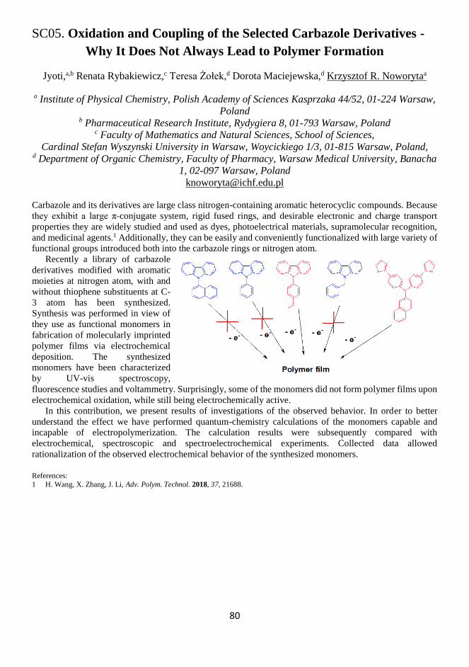
80
SC05. Oxidation and Coupling of the Selected Carbazole Derivatives -
Why It Does Not Always Lead to Polymer Formation
Jyoti,a,b Renata Rybakiewicz,c Teresa Żołek,d Dorota Maciejewska,d Krzysztof R. Noworytaa
a Institute of Physical Chemistry, Polish Academy of Sciences Kasprzaka 44/52, 01-224 Warsaw,
Poland b Pharmaceutical Research Institute, Rydygiera 8, 01-793 Warsaw, Poland
c Faculty of Mathematics and Natural Sciences, School of Sciences,
Cardinal Stefan Wyszynski University in Warsaw, Woycickiego 1/3, 01-815 Warsaw, Poland, d Department of Organic Chemistry, Faculty of Pharmacy, Warsaw Medical University, Banacha
1, 02-097 Warsaw, Poland
Carbazole and its derivatives are large class nitrogen-containing aromatic heterocyclic compounds. Because
they exhibit a large π-conjugate system, rigid fused rings, and desirable electronic and charge transport
properties they are widely studied and used as dyes, photoelectrical materials, supramolecular recognition,
and medicinal agents.1 Additionally, they can be easily and conveniently functionalized with large variety of
functional groups introduced both into the carbazole rings or nitrogen atom.
Recently a library of carbazole
derivatives modified with aromatic
moieties at nitrogen atom, with and
without thiophene substituents at C-
3 atom has been synthesized.
Synthesis was performed in view of
they use as functional monomers in
fabrication of molecularly imprinted
polymer films via electrochemical
deposition. The synthesized
monomers have been characterized
by UV-vis spectroscopy,
fluorescence studies and voltammetry. Surprisingly, some of the monomers did not form polymer films upon
electrochemical oxidation, while still being electrochemically active.
In this contribution, we present results of investigations of the observed behavior. In order to better
understand the effect we have performed quantum-chemistry calculations of the monomers capable and
incapable of electropolymerization. The calculation results were subsequently compared with
electrochemical, spectroscopic and spectroelectrochemical experiments. Collected data allowed
rationalization of the observed electrochemical behavior of the synthesized monomers.
References:
1 H. Wang, X. Zhang, J. Li, Adv. Polym. Technol. 2018, 37, 21688.

81
82
K21. Molecularly Imprinted Polymer Nanoparticles
as Synthetic Antibody Mimics for the Detection and Modulation
of Cellular Function and Immunotherapy
Karsten Haupt, Paulina Medina Rangel, Jingjing Xu, Bernadette Tse Sum Bui
Université de Technologie de Compiègne, CNRS Institute for Enzyme and Cell Engineering,
Compiègne, France
Molecularly imprinted polymers (MIPs, Figure 1) are synthetic antibody mimics that specifically recognize
molecular targets.1,2 They are highly cross-linked polymers synthesized in the presence of the target molecule
acting as a molecular template. This templating induces three-dimensional binding sites in the polymer that
are complementary to the template in size, shape and chemical functionality. The synthetic antibody can
recognize and bind its target with an affinity and selectivity similar to a biological antibody.1
Herein, we demonstrate the potential of MIPs for immunotherapy on the example of a protein cancer
biomarker, a cell-cell recognition protein.3 In addition, we present the use of MIPs for the inhibition or
modulation of cell-virus interactions.4
MIPs were synthesized using an innovative solid-phase synthesis approach in which an epitope of the
biomarkers was immobilized on glass beads (as solid support) via click chemistry. This configuration allows
an oriented immobilization of the template. A short peptide (terminal alkyne or azide functionalized) was
selected as epitope for immobilization on azide-modified glass beads. The epitope was either a terminal
peptide sequence, or an internal peptide. In the latter case, a cyclic version of the peptide was used as the
template. Thermo-responsive MIP nanoparticles were synthesized around the template. Due to the oriented
immobilization of the latter, the binding sites of the resulting MIPs all have the same orientation and a
homogeneous affinity distribution in the nanomolar range, thus MIPs synthesized by the solid-phase
approach can be considered analogous to monoclonal antibodies. The MIP nanoparticles (50 nm) specifically
recognized both the template peptides and the whole proteins. Mutation of a single amino acid in the peptide
sequence resulted in a reduced affinity by three orders of magnitude.
Cell imaging studies were done with fluorescently labeled MIP nanoparticles, using epifluorescence and
confocal fluorescence microscopy. They showed specific binding of the synthetic antibody to the cell surface
targets, since the non-imprinted control polymer did not show any staining, and when the target protein was
hydrolytically removed from the cell, staining was dramatically decreased. They were able to modulate
specific cellular functions.
References: 1 K. Haupt, A. V. Linares, M. Bompart and B. Tse Sum Bui, Top. Curr. Chem., 2012, 325, 1-28. 2 S. Ambrosini, S. Beyazit, K. Haupt and B. Tse Sum Bui, Chem. Commun., 2013, 49, 6746-6748.
3 P. X. Medina Rangel, S. Laclef, J. Xu, M. Panagiotopoulou, J. Kovensky, B. Tse Sum Bui, K. Haupt, Sci. Rep., 2019, 9, 3923.
4 J. Xu, F. Merlier, B. Avalle, V. Vieillard, P. Debré, K. Haupt, B. Tse Sum Bui, ACS Appl. Mater. Interfaces, 2019, 11, 9824-9831.

83

84
K22. Electrosynthesized MIPs for Peptides and Proteins
Frieder W. Scheller,a Aysu Yarman,a Xiaorong Zhang,a Katharina J. Appelt,a Ulla Wollenberger,a
Giorgio Caserta,b Ingo Zebger,b Goksu Ozcelika,c Sevinc Kurbanoglu,c
Sibel A. Ozkan,c Róbert E. Gyurcsányid
a Universität Potsdam, Institute for Biochemistry and Biology, Potsdam, Germany b Technische Universität Berlin, Inst. für ChemieBerlin, Germany
c Ankara University, Dept. of Analytical Chemistry, Ankara, Turkey, d Budapest University of Technology and Economics, Budapest, Hungary
Molecular imprinting is a method to create binding cavities in a polymer for the preferential recognition of
the target. Here, we present two approaches for electrosynthesizing protein-MIPs using scopoletin or o-
phenelenediamine as the functional monomers.
(i) Epitope imprinting: MIPs for the recognition of the N-terminal peptides of human adult hemoglobin
(HbA) or glycated Hb (HbA1c) were synthesized by applying either the mixtures of scopoletin and the
respective terminal pentapeptides or a SAM of the cys-extended N-terminal pentapeptide of the ß-chain in
the MIP synthesis by electropolymerizing scopoletin on the gold electrode. These two peptides are the
analytes in the IFCC (the International Federation of Clinical Chemistry) Reference Method for the
determination of the long-term biomarker for diabetes HbA1c. All steps of MIP-synthesis and rebinding were
analyzed by square wave voltammetry (SWV) of the redox marker ferricyanide and by surface-enhanced
infrared absorption (SEIRA) spectroscopy of both the bound target, i.e., the peptides or the parent protein,
and the polymer poly-scopoletin. Combination of the respective MIPs allows the quantification of both the
glycated and the non-glycated N-terminal peptides of HbA1c and HbA.
(ii) Holoprotein as target: MIP-sensors for the copper containing enzymes laccase and tyrosinase,
cytochrome P450cam and butyrylcholineesterase were prepared by electropolymerizing the mixture of
monomer and target protein. For these highly active enzymes, synthesis of holoprotein-MIPs is the
straightforward approach.
Acknowledgements:
Financial support from Deutsche Forschungsgemeinschaft (DFG) within the framework of the German Excellence Strategy – EXC
2018/1 (UniSysCat), ERA-Chemistry (2014, 6113 andNKFIH OTKA NN117637), Ankara University Erasmus and UBED Programs is acknowledged.
References: 1 J. Erdőssy, V. Horváth, A. Yarman, F. W. Scheller, R. E. Gyurcsányi, Trends Anal. Chem., 2016, 79, 179–190.
2 K. J. Jetzschmann, A. Yarman, L. Rustam, P. Kielb, V. B. Urlacher, A. Fischer, I. M. Weidinger, U. Wollenberger, F. W. Scheller,
Colloids Surf. B Biointerfaces, 2018, 164, 240–246. 3 X. Zhang, A. Yarman, J. Erdőssy, S. Katz, I. Zebger, K. J. Jetzschmann, Z. Altintas, U. Wollenberger, R. E. Gyurcsányi, F. W.
Scheller, Biosens. Bioelectron., 2018, 105, 29–35.
4 A. Yarman, Turk. J. Chem., 2018, 42, ,346-354.

85
86
K23. From Self-organized Monolayers
to Surface Molecular Imprints:
Assay Formats Extending Beyond the Obvious
Agienszka Strallhofer,a Chakpetch Koitio,b Supaporn Klangprapan,b
Kiattawee Choowongkomon,b Peter A. Lieberzeita
a University of Vienna, Faculty for Chemistry, Department of Physical Chemistry, Vienna,
Austria b Faculty of Science, Department of Biochemistry, Kastesart University, Bangkok, Thailand
Sensing and analysis are among the main driving forces of developing novel responsive surface
modifications. To avoid the limitations of biological species, they often make use of different biomimetic
approaches.1 Many of those approaches rely on self-organization at some point, for instance by immobilizing
recognition species (such as aptamers) on sensor surfaces, or generating surface molecularly imprinted
polymers (MIP).
Molecularly imprinted polymers2 have proven useful for selectively binding biological species ranging
from proteins to entire cells.3 One often assumes that they do so by forming non-covalent interactions with
a part of the outer surface of the respective target species. It is possible to assess this claim by comparing the
sensor responses of different selective surfaces on similar devices with each other. Studies on the porcine
respiratory and reproductive syndrome virus (PRRSV) with aptamers and surface MIP revealed that the
former gave rise to somewhat higher selectivity toward two competing species (porcine respiratory virus –
PRV; common swine fewer virus – CSFV). However, surface MIP led to ten times higher sensitivity of the
respective coated quartz crystal microbalances (QCM). Both results indicate that aptamers indeed bind to
clearly defined “receptor” sites, whereas MIP interact with the entire outer shell of the virus. Selectivity of
MIP relies on two different factors, namely geometric fit and surface chemistry, respectively.
Similar comparative studies revealed that both MIP and immobilized thrombin are useful for binding
thrombocytes to the respective sensor surface, be it a QCM or an SPR substrate (SPR: surface plasmon
resonance). The main challenge from the imprinting side in this case is given by the fact that it is by far not
trivial to generate stamps comprising native, i.e. non-activated thrombocytes. However, overcoming this
obstacle allowed us to design sensor materials that give rise to concentration-dependent responses when
exposed to different platelet concentrates. Response times are in the range of around ten minutes. Though
this is slower than current cell counting systems, MIP sensors have one main advantage: sensor signals do
not only depend on cell concentration, but also on the status of thrombocytes: exposing them to anti-
coagulants changes the chemical properties of their surfaces. This leads to decreased sensor effects due to
lower affinity to the respective MIP. We could reproduce such behavior with thrombin immobilized to SPR
chips. Therefore, such sensors allow for assessing the status of the respective cells. Within about ten minutes,
one can hence assess the coagulation ability of the respective cells, which is much faster than in current
clinical diagnostics.
References: 1 M. Hussain, J. Wackerlig, P. A. Lieberzeit, Biosensors, 2013, 3, 89-107.
2 B. T. S Bui, K. Haupt, Anal. Bioanal. Chem., 2010, 398, 2481-2492.
3 K. Eersels, P. Lieberzeit, P. Wagner, ACS Sens., 2016, 1, 1171-1187.

87

88
K24. Combining Molecularly Imprinted Polymers (MIPs)
with Micro (Opto) Electro Mechanical Systems (M(O)EMS)
for Improved Chemical Sensing
Cédric Ayela,a Frank Bokeloh,a Olivier Soppera,b Karsten Hauptc
a Univ. Bordeaux, IMS, CNRS, UMR 5218, Bordeaux INP, ENSCBP, Talence, France b IS2M, CNRS, UMR 7361, Univ. Haute Alsace, Mulhouse, France
c Sorbonne Universités, UTC, CNRS, GEC UMR 7025, Compiègne, France
Molecularly imprinted polymers (MIPs) are tailor-made antibody mimics obtained by a templating process
at the molecular level. These tailor-made polymers have, unlike their natural biological counterparts, a
superior chemical and physical stability and are therefore excellent candidates for integration into sensors
and biochips. Over the years, we have reported several strategies of micro (opto) electro mechanical systems
(M(O)EMS) with integrated MIP pattern, using different micro- and nanofabrication techniques for the
synthesis of 2D and 3D MIP-based sensors. First, we showed the combination of MIPs with resonant silicon
micromembranes enabling the label-free detection of low molecular weight analytes, by means of mass
sensing.1 To go further, we have then reported an all-organic sensor consisting in a simple monolayered free-
standing MIP microcantilever, used once again as mass sensor.2 Merging the mechanical transducer and the
MIP film into a single device reduces the total mass of the device, one important characteristic to increase
their mass sensitivity. Results showed the sensitive and specific binding of the beta-antagonist drug S-
propranolol. In particular, a limit of detection of 100nM was achieved, a successful challenge for label-free
mass sensors. More recently, the technique of 2-photon stereolithography was used to fabricate 2.5D MIP
diffraction gratings and 3D MIP microcantilevers in a single step.3 The synthesis of MIPs by 2-photon
stereolithography enables the fabrication of real 3D structures with a sub-micrometer resolution and
overcomes the limitations of standard 2D microstructures. Diffraction and resonance frequency
measurements allowed demonstrating specific analyte binding (testosterone and Z-L-Phenylalanine) to
diffraction gratings and microcantilevers, respectively. Last, but not least, we showed very recently that MIP
microcantilevers can be synthesized directly at the end of optical fibers.4 This technique was introduced by
Ton et al. using the polymer tip as a fluorescence sensor, that we have adapted for label-free, mass sensing
using the resonance of the MIP tip.5 With this approach, the same optical fiber is used for both the fabrication
of a polymer beam at its cleaved end, and the dynamic mode operation of the cantilever. In particular,
optomechanical coupling enables both actuation of the MIP cantilevers and detection of the resonance,
enabling remote sensing over long distances. This approach is of particular interest for the development of
mass-produced chemical sensors, to be exploited at industrial level.
References:
1 C. Ayela, F. Vandevelde, D. Lagrange, K. Haupt, L. Nicu, Angew. Chem. Intl Ed., 2007, 46, 9271-9274.
2 C. Ayela, G. Dubourg, C. Pellet, K. Haupt, Adv. Mater., 2014, 26, 5876-5879. 3 L. Piedad Chia Gomez, A. Spangenberg, X-A. Ton, Y. Fuchs, F. Bokeloh, J-P. Malval, B. Tse Sum Bui, D. Thuau, C. Ayela, K.
Haupt, O. Soppera, Adv. Mater., 2016, 28, 5931-5937.
4 X-A. Ton, B. Tse Sum Bui, M. Resmini, P. Bonomi, I. Dika, O. Soppera, K. Haupt, Angew. Chem. Intl Ed., 2013, 52, 8317-8321.
5 C. Ayela, F. Bokeloh, K. Haupt, Optical fiber based organic cantilever with integrated transduction, (2018), Patent
EP18162383.6/WO2019110849.

89
90
SC06. Application of Swelling Phenomena
of Molecularly Imprinted Polymers by Specific Interaction
with the Target Molecule for Sensing Technology
Y. Yoshimi, H. Eguchi, Y. Takeda, M. Abe, and Y. Katsumata
a Department of Applied Chemistry, Shibaura Institute of Technology Tokyo, Japan
It has been shown that the faradic current at an electrode grafted with molecularly imprinted polymer (MIP)
is sensitive to the specific target molecule used as the template.1 This phenomenon termed “gate effect” is
applicable to sensors with very high selectivity and short response time. For example, gate effect of heparin
imprinted MIP is sensitive to heparin but insensitive to chondroitin sulfate C (CSC) which has similar
structure as heparin. The response time is less than 60 s.2 But the sensing mechanism is still in a black box.
We investigated the size sensitivity of nanoparticles of molecularly imprinted polymers (MIP-NPs) to a
specific interaction for determination of the mechanism of the gate effect and its feasibility for new
applications.3 Nanoparticles of poly(methacryloxy ethyl trimethylammonium chloride-co-acrylamide-co-
methylenebisacrylamide) imprinted with heparin immobilized on glass beads were synthesized. The diameter
of the MIP-NPs of heparin was increased by the presence of the heparin template but was insensitive to CSC,
the analogue of heparin. The high selectivity of the MIP-NPs was consistent with the selectivity of electrodes
grafted with a heparin-imprinted polymer in our previous studies. The quartz crystal microbalance probes
immobilizing heparin or CSC were sensitive to MIP-NPs, which indicates that the binding ability of MIP-
NP does not discriminate between the template and other glycosaminoglycans. These results indicate that
the size of the MIP-NP is sensitive to the matched binding with the template through the imprinted cavity
and confirm that the sensors using the gate effect of MIP enable more highly specific sensing of the targets
used as a template than those detecting the targets bound with MIP directly. The MIP-NP would be potential
tool for understanding the mechanism of the gate effect. As an expanded the application of MIP-NP, we
revealed that the MIP-NP can be used as an optical probe which can visualize the secretion of the target
secreted from cells.4
References:
1 Y. Yoshimi, R. Ohdaira, C. Iiyama, K. Sakai, Sens. Actuators B, 2001, 73, 49-53. 2 Y. Yoshimi, K. Sato, M. Ohshima, E. Piletska, Analyst, 2013, 138, 5121-5128.
3 Y. Yoshimi, D. Oino, H. Ohira, H. Muguruma, E. Moczko, S. A. Piletsky, Sensors, 2019, 19, 2415. 4 www.shibaura-it.ac.jp/en/news/2018/40180026.html

91
92
SC07. Biomimetic Approaches over Direct Detection
of Antibiotics from Real Samples
Adrian Blidar, Bogdan Feier, Oana Hosu, Gheorghe Melinte, Mihaela Tertis, Cecilia Cristea
Department of Analytical Chemistry, Faculty of Pharmacy, “Iuliu Hațieganu” University of
Medicine and Pharmacy, 4 Pasteur Street, 4000349, Cluj-Napoca, Romania
There are numerous analytical methods for the detection of different classes of antibiotics, but most of them
are relatively expensive and require complex laboratory instruments, leading to the impossibility to perform
field analyses. Electrochemical techniques are a viable alternative due to their sensitivity and accessibility,
both from an economical and logistical point of view, making them suitable for in situ analysis, a big
advantage for environmental and clinical tests.1 β-lactam antibiotics are the most important and the most
widely used group of antibiotics, consisting mainly of two subclasses, penicillins and cephalosporins.
Tetracyclines (TCs) are one of the most common used antibiotics for the treatment of infectious diseases in
veterinary medicine. Therefore, high TCs levels can be found in animal-based food products (e.g. meat, eggs,
milk) and environmental samples (e.g. soil, waste water) due to its high accumulation. The overuse of this
therapeutic class, for prophylactic and treatment purposes, but also in agriculture, assures a constant
exposure, increasing the risk of allergies and resistance to broad-spectrum antibacterial drugs. A part of the
solution for this concerning situation is the development of new and rapid detection methods capable of
monitoring the levels of antibiotics from different matrices.1,2
Using direct approaches, we tested the electrochemical behavior of several members of the penicillins
and cephalosporins classes, recording their electrochemical signals, but this way was unable to distinguish
between molecules from the same subclass. However, using this approach, the quantification of cefalexin
(CFX) and oxacillin was performed.
In order to surpass the limitations in terms of selectivity and sensitivity, the biomimetic approaches were
employed by using molecularly imprinted polymers (MIPs) and aptamers (short single-stranded DNA and
RNA oligonucleotides, artificially selected for their capacity to bind specifically a target molecule). The
biomimetic approach improved the detection of CFX, both in terms of selectivity and sensitivity, leading to
the development of an electrochemical MIP-based sensor for CFX. Another biomimetic strategy allowed the
detection of ampicilin using an electrochemical surface plasmon resonance aptasensor. AMP direct detection
was proved to be unsuccessful due to the low value of the response and also due its similarity to the response
of other penicillins.2 As for the direct detection and quantification of tetracycline, gold was electrodeposited
at glassy carbon electrodes and carbon-based screen-printed electrodes from a HAuCl4 solution to obtain
different 3D-nanostructures (nanoparticles and nanovoids). A ferrocene-labelled aptasensor for TC detection
was developed using the platform with the best analytical performance.
Acknowledgements: This work was supported by a grant of the Romanian National Authority for Scientific Research and Innovation, CCCDI-UEFISCDI,
project number
ERANET-RUS-PLUS-PLASMON-ELECTROLIGHT/46/2018, within PNCDI III.
References:
1 B. Feier, A. Gui, C. Cristea and R. Sandulescu, Anal. Chim. Acta, 2017, 976, 25-34. 2 A. Blidar, B. Feier, M. Tertis, R. Galatus and C. Cristea, Anal. Bioanal. Chem., 2019, 411(5),
1053-1065.
3 B. Feier, I. Ionel, C. Cristea and R. Sandulescu, New J. Chem., 2017, 41, 12947-12955.

93
94
K25. Development of Highly Specific Interfaces
for Photoelectrochemical and Electrocatalytic Reduction
of Carbon Dioxide
Pawel J. Kulesza, Iwona A. Rutkowska
Faculty of Chemistry, University of Warsaw, Poland
Electroreduction of carbon dioxide to simple organic fuels and chemicals is a topic of growing scientific and
technological interest. The reaction provides means for both reducing emissions of CO2 into atmosphere and
storing renewable energy. The presentation will address low-temperature CO2-conversion processes based
on electrocatalytic and photoelectrochemical approaches. Among important issues are choice of the catalytic
or semiconducting materials, their morphology and operating conditions including temperature, solvent,
electrolyte, pH etc. There is a need to improve the reaction dynamics and selectivity toward specific products.
In practical electrolysis cells, the CO2-reduction (at cathode) is accompanied by water oxidation (at anode or
photoanode).
Recently, we have concentrated on the development of hybrid materials by utilizing combination of metal
oxide semiconductors thus capable of effective photoelectrochemical reduction of carbon dioxide. For
example, the combination of conducting polymers, or titanium (IV) oxide, and copper (I) oxide has been
considered before and after sunlight illumination. Application of the hybrid system composed of both above-
mentioned oxides resulted in high current densities originating from photoelectrochemical reduction of
carbon dioxide mostly to methanol (CH3OH) as demonstrated upon identification of final products. Among
important issue is intentional stabilization, activation, and functionalization of the mixed-metal-oxide-based
photoelectrochemcal interface toward better long-term performance and selectivity production of small
organic molecules (C1-C4) and other chemicals. In this respect, ultra-thin films of conducting polymers
(simple or polyoxometallate-derivatized) and supramolecular complexes (with nitrogen containing ligands
and certain transition metal sites), sub-monolayers of metals (Cu, Au), networks of noble metal (Au, Ag)
nanoparticles or layers of robust bacterial biofilms have been considered.

95
96
K26. A Fluorescence-based POCT Device
for Immunosuppressant-drug Monitoring
in Transplanted Patients
A. Giannetti,a S. Tombelli,a C. Trono,a S. Berneschi,a C. Berrettoni,a F. Baldini,a R. Bernini,b
I. A. Grimaldi,b G. Testa,b G. Persichetti,b G. Porro,c G. Quartoc
a CNR-IFAC “Nello Carrara” Institute of Applied Physics, 50019 Sesto Fiorentino (FI), Italy b Institute for Electromagnetic Sensing of the Environment - CNR, Napoli, Italy
c Datamed S.r.l., Peschiera Borromeo (MI), Italy
In the treatment of transplanted patients, therapeutic drug monitoring represents one of the most crucial
aspects for the identification of the correct dosage of immunosuppressants, aiming to ensure the appropriate
medical treatment and to avoid the rejection of the transplanted organ.1 Recent clinical studies have
demonstrated that the better clinical indication for the correct drug administration is provided by the area
under the concentration-time curve (AUC) of immunosuppressants, since this value is better correlated with
efficiency and potential side effects than the through level. Since only protein-unbound (free) drugs can cross
membranes and bind to receptors to produce the required pharmacological effect, free drug concentrations
(2-8% for immunosuppressive drugs) are more closely related to efficacy and also to toxicity compared to
plasma, serum or whole blood concentrations, better reflecting the clinical outcome.
At this aim, a novel point of care testing (POCT) optical device for the detection of blood
immunosuppressant free fraction in transplanted patients was designed and tested, with the body interface
constituted by an intravascular microdialysis catheter (MicroEye®), which provides the dialysate as clinical
sample. The work was undertaken in the framework of the EU project NANODEM (NANOphotonic DEvice
for Multiple therapeutic drug monitoring). The benefit of this device will be an optimized dosage of the
therapeutic drugs to support patient management in a clinical environment.
In order to reach the low limit of detection required by the clinicians and enable the detection of the
therapeutic drug free fraction, a heterogeneous binding inhibition immunoassay has been developed, in a
microfluidic chip, with the use of antibody-coated polystyrene particles decorated with magnetic ferrite
nanograins doped with the fluorescent dye BODIPY-641. The microfluidic optical chip, based on total
internal reflection fluorescence (TIRF) and on fluorescence anisotropy, is constituted by an array of
microfluidic channels the surface of which is chemically modified with the analyte derivative. The excitation
light, coming from an external source, is properly coupled and confined by TIRF into the optical waveguide
constituting the chip, and is guided towards the sensing area.
Calibration curves for cyclosporine A (CyA) and mycophenolic acid (MPA) in dialysis perfusate (20%
Lipofundin) were obtained with limit of detection for CyA and MPA of 0.48 ng/mL and 0.79 ng/mL,
respectively. In addition, real clinical Lipofundin-based microdialysate samples, each containing CyA and
MPA, were tested and results were compared with a novel liquid chromatography–tandem mass spectrometry
(LC-MS/MS) method that was developed within the course of the NANODEM project at the Institute of
Clinical Chemistry and Pathobiochemistry, Klinikum rechts der Isar der TU München.
References: 1 Y. Y. Htun, H. K. Swe, Transplantation Reports, 2019, 4, 100031.
2 B. D. Kahan, P. Keown, G. A. Levy, A. Johnston, Clin. Ther., 2002, 24, 330-350.

97

98
K27. Long-Period Fiber Gratings for Sensing and Biosensing
Marta Janczuk-Richter,a Mateusz Śmietana,b Wojtek J. Bock,c Joanna Niedziółka-Jönssona
a Institute of Physical Chemistry Polish Academy of Sciences, Warsaw, Poland b Institute of Microelectronics and Optoelectronics, Warsaw University of Technology, Warsaw,
Poland c Centre de recherche en photonique, Université du Québec en Outaouais, Canada
The biosensor market is developing fast in recent years mainly due to the growing demand for point-of-care
devices and fast detection solutions that may be applied in healthcare, biodefence, environmental monitoring,
and food industry. Significant improvements have been made in optical label-free biosensors that provide
fast, simple, and accurate analysis. Another advantages, such as small size, low cost, and flexibility, can be
obtained by using optical fiber sensors. Here, we present sensors and biosensors based on long-period fiber
gratings (LPFGs).
LPFG is a periodic modulation of refractive index (RI) of the core of single-mode optical fiber with a
grating period in the order of hundreds of micrometers. This perturbation leads to phase matching between
fundamental core mode and forward propagating cladding modes. It results in several attenuation bands in
the transmitted spectrum which positions depend on the RI of the external medium, as well as the thickness
and optical properties of the film/overlay formed on the LPFG surface.
High sensitivity of LPFGs to external RI (~2000 nm/RIU), that indicates also label-free sensing
capabilities, was obtained by chemical etching of the surface cladding. Additionally, the surface drying effect
on optical response of LPFG was analyzed.1 The results showed that drying done between measurements
performed in liquid significantly influences transmission spectrum of LPFG due to the changes in the
cladding surface. This indicates that any drying should be avoided because it may cause getting misleading
conclusions. Therefore, the flow cell in which the surface drying is limited was designed and used for other
measurements with LPFG-based sensors.
The sensitive LPFGs were used for preparation of highly demanded biosensors for virus detection. First,
surface modification methods for receptor immobilization were tested. Vapor phase silanization was used to
introduce proper functional groups on the surface. The best tested protocols were further used for preparation
of biosensors. By application of antibody-modified LPFG it was possible to detect 5 x 10³ PFU/mL of T7
bacteriophages2 and 1 ng/mL of norovirus virus-like particles in 40-min assay. Moreover, the method of
surface regeneration, which may decrease production costs and enable conducting different measurements
on the same LPFG sensor, was presented.
Finally, we demonstrated the usefulness of thin metal oxide overlays. Tantalum oxide film was used for
the sensitivity enhancement and indium tin oxide coated LPFG was used to perform simultaneous optical
and electrochemical measurements.3,4
References: 1 M. Janczuk-Richter, M. Dominik, M. Koba, P. Mikulic, W. J. Bock, S. Maćkowski, M. Jönsson-Niedziółka, J. Niedziółka-Jönsson
and M. Śmietana, J. Light. Technol., 2019, 37, 4542-4548.
2 M. Janczuk-Richter, M. Dominik, E. Roźniecka, M. Koba, P. Mikulic, W. J. Bock, M. Łoś, M. Śmietana and J. Niedziółka-Jönsson, Sensors Actuators B Chem., 2017, 250, 32-38.
3 M. Piestrzyńska, M. Dominik, K. Kosiel, M. Janczuk-Richter, K. Szot-Karpińska, E. Brzozowska, L. Shao, J. Niedziółka- Jönsson, W. J. Bock and M. Śmietana, Biosens. Bioelectron., 2019, 133, 8-15.
4 M. Janczuk-Richter, M. Piestrzyńska, D. Burnat, P. Sezemsky, V. Stranak, W. J. Bock, R. Bogdanowicz, J. Niedziółka-Jönsson
and M. Śmietana, Sensors Actuators B Chem., 2019, 279, 223-229.

99
100
K28. Sensing with Plasmonically Active Metallic Nanostructures
Sebastian Maćkowskia,b
a Faculty of Physics, Astronomy and Informatics, Nicolaus Copernicus University, Torun, Poland b Baltic Institute of Technology, Gdynia, Poland
Metallic nanostructures, which feature plasmon resonances in the visible spectral range, can be used for
efficient detection of fluorescent species, provided they arrive in sufficient proximity to facilitate strong
plasmonic coupling. In this regard, silver nanowires (AgNWs) and silver island film (SIF) structures are
particularly attractive, as they feature plasmon resonances that span over the whole visible range down to
near infrared.
The presentation will focus on several examples of using either AgNWs or SIF as platforms for detecting
biological species. While silver nanowires are chemically stable in both water and buffer solutions, can be
obtained using straightforward wet chemistry synthesis, as well as their surface can be functionalized
appropriately, silver island films can be fabricated in a controlled way on several surfaces of interest, such
as glass, ITO, plastic, and alike, without compromising the enhancement ability. In this way, both types of
metallic nanostructures are suitable for fluorescence imaging of specific conjugation, either in solution or in
real-time upon deposition on surfaces.
In conclusion, the comprehensive experimental evidence indicates that geometry constrain with proper
surface functionalization, when combined with strong enhancement of the optical response, make both
AgNWs and SIF considerably attractive nanostructures for fast and specific sensing.
Acknowledgements:
Research was partially financed by the National Science Centre (Poland) within the OPUS grants 2016/21/B/ST3/02276, 2017/27/B/ST3/02457, and 2017/26/E/ST3/00209, and project 3/DOT/2016 funded by the City of Gdynia.
References: 1 M. Szalkowski, K. Sulowska J. Grzelak, J. Niedziółka-Jönsson, E. Roźniecka, D. Kowalska, S. Maćkowski, Sensors, 2018, 18,
290.
2 S. Mackowski, J Phys Condens. Matter, 2010, 22, 193102. 3 J. Grzelak, K. Sulowska, A. Lesniewski, E. Rozniecka, M. Janczuk-Richter, L. Richter, M. Los, M. Jönsson-Niedziółka, S.
Maćkowski, J. Niedziółka-Jönsson, Sens. Actuator B-Chem., 2018, 273, 689-695.
4 D. Kowalska, B. Krajnik, M. Olejnik, M. Twardowska, N Czechowski, E Hofmann, S Mackowski, Sci. World J., 2013, 2013, 670412.

101

102
SC08. Localized Surface Plasmon Resonance (LSPR)
on Metal Nanoparticles for Bioanalytical Applications
David Zopf, Angelina Pittner, Cornelia Reuter, Philipp Müller, Ondrej Stranik, Andrea Csaki,
Wolfgang Fritzsche
Leibniz Institute of Photonic Technology (IPHT) Jena, Germany
Metal nanostructures exhibit interesting optical properties based on localizedon localized surfaecsurface
plasmon resonance (LSPR), due to interactions of irradiated light with conduction electrons.1 This property
depends on inherent properties of the nanostructures like composition, size and geometry, but is also
influenced by the refractive index of the surrounding. The latter effect is used for sensing purposes, by using
highly specific complementary biomolecules for bioanalytical applications2 such as DNA-based detection of
pathogens. Target molecule binding will result in a local increase in refractive index leading to a resonance
shift, which represents the sensoric signal that is readout spectroscopically.
Different aspects of bioanalytical applications of LSPR will be presented, such as both top-down
(nanosphere lithography)3 as well as bottom-up (chemical synthesis including microfluidics)4 generation of
metal nanostructures as sensors, including preparation of dedicated assay substrates e.g. in a microarray
arrangement,5 and the subsequent biofunctionalization with DNA in order to realize a highly specific
biosensor. Towards the optical detection, both single-particle6 as well as imaging spectroscopic readout of
arrays7 will be demonstrated. A multiplexed DNA-based pathogen detection scheme based on LSPR is
presented,8 as well as an approach to use gold nanoparticle aggregation for a simple and robuste DNA
detection also for low resource settings.
References:
1 W. Fritzsche, M. Lamy de la Chapelle, Molecular Plasmonics, 2014, Wiley-VCH, Weinheim. 2 A. Csaki, O. Stranik, W. Fritzsche, Exp. Rev. Mol. Diagn., 2018, 18, 279-296.
3 G. Li, D. Zopf, G. Schmidl, W. Fritzsche, O. Stranik, Appl. Phys. Lett., 2016, 109, 163101.
4 M. Thiele, A. Knauer, D. Malsch, A. Csaki, T. Henkel, J.M. Köhler, W. Fritzsche, Lab Chip, 2017, 17, 1487-1495.
5 A. Pittner, S. Wendt, D. Zopf, A. Dathe, N. Grosse, A. Csaki, W. Fritzsche, O. Stranik, Anal. Bioanal. Chem., 2019, 411, 1537-
1547.
6 D. Zopf, A. Pittner, A. Dathe, N. Grosse, A. Csaki, K. Arstila, J. Toppari, W. Schott, D. Dontsov, G. Uhlrich, W. Fritzsche, O. Stranik, ACS Sens. 2019, 4, 335-343.
7 D. Zopf, J. Jatschka, A. Dathe, N. Jahr, W. Fritzsche, O. Stranik, Biosens. Bioelectr., 2016, 81, 287-293.
8 T. Schneider, N. Jahr, J. Jatschka, A. Csaki, O. Stranik, W. Fritzsche, J. Nanopart. Res., 2013, 15, 1531.

103
104
SC09. Optical Microbubble Resonators as Emerging Tools for
Environmental Sensing: the “Safe Water” Project
S. Berneschi,a F. Baldini,a A. Giannetti,a G. Nunzi Conti,a S. Pelli,a S. Tombelli,a C. Trono,a
F. Bettazzi,b I. Palchetti,b Z. Chen,c G. Barillaro,c L. Ferrante Vero,d S. Neri,d G. Virgili,d
L. Derszi,e,f J. Mazuryk,e P. S. Sharma,e W. Kutner,e P. Jorge,g P. Marquesg
a CNR-IFAC “Nello Carrara” Institute of Applied Physics, 50019 Sesto Fiorentino (FI), Italy b Chemistry Department, University of Florence, 50019 Sesto Fiorentino (FI), Italy
c Information Engineering Department, University of Pisa, 56122 Pisa (PI), Italy d West Systems S.r.l., 56025 Pontedera (PI), Italy
e IPC-PAS Institute of Physical Chemistry Polish Academy of Sciences, 01-224 Warsaw, Poland f
ScopeFluidics, 01-796 Warsaw, Poland g INESC-TEC, 4200-465 Porto, Portugal
In the last decades, the rapid growth of the world population and the increase in industrial and agricultural
activities have contributed to a continuous depletion of the water resources quality, endangering the health
of individual citizens especially when water is destined for human consumption. As highlighted in recent
European directives on the quality and monitoring of the water environments (EU 2013/39, EU 2015/495
and EU 2018/840), particular attention is paid to emerging microcontaminants (EMCs such as drugs,
hormones, personal care and life-style products), which, even if present in traces in the aqueous matrices, are
poorly biodegradable and difficult to remove through conventional treatment processes.1,2 Then, once these
contaminants are entered within these aqueous matrices, they remain there and their presence can cause
known or suspected adverse ecological and/or human health effects.3
Therefore, it becomes essential to have an analytical method that can detect the presence of these
pollutants at low concentrations. Based on the synergy among different competences of the consortium
partners in the field of chemistry, electronics, photonics, microfluidics, the aim of the SAFE WATER project
is the realization of a new portable optical instrument for in situ and multiplexing detection of different
EMCs. The core of the device consists in the use of whispering gallery mode (WGM) optical microbubble
resonators (OMBRs), considered as a localized swelling induced in a silica microcapillary by a suitable
fabrication process.4 These hollow-core resonant microcavities support the exclusive properties of the WGM
resonators (i.e.: small mode volumes, high Q factor values, typically > 107 in air, high analytical sensitivity
and extremely low limit of detection LOD up to pg/L) with an embedded microfuidics and their sensing
principle is generally based on a label-free strategy.5
The achievement of this innovative monitoring system requires the realization of the following
objectives: i) development of an OMBR matrix, integrated on a chip, as an optical transduction element
capable of detecting the selected EMCs in parallel; ii) development of chemical/biochemical
functionalization protocols in order to modify the OMBR inner surface using highly specific chemical
biochemical receptors for the selected EMCs; iii) development of the microfluidic circuit for mixing and
sending the liquid sample with its chemical reagents to the OMBR matrix; iv) development of the
optoelectronic system for interrogation of the OMBR matrix and for data processing and management.
In this presentation the general aspects of the project and its progress will be presented and discussed.
References: 1 R. D. Richardson et al. Anal. Chem., 2016, 88, 546-582.
2 R. Meffe et al. Sci. Total Environ., 2014, 481, 280-295.
3 L. Araujo et al. J. Environ. Hum., 2014, 1, 32-38. 4 S. Berneschi et al. Opt. Lett., 2011, 36, 3521-3523.
5 Z. Guo et al. Opt. Express, 2019, 27, 12424-12435.

105

106
Monday, November 11
08:00-09:00 Breakfast
09:00-
10:35
Morning session 1
Chairs: A. Giannetti / Y. Yoshimi
09:00-
09:40
T04 Gary J. Blanchard
Interfacial Free Charge Density Gradients in Room Temperature
Ionic Liquids and Their Potential Applications
09:40-
10:00
K29 Ilaria Palchetti
Novel Materials for Electrochemical Biosensing of Nucleic Acids
10:00-
10:20
K30 Gerd-Uwe Flechsig
Detection of DNA Cross-linking with Cisplatin by Redox-switching of
DNA Viscoelasticity Using EQCM
10:20-
10:35
SC10 Elena V. Suprun
Electrocatalytic Sensing of Protein and DNA Molecules on Prussian Blue
Modified Electrodes
10:35-11:00 Coffee break
11:00-
12:50
Morning session 2
Chairs: K. Haupt / S. Arbault
11:00-
11:20
K31 Marcin Opałło
Scanning Electrochemical Microscopy Detection of the Hydrogen Peroxide
and Hydrogen Generated at Liquid-Liquid Interface
11:20-
11:40
K32 Wojciech Nogala
Nanoscale Mapping of Chemical and Biochemical Activity at Modified
Surfaces
11:40-
12:00
K33 Stefania Rapino
Functional Imaging of Cellular Processes Using Scanning Electrochemical
Microscopy
12:00-
12:20
K34 Vitali Syritski
Molecularly Imprinted Polymers Interfaced with Label-free Transducers:
towards Development of Chemosensors for Medical Diagnostics and
Environmental Monitoring
12:20-
12:35
SC11 Aysu Yarman
Epitope-MIP for Engineered Enzymes
12:35-
12:50
SC12 Yulia Efremenko
Electrical Control of the Receptor Affinity
13:00-14:30 Lunch
14:30-
16:25
Afternoon session 1
Chairs: P. Lieberzeit / C. Cristea
14:30-
14:50
K35 Alexander Kuhn
Electronic Diversion of Enzymes for Carrying Out Unconventional Tasks

107
14:50-
15:10
K36 Mathieu Etienne
Bacterial Biocomposite Materials: a Tool for Optimizing and Studying
Extracellular Electron Transfer Reactions
15:10-
15:30
K37 Tautgirdas Ruzgas
Wireless, Battery-less Biosensor Tags Based on Direct Electron Transfer
Reactions
15:30-
15:50
K38 Ulla Wollenberger
Bioelectrocatalysis and Bioanalytical Applications of Molybdoenzymes
15:50-
16:10
K39 Sławomir Sęk
Electron Transport in Nanoscale Junctions with Helicomimetic Foldamers
16:10-
16:25
SC13 Łukasz Półtorak
Simple Methods for the Electrified Liquid – Liquid Interface Downscaling.
From Design to Sensing Applications
16:25-17:00 Coffee break
17:00-
18:25
Afternoon session 2
Chairs: W. Nogala / S. Rapino
17:00-
17:20
K40 Insung S. Choi
Single-cell Nanoencapsulation
17:20-
17:40
K41 Jenny Emnéus
2D and 3D Lab-on-a-Chip Systems for Life Science Applications
17:40-
17:55
SC14 Maciej Cieplak
Protein imprinting. Better control over deposited polymer structure for
better sensor performance.
17:55-
18:10
SC15 Aleksandra Jaworska
Towards SERS-based Detection of DNA Mutations
18:10-
18:25
SC16 Lidia J. Opuchlik
About Gold Nanotriangles and Their Applications
19:00-20:00 Dinner
21:00- Disco
108
T04. Interfacial Free Charge Density Gradients
in Room Temperature Ionic Liquids
and Their Potential Applications
Gary J. Blanchard
Michigan State University, Department of Chemistry
578 S. Shaw Lane
East Lansing, MI 48824 USA
We have reported the existence of long range order (ca. 100 m) in room temperature ionic liquids (IL)
which is induced by the surface charge of the support on which a film of ionic liquid resides. This long range
order can be modulated by the support surface charge, providing facile control over the sign and magnitude
of the gradient with respect to the surface normal. Our recent work has demonstrated such control as well as
the effect of water on the existence of the gradient. Our findings indicate that the chemical structure of the
ionic liquid constituents can play an important role in mediating the role of water in this class of materials.
Current work is focused on devising novel nonlinear optical strategies for characterization of the charge
density gradient with an eye toward their utilization in interfacial sensing applications.

109
110
K29. Novel Materials for Electrochemical Biosensing
of Nucleic Acids
Ilaria Palchetti
Dipartimento di Chimica Ugo Schiff, Università degli Studi di Firenze, Sesto Fiorentino (FI),
Italy
In comparison with other techniques, bioelectroanalytical sensors permit the analysis of clinically relevant
species with great specificity and very rapidly, being sensitive, highly selective and, in principle, cheap. In
particular, electrochemical genosensors have been intensively studied due to their potential for nucleic acid
testing, as a result of their appropriate sensitivity, multiplexing capability, and small amount of sample
required.
However, the electrocatalytic activity, the electron transfer kinetic and the chemical reactivity of the
electrode material as well as the electrode surface area itself, can affect the biosensor sensitivity and
selectivity and can direct the choice of the detection scheme. Indeed, in order to find the optimal biosensing
strategy, the properties of the electrode surface can be modulated, by using novel materials.
Herein, different approaches based on the use of functional nanostructures and conducting polymers will
be discussed. Recently, we have optimized in situ colloidal chemistry approaches to achieve a high density
and very uniform coverage of reduced graphene oxide (RGO) nanoflakes with Au nanoparticles (NPs).
Graphene is an interesting platform for electrochemical sensing. In particular, its high chemical reactivity
makes it a robust scaffold for manufacturing original and highly functional hybrid nanocomposite materials
with inorganic NPs and by non-covalent methods, exhibiting enhanced properties that the pristine
components do not intrinsically possess. The electrochemical behavior of these novel AuNPs-RGO
nanostructures, have been investigated towards the electrochemical sensing of clinically relevant molecules
and towards the development of DNA-based biosensors.1
Moreover, we have also evaluated approaches based on the use of conducting polymers for
electrochemical and photoelectrochemical sensing of nucleic acids with emphasis on biocompatible systems
and herein preliminary results will be discussed.2
References:
1 C. Ingrosso et al., J. Mater. Chem. B, 2019, 7, 768-777. 2 F. Bettazzi et al., unpublished results.

111

112
K30. Detection of DNA Cross-linking with Cisplatin
by Redox-switching of DNA Viscoelasticity Using EQCM
Gerd-Uwe Flechsig,a,b Sarasi K. K. Galagederab
a Coburg University of Applied Sciences, Coburg, Germany b University at Albany – SUNY, Albany, NY, USA
Recently we have reported on quick changes of the viscoelasticity of DNA layers immobilized on gold
electrodes observed as a response upon redox-switching of ruthenium complexes at the millisecond time
scale by means of electrochemical quartz crystal microbalance (EQCM).1 We have now applied this new
approach with single stranded and double stranded DNA/ 6-mercapto-1-hexanol (MCH) mixed SAMs on
gold electrodes in the presence of hexammine ruthenium(III) (RuHex) to monitor the DNA crosslinking-
modification with cis-diammine dichloroplatinum(II) (cisplatin). Our new EQCM studies confirmed that the
redox response of electrostatically bound RuHex is capable of providing quantitative information regarding
the extent of DNA cross-linking with cisplatin.
The Figure illustrates a time study of decreasing EQCM frequency response of a DNA layer after
crosslinking reaction with 100 µM cisplatin in 10 mM NaClO4 followed by the redox-switching experiment
with 50 µM RuHex in 10 mM Tris buffer at pH 7.5. Left: EQCM frequency signals obtained upon redox-
switching with RuHex. Right: EQCM frequency response at different reaction times with cisplatin.
The drop in EQCM frequency response can probably be interpreted as a loss of viscosity switching ability
caused by the crosslinking of DNA strands. The effect was observed with both ssDNA and dsDNA. Sequence
of DNA oligos also plays a role.
We believe that this method can be used in studies to test the interactions of cancer chemotherapeutic
medications like cisplatin with DNA and ultimately be a new technology for rapid screening of potential
drug candidates and their interactions with DNA in order to inhibit cancer cell growth by forming DNA
adducts.
References:
1 J. Peter, S. K. K. Galagedera and G. U. Flechsig, ChemElectroChem, 2018, 5, 418-424.

113
114
SC10. Electrocatalytic Sensing of Protein and DNA Molecules
on Prussian Blue Modified Electrodes
Elena V. Suprun,a Elena V. Karpova,b Sergey P. Radko,a Arkady A. Karyakinb
a Institute of Biomedical Chemistry, Pogodinskaya Street, 10/8, 119121, Moscow, Russia b Chemistry Faculty of M.V. Lomonosov Moscow State University, Lenin Hills, 1/3, 119991,
Moscow, Russia
The detection of biopolymers such as proteins and DNAs, is of a great significance for fundamental and
applied biomedical studies as well as medical diagnostics. These macromolecules play crucial roles in
biological processes, both in health and disease, and serve in many instances as disease markers.
Electrochemical detection of proteins and DNAs through the direct redox reactions of their monomeric units,
side chains of amino acid residues and nucleotides (direct electrochemistry) benefits from the simplicity, low
cost, speed, and potential for miniaturization, thereby making it especially suitable for ‘point-of-care’ or ‘in-
field’ testing. Moreover, direct electrochemistry allows one to detect biomolecular conformational changes,
mutations, ligand binding, oxidative damage, and post-translational modifications.1-3
At present, it is generally accepted that in proteins only Tyr, Trp, His, Met, Cys, and Cys-Cys residues
can be electrochemically oxidized on solid (carbon) electrodes,2,3 while in DNA molecules all nucleobases
(G, A, T, and C) are known as electoactive.1,4 On carbon electrodes, the oxidation of protein and DNA
molecules takes place at relatively high positive potentials (0.5 V and higher). The major drawback of direct
biopolymer electrochemistry is a low level of registered currents. It is suggested that the specific
electrocatalytic oxidation of amino acids and nucleobases may allow to overcome these problems.
In this work, the electrochemical oxidation of protein and DNA molecules as well as their monomeric
units (α, L-amino acids and nucleobases) was tested on bare and Prussian Blue (PB) modified carbon screen
printed electrodes (SPE) by cyclic voltammetry and flow injection analysis. In contrast to the generally
accepted point of view, the specific electrooxidation of nearly all proteinogenic amino acids, except for Glu,
is revealed with constant potential (0.95 V) flow injection analysis on bare carbon SPEs. Furthermore, PB
was found to catalyzed this electrooxidation. For 20 amino acids out of 21 tested, the electrogenerated Berlin
Green (BG) has been reduced back to PB by amino acids, forming a catalytic cycle that resulted in an
enhancement of oxidation signals. The most effective catalysis – the 54-, 31-, and 11-fold current
enhancement – was observed for Gln, Ser, and His, respectively. The pronounced catalytic effect of
electrogenerated BG on their oxidation has been observed for all proteins tested: human serum albumin,
cytochrome c from equine heart, and equine skeletal muscle myoglobin. The same catalytic effect of PB was
observed toward DNA from herring sperm, oligonucleotides, and nucleobases, while the electrochemical
oxidation of guanosine and adenosine 5′-triphosphates has non been enhanced by PB. Presently, PB seems
to be the only known material able to catalyze the specific electrochemical oxidation of nearly all protein
and DNA molecules under physiological conditions (pH 6.0). Both the increased number of specifically
oxidizable proteinogenic amino acids and the achieved efficient electrocatalysis of amino acids and
nucleobases oxidation obviously opens new horizons for electrochemical detection of biopolymers (proteins,
peptides, DNA, and oligonucleotides).
Acknowledgements: This work was financially supported by the Russian Science Foundation, grant 19-14-00247.
References: 1 V. C. Diculescu, A. M. Chiorcea-Paquim and A. M. Oliveira-Brett, Trends Analyt. Chem., 2016, 79, 23-36.
2 E. V. Suprun, V. V. Shumyantseva and A. I. Archakov, Electrochim. Acta, 2014, 140, 72-82. 3 E. V. Suprun, Trends Analyt. Chem., 2019, 116, 44-60.
4 J. Špaček, A. Daňhel, S. Hasoň and M. Fojta, Electrochem. Commun., 2017, 82, 34-38.

115

116
K31. Scanning Electrochemical Microscopy Detection
of the Hydrogen Peroxide and Hydrogen Generated
at Liquid-Liquid Interface
Marcin Opallo,a Justyna Jedraszko,a Wojciech Nogala,a Wojciech Adamiak,a Hubert Giraultb
a Institute of Physical Chemistry PAS, Warsaw, Poland b Ecole Polytechnique Federale de Lausanne, Sion, Switzerland
An interface between two immiscible fluids, like organic and aqueous solutions, belong to the class of so-
called soft interfaces. It is macro- and microscopically flat, defect-free and a self-healing. Therefore, it is a
perfect platform for catalytic reaction, where reactants (here an electron donor, oxygen and a hydrated
proton) are present in separate phases and only meet at the interface. Oxygen and proton reduction are most
frequently studied at specific type of liquid-liquid interface: interface between two immiscible electrolyte
solution (ITIES). The presence of electrolyte in both phases allows to set potential difference between liquid
phases by appropriate selection of electrolytes and their concentration and/or by application of external
potential difference to the system. Scanning Electrochemical Microscopy (SECM) is a perfect tool for local
detection of the product of two reactions mentioned above: hydrogen peroxide or hydrogen close to the
ITIES, especially when concentraion of the product is relatively low.
In the proposed contribution few examples of SECM application to detection of H2O2 close to ITIES will
be presented. These include oxygen reduction in nonfavourable conditions for proton transfer or in the
presence of catalytic (MoS2) microparticles. We will also demonstrate generation of H2O2 when electrolyte
is absent in organic phase or when organic phase consists only ions (room temperature ionic liquid). We will
present the new method for SECM potentiometric hydrogen detection. Hydrogen was detected by SECM,
when electrolyte is organic phase and also when hydrogen generation is driven by light. Finally, we will
demonstrate that SECM setup allows for regeneration of electron donor in organic phase.

117

118
K32. Nanoscale Mapping of Chemical and Biochemical Activity
at Modified Surfaces
Agata Roguska,a Fatih C. Alcicek,a Marcin Holdynski,a Magdalena Michalak,a
Gunther Wittstock,b Marcin Opallo,a Wojciech Nogalaa
a Institute of Physical Chemistry, Polish Academy of Sciences, Warsaw, Poland b Carl von Ossietzky University of Oldenburg, Oldenburg, Germany
Distribution of active sites at sensing surface influences sensitivity of the sensor. The same concerns
heterogeneous catalysts. Therefore, mapping of heterogeneous activity with high resolution is a current
interest in characterization of active surfaces. Structure–activity relationships retrieved from activity maps
can indicate directions for designing of efficient sensors and catalysts.
Scanning electrochemical microscopy (SECM) is a method for determination of heterogeneous reaction
rates and activity mapping. In all analytical modes of SECM the signal registered at the tip depends on both
sample activity and tip-to-sample distance. Imaging of activity of samples possessing topographic features
is usually performed by recording tip current upon scanning at constant distance above the sample. This
requires distance dependent signal, which is independent of variations in activity at the sample. The most
common approaches are combinations of SECM with AFM,1 shear force microscopy,2 and scanning ion
conductance microscopy.3 These combinations require apparatus extended versus simple SECM and special
tip preparation procedures. Moreover, due to common geometric displacement of distance sensing part
versus electrochemical probe (parallax), the tip-to-sample distance is not maintained precisely implying
inaccuracy in activity mapping. This can be avoided by application of concentric double probes,3 however,
such approach causes difficulties in fabrication of smaller probes for imaging at nanoscale resolution.
In this work we present a method for simultaneous mapping of redox activity and topography with single
SECM probe (a nanoelectrode obtained by chemical vapor deposition of carbon inside quartz nanopipette).
This method relies on hopping probe in SECM feedback mode without tapping.4 Maps of local heterogeneous
pseudo first order electron transfer rate constant at the sample and its topography are constructed by analysis
of tip current-vertical tip displacement relations using developed equations. Proposed method was employed
in analysis of regeneration of reversible mediator at a model sample composed of gold nanostructures
electrodeposited on indium tin oxide (ITO) support. This method is applicable to nanoscale imaging of
activity towards chemically reversible redox processes or processes which stoichiometry allows partial
regeneration of the substrate. The latter group comprises disproportionation. Imaging of activity towards
disproportionation of hydrogen peroxide by immobilized catalase will be also presented.
References: 1 C. Kranz, G. Friedbacher and B. Mizaikoff, Anal. Chem., 2001, 73, 2491-2500.
2 A. Hengstenberg, C. Kranz and W. Schuhmann, Chem. Eur. J., 2000, 6, 1547-1554.
3 Y. Takahashi, A.I. Shevchuk, P. Novak, Y. Murakami, H. Shiku, Y.E. Korchev and T. Matsue, J. Am. Chem. Soc., 2010, 132, 10118-10126.
4 J. Jedraszko, M. Michalak, M. Jönsson-Niedziolka and W. Nogala, J. Electroanal. Chem., 2018, 815, 231-237.

119

120
K33. Functional Imaging of Cellular Processes Using Scanning
Electrochemical Microscopy
Marco Malferrari, Maila Becconi, Simona De Zio, Alice Soldà, Luca Bartolini,
Francesco Paolucci, Stefania Rapino
Dipartimento di Chimica “Giacomo Ciamician”, Università di Bologna,
via F. Selmi 2, 40126 Bologna
Cell functional imaging allows identifying cancer cells and helps to elucidate mechanisms of cancer initiation
and progression. We use scanning electrochemical microscopy (SECM) as a viable and straightforward
technique to detect redox metabolism and redox balance of healthy and cancer cells. We investigated the
diagnostic value of SECM images by: (i) tracking oxidized/reduced glutathione (GSSG/GSH) balance to
sense oncogenic transformations; (ii) revealing the redox state differences between normal and RasV12
expressing cells and (iii) distinguishing lung carcinoma cells from the surrounding normal epithelium of
fresh human biopsies.
It will be shown how to quantify the Warburg effect at single cell level by means of newly developed
ultra-microelectrode biosensors and obtain electrochemical imaging of the production of reactive oxygen
species. SECM can be used to map in real-time the local concentration and time evolution of key player
molecules, such as glucose and lactate, in close proximity of cell membranes and substrates.
References: 1 L. Bartolini, M. Malferrari, F. Lugli, F. Zerbetto, F. Paolucci, P. G. Pelicci, C. Albonetti, S. Rapino, ChemElectroChem, 2018, 5,
2975-2981.
2 T. E. Lin, S. Rapino, H. H. Girault, A. Lesch, Chem. Sci., 2018, 9, 4546-4554. 3 Solda, G. Valenti, M. Marcaccio, M. Giorgio, P. G. Pelicci, F. Paolucci, S. Rapino. ACS Sens., 2017, 2(9), 1310-1318.
4 S. Rapino, R. Marcu, A. Bigi, A. Soldà, M. Marcaccio, F. Paolucci, P. G. Pelicci, M. Giorgio, Electrochim. Acta, 2015, 179, 65-
73.

121
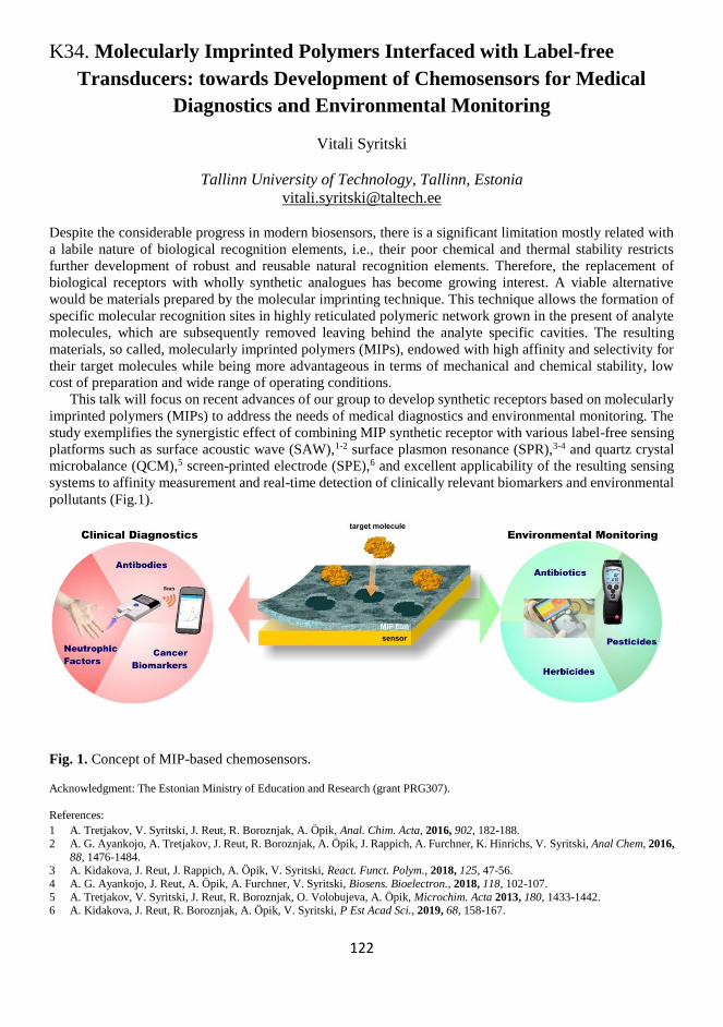
122
K34. Molecularly Imprinted Polymers Interfaced with Label-free
Transducers: towards Development of Chemosensors for Medical
Diagnostics and Environmental Monitoring
Vitali Syritski
Tallinn University of Technology, Tallinn, Estonia
Despite the considerable progress in modern biosensors, there is a significant limitation mostly related with
a labile nature of biological recognition elements, i.e., their poor chemical and thermal stability restricts
further development of robust and reusable natural recognition elements. Therefore, the replacement of
biological receptors with wholly synthetic analogues has become growing interest. A viable alternative
would be materials prepared by the molecular imprinting technique. This technique allows the formation of
specific molecular recognition sites in highly reticulated polymeric network grown in the present of analyte
molecules, which are subsequently removed leaving behind the analyte specific cavities. The resulting
materials, so called, molecularly imprinted polymers (MIPs), endowed with high affinity and selectivity for
their target molecules while being more advantageous in terms of mechanical and chemical stability, low
cost of preparation and wide range of operating conditions.
This talk will focus on recent advances of our group to develop synthetic receptors based on molecularly
imprinted polymers (MIPs) to address the needs of medical diagnostics and environmental monitoring. The
study exemplifies the synergistic effect of combining MIP synthetic receptor with various label-free sensing
platforms such as surface acoustic wave (SAW),1-2 surface plasmon resonance (SPR),3-4 and quartz crystal
microbalance (QCM),5 screen-printed electrode (SPE),6 and excellent applicability of the resulting sensing
systems to affinity measurement and real-time detection of clinically relevant biomarkers and environmental
pollutants (Fig.1).
Fig. 1. Concept of MIP-based chemosensors.
Acknowledgment: The Estonian Ministry of Education and Research (grant PRG307).
References:
1 A. Tretjakov, V. Syritski, J. Reut, R. Boroznjak, A. Öpik, Anal. Chim. Acta, 2016, 902, 182-188.
2 A. G. Ayankojo, A. Tretjakov, J. Reut, R. Boroznjak, A. Öpik, J. Rappich, A. Furchner, K. Hinrichs, V. Syritski, Anal Chem, 2016,
88, 1476-1484. 3 A. Kidakova, J. Reut, J. Rappich, A. Öpik, V. Syritski, React. Funct. Polym., 2018, 125, 47-56.
4 A. G. Ayankojo, J. Reut, A. Öpik, A. Furchner, V. Syritski, Biosens. Bioelectron., 2018, 118, 102-107.
5 A. Tretjakov, V. Syritski, J. Reut, R. Boroznjak, O. Volobujeva, A. Öpik, Microchim. Acta 2013, 180, 1433-1442. 6 A. Kidakova, J. Reut, R. Boroznjak, A. Öpik, V. Syritski, P Est Acad Sci., 2019, 68, 158-167.

123
124
SC11. Epitope-MIP for Engineered Enzymes
Aysu Yarman,a Giorgio Caserta,b Stefan Frielingsdorf,b Eszter Supala,c Ingo Zebger,b
Oliver Lenz,b Róbert E. Gyurcsányi,c Ulla Wollenberger,a Frieder W. Schellera
a Universität Potsdam, Institute for Biochemistry and Biology, Potsdam, Germany b Technische Universität Berlin, Inst. für Chemie Berlin, Germany
c Budapest University of Technology and Economics, Budapest, Hungary
In the “classical” synthesis of molecularly imprinted polymers (MIPs) functional monomers and cross-
linkers are polymerized in the presence of the target analyte. In the next step, recognition sites are formed by
removal of the template. These MIPs have been called plastibodies to stress the analogy of specific binding
by antibodies. Whilst antibodies interact only with a small area of its antigen (the immunodeterminant), MIPs
typically interact with a large part of the surface of the target molecule. Taking advantage of the simpler MIP
synthesis of low-molecular weight molecules short peptide sequences of the C-terminus or N-terminus have
been used in the epitope approach. The resulting MIPs have been shown to recognize both the target peptide
and the holoprotein. This concept has been extended to peptide tags of engineered proteins, sugars of
glycoproteins and even to chemical labels of macromolecules. This approach is closer to the antigen-antibody
reaction because only a small exposed part of the macromolecule is recognized by epitope-shaped cavities
of the MIP.
Here we present epitope- MIPs for the recognition of engineered proteins carrying the Strep-Tag II,
examplified for membrane-bound hydrogenase (MBH) of Ralstonia eutropha and alkaline phosphatase. For
the MIP synthesis the Cys-extended Strep-Tag II was chemisorbed on the gold electrode prior to
electropolymerization of the functional monomer scopoletin. Binding and electrochemical removal of the
peptide could be demonstrated electrochemically by using ferricyanide as a redox marker and by Surface-
Enhanced Infrared Absorption (SEIRA) spectroscopy. Atomic force microscopy was also applied for
monitoring the (different) preparation steps. The redox marker current decreased linearly in the presence of
up to 6 nM peptide and reached saturation at 10 nM upon rebinding of the peptide. Moreover, the Strep-
tagged proteins, MBH and alkaline phosphatase, were successfully bound to the peptide-imprinted layer. KD
values for Strep-Tag II and MBH to the Strep-Tag II-MIP were calculated to be 3.05 and 33.08 nM
respectively, by fitting the binding isotherm to a Langmuir model. Lower KD values showed that the MIP
has a higher affinity than the commercial systems (IBA Life Sciences, Göttingen, Strep-Tag II/Strep-Tactin
= 1 µM). Furthermore, the MIP has an almost 10-fold higher affinity to its target than to MBH.
Changing just one amino acid of the peptide significantly reduced its affinity to the MIP. The cross-
reactivity tests showed that removal of the terminal tryptophan resulted in an almost 20% lower signal
suppression. On the other hand, substitution of a glutamate by the uncharged amino acid glutamine reduced
the binding by 75 % which indicates the important role of electrostatic interactions. The dipeptide aspartam
had only a very low binding affinity. However, binding of Strep-tagged proteins demonstrated that MIP is
highly specific for a group of analytes.
Acknowledgements:
Financial support from Deutsche Forschungsgemeinschaft (DFG) within the framework of the German Excellence Strategy – EXC
2018/1 (UniSysCat) is acknowledged.

125

126
SC12. Electrical Control of the Receptor Affinity
Yulia Efremenko, Vladimir M. Mirsky
Department of Nanobiotechnology, Institute of Biotechnology,
Brandenburgische Technische Universität Cottbus – Senftenberg,
University square 1, 01968 Senftenberg, Germany
A concept of virtual sensor array based on
electrically controlled variation of affinity
properties of the receptor layer was realized on
the base of integrated electrochemical
chemotransistor1-2 containing conducting
polymer as the receptor layer. An electrical
control of the redox-state of the polymer
(polyaniline) was performed in five-electrode
configuration with four electrodes for
conductivity measurements and Ag/AgCl
reference electrode integrated on the same glass
chip. Using an ionic liquid was provided
electrical connection between the reference
electrode and chemosensitive material.3-4
Conductivity measurements demonstrated
potential controlled electrochemical conversions
of the receptor material between different redox-
states. Binding of trimethylamine at three
different potentials, corresponding to these states
was studied. The results demonstrated that both
kinetic- and equilibrium binding properties of the
receptor are controlled by electrical potential thus
providing a possibility to form a virtual sensor
array using only a single sensing element. The concept was applied for monitoring of fish headspace. Using
three characteristics of the sensor response measured at three different redox states of the same sensor
material, we have obtained signals from a virtual sensor array consisting of nine chemosensitive elements.
The sensor displays systematic changes of its nine signals during fish degradation.4 This approach can be
applied also for the electrical control of affinity of immunoglobulins.5 A development of a new materials
with conducting electrically controlled affinity is in progress. Binding energy of conducting polymers with
incorporated a boronic acid functional group was studied by isothermal titration calorimetry, the mostly
perspective receptor – ligand combinations were evaluated.
References: 1 U. Lange, V. M. Mirsky. Multielectrode chemoresistor; US Patent 9217722.
2 U. Lange, V. M. Mirsky. Anal. Chim. Acta, 2011, 687, 7-11.
3 Yu. Efremenko, V. M. Mirsky. Anal. Bioanal. Chem., 2016, 408, 7283 – 7287. 4 Yu. Efremenko, V. M. Mirsky, Sensors Actuators, B, 2016, 241, 652 – 657.
5 M. Vasjari, Yu. Efremenko, V. M. Mirsky, in preparation.

127
128
K35. Electronic Diversion of Enzymes
for Carrying Out Unconventional Tasks
Emmanuel Suraniti,b Aleksandar Karajic,a,c Pascal Merzeau,c Jérôme Roche,d
Sébastien Gounel,c Andrew G. Mark,b Peer Fischer,b Nicolas Mano,c Alexander Kuhna
a Univ. Bordeaux, CNRS UMR 5255, Bordeaux INP, ENSCBP, 16 avenue Pey-Berland, 33600
Pessac, France b Max Planck Institute for Intelligent Systems, Heisenbergstr. 3, 70569 Stuttgart, Germany
c Centre de Recherche Paul Pascal (CRPP), CNRS UMR 5031, Univ. Bordeaux, 115 Avenue du
Docteur Schweitzer, 33600 Pessac, France d CIRIMAT, Université de Toulouse, UPS-INP-CNRS, 118 Route de Narbonne, 31062 Toulouse
Cedex 09, France
Enzymes usually perform the reactions that Nature has made them for. Typically, these thermodynamically
favorable schemes transform spontaneously high energy educts into lower energy products. This is related
to the fact that (bio)chemical systems do not allow the coupling of energy from several simple reactions to
drive a subsequent reaction, which takes place in the same medium and leads to a product with a higher
energy than the one released during the first reaction. Normally, the only way to carry out uphill reactions is
to use an external input of energy.
Here we present a new approach that shows that it is nevertheless possible to perform such energetically
uphill reaction, if the electrons released in an oxidation reaction are temporarily stored in an electromagnetic
system, which is then used to raise the electrons’ potential energy, so that they can power in a second step,
after electrical decoupling, reactions with a higher demand of energy in the same reaction vessel. We
exemplify this general concept with a model system based on a glucose/oxygen biofuel cell to drive
thermodynamically unfavorable water splitting without input of external energy.1 Obviously this diversion
from the original enzymatic task by electronic means can be also applied to other reaction schemes.2
Acknowledgements:
E.S. is grateful for having received a Max-Planck Society scholarship. This project has also received funding from the European
Research Council (ERC) under the European Union's Horizon 2020 research and innovation program (grant agreement n° 741251, ERC Advanced grant ELECTRA). The work has also benefitted from aid of the Laboratory of Excellence AMADEus with the reference
ANR-10-LABX-0042-AMADEUS under the program ‘’Initiative for Excellence IdEx Bordeaux’’ holding the reference ANR-10-
IDEX-0003-02.
References:
1 E. Suraniti, P. Merzeau, J. Roche, S. Gounel, A. G. Mark, P. Fischer, N. Mano, A. Kuhn, Nature Comm., 2018, 9, 3229. 2 A. Karajic, E. Suraniti, P. Merzeau, S. Gounel, N. Mano, A. Kuhn, 2019, in preparation.

129

130
K36. Bacterial Biocomposite Materials: a Tool for Optimizing
and Studying Extracellular Electron Transfer Reactions
Mathieu Etienne
Laboratoire de Chimie Physique et Microbiologie pour les Matériaux et l’Environnement, CNRS
and Université Lorraine, UMR7564, 405, rue de Vandoeuvre, 54600, Villers-lès-Nancy, France
We describe in this work a method to manufacture a biocomposite material by self-assembly of bacteria of
the genus Shewanella oneidensis MR-1 with carbon nanoparticles in the presence of a protein having a
structuring role on the basis of electrostatic interactions. When this material is deposited on the surface of an
electrode, it can be used as an electroactive biofilm of artificial nature, in which the structuring protein can
then play a role in extracellular electron transfer reactions. In a first step of this study, bovine heart
cytochrome c was used for the preparation of an artificial electroactive biofilm catalyzing the oxidation of
formate. We have shown that the electron transfer was at the redox potential of cytochrome c, thus
demonstrating the role of this cytochrome in electron transfer reactions between bacteria and nanoparticles.
We then sought to catalyze a cathodic reaction, taking as an example the reduction of fumarate. The bovine
heart cytochrome c no longer allows the electron transfer for this reaction because it has too high a potential,
but the bacterial cytochromes of type c from the genera Desulfovibrio and Desulfuromonas have made it
possible to demonstrate this catalytic reaction, which demonstrates the great flexibility of this composite
material for the electrochemical study of electroactive bacteria. In the two previous examples, the electron
transfers were mediated by the exogenous cytochrome introduced artificially into the biocomposite. We then
sought to promote direct electron transfer reactions between bacteria and carbonaceous particles by replacing
cytochrome c with protamine,3 which is also known for its structuring role in biological systems. And more
recently, we extended this work to electroactive yeast cells.4 We will discuss the performance of this "living
materials" and the potential of this approach for living cell imprinting and electroanalytical applications.
Acknowledgments:
This work was supported by the French PIA project «Lorraine Universite d’Excellence», reference ANR-15-IDEX-04-LUE.
References:
1 S. Pinck, M. Etienne, M. Dossot, F. P. A. Jorand, Bioelectrochemistry, 2017, 118, 131–138.
2 S. Pinck, M. Xu, R. Clement, E. Lojou, F. P. Jorand, and M. Etienne, Bioelectrochemistry, 2018, 124, 185-194. 3 S. Pinck, F .P. A. Jorand, M. Xu, M. Etienne, ChemElectroChem, 2019, 6, 2398–2406.
4 Manuscript in preparation.

131
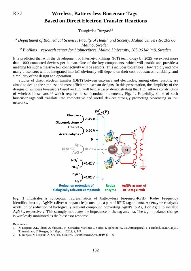
132
K37. Wireless, Battery-less Biosensor Tags
Based on Direct Electron Transfer Reactions
Tautgirdas Ruzgasa,b
a Department of Biomedical Science, Faculty of Health and Society, Malmö University, 205 06
Malmö, Sweden. b Biofilms – research center for biointerfaces, Malmö University, 205 06 Malmö, Sweden
It is predicted that with the development of Internet-of-Things (IoT) technology by 2025 we expect more
than 1000 connected devices per human. One of the key components, which will enable and provide a
meaning for such a massive IoT connectivity will be sensors. This includes biosensors. How rapidly and how
many biosensors will be integrated into IoT obviously will depend on their cost, robustness, reliability, and
simplicity of the design and operation.
Studies of direct electron transfer (DET) between enzymes and electrodes, among other reasons, are
aimed to design the simplest and most efficient biosensor designs. In this presentation, the simplicity of the
designs of wireless biosensors based on DET will be discussed demonstrating that DET allows construction
of wireless biosensors,1,2 which require no semiconductor elements, Fig. 1. Hopefully, some of such
biosensor tags will translate into competitive and useful devices strongly promoting biosensing in IoT
networks.
Fig. 1 Illustrates a conceptual representation of battery-less biosensor-RFID (Radio Frequency
Identification) tag. AgNPs (silver nanoparticles) constitute a part of RFID tag antenna. An enzyme catalyzes
oxidation or reduction of biologically relevant compound converting AgNPs to AgCl or AgCl to metallic
AgNPs, respectively. This strongly modulates the impedance of the tag antenna. The tag impedance change
is wirelessly monitored as the biosensor response.
References:
1 N. Larpant, A.D. Pham, A. Shafaat, J.F . Gonzalez-Martinez, J. Sotres, J. Sjöholm, W. Laiwattanapaisal, F. Faridbod, M.R. Ganjali,
T. Arnebrant, T. Ruzgas, Sci. Reports, 2019, 9, 1-9. 2 T. Ruzgas, N. Larpant, A. Shafaat, J. Sotres, ChemElectroChem, 2019, 6, 1–6.

133

134
K38. Bioelectrocatalysis and Bioanalytical Applications
of Molybdoenzymes
Ulla Wollenberger
University of Potsdam, Institute for Biochemistry and Biology, Liebknechtstrasse 24-25, 14476
Potsdam/Golm, Germany
The phenomenon of bioelectrocatalysis has already been described by Berezin et al more than 30 years ago.1
Bioelectrocatalysis involves the enhancement of an electrode reaction by a (bio)catalytic process and allows
therefore to gain important kinetic information about the catalyst. The presence of the catalyst enables redox
reactions of compounds at electrode to occur at low overpotentials and together with the substrate selectivity
bioelectrocatalysis provides the basis for sensitive and selective electroanalytical devices. The signal
transduction between the catalyst protein and redox electrodes proceeds by direct electronic communication
and by mobile or polymer bound redox mediators. Recent progress in the field is due to advances in enzyme
engineering, surface chemistry and nanotechnology.
In this talk examples of electrochemical studies on molybdenum containing enzymes and biosensors
based on them will be discussed. Molybdenum containing enzymes catalyze important metabolic
conversions such as oxidation of aldehydes, xanthine, and sulfite, or the reduction of DMSO, CO2 and
nitrate.2 More than 50 molybdoenzymes were identified to date. These enzymes have a complex overall
architecture and generally contain multiple redox-active centers to transduce between the two-electron redox
reaction of the substrate at molybdenum cofactor (Moco) and the terminal electron donor/ acceptor site.
Strategies to immobilize protein and enzymes based on self assembled monolayers, detergents and three
dimensional sensor structures were successfully developed in order to achieve an efficient electronic
communication between enzyme and electrode and a high catalyst loading particular for direct electron
transfer and mediatorless bioelectrocatalysis. On the other hand immobilization is achieved in redox
polymers for mediated electron transfer. The lecture will discuss examples of such electrochemical
biosensors, i.e. electrodes for aromatic aldehydes with an aldehyde oxidoreductase,3,4 for sulfite with sulfite
oxidase on nanoparticle modified electrodes,5-7 and a sensor for N-methylamine oxides with an enzyme of
the class of DMSO reductases.8
Acknowledgments: We gratefully acknowledge funding by the DFG under Germany’s Excellence Strategy (Unicat EXC 314/2; UniSyCat EXC 2008/1).
References: 1 I. V. Berezin, Priklad. Biokhimia Mikrobiologia, 1982, 18, 451-453.
2 R. Hille, Chem. Rev., 1996, 96, 2757–2816.
3 A. Badalyan, M. Neumann-Schaal, S. Leimkühler, U. Wollenberger, Electroanalysis, 2013, 25, 101-108. 4 P. Pinyou, A. Ruff, S. Pöller, S. Alsaoub, S. Leimkühler, U. Wollenberger, N. Plumeré, W. Schuhmann, Bioelectrochem., 2016,
109, 24-30.
5 T. Zeng, S. Leimkühler, U. Wollenberger, V. Fourmond, JACS, 2017, 139, 11559−11567. 6 R. Spricigo, R. Dronov, F. Lisdat, S. Leimkühler, F.W. Scheller, U. Wollenberger, Anal. Bioanal. Chem., 2009, 393, 225-233.
7 T. Zeng, J. Koetz, S. Leimkühler, U. Wollenberger, ACS Appl. Mater. Interfaces, 2015, 7, 21487-21494.
8 B. Mitrova, A. F. T. Waffo, P. Kaufmann, C. Iobbi-Nivol, S. Leimkühler, U. Wollenberger, ChemElectroChem, 2019, 6, 1732-
1737.

135
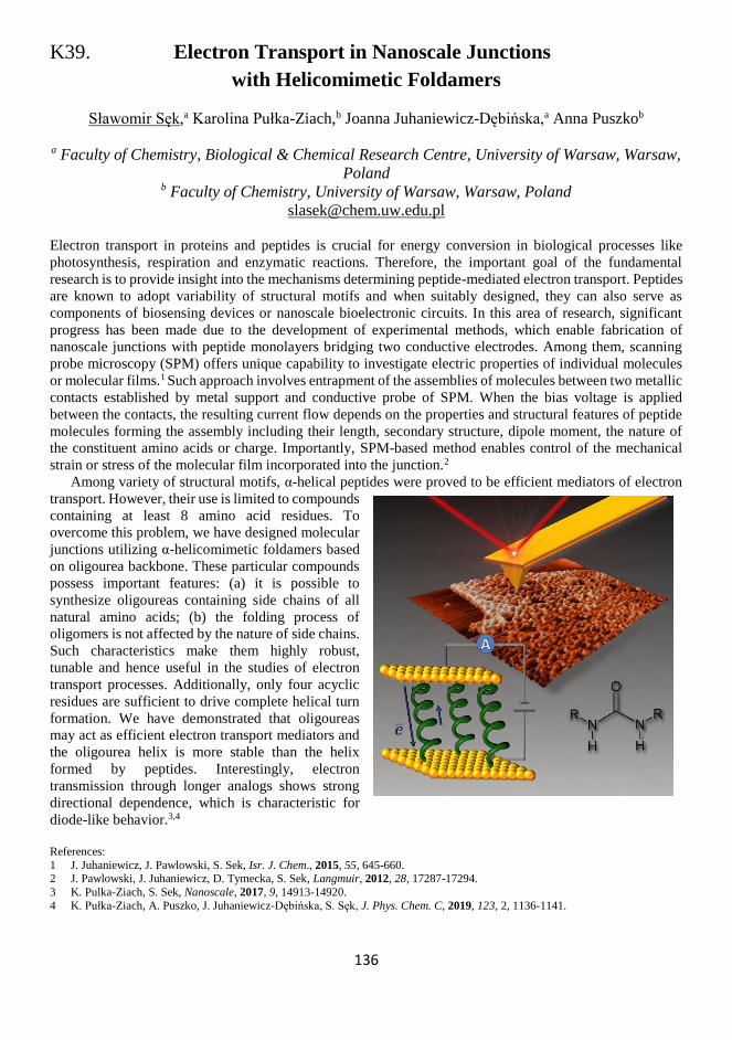
136
K39. Electron Transport in Nanoscale Junctions
with Helicomimetic Foldamers
Sławomir Sęk,a Karolina Pułka-Ziach,b Joanna Juhaniewicz-Dębińska,a Anna Puszkob
a Faculty of Chemistry, Biological & Chemical Research Centre, University of Warsaw, Warsaw,
Poland b Faculty of Chemistry, University of Warsaw, Warsaw, Poland
Electron transport in proteins and peptides is crucial for energy conversion in biological processes like
photosynthesis, respiration and enzymatic reactions. Therefore, the important goal of the fundamental
research is to provide insight into the mechanisms determining peptide-mediated electron transport. Peptides
are known to adopt variability of structural motifs and when suitably designed, they can also serve as
components of biosensing devices or nanoscale bioelectronic circuits. In this area of research, significant
progress has been made due to the development of experimental methods, which enable fabrication of
nanoscale junctions with peptide monolayers bridging two conductive electrodes. Among them, scanning
probe microscopy (SPM) offers unique capability to investigate electric properties of individual molecules
or molecular films.1 Such approach involves entrapment of the assemblies of molecules between two metallic
contacts established by metal support and conductive probe of SPM. When the bias voltage is applied
between the contacts, the resulting current flow depends on the properties and structural features of peptide
molecules forming the assembly including their length, secondary structure, dipole moment, the nature of
the constituent amino acids or charge. Importantly, SPM-based method enables control of the mechanical
strain or stress of the molecular film incorporated into the junction.2
Among variety of structural motifs, α-helical peptides were proved to be efficient mediators of electron
transport. However, their use is limited to compounds
containing at least 8 amino acid residues. To
overcome this problem, we have designed molecular
junctions utilizing α-helicomimetic foldamers based
on oligourea backbone. These particular compounds
possess important features: (a) it is possible to
synthesize oligoureas containing side chains of all
natural amino acids; (b) the folding process of
oligomers is not affected by the nature of side chains.
Such characteristics make them highly robust,
tunable and hence useful in the studies of electron
transport processes. Additionally, only four acyclic
residues are sufficient to drive complete helical turn
formation. We have demonstrated that oligoureas
may act as efficient electron transport mediators and
the oligourea helix is more stable than the helix
formed by peptides. Interestingly, electron
transmission through longer analogs shows strong
directional dependence, which is characteristic for
diode-like behavior.3,4
References:
1 J. Juhaniewicz, J. Pawlowski, S. Sek, Isr. J. Chem., 2015, 55, 645-660. 2 J. Pawlowski, J. Juhaniewicz, D. Tymecka, S. Sek, Langmuir, 2012, 28, 17287-17294.
3 K. Pulka-Ziach, S. Sek, Nanoscale, 2017, 9, 14913-14920.
4 K. Pułka-Ziach, A. Puszko, J. Juhaniewicz-Dębińska, S. Sęk, J. Phys. Chem. C, 2019, 123, 2, 1136-1141.

137
138
SC13. Simple Methods for the Electrified Liquid – Liquid Interface
Downscaling. From Design to Sensing Applications
Lukasz Poltorak,a,b Konrad Rudnicki,a Irene Eggink,b Marnix Hoitink,b
Slawomira Skrzypek,a Ernst J. R. Sudhölter,b Marcel de Puitb,c
a Department of Inorganic and Analytical Chemistry, Faculty of Chemistry, University of Lodz,
Tamka 12, 91-403 Lodz, Poland b Delft University of Technology, Department of Chemical Engineering, Van der Maasweg 9,
2629 HZ Delft, The Netherlands c Netherlands Forensic Institute, The Hague, The Netherlands
Electrochemistry knows the liquid – liquid interface as the interface between two immiscible electrolyte
solutions (ITIES) or electrified or polarized soft junction. On its own, it has a number of inherent and unique
properties. It is self-healing and renewable, can be polarized in a purely ionic manner and allows the study
of the interfacial charge transfer reactions. For the latter, associated currents can be recorded in a form of
interfacial electrons and/or ions transfer. The interfacial modification is a topic on its own. The interface can
receive new properties after being decoration with a smart nano-objects, molecular self-assemblies or by
placing it in an appropriate support.1
The flowing nature of the immiscible liquids used to create the ITIES helps when it comes to interface
miniaturization. This is usually performed in a relatively easy manner with patterned membranes, micro- or
nano-capillaries used as a support. In this respect, the secret is to adjust the chemical properties of the interior
of voids and/or pores within the support so that only one of the liquids will be in favor to wet their surface.
Addition of the second, immiscible phase results in the formation of a miniaturized liquid – liquid interface
bringing a few qualities to the investigated system: (i) ITIES receives additional stability; (ii) downscaling
facilities interfacial modification process; whereas from the electroanalytical point of view (iii) lower
capacitance currents can be translated into lower detectable concentration of analyzed species and (iv)
establishment of the hemispherical diffusion zones improves the analyte mass transport towards the interface,
which results in a higher detection sensitivity.
In this work, I will present two very simple methods used to prepare a micro-capillaries which are
subsequently used as a support for the ITIES. First method, is based on a metal microwire (DIA = 25 µm)
entrapped and dissolved from a glass framework.2 In the second approach, we used the fused silica capillaries
(DIA equal to 5; 10 and 25 µm) that are sliced and embedded in a heat shrinking tube.3 Resulting devices are
then used as the sensors for cocaine determination from a real street samples1-4 or to perform electroanalytical
screen the family of fluoroquinolone antibiotics.5 The fabrication process, comprehensive electroanalytical
description and further challenges will be presented and discussed.
Acknowledgments:
National Science Centre, Poland. Grant number 2018/31/D/ST4/03259
References:
1 L. Poltorak, A. Gamera, G. Herzog, A. Walcarius, App. Mat. Today, 2017, 9, 533–550. 2 L. Poltorak, I. Eggink, M. Hoitink, E. J. R. Sudholter, M De Puit, Anal. Chem., 2018, 90, 8–13.
3 K. Rudnicki, L. Poltorak, S. Skrzypek, E. J. R. Sudhölter, Anal. Chem., 2018, 90, 7112–7116.
4 L. Poltorak, E. J. R. Sudholter, M De Puit, TrAC, 2019, 114, 48–55. 5 K. Rudnicki, L. Poltorak, S. Skrzypek, E.J.R. Sudhölter, Anal. Chim. Acta., 2019, under the second revision round.

139
140
K40. Single-cell Nanoencapsulation
Insung S. Choi
Center for Cell-Encapsulation Research, Department of Chemistry, KAIST, Korea
Nature has developed a fascinating strategy of cryptobiosis for counteracting the stressful, and often lethal,
environmental conditions. For example, certain bacteria sporulate to transform from a metabolically active,
vegetative state to an ametabolic endospore state. The bacterial endospores, encased within tough
biomolecular shells, withstand the extremes of harmful stressors, such as radiation, desiccation, and
malnutrition, for extended periods of time and return to a vegetative state by breaking their protective shells
apart when their environment becomes hospitable for living. Inspired by cryptobiosis found in nature,
researchers have sought to chemically control and tailor the metabolic behaviors of non-spore-forming cells
as well as enhancing their viability against adverse environmental conditions, by forming thin (< 100 nm),
tough artificial shells. These living “cell-in-shell” structures, called artificial spores, enable chemical control
of cell division, protection against physical and chemical stresses, and cell-surface functionalizability, armed
with exogenous properties that are not innate to the cells but are introduced chemically. The field has further
advanced to the stage of chemical sporulation and germination, where cytoprotective shells are formed on
living cells and broken apart on demand. The (degradable) cell-in-shell hybrids are anticipated to find their
applications in various biomedical and bionanotechnological areas, such as tissue engineering,
cytotherapeutics, high-throughput screening, sensors, and biocatalysis, as well as providing a versatile
research platform for single-cell biology.

141
142
K41. 2D and 3D Lab-on-a-Chip Systems
for Life Science Applications
Jenny Emnéus
DTU Bioengineering Kgs. Lyngby, Denmark
jemn@ dtu.dk
The clinical need for functional replacement of tissues and physiologically
relevant drug screening models has fuelled research into engineering in-vitro
tissues. In conventional 2D cell cultures, cells tend to loose key specific
functions that they normally exhibit due to the absence of complex cell-cell,
cell–matrix and cell factor interactions that are usually present in vivo. The
trend thus goes towards culturing cells in a 3D environment. While cells in
vivo are typically no further than 200 µm away from the nearest blood vessel,
enabling their nutrient requirements to be met through diffusion of the
necessary chemical species, the absence of functional vasculature in tissue
engineered constructs poses a significant challenge when attempting to grow
tissues of clinically relevant sizes. An enhanced mechanism for transport of oxygen and nutrients is required
with a growing trend of using porous scaffolds to guide cell assembly into 3D constructs for engineering
different types of tissues. Moreover, depending on the tissue of interest, the scaffold stiffness (Youngs/elastic
modulus) is a vital consideration and varies from tissue to tissue (e.g. Brain 0.1-16 kPa, Liver 5-10 kPa, bone
15-30GPa).
The human population is getting increasingly older. Hence, age-related neurodegenerative diseases like
Parkinson’s disease (PD) are imposing an escalating threat as a medical, economical, and emotional burden.
For decades, PD has been studied in animal and simple cellular models. While in many aspects these models
are irreplaceable, they are inherently limited in others. Low success of clinical trials for new drugs and
therapies has recently turned attention to poor translation of animal experiments to human outcomes. On the
other hand, simple cell cultures lack in vivo tissue complexity. Therefore, a need for a more advanced human
in vitro models has emerged. 2D and 3D electrochemical Lab-on-a-Chip (LOC) systems will be presented
that can be applied in organ-on-a-chip applications. 2D chips are equipped with a planar electrode array
explored for intra and extracellular events using voltammetric and/or impedimetric detection. 3D systems
are equipped with different scaffolds for support and culturing of cells, having: (a) Structured perfusable
channel network, enabling delivery of necessary nutrients and oxygen to the interior of the scaffolds. (b)
Secondary more arbitrary random porous network that can enclose a hydrogel phase with a “nearby” source
of important cell factors, supporting the growth and differentiation of cells. (c) Multiple functions, as a cell
carrier and as combined optical waveguide and electrical sensor for e.g. optogenetic applications. Examples
of 2D and 3D systems applied in relation to drug delivery, as potential brain implants exemplified for
Parkinson´s disease will be presented.
The applicability of the presented systems spans from enzyme/antibody based biosensor array systems
for environmental and diagnostic biomedical bioanalysis to cell- and organ on chip system for better in vivo
mimicking toxicity or drug screening.

143
144
SC14. Protein imprinting.
Better control over deposited polymer structure
for better sensor performance.
Maciej Cieplak,a Marcin Dąbrowski,a Jakub Kałęcki,a Agnieszka Zimińska,a
Krzysztof Noworyta,a Alexander Kuhn,b,c and Piyush Sindhu Sharma a
a Institute of Physical Chemistry, Polish Academy of Sciences (IPC PAS), Kasprzaka 44/52, 01-
224 Warsaw, Poland b Institut des Sciences Moléculaires, University of Bordeaux, Bordeaux INP, ENSCBP, 16 Avenue
Pey Berland, 33607, Pessac, France c CNRS, ISM, UMR 5255, 351 Cours de la Liberation, 33400, Talence, France
Protein imprinting is a challenging task mainly because of their large size. It is very difficult to estimate
which and how many groups on the protein template molecule surface are accessible for binding. To
overcome this drawback, we introduced semi-covalent protein imprinting. We prepared a conducting
molecularly imprinted polymer (MIP) based on bis(2,2-bithien-5-yl)methane for human serum albumin
(HSA) determination. A very high imprinting factor (IF > 20) and selectivity of the devised chemosensor
proved that we the MIP featured molecular cavities of well-defined structure and high affinity to HSA. This
success encouraged us to improve this approach even further. For that, we prepared a macroporous MIP film
with hierarchical nanostructure controlled at three different size scale levels. Introduction of this
nanostructure resulted in extraordinary properties of this recognizing material. Very high selectivity of MIP
sensor was accompanied by sensitivity and detectability at an impressive femtomolar concentration level. To
prove versatility of this approach we also prepared macroporous MIP sensors for more demanding target
proteins, namely, human chorionic gonadotropin (hCG) and follicle-stimulating hormone (FSH).
Acknowledgments:
This research was financially supported by the National Science Centre, grant no. NCN 2017/25/B/ST4/01696 to P.S.S..

145
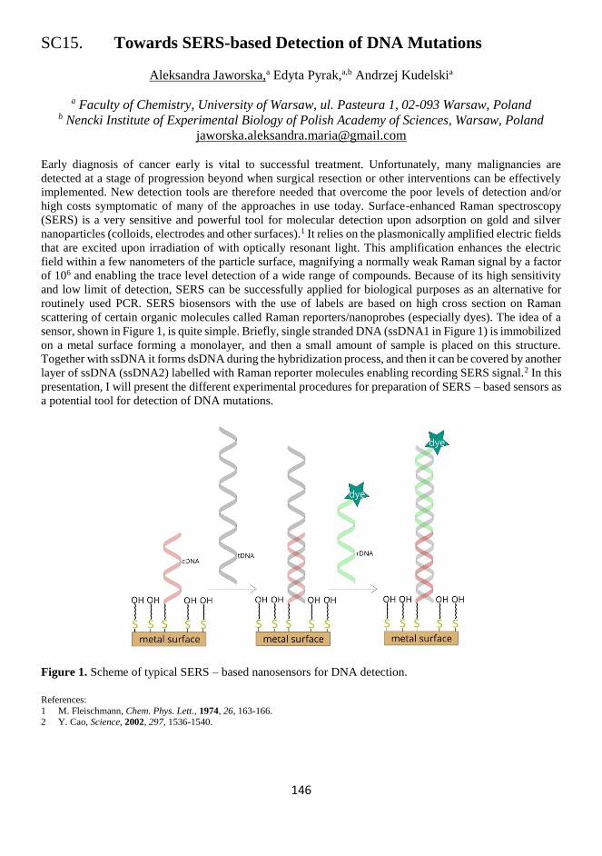
146
SC15. Towards SERS-based Detection of DNA Mutations
Aleksandra Jaworska,a Edyta Pyrak,a,b Andrzej Kudelskia
a Faculty of Chemistry, University of Warsaw, ul. Pasteura 1, 02-093 Warsaw, Poland b Nencki Institute of Experimental Biology of Polish Academy of Sciences, Warsaw, Poland
Early diagnosis of cancer early is vital to successful treatment. Unfortunately, many malignancies are
detected at a stage of progression beyond when surgical resection or other interventions can be effectively
implemented. New detection tools are therefore needed that overcome the poor levels of detection and/or
high costs symptomatic of many of the approaches in use today. Surface-enhanced Raman spectroscopy
(SERS) is a very sensitive and powerful tool for molecular detection upon adsorption on gold and silver
nanoparticles (colloids, electrodes and other surfaces).1 It relies on the plasmonically amplified electric fields
that are excited upon irradiation of with optically resonant light. This amplification enhances the electric
field within a few nanometers of the particle surface, magnifying a normally weak Raman signal by a factor
of 106 and enabling the trace level detection of a wide range of compounds. Because of its high sensitivity
and low limit of detection, SERS can be successfully applied for biological purposes as an alternative for
routinely used PCR. SERS biosensors with the use of labels are based on high cross section on Raman
scattering of certain organic molecules called Raman reporters/nanoprobes (especially dyes). The idea of a
sensor, shown in Figure 1, is quite simple. Briefly, single stranded DNA (ssDNA1 in Figure 1) is immobilized
on a metal surface forming a monolayer, and then a small amount of sample is placed on this structure.
Together with ssDNA it forms dsDNA during the hybridization process, and then it can be covered by another
layer of ssDNA (ssDNA2) labelled with Raman reporter molecules enabling recording SERS signal.2 In this
presentation, I will present the different experimental procedures for preparation of SERS – based sensors as
a potential tool for detection of DNA mutations.
Figure 1. Scheme of typical SERS – based nanosensors for DNA detection.
References:
1 M. Fleischmann, Chem. Phys. Lett., 1974, 26, 163-166. 2 Y. Cao, Science, 2002, 297, 1536-1540.

147
148
SC16. About Gold Nanotriangles and Their Applications
Lidia J. Opuchlik,a Olga A. Swiech,a Michal Kizling,b Renata Bilewicz,a
a Faculty of Chemistry, University of Warsaw, Ludwika Pasteura 1, Warsaw, Poland b College of Inter-Faculty Individual Studies in Mathematic and Natural Sciences (MISMaP),
Stefana Banacha 2C, Warsaw, Poland
Current cancer research is aimed to develop new anticancer therapies as well as to improve the performance
of already discovered ones. We synthesized two types (pH-sensitive and pH-insensitive) of anisotropic
nanotriangles (AuNTs) as carriers for doxorubicin.1 Due to atypical optical properties resulting from
triangular shape, these drug-carrier conjugates are sensitive to light in the near-IR region (in which human
tissues do not absorb) and can induce hyperthermic cytotoxicity (convert light energy to heat). Also, their
optical properties make them suitable for medical imaging and detection. Therefore, they are very interesting
because they can be considered as multifunctional therapy agents – not only efficient drug carriers but also
thermal energy generating agents. We performed detailed studies of prepared conjugates. The optical
properties, shape and size were evaluated by UV-vis-NIR spectroscopy, SEM and DLS. Fluorescence studies
demonstrated different drug release profiles depending on the pH. MTT assay performed on two cell lines
(A549 and HeLa) revealed that gold nanostructures modified with doxorubicin were more toxic than free
doxorubicin. Viability tests and confocal microscopy revealed that the bond between the gold carrier and the
drug determined the pathway of cell death – apoptotic in the case of peptide bonding and sudden and necrotic
when a hydrazone linker was employed.
Another application of gold nanotriangles results from their interesting structural features – multiple
edges and structural defects remaining after synthesis by wet-chemistry – granting them good catalytic
activity. These novel-shaped nanoparticles may be used as effective means for modification of working
electrodes exhibiting multiple edges or curvature effects, all possessing roughened surface area. Therefore,
they can be utilized as supports in catalysis. Glassy carbon electrode coated with AuNTs was used for the
study of oxygen reduction reaction (ORR) in 0.5 M sulfuric acid.2 The applied activation procedure led to a
surface highly active towards oxygen reduction. The advantages: much lower overpotential and larger current
densities of oxygen reduction are ascribed to the unique nanostructures present on the carbon electrode
surface – the gold nanoplates which are rich in edges providing a large population of Au (100) sites with
unsaturated coordination exposed to the solution, and catalytically active. Measurements performed using a
rotating disc electrode, modified with the gold nanoplates, confirmed that ORR proceeds via two separate
steps: oxygen is reduced to hydrogen peroxide, and the peroxide is further reduced in a two-electron
reduction to water.
Currently we are working on multicomponent polymers doped with nanotriangles modified with
cyclodextrin derivatives. Such systems can be utilized in electrochemical sensors or in (bio)electrocatalysis.
The use of cyclodextrin derivatives provides selectivity and enhances binding of analytes. The introduction
of AuNTs, evenly dispersed in the polymer matrix, provides the enhancement of the signal due to the
significant increase of the conductivity of the fabricated material. Additionally, the use of easily-wettable
short oligoethylene glycol for crosslinking and structuring allows binding of the analyte in the whole volume
of the polymer thanks to the uninterrupted diffusion within the material.
References:
1 O. A. Swiech, L. J. Opuchlik, G. Wojciuk, T. M. Stepkowski, M. Kruszewski and R. Bilewicz, RSC Adv., 2016, 6, 31960–31967. 2 L. J. Opuchlik, M. Kizling, P. Bacal and R. Bilewicz, Electroanalysis, 2019, 31, 1–10.

149

150
Tuesday, November 12
08:00-09:00 Breakfast
09:00-
10:30
Morning session 1
Chairs: A. Kuhn / L. Opuchlik
09:00-
09:40
T05 Sergey Shleev
Non-invasive Electrochemical (Bio)sensors Operating in Human
Physiological Fluids
09:40-
10:00
K42 Hanna Radecka
Ultrasensitive Electrochemical Sensors for Exploring of Anion
Recognition Processes at Aqueous/Solid Interface
10:00-
10:15
SC17 Nabila Yasmeen
Electropolymerized Molecularly Imprinted Polymer towards
Chemosensing of an Autism Biomarker
10:15-
10:30
SC18 Luís C. Almeida
Electrosynthesized Poly(catecholamine) Films Modified with
Ethanolamine for Immunosensing
10:30-11:00 Coffee break
11:00-
12:00
Morning session 2
Chairs: S. Shleev / Y. Efremenko
11:00-
11:20
K43 Lo Gorton
Connecting Biological Membranes and Bacterial Cells to Electrodes
through Redox Polymers
11:20-
11:35
SC19 Kamila Łępicka
The poly[NBI-(DTP)2] as an electrode material for the inherently
asymmetric supercapacitor
11:35-11:50 Closing
12:00-13:00 Lunch
13:30- Departures

151

152
T05. Non-invasive Electrochemical (Bio)sensors
Operating in Human Physiological Fluids
Sergey Shleev, Stefan Cirovic, Magnus Falk
Department of Biomedical Science, Faculty of Health and Society,
Malmö University, Malmö, Sweden
A significant effort is being devoted into the development of new healthcare and fitness innovations, driven
by the need imposed by patients and individuals, as well as the possibilities provided by recent electronics,
especially combined with information technology, to evaluate and benchmark personal health parameters.1-
3 By designing non-invasive (bio)sensors operating in human physiological fluids, such as sweat, urine,
saliva, and lachrymal liquid, it is possible to perform continuous, non-invasive monitoring of biomarkers for
assessing human performance, health and wellbeing.4 Such new technologies enable a shift from professional
medical care provided by hospitals to essentially outsourced medical services at point-of-care units and
homes and individuals being able to self-assess, to a much lower cost than what is currently possible.5
The keynote lecture will overview recent progress in the development of non-invasive biosensors
operating ex vivo.6-9 However, it will be mostly focused on electrochemical devices, which have been
designed and tested at the Department of Biomedical Science, Malmö University, Sweden. Specifically, the
following electrochemical (bio)sensors will be discussed: (i) flexible micro(bio)sensors for quantitative
analysis of bioanalytes in a nanovolume of human lachrymal liquid [10], (ii) wireless biosensing systems for
glucose and lactate monitoring in human urine, and (iii) headphone based electronic tongues for direct
continuous sweat analysis (Figure 1).
Figure 1. Electrochemical (bio)sensors developed at the Department of Biomedical Science.
Acknowledgement:
This research was funded by the Swedish Knowledge Foundation (KKS), grant number 20150248.
References:
1 A. Turner, Chem. Soc. Rev., 2013, 42, 3184-3196.
2 M. Chen, Y. Ma, Y. Li, D. Wu, Y. Zhang, C. H. Youn, IEEE Commun. Magazine, 2017, 55, 54-61. 3 J. Kim, A. Campbell, B. de Avila, J. Wang, Nat. Biotech., 2019, 37, 389-406.
4 A. Salim, S. Lim, Biosens. Bioelectron., 2019, 141, 111422.
5 K. Kim, B. Kim, C. Lee, Adv. Mater., 2019, doi:10.1002/adma.201902051. 6 W. Gao, S. Emaminejad, H. Nyein, S. Challa, K. Chen, A. Peck, H. Fahad, H. Ota, H. Shiraki, D. Kiriya, D. H. Lien, G. Brooks,
R. Davis, A. Javey, Nature, 2016, 529, 509-514.
7 D. Gaddes, W. Reeves, S. Tadigadapa, ACS Sensors, 2017, 2, 796-802. 8 C. Liao, C. Mak, M. Zhang, H. Chan, F. Yan, Adv. Mater., 2015, 27, 676-681.
9 R. Tseng, C. C. Chen, S. M. Hsu, H. S. Chuang, Sensors, 2018, 18, 2651.
10 V. Andoralov, S. Shleev, T. Arnebrant, T. Ruzgas, Anal. Bioanal. Chem., 2013, 405, 3871-3879.
Smart headphones with inbuilt e-tongue
Pen-like sensors Smart diapers

153
154
K42. Ultrasensitive Electrochemical Sensors
for Exploring of Anion Recognition Processes
at Aqueous/Solid Interface
Hanna Radecka, Jerzy Radecki
Institute of Animal Reproduction and Food Research, Polish Academy of Sciences,
Tuwima 10, 10-748 Olsztyn, Poland
In recent decade, the intermolecular recognition of anions in water has been attracted the attention of
numerous scientific groups involved in supramolecular chemistry.1,2 The most of literature reports the
recognition of anions in one organic phase. Developing systems for the recognition of anions in water
medium is still a challenging task. The several analytical approaches for anion recognition at aqueous/solid
interface based on the following redox active monolayers deposited on the gold electrodes will be presented:
(i) dipyrromethene –M(II)- dipyrromethene- dipodal anion receptor,3 (ii) terpirydyne–M(II)-
dipyrromethene- dipodal anion receptor, (iii) dipyrromethene –M(II)- dipyrromethene- cyclopeptide4
[M(II)= Cu(II) and Co(II)].
The developed systems were characterized electrochemically using cyclic voltammetry (CV) and
Osteryoung square wave voltammetry (OSWV). Then, they were successfully applied for the
electrochemical recognition of anions (Cl- and SO42-) in highly diluted aqueous medium (in the picomolar
range). The mechanism of communication between the redox centre and receptor anion complex as well as
analytical signal generation will be discussed.
Acknowledgements:
This work was supported by National Science Centre, Poland, No 2016/21/B/ST4/03834.
References:
1 M. J. Langton, C. J. Serpell, P. D. Beer, Angew. Chem. Int. Ed., 2016, 55, 1974-1987.
2 S. Kubik, Chem. Soc. Rev., 2010, 39, 3648-3663. 3 B. Kaur, C. A. Erdmann, M. Daniels, W. Dehaen, Z. Rafiński, H. Radecka, J. Radecki, Anal. Chem., 2017, 89, 12756−12763.
4 P. Gołębiewski, B. Puciłowski, F. Sommer, S. Kubik, M. Daniels, W. Dehaen, U. Sivasankaran, K. G. Kumar, H. Radecka, J.
Radecki, Sensor. Actuat. B Chem., 2019, 285, 536–545.

155
156
SC17. Electropolymerized Molecularly Imprinted Polymer
towards Chemosensing of an Autism Biomarker
Nabila Yasmeen,a Piyush S. Sharma,a Mathieu Etienne,b
Francis D’Souza,c Wlodzimierz Kutner,a,d
a Institute of Physical Chemistry, Polish Academy of Science, Kasprzaka 44/52,
01-224 Warsaw, Poland b Laboratory of Physical Chemistry and Microbiology
for Materials and the Environment,54600 Villers-lès-Nancy, France c Department of Chemistry, University of North Texas, Denton, 1155,
Union Circle, #305070 TX 76203-5017, USA d Department of Mathematics and Natural Sciences, School of Science, Cardinal Stefan
Wyszynski University, Warsaw, Poland
Nowadays, molecularly imprinted polymers (MIPs) are widely used as substitues for biologically derived
antibody-antigen systems for determination of different analytes of interest, such as biocompounds,
metabolites, and even whole bacteria because of their high affinity, sensitivity and durability under harsh
environmental conditions.1-3 Several biocompounds including gamma aminobutyric acid (GABA) is reported
as autism spectrum disorder (ASD) biomarkers.4,5 Therefore, there is an urgence to devise chemosensor for
selective and sensitive determination of the GABA biomarker of ASD.
In the present study, we deposited an MIP film on the interdigitated array (IDA) electrodes by
electropolymerization for fabrication of a chemosensor of ASD biomarkers. The IDA electrodes are
particularly attractive because their geometric features differences from those of conventional electrodes
resulting in different electrochemical behavior. This includes semi-spherical diffusion to each micro band,
fast establishment of a steady-state mass transfer, small iR drop, high redox cycling along with the high
signal-to-noise ratio within a dynamic range of potential.6,7 These all features make electrochemical
measurements on IDA electrodes highly sensitive for analytes at a considerably low detection limits.
For designing of selective molecular cavities in the MIP, bis-bithiophene derivatized functional
monomers were used. The impedance based transduction determined GABA in the micromolar
concentration range. This high detectability indicates chemosensor potential suitability in clinical analysis
applications.
References:
1 Patrycja Lach et al., Anal. Chem., 2019, 91, 7546-7553.
2 Piyush. S. Sharma et al., Trends Anal. Chem., 2013, 51, 146-157. 3 Mahmoud Labib et al., Chem. Rev., 2016, 116, 9001-9090.
4 Xue Ming et al., J. Proteome Res., 2012, 11, 5856-5862.
5 Andre A. S. Goldani et al., J. Mol. Neurosci., 2018, 66, 492-511. 6 Nera Alayo et al., Micro. Chim Acta., 2016, 183, 1633-1639.
7 Fei Liu et al., Anal. Chem., 2016, 86, 7391.

157
158
SC18. Electrosynthesized Poly(catecholamine) Films Modified
with Ethanolamine for Immunosensing
Luís C. Almeida,a Tânia Frade,a Rui D. Correia,a Yu Niu,b Gang Jin,b Jorge P. Correia,a
Ana S. Vianaa
a Centro de Química e Bioquímica, Centro de Química Estrutural, Faculdade de Ciências da
Universidade de Lisboa, Lisboa, Portugal b NML, Institute of Mechanics, Chinese Academy of Sciences, Beijing, China
Polydopamine-based surface modifications have been widely studied to obtain biocompatible and versatile
materials for biomedical, energy and industrial applications.1 The spontaneous oxidative polymerization in
aerated mediums makes it a simple and almost universal method for material coating, enriching the substrate
with quinone groups available for further functionalization by Michael-type addition or Schiff-base
formation.2 In fact, a robust immobilization of biomolecules, that allows the retention of its biological
activity, is a desired feature of the supporting matrix in a biosensors.3 Nonetheless, the control over structural
organization, homogeneity, oxidation state and conductivity of polydopamine thin films is still difficult to
achieve, limiting the technological application of this material. Besides oxygen, other chemical oxidants,4
have been used to increase polymerization rate, obtaining different porosities and chemical structures. More
recently, electrosynthesis has shown to yield better electrochemical transducers in biosensing interfaces than
the standard chemical synthesis,5 highlighting the great potential of electrochemical methods to better tune
the chemical and physical properties of poly(catecholamines).
Hereby, we report a comprehensive study of the electropolymerization of distinct catecholamine
monomers (dopamine, norepinephrine and L-DOPA), showing novel insights of the underlying mechanism
supported by electrogravimetric data. The precise control over the growth charge, mass and optical thickness
allows the synthesis of poly(catecholamine) films with different chemical composition, redox properties,
ionic permeability, wettability and morphology. Real-time Surface Plasmon Resonance assays are conducted
to assess the adsorption of different proteins to the biomimetic films. Furthermore, the ability to inhibit the
non-specific adsorption is achieved by one-step or two-steps modifications of the polymers with
ethanolamine. The antigen-antibody specific interaction was evaluated using immunoglobulin G and its
respective antigen, as model proteins. The powerful combination of several surface characterization
techniques allowed to demonstrate the very promising applications of poly(catecholamine) films in
immunosensors as controllable, reproducible and versatile biorecognition layers.
Acknowledgments:
This work was funded by Fundação para a Ciência e a Tecnologia through PhD scholarship SFRH/BD/129566/2017 and projects: IF/00808/2013/CP1159/CT0003 (POPH, UE-FSE), UID/MULTI/00612/2019 and PTDC/CTM-NAN/0994/2014.
References: 1 J. H. Ryu, P. B. Messersmith and H. Lee, ACS Appl. Mater. Interfaces, 2018, 10, 7523-7540.
2 L. C. Almeida, J. P. Correia, A. S. Viana, Electrochim. Acta, 2018, 263, 480-489.
3 T. O. Paiva, I. Almeida, J. T. Marquês, Y. Niu, W. Liu and G. Jin, Appl. Surf. Sci., 2017, 412, 455-463. 4 F. Ponzio, J. Barthès, J. Bour, M. Michel, P. Bertani, J. Hemmerlé, M. D’Ischia and V. Ball, Chem. Mater., 2016, 28, 4697–4705.
5 L. C. Almeida, R. D. Correia, G. Squillaci, A. Morana, F. La Cara, J. P. Correia and A. S. Viana, Electrochim. Acta, 2019, 319, 462-471.

159
160
K43. Connecting Biological Membranes and Bacterial Cells
to Electrodes through Redox Polymers
Lo Gorton
Department of Biochemistry,
P.O.Box 124, SE-22100 Lund University, Lund, Sweden
We have throughout the years since 2004 pioneered “wiring” both Gram-positive and Gram-negative
bacteria1-4 (and thylakoid membranes5-7) to electrodes using osmium redox polymers (Os RPs) that will
strongly facilitate extracellular electron transfer (EET). Because of the cationic nature of the Os RPs they
will electrostatically interact very strongly with membranes and cells to form hydrogels that will strongly
attach on electrode surfaces and will allow substrates and products to freely diffuse in and out of the hydrogel.
We have investigated the influence of E°’-value and structure of the Os-complexes of the Os RPs on the rate
of electron transfer. However, only recently we have obtained a much more clear picture on how the RPs
and the cells interact and how the interaction changes with time. In these recent investigations we have
“wired” wild type and some mutants of Enterococcus faecalis with both 4 different Os RPs as well as with
a quinone RP.8-11 These recent results will be shown and discussed.
References:
1 K. Hasan, S. A. Patil, D. Leech, L. Gorton, Biochem. Soc. Trans., 2012, 40,1330-1335.
2 S. A. Patil, C. Hägerhäll, L. Gorton, Bioanal. Rev., 2012, 4, 159-192. 3 G. Pankratova and L. Gorton, Curr. Opin. Electrochem., 2017, 5, 193–202.
4 G. Pankratova, L. Hederstedt, L. Gorton, Anal. Chim. Acta, 2019, 1076, 32-47.
5 G. Pankratova, D. Pankratov, K Hasan, H. E. Åkerlund, P. Å. Albertsson, D. Leech, S. Shleev, L. Gorton, Adv. Energy Mater., 2017, 1602285.
6 G. Pankratova, D. Pankratov, L. Gorton, Curr. Opin. Electrochem., accepted.
7 D. Pankratov, G. Pankratova, L. Gorton, “Current Trends in Development of Photosynthetic Bioelectrochemical Systems for Light Energy Conversion”, in “Novel Catalyst Materials for Bioelectrochemical Systems: Fundamentals and Applications”, Eds. L.
Singh, D. M. Mahapatra, H. Liu, ACS Book Ser., 2019 in press.
8 G. Pankratova, K. Hasan, D. Leech. L. Hederstedt, L. Gorton, Electrochem. Commun., 2017, 75, 56–59.
9 G. Pankratova, D. Leech, L. Gorton, L. Hederstedt, Biochemistry, 2018, 57, 4597−4603.
10 G. Pankratova, E. Szypulska, D. Pankratov, D. Leech, L. Gorton, ChemElectroChem, 2019, 6, 110–113.
11 G. Pankratova, D. Pankratov, R. D. Milton, S. D. Minteer, L. Gorton, Adv. Energy Mater., 2019, 1900215.

161
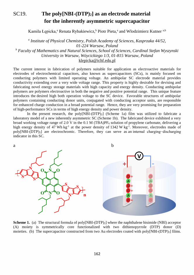
162
SC19. The poly[NBI-(DTP)2] as an electrode material
for the inherently asymmetric supercapacitor
Kamila Łępicka,a Renata Rybakiewicz,b Piotr Pieta,a and Wlodzimierz Kutner a,b
a Institute of Physical Chemistry, Polish Academy of Sciences, Kasprzaka 44/52,
01-224 Warsaw, Poland b Faculty of Mathematics and Natural Sciences, School of Sciences, Cardinal Stefan Wyszynski
University in Warsaw, Wóycickiego 1/3, 01-815 Warsaw, Poland
The current interest in fabrication of polymers suitable for application as electroactive materials for
electrodes of electrochemical capacitors, also known as supercapacitors (SCs), is mainly focused on
conducting polymers with limited operating voltage. An ambipolar SC electrode material provides
conductivity extending over a very wide voltage range. This property is highly desirable for devising and
fabricating novel energy storage materials with high capacity and energy density. Conducting ambipolar
polymers are polymers electroactive in both the negative and positive potential range. This unique feature
introduces the desired high both operation voltage to the SC device. Favorable structures of ambipolar
polymers containing conducting donor units, conjugated with conducting acceptor units, are responsible
for enhanced charge conduction in a broad potential range. Hence, they are very promising for preparation
of high-performance SCs in terms of high energy density and power density.
In the present research, the poly[NBI-(DTP)2] (Scheme 1a) film was utilized to fabricate a
laboratory model of a new inherently asymmetric SC (Scheme 1b). The fabricated device exhibited a very
broad working voltage range of 2.0 V in the 0.1 M (TBA)PF6 solution of propylene carbonate, delivering a
high energy density of 47 Wh kg-1 at the power density of 1342 W kg-1. Moreover, electrodes made of
poly[NBI-(DTP)2] are electrochromic. Therefore, they can serve as an internal charging−discharging
indicator in this SC.
a b
n
Scheme 1. (a) The structural formula of poly[NBI-(DTP)2] where the naphthalene bisimide (NBI) acceptor
(A) moiety is symmetrically core functionalized with two dithienopyrrole (DTP) donor (D)
moieties. (b) The supercapacitor constructed from two Au electrodes coated with poly[NBI-(DTP)2] films.

163

164
Poster Session
Saturday, November 9 20:00-22:00

165

166
P01. Characterization of Molecularly Imprinted Polymer
Fixed Electrode Surface by MIP-Nanoparticle
Masaki Abe,a Haruto Eguchi,a Akihiko Hatano,b Yasuo Yoshimia
a Shibaura Institute of Technology, 3-7-5 Toyosu, Koto-ku, Tokyo 135-8548, Japan b Shibaura Institute of Technology, 307 Fukasaku Minuma-ku, Saitama 337-8570, Japan
A molecularly imprinted polymer (MIP) is molecular-recognition material synthesized by polymerization in
the presence of the target molecule as a template. We are developing highly selective electrochemical sensors
for vancomycin (VCM), which is one of the primary antibacterials, by using VCM-imprinted (methacrylic
acid (MAA)-co-methylenebisacrylamide (MBAA)-co-acrylamide-co-ferrocenyl monomer) on an electrode.1
The ferrocenyl monomer (vinyl ferrocene (VF) or allylaminocarboxypropyonic-3-ferrocene (ACPF)) is
essential for the VCM sensing but the role is still unknown. We also discovered that nanoparticle of MIP
(MIP-NP) is swelled by the specific interaction with the template.2 In this work , we analyzed the size
dependency of the MIP-NP of VCM composed by the same material as the MIP on the electrode in order to
discuss the role of the ferrocenyl group. VCM was immobilized covalently on the aminated
surface of glass beads via glutaraldehyde. MIP-NP was
synthesized at the surface of glass beads by radical
copolymerization of MAA, MBAA and ferrocenyl
monomer (VF, ACPF, or none). MIP-NP was obtained
by washing glass beads surface phosphate buffer saline
including 1.0 M NaCl. The particle diameter was
measured by dynamic scattering spectroscopy (DLS).
The diameter MIP-NP containing ACPF is larger
than that containing VF or that without ferrocenyl
group. The VCM-MIP electrode containing ACPF was
more sensitive to VCM that that containing VF or no
ferrocenyl monomer. The consistency indicates that
the ferrocenyl group of ACPF has strong affinity with
VCM.
References:
1 H. Eguchi et al., Presentation at this conference.
2 Y. Yoshimi, D. Oino, H. Ohira, H. Muguruma, E.Moczko, S. A. Piletsky, Sensors, 2019, 19, 2415.
Fig. 1 Size dependency of the MIP-NP

167
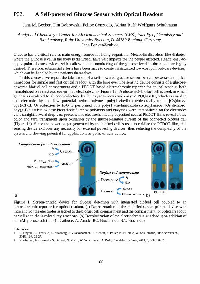
168
P02. A Self-powered Glucose Sensor with Optical Readout
Jana M. Becker, Tim Bobrowski, Felipe Conzuelo, Adrian Ruff, Wolfgang Schuhmann
Analytical Chemistry - Center for Electrochemical Sciences (CES), Faculty of Chemistry and
Biochemistry, Ruhr University Bochum, D-44780 Bochum, Germany
Glucose has a critical role as main energy source for living organisms. Metabolic disorders, like diabetes,
where the glucose level in the body is disturbed, have vast impacts for the people affected. Hence, easy-to-
apply point-of-care devices, which allow on-site monitoring of the glucose level in the blood are highly
desired. Therefore, substantial efforts have been made to create miniaturized low-cost point-of-care devices,1
which can be handled by the patients themselves.
In this context, we report the fabrication of a self-powered glucose sensor, which possesses an optical
transducer for simple and fast optical readout with the bare eye. The sensing device consists of a glucose-
powered biofuel cell compartment and a PEDOT based electrochromic reporter for optical readout, both
immobilized on a single screen-printed electrode chip (Figure 1a). A glucose/O2 biofuel cell is used, in which
glucose is oxidized to glucono-𝛿-lactone by the oxygen-insensitive enzyme PQQ-GDH, which is wired to
the electrode by the low potential redox polymer poly(1-vinylimidazole-co-allylamine)-[Os(dmxy-
bpy)2Cl]Cl. O2 reduction to H2O is performed at a poly(1-vinylimidazole-co-acrylamide)-[Os(dichloro-
bpy)2Cl]/bilirubin oxidase biocathode.2 Redox polymers and enzymes were immobilized on the electrodes
via a straightforward drop-cast process. The electrochemically deposited neutral PEDOT films reveal a blue
color and turn transparent upon oxidation by the glucose-limited current of the connected biofuel cell
(Figure 1b). Since the power output generated by the biofuel cell is used to oxidize the PEDOT film, this
sensing device excludes any necessity for external powering devices, thus reducing the complexity of the
system and showing potential for applications as point-of-care device.
(a) (b)
Figure 1. Screen-printed device for glucose detection with integrated biofuel cell coupled to an
electrochromic reporter for optical readout. (a) Representation of the modified screen-printed device with
indication of the electrodes assigned to the biofuel cell compartment and the compartment for optical readout,
as well as to the involved key-reactions. (b) Decolorization of the electrochromic window upon addition of
50 mM glucose solution (C: Cathode, A: Anode, BC: Biocathode, BA: Bioanode)
References:
1 P. Pinyou, F. Conzuelo, K. Sliozberg, J. Vivekananthan, A. Contin, S. Pöller, N. Plumeré, W. Schuhmann, Bioelectrochem.,
2015, 106, 22-27. 2 S. Alsaoub, F. Conzuelo, S. Gounel, N. Mano, W. Schuhmann, A. Ruff, ChemElectroChem, 2019, 6, 2080-2087.

169

170
P03. Characterizing Escherichia Coli-imprinted Polystyrene Thin Films
with Confocal Raman Microscopy
Birgit Bräuer, Felix Thier, Marius Bittermann, Dieter Baurecht, Peter Lieberzeit,
University of Vienna, Faculty for Chemistry, Department of Physical Chemistry, Vienna, Austria
The gram-negative bacterium Escherichia coli (E.coli) is considered an indicator of food and water safety.1
Thus, various methods have been established to quantify and identify this microorganism, but they either
lack sensitivity and selectivity, or require growth and enrichment of the bacteria. To address these issues,
sensors suitable for direct detection of E.coli in water have been developed using molecularly imprinted
polymers (MIPs) as an E.coli-sensitive layer on Quartz Crystal Microbalances (QCMs).2 Surface
characterization of MIPs is indispensable to ensure reproducibility of the polymer synthesis and assess the
success of imprinting. Established techniques including Optical and Atomic Force Microscopy provide
morphological characterization but no information about chemical composition of the surface.
We utilized Confocal Raman Microscopy to characterize E.coli-imprinted polystyrene synthesized using
a covalent stamp imprinting approach. Performing Raman Image Scans on the polymer allowed us to obtain
optical as well as spatially resolved chemical information about the sample surface. Overlaying Raman TV
images (showing intensity distribution of the Raman signal at 2908 cm-1) of E.coli-imprinted polystyrene
before and after E.coli treatment and the corresponding white light images enabled straightforward
differentiation between imprints, bacteria and polymer based on discrepancies in signal intensity at this
wavenumber (Figure 1 A and B). Atomic Force Microscopy (AFM) was successfully used to assess accuracy
of this distinction (Figure 1C) and to confirm success of the imprinting, which had been suggested by the
Raman Image Scans.
Furthermore, we succeeded in successfully differentiating between two bacteria species, E.coli and L.lactis,
on E.coli-imprinted polystyrene. For that purpose, we applied chemometric pattern recognition (Partial Least
Squares Discriminant Analysis (PLS-DA) on Confocal Raman Images. This provides a basis for further
selectivity studies of E.coli-imprinted polystyrene. The corresponding Raman spectra of the bacteria
apparently exhibit spectral differences that, although they are not visible, can be extracted using PLS-DA
and used to establish a model that can predict the affiliation of a bacterium to the E.coli or L.lactis class.
References: 1 A. Poller, E. Spieker, P. Lieberzeit and C. Preininger, ACS Appl. Mater. Interfaces, 2017, 9, 1129-1135.
2 H. Zhou, D. Yang, N. Ivleva, N. Mirescu, R. Niessner and C. Haisch, Anal. Chem., 2014, 86, 1525-1533.
Figure 2. Overlays of Raman TV and white light images of A: E.coli-imprinted polystyrene and
B: E.coli-imprinted polystyrene treated with E.coli; C: AFM image of E.coli-imprinted
polystyrene treated with E.coli.

171
172
P04. Reconstitution of Mitochondrial Membrane Protein
in Liquid Crystal Phases
Aleksandra Buta,a Ewa Nazaruk,a Milena Krajewska,b Piotr Koprowski,b
Adam Szewczyk,b Renata Bilewicza
a University of Warsaw, Faculty of Chemistry, 1 Pasteur Street, 02-093 Warsaw, Poland b Nencki Institute of Experimental Biology, Polish Academy of Sciences, 3 Pasteur Street,
02-093 Warsaw, Poland
The liquid crystal phases (mesophases) have specific properties and structure, e.g. the presence of a lipid
bilayer surrounded by water channels, and a large internal surface (400m2/g). Such phases can be used to
reconstitute amphiphilic membrane proteins. The immobilization of these molecules in phase provides them
with a lipid environment similar to that of the biological membrane environment in vivo, thanks to which it
is possible to study the structure and mechanism of protein activity.
We isolated a mitochondrial membrane protein - Renal Outer Medullary Potassium Channel 1 (ROMK1)
from the bacterial culture (Escherichia coli). The quality of purification was checked by Western Blot and
the protein concentration was determined by Bicinchoninic acid (BCA) method.
Protein immobilization was performed in mesophases containing monoolein (MO) or monopalmitolein
(MP). These lipids differ from each other by the length of the lipid chain which affects the structure of the
phase and the possibility of packing the protein inside it. The phase structure and the influence of protein
incorporation were checked by Small-angle X-ray scattering (SAXS) method.
Studies of the activity of the ROMK1 protein inserted into the cubic phase include electrochemical
methods. Chronoamperometry measurements will be done at different potentials applied to the electrode
covered with the cubic phase membrane with and without ROMK1. The influence of potassium ion
concentration and of the presence of thalium ion as the inhibitor will be presented.

173
174
P05. Wireless Biosensing of Analytes in Human Urine:
Towards Smart Electronic Diapers
Stefan Cirovic, Magnus Falk, Sergey Shleev
Department of Biomedical Science, Faculty of Health and Society, Malmö University,
Malmö, Sweden
Urinary incontinence is a common health problem among aged people, which require hospital staff to
monitor patients. Leaving incontinence events unmanaged negatively impacts patient´s mental and physical
health. To alleviate the problem, there is a need and a desire to have an incontinence detection system or
device to alert a caregiver when it is time to change diapers. Automatic detection systems can significantly
improve incontinence management, moving from a predominantly manual to an automatic process. A variety
of different systems have already been developed that achieve wireless urinary incontinence monitoring,
detecting the presence of moisture.1,2 Instead of being used solely as wetness sensors, such systems could
greatly benefit from also being able to detect different analytes of interest released in the urine.
By incorporating a biosensor in an electronic diaper, detection of different analytes can be achieved. The
presence of glucose in urine results from the glomerular filtration of more glucose than the renal tubule can
absorb, where abnormally increased amounts typically is associated with the disease diabetes.3 Lactate is
another very important prognostic marker, especially in critical patients, and urinary lactate have been shown
to correlate with blood lactate.4 A variety of other compounds with clinical relevance are also present in
urine, such as urate, ascorbate, cholesterol and oxalate, all of which can be converted by different
oxidoreductases.3-5
We design a wireless biosensing system for glucose and lactate in human urine, detected when present at
elevated levels. To enable the wireless sensing, a previously developed wireless system for detecting
moisture was modified with a conductometric biotransducer, giving a binary response that changes when the
analyte is present at sufficiently high concentrations. The conductometric biotransducer is based on a
Prussian blue – cellulose acetate layer, further modified with either glucose oxidase or lactate oxidase. In the
presence of the substrates of the enzymes, hydrogen peroxide is produced which reacts with the layer,
changing the conductivity of the Prussian blue – cellulose acetate layer from non-conducting to conducting
state, which switches the response of the wireless sensing system. The system enables non-invasive analyte
detection of urine samples, which could be used to design smart electronic diapers. Acknowledgement:
This research was funded by the Swedish Knowledge Foundation (KKS), grant number 20150248.
References:
1 X. Nie, Z. Song, J. Yang, Z. Dengyun, M. Yin, X. Duan, B Jiao, Inter. J. Commun., 2017, 10, 252-263. 2 S. Sajal, Y. Atanasov, B. Braaten, V. Marinov, O. Swenson, IEEE Intern. Conf. Electro/Information Technol., 2014, 542-545.
3 J. Gerich, Diabet. Med., 2010, 27, 136-142.
4 T. Hagen, M. Korson, J. Wolfsdorf, C. Grade, M. Ideas, Mol. Genet. Metab., 2010, 70, 189-195. 5 S. Milardović, I. Kereković, M Nodilo, Talanta, 2008, 77, 222-228.

175
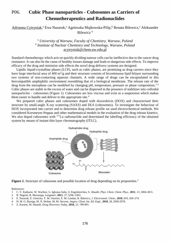
176
P06. Cubic Phase nanoparticles - Cubosomes as Carriers of
Chemotherapeutics and Radionuclides
Adrianna Cytryniak,a Ewa Nazaruk,a Agnieszka Majkowska-Pilip,b Renata Bilewicz,a Aleksander
Bilewicz b
a University of Warsaw, Faculty of Chemistry, Warsaw, Poland b Institute of Nuclear Chemistry and Technology, Warsaw, Poland
Standard chemotherapy which acts on quickly dividing tumour cells can be ineffective due to the cancer drug
resistance. It can also be the cause of healthy tissues damage and leads to dangerous side effects. To improve
efficacy of the drug and minimize side effects the novel drug delivery systems are designed.
Lipidic liquid-crystalline phases (LCP), such as cubic phases, are promising as drug carriers since they
have large interfacial area of 400 m2/g and their structure consists of bicontinuous lipid bilayer surrounding
two systems of non-contacting aqueous channels. A wide range of drugs can be encapsulated in this
biocompatible amphiphilic environment resembling that of a biological membrane. The release rate of the
drug from the mesophase can be modified by changing pH, temperature, pressure or phase composition.1-3
Cubic phases are stable in the excess of water and can be dispersed in the presence of stabilizer into colloidal
nanoparticles – cubosomes (Figure 1). Cubosomes are less viscous and exist as a suspension which makes
them easier to handle and deliver to the appropriate site.4
We prepared cubic phases and cubosomes doped with doxorubicin (DOX) and characterised their
structure by small-angle X-ray scattering (SAXS) and DLS (cubosomes). To investigate the behaviour of
DOX incorporated into carrier and to determine drug release profile we used electrochemical methods. We
considered Korsmeyer-Peppas and other mathematical models in the evaluation of the drug release kinetics.
We also doped cubosomes with 177Lu radionuclide and determined the labelling efficiency of the obtained
system by means of instant thin layer chromatography (ITLC).
Figure 1. Structure of cubosome and possible location of drug depending on its propoerties.5
References:
1 C. V. Kulkarni, W. Wachter, G. Iglesias-Salto, S. Engelskirchen, S. Ahualli, Phys. Chem. Chem. Phys., 2011, 13, 3004-3021. 2 R. Negrini, R. Mezzenga, Langmuir, 2011, 27, 5296–5303.
3 E. Nazaruk, E. Górecka, Y. M. Osornio, E. M. Landau, R. Bilewicz, J. Electroanal. Chem., 2018, 819, 269–274.
4 H. M. G. Barriga, M. N. Holme, M. M. Stevens, Angew. Chem. Int. Ed. Engl., 2018, 58, 2958-2978. 5 Z. Karami, M. Hamidi, Drug Discovery Today, 2016, 21, 789-801.

177
178
P07. Electrochemical Studies of DMPC-cholesterol Lipid Bilayer
Supported on Au(111) Electrode Modified by 1-thio-β-D-glucose
Damian Dziubak, Sławomir Sęk
Biological and Chemical Research Centre, Faculty of Chemistry, University of Warsaw
Żwirki i Wigury 101, 02-089 Warsaw, Poland
Floating lipid bilayer membrane immobilized on the surface of a gold electrode is one of the models which
can imitate the biological membranes. In this approach, the membrane is not directly contacted with the solid
substrate but separated by a water-rich domain. This water reservoir is provided by the presence of the
hydrophilic molecules which are used to pre-modify the electrode surface.
The gold surface modified by 1-thio-β-D-glucose1 is able to mimic the quasi-natural environment of
membranes and prevents transmembrane peptides and proteins from denaturation. It is possible due to
hydrophilic properties of 1-thio-β-D-glucose (β-Tg) and presence of sulfur atoms in molecules, which enable
to create stable and rich in water layer between the electrode and the lipid membrane. This approach has
been used for example to investigate the pore-forming activity of alamethicin in DMPC bilayer,2 or study
properties of colicin E1, a channel-forming protein.3
In this work, we have monitored the process of vesicles spreading and formation of a bilayer consisting
of DMPC and cholesterol (DMPC:Chol) on gold electrode modified with β-Tg by quartz crystal
microbalance technic (QCM). We have also investigated capacitive behavior of Au(111)/β-Tg and Au(111)
/β-Tg with DMPC cholesterol lipid bilayer by AC voltammetry and electrochemical impedance
spectroscopy. Results show that the presence of lipid bilayer lowers the capacitance. However, its value is
noticeably higher than for DMPC cholesterol directly deposited on Au(111) electrode. This is indicative of
the presence of a thin water cushion between the electrode and the membrane. Using electrochemical QCM,
we have investigated the mass changes vs. the applied potential for β-Tg, DMPC:chol lipid bilayer and for
DMPC:Chol membrane on top of β-Tg. Our results confirm that upon desorption, the head groups of the
inner layer detach from the metal and are separated by a thin cushion of water.4 However, the same effect is
also visible for Au(111)/β-Tg/DMPC:Chol.
References:
1 A. H. Kycia, S. Sek, Z. Su, A. R. Merrill, J. Lipkowski, Langmuir, 2011, 27, 13383–13389. 2 F. Abbasi, J. Alvarez-Malmagro, Z. Su, J. J. Leitch, J. Lipkowski, Langmuir, 2018, 34, 13754-13765.
3 Z. Su, D. Ho, A. R. Merrill, J. Lipkowski, Langmuir, 2019, 35, 8452-8459.
4 D. Matyszewska, R. Bilewicz, Z. Su, F. Abbasi, J. J. Leitch, J. Lipkowski, Langmuir, 2016, 32, 1791–1798.

179
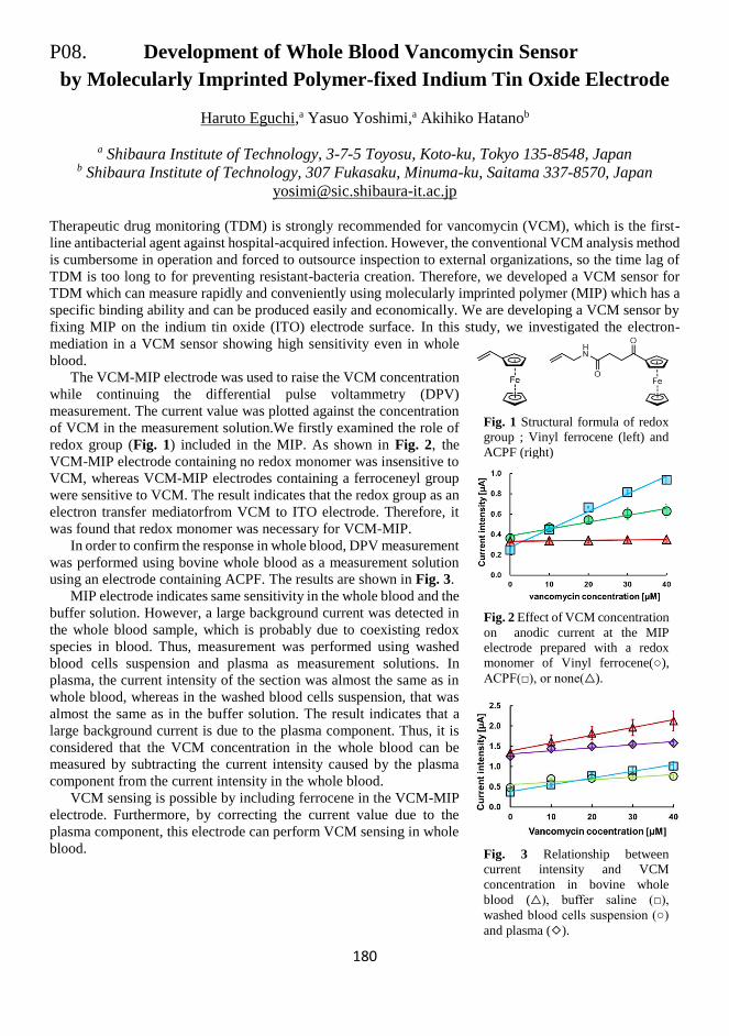
180
P08. Development of Whole Blood Vancomycin Sensor
by Molecularly Imprinted Polymer-fixed Indium Tin Oxide Electrode
Haruto Eguchi,a Yasuo Yoshimi,a Akihiko Hatanob
a Shibaura Institute of Technology, 3-7-5 Toyosu, Koto-ku, Tokyo 135-8548, Japan b Shibaura Institute of Technology, 307 Fukasaku, Minuma-ku, Saitama 337-8570, Japan
Therapeutic drug monitoring (TDM) is strongly recommended for vancomycin (VCM), which is the first-
line antibacterial agent against hospital-acquired infection. However, the conventional VCM analysis method
is cumbersome in operation and forced to outsource inspection to external organizations, so the time lag of
TDM is too long to for preventing resistant-bacteria creation. Therefore, we developed a VCM sensor for
TDM which can measure rapidly and conveniently using molecularly imprinted polymer (MIP) which has a
specific binding ability and can be produced easily and economically. We are developing a VCM sensor by
fixing MIP on the indium tin oxide (ITO) electrode surface. In this study, we investigated the electron-
mediation in a VCM sensor showing high sensitivity even in whole
blood.
The VCM-MIP electrode was used to raise the VCM concentration
while continuing the differential pulse voltammetry (DPV)
measurement. The current value was plotted against the concentration
of VCM in the measurement solution.We firstly examined the role of
redox group (Fig. 1) included in the MIP. As shown in Fig. 2, the
VCM-MIP electrode containing no redox monomer was insensitive to
VCM, whereas VCM-MIP electrodes containing a ferroceneyl group
were sensitive to VCM. The result indicates that the redox group as an
electron transfer mediatorfrom VCM to ITO electrode. Therefore, it
was found that redox monomer was necessary for VCM-MIP. In order to confirm the response in whole blood, DPV measurement
was performed using bovine whole blood as a measurement solution
using an electrode containing ACPF. The results are shown in Fig. 3.
MIP electrode indicates same sensitivity in the whole blood and the
buffer solution. However, a large background current was detected in
the whole blood sample, which is probably due to coexisting redox
species in blood. Thus, measurement was performed using washed
blood cells suspension and plasma as measurement solutions. In
plasma, the current intensity of the section was almost the same as in
whole blood, whereas in the washed blood cells suspension, that was
almost the same as in the buffer solution. The result indicates that a
large background current is due to the plasma component. Thus, it is
considered that the VCM concentration in the whole blood can be
measured by subtracting the current intensity caused by the plasma
component from the current intensity in the whole blood.
VCM sensing is possible by including ferrocene in the VCM-MIP
electrode. Furthermore, by correcting the current value due to the
plasma component, this electrode can perform VCM sensing in whole
blood.
Fig. 3 Relationship between
current intensity and VCM
concentration in bovine whole
blood (△), buffer saline (□),
washed blood cells suspension (○)
and plasma (◇).
Fig. 1 Structural formula of redox
group ; Vinyl ferrocene (left) and
ACPF (right)
Fig. 2 Effect of VCM concentration
on anodic current at the MIP
electrode prepared with a redox
monomer of Vinyl ferrocene(○),
ACPF(□), or none(△).

181

182
P09. The Regeneration of NAD(P)H Cofactor:
Electrochemical Process for Biosynthetic Applications
Loaded by a Functionalized Electrode
Wassim El Housseini,a,b,c Françcois Lapicque,b Elisabeth Lojou,c Neus Vilà,a Alain Walcarius,a
Mathieu Etiennea,*
a Laboratoire de Chimie Physique et Microbiologie pour les Matériaux et l’Environnement
(LCPME), Villers-lès-Nancy, France b Laboratoire Réactions et Génie des Procédés (LRGP), Nancy, France
c Bioénergétique et Ingénierie des Protéines, Marseille, France
There is a growing interest for the regeneration of the cofactor NAD(P)H due to its large-scale biosynthetic
applications and its high price. Different methods including: chemical, electrochemical, photochemical,
microbial, and enzymatic reactions have been developed for this aim. Among all the cited cofactor’s
regeneration methods, the electrochemical regeneration is a continuous electron-transfer process, where the
regeneration of NAD(P)H is loaded only with an electrode. In our work, we have demonstrated an enzymatic
bioreactor for simultaneous electrosynthesis and electricity production.1 In order to perform the bioreactor,
two electrodes are involved: a carbon felt bioanode modified by multi-walled carbon nanotubes (MWCNT)
containing D-sorbitol dehydrogenase immobilized in a silica matrix offering high stability for a long time
and an oxygen gas flow cathode. Two reactions were involved in the bioanodic compartment: the
regeneration of NADH simultaneously followed by the bioconversion of D-sorbitol into D-fructose. In
addition, the bioconversion of D-sorbitol proved that the regenerated NADH was active. The co-
immobilization of the enzyme which helps in reducing waste production and the mediator used (poly
(methylene green)) electrodeposited on the carbon felt/MWCNT electrode) allowed a regioselective
conversion of D-sorbitol with high rates (1.65 mg.day-1.cm-3) and with a peak power of 14.6 µW.cm-3 at 0.1
V without the need for the usage of a membrane.
Having achieved this first point of the study, actually, our work is now focusing on the reverse reaction.
Here, we are studying the regeneration of NADH on the cathodic compartment. To begin our study, an
electrode for this purpose was prepared using carbon bucky paper where a rhodium complex mediator was
either immobilized on the carbon bucky paper surface or used in solution. The covalent immobilization of
the rhodium complex ([Cp*Rh(bpy)Cl]+) on the surface of a bucky paper electrode was achieved by
following a two steps protocol described by us recently.2 A bipyridine ligand was first grafted on the electrode
by electro‐reduction of bipyridyl diazonium cations generated from 4‐amino‐2,2′‐bipyridine, and the
complex was then formed by reaction with [RhCp*Cl2]2+. The work started with the study voltammetric
responses obtained after the addition of a quantity of NAD+ in order to prove the feasibility of the reaction.
Then, the study was continued by measuring the obtained quantity of NADH through UV-visible technique
at 340 nm and pH of 6.3.
References:
1 I. Mazurenko, M. Etienne, G. W. Kohring, F. Lapicque, A, Walcarius, Electrochim. Acta, 2016, 199, 342–348.
2 L. Zhang, M. Etienne, N. Vilà, T. X. H. Le, G. W. Kohring, A. Walcarius, ChemCatChem, 2018, 10 (18), 4067–4073.

183
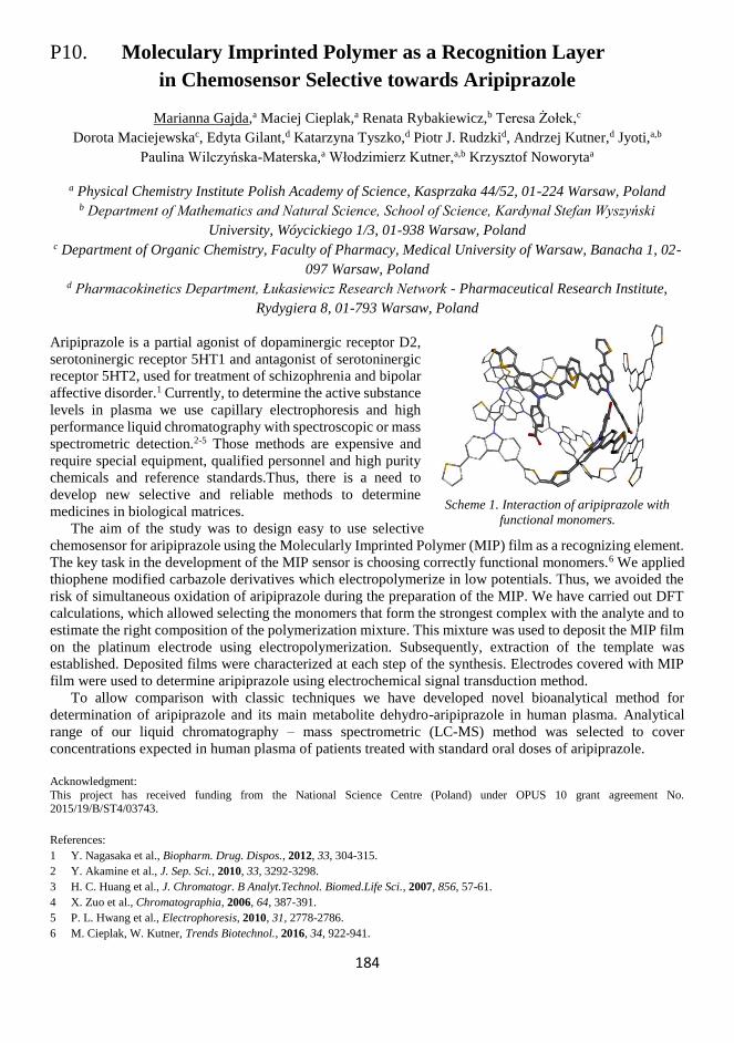
184
P10. Moleculary Imprinted Polymer as a Recognition Layer
in Chemosensor Selective towards Aripiprazole
Marianna Gajda,a Maciej Cieplak,a Renata Rybakiewicz,b Teresa Żołek,c
Dorota Maciejewskac, Edyta Gilant,d Katarzyna Tyszko,d Piotr J. Rudzkid, Andrzej Kutner,d Jyoti,a,b
Paulina Wilczyńska-Materska,a Włodzimierz Kutner,a,b Krzysztof Noworytaa
a Physical Chemistry Institute Polish Academy of Science, Kasprzaka 44/52, 01-224 Warsaw, Poland b Department of Mathematics and Natural Science, School of Science, Kardynal Stefan Wyszyński
University, Wóycickiego 1/3, 01-938 Warsaw, Poland c Department of Organic Chemistry, Faculty of Pharmacy, Medical University of Warsaw, Banacha 1, 02-
097 Warsaw, Poland d Pharmacokinetics Department, Łukasiewicz Research Network - Pharmaceutical Research Institute,
Rydygiera 8, 01-793 Warsaw, Poland
Aripiprazole is a partial agonist of dopaminergic receptor D2,
serotoninergic receptor 5HT1 and antagonist of serotoninergic
receptor 5HT2, used for treatment of schizophrenia and bipolar
affective disorder.1 Currently, to determine the active substance
levels in plasma we use capillary electrophoresis and high
performance liquid chromatography with spectroscopic or mass
spectrometric detection.2-5 Those methods are expensive and
require special equipment, qualified personnel and high purity
chemicals and reference standards.Thus, there is a need to
develop new selective and reliable methods to determine
medicines in biological matrices.
The aim of the study was to design easy to use selective
chemosensor for aripiprazole using the Molecularly Imprinted Polymer (MIP) film as a recognizing element.
The key task in the development of the MIP sensor is choosing correctly functional monomers.6 We applied
thiophene modified carbazole derivatives which electropolymerize in low potentials. Thus, we avoided the
risk of simultaneous oxidation of aripiprazole during the preparation of the MIP. We have carried out DFT
calculations, which allowed selecting the monomers that form the strongest complex with the analyte and to
estimate the right composition of the polymerization mixture. This mixture was used to deposit the MIP film
on the platinum electrode using electropolymerization. Subsequently, extraction of the template was
established. Deposited films were characterized at each step of the synthesis. Electrodes covered with MIP
film were used to determine aripiprazole using electrochemical signal transduction method.
To allow comparison with classic techniques we have developed novel bioanalytical method for
determination of aripiprazole and its main metabolite dehydro-aripiprazole in human plasma. Analytical
range of our liquid chromatography – mass spectrometric (LC-MS) method was selected to cover
concentrations expected in human plasma of patients treated with standard oral doses of aripiprazole.
Acknowledgment:
This project has received funding from the National Science Centre (Poland) under OPUS 10 grant agreement No.
2015/19/B/ST4/03743.
References:
1 Y. Nagasaka et al., Biopharm. Drug. Dispos., 2012, 33, 304-315.
2 Y. Akamine et al., J. Sep. Sci., 2010, 33, 3292-3298.
3 H. C. Huang et al., J. Chromatogr. B Analyt.Technol. Biomed.Life Sci., 2007, 856, 57-61.
4 X. Zuo et al., Chromatographia, 2006, 64, 387-391.
5 P. L. Hwang et al., Electrophoresis, 2010, 31, 2778-2786.
6 M. Cieplak, W. Kutner, Trends Biotechnol., 2016, 34, 922-941.
Scheme 1. Interaction of aripiprazole with
functional monomers.

185
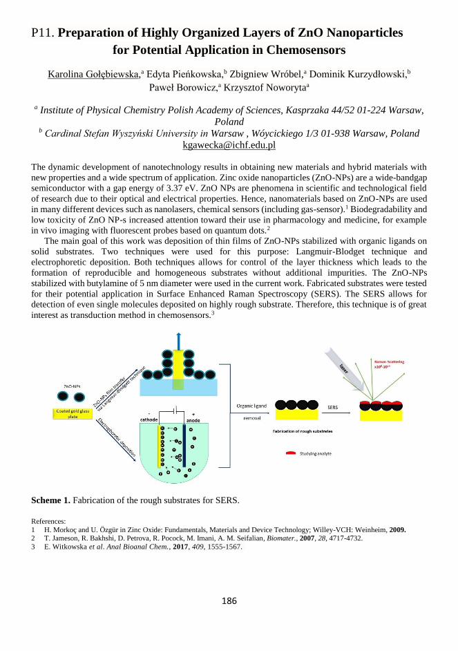
186
P11. Preparation of Highly Organized Layers of ZnO Nanoparticles
for Potential Application in Chemosensors
Karolina Gołębiewska,a Edyta Pieńkowska,b Zbigniew Wróbel,a Dominik Kurzydłowski,b
Paweł Borowicz,a Krzysztof Noworytaa
a Institute of Physical Chemistry Polish Academy of Sciences, Kasprzaka 44/52 01-224 Warsaw,
Poland b Cardinal Stefan Wyszyński University in Warsaw , Wóycickiego 1/3 01-938 Warsaw, Poland
The dynamic development of nanotechnology results in obtaining new materials and hybrid materials with
new properties and a wide spectrum of application. Zinc oxide nanoparticles (ZnO-NPs) are a wide-bandgap
semiconductor with a gap energy of 3.37 eV. ZnO NPs are phenomena in scientific and technological field
of research due to their optical and electrical properties. Hence, nanomaterials based on ZnO-NPs are used
in many different devices such as nanolasers, chemical sensors (including gas-sensor).1 Biodegradability and
low toxicity of ZnO NP-s increased attention toward their use in pharmacology and medicine, for example
in vivo imaging with fluorescent probes based on quantum dots.2
The main goal of this work was deposition of thin films of ZnO-NPs stabilized with organic ligands on
solid substrates. Two techniques were used for this purpose: Langmuir-Blodget technique and
electrophoretic deposition. Both techniques allows for control of the layer thickness which leads to the
formation of reproducible and homogeneous substrates without additional impurities. The ZnO-NPs
stabilized with butylamine of 5 nm diameter were used in the current work. Fabricated substrates were tested
for their potential application in Surface Enhanced Raman Spectroscopy (SERS). The SERS allows for
detection of even single molecules deposited on highly rough substrate. Therefore, this technique is of great
interest as transduction method in chemosensors.3
Scheme 1. Fabrication of the rough substrates for SERS.
References:
1 H. Morkoç and U. Özgür in Zinc Oxide: Fundamentals, Materials and Device Technology; Willey-VCH: Weinheim, 2009. 2 T. Jameson, R. Bakhshi, D. Petrova, R. Pocock, M. Imani, A. M. Seifalian, Biomater., 2007, 28, 4717-4732.
3 E. Witkowska et al. Anal Bioanal Chem., 2017, 409, 1555-1567.

187

188
P12. Spin Filter Properties of Chiral Thin Film Highlighted
by an External Magnetic Field
S. Grecchi,a S. Arnaboldi,a M. Magni,a T. Benincori,b C. Fontanesi,c
R. Cirilli,d S. Cauteruccio,a E. Licandro,a P. R. Mussinia
a Università degli Studi di Milano, Dipartimento di Chimica, Via Golgi 19 20133, Milano, Italy. b Università degli Studi dell’Insubria, Dipartimento di Scienza e Alta Tecnologia, Via
Valleggio11, 22100, Como, Italy c Università degli Studi di Modena, Dipartimento di Ingegneria "Enzo Ferrari", via Vivarelli 10,
41125, Modena, Italy d Istituto Superiore di Sanità, Centro Nazionale per il Controllo e la Valutazione dei Farmaci,
Viale Regina Elena 299, Roma, Italy
The study of the mutual influence between chirality, electron spin and magnetism is a well-documented field
of research, in fact implications span from pure fundamental research, to chemical applications of both
analytical and synthetic character, to multidisciplinary purposes.1,2 In this context the interrelated disciplines
of magnetoelectrochemistry, spintronics and Spin Dependent Electrochemistry (SDE) play a crucial role. In
particular, the combination of spintronics with magneto- electrochemistry, involving truly chiral molecular
spin selectors, was promoted by the discovery of the Chiral Induced Spin Selectivity (CISS) effect by Ron
Naaman and coworkers, observing spin polarization in photo-ejected electrons transmitted through a thin
layer of enantiopure material adsorbed on gold, acting as an electron spin filter.3
Our proposed strategy is a variation of the SDE protocol, in fact the innovative set-up involves i) a non-
ferromagnetic electrode (ITO as working electrode) covered by thin electroactive chiral films as electron
source, ii) achiral redox couples dissolved in aqueous or organic solutions and iii) an external permanent
magnet which was placed perpendicular to the electrode surface, considering as spin filters four different
types of chiral selectors (with different stereogenic elements, i.e. helix, stereogenic axis and chiral
pendant).4-6
A spectacular unforeseen effect was observed performing cyclic voltammetry (CV) experiments under
applied magnetic field, in fact the CV peaks of achiral, chemically reversible redox couples undergo
impressive potential shifts by flipping the magnet orientation (north vs south), with specular results changing
the film configuration.
The importance of these studies includes possible applications in the field of spintronics, electronics,
chemical sensoristic and so on and provides a striking evidence of the spin selectivity properties of chiral
thin films.
References:
1 G. A. Printz, Science, 1998, 282, 1660-1663.
2 P. L. Popa, N. T. Kemp, H. Majjad, G. Dalmas, V. Faramarzi, C. Andreas, R. Hertel, B. Doudin, Proc. Natl. Acad. Sci., 2014, 111, 10433-10437.
3 O. B Dor, S. Yochelis, S. P. Mathew, R. Naaman, Y. Paltiel, Nat. Commun., 2013, 4, 3256-3262.
4 T. Benincori, S. Arnaboldi, M. Magni, S. Grecchi, R. Cirilli, C. Fontanesi, P. R. Mussini, Chem. Sci., 2019, 10, 2750-2757.
5 S. Arnaboldi, S. Cauteruccio, S. Grecchi, T. Benincori, M. Marcaccio, A. Orbelli Biroli, G. Longhi, E. Licandro P. R. Mussini,
Chem. Sci., 2019, 10, 1539-1548.
6 S. Arnaboldi, T. Benincori, A. Penoni, L. Vaghi, R. Cirilli, S. Abbate, G. Longhi, G. Mazzeo, S. Grecchi, M. Panigati, P. R. Mussini, Chem. Sci., 2019, 10, 2708-2717.

189
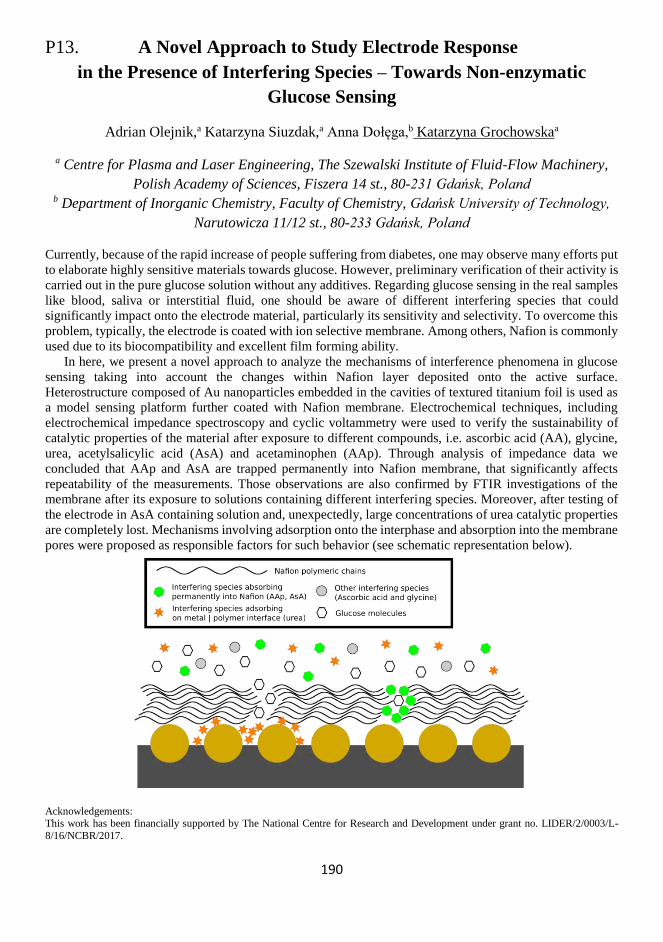
190
P13. A Novel Approach to Study Electrode Response
in the Presence of Interfering Species – Towards Non-enzymatic
Glucose Sensing
Adrian Olejnik,a Katarzyna Siuzdak,a Anna Dołęga,b Katarzyna Grochowskaa
a Centre for Plasma and Laser Engineering, The Szewalski Institute of Fluid-Flow Machinery,
Polish Academy of Sciences, Fiszera 14 st., 80-231 Gdańsk, Poland b Department of Inorganic Chemistry, Faculty of Chemistry, Gdańsk University of Technology,
Narutowicza 11/12 st., 80-233 Gdańsk, Poland
Currently, because of the rapid increase of people suffering from diabetes, one may observe many efforts put
to elaborate highly sensitive materials towards glucose. However, preliminary verification of their activity is
carried out in the pure glucose solution without any additives. Regarding glucose sensing in the real samples
like blood, saliva or interstitial fluid, one should be aware of different interfering species that could
significantly impact onto the electrode material, particularly its sensitivity and selectivity. To overcome this
problem, typically, the electrode is coated with ion selective membrane. Among others, Nafion is commonly
used due to its biocompatibility and excellent film forming ability.
In here, we present a novel approach to analyze the mechanisms of interference phenomena in glucose
sensing taking into account the changes within Nafion layer deposited onto the active surface.
Heterostructure composed of Au nanoparticles embedded in the cavities of textured titanium foil is used as
a model sensing platform further coated with Nafion membrane. Electrochemical techniques, including
electrochemical impedance spectroscopy and cyclic voltammetry were used to verify the sustainability of
catalytic properties of the material after exposure to different compounds, i.e. ascorbic acid (AA), glycine,
urea, acetylsalicylic acid (AsA) and acetaminophen (AAp). Through analysis of impedance data we
concluded that AAp and AsA are trapped permanently into Nafion membrane, that significantly affects
repeatability of the measurements. Those observations are also confirmed by FTIR investigations of the
membrane after its exposure to solutions containing different interfering species. Moreover, after testing of
the electrode in AsA containing solution and, unexpectedly, large concentrations of urea catalytic properties
are completely lost. Mechanisms involving adsorption onto the interphase and absorption into the membrane
pores were proposed as responsible factors for such behavior (see schematic representation below).
Acknowledgements: This work has been financially supported by The National Centre for Research and Development under grant no. LIDER/2/0003/L-
8/16/NCBR/2017.

191

192
P14. Fluorescence Microscopy of mCherry Photoactive Protein
Justyna Grzelak,a Martyna Domagalska,a Artur Ziółkowski,a Karolina Sulowska,a
Joanna Niedziółka-Jӧnsson,b Sebastian Maćkowskia,c
a Faculty of Physics, Astronomy and Informatics, Nicolaus Copernicus University, Torun, Poland b Institute of Physical Chemistry, Polish Academy of Sciences, Warszawa, Poland
c Baltic Institute of Technology, Gdynia, Poland
Photoactive proteins, including photoconvertible and photoswitchable proteins, form a class of structures
with unique and exciting properties.1 Combining them with nanostructures, such as plasmonically active
metallic nanoparticles,2 offers a possibility to construct systems with new functionality.
In this work we study the photophysics of several photoactive proteins using fluorescence microscopy
and spectroscopy. In addition to standard ensemble measurements, we have carried out also single-molecule
studies in order to understand the influence of excitation wavelength on the photostability in various matrices.
Also, we have fabricated a hybrid nanostructure with single proteins attached to silver nanowires.
Fluorescence microscopy proves that the emission of these proteins is increased due to the plasmonic
coupling with excitations in the nanowires. An important step towards controlled assembly of such systems
has been developed by depositing microdroplets on silver nanowires. In this way the optical properties of the
photoactive proteins can be remotely controlled with defined excitation wavelength. Consequently, the
experiments show that it is possible to control the fluorescence of photoactive proteins both via plasmon
resonance and remote excitation with precisely tuned excitation wavelength.
Acknowledgements: Research was partially financed by the National Science Centre (Poland) within the OPUS grants 2016/21/B/ST3/02276,
2017/27/B/ST3/02457, and 2017/26/E/ST3/00209, project 3/DOT/2016 funded by the City of Gdynia, and project nr POWR.03.05.00-
00-Z302/17 “Universitas Copernicana Thoruniensis In Futuro” (2018-2022) co-financed by the European Social Fund – the Operational Programme Knowledge Education Development. Module 5. Interdisciplinary PhD School “Academia Copernicana”.
References:
1 E. A. Rodriguez, R. E. Campbell, J. Y. Lin, M. Z. Lin, A. Miyawaki, A. E. Palmer, X. Shu, J. Zhang, R. Y. Tsien, Trends in
Biochemical Sciences, 2017, 42, 111-129.
2 J. Niedziółka-Jönsson, S. Maćkowski, Materials, 2019, 12(9), 1418.

193

194
P15. Functionalization of Au Electrodes
with Glycosylated Hydrazides for the Electrochemical Detection
of Vibrio cholerae Toxin
C. Guillén Posteguillo, A. Kołodziej, F. Fernández-Trillo, P. Rodríguez
University of Birmingham, Birmingham, United Kingdom
The World Health Organization (WHO) estimates that millions of people fall ill and thousands die every
year after ingesting contaminated food or water;1 most cases of food poisoning are caused by bacteria.
Current standard methods of detection of bacteria, although reliable, present features that make their use
difficult or impossible in rural areas, regions with poor health infrastructure or conflict zones. Furthermore,
phenomena like climate change, globalization or the aging of the world population have increased the risk
of food poisoning, for e.g. in the aftermath of increasingly frequent natural disasters.2 Electrochemical
biosensors represent an alternative thanks to their sensitivity but also simplicity, low-cost and portability.3
In this work the first steps of
development of an electrochemical
biosensor for the detection of the toxin
of Vibrio cholerae in aqueous media are
presented. The design of the device aims
at mimicking the recognition
mechanism of the toxin in the human
intestinal cells.4 Polycrystalline
electrodes have been functionalized
with self-assembled monolayers
(SAMs) of organic molecules
(hydrazides) that will be chemically
linked to galactose. The interaction of
the toxin with the saccharide will induce
changes in the electrodic process on the
electrode surface generating an
analytical signal for the detection of
cholera. This biosensor will serve as a
proof-of-concept for the construction of
analogues for other pathogens.
References:
1 WHO (World Health Organization), online. Food Safety. Last updated on: 31 October 2017. Retrieved from: https://www.who.int/news-room/fact-sheets/detail/food-safety, (accessed 10 March 2019).
2 Tsvangirayi Mukwazhi/Associated Press (2019, March 27). Cholera Outbreak Raises New Danger in Flood-Ravaged
Mozambique. The New York Times. Retrieved from: https://www.nytimes.com/2019/03/27/world/africa/cholera-mozambique-cyclone-idai.html, (accessed 28 March 2019).
3 J. Monzó, I. Insua., F. Fernández-Trillo, P. Rodríguez, Analyst, 2015, 140(21), 7116-7128.
4 J. A. Mertz, J. A. McCann, W. D. Picking, Biochemical and Biophysical Research Communications, 1996, 226(1), 140-144.

195
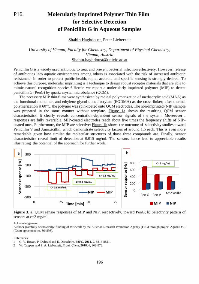
196
P16. Molecularly Imprinted Polymer Thin Film
for Selective Detection
of Penicillin G in Aqueous Samples
Shahin Haghdoust, Peter Lieberzeit
University of Vienna, Faculty for Chemistry, Department of Physical Chemistry,
Vienna, Austria
Penicillin G is a widely used antibiotic to treat and prevent bacterial infection effectively. However, release
of antibiotics into aquatic environments among others is associated with the risk of increased antibiotic
resistance.1 In order to protect public health, rapid, accurate and specific sensing is strongly desired. To
achieve this purpose, molecular imprinting is a technique to design robust receptor materials that are able to
mimic natural recognition species.2 Herein we report a molecularly imprinted polymer (MIP) to detect
penicillin G (PenG) by quartz crystal microbalance (QCM).
The necessary MIP thin films were synthesized by radical polymerization of methacrylic acid (MAA) as
the functional monomer, and ethylene glycol dimethacrylate (EGDMA) as the cross-linker; after thermal
polymerization at 60°C, the polymer was spin-coated onto QCM electrodes. The non-imprinted (NIP) sample
was prepared in the same manner without template. Figure 1a shows the resulting QCM sensor
characteristics: It clearly reveals concentration-dependent sensor signals of the system. Moverover ,
responses are fully reversible. MIP-coated electrodes reach about five times the frequency shifts of NIP-
coated ones. Furthermore, the MIP are selective: Figure 1b shows the outcome of selectivity studies toward
Penicillin V and Amoxicillin, which demonstrate selectivity factors of around 1.5 each. This is even more
remarkable given how similar the molecular structures of those three compounds are. Finally, sensor
characteristics reveal limit of detection at 0.015 mg/ml. The sensors hence lead to appreciable results
illustrating the potential of the approach for further work.
Figure 3. a) QCM sensor responses of MIP and NIP, respectively, toward PenG; b) Selectivity pattern of
sensors at c=2 mg/ml.
Acknowledgement: Authors gratefully acknowledge funding of this work by the Austrian Research Promotion Agency (FFG) through project AquaNOSE
(Grant agreement no. 864893).
References:
1 G. V. Royan, P. Dubruel and E. Daeseleire, JAFC, 2014, 2, 8814-8821. 2 W. Cuypers and P. A. Lieberzeit, Front. Chem, 2018, 6, 268-278.
-500
-300
-100
100
300
0 25 50 75
Sen
sor
resp
on
se [
Hz]
Time [min]
NIP MIP
C= 0.4 mg/mL
C= 0.2 mg/mL
0
200
400
600
800
Pen GSen
sor
resp
on
se [
Hz]
MIP NIP
Pen V
0.3 mg/mL
0.15 mg/mL
b
C= 0.8 mg/mL
Amoxicillin
a
C= 2 mg/mL

197
198
P17. Biofunctionalization of a Microring Resonator
via Hydrosilylation Followed by Copper Free Click Chemistry
Anders Henriksson,a Peter Neubauer,a Mario Birkholzb
a Technische Universität Berlin, Institute of Biotechnology, Chair of Bioprocess Engineering,
Ackerstrasse 76, 13355 Berlin, Germany b IHP - Leibniz-Institut für innovative Mikroelektronik, Im Technologiepark 25, D - 15236
Frankfurt (Oder), Germany
Silicon on insulator (SOI) micro-ring resonators are one of the more interesting techniques developed for
label free sensing during the last decade. It benefits from well-known standardized and scalable
semiconductor fabrication processes, and enables real time responses, minimal footprints and extraordinary
sensitivity. Essential for the development of high-performance sensors is a surface modification strategy that
generates a sensor surface that facilitate both molecular interaction as well as the generation of a large optical
resonance shift. The general surface chemistry approaches are based on ω-functional silanes that binds to the
native oxide of the silicon surface followed by biomolecule immobilization via standard coupling reactions.
While the approach is reliable and simple, this strategy functionalizes not only the active sensor surface but
also the enclosed buried oxide layer (BOX), thus wasting receptor molecules and potentially decreasing the
sensitivity of the device. In this study we have explored a copper free click chemistry approach1 as a
bioconjugation method and the hydrosilylation reaction as an alternative to silane-based surface
functionalization strategies that is selective for the silicon surface.
An electron deficient dialkyne was grafted to the ring resonator via thermal induced hydrosilyation to
form an alkyne terminated surfaces. Green fluorescent protein labeled with azido functionalities was
subsequently coupled to the sensor surface via copper free click chemistry at 40 °C. The functionalization
was followed optically via a resonance shift of the ring resonators and by ellipsometry. The copper free click
chemistry approach was preferred over the state-of-the-art copper catalyzed reaction, as the later resulted in
substantial copper deposition onto the sensor chip.
The presented protocol may be useful for the biofunctionalization of a large variety of SOI microchips,
were functionalization of the silicon oxide are to be avoided.
References: 1 A. Henriksson, G. Friedbacher and H. Hoffmann, Langmuir, 2011, 27(12), 7345-7348.

199
200
P18. On the Role of Cholesterol in Perifosine Action
on Model Lipid Rafts
Joanna Juhaniewicz-Debinska, Kinga Burdach
Faculty of Chemistry, Biological and Chemical Research Centre, University of Warsaw
Zwirki I Wigury 101, 02-089, Warsaw, Poland
Cancer diseases are one of the main causes of death, right after cardiovascular diseases. Their number is
steadily growing despite extensive development of new drugs and therapies. Synthetic antitumor lipids
(ATLs) are a promising group of compounds in cancer chemotherapy.1 They can be divided into two main
classes: alkylphospholipids (APLs) and alkylphosphocholines (APCs). They are structurally similar to a
naturally occurring lysophosphatidylcholine and possess distinct antiproliferative properties in tumor cells.2
APLs and APCs do not interfere with the DNA but incorporate directly into cell membranes, where they
accumulate and disturb lipid metabolism and lipid-dependent signalling pathways.
Perifosine (octadecyl-(1,1-dimethylpiperidinio-4-yl)-phosphate) is one of alkylphospholipids analog
with promising results against a variety of cancers. The studies showed that perifosine accumulates in
specific domains in cell membranes called lipid rafts.3 These membrane microdomains enriched in
cholesterol and sphingolipids are the platform of several signalling pathways, including the Akt pathway,
one of the most frequently hyperactivated signaling pathways in human cancers and, therefore, a very
important target in a global fight against this disease.
The aim of the present study was to assess the effects of perifosine on model lipid membranes reflecting
the lipid composition of natural lipid rafts. Model lipid rafts were composed of 1,2-dioleoyl-sn-glycero-3-
phosphocholine (DOPC), sphingomyelin (SM) and cholesterol (Chol). Firstly, the effect of cholesterol on
perifosine action on model lipid raft was studied. For this purpose, we prepared model lipid rafts with 0 – 50
% cholesterol content. We have also verified the effect of gangliosides on the perifosine – lipid membrane
interactions. Results of Langmuir studies show that perifosine incorporate in model lipid membranes at the
air-water interface and the effect of perifosine increases with increasing cholesterol content. The changes in
the morphology of the lipid layer in the presence of perifosine were also monitored by Brewster Angle
Microscopy. In the next step, solid-supported lipid bilayers were used to obtained more detailed information
on perifosine-lipid interactions. We employed the combination of Langmuir-Blodgett and Langmuir-
Schaefer techniques to immobilized model lipid membranes on the solid support. Quartz crystal
microbalance with dissipation monitoring allowed us to observe the kinetics of the perifosine-membrane
interactions and to verify the mechanism of perifosine-induced perturbations within the lipid membrane.
References: 1 J. A. Pachioni, J. G. Magalhaes, E. J. C. Lima, L. M. Bueno, J. F. Barbosa, M. Malta de Sa, C.O. Rangel- Yagui, J. Pharm. Pharm.
Sci., 2013, 16, 742-759.
2 A. Kostadinova, T. Topouzova-Hristova, A. Momchilova, R. Tzoneva, M. R. Berger, Adv Protein Chem Str., 2015, 101, 27-66. 3 F. Mollinedo, C. Gajate, Future Oncol., 2010, 6, 811-821.
4 S. B. Kondapaka, S. S. Singh, G. P. Dasmahapatra, E. A. Sausville, K. K. Roy, Mol. Cancer Ther., 2003, 2, 1093-1103.

201
202
P19. Synergy of Surface and Semi-covalent Imprinting
for Chemosensing of Gonadotropin Protein Hormone Jakub Kałęcki, Marcin Dąbrowski, Maciej Cieplak, Piyush Sindhu Sharma
Institute of Physical Chemistry, Polish Academy of Sciences,
Kasprzaka 44/52, 01-224 Warsaw, Poland
Family of gonadotropin hormone includes human chorionic gonadotropin (hCG), luteinizing hormone (LH)
and follicle-stimulating hormone (FSH). The latter hormone stimulates the maturation of the primordial germ
cells in women and men. Both lowered and elevated levels of FSH in the blood are associated with disease
states. Therefore, interest in the determination of fertility hormones has steadily increased.
Molecular imprinted polymers (MIPs) are synthetic biomimetic receptors which are capable of binding
target molecules with an affinity and specificity similar to natural receptors. These materials integrated
successfully to transducer surface for fabrication of chemosensors. However, conditions normally used for
imprinting of small molecule can bring conformational change in proteins. Additionally, bulky proteins are
difficult to remove from cross-linked polymer.
Therefore, to enhance the number of binding sites and for easy removal of proteins from imprinted
cavities we adopt combination of surface and semi-covalent imprinting. Towards that, we first immobilize
the protein on silica substrate through an anchor layer. Later, we labelled the immobilized protein with
functional monomer. This labelling was possible through amide chemistry. In final step, we
electrochemically cross-linked functional monomers with excess of cross-linking monomer.
One step further, in current work, we have optimized conditions for preparation of a stable homogeneous
colloidal crystal structure with surface modified silica beads of 500 nm diameter.1 We confirmed regular and
hexagonal packing of silica beads and resulted inverse opal surface imprinted molecular imprinted film by
scanning electron microscope (SEM). Successful modification of silica with protein through imine bonds, as
well as protein derivatizing with functional monomers through amide bonds, were confirmed by X-ray
photoelectron spectroscopy (XPS). The recognition of FSH by the macroporous molecularly imprinted
polymer film was transduced with the help of capacitive impedimetry. Ultra level sensitivity was achieved
by synergetic combination of silica molding, surface and semi-covalent imprinting. This synthetic receptor,
was further used for selective determination of FSH in artificial blood serum samples.
Acknowledgments:
The present research was financially supported by the National Science Centre (Grant No. NCN 2017/25/B/ST4/01696 to P.S.S.).
References:
1 M. Dąbrowski et al., ACS Appl. Mater. Interfaces, 2019, 11, 9265−9276.

203
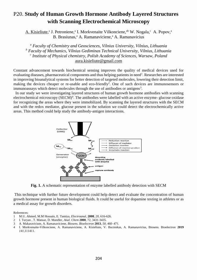
204
P20. Study of Human Growth Hormone Antibody Layered Structures
with Scanning Electrochemical Microscopy
A. Kisieliute,a J. Petroniene,a I. Morkvenaite Vilkonciene,ab W. Nogala,c A. Popov,a
B. Brasiunas,a A. Ramanaviciene,a A. Ramanavicius
a Faculty of Chemistry and Geosciences, Vilnius University, Vilnius, Lithuania b Faculty of Mechanics, Vilnius Gediminas Technical University, Vilnius, Lithuania
c Institute of Physical chemistry, Polish Academy of Sciences, Warsaw, Poland
Constant advancement towards biochemical sensing improves the quality of medical devices used for
evaluating diseases, pharmaceutical components and thus helping patients in need1. Researches are interested
in improving bioanalytical systems for better detection of targeted molecules, lowering their detection limit,
making the devices cheaper or re-usable and eco-friendly2. One of such devices are immunosensors or
immunoassays which detect molecules through the use of antibodies or antigens3.
In our study we were investigating layered structures of human growth hormone antibodies with scanning
electrochemical microscopy (SECM)4. The antibodies were labelled with an active enzyme- glucose oxidase
for recognizing the areas where they were immobilized. By scanning the layered structures with the SECM
and with the redox mediator, glucose present in the solution we could detect the electrochemically active
areas. This method could help study the antibody-antigen interactions.
Fig. 1. A schematic representation of enzyme labelled antibody detection with SECM
This technique with further future development could help detect and evaluate the concentration of human
growth hormone present in human biological fluids. It could be useful for dopamine testing in athletes or as
a medical assay for growth disorders.
References: 1 M.U. Ahmed, M.M Hossain, E. Tamiya, Electroanal, 2008, 20, 616-626.
2 I. Turyan , T. Matsue, D. Mandler, Anal. Chem 2000, 72, 3431-3435.
3 A. Makaraviciute, A. Ramanaviciene, Biosens. Bioelectron 2013, 50, 460–471. 4 I. Morkvenaite-Vilkonciene, A. Ramanaviciene, A. Kisieliute, V. Bucinskas, A. Ramanavicius, Biosens. Bioelectron 2019
141,111411.

205
206
P21. The Influence of Modified Cyclodextrin
on the Anthracycline Binding to DNA
A. Krzak,a M. Majdecki,b O. Święch,a R. Bilewicza
a University of Warsaw, Faculty of Chemistry, Warsaw, Poland b Institute of Organic Chemistry, Polish Academy of Sciences, Warsaw, Poland
Anthracycline antibiotics (ANT), are most commonly used anticancer agents for the treatment of several
types of cancers. Clinical effects of the drugs are connected with modification of the DNA structure primarily
through intercalating complexes and covalent bonding.1,2 However, the clinical application of doxorubicin
has been limited by serious adverse effects.3,4 The specific toxicity of the drug is due to reactive oxygen
species (ROS) produced in redox reactions of anthracycline. The radical oxygen species can interact with
cellular components such as proteins, lipids, carbohydrates or nucleic acids. All of the modifications can
cause mutation of the healthy cells. The toxicity can be reduced e.g. by creating an inclusion complex holding
the anthracycline molecule inside a cyclodextrin (CD) cavity.
The limitation in the use of CD as a carrier of anthracycline drugs is the low stability of the complex. We
showed earlier that appropriate modification of the cyclodextrin can increase the stability of the drug-CD
inclusion complex. The derivatives of β-cyclodextrin with a triazole moiety formed strong inclusion
complexes with ANT with 1:1 stoichiometry. Using voltammetry data we determined the stability constants
of the complexes and we found their dependence on the pH: at pH 7.4, corresponding to the pH of body
fluids, the stability constants were much larger than at pH 5.5, which is characteristic for cancer cells.5-7 The
question was how the modified CD affects the interactions of the drug with DNA – which are crucial for the
therapy.
We present here newly designed nontoxic derivatives of β-cyclodextrin containing both antioxidant and
targeting units in the molecule (lipoic acid, folic acid and galactosamine). We determined first the stabilities
of the complexes of this new group of modified CDs with daunorubicin and doxorubicin, and next we
evaluated the effect of these modified βCDs on the anthracycline interactions with DNA. Interestingly, the
CD ligands were found to strengthen the drug–DNA intercalation both at pH 7.4 and 5.5. In the absence of
cyclodextrins, binding of the anthracyclines to DNA is known to be much weaker at lower pH, which is a
difficulty encountered in the therapies. We demonstrate that when the drug is complexed by appropriately
modified cyclodextrins this effect is eliminated: the values of the stability constants of ANT-DNA are strong
and similar at both pHs, The reasons of this phenomenon, important for potential therapeutic applications of
the CD - based drug delivery, will be discussed.
References:
1 J. B. Chaires, J. E. Herrera, M. J. Waring, Biochemistry, 1990, 29, 6145-6153.
2 G. J. Quigley, A. H. Wang, G. Ughetto, G. van der Marel, J. H. van Boom, A. Rich, Proc. Natl. Acad. Sci., 1980, 77, 7204-7208. 3 G. Takenura, H. Fujiwara, Prog. Cardiocasc. Dis., 2007, 49, 330-352.
4 G. Minotti, P. Menna, E. Salvatorelli, G. Cairo, L. Gianni, Pharmacol. Rev., 2004, 56, 185-229.
5 O. Swiech, P. Dutkiewicz, K. Wójciuk, K. Chmurskim M. Kruszewski, R. Bilewicz, J. Physical. Chemistry B, 2013, 117, 13444-13450.
6 O. Swiech, M. Majdecki, A. Debinski, A. Krzak, T. M. Stępkowski, G. Wójciuk, M. Kruszewski,
R. Bilewicz, Nanoscale, 2016, 8, 16733-16742. 7 A. Krzak, O. Swiech, M. Majdecki, R. Bilewicz, Electrochim. Acta, 2017, 247, 139-148.

207
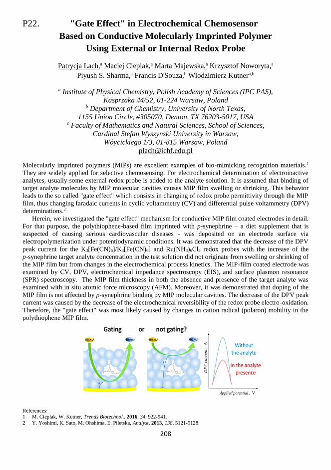
208
P22. "Gate Effect" in Electrochemical Chemosensor
Based on Conductive Molecularly Imprinted Polymer
Using External or Internal Redox Probe
Patrycja Lach,a Maciej Cieplak,a Marta Majewska,a Krzysztof Noworyta,a
Piyush S. Sharma,a Francis D'Souza,b Wlodzimierz Kutnera,b
a Institute of Physical Chemistry, Polish Academy of Sciences (IPC PAS),
Kasprzaka 44/52, 01-224 Warsaw, Poland b Department of Chemistry, University of North Texas,
1155 Union Circle, #305070, Denton, TX 76203-5017, USA c Faculty of Mathematics and Natural Sciences, School of Sciences,
Cardinal Stefan Wyszynski University in Warsaw,
Wóycickiego 1/3, 01-815 Warsaw, Poland
Molecularly imprinted polymers (MIPs) are excellent examples of bio-mimicking recognition materials.1
They are widely applied for selective chemosensing. For electrochemical determination of electroinactive
analytes, usually some external redox probe is added to the analyte solution. It is assumed that binding of
target analyte molecules by MIP molecular cavities causes MIP film swelling or shrinking. This behavior
leads to the so called "gate effect" which consists in changing of redox probe permittivity through the MIP
film, thus changing faradaic currents in cyclic voltammetry (CV) and differential pulse voltammetry (DPV)
determinations.2
Herein, we investigated the "gate effect" mechanism for conductive MIP film coated electrodes in detail.
For that purpose, the polythiophene-based film imprinted with p-synephrine – a diet supplement that is
suspected of causing serious cardiovascular diseases - was deposited on an electrode surface via
electropolymerization under potentiodynamic conditions. It was demonstrated that the decrease of the DPV
peak current for the K3[Fe(CN)6]/K4[Fe(CN)6] and Ru(NH3)6Cl3 redox probes with the increase of the
p-synephrine target analyte concentration in the test solution did not originate from swelling or shrinking of
the MIP film but from changes in the electrochemical process kinetics. The MIP-film coated electrode was
examined by CV, DPV, electrochemical impedance spectroscopy (EIS), and surface plasmon resonance
(SPR) spectroscopy. The MIP film thickness in both the absence and presence of the target analyte was
examined with in situ atomic force microscopy (AFM). Moreover, it was demonstrated that doping of the
MIP film is not affected by p-synephrine binding by MIP molecular cavities. The decrease of the DPV peak
current was caused by the decrease of the electrochemical reversibility of the redox probe electro-oxidation.
Therefore, the "gate effect" was most likely caused by changes in cation radical (polaron) mobility in the
polythiophene MIP film.
References: 1 M. Cieplak, W. Kutner, Trends Biotechnol., 2016, 34, 922-941.
2 Y. Yoshimi, K. Sato, M. Ohshima, E. Piletska, Analyst, 2013, 138, 5121-5128.

209
210
P23. Targeted Nanoparticles for
Diagnostic and Theranostic Applications.
Juan Leva-Bueno,a Ina Meuskens,b , Dirk Linke,b Sally Peyman,b Paul A Millnera
a University of Leeds, UK b University of Oslo, Norway
Antimicrobial resistance (AMR) is a serious threat due to the rise of multidrug-resistant (MDR) bacteria.1
Therefore, new technologies such as biosensing and lab-on-a-chip (LOC) devices have emerged in the last
decades. Biosensing can detect and quantify biological analytes and combine high sensitivity and specificity
with fast response times, portability, low-cost and ease-of-use.2
Two systems were studied through electrochemical impedance spectroscopy (EIS): on the one hand, a
classic immunosensor3 for E. coli was able to detect from 103 cfu/ml to 108 cfu/ml. On the other hand, a novel
extracellular matrix (ECM)-based bioreceptor was investigated using YadA-expressing E. coli (O:8) as
specific analyte.4 ECM proteins are involved in the first-step of infection and it is thought that they can
improve the homogeneity and reproducibility of antibodies as well as minimizing costs.
References:
1 W. Wang et al., Infect Drug Resist., 2018, 11, 1645–1658.
2 L. Soleymani and F. Li, ACS Sens., 2017, 2, 458–467. 3 K. A. Kline, S. Fälker, S. Dahlberg, S. Normark and B. Henriques-Normark, Cell Host Microbe, 2009, 5, 580-592.
4 J. Pizarro-Cerda and P. Cossart Cell, 2006, 124, 715–727.

211

212
P24. Studies of the Charge Transfer Kinetics
and Surface Inhomogeneities
of Enzyme-functionalized Au-Ti Electrode
Wiktoria Lipińska,a Katarzyna Siuzdak,a Jacek Ryl,b Paweł Ślepski,b Katarzyna Grochowskaa
a Centre for Plasma and Laser Engineering, The Szewalski Institute of Fluid-Flow Machinery,
Polish Academy of Sciences, Fiszera 14 st., 80-231 Gdańsk, Poland b Department of Electrochemistry, Corrosion and Material Engineering, Faculty of Chemistry,
Gdańsk University of Technology, Narutowicza 11/12 st., 80-233 Gdańsk, Poland
Due to the fact that nowadays about 400 million people are diabetics and 1.5 million of them die each year,
glucose detection in human body fluids contributes to many clinical tests and development of variety of
biosensors.1,2 Enzymatic electrochemical sensors represent the majority of such devices as they are
considered as reliable. Typically their operating principle is based on potentiometric, amperometric or
impedimetric measurements while the impedimetric approach is thought to be the most sensitive.
Nevertheless, it should be considered that the errors associated with chosen technique affects its performance,
while impedance measurement accuracy highly depends on selection of suitable electric equivalent circuit
(EEC). It should be also noted that surface morphology affects the heterogeneity of distribution of
electrochemical properties and hence the selection of appropriate EEC elements.
Therefore, we propose a novel approach for impedimetric measurements by applying dynamic
electrochemical impedance spectroscopy in galvanostatic mode (g-DEIS), which allows to obtain key
information regarding kinetic changes at the electrode/electrolyte interface during non-stationary processes.3
The investigated material is composed of dimpled Ti plate where each cavity is filled with a single gold
nanoparticle with anchored enzyme: glucose oxidase.4,5 All electrochemical tests are performed in air-
saturated 0.1 M PBS with the addition of different amounts of glucose within the concentration range of: 0-
10 mM. It was shown that the increase of glucose content leads to slow decrease of functionalized layer
capacitance. Additionally, charge transfer resistance changes are more dynamic for glucose concentrations
below 3.9 mM as with the increase of sugar level in electrolyte the enzyme active centers become more
occupied with glucose molecules. Moreover, the disturbance in the kinetics of the charge transfer versus
glucose concentration function was confirmed through direct observation of constant phase element
parameters, especially its exponent α reflecting surface homogeneity. It has been observed that below
concentration of 1.9 mM glucose interaction with enzyme-functionalized electrode negligibly affects the
charge transfer homogeneity, while at higher concentrations the electrode becomes more heterogeneous.
Acknowledgements: This work has been financially support by The National Centre for Research and Development under grant no. LIDER/2/0003/L-
8/16/NCBR/2017.
References:
1 World Health Organization (WHO), Global Report on Diabetes, 2016.
2 J. Mohapatra, B. Ananthoju, V. Nair, A. Mitra, D. Bahadur, N.V. Medhekar and M. Aslam, Appl. Surf. Sci., 2018, 442, 332-341. 3 J. Wysocka, S. Krakowiak, J. Ryl, Electrochim. Acta, 2017, 258, 1-13.
4 K. Grochowska, K. Siuzdak, Ł. Macewicz, F. Skiba, M. Szkoda, J. Karczewski, Ł. Burczyk and G. Śliwiński, Appl. Surf. Sci., 2017,
418, 472-480. 5 K. Grochowska, M. Szkoda, J. Karczewski, G. Śliwiński and K. Siuzdak, Talanta, 2017, 166, 207-214.

213
214
P25. Functional Plasma Polymerized Surfaces for Biosensing
E. Makhneva,a L. Barillas,a Z. Farka,b M. Pastucha,b P. Skladal,b K. Frickea
a Leibniz Institute for Plasma science and Technology (INP), Greifswald, Germany b Central European Institute of Technology (CEITEC), Brno, Czech Republic
Various types of biosensors are convenient tools for fast, economic and user friendly analysis of complex
biological samples. For stable biomodification of the sensing surfaces, a thin functional layer is required to
provide a sufficiently high surface reactivity towards the adopted bioreceptor, as well as good stability in the
presence of sample matrix.
We report on the novel method of preparation of thin reactive plasma polymerized (pp) films for
biosensing applications, using atmospheric pressure plasma jet (APPJ). Our original method for creation of
pp films offers the advantages of a versatile, fast and eco-friendly procedure. The whole procedure takes just
1 min in total and does not require usage of any aggressive chemicals.
Three different types of pp films were developed and characterized. Chemical composition, morphology
and stability in water of the obtained plasma polymerized films were carefully scrutinized. The obtained pp
films provided unique functionality and excellent level of adhesion to the substrates. The tested pp films
exhibited initial thickness losses during the first 24 hours of storage in water. Further prolonged immersion
in water (up to 120 h) did not indicate any significant thickness losses or deterioration of the sensing
properties.
SPR immunosensors were successfully developed, using cost-efficient model pair of AL01 antibody and
HSA antigen. The pp films provided an excellent platform for the efficient immobilization of antibody
molecules and the obtained immunosensors showed selective and high response towards analyte, excellent
regenerability and level of stability. A limit of detection of 50 ng/mL of HSA was achieved for all of the
developed immunosensors. The developed immunosensors provided a linear response in the range of 50
ng/mL to 20 µg/mL concentrations of HSA. The obtained results showed that the plasma polymerized film–
based immunosensors have performance similar to widely used SAM– or CMD–based immunosensors.
Plasma polymerized films though offer a better level of stability and regenerability.

215
216
P26. Chemosensor with Molecularly Imprinted Polymer Film
as the Recognition Unit for Determination of Anticancer Drug Imatinib
Paulina Materska-Wilczynska,a Jyoti,a,b Renata Rybakiewicz,b Teresa Zolek,c
Dorota Maciejewska,c Wlodzimierz Kutner,a,b Krzysztof Noworytaa
a Institute of Physical Chemistry, Polish Academy of Sciences (IPC PAS),
Kasprzaka 44/52, 01-224 Warsaw, Poland b Faculty of Mathematics and Natural Sciences, School of Sciences,
Cardinal Stefan Wyszynski University, Woycickiego 1/3, 01-815 Warsaw, Poland c Department of Organic Chemistry, Faculty of Pharmacy, Warsaw Medical University,
Banacha 1, 02-097 Warsaw, Poland
Electrochemical chemosensor for selective imatinib determination with molecularly imprinted film as
recognition layer was devised, fabricated and tested. For that purpose, we prepared by electropolymerization
a molecularly imprinted polymer (MIP) film serving as the recognition unit of the chemosensor. The MIP
film was based on polycarbazole derivatives.
Imatinib is a 2-phenylaminopyrimidine derivative acting as a specific tyrosine kinase inhibitor. It is
widely used for treatment of chronic myelogenous leukemia, gastrointestinal stromal tumors and many other.
Fast, simple and reliable method of this drug determination along with its principal active metabolites is
important from the point of view of personalized drug dosage as well as pharmacokinetic studies of its
transformations in the patient’s body.
Molecular imprinting involves polymerization of a functional monomer and cross-linking monomer
around a molecular template. Then, after template removal, we obtain molecular cavity which is able to bind
the target molecule. Molecularly imprinted polymers (MIPs)-based chemosensors have numerous
advantages, including high selectivity, stability, low limit of detection, robustness, and low cost.
In order to devise a selective MIP film, we have synthesized a series of electrochemically active carbazole
derivatives which were then used as functional and cross-linking monomers. Interactions of selected
carbazole monomers with the target analyte have been studied by Molecular Mechanics and DFT
calculations. The calculation results allowed for better understanding of the molecular cavity formation
process. Subsequently, the pre-polymerization complex was potentiodynamically electropolymerized
resulting in formation of an electrochemically conductive polymer film. The deposited polymer film was
characterized by spectroscopic, microsopic and electrochemical techniques showing differences between
molecularly imprinted (MIP) and non-imprinted (NIP) polymer films. Extraction of a template was proven
by using differential pulse voltammetry (DPV) and Fourier-transform infrared spectroscopy (FTIR).
The fabricated chemosensor response to imatinib was tested by using voltammetric techniques as well as
electrochemical impedance spectroscopy. The chemosensor responded to changes in imatinib concentration
from 10 to 300 μM. Tests were provided in both organic and aqueous solutions.
Acknowledgements:
The presented work has been supported by Polish National Science Center through grant No. 2015/19/B/ST4/03743.

217
218
P27. Sensing of Nonconjugated Proteins
at Charged Electrode Surfaces
Eva Melníková,a Nasim Izadi,b,c Miroslav Gál,a Veronika Ostatnáb
a Slovak University of Technology, Faculty of Chemical and Food Technology, Department of
Inorganic Technology, Radlinského 9, 812 37 Bratislava, Slovakia b Institute of Biophysics of the Czech Academy of Sciences, Královopolská 135, 61265 Brno,
Czech Republic c Department of Experimental Biology, Faculty of Science, Masaryk University, Kotlářská 2, 611
37 Brno Czech Republic
Different amino acid content and structure strongly affect proteins electrochemical behavior at charged
surface. Ability to catalyze hydrogen evolution (CHER)1 of five selected proteins (namely: human serum
albumin, lysozyme, β-synuclein, histones H2A, and H3) was studied using electrochemical methods such as
cyclic voltammetry (CV), chronopotentiometric stripping analysis (CPS) and phase sensitive alternating
current (AC) voltammetry. Differences in proteins response at charged surface were due to its amino acid
content, mainly cysteine2 (Cys). We found out that appearance of peak H of Cys-containing proteins was at
less negative potential in contrast to proteins not containing Cys residues suggesting easier CHER. Acidic
and basic proteins without Cys after their adsorption at charged interface can be also recognized due to
different CPS response. We also showed that CPS dependences of peak H height and potential of proteins
on accumulation potential differed in comparison to almost equal peak H area.3
Acknowledgements:
This research was supported by the Slovak Research and Development Agency under the contract No. APVV-17-0149, NO. APVV-
17-0183 and by project No. 18-18154S from the Czech Science Foundation.
References:
1 E. Palecek, J. Tkac, M. Bartosik, T. Bertok, V. Ostatna, J. Palecek, Chem. Rev., 2015, 115, 2045-2108.
2 T. Doneux, V. Dorčák, E. Paleček. Langmuir, 2010, 26, 1347-1353.
3 E. Melníková, N. Izadi, M. Gál, V. Ostatná, Electroanalysis, 2019, in press.

219
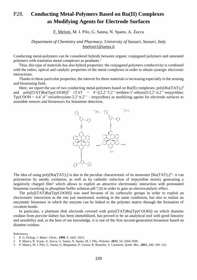
220
P28. Conducting Metal-Polymers Based on Ru(II) Complexes
as Modifying Agents for Electrode Surfaces
F. Meloni, M. I. Pilo, G. Sanna, N. Spano, A. Zucca
Department of Chemistry and Pharmacy, University of Sassari, Sassari, Italy
Conducting metal-polymers can be considered hybrids between organic conjugated polymers and saturated
polymers with transition metal complexes as pendants.1
Thus, this type of materials has also hybrid properties: the conjugated polymers conductivity is combined
with the redox, optical and catalytic properties of the metal complexes in order to obtain synergic electronic
interactions.
Thanks to these particular properties, the interest for these materials is increasing especially in the sensing
and biosensing field.
Here, we report the use of two conducting metal-polymers based on Ru(II) complexes: poly[Ru(TAT)2]2
and poly[(TAT)Ru(TpyCOOH)]3 (TAT = 4’-[(2,2’:5,2’’-terthien-3’-ethynyl]-2,2’:6,2’’-terpyridine;
TpyCOOH = 4,4’,4’’-tricarboxylate-2,2’:6,2’’ – terpyridine) as modifying agents for electrode surfaces to
assemble sensors and biosensors for histamine detection.
The idea of using poly[Ru(TAT)2] is due to the peculiar characteristic of its monomer [Ru(TAT)2]2+: it can
polymerize by anodic oxidation, as well as by cathodic reduction of terpyridine moiety generating a
negatively charged film2 which allows to exploit an attractive electrostatic interaction with protonated
histamine (working in phosphate buffer solution pH 7,0) in order to gain an electrocatalytic effect.
The poly[(TAT)Ru(TpyCOOH)] was used because of its carboxylic groups in order to exploit an
electrostatic interaction as the one just mentioned, working in the same conditions, but also to realize an
enzymatic biosensor in which the enzyme can be linked to the polymer matrix through the formation of
covalent bonds.
In particular, a platinum disk electrode covered with poly[(TAT)Ru(TpyCOOH)] on which diamine
oxidase from porcine kidney has been immobilized, has proved to be an analytical tool with good linearity
and sensibility and, to the best of our knowledge, it is one of the first second-generation biosensor based on
diamine oxidase.
References:
1 P. G. Pickup, J. Mater. Chem., 1999, 9, 1641–1653.
2 P. Manca, R. Scanu, A. Zucca, G. Sanna, N. Spano, M. I. Pilo, Polymer, 2013, 54, 3504-3509. 3 P. Manca, M. I. Pilo, G. Sanna, G. Bergamini, P. Ceroni, R. Boaretto, S. Caramori, Synth. Met., 2015, 200, 109–116.

221
222
P29. Do Fats Matter?
Design of Serum Albumin Bioreceptor for PFOA,
from Toxicological Studies to Biosensor Applications
Giulia Moro,a,b Stefano Liberi,a Elise Daems,b,c Sonia Covaceuszach,d Alberto Cassetta,d
Alessandro Angelini,a Karolien De Wael,b Ligia M. Morettoa
a Department of Molecular Science and Nanosystems, Università Ca’ Foscari, Mestre, Italy b AXES Research Group, Department of Chemistry, University of Antwerp, Antwerp, Belgium c BAMS Research Group, Department of Chemistry, University of Antwerp, Antwerp, Belgium
d Istituto di Cristallografia–CNR, Trieste Outstation, Trieste, Italy
A better understanding of the interactions between perfluroocatanoic acid (PFOA) and human serum albumin
(HSA) can improve the design of protein-based electrochemical biosensors for fluorinated environmental
contaminants, namely per- and poly-fluoroalkyl substances (PFAS). In the last decade, the affinity of PFOA
for fatted and defatted serum albumin was confirmed by numerous toxicological studies.1-3 The results
showed that PFOA can bind strongly to serum albumins, such as HSA, mimicking fatty acids binding and
affecting the functions of these transport proteins. To transpose these findings in the design of bioreceptors,
a multi-analytical study was carried out comparing directly the performances of fatted and defatted HSA in
terms of stability, number of binding sites and affinity towards PFOA and other PFAS . Isothermal titration
calorimetry (ITC) measurements allowed to define the stoichiometry and the affinity constants, while native
nano-electronspray ionization MS (nESI-MS) provided additional information about the stability of the
proteins and the protein-target complex. To identify the binding site and discriminate between fatted and
defatted HSA, vapor diffusion crystallization was performed. All analysis confirmed the higher affinity of
PFOA for the defatted HSA even though the fatted HSA showed an higher stability. Both types of HSA were
immobilized on the graphite-screen printed electrodes4 modified with graphene oxide (GO) or
electropolymerized o-phenylene diamine (oPD). Impedance electrochemical spectroscopy (EIS) and cyclic
voltammetry (CV) were used to characterize the immobilization strategies performances. Also direct
detection by EIS and indirect detection using a redox mediator were compared to evaluate the binding event
between PFOA and HSA. The combination of toxicological data and electrochemistry allowed to devise a
new detection strategy for environmental contaminants, mimicking a biological process happening in vivo
for biosensing purposes.
References:
1 Y. Chen, L. Guo, Arch. Toxicol., 2009, 83, 255–261.
2 L. A. Mac Manus-Spencer, M. L. Tse, P. C. Hebert, H. N. Bischel, R. G. Luthy, Anal. Chem., 2010, 82, 974–981. 3 H. N. Bischel, L. A. Mac Manus-Spencer, R. G. Luthy, Environ. Sci. Technol., 2010, 44, 5263–5269.
4 S. Rengaraj, A. Cruz-Izquierdo, J. L. Scott, M. Di Lorenzo, Sensors and Actuators B, 2018, 265, 50-58.

223
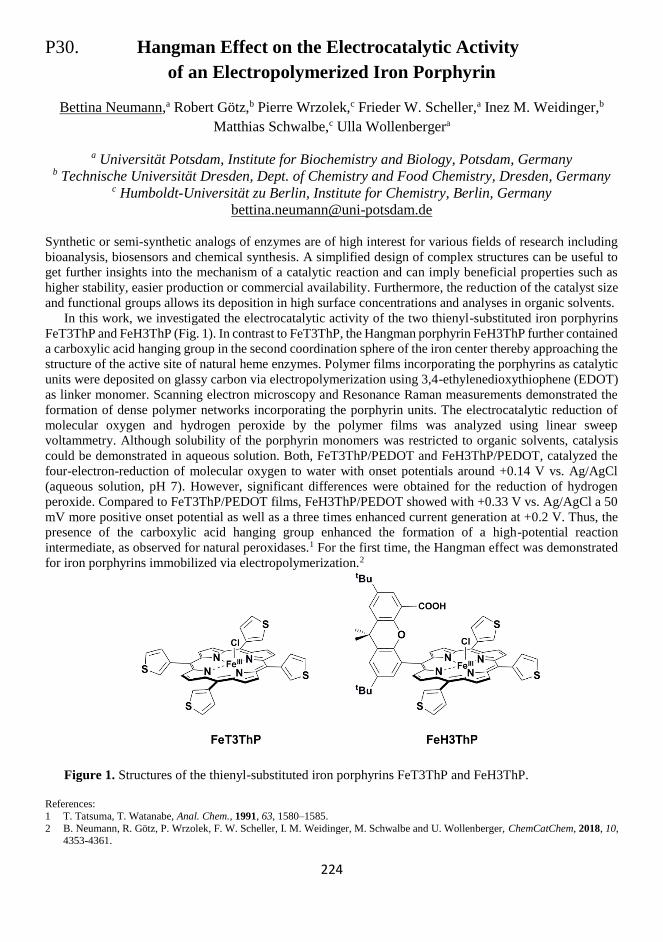
224
P30. Hangman Effect on the Electrocatalytic Activity
of an Electropolymerized Iron Porphyrin
Bettina Neumann,a Robert Götz,b Pierre Wrzolek,c Frieder W. Scheller,a Inez M. Weidinger,b
Matthias Schwalbe,c Ulla Wollenbergera
a Universität Potsdam, Institute for Biochemistry and Biology, Potsdam, Germany b Technische Universität Dresden, Dept. of Chemistry and Food Chemistry, Dresden, Germany
c Humboldt-Universität zu Berlin, Institute for Chemistry, Berlin, Germany
Synthetic or semi-synthetic analogs of enzymes are of high interest for various fields of research including
bioanalysis, biosensors and chemical synthesis. A simplified design of complex structures can be useful to
get further insights into the mechanism of a catalytic reaction and can imply beneficial properties such as
higher stability, easier production or commercial availability. Furthermore, the reduction of the catalyst size
and functional groups allows its deposition in high surface concentrations and analyses in organic solvents.
In this work, we investigated the electrocatalytic activity of the two thienyl-substituted iron porphyrins
FeT3ThP and FeH3ThP (Fig. 1). In contrast to FeT3ThP, the Hangman porphyrin FeH3ThP further contained
a carboxylic acid hanging group in the second coordination sphere of the iron center thereby approaching the
structure of the active site of natural heme enzymes. Polymer films incorporating the porphyrins as catalytic
units were deposited on glassy carbon via electropolymerization using 3,4-ethylenedioxythiophene (EDOT)
as linker monomer. Scanning electron microscopy and Resonance Raman measurements demonstrated the
formation of dense polymer networks incorporating the porphyrin units. The electrocatalytic reduction of
molecular oxygen and hydrogen peroxide by the polymer films was analyzed using linear sweep
voltammetry. Although solubility of the porphyrin monomers was restricted to organic solvents, catalysis
could be demonstrated in aqueous solution. Both, FeT3ThP/PEDOT and FeH3ThP/PEDOT, catalyzed the
four-electron-reduction of molecular oxygen to water with onset potentials around +0.14 V vs. Ag/AgCl
(aqueous solution, pH 7). However, significant differences were obtained for the reduction of hydrogen
peroxide. Compared to FeT3ThP/PEDOT films, FeH3ThP/PEDOT showed with +0.33 V vs. Ag/AgCl a 50
mV more positive onset potential as well as a three times enhanced current generation at +0.2 V. Thus, the
presence of the carboxylic acid hanging group enhanced the formation of a high-potential reaction
intermediate, as observed for natural peroxidases.1 For the first time, the Hangman effect was demonstrated
for iron porphyrins immobilized via electropolymerization.2
Figure 1. Structures of the thienyl-substituted iron porphyrins FeT3ThP and FeH3ThP.
References:
1 T. Tatsuma, T. Watanabe, Anal. Chem., 1991, 63, 1580–1585.
2 B. Neumann, R. Götz, P. Wrzolek, F. W. Scheller, I. M. Weidinger, M. Schwalbe and U. Wollenberger, ChemCatChem, 2018, 10, 4353-4361.

225
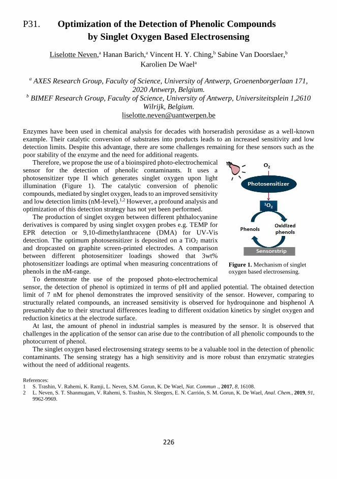
226
P31. Optimization of the Detection of Phenolic Compounds
by Singlet Oxygen Based Electrosensing
Liselotte Neven,a Hanan Barich,a Vincent H. Y. Ching,b Sabine Van Doorslaer,b
Karolien De Waela
a AXES Research Group, Faculty of Science, University of Antwerp, Groenenborgerlaan 171,
2020 Antwerp, Belgium. b BIMEF Research Group, Faculty of Science, University of Antwerp, Universiteitsplein 1,2610
Wilrijk, Belgium.
Enzymes have been used in chemical analysis for decades with horseradish peroxidase as a well-known
example. Their catalytic conversion of substrates into products leads to an increased sensitivity and low
detection limits. Despite this advantage, there are some challenges remaining for these sensors such as the
poor stability of the enzyme and the need for additional reagents.
Therefore, we propose the use of a bioinspired photo-electrochemical
sensor for the detection of phenolic contaminants. It uses a
photosensitizer type II which generates singlet oxygen upon light
illumination (Figure 1). The catalytic conversion of phenolic
compounds, mediated by singlet oxygen, leads to an improved sensitivity
and low detection limits (nM-level).1,2 However, a profound analysis and
optimization of this detection strategy has not yet been performed.
The production of singlet oxygen between different phthalocyanine
derivatives is compared by using singlet oxygen probes e.g. TEMP for
EPR detection or 9,10-dimethylanthracene (DMA) for UV-Vis
detection. The optimum photosensitizer is deposited on a TiO2 matrix
and dropcasted on graphite screen-printed electrodes. A comparison
between different photosensitizer loadings showed that 3wt%
photosensitizer loadings are optimal when measuring concentrations of
phenols in the nM-range.
To demonstrate the use of the proposed photo-electrochemical
sensor, the detection of phenol is optimized in terms of pH and applied potential. The obtained detection
limit of 7 nM for phenol demonstrates the improved sensitivity of the sensor. However, comparing to
structurally related compounds, an increased sensitivity is observed for hydroquinone and bisphenol A
presumably due to their structural differences leading to different oxidation kinetics by singlet oxygen and
reduction kinetics at the electrode surface.
At last, the amount of phenol in industrial samples is measured by the sensor. It is observed that
challenges in the application of the sensor can arise due to the contribution of all phenolic compounds to the
photocurrent of phenol.
The singlet oxygen based electrosensing strategy seems to be a valuable tool in the detection of phenolic
contaminants. The sensing strategy has a high sensitivity and is more robust than enzymatic strategies
without the need of additional reagents.
References:
1 S. Trashin, V. Rahemi, K. Ramji, L. Neven, S.M. Gorun, K. De Wael, Nat. Commun ., 2017, 8, 16108. 2 L. Neven, S. T. Shanmugam, V. Rahemi, S. Trashin, N. Sleegers, E. N. Carrión, S. M. Gorun, K. De Wael, Anal. Chem., 2019, 91,
9962-9969.
Figure 1. Mechanism of singlet
oxygen based electrosensing.

227
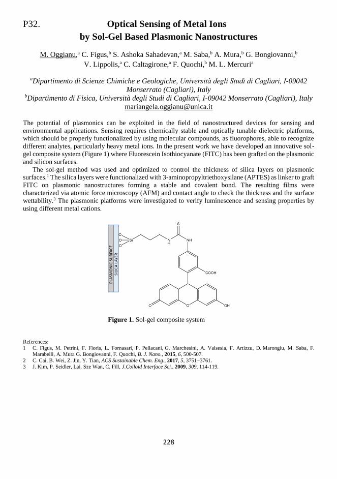
228
P32. Optical Sensing of Metal Ions
by Sol-Gel Based Plasmonic Nanostructures
M. Oggianu,a C. Figus,b S. Ashoka Sahadevan,a M. Saba,b A. Mura,b G. Bongiovanni,b
V. Lippolis,a C. Caltagirone,a F. Quochi,b M. L. Mercuria
aDipartimento di Scienze Chimiche e Geologiche, Università degli Studi di Cagliari, I-09042
Monserrato (Cagliari), Italy bDipartimento di Fisica, Università degli Studi di Cagliari, I-09042 Monserrato (Cagliari), Italy
The potential of plasmonics can be exploited in the field of nanostructured devices for sensing and
environmental applications. Sensing requires chemically stable and optically tunable dielectric platforms,
which should be properly functionalized by using molecular compounds, as fluorophores, able to recognize
different analytes, particularly heavy metal ions. In the present work we have developed an innovative sol-
gel composite system (Figure 1) where Fluorescein Isothiocyanate (FITC) has been grafted on the plasmonic
and silicon surfaces.
The sol-gel method was used and optimized to control the thickness of silica layers on plasmonic
surfaces.1 The silica layers were functionalized with 3-aminopropyltriethoxysilane (APTES) as linker to graft
FITC on plasmonic nanostructures forming a stable and covalent bond. The resulting films were
characterized via atomic force microscopy (AFM) and contact angle to check the thickness and the surface
wettability.3 The plasmonic platforms were investigated to verify luminescence and sensing properties by
using different metal cations.
Figure 1. Sol-gel composite system
References:
1 C. Figus, M. Petrini, F. Floris, L. Fornasari, P. Pellacani, G. Marchesini, A. Valsesia, F. Artizzu, D. Marongiu, M. Saba, F. Marabelli, A. Mura G. Bongiovanni, F. Quochi, B. J. Nano., 2015, 6, 500-507.
2 C. Cai, B. Wei, Z. Jin, Y. Tian, ACS Sustainable Chem. Eng., 2017, 5, 3751−3761.
3 J. Kim, P. Seidler, Lai. Sze Wan, C. Fill, J.Colloid Interface Sci., 2009, 309, 114-119.

229
230
P33. Discrimination of the Glycan Isomers 2,3-Sialyllactose
and 2,6-Sialyllactose Modified with Osmium(VI) Complexes
by Square Wave Voltammetry
Veronika Ostatná, Mojmír Trefulka, Hana Černocká
Institute of Biophysics, The Czech Academy of Science, v.v.i., Královopolská 135, 61265, Brno,
Czech Republic
Carbohydrate components in glycoproteins play a critical role in health and disease and specificity of
glycoprotein biomarkers can be greatly enhanced by the analysis of their sugar components containing
frequently different isomers. Altered glycosylation is a universal feature of cancer cells and certain glycans
are well-known markers of tumor progression.1 Two glycan isomers, 2,3-sialyllactose (3-SL) and 2,6-
sialyllactose (6-SL) frequently appear in glycoproteins connected with cancer. For example, various changes
in the well-known Prostate Specific Antigen (PSA) glycosylation were found at cancer patients, including
the increased amount of 3-SL. We combine glycan modification with osmium(VI) N,N,N’,N’-
tetramethylethylenediamine (Os(VI)tem) and square wave voltammetry for discrimination of 3-SL and 6-SL
regioisomers.2 Covalent adducts of Os(VI)tem with glycans yielded three reduction voltammetric peaks. The
ratio of peak I/peak II heights depends on the content of individual regioisomers in the sample. Experiments
using capillary electrophoresis, inductively coupled plasma mass spectrometry and thin layer
chromatography showed that 6-SL molecule can bind three Os(VI), while the 3-SL only two Os(VI) moieties.
A similar pattern of Os(VI)-modification was found for isomers of sialyl-N-acetyllactosamine and
sialylgalactose.3 Our proposed approach allows the determination of isomer percentage representation in the
mixture after one voltammogram recording. These results show a new appropriate method for the
discrimination of glycan isomers containing terminal sialic acid important for distinguishing between
cancerous and non-cancerous origin of biomarkers. We can conclude that the electrochemical analysis
appeared suitable for the recognition of SL isomers.
Acknowledgements:
This work was supported by the Czech Science Foundation 18-18154S project.
References:
1 E. Palecek, J. Tkac, M. Bartosik, T. Bertok, V. Ostatna, J. Palecek, Chem. Rev., 2015, 115, 2045-2108. 2 M. Trefulka, E. Palecek, Electrochem. Commun., 2017, 85, 19-22.
3 M. Trefulka, H. Černocká, L. Fojt, E. Paleček, V. Ostatná, Anal. Chim. Acta, 2019, 1067, 56-62.

231
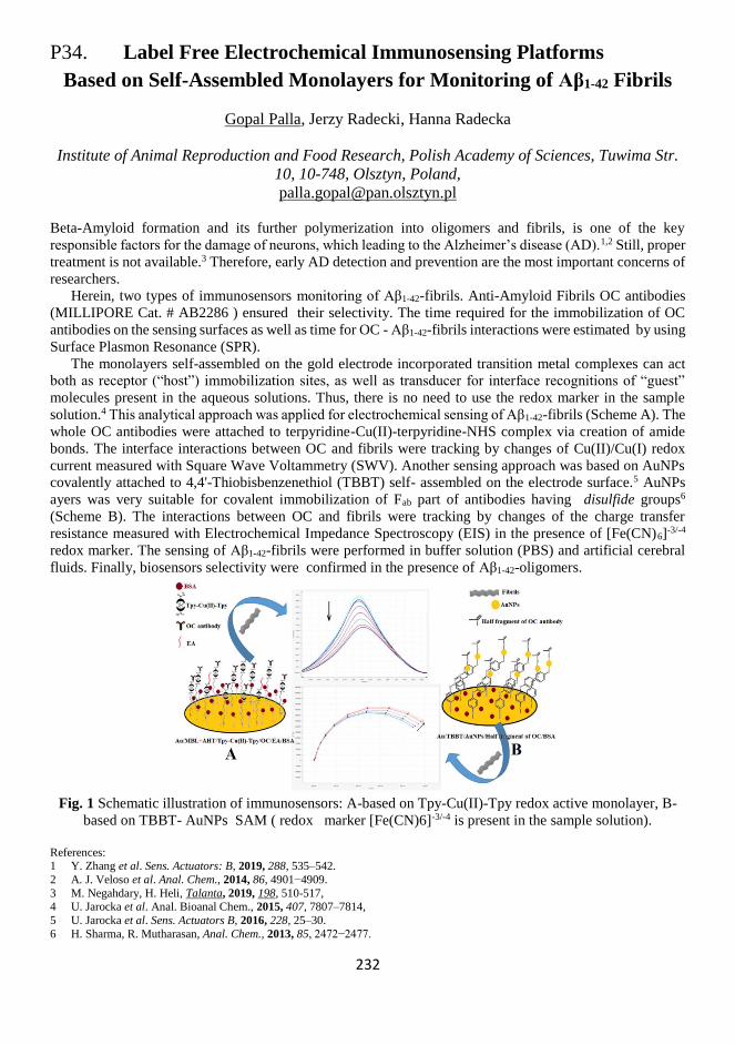
232
P34. Label Free Electrochemical Immunosensing Platforms
Based on Self-Assembled Monolayers for Monitoring of Aβ1-42 Fibrils
Gopal Palla, Jerzy Radecki, Hanna Radecka
Institute of Animal Reproduction and Food Research, Polish Academy of Sciences, Tuwima Str.
10, 10-748, Olsztyn, Poland,
Beta-Amyloid formation and its further polymerization into oligomers and fibrils, is one of the key
responsible factors for the damage of neurons, which leading to the Alzheimer’s disease (AD).1,2 Still, proper
treatment is not available.3 Therefore, early AD detection and prevention are the most important concerns of
researchers.
Herein, two types of immunosensors monitoring of Aβ1-42-fibrils. Anti-Amyloid Fibrils OC antibodies
(MILLIPORE Cat. # AB2286 ) ensured their selectivity. The time required for the immobilization of OC
antibodies on the sensing surfaces as well as time for OC - Aβ1-42-fibrils interactions were estimated by using
Surface Plasmon Resonance (SPR).
The monolayers self-assembled on the gold electrode incorporated transition metal complexes can act
both as receptor (“host”) immobilization sites, as well as transducer for interface recognitions of “guest”
molecules present in the aqueous solutions. Thus, there is no need to use the redox marker in the sample
solution.4 This analytical approach was applied for electrochemical sensing of Aβ1-42-fibrils (Scheme A). The
whole OC antibodies were attached to terpyridine-Cu(II)-terpyridine-NHS complex via creation of amide
bonds. The interface interactions between OC and fibrils were tracking by changes of Cu(II)/Cu(I) redox
current measured with Square Wave Voltammetry (SWV). Another sensing approach was based on AuNPs
covalently attached to 4,4'-Thiobisbenzenethiol (TBBT) self- assembled on the electrode surface.5 AuNPs
ayers was very suitable for covalent immobilization of Fab part of antibodies having disulfide groups6
(Scheme B). The interactions between OC and fibrils were tracking by changes of the charge transfer
resistance measured with Electrochemical Impedance Spectroscopy (EIS) in the presence of [Fe(CN)6]-3/-4
redox marker. The sensing of Aβ1-42-fibrils were performed in buffer solution (PBS) and artificial cerebral
fluids. Finally, biosensors selectivity were confirmed in the presence of Aβ1-42-oligomers.
Fig. 1 Schematic illustration of immunosensors: A-based on Tpy-Cu(II)-Tpy redox active monolayer, B-
based on TBBT- AuNPs SAM ( redox marker [Fe(CN)6]-3/-4 is present in the sample solution).
References: 1 Y. Zhang et al. Sens. Actuators: B, 2019, 288, 535–542.
2 A. J. Veloso et al. Anal. Chem., 2014, 86, 4901−4909.
3 M. Negahdary, H. Heli, Talanta, 2019, 198, 510-517, 4 U. Jarocka et al. Anal. Bioanal Chem., 2015, 407, 7807–7814,
5 U. Jarocka et al. Sens. Actuators B, 2016, 228, 25–30.
6 H. Sharma, R. Mutharasan, Anal. Chem., 2013, 85, 2472−2477.

233
234
P35. Two-photon Stereolithography for the Direct Writing
of 3D MIP Photonic Crystals
Ernesto Paruli, Carlo Gonzato, Karsten Haupt
Génie Enzymatique et Cellulaire, Sorbonne Universités, Université de Technologie de
Compiègne, CNRS Laboratory for Enzyme and Cell Engineering UMR 7025, 60200 Compiègne,
France
Two-photon stereolithography (TPS) has gained an increasing interest as a rapid prototyping technique for
3D micro- and nanostructures.1 Due to the high energy densities required by the two-photon absorption
process, it is possible to spatially confine the polymerization of a photoresist into the very small volume
represented by the focal point of a microscope’s objective using a pulsed laser. This allows in turn for the
direct and precise fabrication of micro-/nano-structures, conversely to conventional
microstereolithography.2 In this study, TPS has been exploited for the direct fabrication of photonic crystals,
which were molecularly imprinted against propranolol as a model template. Molecularly imprinted polymers
(MIPs), which are also known as plastic antibodies3 are polymers able to specifically recognize a target
molecule and as such, they are well suited as recognition elements for sensors.
Thus, TPS was successfully used for the one-step synthesis of an optical sensor, including both a
transducer (i.e. the photonic crystal) and a recognition element (i.e. the MIP), in a single structure.
References:
1 L.P.C. Gomez, A. Spangenberg, X. A. Ton, Y. Fuchs, F. Bokeloh, J. P. Malval, B. Tse Sum Bui, D. Thuau, C. Ayela, K. Haupt, O.
Soppera, Adv. Mater., 2016, 28, 5931-5937. 2 A. Spangenberg, N. Hobeika, F. Stehlin, J. P. Malval, F. Wielder, P. Prabhakaran, P. Baldeck, O. Soppera, Updates in Advanced
Lithography, S. Hosaka, Ed., InTech, 2014, 35−63.
3 K. Haupt, Anal. Chem., 2003, 75, 376A-383A.

235
236
P36. Silanization of Spherically-shaped WGM Resonators
for Cancer Detection
Piotr Paszke,a Rafał Nowaczyński,a,b Kamil Szlachetko,a Andrea Csaki,c
Dorota Anna Pawlaka,d
a Faculty of Chemistry, University of Warsaw, ul. Ludwika Pasteura 1, 02-093 Warsaw, Poland, b Faculty of Materials Science and Engineering, Warsaw University of Technology, Wołoska 141
St., 02-507 Warsaw, Poland c Leibniz Institute of Photonic Technology (IPHT) Jena, Member of the Leibniz Research Alliance
- Leibniz Health Technologies, Albert-Einstein-Straße 9, 07745 Jena, Germany d Institute of Electronic Materials Technology, Wólczyńska 133, 01-919 Warsaw, Poland
Whispering gallery mode (WGM) optical resonators can be used for many applications, such as optical
switches, narrow band lasers or thermo- and biosensors.1,2 Particularly interesting is the application of that
resonators in biosensing, due to the significantly enhanced light-matter interactions.
The aim of our work is to prepare the WGM resonators, which can be used to detect exosomes. Exosomes
are small vesicles which are excreted by every cell in our body to body fluids (for ex. blood and urine). By
detecting exosomes from cancerous cells we want to perform fast, early and non-invasive cancer diagnostics.
We develop WGM microresonators from fused silica and also from low-melting materials (like tellurite
and phosphate glasses). For our current knowledge, we developed for the first time the resonators, based on
phosphate and tellurite glasses doped with both quantum dots and plasmonic nanoparticles. We have
achieved this through using the NanoParticle Direct Doping method,3 which is based on one of the crystal
growth techniques - the micro-pulling down method.
The next stage of our work is to cover the resonators with a biologically active material, that will allow us to
specifically bind to tumor exosomes. The glass silanization procedure was used to functionalize the surface,
allowing the glass to be coated with the antibodies that capture target exosomes. We performed
functionalizations of WGM resonators with the use of GOPS (3-Glycidyloxypropyltrimethoxysilane) silanes
based on works by Moller et al.4 To verify surface coverage by silanes, silanized samples were placed in a
solution of fluorescent oligonucleotide. This work presents the results of silanization tests of WGM
microresonators from various types of glasses.
Acknowledgments:
This project was supported financially by the TEAM programme of the Foundation for Polish Science (No. TEAM/2016-3/29), co-financed by the European Union under the European Regional Development Fund, the MAESTRO Project (No. 2011/02/A/ST5/00471)
operated by National Science Centre and the Era.Net Rus Plus project (No. 4/RUSPLUS-INNO/2015) operated by The National Centre
of Research and Development.
References:
1 J. Ward, O. Benson, Laser & Photonics Rev., 2011, 5.4, 553-570. 2 G. Righini, S. Soria, Sensors, 2016, 16(6), 905.
3 M. Gajc et al., Adv. Funct. Mater., 2013, 23, 3443-3451.
4 R. Möller et al., Nucleic Acids Res., 2000, 28(20), e91.

237

238
P37. Research on Novel Matrix Composites
Based on Reduced Graphene Oxide
for Electrochemical Sensor Construction
Aleksandra Pollap, Karolina Waląg, Jolanta Kochana
Jagiellonian University, Kraków, Poland
Chromatographic and spectroscopic methods belong to the most popular analytical methods. Their
application allows to carry out both qualitative and quantitative analyzes with high accuracy and precision.
Nevertheless, application of these methods is often related with high cost of apparatus and need for special
sample preparation. Therefore, it is worth to note the electroanalytical methods including electrochemical
sensors and biosensors. Their use meets the requirements of analytical procedures, such as simplicity and
speed of determination, accuracy, precision and low detection limit. Modification of electrode surface is an
important issue during electrochemical sensor construction. The main goal of surface modification is to
increase the active area, increase of electrical conductivity as well as improvement of sensor durability.
Consequently, the modification of electrode surface allows to improve analytical parameters of developed
sensor such as sensitivity, limit of detection or selectivity. The purpose of presented work was to carry out
the research on novel matrix composites based on reduced graphene oxide (rGO). Due to structural flexibility
and electrical conductivity rGO is a very attractive material.1 In presented work rGO was used for
modification of graphite electrode surface. During sensor construction titatnia dioxide sol as binding agent
was used. Additionaly, the matrix composite was also enriched with gold nanoparticles increasing the
electrical conductivity and conductive polymer – Nafion increasing the stability of sensor indication. The
experiments were carried out in Fe(II)/Fe(III) redox probe, applying cyclic voltammetry techinque. Obtained
results enabled to indicate the optimal composition of matrix nanocomposite which will be verified for sensor
development for determination of pesticides.
References:
1 B. Y. Zhu, S. Murali, W. Cai, X. Li, J.W. Suk, J.R. Potts and R.S. Ruoff, Adv. Mater., 2010, 22, 3096-3924.

239

240
P38. Electrochemical Molecularly Imprinted Polymer (MIP) Sensor
for Novel Psychoactive Substances
Rosa A.S. Couto,a Séfora S. Costa,a João G. Pacheco,b Cristina Delerue-Matos,b Eduarda
Fernandes,a Félix Carvalho,c Luís M. Gonçalves,d Cecília M.P. Rodrigues,e M. Beatriz Quinaza
a LAQV, REQUIMTE, Laboratory of Applied Chemistry, Department of Chemical Sciences,
Faculty of Pharmacy, University of Porto, Porto, Portugal b LAQV, REQUIMTE, Instituto Superior de Engenharia do Porto (ISEP), Politécnico do Porto,
Porto, Portugal c UCIBIO, REQUIMTE, Laboratory of Applied Chemistry, Department of Chemical Sciences,
Faculty of Pharmacy, University of Porto, Porto, Portugal d Departamento de Química Fundamental, Instituto de Química, Universidade de São Paulo
(USP), São Paulo, Brazil e Research Institute for Medicines (iMed.ULisboa), Faculty of Pharmacy, Universidade de
Lisboa, Lisbon, Portugal
In this work we propose an electrochemical molecularly imprinted polymer (e-MIP) to the selective
quantification of the psychoactive substance 3,4-methylenedioxymethamphetamine (MDMA), also known
as ecstasy (Fig. 1), using screen-printed carbon electrodes (SPCEs).1 The device was constructed using ortho-
phenylenediamine (o-PD) as the MIP’s building monomer and MDMA as template. The step-by-step
construction of the SPCE- MIP sensor was characterized by cyclic voltammetry (CV) and electrochemical
impedance spectroscopy (EIS). The proposed electrochemical sensor worked by directly measuring the
MDMA oxidation signal through square-wave voltammetry (SWV) after an incubation period of 10 min.
Several parameters that could influence the sensor’ sensing performance were optimized, such as the
monomer/template ratio, the number of electropolymerization scanning cycles, and the incubation period.
Under optimized conditions, the sensor exhibited suitable selectivity, repeatability, reproducibility and up to
one month of stable response, with a linear range up to 0.2 mmol L−1. Finally, the developed sensor was
successfully applied to human blood serum and urine samples, showing its potential for application in
medicine and in forensic sciences.
Figure 1. Structural formula of ecstasy.
Acknowledgements:
The work was supported by UID/QUI/50006/2019 with funding from FCT/MCTES through national funds. RASC acknowledges FCT
for her PhD fellowship (PD/BD/127797/2016) from PhD Programme i3DU. AACB (grants 2014/25770-6 and 2015/01491-3) and LMG
(grant 2018/14425-7) thank the São Paulo Research Foundation (FAPESP) for financial support. AACB (grant 309715/2017-2) also
thanks the Brazilian National Research Council (CNPq) for financial support and fellowships. This study was financed in part by the Coordenação de Aperfeiçoamento de Pessoal de Nível Superior - Brasil (CAPES) - Finance Code 001.
References: 1 R. A. S. Couto, S. S. Costa, B. Mounssef, J. G. Pacheco, E. Fernandes, F. Carvalho, C. M. P. Rodrigues, C. Delerue-Matos, A. A.
C. Braga, L. M. Gonçalves, M. B. Quinaz, Sens. Actuator B-Chem., 2019, 290, 378-386.

241
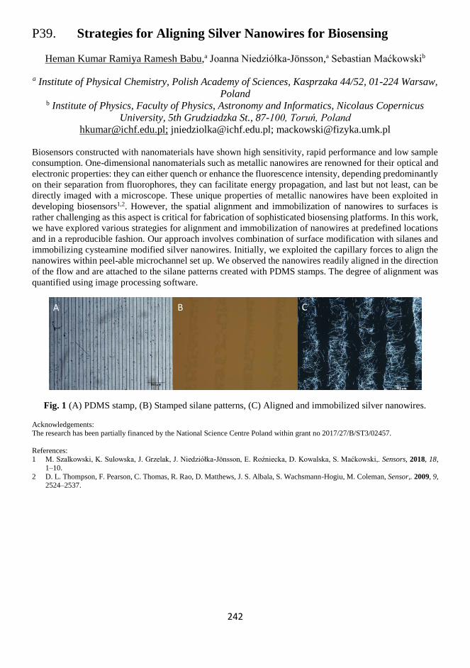
242
P39. Strategies for Aligning Silver Nanowires for Biosensing
Heman Kumar Ramiya Ramesh Babu,a Joanna Niedziółka-Jönsson,a Sebastian Maćkowskib
a Institute of Physical Chemistry, Polish Academy of Sciences, Kasprzaka 44/52, 01-224 Warsaw,
Poland b Institute of Physics, Faculty of Physics, Astronomy and Informatics, Nicolaus Copernicus
University, 5th Grudziadzka St., 87-100, Toruń, Poland
[email protected]; [email protected]; [email protected]
Biosensors constructed with nanomaterials have shown high sensitivity, rapid performance and low sample
consumption. One-dimensional nanomaterials such as metallic nanowires are renowned for their optical and
electronic properties: they can either quench or enhance the fluorescence intensity, depending predominantly
on their separation from fluorophores, they can facilitate energy propagation, and last but not least, can be
directly imaged with a microscope. These unique properties of metallic nanowires have been exploited in
developing biosensors1,2. However, the spatial alignment and immobilization of nanowires to surfaces is
rather challenging as this aspect is critical for fabrication of sophisticated biosensing platforms. In this work,
we have explored various strategies for alignment and immobilization of nanowires at predefined locations
and in a reproducible fashion. Our approach involves combination of surface modification with silanes and
immobilizing cysteamine modified silver nanowires. Initially, we exploited the capillary forces to align the
nanowires within peel-able microchannel set up. We observed the nanowires readily aligned in the direction
of the flow and are attached to the silane patterns created with PDMS stamps. The degree of alignment was
quantified using image processing software.
Fig. 1 (A) PDMS stamp, (B) Stamped silane patterns, (C) Aligned and immobilized silver nanowires.
Acknowledgements: The research has been partially financed by the National Science Centre Poland within grant no 2017/27/B/ST3/02457.
References: 1 M. Szalkowski, K. Sulowska, J. Grzelak, J. Niedziółka-Jönsson, E. Roźniecka, D. Kowalska, S. Maćkowski,. Sensors, 2018, 18,
1–10.
2 D. L. Thompson, F. Pearson, C. Thomas, R. Rao, D. Matthews, J. S. Albala, S. Wachsmann-Hogiu, M. Coleman, Sensor,. 2009, 9, 2524–2537.
A B C

243
244
P40. Different Redox Probes for Electrochemistry
at the Three-phase Junction
Vishwanath R. S, Emilia Witkowska-Nery, Martin Jönsson-Niedziółka
Institute of Physical Chemistry, Polish Academy of Sciences, Warsaw, ul. Kasprzaka 44/52,
Poland
The three-phase electrode (TPE) ion transfer studies at organic/aqueous/electrode interface are essential to
obtain highly important thermodynamic data which determine the lipophilicity, biological and
pharmacological activity of transferring ions/compounds. Also, lipophilic/hydrophilic interface created at
TPE is the simple model of complex biological systems. In TPE configuration, the electron transfer across
the electrode/organic interface is accompanied by the ion transfer from aqueous to organic phase, which
allows to determine the lipophilicity of a large number of inorganic and organic ions in different liquid/liquid
interfaces.1
For cations transfer studies, iodine2 and iron(III) tetraphenyl porphyrine chloride3 have been used so far.
We investigated the reduction of 1-aminoanthraquinone (AQ), and 2,3-dichloro-1,4-naphthoquinone (NQ)
at a three-phase junction formed by n-octyl-2-pyrrolidone/aqueous electrolyte/glassy carbon (GC) working
electrode. The redox behaviour of AQ (in anthracycline drugs) and NQ (in vitamin K) play a significant role
in the biochemistry of living systems. AQ underwent a 1-step, 2-electron reduction resulting in the transfer
of cations from the aqueous phase. NQ reduction occurred in two steps, the first was by a transfer of the NQ•−
radical to the interface with the reduction potential dependent on the anion present in the aqueous phase due
to salting-out effects. After that, the NQ•− radical was reduced to NQ2− dianion resulted in a transfer of cations
from the aqueous phase.4 Importantly, the cation transfer potential is determined by ion-pair formation with
the quinone, rather than the solvation energy of cation in the pure organic solvent.3
Decamethylferrocene has been extensively used for anion transfer studies so far. We synthesised a
mononuclear lipophilic ruthenium(II) complex [Ru(LR)(L)]0 having tridentate 2,6-bis(1-(2-
octyldodecan)benzimidazol-2-yl)pyridine (LR) and (2,6-bis(benzimidazol-2-yl)pyridine) (L) ligands.
Further [Ru(LR)(L)]0 complex was used as a redox probe for the anion transfer studies at
nitrobenzene/aqueous/GC working electrode three-phase interface. The metal associated the oxidation of
[Ru(LR)(L)]0 complex is accompanied by the anion transfer from the aqueous to the organic phase. The
formal oxidation potential of [Ru(LR)(L)]0 depends on the hydrophilicity of the aqueous anion and its
concentration. The prime requirement for anion transfer is the oxidation of [Ru(LR)(L)]0 complex at TPE
before the water oxidation at GC/water interface which was achieved by the deprotonation at N-H sites of L
ligand. Further, the deprotonated sites allow us to examine any proton coupled anion transfer from aqueous
to organic phase.
References: 1 F. Scholz, U. Schröder, R. Gulaboski, A. Doménech-Carbó, Electrochemistry of Immobilized Particles and Droplets, 2nd Edition
2005.
2 V. Mirčeski, R. Gulaboski, F. Scholz, Electrochem. commun., 2002, 4(10), 814–819. 3 F. Scholz, R. Gulaboski, K. Caban, Electrochem. commun., 2003, 5(11), 929–934.
4 R. S. Vishwanath, E. W. Nery, Jönsson-Niedziółka Martin, Electrochim. Acta, 2019, 306, 54–60.

245
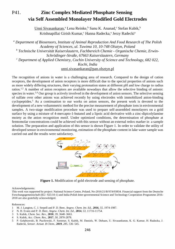
246
P41. Zinc Complex Mediated Phosphate Sensing
via Self Assembled Monolayer Modified Gold Electrodes
Unni Sivasankaran,a Lena Reinke,b Sanu K. Ananad,c Stefan Kubik,b
Krishnapillai Girish Kumar,c Hanna Radecka,a Jerzy Radeckia
a Department of Biosensors, Institute of Animal Reproduction And Food Research of The Polish
Academy of Sciences, ul. Tuwima 10, 10-748 Olsztyn, Poland b Technische Universität Kaiserslautern, Fachbereich Chemie - Organische Chemie, Erwin-
Schrödinger-Straße, 67663 Kaiserslautern, Germany c Department of Applied Chemistry, Cochin University of Science and Technology, 682 022,
Kochi, India
The recognition of anions in water is a challenging area of research. Compared to the design of cation
receptors, the development of anion receptors is more difficult due to the special properties of anions such
as more widely differing structures, their varying protonation states at different pH and low charge to radius
ratios.1,2 A number of anion receptors are available nowadays that allow the selective binding of anionic
species in water.3,4 Our group is actively involved in the development of anion sensors. The selective sensing
of sulfate over other anions was achieved recently by using electrodes with immobilized anion-binding
cyclopeptides.5 As a continuation to our works on anion sensors, the present work is devoted to the
development of a new voltammetric method for the precise measurement of phosphate ions in environmental
samples. A two-stage modification procedure was used to prepare self-assembled monolayers on a gold
surface by using a mixture of 4-mercapto-1-butanol and a lipoic acid derivative with a zinc-dipicolylamine
moiety as the anion recognition motif. Under optimized conditions, the determination of phosphate at
femtomolar concentrations could be achieved with this sensor without an external redox marker in a sample
solution. The preparation and application of this sensor is shown Figure 1. In order to validate the utility of
developed sensor in environmental monitoring, estimation of the phosphate content in lake water sample was
carried out and the results were satisfactory.
Figure 1. Modification of gold electrode and sensing of phosphate.
Acknowledgements:
This work was supported by project: National Science Centre, Poland, No-2016/21/B/ST4/03834. Financial support from the Deutsche Forschungsgemeinschaft (KU- 925/10-1) and India-Polish Inter-governmental Science and Technology Cooperation Programme 2016-
2018 are also gratefully acknowledged.
References: 1 M. J. Langton, C. J. Serpell and P. D. Beer, Angew. Chem. Int. Ed., 2016, 55, 1974-1987.
2 N. H. Evans and P. D. Beer, Angew. Chem. Int. Ed., 2014, 53, 11716-11754. 3 S. Kubik, Chem. Soc. Rev., 2010, 39, 3648–3663.
4 S. Kubik, Acc. Chem. Res., 2017, 50, 2870-2878.
5 P. Gołębiewski, B. Puciłowski, F. Sommer, S. Kubik, M. Daniels, W. Dehaen, U. Sivasankaran, K. G. Kumar, H. Radecka, J. Radecki, Sensor. Actuat. B Chem., 2019, 285, 536–545.

247
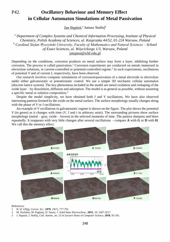
248
P42. Oscillatory Behaviour and Memory Effect
in Cellular Automaton Simulations of Metal Passivation
Jan Stępień,a Janusz Stafiejb
a Department of Complex Systems and Chemical Information Processing, Institute of Physical
Chemistry, Polish Academy of Sciences, ul. Kasprzaka 44/52, 01-224 Warsaw, Poland b Cardinal Stefan Wyszyński University, Faculty of Mathematics and Natural Sciences – School
of Exact Sciences, ul. Wóycickiego 1/3, Warsaw, Poland [email protected]
Depending on the conditions, corrosion products on metal surface may form a layer, inhibiting further
corrosion. The process is called passivation.1 Corrosion experiments are conducted on metals immersed in
electrolyte solutions, in current-controlled or potential-controlled regime.2 In such experiments, oscillations
of potential V and of current I, respectively, have been observed. Our research involves computer simulations of corrosion/passivation of a metal electrode in electrolyte
under either galvanostatic or potentiostatic control. We use a simple 3D stochastic cellular automaton
(discrete lattice system). The key phenomena included in the model are metal oxidation and reshaping of the
oxide layer – by dissolution, diffusion and adsorption. The model is as general as possible, without assuming
a specific metal or solution composition.3 Despite the model simplicity, we have obtained both I and V oscillations. We have also observed
interesting patterns formed by the oxide on the metal surface. The surface morphology usually changes along
with the phase of V or I oscillations. An example of V oscillations in galvanostatic regime is shown on the figure. The plot shows the potential
V (in green) as it changes with time (V, I and t in arbitrary units). The surrounding pictures show surface
morphology (metal – gray, oxide – brown) in the selected moments of time. The pattern sharpens and blurs
repeatedly. It reappears with very little changes after several oscillations – compare A with G or D with H.
We call this the memory effect.
References: 1 H. H. Uhlig, Corros. Sci., 1979, 19(7), 777-791. 2 M. Pavlidou, M. Pagitsas, D. Sazou, J. Solid State Electrochem., 2015, 19, 3207-3217. 3 J. Stępień, J. Stafiej, Cell. Autom., no. 13 in Lecture Notes in Computer Science, 2018, 92-101.

249
250
P43. Real-time Sensing with Elongated Silver Nanoparticles
Karolina Sulowska,a Ewa Różniecka,b Kamil Wiwatowski,a
Joanna Niedziółka-Jӧnsson,b Sebastian Maćkowskia,c
a Faculty of Physics, Astronomy and Informatics, Nicolaus Copernicus University, Torun, Poland b Institute of Physical Chemistry, Polish Academy of Sciences, Warszawa, Poland
c Baltic Institute of Technology, Gdynia, Poland
In this work we focus on applying silver nanowires (AgNWs) as building blocks of a biosensor. The
nanowires feature several key properties that make them suitable for this application,1-3 which include
chemical stability in water and buffer solutions, straightforward synthesis using wet chemistry, as well as
ability to control the functionality via appropriate surface modifications. Importantly, the diameters of the
nanowires, which are around 100 nm and their lengths of around 50 μm, facilitate plasmon resonance and
ability to visualize the nanowires using optical microscopy, respectively.
We tested the sensor ability of the nanowires using natural photosynthetic complexes, Peridinin-
Chlorophyll-Protein (PCP) modified with streptavidin. The size of the PCP complex is 4 nm, i.e. much less
than typical diameters of AgNWs. The absorption and fluorescence spectra of PCP overlap with the
extinction spectrum of AgNWs, facilitating plasmon coupling.4 The experiments were carried out using
advanced fluorescence microscopy and spectroscopy techniques.
Upon modification of the AgNWs surface with biotin, we placed them on a surface. In the next step a
droplet of PCP solution was deposited and fluorescence of an area of 100 x 100 microns was monitored in
real-time mode. We observe specific attachment of the PCP complexes to AgNWs. Importantly, with this
approach we can demonstrate detection of single proteins in real-time. Partially this is due to plasmonic
enhancement of PCP emission, as evidenced by time-resolved fluorescence microscopy. In addition to this
approach, other ways of improving the efficiency of the detection or the limit of detection will be presented.
Research was partially financed by the National Science Centre (Poland) within the OPUS grants 2016/21/B/ST3/02276,
2017/27/B/ST3/02457, and 2017/26/E/ST3/00209, project 3/DOT/2016 funded by the City of Gdynia, and project nr POWR.03.05.00-00-Z302/17 “Universitas Copernicana Thoruniensis In Futuro” (2018-2022) co-financed by the European Social Fund – the Operational
Programme Knowledge Education Development. Module 5. Interdisciplinary PhD School “Academia Copernicana”.
References:
1 M. Szalkowski, K. Sulowska J. Grzelak, J. Niedziółka-Jönsson, E. Roźniecka, D. Kowalska, S. Maćkowski, Sensors, 2018, 18(1), 290.
2 S. Mackowski, J Phys Condens. Matter, 2010, 22(19), 193102.
3 J. Grzelak, K. Sulowska, A. Lesniewski, E. Rozniecka, M. Janczuk-Richter, L. Richter, M. Los, M. Jönsson-Niedziółka, S. Maćkowski, J. Niedziółka-Jönsson, Sens. Actuator B-Chem., 2018, 273, 689-695.
4 D. Kowalska, B. Krajnik, M. Olejnik, M. Twardowska, N. Czechowski, E. Hofmann, S. Mackowski, Scientific World J., 2013, 12,
670412.

251
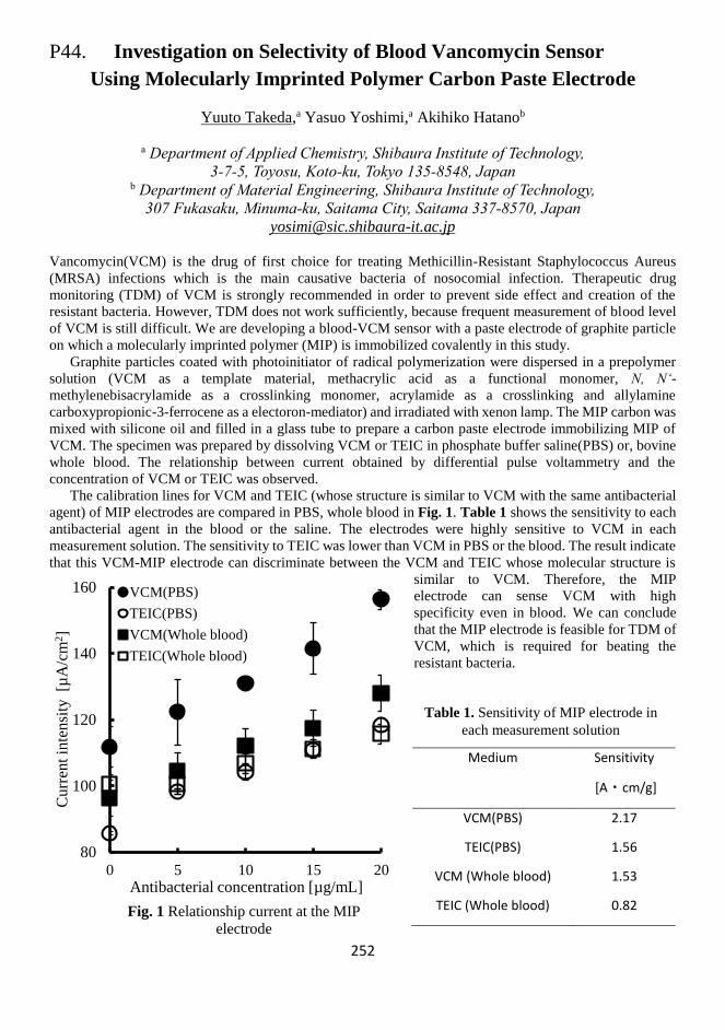
252
P44. Investigation on Selectivity of Blood Vancomycin Sensor
Using Molecularly Imprinted Polymer Carbon Paste Electrode
Yuuto Takeda,a Yasuo Yoshimi,a Akihiko Hatanob
a Department of Applied Chemistry, Shibaura Institute of Technology,
3-7-5, Toyosu, Koto-ku, Tokyo 135-8548, Japan b Department of Material Engineering, Shibaura Institute of Technology,
307 Fukasaku, Minuma-ku, Saitama City, Saitama 337-8570, Japan
Vancomycin(VCM) is the drug of first choice for treating Methicillin-Resistant Staphylococcus Aureus
(MRSA) infections which is the main causative bacteria of nosocomial infection. Therapeutic drug
monitoring (TDM) of VCM is strongly recommended in order to prevent side effect and creation of the
resistant bacteria. However, TDM does not work sufficiently, because frequent measurement of blood level
of VCM is still difficult. We are developing a blood-VCM sensor with a paste electrode of graphite particle
on which a molecularly imprinted polymer (MIP) is immobilized covalently in this study.
Graphite particles coated with photoinitiator of radical polymerization were dispersed in a prepolymer
solution (VCM as a template material, methacrylic acid as a functional monomer, N, N‘-
methylenebisacrylamide as a crosslinking monomer, acrylamide as a crosslinking and allylamine
carboxypropionic-3-ferrocene as a electoron-mediator) and irradiated with xenon lamp. The MIP carbon was
mixed with silicone oil and filled in a glass tube to prepare a carbon paste electrode immobilizing MIP of
VCM. The specimen was prepared by dissolving VCM or TEIC in phosphate buffer saline(PBS) or, bovine
whole blood. The relationship between current obtained by differential pulse voltammetry and the
concentration of VCM or TEIC was observed.
The calibration lines for VCM and TEIC (whose structure is similar to VCM with the same antibacterial
agent) of MIP electrodes are compared in PBS, whole blood in Fig. 1. Table 1 shows the sensitivity to each
antibacterial agent in the blood or the saline. The electrodes were highly sensitive to VCM in each
measurement solution. The sensitivity to TEIC was lower than VCM in PBS or the blood. The result indicate
that this VCM-MIP electrode can discriminate between the VCM and TEIC whose molecular structure is
similar to VCM. Therefore, the MIP
electrode can sense VCM with high
specificity even in blood. We can conclude
that the MIP electrode is feasible for TDM of
VCM, which is required for beating the
resistant bacteria.
Medium Sensitivity
[A・cm/g]
VCM(PBS) 2.17
TEIC(PBS) 1.56
VCM (Whole blood) 1.53
TEIC (Whole blood) 0.82
80
100
120
140
160
0 5 10 15 20
Cu
rren
t in
ten
sity
[µ
A/c
m2]
Antibacterial concentration [µg/mL]
Fig. 1 Relationship current at the MIP
electrode
VCM(PBS)
TEIC(PBS)
VCM(Whole blood)
TEIC(Whole blood)
Table 1. Sensitivity of MIP electrode in
each measurement solution

253
254
P45. Gold Thin-Film Based Photo-electrochemical
Planar DNA Sensors
Saranya Thiruvottriyur Shanmugam, Stanislav Trashin, Karolien De Wael
AXES Research Group, Department of Chemistry, University of Antwerp, Antwerp, Belgium
Gold-sputtered three-electrode planar structures have recently become commercially available as an
attractive platform for development of cost-efficient and disposable biosensors. However, critical aspects
such as preparation and characterization of the gold surface, evaluation of immobilized DNA probes onto
gold substrates and control of their reproducibility are often dismissed, though these parameters directly
affect the characteristics of the sensor.
In the current work, we evaluated the applicability of commercially available sputtered thin-film gold
electrode structures that consist of three planar gold electrodes and can be used for the analysis in a drop of
few microliters. These electrodes were characterized in comparison to gold disk electrodes that are typically
used as a model support for DNA sensors. Three surface pretreatment methods were compared and the
optimal non-tedious pre-treatment procedure using a drop cell connector was chosen. Different methods
including electrochemical approaches for immobilizing oligonucleotide probes on the gold surface were
performed with the aim to minimize/eliminate non-specific adsorption of the oligonucleotides on the gold,
creating a better-defined electrode surface. Surface densities of the immobilized probes through different
approaches were determined using chronocoulometry with ruthenium hexamine. Furthermore, electrodes
with single thiolated probes immobilized by both methods were further evaluated for their ability to capture
the complementary target ssDNA and the detection strategy was based on alkaline phosphatase based
enzymatic assay. The best strategy was chosen and adapted for photo-electrochemical detection of the DNA-
DNA hybridization.1 The detection strategy here is based on a photosensitizer type II which generates singlet
oxygen under light illumination and further reduction of this singlet oxygen at the electrode surface resulting
in the current response. With an intrinsic background elimination feature by switching the light ON/OFF,
this photo-electrochemical strategy provides enhanced sensitivity.1
References:
1 S. Trashin, V. Rahemi, K. Ramji, L. Neven, S. M. Gorun, K. De Wael, Nat. Commun., 2017, 8, 16108.

255
256
P46. Photoelectrochemical Sensing of 4-Nitrophenol
Based on ZnO Nanostructured Sensitized by Carboxylated Perylene
Camila D.M. Verbinnen, Sergio A.S. Machado
Institute of Chemistry of São Carlos, University of São Paulo, IQSC – USP, Av. Trabalhador
São-carlense 400, São Carlos – SP, Brazil
A voltammetric method for the quantification of 4-nitrophenol (4-NP) was proposed using a nanostructured
zinc oxide (ZnO) sensitized by perylene-3,4,9,10-tetracarboxylic acid (PTCA), under UV light illumination.
4-NP is the main degradation product of pesticides ethyl and methyl-paration and it is also classified as a
toxic compound according to U.S. Environmental Protection Agency (U.S. EPA).1 Due to its high stability
in aqueous matrix and its harmful effects to the environment, 4-NP is considered as an ordinary water
pollutant.2 Based on the hazardous nature of 4-NP the concern with public health needs accurate knowledge,
in real time, of the water contamination level, as well as soil and food, with this organic pollutant. In the last
years photoelectrochemical analysis has been explored, increasingly, for the analysis of toxic organic
molecules. In this methodology, a semiconductor material such as ZnO is used to harvest the energy from
UV or visible light and transform it into the electron/hole pair.3 The holes and electrons can react with the
available molecules adsorbed on ZnO surface, like OH- and H2O and generate reactive oxygen species
(ROS). ROS can oxidize the target molecule allowing its quantification by the photocurrent response
generated. The ZnO nanorods were synthesized by a simple electrodeposition method on the surface of
fluorine-doped tin oxide (FTO) glass and the PTCA was synthesized by hydrolysis and neutralization steps.
Afterwards, the PTCA was coated on the ZnO surface by drop. The prepared sensor was characterized by
morphological (XRD, SEM-EDX, RAMAN), optical (UV-vís and diffuse reflectance spectroscopy (DRS))
and electrochemical techniques (chronoamperometry, linear voltammetry and EIS). The absorption
coefficient of ZnO was determined by the DRS optical characterization and, therefore, the bandgap energy
of 2.96 eV was calculated. The photosensitivity of the ZnO_Perylene film was evaluated by linear scanning
measurements and photocurrent transient chronoamperograms in the dark and under illumination, using a
black UV lamp (20 W). After characterization step and at optimum conditions the photosensor was used to
quantify 4-NP. The strategy of PTCA and ZnO interaction enhanced significatively the photocurrent
response. Linear calibration curve was obtained on the range 0.1 – 15 nmolL-1 in a phosphate buffer solution
0.01 molL-1 pH 7.1, with limit of detection (LOD) 0.09 nmolL-1. The ZnO nanorods on FTO surface were
obtained using a simple and fast electrodeposition synthesis and its combination with PTCA were successful
optimized. A sensitive low cost photosensor was developed with the possibility of application "in situ" and
in real-time for the quantification of 4-NP.
References:
1 U.S. Environmental Protection Agency, Ambient water quality criteria for nitrophenols, 1980 (EPA 440/5-80-063). 2 L. Zeng, J. Gong, J. Dan, S. Li, J. Zhang, W. Pu and C. Yang, Chemosphere, 2019, 228, 232-240.
3 S. R. Morrison, Electrochemistry at semiconductor and oxidized metal electrodes, 1980.

257
258
P47. A Detection Scheme for the Quasi-continuous
Monitoring of Biomarkers
Tobias Vöpel,a Lukas Straczek,b Jürgen Oehm,b Nicolas Plumeréa
a Center for Electrochemical Sciences, Ruhr University Bochum, Germany b Faculty of Electrical Engineering and Information Technology, Ruhr University Bochum,
Germany
Testing of vital biomarkers is becoming increasingly important in clinical analysis. Usually, immunoassays
such as ELISA (Enzyme-linked Immunosorbent Assay) are used in which detection is realized by absorption,
fluorescence or luminescence. These assays are however labor-intensive and time-consuming, which is why
they are usually performed in specialized laboratories and rarely as point-of-care (POC) diagnostics.1
Electrochemical detection has the potential to establish POC immunoassays as the diagnostic method of
choice.
Aim of this work is to develop an electrochemical sensor for the automated and quasi-continuous
monitoring of biomarkers at the bedside. Antibodies specific to the biomarker of interest are immobilized on
a gold electrode using thiol-gold bonding.2 For the electrochemical detection of the antibody-antigen
interaction EIS (Electrochemical Impedance Spectroscopy) will be used.3 Antibodies strongly bind to their
antigen upon encounter, that is why an antibody activated surface can only be used for a single binding event.
To achieve a quasi-continuous detection of biomarkers in complex liquids like blood or urine we combine
an array of microelectrodes with the chemical protection of the electrode surfaces with a polymer. In this
detection scheme individual electrodes can be activated for a single binding event in a controlled sequence
to establish monitoring of biomarkers over an extended time period. To remove the protective polymer layer,
the pH will be locally lowered by electrochemical water splitting through a dedicated microelectrode in close
vicinity of the detection electrode. The array of microelectrodes is fabricated using CMOS technology, with
integrated automated evaluation. Data analysis is based on a new method for impedance spectroscopy, which
is particularly robust with regard to noise and can be easily integrated into CMOS circuits.4
Integration of such an immuno-microelectrode array into a central catheter will enable monitoring of
relevant biomarkers in biological liquids (blood, urine).
References:
1 R. Bingisser, C. Cairns, M. Christ, P. Hausfater, B. Lindahl, J. Mair, M. Panteghini, C. Price, P. Venge, Am. J. Emerg. Med., 2012,
30, 1639–1649. 2 A. Kausaite-Minkstimiene, A. Ramanaviciene, J. Kirlyte, A. Ramanavicius, Anal. Chem., 2010, 82, 6401–6408.
3 A. Periyakaruppan, R. P. Gandhiraman, M. Meyyappan, J. E. Koehne, Anal. Chem., 2013, 85, 3858–3863.
4 V. Viswam, R. Bounik, A. Shadmani, J. Dragas, C. Urwyler; J.A. Boos; M. E. J. Obien, J. Muller, Y. Chen, A. Hierlemann., IEEE Trans Biomed Circuits Syst., 2018, 12, 1356–1368.

259
260
P48. Modifying Bacteria with Light-Harvesting Nanoparticles:
Rational Development of Biohybrids for Solar Chemicals and Fuels
Huijie Zhang,a Jessica H. van Wonderen,b Erwin Reisner,c Julea N. Butt,b Lars Jeukena
a School of Biomedical Sciences, University of Leeds,UK b School of Chemistry and School of Biology, University of East Anglia, UK
c Department of Chemistry, University of Cambridge, UK
Inorganic-biological hybrids which combine synthetic light-harvesting nanoparticles and biocatalysts show
great potential to harvest solar energy for chemicals and fuels production in a sustainable way.1,2 Inspired by
natural photosynthesis, we aim to provide proof-of-principle for whole-cell biohybrid photocatalysis
producing value-added chemicals from coupled redox transformations within Gram-negative bacterium
Shewanella oneidensis MR-1 (MR-1). A decaheme protein complex MtrC provides a direct conduit for
bidirectional electron exchange across the MR-1 outer membrane. Single point mutations were performed to
prepare Cys variants of MtrC which can be selectively labelled with light-harvesting electrocatalysts at
different distances to heme. The activities of redox enzymes inside Gram-negative Shewanella bacteria will
be directly coupled to the activities of extracellular photocatalysts by rational labelling of MtrC to deliver
simultaneous production of intra- and extra-cellular chemicals in closed redox cycles. This work will provide
a platform to perform complex, coupled light-driven transformations in oxidation and reduction for the first
time.
References: 1 J. Guo, M. Suástegui, K. K. Sakimoto, V. M. Moody, G. Xiao, D. G. Nocera and N. S. Joshi, Science, 2018, 351, 74-77.
2 N. Kornienko, J. Z. Zhang, K. K. Sakimoto, P. Yang, E. Reisner, Nat. Nanotechnol., 2018, 13, 890-899.

261
262
P49. Plasmonic Enhancement of mCherry Fluorescence
Artur Ziółkowski,a Martyna Domagalska,a Karolina Sulowska,a
Joanna Niedziółka-Jӧnsson,b Justyna Grzelak,a Sebastian Maćkowskia,c
a Faculty of Physics, Astronomy and Informatics, Nicolaus Copernicus University, Torun, Poland b Institute of Physical Chemistry, Polish Academy of Sciences, Warszawa, Poland
c Baltic Institute of Technology, Gdynia, Poland
In this work we study interactions between plasmonic excitations in silver nanowires and photoactive
proteins. The plasmon resonance, characteristic for metallic nanoparticles1 allows for enhancing the
fluorescence emission of properly attached (bio)molecules, making the nanowires attractive structures for
sensing.2,3 In order to facilitate sensing using silver nanowires, it is critical to develop ways of specific
functionalization.
The experiment was based on comparing two geometries of hybrid nanostructures composed of silver
nanowires and mCherry protein, equipped with His-tag. Using fluorescence imaging and spectroscopy we
evaluated plasmonic enhancement for structures, where proteins were deposited directly onto silver
nanowires and where the proteins were specifically attached to them. Such differences in geometry yield
changes in the fluorescence enhancement due to interactions with plasmon resonances in the nanowires.
Understanding the specifics of this interaction is essential for applying silver nanowires as building-blocks
of sensing devices. Additional experiments focused on proteins that can be switched by light will be
presented.
Research was partially financed by the National Science Centre (Poland) within the OPUS grants 2016/21/B/ST3/02276, 2017/27/B/ST3/02457, and 2017/26/E/ST3/00209, project 3/DOT/2016 funded by the City of Gdynia, and project nr POWR.03.05.00-
00-Z302/17 “Universitas Copernicana Thoruniensis In Futuro” (2018-2022) co-financed by the European Social Fund – the Operational
Programme Knowledge Education Development. Module 5. Interdisciplinary PhD School “Academia Copernicana”.
References:
1 J. Niedziółka-Jönsson, S. Maćkowski, Materials, 2019, 12(9), 1418. 2 M. Szalkowski, K. Sulowska, J. Grzelak, J. Niedziółka-Jönsson, E. Roźniecka, D. Kowalska, S. Maćkowski, Sensors, 2018, 18,
290. 3 J. Grzelak, K. Sulowska, A. Lesniewski, E. Rozniecka, M. Janczuk-Richter, L. Richter, M. Los, M. Jönsson-Niedziółka, S.
Maćkowski, J. Niedziółka-Jönsson, Sens. Actuator B-Chem., 2018, 273, 689-695.

263

264
Index
Last name First name(s) Abstract Page
Abe Masaki P01 166
Almeida Luís SC18 158
Arbault Stéphane K20 78
Ayela Cédric K24 88
Bala Camelia K12 56
Becker Jana M. P02 168
Berneschi Simone SC09 104
Blanchard Gary T04 108
Bobacka Johan K06 40
Bräuer Birgit P03 170
Buta Aleksandra P04 172
Butt Julea T02 30
Choi Insung K40 140
Cieplak Maciej SC14 144
Cirovic Stefan P05 174
Conzuelo Felipe K16 64
Cristea Cecilia SC07 92
Cytryniak Adrianna P06 176
De Wael Karolien K10 52
D'Souza Francis T03 74
Dziubak Damian P07 178
Efremenko Yulia SC12 126
Eguchi Haruto P08 180
El Housseini Wassim P09 182
Emnéus Jenny K41 142
Etienne Mathieu K36 130
Flechsig Gerd-Uwe K30 112
Fritzsche Wolfgang SC08 102
Gajda Marianna P10 184
Gebala Magdalena K13 58
Giannetti Ambra K26 96
Gołębiewska Karolina P11 186
Gorton Lo K43 160
Grecchi Sara P12 188
Grochowska Katarzyna P13 190
Grzelak Justyna P14 192
265
Last name First name(s) Abstract Page
Guillén Posteguillo Carlos P15 194
Gyurcsányi Róbert E. T01 22
Haghdoust Shahin P16 196
Haupt Karsten K21 82
Henriksson Anders P17 198
Jachimska Barbara K11 54
Jaworska Aleksandra SC15 146
Jeuken Lars K03 32
Juhaniewicz-Dębińska Joanna P18 200
Kałęcki Jakub P19 202
Kanoufi Frederic K07 42
Kisieliute Aura P20 204
Krysiński Paweł K15 62
Krzak Agata P21 206
Kuhn Alexander K35 128
Kulesza Pawel K25 94
Lach Patrycja P22 208
Lesch Andreas K02 26
Leva Juan P23 210
Lieberzeit Peter Alexander K23 86
Lipińska Wiktoria P24 212
Lisdat Fred K05 38
Lojou Elisabeth K04 34
Łępicka Kamila SC19 162
Maćkowski Sebastian K28 100
Makhneva Ekaterina P25 214
Marken Frank K19 76
Materska-Wilczyńska Paulina P26 216
Mazurenko Ievgen SC01 36
Melníková Eva P27 218
Meloni Francesca P28 220
Millner Paul K09 50
Moro Giulia P29 222
Mussini Patrizia Romana K08 44
Neumann Bettina P30 224
Neven Liselotte P31 226
Niedziółka-Jönsson Joanna K27 98
Nogala Wojciech K32 118
266
Last name First name(s) Abstract Page
Noworyta Krzysztof SC05 80
Oggianu Mariangela P32 228
Opałło Marcin K31 116
Opuchlik Lidia Jagoda SC16 148
Ostatna Veronika P33 230
Palchetti Ilaria K29 110
Palla Gopal P34 232
Paruli Ernesto P35 234
Paszke Piotr P36 236
Plumere Nicolas K01 24
Pollap Aleksandra P37 238
Poltorak Lukasz SC13 138
Quinaz M. Beatriz P38 240
Radecka Hanna Jadwiga K42 154
Rahemi Vanoushe SC02 46
Ramiya Ramesh Babu Heman Kumar P39 242
Rapino Stefania K33 120
Rudregowda
Sarojamma Vishwanath P40 244
Ruzgas Tautgirdas K37 132
Scheller Frieder W. K22 84
Sęk Sławomir K39 136
Shleev Sergey T05 152
Sivasankaran Unni P41 246
Stępień Jan P42 248
Stojek Zbigniew K18 68
Sulowska Karolina P43 250
Suprun Elena SC10 114
Syritski Vitali K34 122
Szczesny Julian SC03 48
Szot-Karpińska Katarzyna K14 60
Takeda Yuuto P44 252
Thiruvottriyur
Shanmugam Saranya P45 254
Verbinnen Camila P46 256
Vöpel Tobias P47 258
Walcarius Alain K17 66
Warszyński Piotr SC04 70
Wollenberger Ulla K38 134

267
Last name First name(s) Abstract Page
Yarman Aysu SC11 124
Yasmeen Nabila SC17 156
Yoshimi Yasuo SC06 90
Zhang Huijie P48 260
Ziółkowski Artur P49 262


See you all
at the 10th International Workshop
of Chemical and Biochemical Sensing,
SMCBS'2021,
in November 2021

269

This book was set and edited by Patrycja Łach and Jakub Kałęcki, Institute of Physical Chemistry, Polish Academy of Sciences Warsaw, Poland Published October 2019 ISBN 978-83-939295-2-8