Embed Size (px)
Citation preview

Case Report
Page 1 of 5
Com
peti n
g in
tere
sts:
non
e de
clar
ed. C
onfl i
ct o
f Int
eres
ts: n
one
decl
ared
. A
ll au
thor
s co
ntrib
uted
to th
e co
ncep
ti on,
des
ign,
and
pre
parati o
n of
the
man
uscr
ipt,
as
wel
l as
read
and
app
rove
d th
e fi n
al m
anus
crip
t. A
ll au
thor
s ab
ide
by th
e A
ssoc
iati o
n fo
r Med
ical
Eth
ics
(AM
E) e
thic
al ru
les
of d
iscl
osur
e.
Licensee OA Publishing London 2013. Creative Commons Attribution Licence (CC-BY)
FOR CITATION PURPOSES: Mavridis IN, Anagnostopoulou S. The ‘8 of Willis’: large anterior communicating artery fenestration associated with posterior circulation anomalies. OA Case Reports 2013 Jan 31;2(1):6.
The ‘8 of Willis’: large anterior communicating artery fenestration associated with posterior circulation anomalies
IN Mavridis*, S Anagnostopoulou
AbstractIntroductionAnomalies and variations of the ant-erior communicating artery are co-mmon. Fenestration has been repor-ted as the most common variation of this artery. Dolichoectasia of intracr-anial arteries is a rare arteriopathy characterised by elongation and wi-dening of the arteries, mostly involv-ing vertebral and basilar arteries. T-his report discusses large anterior c-ommunicating artery fenestration a-ssociated with posterior circulation anomalies.Case ReportWe report here a rare case of large anterior communicating artery fenestration associated with mild dolichoectasia of the basilar artery and marked dolichoectasia of the left posterior inferior cerebellar artery, forming a large vascular loop at the area of cisterna magna, as well as hypoplasia of the left anterior inferior cerebellar artery. The study discusses the details of this case. ConclusionDolichoectasia of intracranial arteri-es is a rare arteriopathy character-ised by elongation and widening of the arteries and disturbance of the laminar blood flow, mostly involving vertebral and basilar arteries.
IntroductionThe anterior communicating artery (ACoA) appears in the human embryo of 18 mm as a reticulated anastomosis between the two ante-rior cerebral arteries (ACAs). At the 24 mm crown-rump length stage, this network fuses to form a single ACoA.
During the foetal period, the artery acquires the same size as that of the ACA. Because of this growth in foetal life, the artery in adults is often of a large size, duplicated or plexiform but rarely absent. Padget noticed occurrence of anomalies in the ACoA to be fairly common and explained their presence on the basis of its embryonic development1.
Congenital anomalies of the intracranial arteries predispose to the formation of saccular aneu-rysms due to an increased haemo-dynamic stress2. Aneurysms of the ACA and ACoA are common, and their microvascular surgical manage-ment requires sound knowledge of the normal and variant vascular anatomy3. Variations and anomalies of the ACA-ACoA-recurrent artery complex are commonly observed when associated with a symptomatic intracranial aneurysm and especially during ACoA aneurysm surgery4,5. Regardless of whether a vascular anomaly has been identified preop-eratively, ACoA aneurysm surgery should be undertaken with that possibility in mind6.
The fenestration has been found to be the most common ACoA varia-tion raising concern as this has been shown to compromise collateral flow and predispose to aneurysm forma-tion3. In contrast, fenestration of the internal carotid artery (ICA) is extremely rare and may be associ-ated with aneurysms arising from the fenestrated segment7.
Dolichoectasia of intracranial arteries is a rare arteriopathy charac-terised by elongation and widening of the arteries and disturbance of the laminar blood flow. It involves mostly the vertebral and basilar arteries. In advanced cases, formation of a fusiform aneurysm is possible.
Intracranial arterial dolichoectasia may be asymptomatic for a long time. However, in many cases, it leads to neurological symptoms associated with haemodynamic disturbance (due to unstable wall clots) and mass effect caused by the widened vessel8.
The most frequently diagnosed complication of vertebrobasilar doli-choectasia (VBD) is the compression of structures adjacent to the verte-bral and basilar arteries. A giant VBD with only slight compressive symp-toms is unusual9. Further, VBD can trigger various clinical symptoms such as posterior circulation stroke (including ischemia of cervical spine, cerebellum and cerebral trunk, as well as occipital lobe syndromes), cranial nerve palsies, subarachnoid or intracerebral haemorrhages and even symptoms of a neoplasm in the posterior fossa and in the cerebello-pontine angle, but there is no effec-tive treatment for their prevention9-12. The clinical symptom that should be stressed is headache, which precedes the occurrence of stroke for several days12. Most arterial compressive lesions have been attributed to VBD and prior reports have concentrated mainly on the pressure effects of basilar artery ectasia. Much less is known about vertebral artery compression of the medulla13.
We report here an asymptomatic case of a large ACoA fenestration associated with posterior circula-tion anomalies, including dolichoec-tasias. These Willis circle anomalies were found during routine dissec-tion by chance. They were observed in a formalin-embalmed cadaver of a 75-year-old female who had died from heart disease. The details of this case as well as a relative litera-ture-based discussion are presented below.
* Corresponding authorEmail: [email protected]
Department of Anatomy, University of Athens School of Medicine, Athens, Greece
Neu
rolo
gy

Case Report
Page 2 of 5
Com
peti n
g in
tere
sts:
non
e de
clar
ed. C
onfl i
ct o
f int
eres
ts: n
one
decl
ared
.A
ll au
thor
s co
ntrib
uted
to th
e co
ncep
ti on,
des
ign,
and
pre
parati o
n of
the
man
uscr
ipt,
as
wel
l as
read
and
app
rove
d th
e fi n
al m
anus
crip
t.A
ll au
thor
s ab
ide
by th
e A
ssoc
iati o
n fo
r Med
ical
Eth
ics
(AM
E) e
thic
al ru
les
of d
iscl
osur
e.
Licensee OA Publishing London 2013. Creative Commons Attribution Licence (CC-BY)
FOR CITATION PURPOSES: Mavridis IN, Anagnostopoulou S. The ‘8 of Willis’: large anterior communicating artery fenestration associated with posterior circulation anomalies. OA Case Reports 2013 Jan 31;2(1):6.
Case reportDissecting the Willis circle arteries of this case’s brain, it was observed that the ACoA was largely fenes-trated (duplicated), forming a second smaller arterial circle just anterior and connected to the Willis circle. Given the fact that the ACoA belongs to the Willis circle, the appearance of this variation could be described as an ‘8 (eight)’ instead of the classical ‘circle’ of Willis (Figure 1). The maximum diameter of the ACoA fenes-tration was approximately 10 mm.
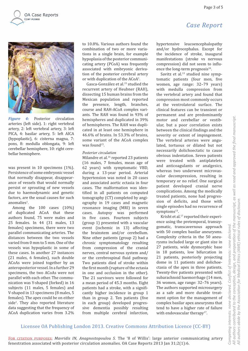
Furthermore, examination of the rest of the cerebral vessels revealed mild dolichoectasia of the basilar artery (mainly dilated and curved to the left side at the pontomedullary junction) (Figure 2) and marked doli-choectasia of the left posterior infe-rior cerebellar artery (PICA), forming a large vascular loop at the area of the cisterna magna (Figures 3 and 4). Hypoplasia of the left anterior infe-rior cerebellar artery (AICA) (Figure 4) was another interesting coex-isting finding. Bilaterally, the inferior
cerebellar arteries of larger diameter (i.e. right AICA and left PICA) were distributed to the cerebellar tonsil.
DiscussionSeriesAnterior circulationKwak et al.14 investigated arterial anomalies accompanying 296 cases of a single ACoA aneurysm and the etiological significance of these anomalies in the development of cerebral aneurysms. Either the ACoA fenestration or the presence of more than two ACoAs was observed in 17 cases (5.7%). As the anomaly was duplicated in some of these cases, the total number of cases with ACoA anomalies resulted to be 26 cases (8.8%) out of 296 cases investigated. This occurrence rate was not higher than those observed in other previ-ously reported cerebral aneurysmal cases and control cases14.
Ogawa et al.6 reported operations performed on 206 patients with ACoA aneurysms using a bifrontal craniotomy and an interhemispheric approach. A total of 44 (21.4%) of these patients had vascular anoma-lies in the vicinity of the ACoA; these included a median artery of the
corpus callosum (MACC) in 27 cases (13.1%), duplication of the ACoA in 20 cases (9.7%) and duplication of the A1 segment of the ACA in one case (0.5%). A retrospective study of the angiograms indicated that diag-nosis of the A1 or ACoA duplication was not possible. The majority of the cases of ACoA aneurysms with MACC (81.5%) showed trifurcation of the ACoA, A2 and MACC6.
Saidi et al.3 evaluated variations of the ACA and ACoA. Thirty-six cadav-eric brains were studied by gross dissection for the pattern of arte-rial blood supply. Unique variations observed include an accessory ACA from the ACoA. Variations of the ACoA were also observed including fenes-tration (26%) and duplication (13%)3.
Kapoor et al.1 studied variations of the Willis circle using brains from 1000 medicolegal autopsy subjects of varying ages (Indian population). In 54.8% of specimens, there were vari-ations in the Willis circle. The circle was deficient in 32 specimens (3.2%). The ACoA was absent in 1.8% cases, duplicate in 10%, triplicate in 1.2% and plexiform in 0.4%. Seventy-four brains (7.4%) had multiple varia-tions. Intracranial saccular aneurysm
Figure 1: The ‘8 of Willis’: large ACoA fenestration. 1: right ICA, 2: left ICA, 3: right ACA, 4: left ACA, 5: fenestrated (duplicated) ACoA, 6: right middle cerebral artery, 7: left middle cerebral artery, 8: right PCoA, 9: left PCoA, 10: right posterior cerebral artery, 11: left posterior cerebral artery, 12: basilar artery, 13: optic chiasm.
Figure 2: Posterior circulation arteries (right side). 1: basilar artery, 2: right vertebral artery, 3: left verte-bral artery, 4: right PICA, 5: right cerebellar tonsil, 6: pons, 7: medulla oblongata, 8: left cerebellar tonsil, 9: optic chiasm.
Figure 3: Area of cisterna magna. 1: left vertebral artery, 2: left PICA (large vascular loop), 3: cisterna magna, 4: medulla oblongata, 5: left cerebellar hemisphere, 6: right cerebellar hemi-sphere.
Case Report
Page 3 of 5
Com
peti n
g in
tere
sts:
non
e de
clar
ed. C
onfl i
ct o
f Int
eres
ts: n
one
decl
ared
. A
ll au
thor
s co
ntrib
uted
to th
e co
ncep
ti on,
des
ign,
and
pre
parati o
n of
the
man
uscr
ipt,
as
wel
l as
read
and
app
rove
d th
e fi n
al m
anus
crip
t. A
ll au
thor
s ab
ide
by th
e A
ssoc
iati o
n fo
r Med
ical
Eth
ics
(AM
E) e
thic
al ru
les
of d
iscl
osur
e.
Licensee OA Publishing London 2013. Creative Commons Attribution Licence (CC-BY)
FOR CITATION PURPOSES: Mavridis IN, Anagnostopoulou S. The ‘8 of Willis’: large anterior communicating artery fenestration associated with posterior circulation anomalies. OA Case Reports 2013 Jan 31;2(1):6.
was present in 10 specimens (1%). Persistence of some embryonic vessel that normally disappear, disappear-ance of vessels that would normally persist or sprouting of new vessels due to haemodynamic and genetic factors, are the usual causes for such anomalies1.
Among the 100 cases (10%) of duplicated ACoA that these authors found, 75 were males and 25 females. In 44 (31 males, 13 females) specimens, there were two parallel communicating arteries. The distances between the two vessels varied from 0 mm to 5 mm. One of the vessels was hypoplastic in some of these cases. In another 27 instances (21 males, 6 females), such double ACoAs were joined together by an anteroposterior vessel. In a further 29 specimens, the two ACoAs were not parallel with each other. The commu-nication was Y-shaped (forked) in 16 subjects (11 males, 5 females) and V-shaped in 13 specimens (8 males, 5 females). The apex could be on either side1. They also reported literature data suggesting that the frequency of ACoA duplication varies from 3.2%
to 10.8%. Various authors found the combination of two or more varia-tions in a single brain, for example hypoplasia of the posterior communi-cating artery (PCoA) was frequently associated with embryonic deriva-tion of the posterior cerebral artery or with duplication of the ACoA1.
Gasca-González et al.15 studied the recurrent artery of Heubner (RAH), dissecting 15 human brains from the Mexican population and reported the presence, length, branches, course and RAH-ACoA complex vari-ants. The RAH was found in 93% of hemispheres and duplicated in 39% of hemispheres. The RAH was dupli-cated in at least one hemisphere in 46.6% of brains. In 53.3% of brains, some variant of the ACoA complex was found15.
Posterior circulationMilandre et al.16 reported 23 patients (16 males, 7 females, mean age of 62 years) with symptomatic VBD, during a 13-year period. Arterial hypertension was noted in 20 cases and associated aortic ectasia in four cases. The malformation was iden-tified in all patients on computed tomography (CT) completed by angi-ography in 19 cases and magnetic resonance imaging (MRI) in seven cases. Autopsy was performed in five cases. Fourteen subjects (group 1) presented with a vascular event (ischemic in 13) affecting the brainstem and/or cerebellum. Nine other patients (group 2) had a chronic symptomatology resulting from compression of the cranial nerves, central nervous system and/or the cerebrospinal fluid pathway. Two patients died of stroke within the first month (rupture of the ectasia in one and occlusion in the other). The 21 survivors were followed for a mean period of 45.3 months. Eight patients had a stroke, with a signifi-cantly higher incidence in group 1 than in group 2. Ten patients (five in each group) developed progres-sive dementia possibly resulting from multiple cerebral infarction,
hypertensive leucoencephalopathy and/or hydrocephalus. Except for the incidence of stroke, inaugural manifestations (stroke vs nervous compression) did not seem to influ-ence the long-term prognosis16.
Savitz et al.13 studied nine symp-tomatic patients (four men, five women, age range: 32–79 years) with medulla compression from the vertebral artery and found that compression most commonly occurs at the ventrolateral surface. The clinical features can be transient or permanent and are predominantly motor and cerebellar or vestib-ular, but a poor correlation exists between the clinical findings and the severity or extent of impingement. The vertebral arteries were angu-lated, tortuous or dilated but not necessarily dolichoectatic to cause obvious indentation. Seven patients were treated with antiplatelets and anticoagulants or analgesics, whereas two underwent microvas-cular decompression, resulting in temporary or no relief. One surgical patient developed cranial nerve complications. Among the medically treated patients, none had progres-sion of deficits, and those with single episodes had no recurrence of symptoms13.
Krisht et al.17 reported their experi-ence using the pretemporal, transzy-gomatic, transcavernous approach with 50 complex basilar aneurysms. Complexity criteria in the 50 aneu-rysms included large or giant size in 27 patients, wide dysmorphic base in 18 patients, low bifurcation in 21 patients, posteriorly projecting dome in 11 patients and dolichoe-ctasia of the apex in three patients. Twenty-five patients presented with subarachnoid haemorrhage (14 men, 36 women, age range: 32–76 years). The authors supported microsurgery as a safe and more durable treat-ment option for the management of complex basilar apex aneurysms that tend to have a higher rate of failure with endovascular therapy17.
Figure 4: Posterior circulation arteries (left side). 1: right vertebral artery, 2: left vertebral artery, 3: left PICA, 4: basilar artery, 5: left AICA (hypoplastic), 6: cisterna magna, 7: pons, 8: medulla oblongata, 9: left cerebellar hemisphere, 10: right cere-bellar hemisphere.

Case Report
Page 4 of 5
Com
peti n
g in
tere
sts:
non
e de
clar
ed. C
onfl i
ct o
f int
eres
ts: n
one
decl
ared
.A
ll au
thor
s co
ntrib
uted
to th
e co
ncep
ti on,
des
ign,
and
pre
parati o
n of
the
man
uscr
ipt,
as
wel
l as
read
and
app
rove
d th
e fi n
al m
anus
crip
t.A
ll au
thor
s ab
ide
by th
e A
ssoc
iati o
n fo
r Med
ical
Eth
ics
(AM
E) e
thic
al ru
les
of d
iscl
osur
e.
Licensee OA Publishing London 2013. Creative Commons Attribution Licence (CC-BY)
FOR CITATION PURPOSES: Mavridis IN, Anagnostopoulou S. The ‘8 of Willis’: large anterior communicating artery fenestration associated with posterior circulation anomalies. OA Case Reports 2013 Jan 31;2(1):6.
Wu et al.11 aimed to validate the feasibility of coil-assisted stent recon-struction in the vascular lumen for the treatment of VBD and to evaluate its long-term effectiveness in preventing ischemic events. They found that endovascular reconstruction with coil-assisted stent placement or stent placement alone in the vascular lumen for the treatment of VBD is technically feasible and can prevent ischemic events in the territory of stented vessels compared with the natural course, though further studies in larger samples are needed11.
CasesAnterior circulationGurdal et al.18 examined 30 cadavers and reported two unusual variations of the ACoA. In the first case, ACoA was duplicated with a fenestrated ACA. In the second case, an oblique ACoA was present. Further, two branches of the oblique ACoA were joined to the right ACA. According to the authors, during neurosurgical exposure of the region for different purposes, knowledge of the vascular variations will increase the success of the procedure18.
Dey and Awad7 reported two cases of ICA fenestration with successful surgical clipping of associated saccular aneurysms. In both instances, the fenestra-tion involved the supraclinoid ICA and the aneurysm arose from the duplicated segment proximal to the origin of the PCoA. The aneu-rysms were more proximal than typical PCoA aneurysms and fenestration was suspected by rota-tional three- dimensional angiography and confirmed at surgery7.
Furthermore, Osonuga et al.19 reported a case of ACoA duplication with no associated vascular varia-tions or aneurysms in other major blood vessels. One ACoA was shorter than the other with external diam-eters of 1.0 mm and 1.4 mm, respec-tively. A quadrangular opening was formed between them19.
Posterior circulationDavous et al.20 reported a 54-year-old man who was affected by three successive infarctions in the vertebro-basilar territory. These infarctions were related to a dolichoectatic basilar artery. Deafness occurred first on the left side and then, after the third infarction, on the right side. The authors underlined that deafness can be observed after a pontine infarc-tion in the territory of the AICA. A dolichoectatic basilar artery can be the source of thrombotic or embolic strokes. Their prevention by anti-aggregant or anticoagulant therapy is suggested20.
Muñoz et al.21 reported a 52-year-old man with rapid jerking move-ments of his head to the right and clonic involuntary twitches involving his left eyelid and cheek. Electromyo-graphic recordings were consistent with left sternocleidomastoid and facial asynchronous myoclonus. MRI showed the presence of a dolichoe-ctatic left vertebral artery displacing the medulla. The authors proposed that direct compression of the XIth and VIIth cranial nerves by the doli-choectatic left vertebral artery might be the mechanism responsible for myoclonus in their patient21.
Campos et al.9 reported a 48-year-old woman who presented a two-month history of continuous buzz and a slight right-sided hearing loss that was followed by a cerebellar ischemic stroke. Brain CT and MRI revealed a marked compression of the brain-stem due to an ectatic, tortuous and partially thrombosed basilar artery. The largest cross-sectional diameter of the basilar artery was 18 mm. They underlined the fact that an unusual giant VBD caused an impressive brain-stem compression with displacement of important structures in an oligo-symptomatic patient. Diagnosis was made only after the occurrence of a stroke. According to the authors, the presence of significant atheroscle-rotic changes and the large basilar artery diameter may indicate a poor outcome9.
Interestingly, Tsutsumi et al.22 reported a 71-year-old female with basilar and bilateral carotid dolich-oectasia manifesting as dysarthria and hemisensory disturbance, which resolved spontaneously within a day. She suffered a brainstem infarc-tion (infarct in the perfusion area of the superior cerebellar artery) 28 months later, manifesting as drowsi-ness, dysarthria and right hemi-paresis. Her consciousness level progressively deteriorated to stupor and she died of acute respiratory failure on the seventh day. Autopsy demonstrated a tear in the lateral wall of the broad-based aneurysm on the ectatic basilar artery and a diffused subarachnoid haemor-rhage. The authors concluded that VBD is a dynamic vasculopathy that may rapidly progress in the affected basilar artery following an indo-lent clinical course. The prognosis for patients with VBD may depend mainly on the pathological changes in the basilar artery22.
Finally, Baran et al.8 reported a case of a 64-year-old female with hyper-tension who was admitted to the hospital with severe non-systemic vertigo and dysarthria, which had lasted for a couple of weeks. Imaging of the brain revealed dolichoectasia of the Willis circle arteries (ICA, verte bral artery, basilar artery, middle cerebral artery) coexisting with a fusiform aneurysm of the basilar artery. The examinations showed an aneurysmal distension of the basilar artery dis placing the brain stem to the right and compressing the other adjacent structures, including the fourth ventricle. The fusiform aneurysm (middle section of the basilar artery) was up to 23 mm in diameter (with width of the opacified lumen of up to 11 mm) with evidence of a thick wall clot and small periph-eral calcifications situated at the vessel wall. The other dilated vessels were accompanied by heavily calci-fied atheroscle rotic plaques8.
Case Report
Page 5 of 5
Com
peti n
g in
tere
sts:
non
e de
clar
ed. C
onfl i
ct o
f Int
eres
ts: n
one
decl
ared
. A
ll au
thor
s co
ntrib
uted
to th
e co
ncep
ti on,
des
ign,
and
pre
parati o
n of
the
man
uscr
ipt,
as
wel
l as
read
and
app
rove
d th
e fi n
al m
anus
crip
t. A
ll au
thor
s ab
ide
by th
e A
ssoc
iati o
n fo
r Med
ical
Eth
ics
(AM
E) e
thic
al ru
les
of d
iscl
osur
e.
Licensee OA Publishing London 2013. Creative Commons Attribution Licence (CC-BY)
FOR CITATION PURPOSES: Mavridis IN, Anagnostopoulou S. The ‘8 of Willis’: large anterior communicating artery fenestration associated with posterior circulation anomalies. OA Case Reports 2013 Jan 31;2(1):6.
ConclusionAnomalies of the ACoA are common and fenestration has been reported as its most common variation. Dolichoe-ctasia of intracranial arteries is a rare arteriopathy characterised by elonga-tion and widening of the arteries and disturbance of the laminar blood flow, mostly involving vertebral and basilar arteries. However, a case combining large ACoA fenestration with mild basilar artery and marked left PICA dolichoectasia, as well as left AICA hypoplasia, is quite unusual. Persis-tence of some embryonic vessels that normally disappear, disappearance of vessels that would normally persist or sprouting of new vessels due to haemodynamic and genetic factors, are probably the usual causes of Willis circle anomalies. Knowledge of such variations and anomalies (together with their potential clinical manifes-tations) is of paramount importance, primarily for neurosurgeons and neuroradiologists.
Abbreviations listACA, anterior cerebral artery; ACoA, anterior communicating artery; AICA, anterior inferior cerebellar artery; CT, computed tomog-raphy; ICA, internal carotid artery; MACC, median artery of the corpus callosum; MRI, magnetic resonance imaging; PCoA, posterior commu-nicating artery; PICA, posterior inferior cerebellar artery; RAH, recurrent artery of Heubner; VBD, vertebrobasilar dolichoectasia.
References1. Kapoor K, Singh B, Dewan LI. Variations in the configuration of the circle of Willis. Anat Sci Int. 2008 Jun;83(2):96–106.2. Karazincir S, Ada E, Sarsilmaz A, Yalçin O, Vidinli B, Sahin E. [Frequency of vascular variations and anomalies accom-panying intracranial aneurysms]. Tani Girisim Radyol. 2004 Jun;10(2):103–9. Turkish.3. Saidi H, Kitunguu PK, Ogeng’O JA.
Variant anatomy of the anterior cerebral artery in adult brains. Afr J Neurol Sci. 2008;27(1):97–105.4. Hannequin P, Peltier J, Destrieux C, Velut S, Havet E, Le Gars D. The inter-optic course of a unique precommunicating anterior cerebral artery with aber-rant origin of an ophthalmic artery: an anatomic case report. Surg Radiol Anat. 2013 Apr;35(3):269–71.5. Weil AG, Bojanowski MW, Scholtes F, Darsaut TE, Signorelli F, Weill A. Angio-graphic pitfall: duplicated tapered A1 segment of the anterior cerebral artery mimicking an anterior communicating artery aneurysm. Interv Neuroradiol. 2011 Jun;17(2):179–82.6. Ogawa A, Suzuki M, Sakurai Y, Yoshi-moto T. Vascular anomalies associated with aneurysms of the anterior communi-cating artery: microsurgical observations. J Neurosurg. 1990 May;72(5):706–9.7. Dey M, Awad IA. Fenestration of supraclinoid internal carotid artery and associated aneurysm: embryogenesis, recognition, and management. World Neurosurg. 2011 Dec;76(6):592.e1–5.8. Baran B, Kornafel O, Guziński M, Sąsiadek M. Dolichoectasia of the circle of Willis arteries and fusiform aneu-rysm of basilar artery–case report and review of the literature. Pol J Radiol. 2012 Apr;77(2):54–9.9. Campos CR, Doria-Netto HL, Souza-Filho AM, Silva Júnior HM. Oligo-symptomatic and giant basilar artery dolichoectasia discovered after a stroke: case report. Arq Neuropsiquiatr. 2007 Jun;65(2A):345–7.10. Weber AL. Magnetic resonance imaging and computed tomography of the internal auditory canal and cerebel-lopontine angle. Isr J Med Sci. 1992 Mar–Apr;28(3–4):173–82.11. Wu X, Xu Y, Hong B, Zhao WY, Huang QH,Liu JM. Endovascular reconstruction for treatment of vertebrobasilar dolichoe-ctasia: long-term outcomes. AJNR Am J Neuroradiol. 2013 Mar;34(3):583–8.12. Jankowicz E, Drozdowski W. [Sponta-neous dissection of carotid and vertebral arteries. II. Vertebral arteries and their branches]. Neurol Neurochir Pol. 1998 Sept–Oct;32(5):1237–45. Polish.13. Savitz SI, Ronthal M, Caplan LR. Verte-bral artery compression of the medulla. Arch Neurol. 2006 Feb;63(2):234–41.14. Kwak R, Niizuma H, Hatanaka M,
Suzuki J. [Anterior communicating artery aneurysms accompanied with anoma-lies or anterior communicating artery]. No To Shinkei. 1978 Nov;30(11):1221–5.Japanese.15. Gasca-González OO, Delgado-Reyes L, Pérez-Cruz JC. Microsurgical anatomy of the extracerebral segment of recurrent artery of Heubner in the Mexican popula-tion. Cir Cir. 2011 May–Jun;79(3):201–6, 219–24. English, Spanish.16. Milandre L, Bonnefoi B, Pestre P, Pellis-sier JF, Grisoli F, Khalil R. [Vertebrobasilar arterial dolichoectasia. Complications and prognosis]. Rev Neurol (Paris). 1991;147(11):714–22. French.17. Krisht AF, Krayenbühl N, Sercl D,Bikmaz K, Kadri PA. Results of microsur-gical clipping of 50 high complexity basilar apex aneurysms. Neurosurgery. 2007 Feb;60(2):242–50;discussion 250–2.18. Gurdal E, Cakmak O, Yalcinkaya M, Uzun I, Cavdar S. Two variations of the anterior communicating artery: a clinical reminder. Neuroanatomy. 2004 Aug;3:32–4.19. Osonuga A, Mohammed KM, Osonuga OA, Ayetey H, Saadaare AV, Oduro JK, et al. A case report on duplicate anterior communicating artery in Ghana. Novel Sci Int J Med Sci. 2012;1(2):32–3.20. Davous P, Tillier JN, Torrent J. [Total deafness after multiple pontine infarc-tions. Dolichoectasia of the basilar trunk]. Rev Neurol (Paris). 1991;147(3):234–7. French.21. Muñoz EJ, Vila N, Valls-Solé J, Tolosa E.Cervical and facial myoclonus associated with dolichoectasia of the left vertebral artery. Mov Disord. 1997 Sep;12(5):790–3.22. Tsutsumi S, Yasumoto Y, Ito M. Atyp-ical megadolichoectasia manifesting as brain infarction rapidly followed by fatal subarachnoid hemorrhage. J Neuroim-aging. 2010 Oct;20(4):376–8.