Embed Size (px)
Citation preview

1
Tethered N-Heterocyclic Carbene-Carboranyl Silver
Complexes for Cancer Therapy
Jordan Holmes,a Rachel J. Kearsey,a Katie A. Paske,a Frances N. Singer,a Suliman Atallah,b
Christopher M. Pask,a Roger M. Phillipsb and Charlotte E. Willans*a
aSchool of Chemistry, University of Leeds, Woodhouse Lane, Leeds, LS2 9JT, UK. Email:
bSchool of Applied Sciences, University of Huddersfield, Queensgate, Huddersfield, HD1 3DH,
UK.

2
ABSTRACT. Silver complexes of tethered N-heterocyclic carbene-carboranyl ligands have been
prepared and fully characterized. The first example of silver bonded directly to the cage of ortho-
carborane has been identified in the solid state. The presence of a carboranyl N-substituent on the
N-heterocyclic carbene significantly enhances the in vitro cytotoxicity of the silver complex
against HCT116 p53+/+ and HCT116 p53-/- colon cancer cells compared to a phenyl derivative.
Conversely, the presence of a carboranyl on the backbone of a xanthine derived N-heterocyclic
carbene decreases the in vitro cytotoxicity of the silver complex compared to its phenyl derivative.
Stability studies on the xanthine-derived ligands and complexes show that decomposition via
deboronation occurs in hydrous dimethyl sulfoxide, which may attribute to the contrasting in vitro
behavior of the carborane-containing complexes.
INTRODUCTION. Both N-heterocyclic carbenes (NHCs)1-2 and carboranes3-6 are distinct
classes of ligand, which have shown interaction with elements across the whole periodic table
through varying coordination modes. Complexes of NHCs and carboranes have displayed broad
ranging applications, including in the areas of catalysis, materials and biomedicine.1, 7-10 Silver is
renowned for its antimicrobial properties,11 with the anticancer effects of silver being recognized
relatively recently.12-13 One of the first silver compounds reported to exhibit cytotoxic properties
against cancer cells was a silver fluorobenzoate dimer.14 Five years later the first AgI-NHC
complexes to display in vitro cytotoxicity were reported by Youngs and co-workers (1, Figure 1),15
and were screened against OVCAR-3 (ovarian), MB157 (breast) and HeLa (cervical) cancer cell
lines. The complexes exhibited greatest cytotoxicity towards the MB157 cell line and had little
effect on the HeLa cell line, highlighting the selectivity towards specific cancers for this type of
complex. Work from our group has extended the natural xanthine-derived NHC ligand class,16 and

3
shown that increased steric bulk on the ligand and increased hydrophilicity of the silver complex
delivers greater cytotoxic effects against the cancer cells screened (2, Figure 1).17
Recently, work from our group and from Lavallo and co-workers has demonstrated that NHCs
and carboranes may be combined into the same ligand architecture. Lavallo has reported NHC
ligands in which carborane clusters are linked directly to the nitrogen atom of the N-heterocycle.18-
21 We have reported ligands with flexible (CH2)n spacers between the NHC and ortho- or nido-
carboranes, which offer versatile coordination to a range of metals (3, Figure 1).22-23 Preparation
of Rh, Ir and Ru complexes of these ligands involves in situ deprotonation by silver oxide prior to
transmetallation on to the transition metals. We envisaged that isolation of the Ag intermediates
of these reactions would provide further insight into their reactivity, in addition to novel Ag
complexes with enhanced tuneability which may be applied in cancer therapeutics. Though Ag-
NHCs have shown significant promise in recent years as potent inhibitors of cancer cells,24 in vivo
stability presents a serious challenge that needs addressing. Carboranes have also attracted
attention for medicinal applications owing to their high catabolic stability.6, 25 Incorporating
carboranes into NHC ligands has the potential to modify the stability, solubility and activity of
these complexes, allowing for further tuning for cancer therapy. Furthermore, the approach offers
the possibility of combined chemotherapy/BNCT and the ability to modify the carborane
derivative for targeted therapy.26

4
Figure 1. Structures of the first cytotoxic AgI-NHCs (1), xanthine-derived AgI-NHCs (2), a
tethered NHC-carboranyl ligand coordinated to RhI (3) and AgI NHC-carboranyl complexes
developed in this work.
RESULTS AND DISCUSSION. Previously we reported that RhI- and IrIII-NHC complexes
could be prepared from ligand precursor L1. Activation of the carborane substituent resulted in a
closo-dicarbadodecaborane anion, which chelated the Rh center through a carbon atom (3, Figure
1) and to the Ir center through a boron atom of the cage. Activation of both the imidazolium NCHN
proton and the carborane CH or BH proton was promoted using silver oxide in CH3CN solvent.
L1 was reacted with Ag2O in anhydrous CD2Cl2 and the reaction monitored using 1H NMR
spectroscopy (Scheme 1). The imidazolium NCHN proton resonance disappeared completely after
9 hours at 40°C, indicating that a Ag-NHC (C1) had formed (Figure 2). The carboranyl CH
resonance is masked by the CH2 resonance at 4.31ppm. A 2D 1H13C HMQC experiment revealed
that the carboranyl resonance is still present after 9 hours reaction time, with strong coupling
observed between this and the carboranyl cage carbon atom at 63.3ppm (Figure 2).

5
Scheme 1. Synthesis of [Ag(NHC)Br]2 C1 with formation of [Ag2(NHC-carboranyl)2] macrocycle
C1’, and synthesis of Ag(NHC)2Br C2 and Ag(NHC)2Br C3.

6
Figure 2. 1H NMR and 1H13C HMQC spectra (300MHz, CD2Cl2) of [Ag(NHC)Br]2 C1. Inset
highlights the coupling between the masked carboranyl CH proton resonance and the carboranyl
cage carbon resonance at 63.3ppm.
Complex C1 appeared very unstable hence it was not possible to obtain further data on this
complex. However, crystals of a second complex, C1’, were found in the product mixture. Single
crystal X-ray diffraction analysis revealed a remarkable structure, in which the Ag is bound to the
NHC of one ligand and the carborane carbon atom of a second ligand, to give a macrocycle (Figure
3). The geometry about each silver atom is slightly distorted linear, with the solid state structure
exhibiting relatively long Ag-Ccarbene bond lengths (2.112(3) Å and 2.128(3) Å). As complex C1
appears unstable, with the solution darkening over time, presumably C1’ is a decomposition
product. As a second product was not observed in the NMR data, it is likely that C1’ is a

7
crystallization effect with concomitant HBr formation, and was found to be too unstable to re-
dissolve and analyze in solution. This is, however, the first structurally elucidated example of silver
bound directly to the cage of ortho-carborane.
Figure 3. Molecular structure of C1’. Ellipsoids are drawn at 50 % probability, and H atoms have
been omitted for clarity. Ag(1)-C(7): 2.112(3)Å; Ag(1)-C(1A): 2.128(3)Å; Ag(1A)-C(7A):
2.097(2)Å; Ag(1A)-C(1): 2.118(2)Å; C(1)-Ag(1A)-C(7A): 177.15(10)°; C(7)-Ag(1)-C(1A):
176.29(10)°.
In an attempt to increase the stability of the Ag-NHC complex, ligand precursor L2 was prepared
in which the steric encumbrance of the N-substituent was decreased from a tbutyl to a mesityl
group (Scheme 1). Reaction of mesityl imidazole with 1-bromoethyl-ortho-carborane resulted in
imidazolium bromide L2, which was fully characterized including through X-ray diffraction
analysis (Figure 4). Reaction of L2 with Ag2O in CH2Cl2 results in Ag(NHC)2Br complex C2,
which was fully characterized using multinuclear NMR spectroscopy, mass spectrometry,
elemental analysis and X-ray diffraction analysis. The structure reveals a T-shape geometry around

8
the Ag center, bound by two NHCs linear to each other, and a Br ion (Figure 4). The Ag-Ccarbene
bond lengths are relatively long (2.117(6) Å and 2.087(5) Å;), though within the normal range for
complexes with T-shaped geometry.27 It is unusual for a Ag-bis(NHC) complex to result from an
imidazolium salt with a coordinating anion, with 2Ag(NHC)X [Ag(NHC)2][AgX2] (X =
coordinating anion) being more common.28 Elemental analysis confirms the Ag(NHC)2Br
stoichiometry in the bulk of C2, which was isolated in 68% yield. AgBr formed in the process (i.e.
to the right of the mono-NHC/bis-NHC equilibrium) would be removed during work-up which
involved filtration of the solution through Celite®.

9
Figure 4. Molecular structures of L2 (top) C2 (middle) and C3 (bottom). Ellipsoids are drawn at
50 % probability, and H atoms have been omitted for clarity. C2 Ag(1)-C(7): 2.117(6)Å; Ag(1)-
C(7A): 2.087(5)Å; Ag(1)-Br(1): 3.0021(8)Å; C(7)-Ag(1)-C(7A): 158.2(2)°; C(7)-Ag(1)-Br(1):
105.39(18)°. C3 Ag(1)-C(7): 2.061(6)Å; Ag(1)-C(7A): 2.070(6)Å; C(7)-Ag(1)-C(7A): 176.4(2)°.
Exchanging the solvent from CH2Cl2 to CH3CN has previously resulted in CH
activation/deprotonation of the carborane cage in addition to imidazolium NCHN deprotonation.23
Ag2O was added to L2 in CD3CN and heated at 40°C, with the mixture being analyzed over time
using 1H NMR spectroscopy. The imidazolium NCHN proton resonance had disappeared after 22

10
hours reaction time, though the carboranyl CH resonance remains present, even after a further 21
hours reaction time and addition of more Ag2O with further heating. This indicates that
deprotonation of the carboranyl CH proton is metal assisted upon formation of a transition metal
complex, whereby a M-NHC (M = Rh, Ir) is formed initially, with subsequent deprotonation of
the carboranyl CH.
C3, which is a non-carborane phenyl derivative of C2, was prepared from L3 in an analogous
manner to C2 for comparison in biological studies. Single crystals of complex C3 were grown via
slow evaporation of Et2O into a concentrated CH2Cl2 solution. In this case the molecular structure
exhibits the more common cationic structure, in which the NHC ligands coordinate in a planar
arrangement with no contact between the silver center and the bromide anion (Figure 4), though
the overall stoichiometry of complexes C2 and C3 is the same.
Silver complexes of NHC ligands derived from natural xanthine precursors have shown in vitro
cytotoxicity against cancer cells in the micromolar range.17 In addition to the low toxicity of
xanthine-derivatives, these compounds have themselves shown medicinal properties against
cancer and other disease.29 We therefore extended our library of NHC-carboranyl silver complexes
to include xanthine-derived ligands. Theobromine was reacted with a range of alkenyl halides to
incorporate alkenyl groups on to the xanthine backbone (T4-T7), with subsequent reactions with
B10H12(CH3CN)2 under anhydrous conditions producing carboranyl imidazole precursors P4-P6
(Scheme 2). Each precursor was methylated using MeI to produce imidazolium salts L4-L6. In
addition, a phenyl derivative, L7, was prepared directly from T7.

11
Scheme 2. Synthesis of xanthine-derived imidazolium salts L4-L7, and AgI-NHCs C5 and C7.
Attempts were made to prepare AgI complexes of the xanthine-derived ligands through reaction
with Ag2O in a range of solvents, though surprisingly deprotonation of the imidazolium salts did
not occur. Reaction of L5 and L7 with AgOAc at room temperature allowed the corresponding
Ag-NHC complexes C5 and C7 to be isolated (Scheme 2), which were fully characterized using
multinuclear NMR spectroscopy and mass spectrometry. Although the mass spectra exhibited
major molecular ion peaks corresponding to [Ag(NHC)2]+, elemental analysis confirmed the
Ag(NHC)OAc stoichiometry, with ligand scrambling in the mass spectrometer being well
documented.30 The molecular structures of complexes C5 and C7 were elucidated by X-ray
diffraction analysis (Figure 5). Crystals suitable for both complexes were obtained via the slow
diffusion of Et2O into concentrated CH2Cl2 solutions. The Ag-Ccarbene bond lengths of 2.067(10)
Å (C5) and 2.060(3) Å (C7) are within the expected range for Ag(NHC)OAc type complexes,17,

12
31 and the geometry about the silver atoms deviate slightly from linearity. Analogous reactions
using ligand precursors L4 and L6 resulted in the formation of a black precipitate rather than the
corresponding Ag-NHCs.
Figure 5. Molecular structures of C5 (top) and C7 (bottom). Ellipsoids are drawn at 50 %
probability, and H atoms have been omitted for clarity. C5 Ag(1)-C(8): 2.067(10)Å; Ag(1)-O(3):

13
2.131(9)Å: C(8)-Ag(1)-O(3): 175.7(4)°; C(2)-C(3)-N(1): 113.2(10)°. C7 Ag(1)-C(8): 2.060(3)Å;
Ag(1)-O(3): 2.104(2)Å: C(8)-Ag(1)-O(3): 171.80(12)°; C(2)-C(3)-N(1): 112.5(3)°.
The in vitro cytotoxicity of silver complexes C2, C3, C5 and C7 against the colon cancer cell
lines HCT116 p53+/+ and HCT116 p53−/− was determined using MTT-based assays involving a 96
hour drug-exposure period (Table 1). The IC50 values for each complex are the same irrespective
of the cell line they are screened against. HCT116 p53−/− does not possess the p53 gene, a key
tumor suppressor protein that is a most commonly mutated gene in cancer patients. Apoptosis
induced by the silver complexes appears to be independent of the p53 gene, which is a desired
feature in the search for new chemotherapeutic compounds.
Entry Complex HCT116 p53+/+
IC50 (μM)
HCT116 p53−/−
IC50 (μM)
1 C2 1.3 ± 0.2 1.5 ± 0.4
2 C3 10.5 ± 4.8 9.8 ± 3.7
3 C5 17.6 ± 2.1 20.2 ± 5.0
4 C7 11.6 ± 4.6 11.3 ± 4.6
Table 1. IC50 (μM) for Ag-NHC complexes screened against HCT116 p53+/+ and HCT116 p53−/−
cell lines. Each value represents the mean ± standard deviation for three independent experiments.
Carborane-containing complex C2 displayed greater cytotoxicity than its phenyl derivative C3
by an order of magnitude (Table 1, entries 1-2). The C2B10Hx cage occupies approximately the
same volume as a rotating phenyl ring, with both the carborane and the phenyl being hydrophobic
entities. We have previously shown that increased sterics around the AgI center enhances the
cytotoxicity of Ag-NHCs, with the three-dimensional structure of the carborane in C2 likely
providing greater steric protection than the phenyl group in C3 and a slower release of AgI over
the drug exposure period. C5, in which the carborane moiety is remote from the AgI center, has
comparable cytotoxicity to its phenyl derivative C7 within error (Table 1, entries 3-4). The steric

14
encumbrance around the AgI centers in C5 and C7 is the same, hence this further corroborates that
steric factors play a pivotal role in the activity of these complexes. In an attempt to understand the
relative stabilities of these complexes which may link to pharmacokinetic differences, a series of
stability studies were carried out.
Complexes C5 and C7 were dissolved in hydrous DMSO-d6 (the same solvent that complexes
are dissolved in prior to cell line studies) and observed over time both visually and using NMR
spectroscopy. The phenyl complex C7 remains stable in hydrous DMSO-d6, with no sign of
decomposition by 1H NMR spectroscopy, even after several weeks. In contrast, complex C5 starts
to decompose within hours, with the formation of black precipitate and a downfield resonance at
9.25ppm in the 1H NMR spectrum indicative of imidazolium formation. In addition, a broad
resonance appears at -3ppm which is characteristic of a bridging proton of the open face of nido-
carborane. After 9 days in hydrous DMSO, complex C5 fully converts to imidazolium-nido-
carborane D5 and black precipitate, presumably Ag0 (Figure 6). Deboronation under such mild
conditions is unusual, though there is precedent. Kahl et al. reported the degradation of a closo-
carboranyl porphyrin to the nido-species in hydrous DMSO.32 Following a series of studies, it was
concluded that deboronation was i) dependent on the ability of the solvent to stabilize nucleophilic
attack by water, and ii) facilitated by electron withdrawing α-substituents. Release of silver during
decomposition upon dissolution in DMSO will lead to the relatively low cytotoxicity of complex
C5 observed in MTT assays.
To expand this understanding to our theobromine-derived carboranyl compounds, imidazoles
P4-P6 and imidazolium salts L4-L6 were dissolved in anhydrous DMSO-d6 (<0.02% H2O) and
monitored by 1H and 11B{1H} NMR spectroscopy. In accordance with Kahl’s findings, no
deboronation occurred in the absence of water after 4 days in solution. To each sample was added

15
40µL D2O, with deboronation being observed in P4 and L4 within a few hours (Figure 6). The
disubstituted carboranyl compounds P5 and L5 were found to be resistant towards deboronation
after 4 days, due to the electron donating nature of the methyl substituent reducing the
electropositive nature of the boron atoms and thus their susceptibility towards nucleophilic attack.
Compounds containing a longer tether between the electron withdrawing theobromine unit and the
cage, P6 and L6, also resist deboronation, again, due to the decreased electropositive nature of the
cage. These results indicate that complex C5, which has a disubstituted cage, might also resist
deboronation, hence it is somewhat surprising that the complex was found to decompose in this
manner in the presence of H2O. The presence of silver may render the theobromine-derived NHC
more electron withdrawing than its imidazole/imidazolium counterparts and result in a more
electropositive carborane cage. The weakly basic acetate present in the complex may also be
involved in the deboronation step, though we are not aware of any literature precedent for this.

16
Figure 6. Reactions of theobromine derived imidazole, imidazolium and NHC with D2O in DMSO
for 48 hours. 100% conversion to D5 after 9 days.
CONCLUSION. In summary, reactions of imidazolium salts bearing carboranyl N-substituents,
either on the N-heterocycle or on the backbone of a xanthine derivative, with basic silver precursors
have been investigated. When the carboranyl substituent is tethered to the heterocycle, the steric
properties of the second N-substituent affect the stability of the resulting silver-NHC complex.
Decomposition of a silver-NHC bearing a sterically encumbering ligand led to isolation of the first
complex in which silver is bonded directly to the cage of ortho-carborane (C1’). Reducing the
steric bulk of the second N-substituent allowed a stable AgI-NHC-carborane complex to be
prepared (C2). A series of xanthine-derived imidazoles and imidazolium salts, in which the
carboranyl substituent was tethered to the backbone, were also prepared. Reactions with AgOAc
found that when the carborane carbon atom possesses an acidic CH proton, a stable silver complex
could not be isolated. However, when this was substituted for a methyl group, the corresponding
AgI-NHC (C5) was prepared in good yield. The in vitro cytotoxicity of the silver complexes were
examined against the colon cancer cells HCT116 p53+/+ and HCT116 p53-/- and compared to the
phenyl derivatives. Complex C2 was found to be more cytotoxic than its phenyl derivative by an
order of magnitude, whereas complex C5 was found to have significantly higher IC50 values than
its phenyl derivative. This was attributed to the instability of complex C5 in hydrous DMSO, which
was found to decompose via deboronation and imidazolium formation.
EXPERIMENTAL SECTION
Anhydrous solvents were prepared by passing over activated alumina to remove water, copper
catalyst to remove oxygen and molecular sieves to remove any remaining water, via the Dow-
Grubbs solvent system, and then freeze-pump-thaw degassed prior to use. Decaborane was

17
purchased from KatChem and all other chemicals used in this work were bought from either Sigma
Aldrich or Alfa and used without further purification. L1 was prepared according to the published
procedure.23 NMR spectra were recorded on a Bruker AV500 or a Bruker DPX300 spectrometer.
1H NMR and 13C{1H} NMR chemical shifts were referenced against residual solvent peaks.
Assignment of 1H and 13C{1H} NMR spectra was aided by the use of 2D 1H1H COSY, 1H13C
HMQC, 1H13C HMBC and 13C{1H} DEPT 135. Mass spectra were collected on a Bruker Daltonics
(micro TOF) instrument operating in the electrospray mode. Elemental analyses were performed
by Mr Stephen Boyer at London Metropolitan University.
X-ray diffraction data were collected on an Agilent SuperNova diffractometer fitted with an
Atlas CCD detector with Mo- Kα radiation (λ = 0.71073 Å) or Cu- Kα (λ = 1.54184 Å). Crystals
were mounted under oil on nylon fibers and data collected at 110, 120 or 293 K. Data sets were
corrected for absorption using a Gaussian integration method, the structures were solved by direct
methods using SHELXS-9733 or intrinsic phasing using SHELXT34 and refined by full-matrix least
squares on F2 using ShelXL2014,35 interfaced through the program Olex2.36 Molecular graphics
for all structures were generated using POV-RAY in the X-Seed program.37
C1/C1’. L1 (15 mg, 0.04 mmol), Ag2O (9 mg, 0.04 mmol) and anhydrous CD2Cl2 (0.5 mL) were
added to a Youngs NMR tube in the Glovebox. The tube was wrapped in foil and kept at room
temperature for 9 hours, with regular agitation (see SI Figure S34 for NMR spectrum). The reaction
mixture was filtered through Celite® and slow diffusion of Et2O into the filtrate produced crystals
of C1 and C1’.
L2. To an ampoule was added mesityl imidazole (100 mg, 0.531 mmol) and 1-bromoethyl-ortho-
carborane (149 mg, 0.591 mmol) and degassed. Anhydrous toluene (5 mL) was added and the
solution was heated at 90°C for 18 hours. The resulting white solid was filtered, washed with

18
toluene, hexane and Et2O. The product was collected as a white solid. Yield: 159 mg, 0.363 mmol
(68 %). 1H NMR (500 MHz, DMSO-d6): δ (ppm) 9.53 (s, 1H, NCHN), 8.18 (s, 1H, NCH), 7.95
(s, 1H, NCH), 7.15 (s, 2H, Ar), 5.38 (s, 1H, carboranyl CH), 4.46 (t, 2H, J = 10 Hz, CH2) 3.12 (t,
2H, J = 10 Hz, CH2) 2.33 (s, 3H, CH3) 2.03 (s, 6H, CH3). 13C NMR (126 MHz, DMSO-d6): δ
(ppm) 140.3 (C), 137.9 (CH), 134.2 (CH), 131.0 (C), 129.3 (C), 123.9 (NCH), 123.1 (NCH), 72.7
(carboranyl-C), 63.4 (carboranyl-CH), 47.7 (CH2), 34.8 (CH2), 20.6 (CH3), 16.9 (CH3). 11B{1H}
NMR (161 MHz, DMSO-d6): δ (ppm) -3.0, -5.5, -9.7, -12.1. HRMS (ESI+): m/z [C16H29B10N2]+
357.3346, calcd for [M − Br]+ 357.3328.
C2. An ampoule was charged with imidazolium precursor L2 (200 mg, 0.46 mmol) and Ag2O
(53 mg, 0.23 mmol) and degassed. Anhydrous CH2Cl2 (5 mL) was added and the mixture was
stirred at room temperature for 24 hours. The solution was filtered through Celite® and the solvent
removed in vacuo. The residue was dissolved in Et2O (5 mL) and the product was precipitated
with hexane (15 mL), filtered and dried in vacuo to give a beige powder. Yield: 141 mg, 0.16
mmol (68 %). 1H NMR (500 MHz, CD2Cl2): δ (ppm) 7.07 (d, J = 1.8 Hz, 1H, NCH), 6.97 (s, 2H,
Ar), 6.87 (d, J = 1.8 Hz, 1H, NCH), 5.33 (br. s, 1H, carboranyl CH), 4.07 (m, 2H, CH2), 3.05 (m,
2H, CH2), 2.37 (s, 3H, CH3), 1.89 (s, 6H, CH3). 13C NMR (126 MHz, CD2Cl2): δ (ppm) 139.6 (C),
136.7 (C), 135.7 (C), 129.7 (CH), 122.7 (NCH), 121.2 (NCH), 73.1 (carboranyl-C), 64.4
(carboranyl-CH), 50.7 (CH2), 37.8 (CH2), 21.4 (CH3), 18.2 (CH3). 11B{1H} NMR (161 MHz,
DMSO-d6): δ (ppm) −2.9, −5.3, −9.4, −12.9. HRMS (ESI+): m/z [C32H56B20AgN4]+ 819.5598,
calcd for [M − Br]+ 819.5562. Anal. Calcd for C32H56B20AgBrN4: C, 42.67; H, 6.27; N, 6.22.
Found: C, 42.49; H6.16; N, 6.17.
L3. To an ampoule was added CH3CN (2 mL), mesityl imidazole (576 mg, 3.09 mmol) and 2-
bromoethyl-benzene (0.42 mL, 3.09 mmol) and the solution was heated at reflux for 18 hours. The

19
reaction was cooled to room temperature and the product precipitated with Et2O (30 mL), filtered
and dried in vacuo to give a white microcrystalline solid. Yield: 886 mg, 2.39 mmol (77 %). 1H
NMR (500 MHz, DMSO-d6): δ (ppm) 9.35 (t, J = 1.5 Hz, 1H, NCHN), 8.12 (t, J = 1.7 Hz, 1H,
NCH), 7.87 (t, J = 1.8 Hz, 1H, NCH), 7.28 (m, 2H, CH), 7.24 (s, 2H, CH), 7.22 (m, 1H, CH), 7.10
(s, 2H, CH), 4.62 (t, J = 6.8 Hz, 2H, CH2), 3.26 (t, J = 6.8 Hz, 2H, CH2), 2.30 (s, 3H, CH3), 1.86
(s, 6H, CH3). 13C NMR (126 MHz, DMSO-d6): δ (ppm) 140.2 (C), 137.2 (NCHN), 136.6 (C),
134.1 (C), 130.9 (C), 129.1 (CH), 128.7 (CH), 128.5 (CH), 126.8 (CH), 123.8 (NCH), 123.1
(NCH), 50.2 (CH2), 34.9 (CH2), 20.5 (CH3), 16.7 (CH3). HRMS (ESI+): m/z [C20H23N2]+ 291.1870,
calcd for [M − Br]+ 291.1856.
C3. An ampoule was charged with L3 (200 mg, 0.54 mmol) and Ag2O (69 mg, 0.30 mmol) and
degassed. Anhydrous CH2Cl2 (5 mL) was added and the mixture stirred at room temperature for
24 hours. The solution was filtered through Celite® and the solvent removed in vacuo. CH3OH
(5 mL) was added, the insoluble particulate filtered and the product was precipitated from the
filtrate with Et2O (30 mL). The product was collected via filtration and dried in vacuo to give a
white powder. Yield: 145 mg, 0.19 mmol (70 %). 1H NMR (500 MHz, CD2Cl2): δ (ppm) 7.20 (s,
1H, NCH), 7.18 (m, 3H, CH), 6.90 (m, 4H, CH), 6.84 (s, 1H, NCH), 4.26 (t, J = 6.5 Hz, 2H, CH2),
3.00 (t, J = 6.5 Hz, 2H, CH2), 2.31 (s, 3H, CH3), 1.74 (s, 6H, CH3). 13C NMR (126 MHz, DMSO-
d6): δ (ppm) 182.9 (carbenic-C), 139.9 (C), 137.7 (C), 136.1 (C), 135.4 (C), 129.6 (CH), 129.4
(CH), 129.1 (CH), 127.4 (CH), 123.0 (NCH), 122.4 (NCH), 53.4 (CH2), 38.0 (CH2), 21.4 (CH3),
17.9 (CH3). HRMS (ESI+): m/z [C40H44AgN4]+ 689.2621, calcd for [M − Br]+ 689.2615. Anal.
Calcd for C, 62.51; H, 5.77; N, 7.29. Found: C, 62.38; H, 5.89; N, 7.17.
T4. To a round-bottom flask was added CH3CN (30 mL), theobromine (3.00 g, 16.70 mmol),
Cs2CO3 (10.80 g, 33.30 mmol) and propargyl bromide (2.27 mL, 25.00 mmol, 80 wt % toluene)

20
and the mixture heated at 80 °C for 18 hours. The reaction was cooled to room temperature and
the solvent removed in vacuo. CH2Cl2 (50 mL) was added, the mixture filtered and H2O (30 mL)
was added to the filtrate, with the organic phase being collected. This was further washed with
H2O (2 × 30 mL), dried over MgSO4, filtered and the solvent removed in vacuo. The off-white
residue was recrystallized from CH2Cl2 (20 mL) with pentane (50 mL) to give a white powder.
Yield: 2.99 g, 13.70 mmol (82 %). 1H NMR (500 MHz, CDCl3): δ (ppm) 7.52 (m, 1H, NCHN),
4.79 (d, J = 2.5 Hz, 2H, CH2), 3.99 (m, 3H, CH3), 3.59 (s, 3H, CH3), 2.17 (t, J = 2.5 Hz, 1H, CH).
13C NMR (126 MHz, CDCl3): δ (ppm) 154.5 (CO), 151.0 (CO), 149.3 (C), 141.9 (NCHN), 107.7
(C), 78.8 (C≡CH), 70.6 (C≡CH), 33.8 (CH3), 30.6 (CH2), 29.9 (CH3). HRMS (ESI+): m/z
[C10H10N4O2Na]+ 241.0700, calcd for [M + Na]+ 241.0696.
P4. To a Schlenk flask was added T4 (1.00 g, 4.58 mmol) and B10H12(CH3CN)2 (927 mg, 4.58
mmol) and degassed. Anhydrous toluene (10 mL) and anhydrous CH3CN (3 mL) were added and
the mixture was slowly heated to 100 °C and kept at this temperature for 18 hours. The reaction
was cooled to room temperature and the solvent removed in vacuo. Et2O (2 × 15 mL) was added
to the product and filtered to remove insoluble material. The filtrate was washed with 1 M NaOH
solution (2 × 10 mL), H2O (2 × 10 mL), dried over MgSO4, filtered and the solvent removed from
the filtrate in vacuo. The residue was recrystallized from Et2O (10 mL) with hexane (30 mL),
filtered and the white solid dried in vacuo. Yield: 807 mg, 2.40 mmol (52 %). 1H NMR (500 MHz,
CDCl3): δ (ppm) 7.56 (s, 1H, NCHN), 4.88 (d, J = 14.9 Hz, 1H, CH2), 4.60 (d, J = 14.9 Hz, 1H,
CH2), 4.20 (br. s, 1H, carboranyl-CH), 3.98 (s, 3H, CH3), 3.58 (s, 3H, CH3). 13C NMR (126 MHz,
CDCl3): δ (ppm) 154.5 (CO), 151.6 (CO), 149.4 (C), 142.6 (NCHN), 107.3 (C), 74.0 (carboranyl-
C), 61.3 (carboranyl-CH), 45.3 (CH2), 33.9 (CH3), 30.2 (CH3). 11B{1H} NMR (161 MHz, CDCl3):

21
δ (ppm) −1.4, −4.8, −10.0, −10.8, −12.9. HRMS (ESI+): m/z [C10H21B10N4O2]+ 337.2670, calcd
for [M + H]+ 337.2667.
T5. Prepared as described for T4 from theobromine (3.00 g, 16.70 mmol), Cs2CO3 (10.80 g,
33.30 mmol) and 1-bromo-2-butyne (1.60 mL, 18.40 mmol) in CH3CN (30 mL). After work up
the product was obtained as a white powder. Yield: 2.95 g, 12.70 mmol (76 %). 1H NMR (500
MHz, CDCl3): δ (ppm) 7.50 (m, 1H, NCHN), 4.72 (q, J = 2 Hz, 2.5 Hz, 2H, CH2), 3.98 (s, 3H,
CH3), 3.58 (s, 3H, CH3), 1.76 (t, J = 2Hz, 3H, CH3). 13C NMR (126 MHz, CDCl3): δ (ppm) 154.7
(CO), 151.1 (CO), 149.1 (C), 141.7 (NCHN), 107.7 (C), 78.3 (C≡C), 73.9 (C≡C), 33.7 (CH3), 31.1
(CH2), 29.9 (CH3), 3.8 (CH3). HRMS (ESI+): m/z [C11H12N4O2Na]+ 255.0857, calcd for [M + Na]+
255.0852.
P5. To a Schlenk flask was added T5 (1.00 g, 4.31 mmol) and B10H12(CH3CN)2 (871 mg, 4.31
mmol) and degassed. Anhydrous toluene (10 mL) and anhydrous CH3CN (3 mL) were added and
the reaction slowly heated to 100 °C and kept at this temperature for 18 hours. The reaction was
cooled to room temperature and the solvent removed in vacuo. The residue was dissolved in a
minimum amount of CH2Cl2 and purified by silica chromatography, which was eluted with ethyl
acetate/hexane 1:2. The product fractions were combined and the solvent removed in vacuo. The
residue was dissolved in Et2O (5 mL) and the product was precipitated as a white powder with
hexane (30 mL), which was filtered and dried in vacuo. Yield: 573 mg, 1.63 mmol (38 %). 1H
NMR (500 MHz, CDCl3): δ (ppm) 7.56 (d, J = 0.6 Hz, 1H, NCHN), 4.86 (d, J = 14.8 Hz, 1H,
CH2), 4.64 (d, J = 14.8 Hz, 1H, CH2), 3.99 (d, J = 0.6 Hz, 3H, CH3), 3.59 (s, 3H, CH3), 2.34 (s,
3H, carboranyl-CCH3). 13C NMR (126 MHz, CDCl3): δ (ppm) 154.7 (CO), 151.5 (CO), 149.5 (C),
142.4 (NCHN), 107.4 (C), 77.4 (carboranyl-C), 75.9 (carboranyl-CCH3), 42.5 (CH2), 33.9 (CH3),
22
30.2 (CH3), 23.5 (carboranyl-CCH3). 11B{1H} NMR (161 MHz, CDCl3): δ (ppm) −3.2, −6.0, −9.9.
HRMS (ESI+): m/z [C11H23B10N4O2]+ 351.2824, calcd for [M + H]+ 351.2824.
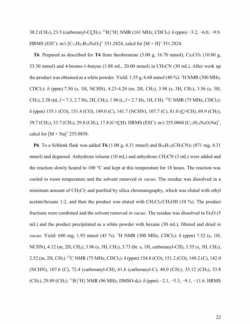
T6. Prepared as described for T4 from theobromine (3.00 g, 16.70 mmol), Cs2CO3 (10.80 g,
33.30 mmol) and 4-bromo-1-butyne (1.88 mL, 20.00 mmol) in CH3CN (30 mL). After work up
the product was obtained as a white powder. Yield: 1.55 g, 6.68 mmol (40 %). 1H NMR (300 MHz,
CDCl3): δ (ppm) 7.50 (s, 1H, NCHN), 4.23-4.20 (m, 2H, CH2), 3.98 (s, 3H, CH3), 3.56 (s, 3H,
CH3), 2.58 (td, J = 7.3, 2.7 Hz, 2H, CH2), 1.96 (t, J = 2.7 Hz, 1H, CH). 13C NMR (75 MHz, CDCl3):
δ (ppm) 155.1 (CO), 151.4 (CO), 149.0 (C), 141.7 (NCHN), 107.7 (C), 81.0 (C≡CH), 69.9 (CH2),
39.7 (CH2), 33.7 (CH3), 29.8 (CH3), 17.8 (C≡CH). HRMS (ESI+): m/z 255.0860 [C11H12N4O2Na]+,
calcd for [M + Na]+ 255.0858.
P6. To a Schlenk flask was added T6 (1.00 g, 4.31 mmol) and B10H12(CH3CN)2 (871 mg, 4.31
mmol) and degassed. Anhydrous toluene (10 mL) and anhydrous CH3CN (3 mL) were added and
the reaction slowly heated to 100 °C and kept at this temperature for 18 hours. The reaction was
cooled to room temperature and the solvent removed in vacuo. The residue was dissolved in a
minimum amount of CH2Cl2 and purified by silica chromatography, which was eluted with ethyl
acetate/hexane 1:2, and then the product was eluted with CH2Cl2/CH3OH (10 %). The product
fractions were combined and the solvent removed in vacuo. The residue was dissolved in Et2O (5
mL) and the product precipitated as a white powder with hexane (30 mL), filtered and dried in
vacuo. Yield: 680 mg, 1.93 mmol (45 %). 1H NMR (300 MHz, CDCl3): δ (ppm) 7.52 (s, 1H,
NCHN), 4.12 (m, 2H, CH2), 3.96 (s, 3H, CH3), 3.73 (br. s, 1H, carboranyl-CH), 3.55 (s, 3H, CH3),
2.52 (m, 2H, CH2). 13C NMR (75 MHz, CDCl3): δ (ppm) 154.8 (CO), 151.2 (CO), 149.2 (C), 142.0
(NCHN), 107.6 (C), 72.4 (carboranyl-CH), 61.6 (carboranyl-C), 40.0 (CH2), 35.12 (CH2), 33.8
(CH3), 29.89 (CH3). 11B{1H} NMR (96 MHz, DMSO-d6): δ (ppm) −2.1, −5.3, −9.1, −11.6. HRMS

23
(ESI+): m/z [C13H23B10N5O2Na]+ 413.2600, calcd for [M + CH3CN + Na − 2H]+ 413.2716. (Note:
only one of the CH2 resonance is observed).
T7. Prepared as described for T4 from theobromine (3.00 g, 16.70 mmol), Cs2CO3 (10.80 g,
33.30 mmol) and benzyl bromide (1.99 mL, 16.70 mmol) in CH3CN (30 mL). After work up the
product was obtained as a white powder. Yield: 3.62 g, 13.40 mmol (80 %). 1H NMR (500 MHz,
CDCl3): δ (ppm) 7.49 (m, 3H, 1H, NCHN), 7.30 (m, 2H, Ar-CH), 7.24 (m, 1H, Ar-CH), 5.19 (s,
2H, CH2), 3.98 (m, 3H, CH3), 3.57 (s, 3H, CH3). 13C NMR (126 MHz, CDCl3): δ (ppm) 155.4
(CO), 151.8 CO), 149.0 (C), 141.7 (NCHN), 137.5 (Ar-C), 129.0 (Ar-CH), 128.5 (Ar-CH), 127.7
(Ar-CH), 107.8 (C), 44.6 (CH2), 33.7 (CH3), 29.9 (CH3). HRMS (ESI+): m/z [C14H14N4O2Na]+
293.1025, calcd for [M + Na]+ 293.1009.
L4. An ampoule was charged with CH3CN (5 mL), P4 (1.17 g, 3.48 mmol) and MeI (6.49 mL,
104.3 mmol) and heated at 80 °C for 7 days. The reaction was cooled to room temperature and the
solvent removed in vacuo. To the residue was added Et2O (30 mL) which was then sonicated, the
solid filtered, washed with Et2O (20 mL) and dried in vacuo to give a white powder. Yield: 631
mg, 1.46 mmol (42 %). 1H NMR (500 MHz, DMSO-d6): δ (ppm) 9.35 (s, 1H, NCHN), 5.11 (br.
s, 1H, carboranyl-CH), 4.81 (d, J = 15.1 Hz, 1H, CH2), 4.58 (d, J = 15.1 Hz, 1H, CH2), 4.16 (s,
3H, CH3), 4.06 (s, 3H, CH3), 3.76 (s, 3H, CH3). 13C NMR (126 MHz, DMSO-d6): δ (ppm) 152.7
(CO), 150.1 (CO), 140.5 (NCHN), 139.9 (C), 107.5 (C), 73.2 (carboranyl-C), 62.4 (carboranyl-
CH), 45.1 (CH2), 37.0 (CH3), 35.8 (CH3), 31.8 (CH3). 11B{1H} NMR (161 MHz, DMSO-d6): δ
(ppm) −2.9, −5.2, −10.0, −11.6, −13.0. HRMS (ESI+): m/z [C11H23B10N4O2]+ 352.2800, calcd for
[M − I]+ 352.2800.
L5. Prepared as described for L4 from P5 (500 mg, 1.43 mmol) and MeI (2.67 mL, 42.6 mmol)
in CH3CN (5 mL). After purification the product was obtained as a white powder. Yield: 287 mg,

24
0.64 mmol (45 %). 1H NMR (500 MHz, DMSO-d6): δ (ppm) 9.34 (s, 1H, NCHN), 4.86 (d, J =
15.4 Hz, 1H, CH2), 4.64 (d, J = 15.4 Hz, 1H, CH2), 4.16 (s, 3H, CH3), 4.07 (s, 3H, CH3), 3.77 (s,
3H, CH3), 2.35 (s, 3H, carboranyl-CCH3). 13C NMR (126 MHz, DMSO-d6): δ (ppm) 152.9 (CO),
150.1 (CO), 140.5 (NCHN), 140.0 (C), 107.4 (C), 77.9 (carboranyl-C), 75.7 (carboranyl-CCH3),
43.0 (CH2), 37.0 (CH3), 35.8 (CH3), 31.8 (CH3), 22.6 (carboranyl-CCH3). 11B{1H} NMR (161
MHz, DMSO-d6): δ (ppm) −2.9, −5.2, −10.0, −11.6, −12.9. HRMS (ESI+): m/z [C11H23B10N4O2]+
366.2963, calcd for [M − I]+ 366.2944.
L6. Prepared as described for L4 from P6 (450 mg, 1.28 mmol) and MeI (2.40 mL, 38.5 mmol)
in CH3CN (5 mL). After purification the product was obtained as a yellow powder. Yield: 189 mg,
0.86 mmol (67 %). 1H NMR (500 MHz, DMSO-d6): δ (ppm) 9.29 (s, 1H, NCHN), 5.38 (br. s, 1H,
carboranyl-CH), 4.14 (s, 1H, CH3), 4.04 (s, 3H, CH3), 3.98 (m, 2H, CH2), 3.72 (s, 3H, CH3). 13C
NMR (75 MHz, DMSO-d6): δ (ppm) 152.8 (CO), 149.7 (CO), 140.0 (NCHN), 139.5 (C), 107.8
(C), 73.0 (carboranyl-CH), 63.6 (carboranyl-C), 36.9 (CH3), 35.6 (CH3), 33.0 (CH2), 31.4 (CH3).
11B NMR (96 MHz, CD3CN): δ (ppm) −2.89, −5.25, −9.97, −11.40, −12.97. HRMS (ESI+): m/z
[C11H23B10N4O2]+ 366.2960, calcd for [M − I]+ 366.2944. (Note: only one of the CH2 resonances
is observed).
L7. An ampoule was charged with CH3CN (5 mL), P7 (1.00 g, 3.70 mmol) and MeI (6.91 mL,
111 mmol) and heated at 80 °C for 100 hours. The reaction was cooled to room temperature and
the solvent removed in vacuo. To the residue was added CH2Cl2 (5 mL) and the product
recrystallized with hexane (30 mL) which was filtered yielding a sticky orange solid. This was
dissolved in CH2Cl2 (5 mL) and hexane (5 mL), and the solvent removed in vacuo to give a fluffy
golden solid, which was highly hygroscopic. Yield: 1.30 g, 3.15 mmol (85 %). 1H NMR (500
MHz, DMSO-d6): δ (ppm) 9.32 (s, 1H, NCHN), 7.33 (m, 4H, Ar-CH), 7.28 (m, 1H, Ar-CH), 5.09
25
(s, 2H, CH2), 4.15 (s, 3H, CH3), 4.07 (s, 3H, CH3), 3.74 (s, 3H, CH3). 13C NMR (126 MHz, DMSO-
d6): δ (ppm) 153.2 (CO), 150.0 (CO), 139.8 (NCHN), 139.6 (C), 136.2 (Ar-C), 128.3 (Ar-CH),
127.7 (Ar-CH), 127.4 (Ar-CH), 107.8 (C), 44.5 (CH2), 36.9 (CH3), 35.7 (CH3), 31.5 (CH3). HRMS
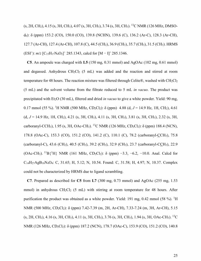
(ESI+): m/z [C15H17N4O2]+ 285.1343, calcd for [M − I]+ 285.1346.
C5. An ampoule was charged with L5 (150 mg, 0.31 mmol) and AgOAc (102 mg, 0.61 mmol)
and degassed. Anhydrous CH2Cl2 (5 mL) was added and the reaction and stirred at room
temperature for 48 hours. The reaction mixture was filtered through Celite®, washed with CH2Cl2
(5 mL) and the solvent volume from the filtrate reduced to 5 mL in vacuo. The product was
precipitated with Et2O (30 mL), filtered and dried in vacuo to give a white powder. Yield: 90 mg,
0.17 mmol (55 %). 1H NMR (500 MHz, CD2Cl2): δ (ppm) 4.88 (d, J = 14.9 Hz, 1H, CH2), 4.61
(d, J = 14.9 Hz, 1H, CH2), 4.21 (s, 3H, CH3), 4.11 (s, 3H, CH3), 3.81 (s, 3H, CH3), 2.32 (s, 3H,
carboranyl-CCH3), 1.95 (s, 3H, OAc-CH3). 13C NMR (126 MHz, CD2Cl2): δ (ppm) 188.4 (NCN),
178.8 (OAc-C), 153.3 (CO), 151.2 (CO), 141.2 (C), 110.1 (C), 78.2 (carboranyl-CCH3), 75.8
(carboranyl-C), 43.6 (CH2), 40.5 (CH3), 39.2 (CH3), 32.9 (CH3), 23.7 (carboranyl-CCH3), 22.9
(OAc-CH3). 11B{1H} NMR (161 MHz, CD2Cl2): δ (ppm) −3.3, −6.2, −10.0. Anal. Calcd for
C14H27AgB10N4O4: C, 31.65; H, 5.12; N, 10.54. Found: C, 31.58; H, 4.97; N, 10.37. Complex
could not be characterized by HRMS due to ligand scrambling.
C7. Prepared as described for C5 from L7 (300 mg, 0.73 mmol) and AgOAc (255 mg, 1.53
mmol) in anhydrous CH2Cl2 (5 mL) with stirring at room temperature for 48 hours. After
purification the product was obtained as a white powder. Yield: 191 mg, 0.42 mmol (58 %). 1H
NMR (500 MHz, CD2Cl2): δ (ppm) 7.42-7.39 (m, 2H, Ar-CH), 7.33-7.24 (m, 3H, Ar-CH), 5.15
(s, 2H, CH2), 4.16 (s, 3H, CH3), 4.11 (s, 3H, CH3), 3.76 (s, 3H, CH3), 1.94 (s, 3H, OAc-CH3). 13C
NMR (126 MHz, CD2Cl2): δ (ppm) 187.2 (NCN), 178.7 (OAc-C), 153.9 (CO), 151.2 (CO), 140.8
26
(C), 137.2 (Ar-C), 129.1 (Ar-CH), 128.9 (Ar-CH), 128.3 (Ar-CH), 110.4 (C), 45.6 (CH2), 40.3
(CH3), 39.1 (CH3), 32.4 (CH3), 22.9 (OAc-CH3). Anal. Calcd for C17H20AgN4O4: C, 45.15; H,
4.46; N, 12.39. Found: C, 45.17; H, 34.10; N, 12.48. Complex could not be characterized by
HRMS due to ligand scrambling. The major molecular ion peak observed corresponds to m/z
[C30H32AgN8O4]+ 675.1602, calcd for [Ag(NHC)2]
+ 675.1592.
MTT Assays. In vitro cell tests were performed in the School of Applied Sciences at the
University of Huddersfield. Cells were incubated in 96-well plates, at 2 × 10³ cells per well in 200
µL of growth media (RPMI 1640 supplemented with 10 % fetal calf serum, sodium pyruvate (1
mM) and L-glutamine (2 mM)). Cells were incubated for 24 hours at 37 °C under an atmosphere
of 5 % CO2 prior to drug exposure. Silver compounds were dissolved in DMSO at a concentration
of 25 mM and diluted with medium to obtain drug solutions ranging from 25 µM to 0.049 µM.
The final DMSO concentration was 0.1 % (v/v) which is non-toxic to cells. Drug solutions were
applied to cells and incubated for 96 hours at 37 °C under an atmosphere of 5 % CO2. The solutions
were removed from the wells and fresh medium added to each well along with 20 µL MTT (5 mg
mL-1), and incubated for 4 hours at 37 °C under an atmosphere of 5 % CO2. The solutions were
removed and DMSO (150 µL) was added to each well to dissolve the purple formazan crystals. A
plate reader was used to measure the absorbance at 540 nm. Lanes containing medium only (no
cells), and cells in medium only (no drug), were used as blanks for the spectrophotometer and 100
% cell survival respectively. Cell survival was determined as the true absorbance of treated cells
divided by the true absorbance of controls and expressed as a percentage. The concentration
required to kill 50 % of cells (IC50) was determined from plots of % survival against drug
concentration. The values presented are an average of three separate runs.
ASSOCIATED CONTENT

27
Supporting Information. NMR spectra and X-ray crystallographic information.
Author Contributions
The manuscript was written through contributions of all authors. All authors have given approval
to the final version of the manuscript.
Funding Sources
The work was funded through the EPSRC (DTA awards to JH and FNS) and the Universities of
Leeds and Huddersfield.
REFERENCES
1. Peris, E., Smart N-Heterocyclic Carbene Ligands in Catalysis. Chem. Rev. 2018, 118
(19), 9988-10031.
2. Hopkinson, M. N.; Richter, C.; Schedler, M.; Glorius, F., An overview of N-heterocyclic
carbenes. Nature 2014, 510 (7506), 485-496.
3. Weller, A., Ligand design: The two faces of carboranes. Nat Chem 2011, 3 (8), 577-578.
4. Grimes, R. N., Chapter 13 - Metallacarboranes of the transition and lanthanide elements.
In Carboranes (Second Edition), Grimes, R. N., Ed. Academic Press: Oxford, 2011; pp 773-
1014.
5. Grimes, R. N., Chapter 15 - Carboranes in catalysis. In Carboranes (Second Edition),
Grimes, R. N., Ed. Academic Press: Oxford, 2011; pp 1037-1052.
6. Grimes, R. N., Chapter 16 - Carboranes in medicine. In Carboranes (Second Edition),
Grimes, R. N., Ed. Academic Press: Oxford, 2011; pp 1053-1082.
7. Liu, W.; Gust, R., Metal N-heterocyclic carbene complexes as potential antitumor
metallodrugs. Chem. Soc. Rev. 2013, 42 (2), 755-73.
8. Grimes, R. N., Carboranes in the chemist's toolbox. Dalton Trans. 2015, 44 (13), 5939-
5956.
9. Mata, J. A.; Hahn, F. E.; Peris, E., Heterometallic complexes, tandem catalysis and
catalytic cooperativity. Chemical Science 2014, 5 (5), 1723-1732.
10. Mercs, L.; Albrecht, M., Beyond catalysis: N-heterocyclic carbene complexes as
components for medicinal, luminescent, and functional materials applications. Chem. Soc. Rev.
2010, 39 (6), 1903-1912.
11. Klasen, H. J., A historical review of the use of silver in the treatment of burns. II.
Renewed interest for silver. Burns 2000, 26 (2), 131-138.
12. Monteiro, D. C. F.; Phillips, R. M.; Crossley, B. D.; Fielden, J.; Willans, C. E., Enhanced
cytotoxicity of silver complexes bearing bidentate N-heterocyclic carbene ligands. Dalton Trans.
2012, 41 (13), 3720-3725.
13. Allison, S. J.; Sadiq, M.; Baronou, E.; Cooper, P. A.; Dunnill, C.; Georgopoulos, N. T.;
Latif, A.; Shepherd, S.; Shnyder, S. D.; Stratford, I. J.; Wheelhouse, R. T.; Willans, C. E.;

28
Phillips, R. M., Preclinical anti-cancer activity and multiple mechanisms of action of a cationic
silver complex bearing N-heterocyclic carbene ligands. Cancer Letters 2017, 403, 98-107.
14. Zhu, H. L.; Zhang, X. M.; Liu, X. Y.; Wang, X. J.; Liu, G. F.; Usman, A.; Fun, H. K.,
Clear Ag-Ag bonds in three silver(I) carboxylate complexes with high cytotoxicity properties.
Inorg. Chem. Commun. 2003, 6 (8), 1113-1116.
15. Medvetz, D. A.; Hindi, K. M.; Panzner, M. J.; Ditto, A. J.; Yun, Y. H.; Youngs, W. J.,
Anticancer Activity of Ag(I) N-Heterocyclic Carbene Complexes Derived from 4,5-Dichloro-
1H-Imidazole. Met Based Drugs 2008, 2008, 384010.
16. Kascatan-Nebioglu, A.; Melaiye, A.; Hindi, K.; Durmus, S.; Panzner, M. J.; Hogue, L.
A.; Mallett, R. J.; Hovis, C. E.; Coughenour, M.; Crosby, S. D.; Milsted, A.; Ely, D. L.; Tessier,
C. A.; Cannon, C. L.; Youngs, W. J., Synthesis from caffeine of a mixed N-heterocyclic carbene-
silver acetate complex active against resistant respiratory pathogens. J. Med. Chem. 2006, 49
(23), 6811-6818.
17. Mohamed, H. A.; Lake, B. R. M.; Laing, T.; Phillips, R. M.; Willans, C. E., Synthesis
and anticancer activity of silver(i)-N-heterocyclic carbene complexes derived from the natural
xanthine products caffeine, theophylline and theobromine. Dalton Trans. 2015, 44 (16), 7563-
7569.
18. Estrada, J.; Lavallo, V., Fusing Dicarbollide Ions with N-Heterocyclic Carbenes.
Angewandte Chemie International Edition 2017, 56, 9906-9909.
19. El-Hellani, A.; Lavallo, V., Fusing N-Heterocyclic Carbenes with Carborane Anions.
Angewandte Chemie International Edition 2014, 53 (17), 4489-4493.
20. Fisher, S. P.; El-Hellani, A.; Tham, F. S.; Lavallo, V., Anionic and zwitterionic
carboranyl N-heterocyclic carbene Au(i) complexes. Dalton Trans. 2016, 45, 9762-9765.
21. Asay, M. J.; Fisher, S. P.; Lee, S. E.; Tham, F. S.; Borchardt, D.; Lavallo, V., Synthesis
of unsymmetrical N-carboranyl NHCs: directing effect of the carborane anion. Chem. Commun.
2015, 51 (25), 5359-5362.
22. Holmes, J.; Pask, C. M.; Fox, M. A.; Willans, C. E., Tethered N-heterocyclic carbene-
carboranes: unique ligands that exhibit unprecedented and versatile coordination modes at
rhodium. Chem. Commun. 2016, 52 (38), 6443-6446.
23. Holmes, J.; Pask, C. M.; Willans, C. E., Chelating N-heterocyclic carbene-carboranes
offer flexible ligand coordination to IrIII, RhIII and RuII: effect of ligand cyclometallation in
catalytic transfer hydrogenation. Dalton Trans. 2016, 45 (40), 15818-15827.
24. Mohamed, H. A.; Willans, C. E., Silver-N-heterocyclic carbene complexes as promising
anticancer compounds. In Organometallic Chemistry: Volume 39, The Royal Society of
Chemistry: 2014; Vol. 39, pp 26-50.
25. Valliant, J. F.; Guenther, K. J.; King, A. S.; Morel, P.; Schaffer, P.; Sogbein, O. O.;
Stephenson, K. A., The medicinal chemistry of carboranes. Coord. Chem. Rev. 2002, 232 (1–2),
173-230.
26. Lai, C. H.; Lin, Y. C.; Chou, F. I.; Liang, C. F.; Lin, E. W.; Chuang, Y. J.; Lin, C. C.,
Design of multivalent galactosyl carborane as a targeting specific agent for potential application
to boron neutron capture therapy. Chem. Commun. 2012, 48 (4), 612-614.
27. Wan, K. Y.; Lough, A. J.; Morris, R. H., Transition Metal Complexes of an (S,S)-1,2-
Diphenylethylamine-Functionalized N-Heterocyclic Carbene: A New Member of the
Asymmetric NHC Ligand Family. Organometallics 2016, 35 (11), 1604-1612.

29
28. Hintermair, U.; Englert, U.; Leitner, W., Distinct Reactivity of Mono- and Bis-NHC
Silver Complexes: Carbene Donors versus Carbene–Halide Exchange Reagents.
Organometallics 2011, 30 (14), 3726-3731.
29. Daly, J. W., Caffeine analogs: biomedical impact. Cellular and Molecular Life Sciences
2007, 64 (16), 2153-2169.
30. Garrison, J. C.; Youngs, W. J., Ag(I) N-heterocyclic carbene complexes: Synthesis,
structure, and application. Chem. Rev. 2005, 105 (11), 3978-4008.
31. Panzner, M. J.; Hindi, K. M.; Wright, B. D.; Taylor, J. B.; Han, D. S.; Youngs, W. J.;
Cannon, C. L., A theobromine derived silver N-heterocyclic carbene: synthesis, characterization,
and antimicrobial efficacy studies on cystic fibrosis relevant pathogens. Dalton Trans. 2009,
(35), 7308-7313.
32. Schaeck, J. J.; Kahl, S. B., Rapid Cage Degradation of 1-Formyl- and 1-
Alkyloxycarbonyl-Substituted 1,2-Dicarba-closo-dodecaboranes by Water or Methanol in Polar
Organic Solvents. Inorg. Chem. 1999, 38 (1), 204-206.
33. Sheldrick, G., A short history of SHELX. Acta Crystallographica Section A 2008, 64 (1),
112-122.
34. Sheldrick, G., SHELXT - Integrated space-group and crystal-structure determination.
Acta Crystallographica Section A 2015, 71 (1), 3-8.
35. Sheldrick, G., Crystal structure refinement with SHELXL. Acta Crystallographica
Section C 2015, 71 (1), 3-8.
36. Dolomanov, O. V.; Bourhis, L. J.; Gildea, R. J.; Howard, J. A. K.; Puschmann, H.,
OLEX2: a complete structure solution, refinement and analysis program. Journal of Applied
Crystallography 2009, 42 (2), 339-341.
37. Barbour, L. J., X-Seed — A Software Tool for Supramolecular Crystallography. Journal
of Supramolecular Chemistry 2001, 1 (4), 189-191.

30
For TOC Only