Embed Size (px)
Citation preview

OECD/OCDE 432 Adopted:
13 April 2004
OECD GUIDELINE FOR TESTING OF CHEMICALS
In Vitro 3T3 NR U phototoxicity test
INTRODUCTION
I . Phototoxicity is defined as a toxic response from a substance applied to the body which is either elicited or increased (apparent at lower close levels) after subsequent exposure to light, or that is induced by ski n irradiation after systemic administration of a substance.
2. The in vitro 3T3 NRU phototoxicity test is used to identify the phototoxic potential of a test substance induced by the excited chemical after exposure to light. The test evaluates photo-cytotoxicity by the relative reduction in viability of cells exposed to the chemical in the presence versus absence of light. Substances identified by thi s test are likely to be phototoxic in vivo , following systemic application and distribution to the skin , or after topical application .
3 . Definition s used are provided in Annex I.
JNITJAI~ CONSIDERATION
4. Many types of chemicals have been reported to induce phototoxic effects (1)(2)(3)(4). Their common feature is their ability to absorb light energy within the sunlight range. According to the first law of photochemi stry (Grotthaus-Draper Law), photoreaction requires sufficient absorption of light quanta . Thus , before biological testing is considered, a UY/vis absorption spectrum of the test chemical must be determi ned according to OECD Test Guideline I 01. It has been suggested that if the molar extinction/abso rption coefficient is less than 10 litre x mor 1 x cm- 1 the chemical is unlikely to be photoreactive . Such chemical may not need to be tested in the in vitro 3T3 NRU phototoxicity test or any other biological test for adverse photochemical effects (I )(5) . See also Annex 2.
5 . The reliability and relevance of the in vitro 3T3 NRU phototoxicity test was recently evaluated (6)(7)(8)(9). The in vitro 3T3 NRU phototoxicity test was shown to be predictive of acute phototoxicity effects in animals and humans in vivo. The test is not designed to predict other adverse effects that may arise from combined action of a chemical and light, for example , it does not address photogenotoxicity , photoallergy, or photocarcinogenicity, nor does it allow an assessment of phototoxic potency . In addition, the test has not been designed to address indirect mechanisms of phototoxicity, effects of metabolites of the test substance, or effects of mixtures.
6. Whereas the use of metabolising systems is a general requirement for all in vitro tests for the prediction of genotoxic and carcinogenic potential, up to now, in the case of phototoxicology, there are only rare examples where metabolic transformation is needed for the chemical to act as a phototoxin in vivo or in vitro. Thus , it is neither considered necessary nor scientifically justified for the present test to be performed with a metabolic activation system.
1/ 15

432 OECD/OCDE
PRINCIPLE OF THE TEST METHOD
7. The in vitro 3T3 NRU phototoxicity test is based on a comparison of the cytotoxicity of a chemical when tested in the presence and in the absence of exposure to a non-cytotoxic dose of simulated solar light. Cytotoxicity in this test is expressed as a concentration-dependent reduction of the uptake of the vital dye Neutral Red when measured 24 hours after treatment with the test chemical and irradiation (10) . NR is a weak cationic dye that readily penetrates cell membranes by non-diffusion, accumulating intracellularly in Iysosomes . Alterations of the cell surface of the sensitive lysosomal membrane lead to lysosomal fragility and other changes that gradually become irreversible . Such changes brought about by the action of xenobiotics result in a decreased uptake and binding of NR . It is thus possible to distinguish between viable, damaged or dead cells, which is the basis of this test.
8. Balb/c 3T3 cells are maintained in culture for 24 h for formation of monolayers. Two 96-well plates per test chemical are pre-incubated with eight different concentrations of the test substance for I h. Thereafter one of the two plates is exposed to the highest non -cytotoxic irradiation dose whereas the other plate is kept in the dark. In both plates the treatment medium is then replaced by culture medium and after ano ther 24 h of incubation cell viability is de termined by Ne utral Red uptake . Cell viability is expressed as percentage of untreated solvent controls and is calculated for each test concentration. To predict the phototoxic potential, the concentration responses obtained in the presence and in the absence of irradiation are compared, usually at the IC50 level, i.e ., the concentration reducing cell viability to 50% compared to the untreated controls .
DESCRIPTION OF THE TEST METHOD
Preparations
Cells
9 . A permanent mouse fi broblast cell line, Balb/c 3T3, clone 31, either from the American Type Culture Collection (ATCC), Manassas, VA, USA, or from the European Collection of Cell Cultures (ECACC), Salisbury, Wiltshire, UK, was used in the validation study, and therefore it is recommended that cells be obtained from a well qualified cell depository. Other cells or cell lines may be used with the same tes t procedure if culture conditions are adapted to the specific needs of the cells , but equivalency must be demonstrated.
10. Cells should be checked regularly for the absence of mycoplasma contami nation and only used if none is found ( I I) .
11. It is important that UV sensitivity of the cells is checked regularly according to the quality control procedure described in this guideline . Because the UVA sensitivity of cells may increase with the number of passages, Balb/c 3T3 cells of the lowest obtainable passage number, preferably less than 100, should be used . (See paragraph 29 and Annex 3).
Media and culture conditions
12. Appropriate culture media and incubation conditions should be used for routine cell passage and during the test procedure, for example, for Balb/c 3T3 cells these are DMEM (Dulbecco's Modified
2/ 15

OECD/OCDE 432
Eagle's Medium) supplemented with 10% new-born calf serum, 4 mM glutamine, penicillin (100 IU), and streptomycin (100 J..lg/mL), and humidified incubation at 37° C, 5-7.5% C02 depending on the buffer (see paragraph 17). It is particularly important that cell culture conditions assure a cell cycle time within the normal historical range of the cells or cell line used.
Preparation of cultures
13. Cells from frozen stock cultures are seeded in culture medium at an appropriate density and subcultured at least once before they are used in the in vitro 3T3 NRU phototoxicity test.
14. Cells used for the phototoxicity test are seeded in culture medium at the appropriate density so that cultures will not reach confluence by the end of the test, i.e ., when cell viabi lity is determined 48 h after seeding of the cells. For Balb/c 3T3 cells grown in 96-well plates, the recommended cell seeding density is I x l 04 cells per well.
15. For each test chemical cells are seeded identically in two separate 96-well plates , which are then taken concurrently through the entire test procedure under identical culture conditions except for the time period where one of the plates is irradiated ( +lrr) and the other one is kept in the dark ( -Irr).
Preparation of test substance
16. Test substances must be prepared fresh, immediately prior to use unless data demonstrate their stability in storage. It is recommended that all chemical handling and the initial treatment of cells be performed under light conditions that would avoid photoactivation or degradation of the test substance prior to irradiation.
17. Test chemicals shall be dissolved in buffered salt solutions, for example Earle's Balanced Salt Solu tion (EBSS) , or other physiologically balanced buffer solutions, which must be free from protein components and light absorbing components (e.g ., pH-indicator colours and vitamins) to avoid interference during irradiation. Since during irradiation cells are kept for about 50 minutes outside of the C02 incubator, care has to be taken to avoid alkalisation. If weak buffe rs like EBSS are used this can be achieved by incubating the cells at 7.5% C02. If the cells are incubated at 5% C02 only, a stronger buffer should be selected .
18 . Test chemicals of limited solubility in water should be dissolved in an appropriate solvent. If a solvent is used it must be present at a constant volume in all cultures, i .e . in the negative (solvent) controls as well as in all concentrations of the test chemical , and be noncytotoxic at that concentration. Test chemical concentrations should be selected so as to avoid precipitation or cloudy solutions.
19. Dimethylsulphoxide (DMSO) and ethanol (EtOH) are the recommended solvents. Other solvents of low cytotoxicity may be appropriate. P1ior to use, all solvents should be assessed for specific properties, for example, reaction with the test chemical, quenching of the phototoxic effect, radical scavenging properties and/or chemical stability in the solvent.
20. Vortex mixing and/or sonication and/or warming to appropriate temperat ures may be used to aid solubilisation unless this would affect the stability of the test chemical.
3/ 15
432 OECD/OCDE
Irradiation Conditions
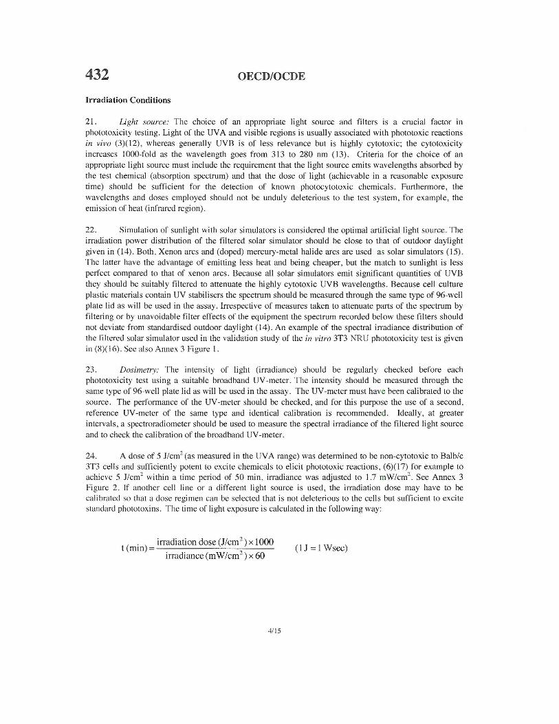
21 . Light source: The choice of an appropriate light source and filters is a crucial factor in phototoxicity testing. Light of the UV A and visible regions is usually associated with phototoxic reactions in vivo (3)(12), whereas generally UVB is of less relevance but is highly cytotoxic; the cytotoxicity increases 1000-fold as the wavelength goes from 313 to 280 nm (13). Criteria for the choice of an appropriate light source must include the requirement that the light source emits wavelengths absorbed by the test chemical (absorption spectrum) and that the dose of light (achievable in a reasonable exposure time) should be sufficient for the detection of known photocytotoxic chemicals. Furthermore, the wavelengths and doses employed should not be unduly deleterious to the test system, for example, the emission of heat (infrared region).
22. Simulation of sunlight with solar simu lators is considered the optimal artificial light source. The irradiation power distribution of the filtered solar simulator should be close to that of outdoor daylight given in (14). Both , Xenon arcs and (doped) mercury-metal halide arcs are used as solar simulators (15). The latter have the advantage of emitting less heat and being cheaper, but the match to sunlight is less perfect compared to that of xenon arcs. Because all solar simulators emit significant quantities of UVB they should be suitably filtered to attenuate the highly cytotoxic UVB wavelengths. Because cell culture plastic materials contain UV stabilisers the spectrum should be measured through the same type of 96-well plate lid as will be used in the assay . Irrespective of measures taken to attenuate parts of the spectrum by filtering or by unavoidable filter effects of the equipment the spectrum recorded below these filters should not deviate from standardised outdoor daylight (14). An example of the spectral in·adiance distribution of the filtered solar simulator used in the validation study of the in vitro 3"1'3 NRU phototoxicity test is given in (8)( 16). Sec also Annex 3 Figure I.
23. Dosimetry: The intensity of light (irradiance) should be regularly checked before each phototoxicity test using a suitable broadband UV-meter. T he intensity should be measured through the same type of 96-well plate lid as will be used in the assay. The UV -meter must hav e been calibrated to the source. The performance of the UV-meter should be checked, and for this purpose the use of a second, reference UV-meter of the same type and identical calibration is recommended. Ideally, at greater intervals, a spectroradiometer should be used to measure the spectral irradiance of the filtered light source and to check the calibration of the broadband UV -meter.
24. A dose of 5 J/cm2 (as measured in the UVA range) was determined to be non-cytotoxic to Balb/c 3T3 cells and sufficiently potent to excite chemicals to elicit phototoxic reactions, (6)(17) for example to achieve 5 J/cm2 within a time period of 50 min, irradiance was adjusted to 1.7 rnW/crn 2
. See Annex 3 Figure 2. If another cell line or a different light source is used, the irradiation dose may have to be calibrated so that a dose regimen can be selected that is not deleterious to the cells but sufficient to excite standard phototoxins. T he time of light exposure is calculated in the following way:
. ) irradiation dose (J/cm 2 ) x 1000
t (mtn = ? (I J = I Wsec)irradiance(mW/cm-) x 60
4115

OECD/OCDE 432 Test conditions
Test substance concentrations
25. The ranges of concentrations of a chemical tested in the presence ( +lrr) and in the absence ( -lrr) of light should be adequately determined in dose range-finding experiments . It may be useful to assess solubility initially and at 60 min (or whatever treatment time is to be used), as solubility can change during time or during the course of exposure. To avoid toxicity induced by improper culture conditions or by highly acidic or alkaline chemicals, the pH of the cell cultures with added test chemical should be in the range 6.5 - 7.8.
26. The highest concentration of the test substance should be within physiological test conditions, for example osmotic and pH stress should be avoided. Depending on the test chemical, it may be necessary to consider other physico-chemical properties as factors limiting the highest test concentration. For relatively insoluble substances that are not toxic at concentrations up to the saturation point the highest achievable concentration should be tested . In general, precipitation of the test chemical at any of the test concentrations should be avoided. The maximum concentration of a test substance should not exceed 1000 ~g/mL; osmolality should not exceed I 0 mmolar. A geometric dilution series of 8 test substance concentrations with a constant dilution factor should be used (sec paragraph 47).
27. If there is information (from a range finding experiment) that the test chemical is not cytotoxic up to the limit concentration in the dark experiment (-lrr), but is highly cytotoxic wht:n irradiated (+lrr), the concentration ranges to be selected for the ( +Irr) experiment may differ from those selected for the (-lrr) experiment to fulfil the requirement of adequate data quality.
Controls
28. Radiation senslf!Vlty of the cells, establishing of historical data: Cells should be checked regularly (about every fifth passage) for sensitivity to the light source by assessing their viability following exposure to increasing doses of irradiation. Several doses of irradiation, including levels substantially greater than those used for the 3T3 NRU phototoxicity test should be used in this assessment. These closes are easiest quantitated by measurements of UY parts of the light source. Cells arc seeded at the density used in the in vitro 3T3 NRU phototoxicity test and irradiated the next clay. Cell viability is then determined one clay later using Neutral Reel uptake. It should be demonstrated that the resulting highest non-cytotoxic close (e.g. in the validation study: 5 J/cm 2 [UVAJ) was sufficient to classify the reference chemicals (Table I) correctly.
29 . Radiation sensitivity, check of current test: The test meets the quality criteria if the irradiated negative/solvent controls show a viability of more than 80% when compared with non-irradiated negative/solvent control.
30. Viability of solvent controls: The absolute optical density (00540 NRu) of the Neutml Reel extracted from the solvent controls indicates whether the l.xl04 cells seeded per well have grown with a normal doubling time during the two days of the assay. A test meets the acceptance criteria if the mean 00540 NRU of the untreated controls is ;::: 0.4 (i.e. approximately twenty times the background solvent absorbance).
31. Positive control: A known phototoxic chemical shall be tested concurrently with each in vitro 3T3 NRU phototoxicity test. Chlorpromazine (CPZ) is recommended. For CPZ tested with the standard
5/15

432 OECD/OCDE
protocol in the in vitro 3T3 NRU phototoxicity test, the following test acceptance criteria were defined: CPZ irradiated (+lrr): IC50 = 0.1 to 2.0 )..lg/ml, CPZ non-irradiated (-lrr): IC50 = 7.0 to 90.0 ~tg/mL. T he Photo Irritation Factor (PIF), should be > 6. T he historical performance of the positive control should be monitored.
32. Other phototoxic chemicals, suitable for the chemical class or solubility characteristics of the chemical being evaluated, may be used as the concurrent positive controls in place of chlorpromazine.
Test procedure (6)(7)(8)(16)(17):
lst day:
33. Dispense 100 )..lL culture medium into the peripheral wells of a 96-well tissue culture microliter plate(= blanks). In the remaining wells, dispense 100 J..lL of a cell suspension of l xl05 cells/mL in culture medium(= I x 104 cells/well). Two plates should be prepared for each series of individual test substance concentrations, and for the solvent and positive controls.
34. Incubate cells for 24 h (see paragraph 12) until they form a half confluent monolayer. This incubation period allows for cell recovery, adherence, and exponential growth.
2nd day:
35. After incubation, decant culture medium from the cells and wash gently with 150 J..lL of the buffered solution used for incubation . Add 100 ~LL of the buffer containing the appropriate concentration of tes t chemical or solvent (solvent con tro l). Apply 8 different concentrations of the test chemical. Incubate cells with the test substance in the dark for 60 minutes (sec paragraphs 12 and 17).
36. From the two plates prepared for each series of test substance concentrations and the controls, one is selected, generally at random, for the determination of cytotoxicity (-lrr) (i.e., the control plate), and one (the treatment plate) for the determination of photocytotoxicity (+lrr).
37. To perform the +lrr exposure, irradiate the cells at room temperature for approximately 50 minutes through the lid of the 96-wcll plate with the hi ghest dose of radiation that is non-cytotoxic (see also Annex 3). Keep non-irradiated plates (-In·) at room temperature in a dark box for 50 min(= light exposure time).
38. Decant test solution and carefully wash twice with 150 J..lL of the buffered solution used for incubation, but not containing the test material. Replace the buffer with culture medium and incubate (sec paragraph 12) overnight (18-22 h) .
3rd day:
Microscopic evaluation
39 . Cells should be examined for growth , morphology, and integrity of the monolayer using a phase contrast microscope. Changes in cell morphology and effects on cell growth should be recorded.
6115

OECD/OCDE 432
Neutral Red Uptake test
40. Wash the cells with 150 1--!L of the pre-warmed buffer. Remove the was hing solution by gentle tapping . Add 100 1--!L of a 50 pg/mL Neutral Red (NR) (3-amino-7-dimethylamino-2-methylphenazine hydrochloride, CAS number 553-24-2; C.!. 50040) in medium without serum (16) and incubate as described in paragraph 12, for 3 h.
41 . After incubation, remove the NR medium, and wash cells with 150 1--!L of the buffer. Decant and remove excess buffer by blotting or centrifugation .
42. Add exactly !50 1--!L NR desorb solution (freshly prepared 49 parts water+ 50 part~ ethanol+ I part acetic acid) .
43. Shake the microliter plate gently on a microliter plate shaker for lO min until NR has been extracted from the cells and has formed a homogeneous solution .
44. Measure the optical density of the NR extract at 540 nm in a spectrophotometer, using blanks as a reference . Save data in an appropriate electronic file format for subsequent analysis .
DATA AND REPORTI NG
Quality and quantity of data
45. The test data should allow a meaningful analysis of the concentration-response obtained in the presence and in the absence of irradiation, and if possible the concentration of test chemical by which cell viability is reduced to 50% (IC50). If cytotoxicity is found, both the concentration range and the intercept of individual concentrations shall be set in a way to allow the fit of a curve to the experimental data .
46. For both clearly positive and clearly negative results (see paragraph 53), the primary experiment, supp01ted by one or more preliminary close range-finding experiment(s) , may be sufficient.
47 . Equivocal, borderline, or unclear results should be clarified by further testing (see also paragraph 56). In such cases, modification of expe1imental conditions should be considered. Experimental conditions that might be modified include the concentration range or spacing, the pre-incubation time, and the irradiation-exposure time . A shorter exposure time may be appropriate for water-unstable chemicals .
Evaluation of results
48. To enable evaluation of the data, a Photo-Irritation-Factor (PIF) or Mean Photo Effect (MPE) may be calculated.
49 . For the calculation of the measures of photocytotoxicity (see below) the set of discrete doseresponse values has to be approximated by an appropriate continuous dose-response curve (model ) . Fitting of the curve to the data is commonly performed by a non-linear regression met hod (18). To assess the inlluence of data variabil ity on the fitted curve a bootstrap procedure is recommended.
7115

432 OECD/OCDE
50 . A Photo-Irritation-Factor (P I F) is calculated using the following fommla:
PIF = IC50(-Irr) ICso( +lrr)
If an IC50 in the presence or absence of ligh t cannot be calculated, a PIF cannot be determined for the test material .
51 . The mean photo effect (MPE) is based on comparison of the complete concentration response curves ( 19) . It is defined as the weighted average across a representative set of photo effect values
11"'w. PE~ l ~i MPE = ..:..'--'-1 --
The photo effect (PEe) at any concentration (C) is defined as the product of the response effect (REc) and the dose effect (DEc) i.e. PEe = REc x DEc. The response effect (REc) is the difference between the responses observed in the absence and presence of light, i.e . REc = Rc (-Irr) - Rc (+Irr). The dose-effect is given by
C/C * -1~ ICIC *+ 1
where C* represents the equivalence concentration, i.e. the concentration at which the +Irr response equals the -lrr response at concentration C. If C* cannot be detem1ined because the response values of the +Irr curve are systematically higher or lower than Rc( -Irr) the dose effect is set to 1. The weighting factors w; arc given by the highest response value, i.e . w; =MAX {Ri (+lrr), Ri (-Irr)}. The concentration grid Ci is chosen such that the same number of points falls into each of the concentration intervals defined by the concentration values used in the experiment. The calculation of MPE is restricted to the maximum concentration value at which at least one of the two curves still exhibits a response value of at least 10%. If this maximum concentration is higher than the highest concentration used in the +Irr experiment the residual part of the +Irr curve is set to the response value "0". Depending on whether the MPE value is larger than a properly chosen cut-off value (MPEc = 0 .15) or not, the chemical is classified as phototoxic.
52. A software package for the calculation of the PIF and MPE is available from the Secretariat (20).
Interpretation of Results
53 . Based on the validation study (8), a test substance with a PIF < 2 or an MPE < 0.1 predicts: "no phototoxicity". A PIF >2 and< 5 or an MPE > 0.1 and< 0.15 predicts: "probable phototoxicity" and a PIF > 5 or an MPE > 0.15 predicts: "phototoxicity" .
54. For any laboratory initially establishing this assay, the reference materials listed in Table should be tested prior to the testing of test substances for phototoxicity. PIF or MPE values should be close to the values mentioned in Table 1.
8/15
I
OECD/OCDE 432
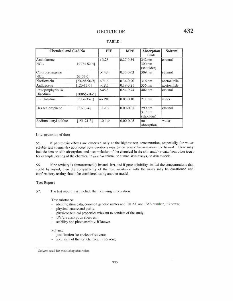
TABLE 1
Chemical and CAS No PIF MPE Absorption Solvent' Peak
Amiodarone >3.25 0.27-0.54 242nm ethanol HCL 119774-82-41 300nm
(shoulder) Chloropromazine >14 .4 0.33-0.63 309nm ethanol HCL 169-09-01 Norlloxacin 170458-96-71 >71.6 0.34-0.90 316 nm acetonitrile Anthracene 1120-12-71 >18.5 0.19-0.81 356nm acetonitrile Protoporphyrin IX, >45.3 0.54-0.74 402nm ethanol Disodium 150865-01 -51
L- Histidine [7006-35-11 no PIF 0.05-0.10 211 nm water
Hexachlorophene 170-30-41 1.1-1.7 0.00-0.05 299nm ethanol 317 nm (shoulder)
Sodium Iaury! sulfate [151-21-31 1.0-1.9 0 .00-0.05 no water absorption
Interpretation of data
55. If phototoxic effects are observed only at the highest test concentratio n, (especially for water soluble test chemicals) additional considerations may be necessary for assessment of hazard. These may include data on skin absorption, and accumulation of the chemical in the skin and I or data from other tests, for example, testing of the chemical in in vitro animal or human ski n assays, or skin models .
56. If no toxicity is demonstrated (+lrr and -Irr), and if poor solubility limited the concentrations that could be tested, then the compatibility of the test substance with the assay may be questioned and confirmatory testing should be considered using another model.
Test Report
57. The test report must include the following information:
Test substance: - identification data, common generic names and IUPAC and CAS number, if known ; - physical nature and purity; - physicochemical properties relevant to conduct of the study;
UY/vis absorption spectrum; - stability and photostability, if known.
Solvent: - justification for choice of solvent; - solubility of the test chemical in solvent;
1 Solvent used for measuring absorption
9/ 15

432 OECD/OCDE
percentage of solvent present in treatment medium.
Cells: - type and source of cells;
absence of mycoplasma; cell passage number, if known; Radiation sensitivity of cells , detem1ined wi th the irradiation equipment used in the in vitro 3T3 NRU phototoxicity test.
Test conditions (I); incubation before and after treatment: type and composition of culture medium; incubation conditions (C02 concentration; temperature; humidity); duration of incubation (pre-treatment; post-treatment).
Test conditions (2) ; treatment with the chemical: rationale for selection of concentrations of the test chemical used in the presence and in the absence of inadiation ; in case of limited solubility of the test chemical and absence of cytotoxicity: rationale for the highest concentration tested; type and composition of treatment medium (buffered salt solution) ; duration of the chemical treatment.
Test conditions (3); irradiation : rationale for selection of the light source used; manufacturer and type of light source and radiometer ;
- spectral irradiance characteristics of the light source ; transmission and absorption characteristics of the fi lter(s) used; characteristics of the radiometer and details on its calibration; distance of the light source from the test system; UVA iiTadiance at this distance , expressed in m W/cm2
;
duration of the UV/vis light exposure; UVA dose (irradiance x time), expressed in J/cm2
;
temperature of cell cultures during irradiation and cell cultures concurrently kept in the dark.
Test conditions (4); Neutral Red viability test: composition of Neutral Red treatment medium; duration of Neutral Red incubation ; incubation conditions (C02 concentration ; temperature; humidity); Neutral Red extraction condi tion s (extractant; duration); wavelength used for spectrophotometric reading of Neutral Red optical density ; second wavelength (reference), if used ; content of spectrophotometer blank, if used.
Results: cell viability obtained at each concentration of the test chemical, expressed in percent viability of mean , concunent solvent controls;
- concentration response curves (test chemical concentration vs. relative cell viability) obtained in concurrent +hT and -Irr experiments;
- analysis of the concentration-response curves: if possible, computation/calculation of IC50
10115

OECD/OCDE 432 ( +lrr) and IC50 ( -lrr);
comparison of the two concentration response curves obtained in the presence and in the absence of irradiation, either by calculation of the Photo-Inhibition-Factor (PIF), or by calculation of the Mean-Photo-Effect (MPE);
test acceptance criteria; concurrent solvent control ; absolute viability (optical density of Neutral Red extract) of irradiated and non-irradiated cells; historic negative and solvent control data; means and standard deviations; test acceptance criteria; concurrent positive control ; !Cso(+lrr) and IC50( -lrr) and PIF/MPE of positive control chemical; historic positive control chemical data: IC50( +Irr) and IC50( -Irr) and PIF/MPE; means and standard deviations.
Discussion of the results.
Conclusions.
LITER ATURE
(I) Lovell W .W. (1993). A scheme for in vitro screening of substances for photoallergenic potential. Toxic . In Vitro 1: 95-102.
(2) Santamaria, L. and Prino , G. (1972). List of the photodynamic substances. In "Research Progress in Organic, Biological and Medicinal Chemistry" Vol. 3 part I . North Holland Publishing Co. Amsterdam . p XI-XXXV.
(3) Spielmann, H., Lovell, W .W ., Holzle, E., Johnson, B .E., Maurer, T., Miranda, M.A ., Pape, W J .W ., Sapora, 0 ., and Sladowski, D. (1994). In vitro phototoxicity testing: The report and recommendations of ECVAM Workshop 2. ATLA , 22 . 314-348.
(4) Spikes , J.D . (1989). Photosensitization . In "The science of Photobiology" Edited by K.C. Smith . Plenum Press, New York . 2nd edition, p 79- 110.
(5) OECD ( 1997) Environmental Health and Safety Publications, Series on Testing and Assessment No.7 "Guidance Document On Direct Phototransformation Of Chemicals In Water" Environment Directorate, OECD , Paris .
(6) Spielmann, H ., Balls, M ., m-iring , B ., Holzhi.itter, H.G ., Kalweit, S ., Klecak, G., L'Eplattenier, H., Liebsch, M., Lovell, W.W., Maurer, T., Moldenhauer. F. Moore. L., Pape, W., Pfannbecker, U., Potthast, .J ., De Silva, 0., Steiling, W ., and Will shaw, A. (1994). EEC/COLI PA project on in vitro phototoxicity testing: First results obtained with a Balb/c 3T3 cell phototoxicity assay. Toxic . In Vitro lL 793-796 .
(7) Anon (1998). Statement on the scientific validity of the 3T3 NRU PT test (an in vitro test for phototoxicity), European Commission, Joint Research Centre: ECVAM and DGXI/E/2, 3 November 1997, ATLA, 22., 7-8.
I 1115

432 OECD/OCDE
(8) Spielmann, H., Balls, M., Dupuis, J., Pape, W J .W ., Pechovitch , G. De Silva, 0 ., Holzhiitter, H.G., Clothier, R., Desolle, P., Gerberick, F., Liebsch, M., Lovell, W.W ., Maurer, T., Pfannenbecker, U., Potthast, J. M ., Csato, M., Sladowski, D ., Stciling, W., and Brantom, P. (1998) . The international EU/COLIPA In vitro phototoxicity validation study: results of phase II (blind trial), part 1: the 3T3 NRU phototoxicity test. Toxic . In Vitro _12, 305-327.
(9) OECD (2002) Extended Expert Consultation Meeting on The In Vitro 3T3 NRU Phototoxicity Test Guideline Proposal, Berlin, 30'"- 31 '' October 2001, Secretariat's Final Summary Report, 15'11 March 2002, OECD ENV/EHS, available upon request from the Secretariat.
(I 0) Borenfreund, E ., and Puern er, J .A . ( 1985) . Toxicity determination in alterations and neutral red absorption . Toxicology Lett., 21, 119-124.
vitro by morphological
(I I ) Hay, R ..J . (1988) The seed stock concept and quality control for cell lines. Analytical Biochemistry 171' 225-237.
(12) Lambert LA, Warner W.G., and Kornhauser A . (1996) Animal models for phototoxicity testing. In "Dermatotoxicology", edited by F.N. Marzulli and H.l. Maibach . Taylor & Francis, Washington DC. 5th Edition, p 515 -530 .
(13) T yrrell R.M., Pidoux M (1987) Action spectra for human skin cells: estimates of the relative cytotoxicity of the middle ultraviolet, near ultraviolet and violet regions of sunlight on epidermal keratinocytes . Cancer Res., :!1, 1825-1829.
(14) ISO 10977 . (1993). Photography - Processed photographic colour films and paper prints - Methods for measuring image stability.
(15) Sunscreen Testing (UV .B) TECHNICALREPORT, CIE, International Commission on Illumnation, Publication No. 90, Vienna, 1993, ISBN 3 900 734 275.
(16) ZEBET/ ECVAM/COLIPA - Standard Operating Procedure: In Vitro 3T3 NR U Phototoxicity Test . Final Ve rsi on , 7 September, 1998. 18 pgs.
(17) Spielmann, H., Balls, M ., Dupuis, J., Pape , W J .W., De Silva, 0., Holzhiitter, H.G., Gerberick, F., Liebsch, M ., Lovell, W.W., and Pfannenbecker, U. (1998) A study on UV filter chemicals from Annex VII of the Europc<m Union Directive 761768/EEC. in the in vitro 3'1'3 NRU phototoxicity test. AT LA 26,679708.
(18) Holzhiitter, H.G., and Quedenau, .1. (1995) Mathematical modeling of cellular responses to external signals. J. Bioi. Systems}, 127-138 .
(19) Holzhiittcr, H .G . ( 1997) . A general measure of in vitro phototoxicity derived from pairs of doseresponse curves and its use for predicting the in vivo phototoxicity of chemicals . ATLA, 25,445462 .
(20) hnu://www .oecQ_,_g_rgL<jocument/55/Cl.?.140,cn 2649 34377 2349687 l I I l.OO .html
12115

OECD/OCDE 432 ANNEXl
DEFINITI ONS
lrradiance: the intensity of ultraviolet (UV) or visible light incident on a surface , measured in W/m~ or mW/cm~ .
Dose of light: the quantity(= intensity x time) of ultraviolet (UV) or visible radiation incident on a surface, expressed in Joules(= W x s) per surface area, for example, J/m~ or J/cm2
•
UV light wavebands: the designations recommended by the CIE (Commission lnternationale de L' Eclairage) are: UVA (315-400nm) UVB (280-3 15nm) and UVC (100-280nm) . Other designations are also used; the division between UVB and UVA is often placed at 320nm, and the UVA may be divided into UV-A I and UV -A2 with a division made at about 340nm .
Cell viability: parameter measuring total activity of a cell population (e .g., uptake of the vital dye Neutral Reel into cellular lysosom es), which , depending on the endpoint measured an d the test design used, correlates with the total number and/or vitality of the cells.
Relative cell viability : cell viability expressed in relation to solvent (negative) controls which have been taken through the whole test procedure (either +lrr or -Irr) but not treated with test chemical.
PIF (Photo-Irri tation-Factor): fac tor generated by comparing two equally effective cytotoxic concentrations (IC50) of the test chemical obtained in the absence (-lrr) and in the presence (+lrr) of a non-cytotoxic irradiation wi th UVA/vis light.
IC2i!: the concentration of the test chemical by which the cell viability is reduced by 50%
MPE (Mean -Photo-Effect): measurement derived from mathematical analysis of the concentration response curves obtained in the absence ( -Irr) and in the presence ( +lrr) of a non-cytotoxic irradiation with UV A/vis light.
Phototoxieity: acute toxic response that is elicited after the first exposure of skin to certain chemicals and su bsequent exposure to light, or that is ind uced similarly by skin irradiation after systemic admi nistration of a chemical.
13115

Initial Evaluation of the Physical, Chemical , and Toxicological Properties of the Test Substance • Physico-chemical properties • Chemical structure , structural alerts • UV/vis- absorpti on • QSAR - photochemistry • General toxicity (including kinetics and metabolism)
1 No
UY/vis Absorptionabsorption spectra
in appropriate solvent (e.g . OECD
TG 101)
Phototoxicity Testing not considered
necessary
In Vitro 3T3 NRU Phototoxicity Test and/or other methods if necessary
432 OECD/OCDE
ANNEX2
Role of the Jf3 NRU Pr in a sequential approach to the phototoxicity testing of chemicals
14/ 15

OECD/OCDE 432
ANNEX 3
Figure 1: Spectral power distribution of a filtered solar simulator
lrradiancc jmW/cm2 j100
UVB UVA visible light
10
0,1
0,01
0,001
-SOL 500 i H2 Filter
SOL 500 i H1 Filter
-~ - -- SOL 500 iH1 Filter+ lid of 96-well plate
250 300 350 400 450 500 550 600 650 700
Wavelength jnmj
Figure 2: Irradiation sensiti vity of Balb/c 3T3 cells (as measured in the UVA range)
cell viability [% Neutral Red uptake of dark controls]
120
60
40
0 I 1 II I r · 1 -1
0 50 100 150 200
irradition time [minutes] (10 min= 1 Joule UVA/cm2)
15/15
(see paraf?raph 22)
Figure I gives an example of an acceptable spectral power dis tribution of a filtered solar simulator. It is from the doped metal halide source used in the val idation trial of the 3T3 NRU PT (6)(8)(17). The effect of t wo different filters and the additional filtering effect of the lid of a 96-well cell culture plate are shown. The H2 filter was only used with test systems that can tolerate a higher amoun t of UVB (skin model test and red blood cell photo-hemolysis test). In the 3T3 NRU-PT the HI filter was used. The figure shows that additional filtering effe ct of the plate lid is mainly observed in the UVB range, still leaving enough UVB in the irrad iation spectrum to excite chemicals typically absorbing in the UVB range. like Amiodarone (see Table 1).
(see paraKraphs 24, 28. 29)
Sensitivity of Balb/c 3T3 cells to irradiation with the solar simu lator used in the validation trial of the 3T3NRU-Phototoxicity Test, as mea~ured in the UVA range. Figure sho ws the results obtained in 7 different laboratories in the prevalidation study (1). While the two curves with open symbols were obtained with aged cells (high number of passages), that had to be rep I aced by new cell stocks the curves with bold symbols show cells with acceptable irradiation tolerance. From these data the highest non-cytotoxic irradiation dose of 5 J/cm2 was derived (vertical dashed line). The horizontal dashed line shows in addition the maximum acceptable irradiation effect given in paragraph 29 .


![Increased Growth of NIH/3T3 Cells by Transfection with Human pi … · [CANCER RESEARCH 52. 428-436. January 15. 1992] Increased Growth of NIH/3T3 Cells by Transfection with Human](https://img.dokumen.tips/doc/110x75/60b9042eb86d34336e7964ac/increased-growth-of-nih3t3-cells-by-transfection-with-human-pi-cancer-research.jpg)