Embed Size (px)
Citation preview

Temporal and Infratemporal Fossa Head and Neck anatomy dr. Ahmed Almusawi
Descriptive anatomy of temporal and infratemporal fossa with explaining the bony
framework of these fossa and describing the contents of each fossae including the nerves,
blood vessels and muscles

Temporal and Infratemporal Fossa Head and Neck Anatomy
1
TEMPORAL AND INFRATEMPORAL FOSSAE The temporal and infratemporal fossae are interconnected spaces on the lateral side of the head. Their boundaries are formed by bone and soft tissues. The temporal fossa is superior to the infratemporal fossa, above the zygomatic arch, and communicates with the infratemporal fossa below through the gap between the zygomatic arch and the more medial surface of the skull. The infratemporal fossa is a wedge-shaped space deep to the masseter muscle and the underlying ramus of the mandible. Structures that travel between the cranial cavity, neck, pterygopalatine fossa, and floor of the oral cavity, floor of the orbit, temporal fossa, and superficial regions of the head pass through it.
Figure 1: the temporal and infratemporal fossa Relation to the four muscles of mastication (masseter, temporalis, medial pterygoid, and lateral pterygoid) that move the lower jaw at the temporomandibular joint,
Masseter is lateral to the infratemporal fossa two (medial and lateral pterygoid) are in the infratemporal fossa, One fills the temporal fossa.

Temporal and Infratemporal Fossa Head and Neck Anatomy
2
Bony framework Bones that contribute significantly to the boundaries of the temporal and infratemporal fossae include the temporal, zygomatic, and sphenoid bones, and the maxilla and mandible. Parts of the frontal and parietal bones are also involved. Temporal bone The squamous part of the temporal bone forms part of the bony framework of the temporal and infratemporal fossae. The tympanic part of the temporal bone forms the posteromedial corner of the roof of the infratemporal fossa, and also articulates with the head of the mandible to form the temporomandibular joint. Sphenoid bone The parts of the sphenoid bone that form part of the bony framework of the infratemporal fossa are the lateral plate of the pterygoid process and the greater wing. The greater wing also forms part of the medial wall of the temporal fossa. The greater wings extend one on each side from the body of sphenoid. They project laterally from the body and curve superiorly. The inferior and lateral surfaces form the roof of the infratemporal fossa and the medial wall of the temporal fossa, respectively. The sharply angled boundary between the lateral and inferior surfaces of the greater wing is the infratemporal crest. Two apertures the (foramen ovale and the foramen spinosum) pass through the base of the greater wing and allow the mandibular nerve [V3] and the middle meningeal artery, respectively, to pass between the middle cranial fossa and infratemporal fossa. In addition, one or more small sphenoidal emissary foramina penetrate the base of the greater wing anteromedial to the foramen ovale and allow emissary veins to pass between the pterygoid plexus of veins in the infratemporal fossa and the cavernous sinus in the middle cranial fossa. Maxilla The posterior surface of the maxilla contributes to the anterior wall of the infratemporal fossa. This surface is marked by a foramen for the posterior superior alveolar nerve and vessels. The superior margin forms the inferior border of the

Temporal and Infratemporal Fossa Head and Neck Anatomy
3
inferior orbital fissure. Zygomatic bone The zygomatic bone is a quadrangular-shaped bone that forms the palpable bony prominence of the cheek:
a maxillary process extends anteromedially to articulate with the zygomatic process of the maxilla;
a frontal process extends superiorly to articulate with the zygomatic process of the frontal bone;
a temporal process extends posteriorly to articulate with the zygomatic process of the temporal bone to complete the zygomatic arch.
One or more small foramina on the temporal fossa surface of the plate where it attaches to the frontal process are for terminal branches of the zygomaticotemporal nerve. Ramus of the mandible The ramus of the mandible is quadrangular in shape and has medial and lateral surfaces and condylar and coronoid processes The lateral surface of the ramus of mandible is generally smooth except for the presence of a few obliquely oriented ridges. Most of the lateral surface provides attachment for the masseter muscle. The temporal fossa is a narrow fan-shaped space that covers the lateral surface of the skull Boundaries
its upper margin is defined by a pair of temporal lines that arch across the skull from the zygomatic process of the frontal bone to the supramastoid crest of the temporal bone;
it is limited laterally by the temporal fascia, which is a tough fan-shaped aponeurosis overlying the temporalis muscle and attached by its outer margin to the superior temporal line and by its inferior margin to the zygomatic arch;
anteriorly, it is limited by the posterior surface of the frontal process of the

Temporal and Infratemporal Fossa Head and Neck Anatomy
4
zygomatic bone and the posterior surface of the zygomatic process of the frontal bone, which separate the temporal fossa behind from the orbit in front;
its inferior margin is marked by the zygomatic arch laterally and by the infratemporal crest of the greater wing of the sphenoid medially between these two features, the floor of the temporal fossa is open medially to the infratemporal fossa and laterally to the region containing the masseter muscle.
Contents 1. The major structure in the temporal fossa is the temporalis muscle.
Figure 2: Temporal and infratemporal boundaries
2. Deep temporal nerves The deep temporal nerves, usually two in number, originate from the anterior trunk of the mandibular nerve [V3] in the infratemporal fossa. They pass superiorly and around the infratemporal crest of the greater wing of the sphenoid to enter the temporal fossa deep to the temporalis muscle, and supply the temporalis muscle.
3. Zygomaticotemporal nerve
The zygomaticotemporal nerve is a branch of the zygomatic nerve. The zygomatic nerve

Temporal and Infratemporal Fossa Head and Neck Anatomy
5
is a branch of the maxillary nerve [V2], which originates in the pterygopalatine fossa. The zygomaticotemporal nerve enters the temporal fossa through one or more small foramina on the temporal fossa surface of the zygomatic bone. Branches of the zygomaticotemporal nerve pass superiorly between the bone and the temporalis muscle to penetrate the temporal fascia and supply the skin of the temple 4. Deep temporal arteries
Normally two in number, these vessels originate from the maxillary artery in the infratemporal fossa and travel with the deep temporal nerves around the infratemporal crest of the greater wing of the sphenoid to supply the temporalis muscle. They anastomose with branches of the middle temporal artery. 5. Middle temporal artery
The middle temporal artery originates from the superficial temporal artery just superior to the root of the zygomatic arch between this structure and the external ear. It penetrates the temporalis fascia, passes under the margin of the temporalis muscle, and travels superiorly on the deep surface of the temporalis muscle. The middle temporal artery supplies temporalis and anastomoses with branches of the deep temporal arteries.
Figure 3: contents of temporal fossa

Temporal and Infratemporal Fossa Head and Neck Anatomy
6
Infratemporal fossa
Figure 4: infratemporal fossa boundaries and contents The wedge-shaped infratemporal fossa is inferior to the temporal fossa and between the ramus of the mandible laterally and the wall of the pharynx medially. It has a roof, a lateral wall, and a medial wall, and is open to the neck posteroinferiorly
Boundaries the roof is formed by the inferior surfaces of the greater wing of the sphenoid
and the temporal bone, contains the foramen spinosum, foramen ovale, and the petrotympanic fissure, and lateral to the infratemporal crest of the greater wing of the sphenoid, is open superiorly to the temporal fossa;
the lateral wall is the medial surface of the ramus of the mandible, which contains the opening to the mandibular canal;
the medial wall is formed anteriorly by the lateral plate of the pterygoid process and more posteriorly by the pharynx and by two muscles of the soft palate (tensor and levator veli palatini muscles), and contains the pterygomaxillary fissure anteriorly, which allows structures to pass between the infratemporal and pterygopalatine fossae;
Temporal and Infratemporal Fossa Head and Neck Anatomy
7
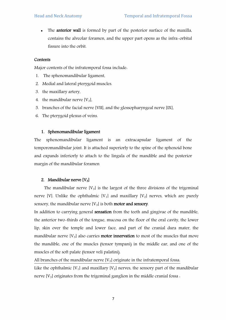
The anterior wall is formed by part of the posterior surface of the maxilla, contains the alveolar foramen, and the upper part opens as the infra-orbital fissure into the orbit.
Contents Major contents of the infratemporal fossa include: 1. The sphenomandibular ligament, 2. Medial and lateral pterygoid muscles. 3. the maxillary artery, 4. the mandibular nerve [V3], 5. branches of the facial nerve [VII], and the glossopharyngeal nerve [IX], 6. The pterygoid plexus of veins.
1. Sphenomandibular ligament
The sphenomandibular ligament is an extracapsular ligament of the temporomandibular joint. It is attached superiorly to the spine of the sphenoid bone and expands inferiorly to attach to the lingula of the mandible and the posterior margin of the mandibular foramen
2. Mandibular nerve [V3] The mandibular nerve [V3] is the largest of the three divisions of the trigeminal nerve [V]. Unlike the ophthalmic [V1] and maxillary [V2] nerves, which are purely sensory, the mandibular nerve [V3] is both motor and sensory. In addition to carrying general sensation from the teeth and gingivae of the mandible, the anterior two-thirds of the tongue, mucosa on the floor of the oral cavity, the lower lip, skin over the temple and lower face, and part of the cranial dura mater, the mandibular nerve [V3] also carries motor innervation to most of the muscles that move the mandible, one of the muscles (tensor tympani) in the middle ear, and one of the muscles of the soft palate (tensor veli palatini). All branches of the mandibular nerve [V3] originate in the infratemporal fossa. Like the ophthalmic [V1] and maxillary [V2] nerves, the sensory part of the mandibular nerve [V3] originates from the trigeminal ganglion in the middle cranial fossa :

Temporal and Infratemporal Fossa Head and Neck Anatomy
8
the sensory part of the mandibular nerve [V3] drops vertically through the foramen ovale and enters the infratemporal fossa between the tensor veli palatini muscle and the upper head of the lateral pterygoid muscle;
the small motor root of the trigeminal nerve [V] passes medial to the trigeminal ganglion in the cranial cavity, then passes through the foramen ovale and immediately joins the sensory part of the mandibular nerve [V3].
Branches Soon after the sensory and motor roots join, the mandibular nerve [V3] gives rise to a small meningeal branch and to the nerve to medial pterygoid, and then divides into anterior and posterior trunks:
branches from the anterior trunk are the buccal, masseteric, and deep temporal nerves, and the nerve to lateral pterygoid, all of which, except the buccal nerve (which is predominantly sensory) are motor nerves;
Branches from the posterior trunk are the auriculotemporal, lingual, and inferior alveolar nerves, all of which, except a small nerve (nerve to mylohyoid) that branches from the inferior alveolar nerve, are sensory nerves.
Meningeal branch The meningeal branch originates from the medial side of the mandibular nerve [V3] and ascends to leave the infratemporal fossa with the middle meningeal artery and re-enter the cranial cavity through the foramen spinosum. It is sensory for the dura mater, mainly of the middle cranial fossa, and also supplies the mastoid cells that communicate with the middle ear. Nerve to medial pterygoid The nerve to medial pterygoid also originates medially from the mandibular nerve [V3]. It descends to enter and supply the deep surface of the medial pterygoid muscle. Near its origin from the mandibular nerve [V3], it has two small branches:
one of these supplies the tensor veli palatini;

Temporal and Infratemporal Fossa Head and Neck Anatomy
9
Figure 5: mandibular nerve and its branches
the other ascends to supply the tensor tympani muscle, which occupies a small bony canal above and parallel to the pharyngotympanic tube in the temporal
Buccal nerve The buccal nerve is a branch of the anterior trunk of the mandibular nerve [V3]. It is predominantly a sensory nerve, but may also carry the motor innervation to the lateral pterygoid muscle and to part of the temporalis muscle. The buccal nerve passes laterally between the upper and lower heads of lateral pterygoid and then descends around the anterior margin of the insertion of temporalis muscle to the anterior margin of the ramus of mandible, often slipping through the tendon of temporalis. It continues into the cheek lateral to the buccinator muscle to supply general sensory nerves to the adjacent skin and oral mucosa and the buccal gingivae of the lower molars. Masseteric nerve The masseteric nerve is a branch of the anterior trunk of the mandibular nerve [V3]. It passes laterally over the lateral pterygoid muscle and through the mandibular notch to penetrate and supply the masseter muscle.

Temporal and Infratemporal Fossa Head and Neck Anatomy
10
Deep temporal nerves The deep temporal nerves, usually two in number, originate from the anterior trunk of the mandibular nerve [V3]. They pass laterally above the lateral pterygoid muscle and curve around the infratemporal crest to ascend in the temporal fossa and supply the temporalis muscle from its deep surface. Nerve to lateral pterygoid The nerve to lateral pterygoid may originate directly as a branch from the anterior trunk of the mandibular nerve [V3] or from its buccal branch. From its origin, it passes directly into the deep surface of the lateral pterygoid muscle. Auriculotemporal nerve The auriculotemporal nerve is the first branch of the posterior division of the mandibular nerve [V3] and originates as two roots, which pass posteriorly around the middle meningeal artery ascending from the maxillary artery to the foramen spinosum. The auriculotemporal nerve passes first between the tensor veli palatini muscle and the upper head of lateral pterygoid muscle, and then between the sphenomandibular ligament and the neck of mandible. It curves laterally around the neck of mandible and then ascends deep to the parotid gland between the temporomandibular joint and ear.
Figure 6: mandibular nerve lateral view (auriculotemporal branch)

Temporal and Infratemporal Fossa Head and Neck Anatomy
11
Figure 7: mandibular nerve (anterior view)
The terminal branches of the auriculotemporal nerve carry general sensation from skin over a large area of the temple. In addition, the auriculotemporal nerve contributes to sensory innervation of the external ear, the external auditory meatus, tympanic membrane, and temporomandibular joint. It also delivers postganglionic parasympathetic nerves from the glossopharyngeal nerve [IX] to the parotid gland. Lingual nerve The lingual nerve is a major sensory branch of the posterior trunk of the mandibular nerve [V3]. It carries general sensation from the anterior two-thirds of the tongue, oral mucosa on the floor of the oral cavity, and lingual gingivae associated with the lower teeth. The lingual nerve is joined high in the infratemporal fossa by the chorda tympani

Temporal and Infratemporal Fossa Head and Neck Anatomy
12
branch of the facial nerve [VII], which carries:
taste from the anterior two-thirds of the tongue; Parasympathetic fibers to all salivary glands below the level of the oral fissure.
The lingual nerve first descends between the tensor veli palatini muscle and the lateral pterygoid muscle, where it is joined by the chorda tympani nerve, and then descends across the lateral surface of the medial pterygoid muscle to enter the oral cavity. The lingual nerve enters the oral cavity between the posterior attachment of the mylohyoid muscle to the mylohyoid line and the attachment of the superior constrictor of the pharynx to the pterygomandibular raphe. As the lingual nerve enters the floor of the oral cavity it is in a shallow groove on the medial surface of the mandible immediately inferior to the last molar tooth. In this position, it is palpable through the oral mucosa and in danger when operating on the molar teeth and gingivae. The lingual nerve passes into the tongue on the lateral surface of the hyoglossus muscle where it is attached to the submandibular ganglion. which contains the secondary cell bodies for the parasympathetic nerves of the chorda tympani nerve carried from the infratemporal fossa into the floor of the oral cavity on the lingual nerve. Inferior alveolar nerve The inferior alveolar nerve, like the lingual nerve, is a major sensory branch of the posterior trunk of the mandibular nerve [V3]. In addition to innervating all lower teeth and much of the associated gingivae, it also supplies the mucosa and skin of the lower lip and skin of the chin. It has one motor branch, which innervates the mylohyoid muscle and the anterior belly of the digastric muscle. The inferior alveolar nerve originates deep to the lateral pterygoid muscle from the posterior trunk of the mandibular nerve [V3] in association with the lingual nerve. It descends on the lateral surface of the medial pterygoid muscle, passes between the sphenomandiular ligament and the ramus of mandible, and then enters the mandibular canal through the mandibular foramen. Just before entering the mandibular foramen, it gives origin to the nerve to mylohyoid, which lies in the mylohyoid groove inferior to the foramen and continues anteriorly below the floor of the oral cavity to innervate the mylohyoid muscle and the anterior belly of the digastric muscle.

Temporal and Infratemporal Fossa Head and Neck Anatomy
13
The inferior alveolar nerve passes anteriorly within the mandibular canal of the lower jaw. The mandibular canal and its contents are inferior to the roots of the molar teeth, and the roots can sometimes curve around the canal making extraction of these teeth difficult. The inferior alveolar nerve supplies branches to the three molar and second premolar teeth and associated labial gingivae, and then divides into its two terminal branches:
The incisive nerve, which continues in the mandibular canal to supply the first premolar, incisor, and canine teeth, and related gingivae.
The mental nerve, which exits the mandible through the mental foramen and supplies the lower lip and chin. The mental nerve is palpable and sometimes visible through the oral mucosa adjacent to the roots of the premolar teeth.
3. Chorda tympani and the lesser petrosal nerve Branches of two cranial nerves join branches of the mandibular nerve [V3] in the infratemporal fossa. These are the chorda tympani branch of the facial nerve [VII] and the lesser petrosal nerve, a branch of the tympanic plexus in the middle ear, which had its origin from a branch of the glossopharyngeal nerve [IX]. Chorda tympani The chorda tympani carry taste from the anterior two-thirds of the tongue and parasympathetic innervation to all salivary glands below the level of the oral fissure. The chorda tympani originates from the facial nerve [VII] within the temporal bone and in association with the mastoid wall of the middle ear, passes anteriorly through a small canal, and enters the lateral aspect of the middle ear. As it continues anterosuperiorly across the middle ear, it is separated from the tympanic membrane by the handle of malleus. It leaves the middle ear through the medial end of the petrotympanic fissure, enters the infratemporal fossa, descends medial to the spine of the sphenoid and then to the lateral pterygoid muscle, and joins the lingual nerve. Preganglionic parasympathetic fibers carried in the chorda tympani synapse with postganglionic parasympathetic fibers in the submandibular ganglion, which 'hangs off' the lingual nerve in the floor of the oral cavity. Postganglionic parasympathetic fibers leave the submandibular ganglion and either:

Temporal and Infratemporal Fossa Head and Neck Anatomy
14
re-enter the lingual nerve to travel with its terminal branches to reach target tissues;
Pass directly from the submandibular ganglion into glands.
The taste (SA) fibers do not pass through the ganglion and are distributed with terminal branches of the lingual nerve.
Figure 8: lingual nerve division
4. Lesser petrosal nerve The lesser petrosal nerve carries mainly parasympathetic fibers destined for the parotid gland. The pre-ganglionic parasympathetic fibers are located in the glossopharyngeal nerve [IX] as it exits the jugular foramen at the base of the skull. Branching from the glossopharyngeal nerve [IX] either within or immediately outside the jugular foramen is The tympanic nerve. The tympanic nerve re-enters the temporal bone through a small foramen on the ridge of bone separating the jugular foramen from the carotid canal and ascends through a small bony canal (inferior tympanic canaliculus) to the promontory located on the labyrinthine wall of the middle ear. Here it participates in the formation of the tympanic plexus. The lesser petrosal nerve is a branch of this plexus.

Temporal and Infratemporal Fossa Head and Neck Anatomy
15
It is also possible to anesthetize the infra-orbital, mental, incisive, and buccal nerves, depending on where the anesthesia is needed. The lesser petrosal nerve contains mainly preganglionic parasympathetic fibers. It leaves the middle ear and enters the middle cranial fossa through a small opening on the anterior surface of the petrous part of the temporal bone just lateral and inferior to the opening for the greater petrosal nerve of the facial nerve [VII]. The lesser petrosal nerve then passes medially and descends through the foramen ovale with the mandibular nerve [V3]. In the infratemporal fossa, the preganglionic parasympathetic fibers synapse with cell bodies of postganglionic parasympathetic fibers in the otic ganglion located on the medial side of the mandibular nerve [V3] around the origin of the nerve to medial pterygoid. Postganglionic parasympathetic fibers leave the otic ganglion and join the auriculotemporal nerve, which carries them to the parotid gland.
5. Maxillary artery The maxillary artery is the largest branch of the external carotid artery in the neck and is a major source of blood supply for the nasal cavity, the lateral wall and roof of the oral cavity, all teeth, and the dura mater in the cranial cavity. It passes through and supplies the infratemporal fossa and then enters the pterygopalatine fossa, where it gives origin to terminal branches. The maxillary artery originates within the substance of the parotid gland and then passes forward, between the neck of mandible and sphenomandibular ligament, into the infratemporal fossa. It ascends obliquely through the infratemporal fossa to enter the pterygopalatine fossa by passing through the pterygomaxillary fissure. This part of the vessel may pass either lateral or medial to the lower head of lateral pterygoid. If it passes medial to the lower head, the maxillary artery then loops laterally between the upper and lower heads of lateral pterygoid to access the pterygomaxillary fissure. Branches Branches of the maxillary artery are as follows:
the first part of the maxillary artery (the part between the neck of mandible and the sphenomandibular ligament) gives origin to two major branches (the middle meningeal and inferior alveolar arteries) and a number of smaller branches

Temporal and Infratemporal Fossa Head and Neck Anatomy
16
(deep auricular, anterior tympanic, and accessory meningeal); the second part of the maxillary artery (the part related to the lateral pterygoid
muscle) gives origin to deep temporal, masseteric, buccal, and pterygoid branches, which course with branches of the mandibular nerve [V3];
the third part of the maxillary artery is in the pterygopalatine fossa.
Middle meningeal artery The middle meningeal artery ascends vertically from the maxillary artery and passes through the foramen spinosum to enter the cranial cavity. In the infratemporal fossa, it passes superiorly between the sphenomandibular ligament on the medial side and the lateral pterygoid muscle on the lateral side. Just inferior to the foramen spinosum, it passes between the two roots of the auriculotemporal nerve at their origin from the mandibular nerve [V3]. The middle meningeal artery is the largest of the meningeal vessels and supplies much of the dura mater, bone, and related bone marrow of the cranial cavity walls. Within the cranial cavity, the middle meningeal artery and its branches travel in the periosteal (outer) layer of dura mater, which is tightly adherent to the bony walls. As major branches of the middle meningeal artery pass superiorly up the walls of the cranial cavity, they can be damaged by lateral blows to the head. When the vessels are torn, the leaking blood, which is under arterial pressure, slowly separates the dura mater from its attachment to the bone, resulting in an extradural hematoma.
figure 9: blood supply of temporal and infratemporal region

Temporal and Infratemporal Fossa Head and Neck Anatomy
17
Inferior alveolar artery The inferior alveolar artery descends from the maxillary artery to enter the mandibular foramen and canal with the inferior alveolar nerve. It is distributed with the inferior alveolar nerve and supplies all lower teeth, and contributes to the supply of the buccal gingivae, chin, and lower lip. Before entering the mandible, the inferior alveolar artery gives origin to a small mylohyoid branch, which accompanies the nerve to mylohyoid. Deep auricular, anterior tympanic, and accessory meningeal arteries The deep auricular, anterior tympanic and accessory meningeal arteries are small branches from the first part of the maxillary artery and contribute to the blood supply of the external acoustic meatus, deep surface of the tympanic membrane, and cranial dura mater, respectively. The accessory meningeal branch also contributes small branches to surrounding muscles in the infratemporal fossa before ascending through the foramen ovale into the cranial cavity to supply dura mater. Branches from the second part Deep temporal arteries, usually two in number, originate from the second part of the maxillary artery and travel with the deep temporal nerves to supply the temporalis muscle in the temporal fossa. Numerous pterygoid arteries also originate from the second part of the maxillary artery and supply the pterygoid muscles. The masseteric artery, also from the second part of the maxillary artery, accompanies the masseteric nerve laterally through the mandibular notch to supply the masseter muscle. The buccal artery is distributed with the buccal nerve and supplies skin, muscle, and oral mucosa of the cheek. Pterygoid plexus The pterygoid plexus is a network of veins between the medial and lateral pterygoid muscles, and between the lateral pterygoid and temporalis muscles Veins that drain regions supplied by arteries branching from the maxillary artery in the infratemporal fossa and pterygopalatine fossa connect with the pterygoid plexus. These

Temporal and Infratemporal Fossa Head and Neck Anatomy
18
tributary veins include those that drain the nasal cavity, roof and lateral wall of the oral cavity, all teeth, and muscles of the infratemporal fossa, paranasal sinuses, and nasopharynx. In addition, the inferior ophthalmic vein from the orbit drains through the inferior orbital fissure into the pterygoid plexus. Significantly, small emissary veins often connect the pterygoid plexus in the infratemporal fossa to the cavernous sinus in the cranial cavity. These emissary veins, which pass through the foramen ovale, through the cartilage that fills the foramen lacerum, and through a small sphenoidal foramen on the medial side of the lateral plate of the pterygoid process at the base of the skull, are a route by which infections can spread into the cranial cavity from structures, such as the teeth, that are drained by the pterygoid plexus. Also, because there are no valves in veins of the head and neck, anesthetic inadvertently injected under pressure into veins of the pterygoid plexus can backflow into tissues or into the cranial cavity. The pterygoid plexus connects:
posteriorly, via a short maxillary vein, with the retromandibular vein in the neck;
anteriorly, via a deep facial vein, with the facial vein on the face.
figure 10: venous drainage of temporal and infratemporal fossa


![Infratemporal Abscess in an Adolescent Following a Dental ... · of an infratemporal fossa abscess was 16.5 days with a range from 2 to 60 days [5]. A more definitive diagnosis of](https://img.dokumen.tips/doc/110x75/5edf2799ad6a402d666a815c/infratemporal-abscess-in-an-adolescent-following-a-dental-of-an-infratemporal.jpg)