Embed Size (px)
Citation preview
IEEE
Proo
f
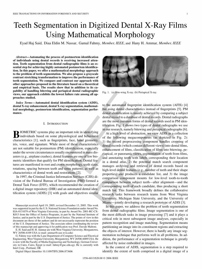
IEEE TRANSACTIONS ON INFORMATION FORENSICS AND SECURITY 1
Teeth Segmentation in Digitized Dental X-Ray FilmsUsing Mathematical Morphology
Eyad Haj Said, Diaa Eldin M. Nassar, Gamal Fahmy, Member, IEEE, and Hany H. Ammar, Member, IEEE
Abstract—Automating the process of postmortem identificationof individuals using dental records is receiving increased atten-tion. Teeth segmentation from dental radiographic films is an es-sential step for achieving highly automated postmortem identifica-tion. In this paper, we offer a mathematical morphology approachto the problem of teeth segmentation. We also propose a grayscalecontrast stretching transformation to improve the performance ofteeth segmentation. We compare and contrast our approach withother approaches proposed in the literature based on a theoreticaland empirical basis. The results show that in addition to its ca-pability of handling bitewing and periapical dental radiographicviews, our approach exhibits the lowest failure rate among all ap-proaches studied.
Index Terms—Automated dental identification system (ADIS),dental X-ray enhancement, dental X-ray segmentation, mathemat-ical morphology, postmortem identification, segmentation perfor-mance.
I. INTRODUCTION
B IOMETRIC systems play an important role in identifyingindividuals based on some physiological and behavioral
characteristics [1], such as fingerprints, face, hand geometry,iris, voice, and signature. While most of these characteristicsare not suitable for postmortem (PM) identification, especiallyunder the severe circumstances usually encountered in mass dis-asters (e.g., airplane crashes), dental features are one of few bio-metric identifiers that qualify for PM identification. Dental fea-tures are manifested in root and crown morphology, teeth sizes,rotations, spacing between teeth and sinus patterns, as well ascharacteristics of dental work and restorations [2].
In 1997, the Criminal Justice Information Services (CJIS) di-vision of the Federal Bureau of Investigation (FBI) formed aDental Task Force (DTF), which recommended the creation ofa digital image repository (DIR) and an automated dental iden-tification system (ADIS) [3] with goals and objectives similar
Manuscript received April 19, 2005; revised December 13, 2005. This workwas supported in part by the U.S. National Science Foundation under Award No.EIA-0131079 of West Virginia University, in part by Award No. 2001-RC-CX-K013 from the Office of Justice Programs, in part by the National Institute ofJustice, and in part by the U.S. Department of Justice. The points of view in thisdocument are those of the authors and do not necessarily represent the positionof the U.S. Department of Justice. The associate editor coordinating the reviewof this manuscript and approving it for publication was Prof. Davide Maltoni.
E. H. Said and H. H. Ammar are with West Virginia University, Morgantown,WV 26506-6109 USA (e-mail: [email protected])
G. Fahmy was with the Lane Department of Computer Science and ElectricalEngineering, West Virginia University, Morgantown, WV 26506-6009 USA. Heis now with the Faculty of Media Engineering and Technology, German Univer-sity in Cairo, Cairo, Egypt (e-mail: [email protected]). He is currently withIntel Corp., Portland, OR
Digital Object Identifier 10.1109/TIFS.2006.873606
Fig. 1. (a) Bite-wing X-ray. (b) Periapical X-ray.
to the automated fingerprint identification system (AFIS) [4]but using dental characteristics instead of fingerprints [5]. PMdental identification is mainly achieved by comparing a subjectdental record to a database of dental records. Dental radiographsare the most common forms of dental records used in PM iden-tification. Fig. 1 shows two types of dental radiographs we usein our research, namely bitewing and periapical radiographs [6].
At a high level of abstraction, we view ADIS as a collectionof the following megacomponents (as depicted in Fig. 2):1) the record preprocessing component handles cropping ofdental records (which contain different views) into dental films,enhancement of films, classification of films into bitewing, pe-riapical, or panoramic views, segmentation of teeth from films,and annotating teeth with labels corresponding their locationin a dental atlas, 2) the potential match search componentmanages archiving and retrieval of dental records based onhigh-level dental features (e.g., number of teeth and their shapeproperties) and produces a candidate list, and 3) the imagecomparison component mounts for low-level tooth-to-toothcomparison between subject teeth—after alignment—and thecorresponding teeth of each candidate, thus producing a shortmatch list. This framework broadly defines the collaborativeresearch tasks between research teams from West VirginiaUniversity, Michigan State University, and the University ofMiami—jointly developing a research prototype of ADIS [3].
In this paper, we address the problem of teeth segmentationfrom dental radiographic films. Image segmentation is one ofthe most difficult tasks in image processing [7] and it plays acritical role in most subsequent image analyses, especially inpattern recognition and image matching. Segmentation meanspartitioning an image into its constituent regions and extractingthe objects of interest. However, there is hardly any image seg-mentation technique that performs well in all problems. In ad-dition, the performance of a segmentation technique is greatlyaffected by noise embedded in images.
In the context of ADIS, segmentation is a step required toidentify the extent of teeth comprised in a digital image of a
1556-6013/$20.00 © 2006 IEEE

IEEE
Proo
f
2 IEEE TRANSACTIONS ON INFORMATION FORENSICS AND SECURITY
Fig. 2. Block diagram illustrating the architecture of ADIS.
dental radiographic film. At a finer level of detail, segmentationalso serves in decomposing a tooth into a crown area and a rootarea. Each segmented tooth represents a region of interest (ROI)that contains distinctive features used in the subsequent steps ofidentification. We define a qualified ROI as a rectangular area inthe image of dental film that bounds one tooth.
We tackle the problem of teeth segmentation using a mathe-matical-morphology (MM) approach. Our approach offers fullyautomated teeth segmentation, and is meant to reduce segmen-tation errors due to inherent and extrinsic background noise.Our interest in MM stems from its powerful capabilities forextracting a variety of shapes and image structures [7]. Mor-phological filtering [8] serves in a wide range of applicationsin image processing and analysis, such as feature extraction,shape representation and description, shape recognition, shapesmoothing, enhancement, and noise suppression [8] to mentiona few.
We set the following objectives for teeth segmentation: 1) toautomatically extract as many qualified ROIs as possible; 2) tooperate on bitewing and periapical views; and 3) in the worstcase scenario, to extract at least one qualified ROI from eachfilm. The qualified ROI shows one tooth as a whole.
In Section II, we briefly review the relevant literature. InSection III, we present our teeth segmentation algorithm, andthen in Section IV, we present a grayscale contrast stretchingtransformation that improves the definition of teeth versusbackground and the segmentation performance. In Section V,we report our experimental results and compare the perfor-mance of the proposed approach to other recently publishedapproaches. Finally, in Section VI, we conclude the paper andlay out plans for future work.

IEEE
Proo
f
SAID et al.: TEETH SEGMENTATION IN DIGITIZED DENTAL X-RAY FILMS 3
TABLE ISUMMARY OF COMPARISON BETWEEN THREE TEETH SEGMENTATION ALGORITHMS [13], [14], AND [15]
II. BACKGROUND
Several radiograph and magnetic-resonance (MR) image seg-mentation approaches have been presented in the last decade.In [9], a fully automated technique using Markov random fieldswas proposed for (MR) images. Noise, inhomogeneity, andstructure thickness have a negative impact on the performanceof the algorithm, and they tend to increase the segmentationerror. In [10], the Hopfield neural network based on patternclassification using the fuzzy c-means algorithm was proposed.The computation time and finding the global minimum of theobjective function affect performance. In [11], the segmen-tation approach is based on analyzing isolable-contour mapsto identify coherent regions corresponding to main objects.In [12], the segmentation is based on an improved watershedtransform that uses the prior information instead of usualgradient calculations. Although high accuracy was reported inthe approaches presented in [11] and [12], user interaction isneeded to select interesting regions.
There are few researches dedicated to the problem of dentalradiograph image segmentation. In [13], Jain and Chen separatethe upper jaw and the lower jaw in the bitewing and panoramicdental images by detecting the gap valley between them usingthe Y-axis projection histogram. Afterwards, the technique iso-lates each tooth from its neighbors in each jaw by detectingthe gaps between them using intensity integral projection. Thisapproach is semiautomated since an initial valley gap point isrequired to detect the gap valley between the upper and lowerjaw. We found from our experiments that the segmentation out-come may vary if we change the position of the selected initialvalley gap point. Fig. 3 shows two different segmentation resultsproduced by choosing two different initialization points both ofwhich are on the valley gap; the choice in Fig. 3(a) leads to per-fect segmentation while the choice in Fig. 3(b) leads to totalsegmentation failure.
In [14], Nomir and Abdel-Mottaleb introduce a fully auto-mated approach for dental X-ray images. The technique dependson applying the following stages: 1) iterative threshold to dividethe image into two parts—teeth and background; 2) adaptive
Fig. 3. Two different results for the same image. (a) Completely segmentedimage. (b) Failed segmented image.
threshold in order to increase the accuracy and remove teeth in-terfering; 3) horizontal integral projection in order to separatethe upper jaw from the lower jaw; 4) and, finally, vertical inte-gral projection in order to separate each individual tooth.
Another fully automated approach for dental X-ray imagesis introduced by Zhou and Abdel-Mottaleb [15]. The techniquedepends on improving the image contrast by applying morpho-logical transformation, and then using the window-based adap-tive threshold and integral projection to segment the teeth andseparate the upper and lower jaw.
Table I shows a brief comparison between the three algo-rithms based on underlying principles, type of dental film viewsthey are reported to operate on, and the level of automation theyachieve.
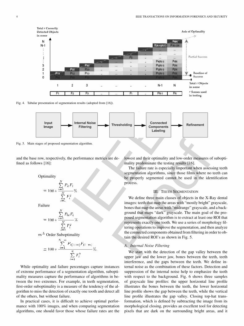
In [16], Nassar et al. present a metrics-based object countingapproach for the empirical assessment of image segmentation.To evaluate the performance of the segmentation algorithm, ref-erence images are used to record the outcome of the experimentin a tabular form as shown in Fig. 4.
Each cell of the results table contains the number of in-stances where the segmentation algorithm correctly detectsobjects out of objects that are present in reference image, with
, where is the number of reference imagesthat contain exactly objects. The results table is used in deter-mining metrics of optimality, suboptimality, and failure basedon the relative weights of the main diagonal, the subdiagonals,
IEEE
Proo
f
4 IEEE TRANSACTIONS ON INFORMATION FORENSICS AND SECURITY
Fig. 4. Tabular presentation of segmentation results (adopted from [16]).
Fig. 5. Main stages of proposed segmentation algorithm.
and the base row, respectively, the performance metrics are de-fined as follows [16]:
Optimality
Failure
Order Suboptimality
While optimality and failure percentages capture instancesof extreme performance of a segmentation algorithm, subopti-mality measures capture the performance of algorithms in be-tween the two extremes. For example, in teeth segmentation,first-order suboptimality is a measure of the tendency of the al-gorithm to miss the detection of exactly one tooth and detect allof the others, but without failure.
In practical cases, it is difficult to achieve optimal perfor-mance with 100% images, and when comparing segmentationalgorithms, one should favor those whose failure rates are the
lowest and their optimality and low-order measures of subopti-mality predominate the testing results [16].
The failure rate is especially important when assessing teethsegmentation algorithms, since those films where no teeth canbe properly segmented cannot be used in the identificationprocess.
III. TEETH SEGMENTATION
We define three main classes of objects in the X-Ray dentalimages: teeth that map the areas with “mostly bright” grayscale,bones that map the areas with “midrange” grayscale, and a back-ground that maps “dark” grayscale. The main goal of the pro-posed segmentation algorithm is to extract at least one ROI thatrepresents exactly one tooth. We use a series of morphology fil-tering operations to improve the segmentation, and then analyzethe connected components obtained from filtering in order to ob-tain the desired ROI’s as shown in Fig. 5.
A. Internal Noise Filtering
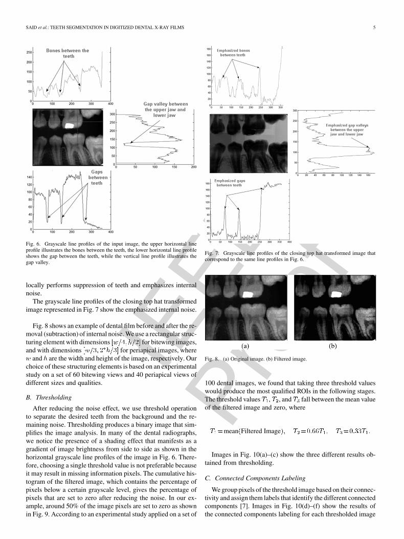
We start with the detection of the gap valley between theupper jaw and the lower jaw, bones between the teeth, teethinterference, and the gaps between the teeth. We define in-ternal noise as the combination of these factors. Detection andsuppression of the internal noise help to emphasize the teethwith respect to the background. Fig. 6 shows three samplesof grayscale line profiles: the upper horizontal line profileillustrates the bones between the teeth, the lower horizontalline profile shows the gap between the teeth, while the verticalline profile illustrates the gap valley. Closing top-hat trans-formation, which is defined by subtracting the image from itsmorphological closing, provides an excellent tool for detectingpixels that are dark on the surrounding bright areas, and it
IEEE
Proo
f
SAID et al.: TEETH SEGMENTATION IN DIGITIZED DENTAL X-RAY FILMS 5
Fig. 6. Grayscale line profiles of the input image, the upper horizontal lineprofile illustrates the bones between the teeth, the lower horizontal line profileshows the gap between the teeth, while the vertical line profile illustrates thegap valley.
locally performs suppression of teeth and emphasizes internalnoise.
The grayscale line profiles of the closing top hat transformedimage represented in Fig. 7 show the emphasized internal noise.
Fig. 8 shows an example of dental film before and after the re-moval (subtraction) of internal noise. We use a rectangular struc-turing element with dimensions for bitewing images,and with dimensions for periapical images, where
and are the width and height of the image, respectively. Ourchoice of these structuring elements is based on an experimentalstudy on a set of 60 bitewing views and 40 periapical views ofdifferent sizes and qualities.
B. Thresholding
After reducing the noise effect, we use threshold operationto separate the desired teeth from the background and the re-maining noise. Thresholding produces a binary image that sim-plifies the image analysis. In many of the dental radiographs,we notice the presence of a shading effect that manifests as agradient of image brightness from side to side as shown in thehorizontal grayscale line profiles of the image in Fig. 6. There-fore, choosing a single threshold value is not preferable becauseit may result in missing information pixels. The cumulative his-togram of the filtered image, which contains the percentage ofpixels below a certain grayscale level, gives the percentage ofpixels that are set to zero after reducing the noise. In our ex-ample, around 50% of the image pixels are set to zero as shownin Fig. 9. According to an experimental study applied on a set of
Fig. 7. Grayscale line profiles of the closing top hat transformed image thatcorrespond to the same line profiles in Fig. 6.
Fig. 8. (a) Original image. (b) Filtered image.
100 dental images, we found that taking three threshold valueswould produce the most qualified ROIs in the following stages.The threshold values , , and fall between the mean valueof the filtered image and zero, where
mean Filtered Image
Images in Fig. 10(a)–(c) show the three different results ob-tained from thresholding.
C. Connected Components Labeling
We group pixels of the threshold image based on their connec-tivity and assign them labels that identify the different connectedcomponents [7]. Images in Fig. 10(d)–(f) show the results ofthe connected components labeling for each thresholded image

IEEE
Proo
f
6 IEEE TRANSACTIONS ON INFORMATION FORENSICS AND SECURITY
Fig. 9. Cumulative histogram of the filtered image.
Fig. 10. (a)–(c) Thresholded images. (e)–(g) Result of connected componentlabeling for (a)–(c). (g)–(i) Qualified ROIs generated from (e)–(g).
TABLE IIRULES USED IN REFINEMENT STAGE TO DETERMINE THE QUALIFIED ROIS
shown in Fig. 10(a)–(c), respectively. The connected compo-nents can be attributed to: 1) teeth that are considered as ROIs;2) more than one tooth because of teeth interference, fillings, orhigh-intensity bone structures; 3) background or bones; and 4)part of the tooth such as the crown or root.
D. Refinement
The purpose of refinement is to analyze the connected com-ponents based on geometric properties (area and dimension) and
Fig. 11. (a) Original image. (b)–(i) Results of segmentation.
Fig. 12. (a) Original image. (b) Filtered image. (c) Thresholded images. (d)Result of connected component labeling. (e)–(h) qualified ROIs.
then to eliminate the unqualified objects. Table II shows the rulesused for refinement based on an experimental study applied on100 images. We classify the image type (bitewing or periapical)according to dental image classification proposed in [15]. Im-ages in Fig. 10(g)–(i) show the qualified ROIs. If two or morequalified ROIs are generated from the three different thresholdsfor the same tooth, we unify them to generate the single ROI. Weapply the union operation on two ROIs if the centriod of eachROI belongs to the other and their intersection is at least 80%of the smaller ROI. The union of qualified ROIs represents thefinal results of segmentation as shown in Fig. 11.
The previous example shows the segmentation stages that ap-plied to bitewing dental images. Fig. 12 shows the results of seg-mentation stages applied on periapical dental images.
Figs. 13–15 show some samples of image segmentationresults. The images in Fig. 13 have fully succeeded; the imagesin Fig. 14 have partially succeeded, while the images in Fig. 15have failed to give any ROI. In Figs. 13 and 14, there are tworows of images—the upper row shows the original images,while the lower row shows teeth obtained from segmentation.It is obvious that the better quality the dental image is, the moreROIs can be extracted from that image. We define the factorsthat introduce difficulties in segmentation as follows: 1) imageblurring [Fig. 15(a) and (f)]; 2) fillings [Fig. 15(b)]; 3) teethinterfering [Figs. 15(b), (c), (f), and (h)]; 4) image scan quality[Fig. 15(c)]; and 5) the intensity of the bones is very close tointensity of the teeth [Fig. 15(e)–(h)].
IV. DENTAL FILM GRAYSCALE CONTRAST ENHANCEMENT
As we mentioned in Section III, the histogram analysis ofdental radiographic films supports the intuition that most teethareas (except for the pulp tissue) predominantly concentratein upperband grayscales, while the areas of support bones andgums appear around midrange grayscales, and the air-gap areasare confined to the lower band of grayscales. Due to aging of

IEEE
Proo
f
SAID et al.: TEETH SEGMENTATION IN DIGITIZED DENTAL X-RAY FILMS 7
Fig. 13. Examples of completely successful images and their segmentation outcomes.
Fig. 14. Examples of partially successful succeeded images and their segmentation outcomes.
Fig. 15. Examples of completely failed images where no teeth can be extracted.
their chemicals, radiographic films tend to lose their contrastover time. Therefore, in dental radiographs that are scanned afterexhibiting contrast decay, it is difficult to identify clear bound-aries between the grayscale bands of teeth, support bones/gums,and air gaps. In addition, the lack of a unified standard for scan-ning and digitization of dental radiographic films exacerbatesthe identification of the grayscale bands of concentration ofthese objects.
To improve segmentation of teeth from low-contrast dentalfilms, we propose to apply a preprocessing contrast stretchingstep using a parametric sigmoid transform. Thus, we map aninput grayscale to , according to the equa-tion shown at the bottom of the page, where and desig-nate compression and expansion factors, respectively; is athreshold grayscale between the compression and the expansionregions of ; is the image of under , , and
are the minimum and maximum grayscales of the inputimage, respectively. We choose such that its proportion-
ality to the new grayscale range [0, 255] resembles that ofto the grayscale range of the input image. Thus
Let denote the cumulative grayscale histogram of the inputimage (I). To determine the values of , , and , we analyze
to first determine two marker grayscales and as fol-lows:
where
To choose suitable values for and , we studied severaldental radiographic films with variability in their brightness andcontrast. We varied these parameters and observed that most of
IEEE
Proo
f
8 IEEE TRANSACTIONS ON INFORMATION FORENSICS AND SECURITY
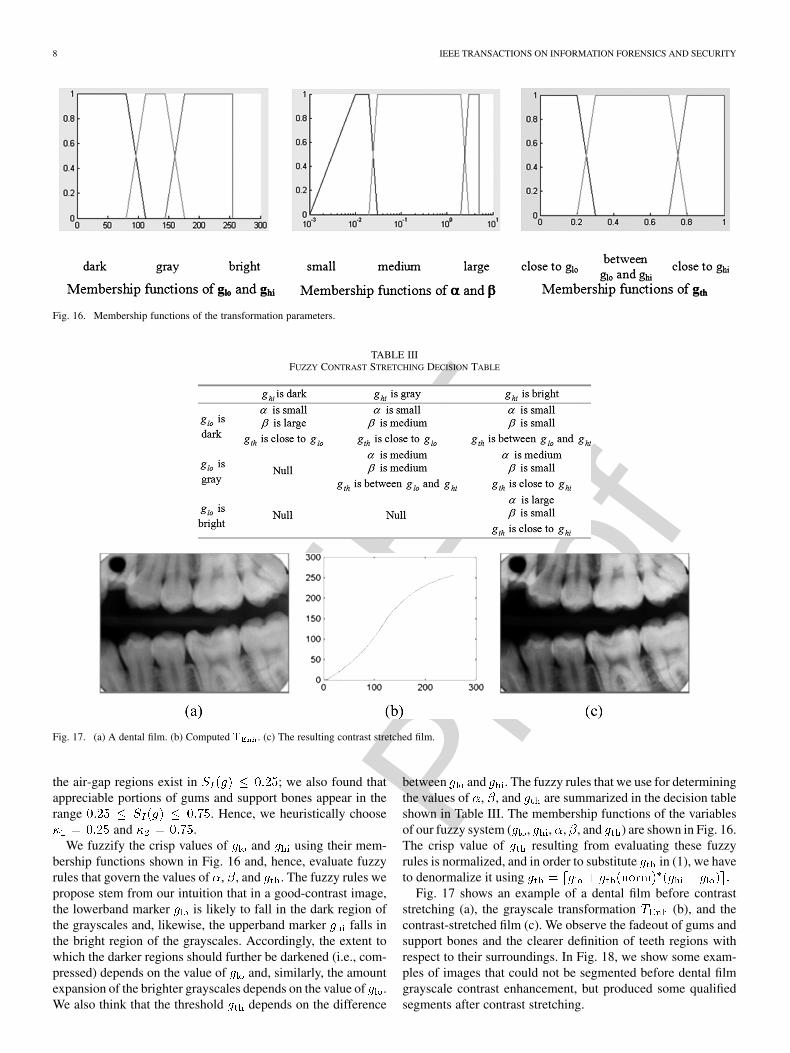
Fig. 16. Membership functions of the transformation parameters.
TABLE IIIFUZZY CONTRAST STRETCHING DECISION TABLE
Fig. 17. (a) A dental film. (b) Computed T . (c) The resulting contrast stretched film.
the air-gap regions exist in ; we also found thatappreciable portions of gums and support bones appear in therange . Hence, we heuristically choose
and .We fuzzify the crisp values of and using their mem-
bership functions shown in Fig. 16 and, hence, evaluate fuzzyrules that govern the values of , , and . The fuzzy rules wepropose stem from our intuition that in a good-contrast image,the lowerband marker is likely to fall in the dark region ofthe grayscales and, likewise, the upperband marker falls inthe bright region of the grayscales. Accordingly, the extent towhich the darker regions should further be darkened (i.e., com-pressed) depends on the value of and, similarly, the amountexpansion of the brighter grayscales depends on the value of .We also think that the threshold depends on the difference
between and . The fuzzy rules that we use for determiningthe values of , , and are summarized in the decision tableshown in Table III. The membership functions of the variablesof our fuzzy system ( , , , , and ) are shown in Fig. 16.The crisp value of resulting from evaluating these fuzzyrules is normalized, and in order to substitute in (1), we haveto denormalize it using
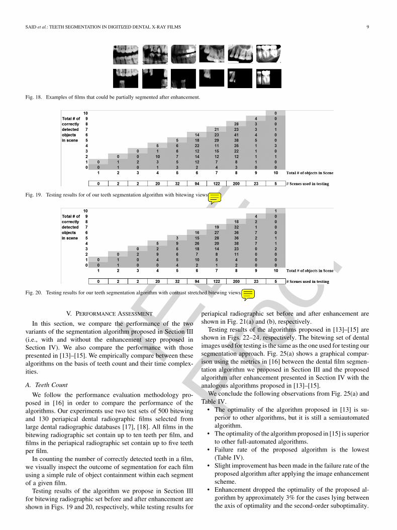
Fig. 17 shows an example of a dental film before contraststretching (a), the grayscale transformation (b), and thecontrast-stretched film (c). We observe the fadeout of gums andsupport bones and the clearer definition of teeth regions withrespect to their surroundings. In Fig. 18, we show some exam-ples of images that could not be segmented before dental filmgrayscale contrast enhancement, but produced some qualifiedsegments after contrast stretching.
IEEE
Proo
f
SAID et al.: TEETH SEGMENTATION IN DIGITIZED DENTAL X-RAY FILMS 9
Fig. 18. Examples of films that could be partially segmented after enhancement.
Fig. 19. Testing results for of our teeth segmentation algorithm with bitewing views.
Fig. 20. Testing results for our teeth segmentation algorithm with contrast stretched bitewing views.
V. PERFORMANCE ASSESSMENT
In this section, we compare the performance of the twovariants of the segmentation algorithm proposed in Section III(i.e., with and without the enhancement step proposed inSection IV). We also compare the performance with thosepresented in [13]–[15]. We empirically compare between thesealgorithms on the basis of teeth count and their time complex-ities.
A. Teeth Count
We follow the performance evaluation methodology pro-posed in [16] in order to compare the performance of thealgorithms. Our experiments use two test sets of 500 bitewingand 130 periapical dental radiographic films selected fromlarge dental radiographic databases [17], [18]. All films in thebitewing radiographic set contain up to ten teeth per film, andfilms in the periapical radiographic set contain up to five teethper film.
In counting the number of correctly detected teeth in a film,we visually inspect the outcome of segmentation for each filmusing a simple rule of object containment within each segmentof a given film.
Testing results of the algorithm we propose in Section IIIfor bitewing radiographic set before and after enhancement areshown in Figs. 19 and 20, respectively, while testing results for
periapical radiographic set before and after enhancement areshown in Fig. 21(a) and (b), respectively.
Testing results of the algorithms proposed in [13]–[15] areshown in Figs. 22–24, respectively. The bitewing set of dentalimages used for testing is the same as the one used for testing oursegmentation approach. Fig. 25(a) shows a graphical compar-ison using the metrics in [16] between the dental film segmen-tation algorithm we proposed in Section III and the proposedalgorithm after enhancement presented in Section IV with theanalogous algorithms proposed in [13]–[15].
We conclude the following observations from Fig. 25(a) andTable IV.
• The optimality of the algorithm proposed in [13] is su-perior to other algorithms, but it is still a semiautomatedalgorithm.
• The optimality of the algorithm proposed in [15] is superiorto other full-automated algorithms.
• Failure rate of the proposed algorithm is the lowest(Table IV).
• Slight improvement has been made in the failure rate of theproposed algorithm after applying the image enhancementscheme.
• Enhancement dropped the optimality of the proposed al-gorithm by approximately 3% for the cases lying betweenthe axis of optimality and the second-order suboptimality.

IEEE
Proo
f
10 IEEE TRANSACTIONS ON INFORMATION FORENSICS AND SECURITY
Fig. 21. (a) Testing results for of our teeth segmentation algorithm with periapical views. (b) Testing results for of our teeth segmentation algorithm with contrast-stretched periapical views.
Fig. 22. Testing results of Jain and Chen segmentation algorithm [13].
Fig. 23. Testing results of the Nomir and Abdel-Mottaleb segmentation algorithm [14].
• Enhancement increased the percentage of the cases lie be-tween optimality and fourth-order suboptimality to 83%.
While the algorithms proposed in [13]–[15] do not work withthe periapical dental radiographs, Fig. 25(b) shows a graphicalcomparison using the metrics in [16] between the proposedalgorithm and the proposed algorithm with enhancement. Itis clear that the enhancement decreases the failure rate from9.4% to 6.7%, and it improves the optimality and first-ordersuboptimality.
B. Time Complexity
To compare the time complexities of the proposed algorithmand those proposed in [13]–[15], we used 40 bitewing films withdifferent dimensions. We invoked MATLAB implementationsof each algorithm on an Intel Pentium 4 2.00-GHz, 512-MBDRAM platform. Table V summarizes the outcome of the timecomplexity comparison between the four teeth segmentation al-gorithms: is the image height, is the image width, and isthe size of the window used in adaptive thresholding.
Fig. 26 records the execution times of the four teeth segmen-tation algorithms for each of the 40 bitewing films. Based onFig. 26 and Table V, we observe that: 1) the proposed algorithmis the fastest among the other algorithms; 2) the difference inthe average time execution between the proposed algorithm, al-gorithm [13], and algorithm [15] is small; and 3) the time com-plexity of the algorithm [14] is significant compared to the otheralgorithms.
VI. CONCLUSION AND FUTURE WORK
We presented an automated dental image segmentationalgorithm that handles bitewing and periapical dental imagesbased on mathematical morphology. The proposed algorithmincludes: 1) noise filtering, 2) thresholding to isolate the teethfrom the background, and 3) analyzing connect componentslabeling to determine the qualified ROIs based on geometricalproperties. We introduced the difficulties that face the proposedalgorithm. These difficulties are image blurring, fillings, teethinterfering, image scan quality, and very low contrast betweenbones and teeth intensity.

IEEE
Proo
f
SAID et al.: TEETH SEGMENTATION IN DIGITIZED DENTAL X-RAY FILMS 11
Fig. 24. Testing results of the Zhou and Abdel-Mottaleb segmentation algorithm [15].
Fig. 25. (a) Performance comparison between bitewing segmentation algorithms. (b) Performance comparison between the proposed algorithm and proposedalgorithm with enhancement for periapical views.
TABLE IVCOMPARISON BETWEEN THE FAILURE RATES OF THE VARIANT TEETH SEGMENTATION ALGORITHMS
TABLE VCOMPARISON BETWEEN THE TIME COMPLEXITIES OF THE FOUR TEETH
SEGMENTATION ALGORITHMS
We also presented a grayscale contrast stretching transfor-mation to improve the performance of teeth segmentation. Ap-plying it prior to segmentation increases the optimality and first-order suboptimality of the periapical image segmentation. It also
drops the failure rate of bitewing and periapical image segmen-tation, which is one of the main objectives of our algorithm.
We also presented a performance comparison between vari-ants of the bitewing dental image segmentation. The resultsshow that 1) the proposed algorithm has the lowest failure ratein terms of the segmentation result, and it is the fastest in termsof time complexity, and it can handle both bitewing and peri-apical images; 2) the algorithm proposed in [13] has the highestoptimality, but it is still a semiautomated algorithm and its per-formance is sensitive to the manually selected initial valley gap-point; and 3) the algorithm proposed in [15] has the highest op-timality among the other full automated algorithms; however, itonly handles the bitewing images.
Our plan for the future is to develop the segmentation algo-rithm in order to improve handling poor quality images and toinclude the panoramic dental radiograph views in the segmen-tation process.

IEEE
Proo
f
12 IEEE TRANSACTIONS ON INFORMATION FORENSICS AND SECURITY
Fig. 26. Time complexity distribution of 40 images for variants algorithms.
ACKNOWLEDGMENT
The authors wish to thank Dr. A. K. Jain and H. Chen ofMichigan State University as well as Dr. M. Abdel-Mottaleb,O. Nomir, and J. Zhou of the University of Miami for providingthe authors with implementations of their referenced teeth seg-mentation algorithms. The authors also acknowledge the helpof the Criminal Justice Information Services (CJIS) division ofthe Federal Bureau of Investigation in developing the ADIS ar-chitecture and in providing the dental image database.
REFERENCES
[1] A. K. Jain, L. Hong, and S. Pankanti, “Biometrics: promising frontiersfor emerging identification market,” in Comm. ACM, Feb. 2000, pp.91–98.
[2] P. Stimson and C. Mertz, Forensic Dentistry. Boca Raton, FL: CRC,1997.
[3] G. Fahmy, D. Nassar, E. Haj-Said, H. Chen, O. Nomir, J. Zhou, R.Howell, H. H. Ammar, M. Abdel-Mottaleb, and A. K. Jain, “Towardan automated dental identification system (ADIS),” in Proc. Int. Conf.Biometric Authentication, Hong Kong, China, Jul. 2004, pp. 789–796.
[4] R. Khanna and W. Shen, “Automated fingerprint identification system(AFIS) benchmarking using the National Institute of Standards andTechnology (NIST) special database 4,” in Proc. Inst. Elect. Electron.Eng. 28th Annu. Int. Carnahan Conf., Albuquerque, NM, Oct. 12–14,1994, pp. 188–194.
[5] D. E. Nassar, “A prototype Automatic Dental Identification System(ADIS),” M.Sc. dissertation, Dept. Elect. Comput. Eng., West VirginiaUniv., Morgantown, Apr. 2001.
[6] S. White and M. Pharoah, Oral Radiology Principles and Interpreta-tion, 4th ed. St. Louis, MO: Mosby, 2000.
[7] R. C. Gonzales and R. E. Woods, Digital Image Processing, 2nd ed.Upper Saddle River, NJ: Prentice-Hall, 2002.
[8] P. Maragos and R. W. Schafer, “Morphological systems for multidi-mensional signal processing,” Proc. IEEE, vol. 78, no. 4, pp. 690–710,Apr. 1990.
[9] K. Held, E. R. Kops, B. J. Krause, W. M. Wells, and R. Kikinis,“Markov random field segmentation of brain MR images,” IEEETrans. Med. Imag., vol. 16, no. 6, pp. 878–886, Dec. 1997.
[10] J.-S. Lin, K.-S. Cheng, and C.-W. Mao, “A fuzzy Hopfield neural net-work for medical image segmentation,” IEEE Trans. Nucl. Sci., vol. 43,no. 4, pp. 2389–2398, Aug. 1996.
[11] S. Shiffman, G. D. Rubin, and S. Naple, “Medical image segmentaionusing analysis of isolable—Contour maps,” IEEE Trans. Med. Imag.,vol. 19, no. 11, pp. 1064–1074, Nov. 2000.
[12] V. Grau, A. U. J. Mewes, M. Alcaniz, R. Kikinis, and S. K. Warfield,“Improved watershed transform for medical image segmentationusing prior information,” IEEE Trans. Med. Imag., vol. 23, no. 4, pp.447–458, Apr. 2004.
[13] A. K. Jain and H. Chen, “Matching of dental X-ray images for humanidentification,” Patt. Recognit., vol. 37, pp. 1519–1532, 2004.
[14] O. Nomir and M. Abdel-Mottaleb, “A system for human identifi-cation from X-ray dental radiographs,” Patt. Recognit., vol. 38, pp.1295–1305, 2005.
[15] J. Zhou and M. Abdel-Mottaleb, “A content-based system for humanidentification based on bitewing dental X-ray images,” Patt. Recognit.,to be published.
[16] D. E. M. Nassar, F. U. Chaudhry, and H. H. Ammar, “On performanceevaluation of image segmentation algorithms: Success is not all ornone,” in Proc. 1st Int. Comput. Eng. Conf. , Cairo, Egypt, Dec. 2004,pp. 354–359.
[17] CJIS Division, “Washington state patrol missing and unidentifiedperson unit,” Digitized Dental Images (Database) May 2000.
[18] CJIS Division—ADIS project, Digitized Radiographic Images (Data-base) Aug. 2002.
Eyad Haj Said received the M.Sc. degree in computer engineering from StevensInstitute of Technology, Hoboken, NJ, and is currently pursuing the Ph.D. degreein the Lane Department of Computer Science and Electrical Engineering, WestVirginia University (WVU), Morgantown.
Currently, he is on the Research Assistant Team inthe Automated Dental Identification System (ADIS) project with WVU. He wasa Software Engineer with Net-Centric Company, Ottawa, ON, Canada, from2003 to 2004, and Teaching Assistant with New York University, New York,from 2001 to 2002.
Diaa Eldin M. Nassar received the B.Sc. degree from Cairo University, Cairo,Egypt, in 1994, the M.Sc. degree in electrical engineering from West VirginiaUniversity in 2001, and the Ph.D. degree in the Lane Department of ComputerScience and Electrical Engineering, West Virginia University (WVU), Morgan-town, in 2005.
His research interests include digital image pro-cessing, biometric systems, pattern recognition, and software quality assess-ment. He was also with Halliburton Company and is currently with Intel Cor-poration, Portland, OR.
Gamal Fahmy (M’00) was born in Leeds, U.K., in1973. He received the B.Sc. and M.Sc. degrees fromAssiut University, Assiut, Egypt, in 1996 and 1998,respectively, and the Ph.D. degree in electrical en-gineering from Arizona State University, Tempe, in2003.
Currently, he is with the Faculty of Media Engi-neering and Technology at the German Universityin Cairo, Cairo, Egypt. From 2003 to 2005, he wasa Research Assistant Professor with West VirginiaUniversity, Morgantown, where he worked on several
identification and recognition projects in collaboration with different federalagencies in the United States, such as the Federal Bureau of Investigation,the National Institute of Justice, Transportation Security Administration, andthe Department of Homeland Security. His research interests include imagesuper-resolution, perceptual image compression, human vision, and biometrics(IRIS recognition and 3-D face recognition).
Dr. Fahmy has served as a technical committee member and reviewer forseveral international conferences.

IEEE
Proo
f
SAID et al.: TEETH SEGMENTATION IN DIGITIZED DENTAL X-RAY FILMS 13
Hany H. Ammar (M’85) is a Professor of Com-puter Engineering in the Department of ComputerScience and Electrical Engineering, West VirginiaUniversity (WVU), Morgantown. He is the Directorof the Software Architectures and High PerformanceComputing Lab at WVU and leads several projectsfunded by the National Science Foundation (NSF)under the Digital Government and ITR programsand NASA Office of Safety and Mission Assurance(OSMA) Software Assurance Research Program(SARP) managed through the NASA Independent
Verification and Validation (IV&V) Facility, Fairmont, WV. His researchinterests are in identification technology and software engineering. He recentlycoauthored a book on Pattern-Oriented Analysis and Design (Addison-Wesley,Reading, MA) and has published many articles in prestigious journals andconference proceedings.
Dr. Ammar is currently serving in the program and steering committees ofseveral professional conferences and workshops. He is a member of the ACM.