Embed Size (px)
Citation preview

71Revista Facultad de Odontología Universidad de Antioquia - Vol. 28 N.o 1 - Segundo semestre, 2016
ABSTRACT. Introduction: the objective of this study was to use real-time qPCR to identify and quantify the Streptococcus mutans species in samples of saliva and dental biofilm. Methods: 27 children were randomly chosen with the following criteria: 8 years of age, low socio-economic levels, residing in the northern metropolitan area of Santiago de Chile; they were asked to attend an appointment while fasting with no teeth brushing for at least 12 hours, in order to collect non-stimulated saliva and a pool of supragingival dental biofilm of all the mesio-vestibular sides of anterior and posterior teeth. The amount of S. mutans in the samples was quantified by qPCR using primers that amplify a fragment of the gtfB gene of S. mutans. Results: the amplification showed 98% efficiency with a fluorescence of 3.36 cycles. The melting curve presented a single maximum at the same temperature for all samples. Conclusion: the methodology allows the specific identification and quantification of gene gtfB of S. mutans in saliva and dental biofilm in a quick and reliable manner, contributing to the identification of individual cariogenic risk.Key words: S. mutans, qPCR, gtfB gen, dental biofilm, saliva.Moncada G, Duperat L, Palma P, Corsini G, Neira M, Oliveira Junior O et al. Real-time quantitative polymerase chain reaction (qPCR) for the identification and quantification of Streptococcus mutans in saliva and dental biofilm in children. Rev Fac Odontol Univ Antioq 2016; 28(1): 71-94. DOI: http://dx.doi.org/10.17533/udea.rfo.v28n1a4
RECIBIDO: AGOSTO 19/2014 - ACEPTADO: JULIO 19/2016
1 Adscrito al Proyecto Fonis SAE 13i202052 DDS, PhD, Escuela Dental, Universidad Mayor de Santiago, Chile3 DDS, Química, Departamento de Ciencias Básicas, Escuela Dental,
Universidad de Chile4 DDS, Microbiología, Departamento de Patología Oral, Escuela Dental,
Universidad de Chile5 BQ Centro de Investigación Biomédica, Facultad de Ciencias de la
Salud, Universidad Autónoma de Chile6 BQ, Química, Departamento de Ciencias Básicas, Escuela Dental,
Universidad de Chile7 DDS, Química, Departamento de Ciencias Básicas, Escuela Dental,
Universidad de Chile8 DDS PhD, Universidade Estadual Paulista, UNESP, Escuela Dental,
Departamento de Odontología Restauradora, Araraquara, Brasil9 DDS PhD Cariología, Escuela Dental, Universidad de Chile10 DDS MS Departamento de Odontología Restauradora, Ciencias
Odontológicas, División de Operatoria Dental, College of Dentistry, University of Florida
11 BQ Química, Departamento de Ciencias Básicas, Escuela Dental, Universidad de Chile
TÉCNICA DE REACCIÓN DE POLIMERASA EN CADENA (QPCR) EN TIEMPO REAL PARA LA IDENTIFICACIÓN Y CUANTIFICACIÓN DE STREPTOCOCCUS
MUTANS EN SALIVA Y BIOPELÍCULA DENTARIA DE NIÑOS1
REAL-TIME QUANTITATIVE POLYMERASE CHAIN REACTION (QPCR) FOR THE IDENTIFICATION AND QUANTIFICATION OF STREPTOCOCCUS MUTANS IN
SALIVA AND DENTAL BIOFILM IN CHILDREN1
GUSTAVO MONCADA2, LORENA DEL CARMEN DUPERAT 3, PATRICIA PALMA4, GINO CORSINI5, MIGUEL NEIRA6,
EVELYN REYES7, OSMIR BATISTA OLIVEIRA JUNIOR8, SIMONE FALEIROS9, VALERIA GORDAN10, ISMAEL YÉVENES11
RESUMEN. Introducción: el objetivo del presente estudio consistió en implementar la técnica de qPCR en tiempo real para identificar y cuantificar la especie Streptococcus mutans en muestras de saliva y biopelícula dentaria. Métodos: se seleccionaron al azar 27 niños de 8 años de edad, de nivel socio-económico bajo del área norte de la región metropolitana de Santiago de Chile, que se citaron en ayunas y sin cepillado durante al menos 12 horas, para colectar saliva no estimulada y un pool de biopelícula dentaria supragingival de todas las caras mesio-vestibulares de dientes anteriores y posteriores. Se cuantificó la cantidad de S. mutans en las muestras mediante qPCR empleando partidores que amplifican un fragmento del gen gtfB de S. mutans. Resultados: la amplificación presentó 98% de eficiencia con delta de fluorescencia de 3,36 ciclos. La curva de fusión (melting) presentó un solo máximo a una misma temperatura para todas las muestras. Conclusión: la metodología permite la identificación y cuantificación específica del gen gtfB de S. mutans en muestras de saliva y biopelícula dentaria, de forma rápida y exacta, aportando a la determinación del riesgo cariogénico individual.Palabras clave: S. mutans, qPCR, gen gtfB, biopelícula dentaria, saliva.Moncada G, Duperat L, Palma P, Corsini G, Neira M, Oliveira Junior O et al. Técnica de reacción de polimerasa en cadena (qPCR) en tiempo real para la identificación y cuantificación de Streptococcus Mutans en saliva y biopelícula dentaria de niños. Rev Fac Odontol Univ Antioq 2016; 28(1): 71-94. DOI: http://dx.doi.org/10.17533/udea.rfo.v28n1a4
1 Appointed to Fonis Project SAE 13i202052 DDS, PhD, Dental School, Universidad Mayor, Santiago, Chile3 DDS, Chemistry, Basic Science Department, Dental School,
Universidad de Chile4 DDS, Microbiology, Departament of Oral Pathology, Dental School,
Universidad de Chile5 BQ Centro de Investigación Biomédica, School of Health Sciences,
Universidad Autónoma de Chile6 BQ, Chemistry, Basic Science Department, Dental School, Universidad
de Chile7 DDS, Chemistry, Basic Science Department, Dental School,
Universidad de Chile8 DDS PhD, Universidade Estadual Paulista, UNESP, School of Dentistry,
Department of Restorative Dentistry, Araraquara, Brazil9 DDS PhD Cariology, Dental School, Universidad de Chile10 DDS MS Department of Restorative Dentistry, Division of Operative
Dentistry Department, College of Dentistry, University of Florida11 BQ Chemistry, Basic Science, Dental School, Universidad de Chile
SUBMITTED: AUGUST 19/2014 - ACCEPTED: JULY 19/2016

72
TÉCNICA DE REACCIÓN DE POLIMERASA EN CADENA (QPCR) EN TIEMPO REAL PARA LA IDENTIFICACIÓN Y CUANTIFICACIÓN DE STREPTOCOCCUS MUTANS EN SALIVA Y BIOPELÍCULA DENTARIA DE NIÑOS
Revista Facultad de Odontología Universidad de Antioquia - Vol. 28 N.o 1 - Segundo semestre, 2016
INTRODUCCIÓN
La caries dental es aún la enfermedad crónica más pre-valente en los seres humanos.1 Su frecuencia se incre-menta constantemente con la edad hasta alcanzar a casi el 100% de la población entre 65 y 70 años de edad en Chile; en los niños en edad escolar afecta al 85% en Chile.2-4
Es una enfermedad de etiología multifactorial, micro-biológicamente inducida, asociada a la fermentación de hidratos de carbono. Es definida como el desbalance mineral y proteico de los dientes, con pérdida neta de minerales en forma irreversible cuando las lesiones se encuentran en estado cavitado. Factores como ocasión, frecuencia y tipo de ingesta de alimentos, deficiencias en las técnicas de control de la biopelícula dentaria, ba-jos niveles de fluoruros en el ambiente oral y baja tasa de secreción salival constituyen algunos de los deter-minantes ecológicos que modulan las variaciones de la biopelícula.5
La biopelícula oral de pacientes libres de lesiones de caries está constituida por más de mil especies diferentes; sin embargo, los pacientes con enfermedad activa de caries presentan reducción de más del 70% de las especies, mostrando predominio de la microbiota acidogénica asociada con el inicio y progresión de la enfermedad.5 La bacteria Streptococcus mutans es una de las especies tradicionalmente responsables del inicio de las lesiones, junto a otras especies productoras de ácidos, como Streptococcus sobrinus, Veillonella y Lactobacillus spp.6-8 También se reconoce la microbiota productora de álcalis que conduce al balance del pH y del microbioma oral, como son Streptococcus sanguinis, Streptococcus gordonii, Streptococcus salivarius y A naeslundii, entre otros.9-11
La actividad deletérea de la especie S. mutans se asocia con su capacidad para producir ácido láctico a partir de la sacarosa, llegando a niveles capaces de provocar la pérdida neta de minerales de los dientes. 7, 12-14
INTRODUCTION
Tooth decay is still the most prevalent chronic disease in humans.1 Its frequency steadily increases with age, affecting almost 100% of the Chilean population aged 65 to 70 years; it affects 85% school-age children in Chile.2-4
This disease has a multifactorial etiology, which is microbiologically induced and associated to the fermentation of carbon hydrates. It is defined as an imbalance of proteins and minerals in teeth, with irreversible loss of minerals when lesions are in their cavitated state. Factors such as time, frequency, and type of food intake, deficiencies in techniques of dental biofilm control, low levels of fluoride in the oral environment, and low rate of salivary secretion are some of the ecological determinants influencing variations in biofilm.5
The oral biofilm in caries-free patients contains more than one thousand different species; however, patients with active caries have a reduction of more than 70% of species, with dominance of the acidogenic microbiota associated with the onset and progression of the disease.5 The bacterium Streptococcus mutans is one of the species traditionally responsible for the onset of lesions, along with other acid-producing species, such as Streptococcus sobrinus, Veillonella and Lactobacillus spp.6-8 Other alkalis-producing microbiota has been identified as it influences pH balance and the oral microbiome, such as Streptococcus sanguinis, Streptococcus gordonii, Streptococcus salivarius and A naeslundii, to name just a few.9-11
The deleterious activity of S. mutans spp. is associated with its ability to produce lactic acid from sucrose, reaching levels capable of causing the loss of minerals in teeth.7, 12-14

73
REAL-TIME QUANTITATIVE POLYMERASE CHAIN REACTION (QPCR) FOR THE IDENTIFICATION AND QUANTIFICATION OF STREPTOCOCCUS MUTANS IN SALIVA AND DENTAL BIOFILM IN CHILDREN
Revista Facultad de Odontología Universidad de Antioquia - Vol. 28 N.o 1 - Segundo semestre, 2016
El Streptococcus es el género bacteriano más numeroso en la cavidad oral. Según su filogenia, se clasifica en tres grupos (bovis, pneumonia y viridans),15 y a su vez el grupo viridans se divide en otros cinco grupos: mitis, mutans, salivarius, anginosus y sanguinis.15 El grupo Mutans Streptococci (MS) es clasificado, según la se-cuencia nucleotídica de su RNA ribosomal 16S, en ocho especies (mutans, sobrinus, cricetus, dawnei, ferus, macacae, ratti y hyovaginalis); de estos, S. mutans es considerada la especie más dominante en la cavidad oral.16
S. mutans es una especie cocácea, Gram positiva, que se agrupa en cadenas; es inmóvil, catalasa negativo, rá-pido productor de ácido láctico, y su hábitat usual es la superficie dentaria, aunque también ha sido detectado en válvulas cardiacas y placas de ateroma en los grandes vasos.17
La virulencia de S. mutans se caracteriza por su acido-génesis, aciduria, acidofilia, síntesis de polisacáridos extracelulares (PEC), síntesis de polisacáridos intracelu-lares, síntesis de proteínas lectinas que ligan al glucano (Gpbs), adhesinas, proteínas asociadas a la pared celu-lar (WapA) y bacteriocinas. La síntesis de polisacáridos extracelulares es un factor destacado de su virulencia, dado que antes de que la sacarosa ingrese a la célula, un porcentaje es transformado por las exoenzimas, glu-cosiltransferasas (Gtfs) y fructosiltransferasas (Ftf), que metabolizan la molécula de sacarosa en monosacáridos, transfiriendo cada fracción a una molécula receptora y formando polímeros que se pueden difundir en la biope-lícula o permanecer asociados a S. mutans.18, 19
S. mutans posee tres tipos distintos de genes gtf que expresan actividad de glucosiltransferasa. Los ge-nes gtfB y gtfC comparten un mismo operón y co-difican las enzimas GtfB y GtfC, y el gen gtfD, que no está en el locus gftBC, codifica para la enzima GtfD. Estas enzimas (Gtfs) condicionan el rol agresivo de la biopelícula, mediante la producción de glucanos a partir de sacarosa, que se unen al esmalte, propor-cionando sitios de colonización y matriz insoluble.16 GtfB sintetiza glucanos insolubles que se adhieren a la
Streptococcus is the most abundant bacterial genus in the oral cavity. According to its phylogeny, it is classified into three groups (bovis, pneumonia and viridans),15 and the viridans group is divided into five groups: mitis, mutans, salivarius, anginosus, and sanguinis.15 The Mutans Streptococci group (MS) is classified, according to the nucleotide sequence of its 16S ribosomal RNA, in eight species (mutans, sobrinus, cricetus, dawnei, ferus, macacae, ratti, and hyovaginalis); of these, S. mutans is considered as the predominant species in the oral cavity.16
S. mutans is a Gram-positive coccus which groups itself in chains; it is motionless, catalase negative, a fast producer of lactic acid, and its usual habitat is the dental surface, although it has also been detected in heart valves and in atheromatous plaques of large blood vessels.17
The virulence of S. mutans is characterized by its acidogenesis, aciduria, acidophile, synthesis of extracellular polysaccharides (EPS), synthesis of intracellular polysaccharides, synthesis of glucan-binding proteins (Gpbs), adhesins, wall-associated protein A (WapA), and bacteriocins. The synthesis of extracellular polysaccharides is a prominent factor of its virulence, since even before the sucrose enters the cell, a percentage is transformed by exoenzymes, glucosyltransferases (Gtfs) and fructosiltransferasas (Ftf), which metabolize sucrose in monosaccharide molecules, transferring each fraction to a receiving molecule and forming polymers that can be spread in the biofilm or remain associated with S. mutans.18, 19
S. mutans has three different types of gtf genes expressing glucosyltransferase activity. Genes gtfB and gtfC share a same operon, encoding enzymes GtfB and GtfC as well as gene gtfD, which is not in the gftBC locus, encoding for enzyme GtfD. These enzymes (Gtfs) condition the aggressive role of biofilm through the production of glucans from sucrose, which adhere to the enamel, providing sites of colonization and insoluble matrix.16 GtfB synthesizes insoluble glucans that adhere to the

74
TÉCNICA DE REACCIÓN DE POLIMERASA EN CADENA (QPCR) EN TIEMPO REAL PARA LA IDENTIFICACIÓN Y CUANTIFICACIÓN DE STREPTOCOCCUS MUTANS EN SALIVA Y BIOPELÍCULA DENTARIA DE NIÑOS
Revista Facultad de Odontología Universidad de Antioquia - Vol. 28 N.o 1 - Segundo semestre, 2016
superficie dentaria. GtfC produce glucanos solubles e in-solubles fácilmente metabolizables. S. mutans produce tres enzimas diferentes para actuar sobre el mismo sus-trato y formar polisacáridos; este hecho determina que S. mutans contribuya a la formación y composición de la biopelícula cariogénica, a diferencia de otros microor-ganismos orales.18, 20
Tradicionalmente, altos recuentos de S. mutans en muestras de saliva y biopelícula dentaria se han rela-cionado con la actividad de la caries. Sin embargo, a pesar de que esta evidencia ha sido cuestionada duran-te los últimos años, existen numerosos reportes en los que mayores recuentos de esta bacteria se detectan en individuos con caries, razón por la cual el recuento de S. mutans se ha utilizado como un indicador de riesgo cariogénico.21-23
Inicialmente, la cuantificación se desarrolló mediante el cultivo tradicional, pero con los avances tecnológicos se ha demostrado que la reacción en cadena de la polime-rasa (PCR) en tiempo real puede ser una metodología sensible y rápida, lo que ha revolucionado el campo de las formas de cuantificación.24, 25-28 Esta técnica basada en fluorescencia,15, 29 a diferencia de la cuantificación por cultivo, no requiere personal entrenado para detectar colonias compatibles con S. mutans, las cuales adicio-nalmente necesitan confirmación a nivel de especie, que puede ser mediante pruebas bioquímicas o moleculares, considerando que este laborioso proceso de cultivo, aislamiento e identificación demanda mayor tiempo y recursos, y comparada con la cuantificación por qPCR resulta más rápida, económica, precisa y sensible; su único inconveniente consiste en que no discrimina la cuantificación entre células vivas y muertas.24, 30-31
Por otra parte, esta técnica mejorada de la PCR de tiem-
po final o convencional, que es un ensayo cualitativo,
permite realizar mediciones cualitativas y cuantitativas
de genes específicos en una muestra de ácido nucleico.
Un ejemplo de ello es SYBR-Green, que corresponde a un
método de detección fluorométrica, diseñado para ampli-
ficar una secuencia de DNA que no exceda los 250 pb,24
dental surface. GtfC produces soluble and non-soluble glucans that are easily metabolizable. S. mutans produces three different enzymes to act on the same substrate to form polysaccharides; this fact allows S. mutans to contribute to the formation and composition of cariogenic biofilm, unlike other oral microorganisms.18, 20
Traditionally, high counts of S. mutans in saliva and dental biofilm have been linked to carious activity. However, while this evidence has been challenged over the past years, there are numerous reports claiming that higher counts of this bacteria are detected in individuals with caries, and thus the count of S. mutans has been used as an indicator of cariogenic risk.21-23
In the past, quantification was conducted through traditional cultivation, but technological advances have demonstrated that real-time polymerase chain reaction (PCR) can be a sensitive, quick methodology, revolutionizing the field of quantification methods.24, 25-28 Unlike quantification by cultivation, this fluorescence-based technique15, 29 does not require trained staff to detect colonies compatible with S. mutans, which needs an additionally step of species confirmation, which can be done through biochemical or molecular tests; this arduous process of cultivation, isolation, and identification requires more time and resources, and compared against quantification by qPCR is faster, inexpensive, accurate, and sensitive; its only drawback is that it does not discriminate quantification between living and dead cells.24, 30-31
On the other hand, this improved technique of conventional PCR, which is a qualitative test, allows conducting qualitative and quantitative measurements of specific genes in a sample of nucleic acid. An example of this is SYBR-Green, a fluorometric detection method designed to amplify a DNA sequence not exceeding 250 pb,24

75
REAL-TIME QUANTITATIVE POLYMERASE CHAIN REACTION (QPCR) FOR THE IDENTIFICATION AND QUANTIFICATION OF STREPTOCOCCUS MUTANS IN SALIVA AND DENTAL BIOFILM IN CHILDREN
Revista Facultad de Odontología Universidad de Antioquia - Vol. 28 N.o 1 - Segundo semestre, 2016
donde el reactivo se une inespecíficamente a la doble hebra de DNA produciendo fluorescencia cuantitativa proporcional al número de moléculas iniciales,29, 32-35 lo que da como resultado una cuantificación exacta, sensi-ble y rápida.
La utilización de la biología molecular en cariología tiene como objetivo desarrollar herramientas para la detección precoz de factores de riesgo que desequilibran el eco-sistema oral, para implementar enfoques preventivos y establecer medidas protectoras que eviten la aparición de lesiones de caries y su posible transmisión.
Basado en esta información, el objetivo de este trabajo fue implementar la metodología de qPCR en tiempo real para la detección y cuantificación de la especie Streptococcus mutans en muestras congeladas de saliva y biopelícula dentaria provenientes de niños de 8 años de edad, basado en la amplificación de un fragmento del gen gtfB.
MATERIALES Y MÉTODOS
El protocolo de esta investigación fue aprobado por el Comité Ético Científico de la Facultad de Odontología de la Universidad de Chile, mediante acta 012-13 W.
Para este estudio se utilizaron las cepas bacterianas des-critas en la tabla 1, y los partidores para las amplificacio-nes tanto de PCR convencional (tiempo final) como de qPCR que se detallan en la tabla 2.
where the reagent non-specifically binds to a double DNA strand producing quantitative fluorescence proportional to the initial number of molecules,29, 32-35 resulting in an accurate, sensitive, and fast quantification.
The use of molecular biology in cariology is intended to develop tools for the early detection of risk factors that destabilize the oral ecosystem, in order to implement preventive approaches and to establish protective measures to prevent the appearance of carious lesions and their possible transmission.
Based on this information, the objective of this research project was to implement the methodology of real-time qPCR to detect and quantify Streptococcus mutans spp. in frozen samples of saliva and dental biofilm from children 8 years of age, based on the amplification of a fragment of the gtfB gene.
MATERIALS AND METHODS
The protocol of this research project was approved by the Scientific Ethical Committee of Universidad de Chile School of Dentistry, through Affidavit 012-13 W.
This study used the bacterial strains described in table 1, and the amplification primers for both conventional PCR (end-point) and qPCR as described in table 2.
Tabla 1. Cepas bacterianas
Bacteria Características Fuente
Streptococcus mutans UA159 Cocáceo Gram positivo, aerobio facultativo, acidófilo, acidúrico, acidogénico. Centro de Investigación BiomédicaUniversidad Autónoma de Chile.
Streptococcus sanguinis ATTC 10556 Cocáceo Gram positivo, aerobio facultativo. Centro de Investigación Biomédica
Universidad Diego Portales.
Escherichia coli DH5α Bacilo Gram negativo, anaerobio facultativo, móvil. Centro de Investigación BiomédicaUniversidad Autónoma de Chile.

76
TÉCNICA DE REACCIÓN DE POLIMERASA EN CADENA (QPCR) EN TIEMPO REAL PARA LA IDENTIFICACIÓN Y CUANTIFICACIÓN DE STREPTOCOCCUS MUTANS EN SALIVA Y BIOPELÍCULA DENTARIA DE NIÑOS
Revista Facultad de Odontología Universidad de Antioquia - Vol. 28 N.o 1 - Segundo semestre, 2016
Tabla 2. Secuencias de partidores utilizados para reacciones de PCR y qPCR, pre-
viamente reportados por Yoshida y colaboradores36 y Gordan y colaboradores.9
Partidor SecuenciaSmut 3368-F 5`-GCCTACAGCTCAGAGATGCTATTCT-3`
Smut 3481-R 5`-GCCATACACCACTCATGAATTGA-3`
16Sr RNA-F 5`-ACTACGTGCCAGCAGCC-3`
16Sr RNA-R 5`-CCTAATCTATGGGACCATCAGG-3`
Muestra
Para este estudio se seleccionaron al azar (Random Numbers Generation, Microsoft 2007, Seattle, Washin-gton, USA) 27 niños de 8 años, de ambos sexos, pro-venientes de escuelas municipales del área norte de la región metropolitana de Santiago, Chile, que cumplían con los siguientes criterios:
Criterios de inclusión: a) tener 8 años de edad, b) estar sistémicamente sano, c) no presentar contraindicacio-nes para participar en un examen dental.
Criterios de exclusión: a) pacientes con flujo salival re-ducido, b) pacientes con enfermedades sistémicas que impidan su tratamiento dental, c) pacientes que hayan estado en terapia antibiótica, con esteroides o con colu-torio oral, durante los últimos tres meses.
Los padres de los niños que cumplían con los criterios de inclusión, y que aceptaron que sus hijos participaran en el estudio, firmaron el formulario de asentimiento in-formado.
Table 1. Bacterial strains
Bacteria Features Source
Streptococcus mutans UA159 Gram-positive coccus, facultative aerobe, acidophilus, aciduric, acidogenic. Biomedical Research CenterUniversidad Autónoma de Chile.
Streptococcus sanguinis ATTC 10556
Gram-positive coccus, facultative aerobe, Biomedical Research CenterUniversidad Diego Portales.
Escherichia coli DH5α Gram-negative bacillus, facultative aerobe, mobile.
Biomedical Research Center
Universidad Autónoma de Chile.
Table 2. Sequences of primers used for PCR and qPCR, previously
reported by Yoshida et al36 and Gordan et al.9
Primer Sequence
Smut 3368-F 5’-GCCTACAGCTCAGAGATGCTATTCT-3’
Smut 3481-R 5’-GCCATACACCACTCATGAATTGA-3’
16Sr RNA-F 5’-ACTACGTGCCAGCAGCC-3’
16Sr RNA-R 5’-CCTAATCTATGGGACCATCAGG-3’
Sample
This study involved a random selection (Random Numbers Generation, Microsoft 2007, Seattle, Washington, USA) of 27 children of both sexes aged 8 years, from local schools in the northern metropolitan area of Santiago, Chile, who met the following criteria:
Inclusion criteria: a) 8 years of age, b) systemically healthy, c) with no contraindications to participate in a dental examination.
Exclusion criteria: a) patients with reduced salivary flow, b) patients with systemic diseases preventing them from dental treatment, c) patients who have been on antibiotic therapy, with steroids or mouthwash, during the past three months.
The parents of children meeting the inclusion criteria signed an informed consent form accepting their children’s participation in the study.

77
REAL-TIME QUANTITATIVE POLYMERASE CHAIN REACTION (QPCR) FOR THE IDENTIFICATION AND QUANTIFICATION OF STREPTOCOCCUS MUTANS IN SALIVA AND DENTAL BIOFILM IN CHILDREN
Revista Facultad de Odontología Universidad de Antioquia - Vol. 28 N.o 1 - Segundo semestre, 2016
Obtención de muestras y procedimiento
Los niños examinados debían abstenerse de cualquier procedimiento de higiene oral e ingesta de alimentos du-rante las últimas 12 horas previas a la toma de muestras.9
Obtención de saliva: Se colectaron 5 mL de saliva no estimulada, en la mañana, en un tubo de plástico estéril de 15 mL, mantenido a 4 °C hasta su traslado al laboratorio de química (Facultad de Odontología, Universidad de Chile). La saliva se transfirió a tubos de centrífuga de 1,5 mL para su posterior análisis en el laboratorio de Microbiología (Facultad de Odontología, Universidad de Chile).
Obtención de biopelícula supragingival dental: La biopelícula se colectó en todas las superficies dentarias mesio-vestibulares de dientes anteriores y posteriores, temporales y permanentes, con curetas estériles (GR 4/5, Gracey finishing curettes, Ransom and Randolph, Toledo, OH, USA). Este pool se transfirió a tubos de centrífuga de 1,5 mL estériles que contenían 500 µL de solución de K2HPO4 10 mM (pH 7), y se almacenó a 4 °C hasta su traslado al laboratorio.
Conservación de las muestras: Las muestras de saliva y biopelícula dentaria previamente rotuladas se almacena-ron a –80 °C para su posterior procesamiento.
Extracción de DNA genómico de saliva y
biopelícula
Para la extracción de DNA se empleó el sistema Mas-terPureTM DNA purification, Epicentre® (Ilumina Co, San Diego, CA, USA). Las muestras de saliva y biope-lícula dental se descongelaron a temperatura ambien-te y se resuspendieron mediante agitación en vortex (Cole-Parmer Vortex Mixer, Vernon Hills, IL, USA) por 30 seg. Se depositaron 150 µL de saliva o biopelícula dental en tubos de centrífuga de 1,5 mL y se adiciona-ron 150 µL de solución (2x) T y C (MasterPureTM). Se agregaron 5 µL de lisozima (20 mg/mL) (Sigma-Aldrich, St. Louis, MO, USA) y se incubó por 1 hora a 37 °C.
Sample collection and processing
The examined children were required to refrain from any oral hygiene procedure and food intake during the 12 hours prior to sampling.9
Saliva collection: 5 mL of non-stimulated saliva were collected in the morning in a 15 mL sterile plastic tube, which was maintained at 4 °C until brought to the Chemistry Laboratory (School of Dentistry, Universidad de Chile). The saliva was transferred to 1.5 mL centrifuge tubes for further analysis in the Microbiology Laboratory (School of Dentistry, Universidad de Chile).
Collection of supragingival dental biofilm: biofilm was collected from all the mesio-vestibular surfaces of temporary and permanent anterior and posterior teeth, using sterile curettes (GR 4/5, Gracey finishing curettes, Ransom and Randolph, Toledo, OH, USA). This pool was brought to 1.5 mL sterile centrifugal tubes containing 500 µL of K2HPO4 10 mM solution (pH 7) and stored at 4 °C until being transferred to the laboratory.
Conservation of samples: the saliva and dental biofilm samples adequately labeled were stored at –80 °C for further processing.
Extraction of genomic DNA from saliva and
biofilm
DNA extraction was conducted with the MasterPureTM DNA purification system, Epicentre® (Ilumina Co, San Diego, CA, USA). The samples of saliva and dental biofilm were defrosted to room temperature and re-suspended by agitation in vortex (Cole-Parmer Vortex Mixer, Vernon Hills, IL, USA) for 30 sec. 150 µL of saliva or dental biofilm were put in 1.5 mL centrifuge tubes, adding 150 µL of solution (2lx) T & C (MasterPureTM) and 5 µL of lysozyme (20 mg/mL) (Sigma-Aldrich, St. Louis, MO, USA) and incubating for 1 hour at 37 °C.
78
TÉCNICA DE REACCIÓN DE POLIMERASA EN CADENA (QPCR) EN TIEMPO REAL PARA LA IDENTIFICACIÓN Y CUANTIFICACIÓN DE STREPTOCOCCUS MUTANS EN SALIVA Y BIOPELÍCULA DENTARIA DE NIÑOS
Revista Facultad de Odontología Universidad de Antioquia - Vol. 28 N.o 1 - Segundo semestre, 2016
Posteriormente se adicionó 1 µL de proteinasa K (50 mg/mL) (Sigma-Aldrich, St. Louis, MO, USA), se incu-bó a 65 °C durante una hora agitando cada 5 min, se agregaron 2 µL RNAsa (5 mg/mL) (Sigma-Aldrich, San Luis, MO, USA) y se incubó 5 min a 37 °C. Posterior-mente los tubos se depositaron en hielo durante 3 a 5 min para luego agregar 150 µL de amortiguador MPC (MasterPureTM) y se agitó en vortex por 10 seg. Luego se centrifugó a 13.000 rpm durante 10 min a 37 °C y se transfirieron 400 µL del sobrenadante a un nuevo tubo. Se agregaron 400 µL de isopropanol puro, se mezcló por inversión y se centrifugó a 13.000 rpm durante 10 min a 4 °C. A continuación se eliminó el isopropanol, se lavó con 150 µL de etanol (75%), y se volvió a centrifugar a 13.000 rpm por 5 min a 4 °C. El precipitado se secó a temperatura ambiente y se rehidrató en 45 µL de agua ultrapura estéril. El DNA obtenido se almacenó a –20 °C.
Extracción de DNA genómico de
Streptococcus mutans UA159
Para elaborar una curva estándar de cuantificación, se realizó el cultivo de Streptococcus mutans UA159. Esta bacteria creció en condiciones microaerofílicas a 37 °C en caldo TSB (Oxoid, Hampshire, UK) durante 48 horas. Se centrifugaron 3 mL del cultivo a 13.000 rpm durante 5 min. Se eliminó el sobrenadante y se agregaron 300 µL de solución T y C (sistema MasterPureTM). Se continuó el protocolo del mismo modo descrito anteriormente.
Extracción de DNA genómico de S. sanguinis
Como control de especificidad de los partidores en las reacciones de PCR, se empleó DNA genómico de cepa Streptococcus sanguinis ATTC10556. Esta bacteria cre-ció en condiciones microaerofílicas a 37 °C en caldo TSB (Oxoid) durante 48 horas, y para la extracción del DNA genómico de S. sanguinis se realizó idéntico proce-dimiento al descrito para S. mutans.
Then, 1µL of proteinase K was added (50 mg/mL) (Sigma-Aldrich, St. Louis, MO, USA), incubating at 65 °C for 1 hour and stirring every 5 min; 2 µL of RNase were added (5 mg/mL) (Sigma-Aldrich, St. Louis, MO, USA), incubating for 5 min at 37 °C. The tubes were later deposited on ice for 3 to 5 min before adding 150 µL of MPC buffer (MasterPureTM), stirring in vortex for 10 sec and centrifuging to 13,000 rpm for 10 min at 37 °C; 400 µL of the supernatant were transferred to a new tube. 400 µL of pure isopropanol were added, mixing by inversion and centrifuging at 13,000 rpm for 10 min at 4 °C. Then the isopropanol was removed, rinsing with 150 µL of ethanol (75%), centrifuging again at 13,000 rpm for 5 min at 4 °C. The precipitate was left to dry at room temperature and rehydrated in 45 µL ultrapure sterile water. The obtained DNA was stored at –20 °C.
Extraction of genomic DNA of Streptococcus
mutans UA159
To obtain a standard quantification curve, Streptococcus mutans UA159 was cultivated. This bacterium grew in microaerophilic conditions at 37 °C in TSB broth (Oxoid, Hampshire, UK) for 48 hours. 3 mL of culture were centrifuged at 13,000 rpm for 5 min. The supernatant was removed adding 300 µL of T & C solution (MasterPure™
system). The protocol continued as described above.
Extraction of genomic DNA of S. sanguinis
Genomic DNA of Streptococcus sanguinis ATTC10556 strain was used as control of specificity of primers in the PCR reactions. This bacterium grew in microaerophilic conditions at 37 °C in TSB broth (Oxoid) for 48 hours, and genomic DNA of S. sanguinis was extracted using an identical procedure as described for S. mutans.

79
REAL-TIME QUANTITATIVE POLYMERASE CHAIN REACTION (QPCR) FOR THE IDENTIFICATION AND QUANTIFICATION OF STREPTOCOCCUS MUTANS IN SALIVA AND DENTAL BIOFILM IN CHILDREN
Revista Facultad de Odontología Universidad de Antioquia - Vol. 28 N.o 1 - Segundo semestre, 2016
Extracción de DNA genómico de E. coli
Como control negativo para las reacciones de PCR se empleó DNA genómico de la cepa Escherichia coli DH5α, que se obtuvo a partir de un cultivo de E. coli en agar Mc Conkey (Becton Dickinson®, NJ, USA). Se selec-cionó una colonia y se inoculó en 5 mL de caldo nutri-tivo, los cuales se hicieron crecer en agitación a 37 °C durante 12 h. Posteriormente, a partir de 3 mL de cultivo, se extrajo DNA genómico siguiendo el protocolo descrito para S. mutans.
Separación de DNA mediante electroforesis
El análisis de integridad de las muestras de DNA se rea-lizó mediante separación electroforética (Major Science MJ 105S/MT108, Saratoga, CA, USA) en gel de agarosa al 1% empleando amortiguador TAE 0,5X (Winkler, Lam-pa, Santiago, Chile). Una vez cumplido el tiempo, se tiñó con bromuro de etidio por 5-10 min y se visualizó en el transiluminador UV a 254 nm.
Comprobación de partidores en PCR tiempo final
Para comprobar que el DNA amplificado correspondía a DNA bacteriano, se realizó PCR en tiempo final (TF) con partidores universales para bacterias 16Sr RNA (tabla 2), los cuales generan un fragmento de DNA de 296-350 pb.8
Para el procedimiento se realizó una mezcla maestra (master mix) para PCR (5 µL de amortiguador de reacción 10X, 4 µL de MgCl2 25mM, 1 µL de dNTPs 10mM c/u, 1µL de partidor 16Sr RNA-F 25 pmol/µL, 1µL de partidor 16Sr RNA-R 25 pmol/µL, 33,5 µL de agua ultrapura, 0,5 µL de Taq polimerasa 5UI/µL (Thermo Fisher Scientific, Waltham, MA, USA) y 4 µL de DNA purificado: DNA genómico de S. mutans, S. sanguinis, E. coli, saliva y biopelícula dental). Las reacciones de PCR se realizaron en termociclador multigene gradient (Labnet® Edison, NJ, USA), empleando el siguiente programa: 94 °C durante 5 min, seguido por 40 ciclos
Extraction of genomic DNA of E. coli
Genomic DNA of Escherichia coli DH5α strain was used as negative control for the PCR reactions. This strain was obtained from a culture of E. coli in Mc Conkey agar (Becton Dickinson®, NJ, USA). A culture was selected and inoculated in 5 mL of nutrient broth, and grown by shaking at 37 °C for 12 hours. Subsequently, using 3 mL of culture, genomic DNA was extracted by following the protocol described for S. mutans.
Separation of DNA by electrophoresis
The analysis of integrity of DNA samples was made by electrophoretic separation (Major Science MJ 105S/MT108, Saratoga, CA, USA) in 1% agarose gel using TAE 0,5X buffer (Winkler, Lampa, Santiago, Chile). Once completed, it was stained with ethidium bromide for 5-10 min observing in UV transilluminator at 254 nm.
Verification of primers in end-point PCR
To verify that the amplified DNA corresponded to bacterial DNA, end-point PCR was conducted with universal primers for bacteria 16Sr RNA (table 2), which produce a fragment of DNA of 296-350 pb.8
This procedure implied making a master mix for PCR (5 µL of reaction buffer 10X, 4 µL of MgCl2
25 mM, 1 µL of dNTPs 10 mM each, 1µL of primer 16Sr RNA-F 25 pmol/µL, 1µL of primer 16Sr RNA-R 25 pmol/µL, 33.5 µL ultrapure water, 0.5 µL Taq polymerase 5UI/µL (Thermo Fisher Scientific, Waltham, MA, USA) and 4 µL of purified DNA: genomic DNA of S. mutans, S. sanguinis, E. coli, saliva and dental biofilm). The PCR reactions were performed in thermal cycler multigene gradient (Labnet® Edison, NJ, USES), using the following plan: 94 °C during 5 min, followed by 40 cycles
80
TÉCNICA DE REACCIÓN DE POLIMERASA EN CADENA (QPCR) EN TIEMPO REAL PARA LA IDENTIFICACIÓN Y CUANTIFICACIÓN DE STREPTOCOCCUS MUTANS EN SALIVA Y BIOPELÍCULA DENTARIA DE NIÑOS
Revista Facultad de Odontología Universidad de Antioquia - Vol. 28 N.o 1 - Segundo semestre, 2016
de 94 °C durante 30 seg. 50 °C durante 1 min, 72 °C durante 1 min, y una extensión final a 72 °C durante 10 min.
Igual procedimiento se utilizó para determinar la es-pecificidad de los partidores para S. mutans (tabla 2). Empleando el siguiente programa: 94 °C durante 5 min, seguido por 40 ciclos de 94 °C durante 30 seg, 60 °C durante 30 seg, 72 °C durante 1 min, y una extensión final a 72 °C durante 10 min, se obtuvo un fragmento de 114 pb.
Cuantificación de DNA total de saliva y
biopelícula dental
Para determinar la concentración del DNA total de las muestras de DNA genómico de saliva, biopelícula dental y S. mutans UA159, se realizó la cuantificación mediante espectrofotometría por microplacas (Take3 Modelo Epoch, BiotekR, Winooski, VT, USA). Se tomaron 2 µL de cada muestra por duplicado y los valores obtenidos de DNA total se llevaron a un volumen final de 20 ng/µL.
Cuantificación de S. mutans mediante qPCR
Para la curva estándar se preparó una dilución seriada de factor 10 y se tomaron cinco veces 1 µL de DNA de S. mutans UA159 más 9 µL de agua miliQ. Se realizó una mezcla maestra para la reacción de qPCR (10 µL de Mix 2X SYBR Green (Thermo Fisher Scientific, Waltham, MA, USA), 0,5 µL de partidor Smut 3368-F 25 pmol, 0,5 µL de partidor Smut 3481-R 25 pmol, 4 µL de agua miliQ, y 5 µL de DNA de las muestras). Para los controles sin templado (NTC) se utilizaron 5 µL de agua miliQ.
En cada pocillo se cargaron 15 µL de mezcla maestra más 5 µL de DNA de S. mutans; las mediciones se hi-cieron por duplicado para los controles (NTC), la cons-trucción de la curva estándar y la cuantificación de cada muestra. Posteriormente se cubrió la placa con sello óp-tico, según instrucciones del fabricante.
of 94 °C during 30 sec. 50 °C during 1 min, 72 °C during 1 min, and a final extension at 72 °C during 10 min.
The same procedure was used to determine the specificity of primers for S. mutans (table 2), using the following program: 94 °C for 5 min, followed by 40 cycles of 94 °C for 30 sec, 60 °C for 30 sec, 72 °C for 1 min, and a final extension at 72 °C for 10 min, obtaining a fragment of 114 pb.
Quantification of total DNA of saliva and
dental biofilm
To determine the total concentration of DNA of samples of genomic DNA from saliva, dental biofilm, and S. mutans UA159, the quantification was carried out with spectrophotometry by microplates (Take3 Model Epoch, BiotekR, Winooski, VT, USA). 2 µL of each sample were taken in duplicate and the obtained values of total DNA were brought to a final volume of 20 ng/µL.
Quantification of S. mutans using qPCR
For the standard curve, a serial dilution of factor 10 was prepared and 1 µL of DNA of S. mutans UA159 was taken five times, in addition to 9 µL of miliQ water. A master mix was prepared for qPCR reaction (10 µL of Mix 2X SYBR Green (Thermo Fisher Scientific, Waltham, MA, USA), 0.5 µL of primer Smut 3368-F 25 pmol, 0.5 µl of primer Smut 3481-R 25 pmol, 4 µL of miliQ water, and 5 µL of DNA from the samples). 5 µL of miliQ water were used for No Template Controls (NTC).
Each recipient was loaded with 15 µL of master mix plus 5 µL of DNA of S. mutans; the measurements were duplicated for controls (NTC), the construction of the standard curve, and the quantification of each sample. Finally, the plaque was covered with optical sealing, according to the manufacturer’s instructions.
81
REAL-TIME QUANTITATIVE POLYMERASE CHAIN REACTION (QPCR) FOR THE IDENTIFICATION AND QUANTIFICATION OF STREPTOCOCCUS MUTANS IN SALIVA AND DENTAL BIOFILM IN CHILDREN
Revista Facultad de Odontología Universidad de Antioquia - Vol. 28 N.o 1 - Segundo semestre, 2016
La amplificación del DNA se realizó en termociclador ECO Real Time (Illumina®, SD, Cal, USA), empleando el siguiente perfil térmico: 95 °C durante 5 min., seguido por 30 ciclos de 95 °C durante 30 seg., 66 °C durante 30 seg., 72 °C durante 1 min, y una extensión final a 72 °C durante 10 min.
RESULTADOS
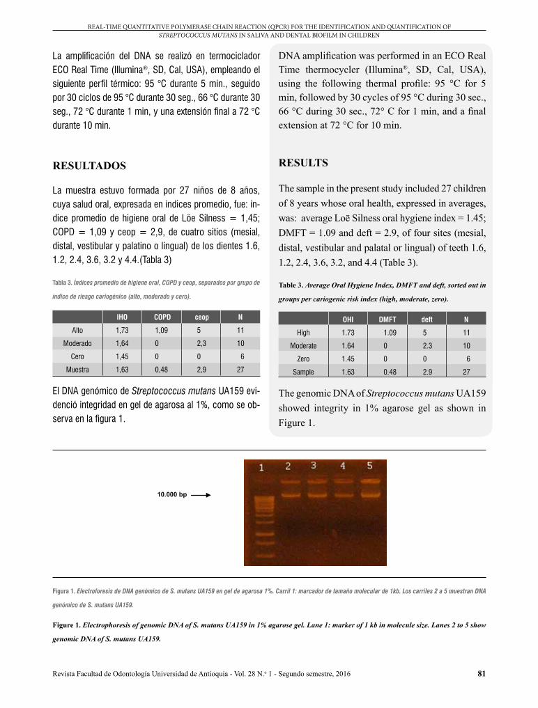
La muestra estuvo formada por 27 niños de 8 años, cuya salud oral, expresada en índices promedio, fue: ín-dice promedio de higiene oral de Löe Silness = 1,45; COPD = 1,09 y ceop = 2,9, de cuatro sitios (mesial, distal, vestibular y palatino o lingual) de los dientes 1.6, 1.2, 2.4, 3.6, 3.2 y 4.4.(Tabla 3)
Tabla 3. Índices promedio de higiene oral, COPD y ceop, separados por grupo de
índice de riesgo cariogénico (alto, moderado y cero).
IHO COPD ceop N
Alto 1,73 1,09 5 11
Moderado 1,64 0 2,3 10
Cero 1,45 0 0 6
Muestra 1,63 0,48 2,9 27
El DNA genómico de Streptococcus mutans UA159 evi-denció integridad en gel de agarosa al 1%, como se ob-serva en la figura 1.
DNA amplification was performed in an ECO Real Time thermocycler (Illumina®, SD, Cal, USA), using the following thermal profile: 95 °C for 5 min, followed by 30 cycles of 95 °C during 30 sec., 66 °C during 30 sec., 72° C for 1 min, and a final extension at 72 °C for 10 min.
RESULTS
The sample in the present study included 27 children of 8 years whose oral health, expressed in averages, was: average Loë Silness oral hygiene index = 1.45; DMFT = 1.09 and deft = 2.9, of four sites (mesial, distal, vestibular and palatal or lingual) of teeth 1.6, 1.2, 2.4, 3.6, 3.2, and 4.4 (Table 3).
Table 3. Average Oral Hygiene Index, DMFT and deft, sorted out in
groups per cariogenic risk index (high, moderate, zero).
OHI DMFT deft N
High 1.73 1.09 5 11
Moderate 1.64 0 2.3 10
Zero 1.45 0 0 6
Sample 1.63 0.48 2.9 27
The genomic DNA of Streptococcus mutans UA159 showed integrity in 1% agarose gel as shown in Figure 1.
10.000 bp
Figura 1. Electroforesis de DNA genómico de S. mutans UA159 en gel de agarosa 1%. Carril 1: marcador de tamaño molecular de 1kb. Los carriles 2 a 5 muestran DNA
genómico de S. mutans UA159.
Figure 1. Electrophoresis of genomic DNA of S. mutans UA159 in 1% agarose gel. Lane 1: marker of 1 kb in molecule size. Lanes 2 to 5 show
genomic DNA of S. mutans UA159.

82
TÉCNICA DE REACCIÓN DE POLIMERASA EN CADENA (QPCR) EN TIEMPO REAL PARA LA IDENTIFICACIÓN Y CUANTIFICACIÓN DE STREPTOCOCCUS MUTANS EN SALIVA Y BIOPELÍCULA DENTARIA DE NIÑOS
Revista Facultad de Odontología Universidad de Antioquia - Vol. 28 N.o 1 - Segundo semestre, 2016
La utilización de los partidores universales demostró la presencia y amplificación de DNA bacteriano por medio de PCR convencional (a tiempo final) con los partidores universales para 16Sr RNA.
La utilización de los partidores Smut 3368-F y Smut 3481-R (Figura 2) evidenció especificidad median-te la obtención de un amplicón del tamaño esperado (114 pb),20 puesto que dichos partidores sólo fueron de-tectados en las muestras de DNA de S. mutans UA159 y en DNA de saliva y biopelícula dental.
The use of universal primers demonstrated the presence and amplification of bacterial DNA by conventional PCR (end-point) with universal primers for 16Sr RNA.
The use of Smut 3368-F and Smut 3481-R primers (Figure 2) showed specificity by obtaining an amplicon of expected size (114 pb),20 since these primers were only detected in the DNA samples of S. mutans UA159 and in DNA from saliva and dental biofilm.
114 bp
100 bp
Figura 2. Separación mediante electroforesis en gel de agarosa al 2% de fragmento de 114 bp del gen gtfB amplificado mediante PCR. El carril 1 corresponde a estándar
de tamaño molecular de 1 kb. Los carriles 2 y 3 corresponden a amplificación empleando DNA de S. mutans UA159. Los carriles 4 y 5 corresponden a amplificaciones
empleando DNA de muestras de saliva y biopelícula dental (C6 S, C6 PB). Los carriles 6 y 7 corresponden a amplificaciones empleando DNA de S. sanguinis. Los
carriles 8 al 10 corresponden a amplificaciones empleando DNA de E. coli. El carril 11 corresponde a una amplificación empleando agua ultrapura (miliQ). El carril 12
corresponde a un estándar de tamaño molecular de 50 bp.
Figure 2. Electrophoretic separation of a fragment of 114 bp of gene gtfB in 2% agarose gel amplified by means of PCR. Lane 1 corresponds
to a standard molecular size of 1 kb. Lanes 2 and 3 correspond to amplification using DNA of S. mutans UA159. Lanes 4 and 5 correspond to
amplifications using DNA from saliva and dental biofilm samples (S C6, C6 BF). Lanes 6 and 7 correspond to amplifications using DNA of
S. sanguinis. Lanes 8 to 10 correspond to amplifications using DNA from E. coli. Lane 11 corresponds to amplification using ultrapure water
(miliQ). Lane 12 corresponds to a standard molecular size of 50 bp.
La curva estándar se construyó a partir de la dilución del DNA aislado de S. mutans UA159, cuya concentración inicial fue de 128 ng/µL (Figura 8). En la curva de fusión (o melting) para la amplificación del DNA de S. mutans UA159 (Figura 3) se observa un solo pico de amplifica-ción, a una misma temperatura para todas las dilucio-nes. Los duplicados emiten el despegue de fluorescen-cia logrando la cuantificación de las muestras con un
The standard curve was designed based on the dilution of DNA isolated from S. mutans UA159, whose initial concentration was 128 ng/µL (Figure 8). The melting curve for the amplification of DNA of S. mutans UA159 (Figure 3) shows a single amplification pick, to the same temperature for all the dilutions. Duplicates issue fluorescence, achieving the quantification of samples with a

83
REAL-TIME QUANTITATIVE POLYMERASE CHAIN REACTION (QPCR) FOR THE IDENTIFICATION AND QUANTIFICATION OF STREPTOCOCCUS MUTANS IN SALIVA AND DENTAL BIOFILM IN CHILDREN
Revista Facultad de Odontología Universidad de Antioquia - Vol. 28 N.o 1 - Segundo semestre, 2016
Y ax
is: D
eriv
ativ
e flu
ores
cenc
e
0.18
0.16
0.14
0.12
0.10
0.08
0.06
0.04
0.02
0.00
X axis: Temperature (ºC)
65 70 75 80 85 90
Y ax
is: F
luor
esce
nce
0.55
0.50
0.45
0.40
0.35
0.30
0.25
0.20
0.15
0.10
0.05
0.00
-0.05
X axis: Cycle
0 5 10 15 20 25 30
delta de 3,36; es decir, entre cada ciclo de amplificación en el que se detecta fluorescencia existe una diferencia de 3,36 ciclos (Figura 4).
delta of 3.36; i.e. between each amplification cycle in which fluorescence is detected there is a difference of 3.36 cycles (Figure 4).
Figura 3. Curva de fusión para un fragmento de 114 bp del gen gtfB. Se observa la única temperatura de amplificación de las diluciones seriadas de DNA de S. mutans
UA159.
Figure 3. Melting curve for a fragment of 114 bp of gtfB gene. Note the only amplification temperature of serial dilutions of DNA of S. mutans
UA159.
Figura 4. Gráfico de concentración por ciclo de amplificación para un fragmento de 114 bp del gen gtfB empleando diluciones seriadas de DNA genómico de S. mutans
UA159.
Figure 4. Graph of concentration per amplification cycle for a fragment of 114 bp of gtfB gene using serial dilutions of genomic DNA of S.
mutans UA159.
84
TÉCNICA DE REACCIÓN DE POLIMERASA EN CADENA (QPCR) EN TIEMPO REAL PARA LA IDENTIFICACIÓN Y CUANTIFICACIÓN DE STREPTOCOCCUS MUTANS EN SALIVA Y BIOPELÍCULA DENTARIA DE NIÑOS
Revista Facultad de Odontología Universidad de Antioquia - Vol. 28 N.o 1 - Segundo semestre, 2016
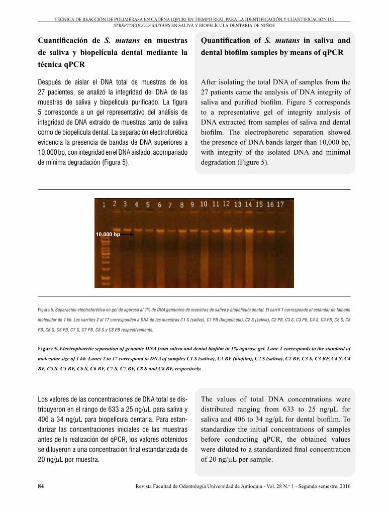
Cuantificación de S. mutans en muestras de saliva y biopelícula dental mediante la técnica qPCR
Después de aislar el DNA total de muestras de los 27 pacientes, se analizó la integridad del DNA de las muestras de saliva y biopelícula purificado. La figura 5 corresponde a un gel representativo del análisis de integridad de DNA extraído de muestras tanto de saliva como de biopelícula dental. La separación electroforética evidencia la presencia de bandas de DNA superiores a 10.000 bp, con integridad en el DNA aislado, acompañado de mínima degradación (Figura 5).
Quantification of S. mutans in saliva and dental biofilm samples by means of qPCR
After isolating the total DNA of samples from the 27 patients came the analysis of DNA integrity of saliva and purified biofilm. Figure 5 corresponds to a representative gel of integrity analysis of DNA extracted from samples of saliva and dental biofilm. The electrophoretic separation showed the presence of DNA bands larger than 10,000 bp, with integrity of the isolated DNA and minimal degradation (Figure 5).
10.000 bp
Figura 5. Separación electroforética en gel de agarosa al 1% de DNA genómico de muestras de saliva y biopelícula dental. El carril 1 corresponde al estándar de tamaño
molecular de 1 kb. Los carriles 2 al 17 corresponden a DNA de las muestras C1 S (saliva), C1 PB (biopelícula), C2 S (saliva), C2 PB, C3 S, C3 PB, C4 S, C4 PB, C5 S, C5
PB, C6 S, C6 PB, C7 S, C7 PB, C8 S y C8 PB respectivamente.
Figure 5. Electrophoretic separation of genomic DNA from saliva and dental biofilm in 1% agarose gel. Lane 1 corresponds to the standard of
molecular size of 1 kb. Lanes 2 to 17 correspond to DNA of samples C1 S (saliva), C1 BF (biofilm), C2 S (saliva), C2 BF, C3 S, C3 BF, C4 S, C4
BF, C5 S, C5 BF, C6 S, C6 BF, C7 S, C7 BF, C8 S and C8 BF, respectively.
Los valores de las concentraciones de DNA total se dis-tribuyeron en el rango de 633 a 25 ng/µL para saliva y 406 a 34 ng/µL para biopelícula dentaria. Para estan-darizar las concentraciones iniciales de las muestras antes de la realización del qPCR, los valores obtenidos se diluyeron a una concentración final estandarizada de 20 ng/µL por muestra.
The values of total DNA concentrations were distributed ranging from 633 to 25 ng/μL for saliva and 406 to 34 ng/μL for dental biofilm. To standardize the initial concentrations of samples before conducting qPCR, the obtained values were diluted to a standardized final concentration of 20 ng/µL per sample.
85
REAL-TIME QUANTITATIVE POLYMERASE CHAIN REACTION (QPCR) FOR THE IDENTIFICATION AND QUANTIFICATION OF STREPTOCOCCUS MUTANS IN SALIVA AND DENTAL BIOFILM IN CHILDREN
Revista Facultad de Odontología Universidad de Antioquia - Vol. 28 N.o 1 - Segundo semestre, 2016
Y ax
is: D
eriv
ativ
e flu
ores
cenc
e
0.20
0.18
0.16
0.14
0.12
0.10
0.08
0.06
0.04
0.02
0.00
0.60
0.55
0.50
0.45
0.40
0.35
0.30
0.25
0.20
0.15
0.10
0.05
0.00
-0.05
X axis: Temperature (ºC)
65 70 75 80 85 90
Y ax
is: Δ
R
X axis: Cycle
0 5 10 15 20 25 30
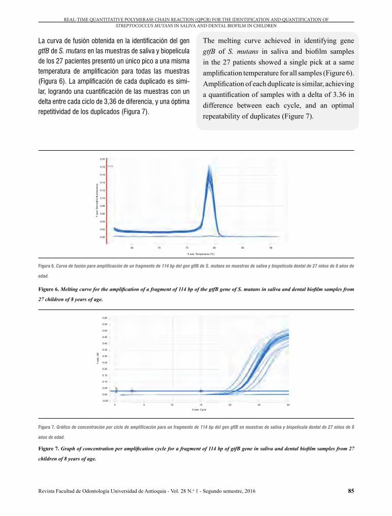
La curva de fusión obtenida en la identificación del gen gtfB de S. mutans en las muestras de saliva y biopelícula de los 27 pacientes presentó un único pico a una misma temperatura de amplificación para todas las muestras (Figura 6). La amplificación de cada duplicado es simi-lar, logrando una cuantificación de las muestras con un delta entre cada ciclo de 3,36 de diferencia, y una óptima repetitividad de los duplicados (Figura 7).
The melting curve achieved in identifying gene gtfB of S. mutans in saliva and biofilm samples in the 27 patients showed a single pick at a same amplification temperature for all samples (Figure 6). Amplification of each duplicate is similar, achieving a quantification of samples with a delta of 3.36 in difference between each cycle, and an optimal repeatability of duplicates (Figure 7).
Figura 6. Curva de fusión para amplificación de un fragmento de 114 bp del gen gtfB de S. mutans en muestras de saliva y biopelícula dental de 27 niños de 8 años de
edad.
Figure 6. Melting curve for the amplification of a fragment of 114 bp of the gtfB gene of S. mutans in saliva and dental biofilm samples from
27 children of 8 years of age.
Figura 7. Gráfico de concentración por ciclo de amplificación para un fragmento de 114 bp del gen gtfB en muestras de saliva y biopelícula dental de 27 niños de 8
años de edad.
Figure 7. Graph of concentration per amplification cycle for a fragment of 114 bp of gtfB gene in saliva and dental biofilm samples from 27
children of 8 years of age.

86
TÉCNICA DE REACCIÓN DE POLIMERASA EN CADENA (QPCR) EN TIEMPO REAL PARA LA IDENTIFICACIÓN Y CUANTIFICACIÓN DE STREPTOCOCCUS MUTANS EN SALIVA Y BIOPELÍCULA DENTARIA DE NIÑOS
Revista Facultad de Odontología Universidad de Antioquia - Vol. 28 N.o 1 - Segundo semestre, 2016
Los valores obtenidos en la cuantificación del DNA de S. mutans en muestras de saliva y biopelícula dental por qPCR se muestran en la tabla 4. Con base en el tamaño del genoma de S. mutans se calculó el número de bacte-rias en mL de saliva y biopelícula dental.35
The values obtained by quantifying DNA of S. mutans in saliva and dental biofilm samples by qPCR are shown in table 4. The number of bacteria in mL of saliva and dental biofilm was calculated based on the size of the S mutans genome.35
Tabla 4. Cuantificación de S. mutans en muestras de saliva y biopelícula dental de niños de 8 años de edad mediante la técnica de qPCR.
Muestra Concentración de DNA de S. mutans en saliva (ng/µL)
Concentración de DNA de S. mutans en biopelícula dental (ng/µL)
Nº de S. mutans en saliva /mL
Nº de S. mutans en biopelícula dental /mL
C1 5 0,15 1,6x10⁷ 4,89x10⁵
C4 3,1 4 1x10⁷ 13x106
C5 0,8 0,9 2,6x10⁶ 2,9x10⁶
C6 0,12 0,32 3,9x10⁵ 1x10⁶
C7 0,4 2,1 1,1x10⁶ 6,8x10⁶
C8 0,5 0,6 1,6x10⁶ 1,9x10⁶
C9 0,08 0.03 2,6x10⁵ 9,7x10⁴
C10 1,62 0,07 5,2x10⁶ 2,2x10⁵
C11 0,1 0,14 3,2x10⁵ 4,5x10⁵
C12 0,05 0,09 1,6x10⁵ 2,9x10⁵
C13 0,13 0,4 4,2x10⁵ 1,3x10⁶
C14 0,14 5,125 4,5x10⁵ 16x107
C16 0,35 2,4 1,1x10⁶ 7,8x10⁶
C17 0 0,07 0 2,2x10⁵
C18 0,02 0,02 6,5x10⁴ 6,5x10⁴
C20 0,03 0,03 9,7x10⁴ 9,7x10⁴
B26 0,02 0,02 6,5x10⁴ 6,5x10⁴
B27 0,02 0,06 6,5x10⁴ 1,9x10⁵
B28 0,02 0,03 6,5x10⁴ 9,7x10⁴
B29 0,02 0,01 6,5x10⁴ 3,2x10⁴
B21 0,02 0,01 6,5x10⁴ 3,2x10⁴
B25 0 0 0 0
B2 0 0,1 0 3,2x10⁵
B4 0,01 1,6 3,2x10⁴ 5,2x10⁶
A7 0 0 0 0
A14 0 0 0 0
A21 0 0,16 0 5,2x10⁵
0,47 0,71 14,85x10⁵ 74,24x10⁵

87
REAL-TIME QUANTITATIVE POLYMERASE CHAIN REACTION (QPCR) FOR THE IDENTIFICATION AND QUANTIFICATION OF STREPTOCOCCUS MUTANS IN SALIVA AND DENTAL BIOFILM IN CHILDREN
Revista Facultad de Odontología Universidad de Antioquia - Vol. 28 N.o 1 - Segundo semestre, 2016
Table 4. Quantification of S. mutans in saliva and dental biofilm samples from 8-year-old children through the qPCR technique.
Sample Concentration of DNA of S. mutans in saliva (ng/µL)
Concentration of DNA of S. mutans in dental biofilm (ng/µL)
Number of S. mutans in saliva /mL
Nº of S. mutans in dental biofilm /mL
C1 5 0.15 1.6 x 10⁷ 4.89 x 10⁵
C4 3.1 4 1 x 10⁷ 13 x 106
C5 0.8 0.9 2.6 x 10⁶ 2.9 x 10⁶
C6 0.12 0.32 3.9 x 10⁵ 1 x 10⁶
C7 0.4 2.1 1.1 x 10⁶ 6.8 x 10⁶
C8 0.5 0.6 1.6 x 10⁶ 1.9 x 10⁶
C9 0.08 0.03 2.6 x 10⁵ 9.7 x 10⁴
C10 1.62 0.07 5.2 x 10⁶ 2.2 x 10⁵
C11 0.1 0.14 3.2 x 10⁵ 4.5 x 10⁵
C12 0.05 0.09 1.6 x 10⁵ 2.9 x 10⁵
C13 0.13 0.4 4.2 x 10⁵ 1.3 x 10⁶
C14 0.14 5.125 4.5 x 10⁵ 16 x 107
C16 0.35 2.4 1.1 x 10⁶ 7.8 x 10⁶
C17 0 0.07 0 2.2 x 10⁵
C18 0.02 0.02 6.5 x 10⁴ 6.5 x 10⁴
C20 0.03 0.03 9.7 x 10⁴ 9.7 x 10⁴
B26 0.02 0.02 6.5 x 10⁴ 6.5 x 10⁴
B27 0.02 0.06 6.5 x 10⁴ 1.9 x 10⁵
B28 0.02 0.03 6.5 x 10⁴ 9.7 x 10⁴
B29 0.02 0.01 6.5 x 10⁴ 3.2 x 10⁴
B21 0.02 0.01 6.5 x 10⁴ 3.2 x 10⁴
B25 0 0 0 0
B2 0 0.1 0 3.2 x 10⁵
B4 0.01 1.6 3.2 x 10⁴ 5.2 x 10⁶
A7 0 0 0 0
A14 0 0 0 0
A21 0 0.16 0 5.2 x 10⁵
0.47 0.71 14.85 x 10⁵ 74.24 x 10⁵
La linealidad obtenida para la curva estándar empleada en la cuantificación mediante qPCR (Figura 8) da cuenta de un 98% de eficiencia de la técnica. Por otra parte, el comportamiento de los controles sin templado (NTC) muestra una fluorescencia muy por debajo del umbral mínimo en que fueron cuantificadas las muestras con templado, lo que valida los resultados de cuantificación obtenidos con esta técnica (Figura 9).
The linearity achieved for the standard curve used in quantifying by qPCR (Figure 8) accounts for 98% technique efficiency. On the other hand, the behavior of No Template Controls (NTC) shows fluorescence far below the minimum threshold in which the templated samples were quantified, validating the quantification results obtained with this technique (Figure 9).

88
TÉCNICA DE REACCIÓN DE POLIMERASA EN CADENA (QPCR) EN TIEMPO REAL PARA LA IDENTIFICACIÓN Y CUANTIFICACIÓN DE STREPTOCOCCUS MUTANS EN SALIVA Y BIOPELÍCULA DENTARIA DE NIÑOS
Revista Facultad de Odontología Universidad de Antioquia - Vol. 28 N.o 1 - Segundo semestre, 2016
Y ax
is: C
q
23.0
22.5
22.0
21.5
21.0
20.5
20.0
19.5
19.0
X axis: Quantity
2 x 10³ 4 x 10³ 6 x 10³ 8 x10³ 104 2 x x104
Y ax
is: Δ
R
0.05
0.04
0.03
0.02
0.01
0.00
-0.01
-0.02
-0.03
X axis: Cycle
0 5 10 15 20 25
Figura 8. Curva estándar. Gráfica de concentracion de DNA a partir del valor cq (ciclo de cuantificación) de la muestra desconocida.
Figure 8. Standard curve. Graph of concentration of DNA starting from the value of cycle of quantification (cq) of the unknown sample.
Figura 9. Gráfica de comportamiento de las muestras sin templado (NTC).
Figure 9. Graph of behavior of non-templated samples (NTC).
DISCUSSION
The findings yielded by qPCR amplification showed a high percentage of efficiency and a reasonable delta in between cycles. The analysis of these parameters allows validating the results thanks to the sensitivity and accuracy of this technique, as previously published,24-27 with similar methodology. It is therefore possible to claim
DISCUSIÓN
Los resultados obtenidos de la amplificación mediante qPCR mostraron un alto porcentaje de eficiencia y un razonable delta entre cada ciclo. El análisis de estos parámetros permite validar los resultados obtenidos, basados en la sensibilidad y exactitud de la técnica, tal como han sido publicados previamente,24-27 con metodología similar. Por lo tanto, es posible afirmar
89
REAL-TIME QUANTITATIVE POLYMERASE CHAIN REACTION (QPCR) FOR THE IDENTIFICATION AND QUANTIFICATION OF STREPTOCOCCUS MUTANS IN SALIVA AND DENTAL BIOFILM IN CHILDREN
Revista Facultad de Odontología Universidad de Antioquia - Vol. 28 N.o 1 - Segundo semestre, 2016
que la metodología presentada identifica y cuantifica la especie Streptococcus mutans en muestras de saliva y biopelícula dental.
Las metodologías tradicionales de identificación y cuantificación microbiana utilizan el método de cultivo en placa para detectar la cantidad o la presencia de S. mutans, lo cual implica que las muestras se procesen en forma inmediata y no se puedan almacenar por tiempos muy prolongados, imposibilitando que estas se congelen para su acopio y posterior análisis. Adicionalmente, el método de cultivo presenta limitaciones que afectan la precisión y coherencia en la evaluación de la infección por S. mutans. Los cultivos muestran ciertos inconvenientes, como un laborioso y prolongado tiempo de procesamiento microbiológico y un menor nivel de sensibilidad. Estas limitaciones dificultan que los métodos de cultivo tradicionales proporcionen evaluaciones microbianas precisas para la identificación y cuantificación de S. mutans que se asocia con la determinación de susceptibilidad de caries para los individuos en situación de riesgo.37
Además, existe el inconveniente relacionado con el al-macenamiento o preservación de los medios de cultivo, que debe garantizar la viabilidad en el tiempo; se debe reducir al mínimo el riesgo de contaminación y permitir que la pureza del cultivo permanezca inalterable.38
El traslado de muestras congeladas es el método prefe-rido para transportar y almacenar grandes colecciones de especies microbianas. Estas condiciones aumentan la complejidad y el riesgo de perder la viabilidad de las células para realizar cuantificación mediante la técnica de cultivo tradicional.39
Por otra parte, el PCR convencional, o TF, es una técnica cualitativa que solo permite indicar presencia o ausencia de un determinado fragmento de DNA y por ende asociar la presencia o ausencia de un determinado microorga-nismo. En cambio, la qPCR es una técnica rápida con la que se pueden obtener resultados cualitativos y cuanti-tativos con facilidad, y que pueden provenir de muestras almacenadas por tiempos muy prolongados.40
that this methodology identifies and quantifies Streptococcus mutans spp. in saliva and dental biofilm.
The traditional methods for microbial identification and quantification used the method of cultivation on plaque to detect the presence or quantity of S. mutans, which implies processing samples immediately without the possibility of storing them for long periods of times or freezing them for later analysis. In addition, the culture method has limitations that affect accuracy and consistency in evaluating infection by S. mutans. Cultures show certain disadvantages, such as extended time for microbiological processing and a lower level of sensitivity. These limitations make it difficult for traditional culture methods to provide accurate microbial evaluations to identify and quantify S. mutans associated with caries susceptibility in individuals at risk.37
In addition, there is the drawback related to storage or preservation of culture agents, which must ensure viability over time; contamination risks should be minimal ensuring that culture purity remains unaffected.38
The transportation of frozen samples is the preferred method for moving and storing large collections of microbial species. These conditions increase complexity as well as the risk of losing the viability of cells for quantification with the traditional culture technique.39
On the other hand, conventional PCR is a qualitative technique that only allows indicating the presence or absence of a specific fragment of DNA and therefore associating it with presence or absence of a particular microorganism. Instead, qPCR is a quick technique that easily provides qualitative and quantitative results that can come from samples stored for long periods of time.40

90
TÉCNICA DE REACCIÓN DE POLIMERASA EN CADENA (QPCR) EN TIEMPO REAL PARA LA IDENTIFICACIÓN Y CUANTIFICACIÓN DE STREPTOCOCCUS MUTANS EN SALIVA Y BIOPELÍCULA DENTARIA DE NIÑOS
Revista Facultad de Odontología Universidad de Antioquia - Vol. 28 N.o 1 - Segundo semestre, 2016
La qPCR, a pesar de ser más compleja y utilizar equi-pos de mayor costo y mayor tecnología que los cultivos tradicionales de dilución en serie, es una técnica que, una vez establecida, resulta de menor costo y de mayor rapidez y precisión.24 A su vez, estas ventajas permiten reducir el costo de laboratorio cuando este examen es solicitado al paciente como complemento para la deter-minación del riesgo cariogénico individual. Sin embargo, la determinación de la masa inicial de la muestra (saliva y placa bacteriana colectada) es considerada una etapa indispensable para el establecimiento del recuento bac-teriano, hecho que determina una debilidad del presente estudio.
La extracción de DNA realizada a partir de las muestras de saliva y biopelícula mostró cierto nivel de degrada-ción, a diferencia del observado en las muestras de cul-tivo puro de S. mutans UA159. La explicación de este fenómeno se encuentra en la diversidad y abundancia del ecosistema oral. Para la resolución de este fenómeno se utilizó RNAasa, lo que permitió aumentar la integridad de las muestras.
La elección del gen blanco, según Ono y colaborado-res,41 se debe a que se describe al gen spaP como el que presenta la mayor sensibilidad para la detección de S mutans. Sin embargo, un reciente estudio de cepas provenientes de la población chilena reporta la presencia del gen gtfB en el 100% de las cepas de S. mutans,45 mientras que solo el 63% muestra la presencia del gen spaP,42-44 razón que explica la elección del gen gtfB para el presente estudio. Sin embargo, este hecho minimiza, pero no garantiza, los defectos en la detección, debido a la posibilidad de encontrar polimorfismo.
La etiopatogenia de la caries dental se ha vinculado tradi-cionalmente con los desequilibrios de la biopelícula den-taria; 1, 12 sin embargo, para la definición de riesgo de la enfermedad se utilizan conteos de UFC de S. mutans en saliva. Esta dicotomía se basaba en la fácil medición vo-lumétrica de la muestra de saliva, respecto de la dificultad para determinar la cantidad de biopelícula dentaria. Actual-mente, con las técnicas moleculares es posible cuantifi-car dicho riesgo en la biopelícula dentaria; por lo tanto, es
Despite being more complex and requiring costly and more advanced equipment in comparison to the traditional method of cultivation in dilution series, once stablished, qPCR results in lower cost and greater speed and accuracy.24 These advantages also allow reducing laboratory costs when patients are requested this test as a complement to determine individual cariogenic risk. However, determining the initial sample mass (collected saliva and bacterial plaque) is an essential stage for establishing bacterial count —a fact that appears as a weakness of the present study—.
The extraction of DNA from saliva and biofilm samples showed some level of degradation, contrasting with the observations in samples of pure culture of S. mutans UA159. This phenomenon may be explained by the diversity and abundance of the oral ecosystem. RNAasa was used to overcome this problem, thus increasing the integrity of samples.
According to Ono et al,41 the white gen is chosen because the spaP gene is described as the one with the greatest sensitivity to detect S mutans. However, a recent study on strains in Chilean population reported the presence of the gtfB gene in 100% of S. mutans strains,45 while only 63% shows the presence of the spaP gene,42-44 which explains the choice of the gtfB gene for this study. However, this fact minimizes, but does not guarantee, detection defects, due to the possibility of finding polymorphism.
The etiology of dental caries has been traditionally linked to imbalances in dental biofilm; 1, 12 however, counts of CFU of S. mutans in saliva are used to define disease risk. This dichotomy used to be based on the ease of volumetric measurement of saliva samples compared to the difficulty in determining the amount of dental biofilm. Currently, molecular techniques allow quantifying such risk in dental biofilm; therefore, it is
91
REAL-TIME QUANTITATIVE POLYMERASE CHAIN REACTION (QPCR) FOR THE IDENTIFICATION AND QUANTIFICATION OF STREPTOCOCCUS MUTANS IN SALIVA AND DENTAL BIOFILM IN CHILDREN
Revista Facultad de Odontología Universidad de Antioquia - Vol. 28 N.o 1 - Segundo semestre, 2016
recomendable obtener muestras de biopelícula, dado que esta constituye el nicho natural de S. mutans en la cavidad oral. El presente estudio mostró mayor canti-dad de S. mutans en biopelícula (media 74,24x10⁵) que en saliva (media 14,85x10⁵), lo que constituye una herramienta de mayor precisión para la identificación y cuantificación de S. mutans en biopelícula en la etapa de evaluación del riesgo cariogénico.
Finalmente, la adecuada distribución de las dilucio-nes sobre la curva de estandarización indicó precisión procedimental y metodológica en la construcción de la gráfica (se utilizaron los cinco puntos recomendados), lo cual permitió validar las posteriores cuantificaciones efectuadas para cada una de las muestras. Similar rigu-rosidad se observó en el comportamiento de la fluores-cencia bajo el umbral, al analizar los controles sin tem-plado, lo que evidenció la ausencia de contaminaciones externas durante los procesos, así como la inexistencia de dímeros de partidores.
CONCLUSIONES
La metodología desarrollada permite identificar y cuanti-ficar S. mutans en muestras de saliva y biopelícula den-tal almacenadas en frío, de manera específica, sensible y rápida, utilizando partidores específicos. La técnica de qPCR posibilita un mayor volumen de procesamiento de muestras, lo que permite ahorrar tiempo en la obtención de los resultados.
AGRADECIMIENTOS
La presente investigación fue posible gracias a fondos otorgados por FONIS-CONICYT Chile al proyecto SA-13i20205.
CONFLICTO DE INTERÉS
Los autores declaran no presentar ningún conflicto de interés directo o indirecto con ninguno de los materiales y equipos utilizados en el presente estudio.
recommended to obtain biofilm samples, since this is the natural niche of S. mutans in the oral cavity. The present study showed greater amount of S. mutans in biofilm (average 74.24 x 10⁵) that saliva (average 14, 85 x 105), making it a tool of greater precision for the identification and quantification of S. mutans in biofilm during the stage of evaluation of cariogenic risk.
Finally, the appropriate distribution of dilutions on the standardization curve showed the procedural and methodological precision in creating the graph (the five recommended points were used), which allowed validating subsequent quantifications performed for each sample. The same accuracy was observed in the behavior of fluorescence under the threshold in analyzing No Template Controls, demonstrating the absence of external contaminations during the processes, as well as the absence of primer dimers.
CONCLUSIONS
The used methodology allows identifying and quantifying S. mutans in saliva and dental biofilm samples stored in cold, in a specific, sensitive and quick manner, using specific primers. The qPCR technique enables processing a greater volume of samples, which saves time in obtaining results.
ACKNOWLEDGMENTS
This research was possible thanks to funding provided by FONIS-CONICYT Chile to project SA-13i20205.
CONFLICT OF INTEREST
The authors declare not having any type of direct or indirect conflict of interest with any of the materials and equipment used in the present study.

92
TÉCNICA DE REACCIÓN DE POLIMERASA EN CADENA (QPCR) EN TIEMPO REAL PARA LA IDENTIFICACIÓN Y CUANTIFICACIÓN DE STREPTOCOCCUS MUTANS EN SALIVA Y BIOPELÍCULA DENTARIA DE NIÑOS
Revista Facultad de Odontología Universidad de Antioquia - Vol. 28 N.o 1 - Segundo semestre, 2016
CORRESPONDENCIA
Gustavo Moncada, DDSUniversidad Mayor(+56 2) 2328 [email protected] Bernardo O’Higgins 2013Santiago, Chile
CORRESPONDING AUTHOR
Gustavo Moncada, DDSUniversidad Mayor(+56 2) 2328 [email protected] Bernardo O’Higgins 2013Santiago, Chile
1. Selwitz RH, Ismail AI, Pitts NB. Dental caries. Lancet 2007; 369(9555): 51-59
2. Petersen PE. The World Oral Health Report 2003: continuous improvement of oral health in the 21st century—the approach of the WHO Global Oral Health Programme. Community Dent Oral Epidemiol 2003; 31 Suppl 1: 3-23.
3. Petersen PE, Bourgeois D, Ogawa H, Estupinan-Day S, Ndiaye C. The global burden of oral diseases and risks to oral health. Bull World Health Organ 2005; 83(9): 661-669.
4. Urzua I, Mendoza C, Arteaga O, Rodríguez G, Cabello R, Faleiros S et al. Dental caries prevalence and tooth loss in Chilean adult population: first national dental examination survey. Int J Dent 2012; 2012. http://dx.doi.org/10.1155/2012/810170.
5. Marsh P, Martin M, Lewis M, Williams D. Oral microbiology. 5 ed. Londres: Churchill Livingstone; 2009.
6. Simón-Soro A, Belda-Ferre P, Cabrera-Rubio R, Alcaraz LD, Mira A. A tissue-dependent hypothesis of dental caries. Caries Res 2013; 47(6): 591-600.
7. Burne RA. Oral streptococci... products of their environment. J Dent Res. 1998; 77(3): 445-452.
8. Rupf S, Merte K, Eschrich K, Kneist S. Streptococcus sobrinus in children and its influence on caries activity. Eur Arch Paediatr Dent 2006; 7(1): 17-22.
9. Gordan VV, Garvan CW, Ottenga ME, Schulte R, Harris PA, McEdward D et al. Could alkali production be considered an approach for caries control? Caries Res 2010; 44(6): 547-554.
REFERENCIAS / REFERENCES
10. Liu Y, Dong Y, Chen YY, Burne RA. Environmental and growth phase regulation of the Streptococcus gordonii arginine deiminase genes. Appl Environ Microbiol 2008; 74(16): 5023-5030.
11. Dong Y, Chen YY, Burne RA. Control of expression of the arginine deiminase operon of Streptococcus gordonii by CcpA and Flp. J Bacteriol 2004; 186(8): 2511-2514.
12. Fejerskov O. Changing paradigms in concepts on dental caries: consequences for oral health care. Caries Res 2004; 38(3): 182-191.
13. Nauntofte B, Tenovuo JO, Lagerlöf F. Secretion and composition of saliva. En: Fejerskov O, Kidd E (eds). 1 ed. Dental caries: the disease and its clinical management. Oxford: Blackwell; 2003. p. 7-27
14. Fejerskov O. Different concepts of dental caries and their implications. 2 ed. Copenhagen: Munksgaard; 1994.
15. Facklam R. What happened to the streptococci: overview of taxonomic and nomenclature changes. Clin Microbiol Rev 2002; 15(4): 613-630.
16. Nakano K, Nomura R, Nakagawa I, Hamada S, Ooshima T. Demonstration of Streptococcus mutans with a cell wall polysaccharide specific to a new serotype, k, in the human oral cavity. J Clin Microbiol 2004; 42(1): 198-202.
17. Nakano K, Inaba H, Nomura R, Nemoto H, Takeda M, Yoshioka H et al. Detection of cariogenic Streptococcus mutans in extirpated heart valve and atheromatous plaque specimens. J Clin Microbiol 2006; 44(9): 3313-3317.
18. Bowen WH, Koo H. Biology of Streptococcus mutans-derived glucosyltransferases: role in extracellular matrix formation of cariogenic biofilms. Caries Res 2011; 45(1): 69-86.

93
REAL-TIME QUANTITATIVE POLYMERASE CHAIN REACTION (QPCR) FOR THE IDENTIFICATION AND QUANTIFICATION OF STREPTOCOCCUS MUTANS IN SALIVA AND DENTAL BIOFILM IN CHILDREN
Revista Facultad de Odontología Universidad de Antioquia - Vol. 28 N.o 1 - Segundo semestre, 2016
19. Yano A, Konno N, Imai S, Kato H. Inhibitory effects of polysaccharides on the cariogenic activities of Streptococcus mutans. Biosci Biotechnol Biochem 2012; 76(12): 2313-2316.
20. Napimoga MH, Höfling JF, Klein MI, Kamiya RU, Gonçalves RB. Transmission, diversity and virulence factors of Streptococcus mutans genotypes. J Oral Sci 2005; 47(2): 59-64.
21. Senneby A, Mejàre I, Sahlin NE, Svensäter G, Rohlin M. Diagnostic accuracy of different caries risk assessment methods. A systematic review. J Dent 2015; 43(12): 1385-93.
22. Karjalainen S, Tolvanen M, Pienihäkkinen K, Söderling E, Lagström H, Simell O et al. High sucrose intake at 3 years of age is associated with increased salivary counts of mutans streptococci and lactobacilli, and with increased caries rate from 3 to 16 years of age. Caries Res 2015; 49(2): 125-132.
23. Twetman L, Twetman S. Comparison of two chair-side tests for enumeration of Mutans Streptococci in saliva. Oral Health Dent Manag 2014; 13(3): 580-583.
24. Childers NK, Osgood RC, Hsu KL, Manmontri C, Momeni SS, Mahtani HK et al. Real-time quantitative polymerase chain reaction for enumeration of Streptococcus mutans from oral samples. Eur J Oral Sci 2011; 119(6): 447-454.
25. Al-Robaiy S, Rupf S, Eschrich K. Rapid competitive PCR using melting curve analysis for DNA quantification. Biotechniques 2001; 31(6): 1382-1386, 1388.
26. Rupf S, Merte K, Eschrich K. Quantification of bacteria in oral samples by competitive polymerase chain reaction. J Dent Res 1999; 78(4): 850-856.
27. Rupf S, Merte K, Kneist S, Al-Robaiy S, Eschrich K. Comparison of different techniques of quantitative PCR for determination of Streptococcus mutans counts in saliva samples. Oral Microbiol Immunol 2003; 18(1): 50-53.
28. Chen Z, Saxena D, Caufield PW, Ge Y, Wang M, Li Y. Development of species-specific primers for detection of Streptococcus mutans in mixed bacterial samples. FEMS Microbiol Lett 2007; 272(2): 154-162.
29. Kubista M, Andrade JM, Bengtsson M, Forootan A, Jonák J, Lind K et al. The real-time polymerase chain reaction. Mol Aspects Med 2006; 27(2-3): 95-125.
30. Sloots TP, Nissen MD, Ginn AN, Iredell JR. Rapid identification of pathogens using molecular techniques. Pathology 2015; 47(3): 191-198.
31. Atieh MA. Accuracy of real-time polymerase chain reaction versus anaerobic culture in detection of Aggregatibacter actinomycetemcomitans and Porphyromonas gingivalis: a meta-analysis. J Periodontol 2008; 79(9): 1620-1629.
32. Karsai A, Müller S, Platz S, Hauser MT. Evaluation of a homemade SYBR green I reaction mixture for real-time PCR quantification of gene expression. Biotechniques 2002; 32(4): 790-792, 794-796.
33. Bustin SA. Quantification of mRNA using real-time reverse transcription PCR (RT-PCR): trends and problems. J Mol Endocrinol 2002; 29(1): 23-39.
34. Tricarico C, Pinzani P, Bianchi S, Paglierani M, Distante V, Pazzagli M et al. Quantitative real-time reverse transcription polymerase chain reaction: normalization to rRNA or single housekeeping genes is inappropriate for human tissue biopsies. Anal Biochem 2002; 309(2): 293-300.
35. Taylor SC, Mrkusich EM. The state of RT-quantitative PCR: firsthand observations of implementation of minimum information for the publication of quantitative real-time PCR experiments (MIQE). J Mol Microbiol Biotechnol 2014; 24(1): 46-52.
36. Yoshida A, Suzuki N, Nakano Y, Kawada M, Oho T, Koga T. Development of a 5’ nuclease-based real-time PCR assay for quantitative detection of cariogenic dental pathogens Streptococcus mutans and Streptococcus sobrinus. J Clin Microbiol 2003; 41(9): 4438-4441.
37. Boutaga K, van Winkelhoff AJ, Vandenbroucke-Grauls CM, Savelkoul PH. Comparison of real-time PCR and culture for detection of Porphyromonas gingivalis in subgingival plaque samples. J Clin Microbiol 2003; 41(11): 4950-4954.
38. Weng T, Jin N, Liu L. Differentiation between amplicon polymerization and nonspecific products in SYBR green I real-time polymerase chain reaction. Anal Biochem 2005; 342(1): 167-169.
39. Morgan CA, Herman N, White PA, Vesey G. Preservation of micro-organisms by drying; a review. J Microbiol Methods 2006; 66(2): 183-193.
40. Espy MJ, Uhl JR, Sloan LM, Buckwalter SP, Jones MF, Vetter EA et al. Real-time PCR in clinical microbiology: applications for routine laboratory testing. Clin Microbiol Rev 2006; 19(1): 165-256.
41. Ono T, Hirota K, Nemoto K, Fernandez EJ, Ota F, Fukui K. Detection of Streptococcus mutans by PCR amplificaction of spaP gene. J Med Microbiol 1994; 41(4): 231-235.
94
TÉCNICA DE REACCIÓN DE POLIMERASA EN CADENA (QPCR) EN TIEMPO REAL PARA LA IDENTIFICACIÓN Y CUANTIFICACIÓN DE STREPTOCOCCUS MUTANS EN SALIVA Y BIOPELÍCULA DENTARIA DE NIÑOS
Revista Facultad de Odontología Universidad de Antioquia - Vol. 28 N.o 1 - Segundo semestre, 2016
42. Hata S, Hata H, Miyasawa-Hori H, Kudo A, Mayanagi H. Quantitative detection of Streptococcus mutans in the dental plaque of Japanese preschool children by real-time PCR. Lett Appl Microbiol 2006; 42(2): 127-131.
43. Choi EJ, Lee SH, Kim YJ. Quantitative real-time polymerase chain reaction for Streptococcus mutans and Streptococcus sobrinus in dental plaque samples and its association with early childhood caries. Int J Paediatr Dent 2009; 19(2): 141-147.
44. Durán-Contreras GL, Torre-Martínez HH, de la Rosa EI, Hernández RM, de la Garza Ramos M. spaP gene of Streptococcus mutans in dental plaque and its relationship with early childhood caries. Eur J Paediatr Dent 2011; 12(4): 220-224.
45. Vásquez S, Lobos O, Padilla C. Presencia de genes de virulencia gtfB y spaP en Streptococcus mutans aislados desde saliva y su relación con el índice COPD y ceod. Rev Clin Periodoncia Implantol Rehabil Oral 2014; 7(2): 65-71.