Embed Size (px)
Citation preview

Fungal Diversity
Taxonomic revision of the genus Cladosporium s.l. 4. Species reallocated to Asperisporium, Dischloridium, Fusicladium, Passalora, Pseudoasperisporium and Stenella Konstanze Schubert* and Uwe Braun Martin-Luther-Universität, FB. Biologie, Institut für Geobotanik und Botanischer Garten, Neuwerk 21, D-06099 Halle (Saale), Germany Schubert, K. and Braun, U. (2005). Taxonomic revision of the genus Cladosporium s.l. 4. Species reallocated to Asperisporium, Dischloridium, Fusicladium, Passalora, Pseudo-asperisporium and Stenella. Fungal Diversity 20: 187-208. Cladosporium caesalpiniae (nom. inval.) is validated as Fusicladium caesalpiniae, the new combinations Dischloridium gloeosporioides, D. livistonae, Fusicladium aromaticum, F. myrticola, Passalora gynoxidicola, Pseudoasperisporium puccinioides and Stenella lonicericola are introduced and detailed descriptions, illustrations and comments are provided. Cladosporium sphaeroideum is reduced to synonymy with Passalora graminis. The herbarium name Cladosporium pygmaeum proved to be conspecific with Asperisporium minutulum and Cladosporium pelliculosum is identical with Passalora lobeliae-cardinalis. Gloeosporium cladosporioides is reduced to synonym with Colletotrichum gloeosporioides s.l. Key words: hyphomycetes, new combinations and species, taxonomy. Introduction
Based on molecular sequence data as well as re-examinations and reassessments of morphological characters of the conidiogenous loci and conidial hila (David, 1997; Braun et al., 2003), the genus Cladosporium Link has been confined to anamorphs of the ascomycetous genus Davidiella Crous & U. Braun (= Mycosphaerella Johanson p.p.), characterized by having pigmented conidiophores with coronate conidiogenous loci, i.e. composed of a central convex dome surrounded by a raised periclinal rim, and pigmented conidia formed in acropetal chains. A recently published Cladosporium checklist contains 772 names (Dugan et al., 2004), showing the urgent need for a comprehensive revision of the heterogeneous genus Cladosporium s.l. During the course of re-examinations of type material and additional collections referred to this genus, numerous species proved to be non-congeneric with Cladosporium s.s. so that they have to be reallocated to other genera. * Corresponding author: K. Schubert; e-mail: [email protected]
187

Following the concept of former papers of this series dealing with morphotaxonomic monographic studies within the genus Cladosporium Link (Schubert and Braun, 2004, 2005; Schubert, 2005), some excluded species are re-assessed, re-described, illustrated and discussed, based on re-examinations of type material and additional collections.
Materials and methods All collections were mounted in distilled water and examined and measured by standard light microscopy (Olympus BX 50, Hamburg, Germany). The collections examined are deposited at the herbaria B, BPI, CUP, DAOM, HAL, HBG, K, M, NY, PPMH (abbreviations according to Holmgren et al., 1990). Re-assessed Cladosporium species Asperisporium minutulum (Sacc.) Deighton, in Ellis, More Dematiaceous Hyphomycetes: 242 (1976).
≡ Fusicladium minutulum Sacc., Nuovo Giorn. Bot. Ital.27: 85 (1920). = Cladosporium pygmaeum Ellis & Everh., in herb. (in Exs.: Flora Sequoia Gigantea
Region, No. 1235, nom. nud.). Material examined: on Vitis californica (Vitaceae), USA, California, Amador Co., Pine
Grove, Jul. 1893, G.E. Hansen, No. 1235 (B 70-6691-70-6692; BPI 427408-427409; NY), authentic material for C. pygmaeum.
Illustration: Ellis (1976: 242, Fig. 182B). On living leaves, hypophyllous as olivaceous-brown or brown patches,
punctiform to extended, mostly irregular in shape, sometimes covering large areas of the leaf surface. Colonies hypophyllous, loose to dense, mostly effuse, punctiform, short, olivaceous-brown or brown, somewhat velvety, reminiscent of erumpent rust sori. Mycelium internal, subcuticular to intraepidermal, branched, 2.5-5 µm wide, septate, with small swellings and constrictions, subhyaline to pale olivaceous-brown, smooth, walls somewhat thickened. Stromata variable in shape and size, small to extended, 25-90 µm wide and 15-45 µm deep, attenuated towards the base, intraepidermal, composed of densely aggregated polygonal cells, pale yellowish olivaceous to pale olivaceous-brown, smooth, walls thickened. Conidiophores numerous, in dense fascicles, arising from stromata, emerging through stomata or erumpent through the cuticle, forming sporodochial conidiomata, erect, straight to slightly flexuous, short cylindrical or conical, unbranched, 11-30 × 5-7 µm, 0-1-septate, medium pale olivaceous-brown to olivaceous-brown, somewhat paler at the apices, thick-walled, almost smooth to irregularly rough-walled, outer wall seems to detach in irregular small plates. Conidiogenous cells integrated, terminal and
188

Fungal Diversity
intercalary or conidiophores usually reduced to conidiogenous cells, proliferation sympodial, with numerous conspicuous conidiogenous loci, often 5-10 or more scars per conidiogenous cell, appearing somewhat rugose, loci protuberant, truncate to slightly convex, 1-2 µm wide, thickened, darkened-refractive. Conidia formed solitary, straight, broadly ellipsoid to subspherical, 10-23 × (6-)8-13 µm, 0-2-septate, mostly with a single median septum, often slightly to distinctly constricted at the septum, pale to medium olivaceous-brown, thick-walled, almost smooth to usually distinctly irregularly rough-walled, somewhat reticulate, outer wall (exospore) apparently splitting and detaching in irregular plates, apex and base broadly rounded or attenuated towards the base, hila more or less truncate, 1-2 µm wide, thickened, slightly to distinctly darkened-refractive.
Notes: The examination of original material of the herbarium name Cladosporium pygmaeum revealed a passalora-like hyphomycete with conspicuous, more or less planate, thickened and darkened-refractive conidiogenous loci and hila and verrucose or somewhat rugose conidia, which proved to be conspecific with Asperisporium minutulum. Asperisporium Maubl. is very close to Passalora Fr. emend. The two genera are only tentatively maintained, based on differences in the ornamentation of the conidial surface. Molecular data are urgently needed for proving the taxonomic status of Asperisporium (Crous and Braun, 2003).
Asperisporium vitiphyllum (Speschnew) Deighton, the second species of the genus Asperisporium occurring on Vitis has smaller and above all narrower conidia (7-10 µm wide) and is recorded from Asia and South Africa (Ellis, 1976). Dischloridium gloeosporioides (G.F. Atk.) U. Braun & K. Schub., comb. nov. (Fig. 1)
≡ Cladosporium gloeosporioides G.F. Atk., Cornell Univ. Sci. Bull. 3(1): 39 (1897). Material examined: on Hypericum stans (= Ascyrum stans, Hypericaceae), USA,
Alabama, Lee Co., Auburn, 29 Aug. 1891, G.F. Atkinson (CUP-A 2064# 1, 2064 # 2; syntypes); on Hypericum mutilum, USA, Alabama, Lee Co., Auburn, 2 Sept. 1891, B.M. Duggar (CUP-A 2170; lectotype of C. gloeosporioides, selected here); on Hypericum virginicum, USA, Wisconsin, Madison, 13 Sept. 1912, J.J. Davis, Syd., F. exot. exs. 99 (B 70-6449, M-57636); USA, Massachusets, 21 Sept. 1883, C.E. Cummings, ex herb. Seymour (NY), as ‘Cercospora sp.’; USA, New York, near Lisbon, 27 Jul. 1889, B.D. Halsted (NY), as ‘Cercospora sp.’.
On living stems and leaves; leaf spots amphigenous, subcircular to irregular, 0.5-20 mm wide, rarely confluent, pale to medium brown or somewhat reddish brown, margin indefinite or narrow, dark brown, sometimes with an irregular brownish halo. Caespituli on stems or on leaves, amphigenous, punctiform, brown. Mycelium internal; hyphae sparingly
189
branched, 2-6 µm wide, septate, subhyaline to pale brown, smooth. Stromata immersed, 10-50 µm diam., brown, composed of swollen hyphal cells with somewhat thickened walls. Conidiophores in small to moderately large fascicles, loose to dense, arising from stromata, rarely solitary, erumpent, erect, straight to slightly flexuous, subcylindrical-oblong, often somewhat narrowed towards the apex, unbranched, 50-140 × 3-6(-8) µm, pluriseptate, pale to medium dark brown, paler towards the apex, tips mostly subhyaline, smooth, wall slightly thickened. Conidiogenous cells integrated, terminal, 10-50 µm long, unilocal, monophialidic, locus rather inconspicuous, minute, about 1 µm wide. Conidia solitary, dry, obovoid, ellipsoid-ovoid to short subcylindrical, 8-20 × 3-6 µm, aseptate, hyaline or subhyaline, smooth, but content sometimes somewhat granulose or with small oil drops, thin-walled, apex rounded, base rounded to subtruncate (in herbarium specimens the conidia seemed to be formed singly, but there is probably a succession of conidia, at least in culture, which is, however, not very obvious on the natural substrate).
Notes: Cladosporium gloeosporioides is quite distinct from Cladosporium s. str. by having monophialidic conidiogenous cells and has to be excluded. Based on stromata, fasciculate, pigmented conidiophores with paler tips, monophialidic conidiogenous cells and aseptate, hyaline conidia, this species can be reallocated to the genus Dischloridium B. Sutton (Sutton, 1977). Cladosporium gloeosporioides is very close to the saprobic Dischloridium tenuisporium Hol.-Jech. (Holubová-Jechová 1987), described from Cuba on dead leaves of Clusia rosea, but the latter species differs in having percurrently proliferating conidiophores and conidia often with a small, truncate basal papilla. The genus Dischloridium is heterogeneous, and the delimitation between Monilochaetes Halst. and Dischloridium is not yet settled (Holubová-Jechová, 1990; Rong and Gams, 2000). Rong and Gams (2000) tentatively confined Dischloridium to taxa with densely caespitose conidiophores arising from stromata immersed in the plant substratum. Dischloridium gloeosporioides and D. tenuisporium are well-characterized by having conidiogenous cells with very narrow channels and inconspicuous collarettes, differing in these features from other species of the genus, including D. laeense (Matsush.) B. Sutton, the type species. If they are congeneric with the latter species, is not yet clear.
Braun (2000a) described and illustrated Fusicladium livistonae P. Karst., excluded it from Fusicladium Bonord., but hesitated to propose any reallocation. Fusicladium livistonae, a saprobic hyphomycete known from Russia (Karelia) on dead petioles of Livistona chinensis, is morphologically very close to Dischloridium gloeosporioides, but ecologically quite distinct. This species can also be assigned to Dischloridium as currently circumscribed:
190

Fungal Diversity
191
Fig. 1. Dischloridium gloeosporioides (based on lectotype material). Conidiophores and conidia. Bar = 10 µm. U. Braun del.
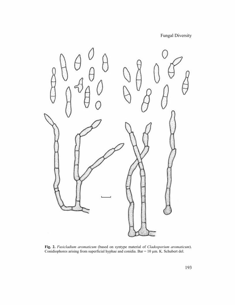
Dischloridium livistonae (P. Karst.) U. Braun & K. Schub., comb. nov. [Bas.: Fusicladium livistonae P. Karst., Hedwigia 30: 302, 1891. Material examined: on dead petioles of Livistona chinensis (Palmae), Russia, Karelia australis, Vyborg, Liimatta, Sept. 1891, A. Tesleff (H 4252), holotype. Description and illustration: Braun, 2000a, p. 38 and p. 41, Fig. 8]. The name Gloeosporium cladosporioides Ellis & Halst. (J. Mycol. 6: 34, 1890), described from North America on Hypericum mutilum, has been confused with Cladosporium gloeosporioides. Atkinson (1897) clearly stated that the two fungi are quite distinct. In his revision of species referred to Gloeosporium, Arx (1970) classified Gloeosporium cladosporioides as a doubtful species, since he could not find any fructification in the type material. Several syntype collections of this species from NY have been examined, viz., four duplicates of ‘Ellis & Everh., North American Fungi 2438’ and two samples with hand-written labels (Hypericum mutilum, USA, New Jersey, Terra Cotta Bank, Jul. 1889, B.D. Halsted). In two of the exsiccatae sufficient conidiomata agreeing with the original diagnosis of G. cladosporioides have been found. This fungus is a true acervular coelomycete belonging to Colletotrichum gloeosporioides (Penz.) Sacc. s.l. (= C. gloeosporioides complex, sensu Sutton, 1980). The conidia are relatively small, (10-)12-16 × (2.5-)3-4.5(-5) µm; some brown setae have been observed. This species seems to be confined to North America. A final conclusion on its taxonomic status is not possible without any cultures, inoculation experiments or molecular data. Therefore, it is tentatively considered a synonym of Colletotrichum gloeosporioides s.l. If detailed examinations show that it is a separate species confined to Hypericum spp. in North America, the basionym Gloeosporium cladosporioides would be available. Fusicladium aromaticum (Ellis & Everh.) K. Schub. & U. Braun, comb. nov. (Fig. 2)
≡ Cladosporium aromaticum Ellis & Everh., Proc. Acad. Nat. Sci. Philadelphia 47(3): 439 (1895).
= Cladosporium nervale Ellis & Dearn., in Bartholomew, Fungi columbiani, Cent. XXI, No. 2010 (1905).
Material examined: on Rhus aromatica (Anacardiaceae), USA, California, Pasadena, 31 Aug. 1894, McClatchie, ex herb. Ellis (BPI 426124; syntype of C. aromaticum); USA, Wisconsin, Dane Co., Madison, 31 Aug. 1957, H.C. Greene (BPI 426123); Rhus copallina, USA, Wisconsin, Columbia Co., Lodi, 6 Aug. 1946, H.C. Greene (BPI 426125); Dane Co., Madison, 21 Sept. 1960, H.C. Greene (BPI 426126); Rhus glabra, USA, Iowa, Steamboat Rock, 9 Sept. 1913, J.C. Anderson (BPI 426130); Nebraska, Valentine, 31 Aug. 1910, J.M. Bates, Bartholomew, Fungi columbiani 3415 (BPI 426127, 426132; HBG); Wisconsin, Dane Co., Madison, 30 Aug. 1955, H.C. Greene (BPI 426129); Green Co., near Brodhead, 26 Jul. 1963, H.C. Greene (BPI 426128); Waukesha Co., Eagleville, 27 Jul. 1941, H.C. Greene (BPI 426131); Rhus typhina, Canada, Ontario, Middlesex Co., London, Jul./Aug. 1904, J. Dearness,
192
Fungal Diversity
193
Fig. 2. Fusicladium aromaticum (based on syntype material of Cladosporium aromaticum). Conidiophores arising from superficial hyphae and conidia. Bar = 10 µm. K. Schubert del.
Bartholomew, Fungi columbiani 2010 (HBG, NY; syntypes of C. nervale); 28 Jul. 1945, W.D. Sutton (DAOM 15521); Lake Timagami, Bear Island, 15 Aug. 1930, J.L. Conners (DAOM 1516); H.S. Jackson et al. (DAOM 1188/ 81525); Ottawa, Dow’s Lake, 30 Aug. 1944, D.B.O. Savile (DAOM); USA, Wisconsin, Dane Co., Madison, 25 Aug. 1945, H.C. Greene (BPI 426133); North America, East-Springbank, 27 May 1904 (DAOM); 9/12 Aug. 1904 (DAOM). On living leaves and petioles, without distinct leaf spots, forming pale to dark olivaceous-brown to brown or even blackish, dingy discolourations, often along leaf veins. Colonies epiphyllous, hypophyllous or amphigenous, punctiform to extended, loose to dense, caespitose, effuse, dark olivaceous-brown, dark brown to slightly red-brown. Mycelium external, superficial; hyphae branched, 2-4 µm wide, septate, not constricted at the septa, pale to medium brown, smooth, walls thickened, often slightly swollen and darker at the base of the conidiophores. Conidiophores solitary or in small groups, but not distinctly fasciculate, arising terminally or laterally from creeping hyphae or swollen hyphal cells, erect, straight to slightly curved, often geniculate-sinuous, unbranched or once branched, 25-150 × 4.5-6(-7) µm, 1-8-septate, occasionally constricted at the septa, medium to dark brown, paler towards the apex, smooth, walls slightly thickened, often somewhat swollen at the base, up to 11 µm wide, occasionally enteroblastically proliferating (after a period of unfavourable conditions when growth has stopped and then resumed, visible as discontinuity in pigmentation and thickness of the wall). Conidiogenous cells integrated, terminal or intercalary, ellipsoid-subcylindrical to cylindrical, 7-28 µm long, proliferation sympodial, usually with a single conidiogenous locus, rarely with a second, intercalary conidiogenous loci often located near a septum, subdenticulate to denticulate, truncate to slightly convex, 1.5-2 µm wide, unthickened or almost so, somewhat refractive to slightly darkened-refractive, sometimes apical conidiogenous cell detaching and then acting like ramoconidia s. str., broadly truncate at the base. Conidia solitary or in unbranched, very rarely branched chains, straight, ellipsoid, fusiform to cylindrical, (5-)7-32 × (3-)4.5-7(-8) µm, 0-2(-3)-septate, not constricted or slightly constricted at the septa, very pale to pale brown, smooth, walls only slightly thickened, apex rounded to pointed, hila truncate to slightly convex, 1.5-2 µm wide, unthickened or almost so, somewhat refractive to slightly darkened-refractive.
Notes: The conidiogenous loci in this species are quite distinct from those of true Cladosporium species and agree well with the concept of Fusicladium Bonord. emend. (Schubert et al., 2003) by being denticle-like, apically truncate and unthickened. Fusicladium aromaticum is the first member of this genus on a host belonging to the Anacardiaceae. Within the genus Fusicladium there are only a few species occasionally forming external,
194

Fungal Diversity
Fig. 3. Fusicladium caesalpiniae (based on holotype material). Conidiophores and conidia. Bar = 10 µm. K. Schubert del. superficial hyphae. Fusicladium caducum (Davis) K. Schub. & U. Braun, described on Betula nigra, differs in having usually multilocal conidiogenous cells and smaller, usually aseptate conidia, 6.5-18 µm long. Furthermore, the superficial creeping hyphae are often irregularly rough-walled (Schubert, 2005). Fusicladium scillae (Deighton) U. Braun & K. Schub. is quite distinct by forming fasciculate narrower conidiophores, (1.5-)2-4 µm wide, only occasionally arising from superficial hyphae, narrower conidia, 2.5-4 µm wide, and narrower conidiogenous loci and hila, 1-1.5 µm wide. Fusicladium humile (Davis) K. Schub. & U. Braun forming distinct stromata and solitary or fasciculate conidiophores has somewhat wider conidiogenous loci and hila, 1-3(-3.5) µm wide (Schubert et al., 2003). Cladosporium nervale, which proved to be conspecific with Fusicladium aromaticum, was reported from China as causal agent of leaf spots on Rhodea
195
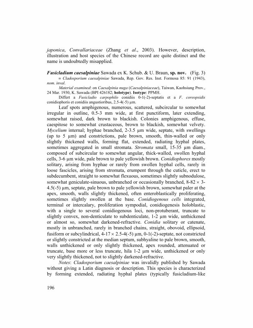
japonica, Convallariaceae (Zhang et al., 2003). However, description, illustration and host species of the Chinese record are quite distinct and the name is undoubtedly misapplied. Fusicladium caesalpiniae Sawada ex K. Schub. & U. Braun, sp. nov. (Fig. 3)
≡ Cladosporium caesalpiniae Sawada, Rep. Gov. Res. Inst. Formosa 85: 91 (1943), nom. inval.
Material examined: on Caesalpinia nuga (Caesalpiniaceae), Taiwan, Kaohsiung Prov., 24 Mar. 1930, K. Sawada (BPI 426182; holotype). Isotype: PPMH.
Differt a Fusicladio carpophilo conidiis 0-1(-2)-septatis et a F. coreopsidis conidiophoris et conidiis angustioribus, 2.5-4(-5) µm.
Leaf spots amphigenous, numerous, scattered, subcircular to somewhat irregular in outline, 0.5-3 mm wide, at first punctiform, later extending, somewhat raised, dark brown to blackish. Colonies amphigenous, effuse, caespitose to somewhat crustaceous, brown to blackish, somewhat velvety. Mycelium internal; hyphae branched, 2-3.5 µm wide, septate, with swellings (up to 5 µm) and constrictions, pale brown, smooth, thin-walled or only slightly thickened walls, forming flat, extended, radiating hyphal plates, sometimes aggregated in small stromata. Stromata small, 15-35 µm diam., composed of subcircular to somewhat angular, thick-walled, swollen hyphal cells, 3-6 µm wide, pale brown to pale yellowish brown. Conidiophores mostly solitary, arising from hyphae or rarely from swollen hyphal cells, rarely in loose fascicles, arising from stromata, erumpent through the cuticle, erect to subdecumbent, straight to somewhat flexuous, sometimes slightly subnodulose, somewhat geniculate-sinuous, unbranched or occasionally branched, 8-82 × 3-4.5(-5) µm, septate, pale brown to pale yellowish brown, somewhat paler at the apex, smooth, walls slightly thickened, often enteroblastically proliferating, sometimes slightly swollen at the base. Conidiogenous cells integrated, terminal or intercalary, proliferation sympodial, conidiogenesis holoblastic, with a single to several conidiogenous loci, non-protuberant, truncate to slightly convex, non-denticulate to subdenticulate, 1-2 µm wide, unthickened or almost so, somewhat darkened-refractive. Conidia solitary or catenate, mostly in unbranched, rarely in branched chains, straight, obovoid, ellipsoid, fusiform or subcylindrical, 4-17 × 2.5-4(-5) µm, 0-1(-2)-septate, not constricted or slightly constricted at the median septum, subhyaline to pale brown, smooth, walls unthickened or only slightly thickened, apex rounded, attenuated or truncate, base more or less truncate, hila 1-2 µm wide, unthickened or only very slightly thickened, not to slightly darkened-refractive. Notes: Cladosporium caesalpiniae was invalidly published by Sawada without giving a Latin diagnosis or description. This species is characterized by forming extended, radiating hyphal plates (typically fusicladium-like
196

Fungal Diversity
growth), fusiform or subcylindrical conidia with truncate, unthickened or almost so, somewhat darkened-refractive conidiogenous hila. Based on these characteristics, C. caesalpiniae has to be assigned to Fusicladium emend. (Schubert et al., 2003). It is the first member of this genus on a host belonging to the Caesalpiniaceae. Fusicladium brevipes Ellis & Everh., F. lathyrinum (Ellis & Galloway) S. Hughes & Piroz., F. pisicola Linford and F. psoraleae (Ellis & Barthol.) S. Hughes & Piroz., species occurring on hosts belonging to the Fabaceae, are quite distinct by always having short conidiophores, fasciculate or aggregated, sometimes even in compact layers, wider conidiogenous loci and hila, and longer and wider solitary conidia with different conidial surface ornamentations [conidia 15-35 × 7-13 µm, smooth to finely asperulate in F. brevipes; 15-28 × 6-10 µm, coarsely verrucose in F. lathyrinum; 12-28(-32) × 6-10(-14) µm, asperulate, coarsely verrucose to echinulate in F. pisicola; and 10-27(-34) × 6-10(-11) µm, smooth to coarsely verrucose, rugose in F. psoraleae].
Fusicladium caesalpiniae differs from all morphologically similar taxa in having 0-1(-2)-septate conidia [usually aseptate in F. carpophilum (Thüm.) Oudem., F. asperatum K. Schub. & U. Braun and F. viticis M.B. Ellis], somewhat narrower conidiophores [versus 3.5-5(-6) µm in F. coreopsidis (H.C. Greene) K. Schub. & U. Braun] and narrower conidia [versus 3.5-5(-6) µm in F. coreopsidis; 4-6(-8.5) µm in F. crataegi Aderh.; 4-7 µm in F. cerasi (Rabenh.) Erikss.; and 5-7 µm in F. viticis] (Schubert et al., 2003; Schubert, 2005). Fusicladium myrticola (Bubák) K. Schub. & U. Braun, comb. nov. (Fig. 4)
≡ Cladosporium myrticola Bubák, in Bubák & Kabát, Ann. Mycol. 13: 113 (1915), as ‘myrticolum’.
Material examined: on leaves of Myrtus communis (Myrtaceae), Italy, Tyrol, Gries near Bozen, 30 May 1914, Dr. W. Pfaff (BPI 427273; holotype).
Leaf spots epiphyllous, scattered, at first punctiform, becoming subglobose, 1-7 µm diam., later confluent, pale to medium olivaceous or olivaceous-brown, occasionally somewhat reddish, definite margin lacking. Colonies epiphyllous, short caespitose, loose, effuse, pale to medium brown, somewhat velvety. Mycelium internal, subcuticular; hyphae not or only sparingly branched, (1-)2-4(-5) µm wide, septate, subhyaline to pale olivaceous-brown, smooth, walls almost unthickened. Stromata absent. Conidiophores solitary or in small loose groups, scattered, arising from swollen hyphal cells, erumpent through the cuticle, erect, straight to slightly flexuous, unbranched or rarely branched, 25-82 × 3.5-5 µm, 0-3-septate, sometimes slightly constricted at the septa, olivaceous-brown, somewhat paler towards the apex, smooth, walls slightly thickened, almost thin-walled near the
197
apex, sometimes slightly swollen at the base, up to 7 µm wide. Conidiogenous cells integrated, terminal or conidiophores reduced to conidiogenous cells, 9-38 µm long, proliferation sympodial, holoblastic, with a single or only few conidiogenous loci, truncate to slightly convex, non-denticulate to subdenticulate, 1.5-2 µm wide, unthickened or almost so, not or only slightly darkened, somewhat refractive. Conidia solitary, straight, obovoid, fusiform to oblong, 8.5-19(-25) × 3.5-5.5(-6) µm, 0(-1)-septate, subhyaline to pale olivaceous, smooth, walls unthickened or almost so, apex rounded or slightly pointed, slightly attenuated towards the base, hila truncate, obtuse, 1.5-2 µm wide, unthickened or almost so, not or only somewhat refractive or darkened.
Notes: Based on the structure of the conidiogenous loci and hila (truncate, unthickened or almost so, not or only somewhat darkened-refractive), this species has to be excluded from Cladosporium s. str. and reallocated to Fusicladium emend. It is the first Fusicladium on a host belonging to the Myrtaceae. Morphologically allied taxa with similar conidiophores and conidia are quite distinct by having usually catenate conidia (F. carpophilum, F. levieri Magnus, F. virgaureae Ondřej), wider conidiogenous loci and hila (1.5-3 µm wide in F. levieri; 2-3 µm diam. in F. scribnerianum (Cavara) M.B. Ellis) and septate conidia (0-2-septate in F. levieri and 0-3-septate in F. scribnerianum).
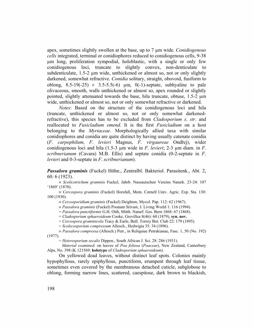
Passalora graminis (Fuckel) Höhn., Zentralbl. Bakteriol. Parasitenk., Abt. 2, 60: 6 (1923).
≡ Scolicotrichum graminis Fuckel, Jahrb. Nassauischen Vereins Naturk. 23-24: 107 ‘1869’ (1870).
≡ Cercospora graminis (Fuckel) Horsfall, Mem. Cornell Univ. Agric. Exp. Sta. 130: 100 (1930).
≡ Cercosporidium graminis (Fuckel) Deighton, Mycol. Pap. 112: 62 (1967). ≡ Passalora graminis (Fuckel) Poonam Srivast, J. Living World 1: 116 (1994). = Passalora punctiformis G.H. Otth, Mitth. Naturf. Ges. Bern 1868: 67 (1868). = Cladosporium sphaeroideum Cooke, Grevillea 8(46): 60 (1879), syn. nov. = Cercospora graminicola Tracy & Earle, Bull. Torrey Bot. Club 22: 179 (1895). = Scolecosporium compressum Allesch., Hedwigia 35: 34 (1896). ≡ Passalora compressa (Allesch.) Petr., in Reliquiae Petrakianae, Fasc. 1, 50 (No. 192)
(1977). = Heterosporium secalis Dippen., South African J. Sci. 28: 286 (1931).
Material examined: on leaves of Poa foliosa (Poaceae), New Zealand, Canterbury Alps, No. 398 (K 121569; holotype of Cladosporium sphaeroideum).
On yellowed dead leaves, without distinct leaf spots. Colonies mainly hypophyllous, rarely epiphyllous, punctiform, erumpent through leaf tissue, sometimes even covered by the membranous detached cuticle, subglobose to oblong, forming narrow lines, scattered, caespitose, dark brown to blackish,
198

Fungal Diversity
Fig. 4. Fusicladium myrticola (based on holotype material). Conidiophores and conidia. Bar = 10 µm. K. Schubert del.
sometimes confluent, velvety. Mycelium immersed. Stromata small to extended, up to 300 µm long, composed of subglobose, pale to medium brown, dense cells. Conidiophores densely fasciculate, numerous, arising from stromata, erumpent through the cuticle, erect, straight to slightly curved, unbranched, up to 45 µm long, 6-10 µm wide, aseptate or with only a few septa, medium to dark brown, somewhat paler towards the apex, apex pale brown, smooth to minutely verruculose near the apex, walls slightly thickened. Conidiophores mostly reduced to conidiogenous cell or conidiogenous cells integrated, terminal, proliferation sympodial, conidiogenous loci planate, truncate to slightly convex, 3.5-7 µm wide, thickened and darkened-refractive.
199
Conidia formed solitary, straight, broadly ellipsoid, fusiform to obclavate, (8-) 13-48 × 8-15 µm, 0-1-septate, pale brown, almost smooth to verruculose, walls slightly thickened, apex rounded or pointed, hila truncate to slightly convex, 3.5-6 µm wide, thickened, darkened-refractive.
Notes: The type collection of Cladosporium sphaeroideum agrees very well with Passalora graminis (Chupp, 1954) and has to be reduced to synonymy with the latter species.
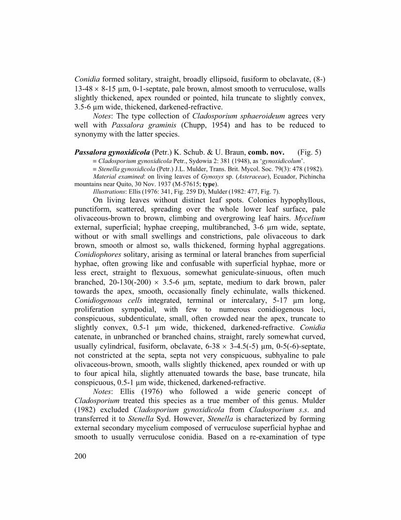
Passalora gynoxidicola (Petr.) K. Schub. & U. Braun, comb. nov. (Fig. 5)
≡ Cladosporium gynoxidicola Petr., Sydowia 2: 381 (1948), as ‘gynoxidicolum’. ≡ Stenella gynoxidicola (Petr.) J.L. Mulder, Trans. Brit. Mycol. Soc. 79(3): 478 (1982). Material examined: on living leaves of Gynoxys sp. (Asteraceae), Ecuador, Pichincha
mountains near Quito, 30 Nov. 1937 (M-57615; type). Illustrations: Ellis (1976: 341, Fig. 259 D), Mulder (1982: 477, Fig. 7). On living leaves without distinct leaf spots. Colonies hypophyllous,
punctiform, scattered, spreading over the whole lower leaf surface, pale olivaceous-brown to brown, climbing and overgrowing leaf hairs. Mycelium external, superficial; hyphae creeping, multibranched, 3-6 µm wide, septate, without or with small swellings and constrictions, pale olivaceous to dark brown, smooth or almost so, walls thickened, forming hyphal aggregations. Conidiophores solitary, arising as terminal or lateral branches from superficial hyphae, often growing like and confusable with superficial hyphae, more or less erect, straight to flexuous, somewhat geniculate-sinuous, often much branched, 20-130(-200) × 3.5-6 µm, septate, medium to dark brown, paler towards the apex, smooth, occasionally finely echinulate, walls thickened. Conidiogenous cells integrated, terminal or intercalary, 5-17 µm long, proliferation sympodial, with few to numerous conidiogenous loci, conspicuous, subdenticulate, small, often crowded near the apex, truncate to slightly convex, 0.5-1 µm wide, thickened, darkened-refractive. Conidia catenate, in unbranched or branched chains, straight, rarely somewhat curved, usually cylindrical, fusiform, obclavate, 6-38 × 3-4.5(-5) µm, 0-5(-6)-septate, not constricted at the septa, septa not very conspicuous, subhyaline to pale olivaceous-brown, smooth, walls slightly thickened, apex rounded or with up to four apical hila, slightly attenuated towards the base, base truncate, hila conspicuous, 0.5-1 µm wide, thickened, darkened-refractive.
Notes: Ellis (1976) who followed a wide generic concept of Cladosporium treated this species as a true member of this genus. Mulder (1982) excluded Cladosporium gynoxidicola from Cladosporium s.s. and transferred it to Stenella Syd. However, Stenella is characterized by forming external secondary mycelium composed of verruculose superficial hyphae and smooth to usually verruculose conidia. Based on a re-examination of type
200

Fungal Diversity
Fig. 5. Passalora gynoxidicola (based on type material). Conidiophores and conidia. Bar = 10 µm. K. Schubert del.
material, this species is better placed in Passalora since the superficial hyphae and conidia are consistently smooth-walled. This is the first Passalora on Gynoxis.
Passalora lobeliae-cardinalis (Schwein.) U. Braun & Crous, in Crous &
Braun, Mycosphaerella and its anamorphs: 1. Names published in Cercospora and Passalora, CBS Biodiversity Ser. 1: 254 (2003).
201
≡ Caeoma lobeliae-cardinalis Schwein., Trans. Amer. Phil. Soc., Ser. 2, 4: 291 (1832). ≡ Mycovellosiella lobeliae-cardinalis (Schwein.) Deighton, Trans. Brit. Mycol. Soc. 86:
637 (1986). = Cercospora ochracea Sacc. & Malbr., Michelia 2: 128 (1880). = Cercospora lobeliicola Solheim, Illinois Biol. Monogr. 12: 64 (1929). = Cercospora diffusa auct. = Cladosporium pelliculosum Berk. & M.A. Curtis, in herb., syn. nov. Material examined: on leaves of Lobelia puberulosa (Campanulaceae), USA, South
Carolina, No. 1742 (K 121567; original material of Cladosporium pelliculosum). Illustration: Deighton (1986: 639, Fig. 2). Without distinct leaf spots, hypophyllous as yellowish brown to dark
brown discolourations without specific shape and size. Colonies hypophyllous, punctiform to effuse, caespitose, loose to dense, brown to somewhat reddish brown, villose. Primary mycelium internal. Stromata absent or small, up to 20 µm wide, pale brown. Secondary mycelium not observed. Conidiophores in small, dense fascicles, arising from stromata, emerging through stomata, erect, straight or subdecumbent, more or less flexuous, often slightly geniculate-sinuous, once branched or often several times, intertwined, up to 240 µm long or even longer, 3.5-5.5 µm wide, pluriseptate, sometimes slightly constricted at the septa, pale reddish to reddish or reddish brown, smooth, walls only slightly thickened. Conidiogenous cells integrated, terminal and intercalary, proliferation sympodial, with a single or few conidiogenous loci, often situated on small lateral shoulders, subdenticulate, truncate to slightly convex, 1-1.5 µm wide, somewhat thickened, darkened-refractive. Conidia solitary, ellipsoid, fusiform to mostly obclavate, more or less straight, 8-50 × 3.5-5.5 µm, 1-5-septate, mostly 2-3-septate, not constricted or slightly constricted at the septa, pale reddish, smooth, walls slightly thickened, apex rounded or pointed, somewhat attenuated towards the base, hila truncate to slightly convex, 1-1.5 µm wide, thickened, darkened-refractive.
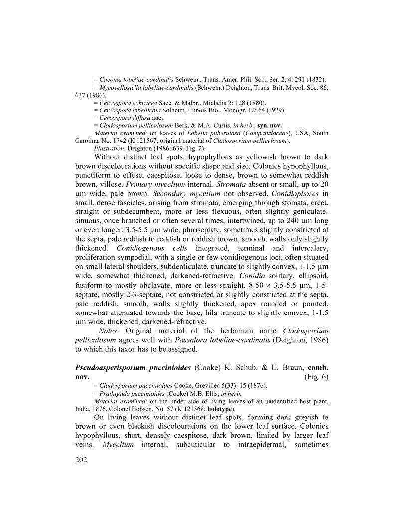
Notes: Original material of the herbarium name Cladosporium pelliculosum agrees well with Passalora lobeliae-cardinalis (Deighton, 1986) to which this taxon has to be assigned. Pseudoasperisporium puccinioides (Cooke) K. Schub. & U. Braun, comb. nov. (Fig. 6)
≡ Cladosporium puccinioides Cooke, Grevillea 5(33): 15 (1876). ≡ Prathigada puccinioides (Cooke) M.B. Ellis, in herb. Material examined: on the under side of living leaves of an unidentified host plant,
India, 1876, Colonel Hobsen, No. 57 (K 121568; holotype). On living leaves without distinct leaf spots, forming dark greyish to brown or even blackish discolourations on the lower leaf surface. Colonies hypophyllous, short, densely caespitose, dark brown, limited by larger leaf veins. Mycelium internal, subcuticular to intraepidermal, sometimes
202

Fungal Diversity
Fig. 6. Pseudoasperisporium puccinioides (based on holotype material). Fascicle of conidiophores, conidiogenous cells and conidia. Bar = 10 µm. K. Schubert del. substomatal. Stromata small to well-developed, 25-75 µm diam., compact, composed of subcircular to somewhat angular, polygonal cells, 5-8 µm wide, olivaceous to olivaceous-brown, smooth, thick-walled. Conidiophores densely fasciculate, in small to large fascicles, forming sporodochial conidiomata, erumpent through the cuticle or emerging through stomata, erect, straight to
203
slightly flexuous, cylindrical, unbranched, 10-40(-80) × 5-8 µm, aseptate, olivaceous to olivaceous-brown, somewhat paler towards the apex, smooth or almost so to somewhat rough-walled, walls somewhat thickened; conidiophores usually reduced to conidiogenous cells, unilocal, determinate or occasionally proliferation sympodial, with a second conidiogenous locus, conidiogenesis holoblastic, loci more or less truncate, 1.5-3 µm wide, unthickened, not darkened but somewhat refractive. Conidia solitary or occasionally in unbranched chains, straight to somewhat curved, obclavate, fusiform to subcylindrical, 22-48 × 6-9 µm, 0-1(-2)-septate, septum more or less median, usually not constricted, pale olivaceous to pale olivaceous-brown, verruculose to irregularly rough-walled, walls thickened, often somewhat attenuated towards the apex, apex rounded, base truncate to somewhat obconically truncate, hila 1.5-3 µm wide, unthickened, not darkened but somewhat refractive.
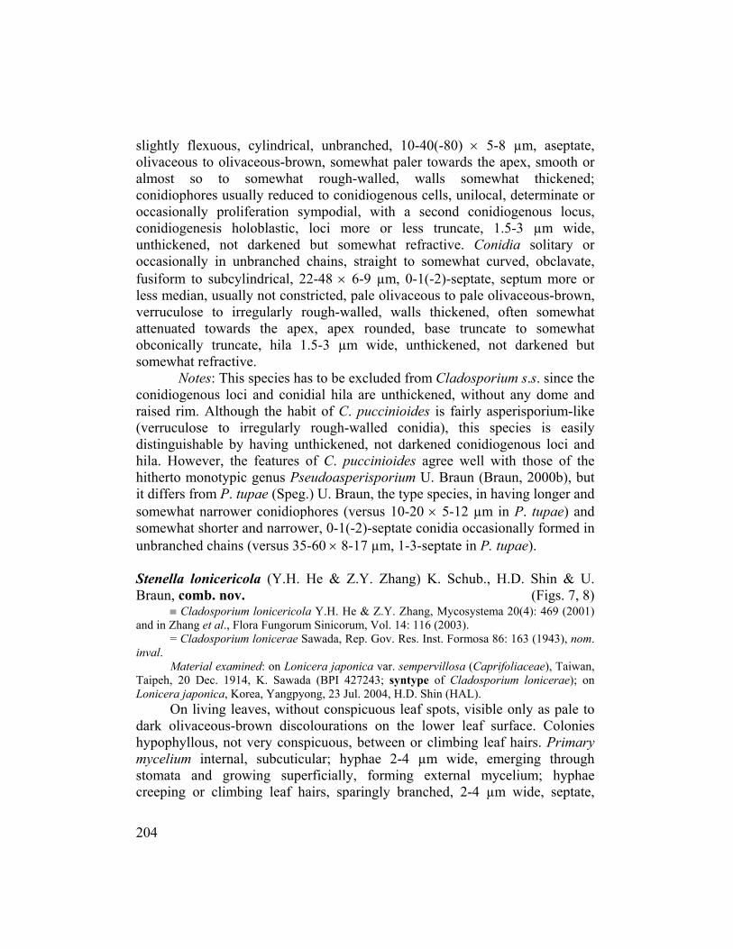
Notes: This species has to be excluded from Cladosporium s.s. since the conidiogenous loci and conidial hila are unthickened, without any dome and raised rim. Although the habit of C. puccinioides is fairly asperisporium-like (verruculose to irregularly rough-walled conidia), this species is easily distinguishable by having unthickened, not darkened conidiogenous loci and hila. However, the features of C. puccinioides agree well with those of the hitherto monotypic genus Pseudoasperisporium U. Braun (Braun, 2000b), but it differs from P. tupae (Speg.) U. Braun, the type species, in having longer and somewhat narrower conidiophores (versus 10-20 × 5-12 µm in P. tupae) and somewhat shorter and narrower, 0-1(-2)-septate conidia occasionally formed in unbranched chains (versus 35-60 × 8-17 µm, 1-3-septate in P. tupae). Stenella lonicericola (Y.H. He & Z.Y. Zhang) K. Schub., H.D. Shin & U. Braun, comb. nov. (Figs. 7, 8)
≡ Cladosporium lonicericola Y.H. He & Z.Y. Zhang, Mycosystema 20(4): 469 (2001) and in Zhang et al., Flora Fungorum Sinicorum, Vol. 14: 116 (2003).
= Cladosporium lonicerae Sawada, Rep. Gov. Res. Inst. Formosa 86: 163 (1943), nom. inval.
Material examined: on Lonicera japonica var. sempervillosa (Caprifoliaceae), Taiwan, Taipeh, 20 Dec. 1914, K. Sawada (BPI 427243; syntype of Cladosporium lonicerae); on Lonicera japonica, Korea, Yangpyong, 23 Jul. 2004, H.D. Shin (HAL). On living leaves, without conspicuous leaf spots, visible only as pale to dark olivaceous-brown discolourations on the lower leaf surface. Colonies hypophyllous, not very conspicuous, between or climbing leaf hairs. Primary mycelium internal, subcuticular; hyphae 2-4 µm wide, emerging through stomata and growing superficially, forming external mycelium; hyphae creeping or climbing leaf hairs, sparingly branched, 2-4 µm wide, septate,
204

Fungal Diversity
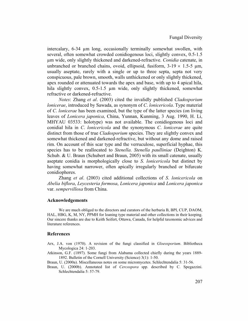
Fig. 7. Stenella lonicericola (based on syntype material of Cladosporium lonicerae). Conidiophores and conidia. Bar = 10 µm. K. Schubert del. septa not very conspicuous, sometimes slightly constricted at the septa, pale to medium olivaceous or brown, walls slightly thickened, verruculose, but at the base of conidiophores smooth or almost so, often somewhat swollen and darker, medium to medium dark brown, concolourous with conidiophores. Conidiophores solitary, arising from superficial hyphae, mostly as lateral branches, erect, straight to flexuous, cylindrical-oblong or often filiform, unbranched, 22-350 × 3-5.5 µm, pluriseptate, occasionally slightly constricted at the septa, medium to medium dark brown, not or somewhat paler towards
205

Fig. 8. Stenella lonicericola (based on the HAL collection from Korea). Conidiophores and conidia. Bar = 10 µm. K. Schubert del.
the apex, smooth, thick-walled, sometimes even two-layered, somewhat attenuated towards apex, sometimes slightly swollen, occasionally enteroblastically proliferating. Conidiogenous cells integrated, terminal, rarely
206
Fungal Diversity
intercalary, 6-34 µm long, occasionally terminally somewhat swollen, with several, often somewhat crowded conidiogenous loci, slightly convex, 0.5-1.5 µm wide, only slightly thickened and darkened-refractive. Conidia catenate, in unbranched or branched chains, ovoid, ellipsoid, fusiform, 3-19 × 1.5-5 µm, usually aseptate, rarely with a single or up to three septa, septa not very conspicuous, pale brown, smooth, walls unthickened or only slightly thickened, apex rounded or attenuated towards the apex and base, with up to 4 apical hila, hila slightly convex, 0.5-1.5 µm wide, only slightly thickened, somewhat refractive or darkened-refractive.
Notes: Zhang et al. (2003) cited the invalidly published Cladosporium lonicerae, introduced by Sawada, as synonym of C. lonicericola. Type material of C. lonicerae has been examined, but the type of the latter species (on living leaves of Lonicera japonica, China, Yunnan, Kunming, 3 Aug. 1990, H. Li, MHYAU 03533: holotype) was not available. The conidiogenous loci and conidial hila in C. lonicericola and the synonymous C. lonicerae are quite distinct from those of true Cladosporium species. They are slightly convex and somewhat thickened and darkened-refractive, but without any dome and raised rim. On account of this scar type and the verruculose, superficial hyphae, this species has to be reallocated to Stenella. Stenella paulliniae (Deighton) K. Schub. & U. Braun (Schubert and Braun, 2005) with its small catenate, usually aseptate conidia is morphologically close to S. lonicericola but distinct by having somewhat narrower, often apically irregularly branched or bifurcate conidiophores.
Zhang et al. (2003) cited additional collections of S. lonicericola on Abelia biflora, Leycesteria formosa, Lonicera japonica and Lonicera japonica var. sempervillosa from China. Acknowledgements
We are much obliged to the directors and curators of the herbaria B, BPI, CUP, DAOM, HAL, HBG, K, M, NY, PPMH for loaning type material and other collections in their keeping. Our sincere thanks are due to Keith Seifert, Ottawa, Canada, for helpful taxonomic advices and literature references. References Arx, J.A. von (1970). A revision of the fungi classified in Gloeosporium. Bibliotheca
Mycologica 24: 1-203. Atkinson, G.F. (1897). Some fungi from Alabama collected chiefly during the years 1889-
1892. Bulletin of the Cornell University (Science) 3(1): 1-50. Braun, U. (2000a). Miscellaneous notes on some micromycetes. Schlechtendalia 5: 31-56. Braun, U. (2000b). Annotated list of Cercospora spp. described by C. Spegazzini.
Schlechtendalia 5: 57-79.
207

Braun, U., Crous, P.W., Dugan, F.M., Groenewald, J.Z. and Hoog, G.S. de (2003). Phylogeny and taxonomy of cladosporium-like hyphomycetes, including Davidiella gen. nov., the teleomorph of Cladosporium s.str. Mycological Progress 2(1): 3-18.
Chupp, C. (1954). A monograph of the fungus genus Cercospora. Ithaca, New York. Published by the author.
Crous, P.W. and Braun, U. (2003). Mycosphaerella and its anamorphs: 1. Names published in Cercospora and Passalora. CBS Biodiversity Ser. 1: 1-571.
David, J.C. (1997). A contribution to the systematics of Cladosporium. Revision of the fungi previously referred to Heterosporium. Mycological Papers 172: 1-157.
Deighton, F.C. (1986). Misidentification of Cercospora effusa. Transactions of the British Mycological Society 86(4): 637-641.
Dugan, F.M., Schubert, K. and Braun, U. (2004). Check-list of Cladosporium names. Schlechtendalia 11: 1-103.
Ellis, M.B. (1976). More dematiaceous hyphomycetes. Commonwealth Mycological Institute, Kew, UK.
Holmgren, P.K., Holmgren, N.H. and Barnett, L.C. (1990). Index Herbariorum, Part 1: The Herbaria of the World. 8th edn. New York Botanical Garden, New York.
Holubová-Jechová, V. (1987). Studies on hyphomycetes from Cuba V. Six new species of dematiaceous hyphomycetes from Havana Province. Česká Mykologie 41: 29-36.
Holubová-Jechová, V. (1990). Problems in the taxonomy of the dematiaceous hyphomycetes. Studies in Mycology 32: 41-48.
Mulder, J.L. (1982). New species and combinations in Stenella. Transactions of the British Mycological Society 79: 469-478.
Rong, I.H. and Gams, W. (2000). The hyphomycete genera Exochalara and Monilochaetes. Mycotaxon 76: 451-462.
Schubert, K. (2005). Taxonomic revision of the genus Cladosporium s. lat. 3. A revision of Cladosporium species described by J.J. Davis and H.C. Greene (WIS). Mycotaxon 92: 55-76.
Schubert, K. and Braun, U. (2004). Taxonomic revision of the genus Cladosporium s. lat. 2. Morphotaxonomic examination of Cladosporium species occurring on hosts of the families Bignoniaceae and Orchidaceae. Sydowia 56(2): 296-317.
Schubert, K. and Braun, U. (2005). Taxonomic revision of the genus Cladosporium s. lat. 1. Species reallocated to Fusicladium, Parastenella, Passalora, Pseudocercospora and Stenella. Mycological Progress 4: 101-109.
Schubert, K., Ritschel, A. and Braun, U. (2003). A monograph of Fusicladium s.lat. (hyphomycetes). Schlechtendalia 9: 1-132.
Sutton, B.C. (‘1976’, published 1977). Species of Hemibeltrania Piroz. and Dischloridium gen. nov. Kavaka 4: 43-50.
Sutton, B.C. (1980). The Coelomycetes. Fungi Imperfecti with Pycnidia, Acervuli and Stromata. Commonwealth Mycological Institute, Kew, UK.
Zhang, Z.Y., Liu, Y.L., Zhang, T., Li, T.F., Wang, G., Zhang, H., He, Y.H. and Peng, H.H. (2003). Flora Fungorum Sinicorum, Vol. 14, Cladosporium, Fusicladium, Pyricularia. Beijing.
(Received 3 March 2005; accepted 20 September 2005)
208