Embed Size (px)
Citation preview

Targeting and function of CAH1 -Characterization of a novel protein pathway to the
plant cell chloroplast
STEFAN BURÉN
Akademisk avhandling
som med vederbörligt tillstånd av rektorsämbetet vid Umeå universitet föravläggande av Teknologie doktorsexamen i Växters cell- och molekylärbiologi,
framläggs till offentligt försvar i KB3A9, KBC-huset, Umeå Universitet,fredagen den 29 januari 2010 klockan 10.00.
Avhandlingen kommer att försvaras på engelska.
Fakultetsopponent:Dr. Muriel BardorUniversité de Rouen
Mont-Saint-Aignan, France
Umeå Plant Science CentreDepartment of Plant Physiology
Umeå universitySweden 2010
Targeting and function of CAH1 -Characterization of a novel protein pathway to the plant cell chloroplast
Stefan BurénUmeå Plant Science Centre, Department of Plant Physiology, Umeå University
ISBN: 978-91-7264-933-0
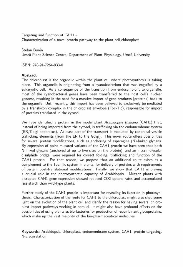
AbstractThe chloroplast is the organelle within the plant cell where photosynthesis is takingplace. This organelle is originating from a cyanobacterium that was engulfed by aeukaryotic cell. As a consequence of the transition from endosymbiont to organelle,most of the cyanobacterial genes have been transferred to the host cell’s nucleargenome, resulting in the need for a massive import of gene products (proteins) back tothe organelle. Until recently, this import has been believed to exclusively be mediatedby a translocon complex in the chloroplast envelope (Toc-Tic), responsible for importof proteins translated in the cytosol.
We have identified a protein in the model plant Arabidopsis thaliana (CAH1) that,instead of being imported from the cytosol, is trafficking via the endomembrane system(ER/Golgi apparatus). At least part of the transport is mediated by canonical vesicletrafficking elements (from the ER to the Golgi). This novel route offers possibilitiesfor several protein modifications, such as anchoring of asparagine (N)-linked glycans.By expression of point mutated variants of the CAH1 protein we have seen that bothN-linked glycans (anchored at up to five sites on the protein), and an intra-moleculardisulphide bridge, were required for correct folding, trafficking and function of theCAH1 protein. For that reason, we propose that an additional route exists as acomplement to the Toc-Tic system in plants, for delivery of proteins with requirementsof certain post-translational modifications. Finally, we show that CAH1 is playinga crucial role in the photosynthetic capacity of Arabidopsis. Mutant plants withdisrupted CAH1 gene expression showed reduced CO2 uptake rates and accumulatedless starch than wild-type plants.
Further study of the CAH1 protein is important for revealing its function in photosyn-thesis. Characterization of the route for CAH1 to the chloroplast might also shed somelight on the evolution of the plant cell and clarify the reason for having several chloro-plast import pathways working in parallel. It might also have profound effects on thepossibilities of using plants as bio-factories for production of recombinant glycoproteins,which make up the vast majority of the bio-pharmaceutical molecules.
Keywords: Arabidopsis, chloroplast, endomembrane system, CAH1, protein targeting,N-glycosylation

Targeting and function of CAH1 -Characterization of a novel protein pathway to the
plant cell chloroplast
STEFAN BURÉN
Umeå Plant Science CentreDepartment of Plant Physiology
Umeå universitySweden 2010

© Stefan Burén, 2010
Umeå Plant Science CentreDepartment of Plant PhysiologyUmeå UniversitySE-901 87 UmeåSweden
ISBN: 978-91-7264-933-0
Cover by: Cristina Ortega VillasantePrinted by: Arkitektkopia, Umeå, 2010

v
Till Peter

vii
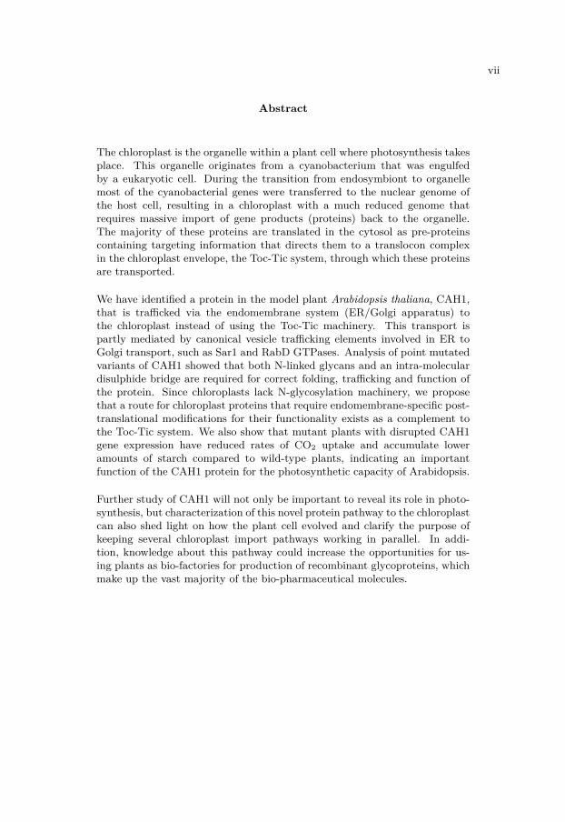
Abstract
The chloroplast is the organelle within a plant cell where photosynthesis takesplace. This organelle originates from a cyanobacterium that was engulfedby a eukaryotic cell. During the transition from endosymbiont to organellemost of the cyanobacterial genes were transferred to the nuclear genome ofthe host cell, resulting in a chloroplast with a much reduced genome thatrequires massive import of gene products (proteins) back to the organelle.The majority of these proteins are translated in the cytosol as pre-proteinscontaining targeting information that directs them to a translocon complexin the chloroplast envelope, the Toc-Tic system, through which these proteinsare transported.
We have identified a protein in the model plant Arabidopsis thaliana, CAH1,that is trafficked via the endomembrane system (ER/Golgi apparatus) tothe chloroplast instead of using the Toc-Tic machinery. This transport ispartly mediated by canonical vesicle trafficking elements involved in ER toGolgi transport, such as Sar1 and RabD GTPases. Analysis of point mutatedvariants of CAH1 showed that both N-linked glycans and an intra-moleculardisulphide bridge are required for correct folding, trafficking and function ofthe protein. Since chloroplasts lack N-glycosylation machinery, we proposethat a route for chloroplast proteins that require endomembrane-specific post-translational modifications for their functionality exists as a complement tothe Toc-Tic system. We also show that mutant plants with disrupted CAH1gene expression have reduced rates of CO2 uptake and accumulate loweramounts of starch compared to wild-type plants, indicating an importantfunction of the CAH1 protein for the photosynthetic capacity of Arabidopsis.
Further study of CAH1 will not only be important to reveal its role in photo-synthesis, but characterization of this novel protein pathway to the chloroplastcan also shed light on how the plant cell evolved and clarify the purpose ofkeeping several chloroplast import pathways working in parallel. In addi-tion, knowledge about this pathway could increase the opportunities for us-ing plants as bio-factories for production of recombinant glycoproteins, whichmake up the vast majority of the bio-pharmaceutical molecules.

viii
Sammanfattning
Kloroplasten är den organell i växtcellen där fotosyntesen sker. Denna organellhärstammar från en cyanobakterie som togs upp av en eukaryot cell. Underomvandlingen från endosymbiont till organell har de flesta av den ursprungli-ga cyanobakteriens gener flyttats över till växtcellens eget kärngenom, vilketresulterat i en kloroplast som endast kan producera ett fåtal av de proteinerden behöver och som istället kräver att en mängd genprodukter (proteiner)transporteras tillbaka till organellen. De flesta av dessa proteiner syntetiserasi cytosolen som polypeptider innehållande en speciell signal för kloroplasten,och tranporteras över kloroplastens dubbelmembran (envelop) med hjälp avett specifikt importsystem (Toc-Tic).
Vi har identifierat ett protein i modellväxten Arabidopsis thaliana (CAH1)som istället för att använda Toc-Tic tranporteras via det endomembrana sys-temet (ER/Golgi). Transporten sker delvis med hjälp av faktorer involveradei normal vesikeltransport, t.ex. Sar1 och RabD GTPaser (mellan ER och Gol-gi). Genom att uttycka och analysera punktmuterade varianter av CAH1 harvi kunnat visa att både sockergrupper kopplade till proteinet, samt en internsvavelbrygga, är nödvändiga för korrekt veckning, transport och funktion avproteinet. Då kloroplasten saknar eget maskineri för att koppla sådana sock-ergrupper till proteiner så föreslår vi att anledningen till att denna rutt exis-terar, som ett komplement till Toc-Tic, är för att proteiner beroende av dennatyp av modifiering ska kunna finnas i kloroplasten. Vi visar också att muter-ade växter som inte kan uttrycka genen som kodar för CAH1 uppvisar lägreupptag av CO2, samt ackumulerar mindre stärkelse än vildtypplantor, vilketantyder att CAH1 har en viktig funktion för den fotosyntetiska förmågan hosArabidopsis.
För att kunna fastställa den exakta funktionen för CAH1 kommer ytterligastudier att vara nödvändiga. En fördjupad karaktärisering av transportvägensom CAH1 följer till kloroplasten kan dessutom ge kunskap om hur växtcellenuppkom, samt besvara varför flera importvägar arbetar till synes parallelltmed varandra. Kunskap om denna transportväg kan även bidra med använd-bar information i försöken att nyttja växter till att uttrycka rekombinantaN-glykosylerade proteiner, t. ex. antikroppar och vacciner.

LIST OF PAPERS ix
List of papers
The thesis is based on the following papers, which will be referred to in thetext by their Roman numerals. Paper I is printed with kind permission fromNature Publishing Group.
I. Arsenio Villarejo, Stefan Burén, Susanne Larsson, Annabelle Déjardin,Magnus Monné, Charlotta Rudhe, Jan Karlsson, Stefan Jansson, PatriceLerouge, Norbert Rolland, Gunnar von Heijne, Markus Grebe, LaszloBako and Göran Samuelsson (2005) Evidence for a protein transportedthrough the secretory pathway en route to the higher plant chloroplast.Nature Cell Biology 7: 1224-1231.
II. Stefan Burén, Cristina Ortega-Villasante, Göran Samuelsson, LaszloBako and Arsenio Villarejo: Optimization of the 2A peptide coexpres-sion system to study trafficking of the plastid N-glycoprotein CAH1 inArabidopsis thaliana. (manuscript)
III. Stefan Burén, Cristina Ortega-Villasante, Amaya Blanco-Rivero, An-drea Martínez-Bernardini, Tatiana Shutova, Laszlo Bako, Arsenio Vil-larejo, Göran Samuelsson: N-glycosylation is required for trafficking andactivity of a chloroplast localized carbonic anhydrase (CAH1) in Ara-bidopsis thaliana. Submitted to JBC
IV. Stefan Burén*, Amaya Blanco-Rivero*, Cristina Ortega-Villasante, GöranSamuelsson, Arsenio Villarejo: Specific suppression of the chloroplastN-glycosylated carbonic anhydrase (CAH1) has major impact on thephotosynthetic performance of Arabidopsis thaliana. (manuscript)* These authors made equal contributions

x
Abbreviations
AmyI-1 α-Amylase isoform I-1Arf1 ADP-ribosylation factor 1β-ME β-mercaptoethanolBFA Brefeldin ABiP Binding ProteinCA Carbonic AnhydraseCAH1 Carbonic Anhydrase 1 in Arabidopsis thalianaCHX CycloheximideCNX CalnexinConA Concanavalin ACRT CalreticulinER Endoplasmic ReticulumERES ER-Export SiteERQC ER-Quality ControlEST Expressed Sequence TagFACS Fluorescence-Activated Cell SortingGFP/CFP/YFP Green/Cyan/Yellow Fluorescent ProteinGlcNAc N-AcetylglucosamineGNT I N-Acetylglucosaminyl Transferase IGT UDP-glucose:glycoprotein Glucosyl TransferaseGUS β-GlucuronidaseHA-CAH1 HA-tagged wild type CAH1IP ImmunoprecipitationKDEL ER retention signalNPP1 Nucleotide Pyrophosphate/Phosphodiesterase 1OST Oligosaccharyl TransferasePLAM Plastid Associated MembranePNGase F Peptide-N-Glycosidase FRabD2a Member of plant D subclass of the Rab family of small GTPasesRubisco Ribulose-1,5-bisphosphate carboxylase oxygenaseSar1 Secretion associated, ras-related protein1 (small GTPase)SP Signal Peptide for the ERSPP Stromal Processing PeptidaseSRP Signal Recognition ParticleTat Twin-arginine tranlocationToc-Tic Translocon of outer/inner chloroplast envelope membraneTP Transit Peptidewt wild type

Contents
List of papers . . . . . . . . . . . . . . . . . . . . . . . . . . . . . . . ixAbbreviations . . . . . . . . . . . . . . . . . . . . . . . . . . . . . . . x
Contents xi
1 Background 31.1 Central dogma . . . . . . . . . . . . . . . . . . . . . . . . . . . 31.2 Chloroplast . . . . . . . . . . . . . . . . . . . . . . . . . . . . . 4
1.2.1 Photosynthesis . . . . . . . . . . . . . . . . . . . . . . 51.2.2 Chloroplast evolution . . . . . . . . . . . . . . . . . . . 61.2.3 Protein import into chloroplasts . . . . . . . . . . . . . 8
1.3 Endomembrane system . . . . . . . . . . . . . . . . . . . . . . 101.3.1 Endoplasmic reticulum . . . . . . . . . . . . . . . . . . 101.3.2 Golgi apparatus . . . . . . . . . . . . . . . . . . . . . . 111.3.3 Protein trafficking between the ER and the Golgi ap-
paratus . . . . . . . . . . . . . . . . . . . . . . . . . . . 111.3.4 Protein folding and post-translational modification in
the ER and Golgi . . . . . . . . . . . . . . . . . . . . . 131.4 Carbonic anhydrases . . . . . . . . . . . . . . . . . . . . . . . . 17
1.4.1 The . . . . . . . . . . . . . . . . . . . . . . . . . . . . 18
2 Results achieved 192.1 Paper I . . . . . . . . . . . . . . . . . . . . . . . . . . . . . . . 19
2.1.1 CAH1 is lacking N-terminal transit peptide for target-ing to the chloroplast . . . . . . . . . . . . . . . . . . . 20
2.1.2 CAH1 contains an N-terminal signal peptide for theER, but is localized to the chloroplast . . . . . . . . . 23
2.1.3 Chloroplast localized CAH1 is N-glycosylated in the ER 232.1.4 Attachment of complex type glycans to CAH1 suggest
a route to the chloroplast via the Golgi apparatus . . . 252.1.5 Trafficking of CAH1 is blocked by Brefeldin A . . . . . 26
2.2 Manuscript II . . . . . . . . . . . . . . . . . . . . . . . . . . . 262.2.1 Development of an optimized 2A peptide co-expression
system . . . . . . . . . . . . . . . . . . . . . . . . . . . 282.2.2 Released GTPase proteins were functional . . . . . . . 302.2.3 Endo H resistance assays can reveal information about
trafficking . . . . . . . . . . . . . . . . . . . . . . . . . 312.2.4 Potential areas of application for the 2A peptide co-
expression system . . . . . . . . . . . . . . . . . . . . . 322.3 Manuscript III . . . . . . . . . . . . . . . . . . . . . . . . . . . 34
2.3.1 CAH1 harbours four or five N-glycans . . . . . . . . . 352.3.2 A disulphide bridge is important for folding and ER-
export of CAH1 . . . . . . . . . . . . . . . . . . . . . . 362.3.3 Non-glycosylated CAH1 forms aggregates . . . . . . . 372.3.4 Evolutionary aspects of CAH1 . . . . . . . . . . . . . . 37
xi

xii CONTENTS
2.4 Manuscript IV . . . . . . . . . . . . . . . . . . . . . . . . . . . 382.4.1 Starch content and CO2 exchange rates are reduced in
CAH1 mutants . . . . . . . . . . . . . . . . . . . . . . 39
3 Conclusions 413.1 Future perspectives . . . . . . . . . . . . . . . . . . . . . . . . 42
4 Acknowledgement 45
5 References 47

Preface
A well-known fact, emphasized in many recent dissertations, is that plantscannot move. At least not very fast (Sjödin, 2007). Another importantfeature in which plants differ from animals is their ability to fix inorganiccarbon into organic molecules. This process, photosynthesis, takes place inthe chloroplast, which is an organelle within the plant cell. In addition toconverting otherwise inaccessible carbon dioxide into sugars, photosynthesissplits water to get the reducing power needed for carbon fixation. As a by-product from this reaction molecular oxygen is released into the atmosphereand subsequently used by you and me when breathing. In addition to theapparent reasons to study this remarkable process, photosynthetic researchhas gained an increased interest the last years because of the ambition to ex-change an oil-based economy for a society where renewable energy resourcesare used.
The aim of this thesis has not been photosynthesis specifically, but rather thestudy of a protein located within the photosynthetic organelle, the chloroplast.Although some recent results have indicated that this protein is involvedin or connected to photosynthesis or photosynthetic processes, the focus ofthe results presented here concern aspects that make this protein unique, orat least different, of most other chloroplast proteins, e.g. its intracellulartransport and biochemical properties.
While many recent dissertations have dealt with large scale genomic, pro-teomic or metabolomic data analysis, this thesis focuses on one gene/protein,with the ambition to draw general conclusions from the obtained information.
1

Chapter 1
Background
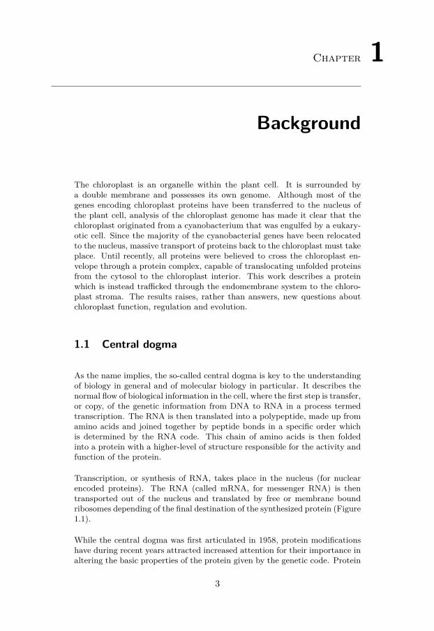
The chloroplast is an organelle within the plant cell. It is surrounded bya double membrane and possesses its own genome. Although most of thegenes encoding chloroplast proteins have been transferred to the nucleus ofthe plant cell, analysis of the chloroplast genome has made it clear that thechloroplast originated from a cyanobacterium that was engulfed by a eukary-otic cell. Since the majority of the cyanobacterial genes have been relocatedto the nucleus, massive transport of proteins back to the chloroplast must takeplace. Until recently, all proteins were believed to cross the chloroplast en-velope through a protein complex, capable of translocating unfolded proteinsfrom the cytosol to the chloroplast interior. This work describes a proteinwhich is instead trafficked through the endomembrane system to the chloro-plast stroma. The results raises, rather than answers, new questions aboutchloroplast function, regulation and evolution.
1.1 Central dogma
As the name implies, the so-called central dogma is key to the understandingof biology in general and of molecular biology in particular. It describes thenormal flow of biological information in the cell, where the first step is transfer,or copy, of the genetic information from DNA to RNA in a process termedtranscription. The RNA is then translated into a polypeptide, made up fromamino acids and joined together by peptide bonds in a specific order whichis determined by the RNA code. This chain of amino acids is then foldedinto a protein with a higher-level of structure responsible for the activity andfunction of the protein.
Transcription, or synthesis of RNA, takes place in the nucleus (for nuclearencoded proteins). The RNA (called mRNA, for messenger RNA) is thentransported out of the nucleus and translated by free or membrane boundribosomes depending of the final destination of the synthesized protein (Figure1.1).
While the central dogma was first articulated in 1958, protein modificationshave during recent years attracted increased attention for their importance inaltering the basic properties of the protein given by the genetic code. Protein
3

4 CHAPTER 1. BACKGROUND
Transcription
Translation
DNA
RNA RNA
Protein
RNA polymerase
Nucleus Cytoplasm
Ribosome
Figure 1.1. The central dogma of biology. The genetic information encodedin the DNA sequence is copied to RNA in the cell nucleus by RNA polymerase ina process termed transcription. The RNA is then transported to the cytosol, wherethe genetic information is translated into a protein by the ribosome.
modifications can happen during or after synthesis of the polypeptide. Exam-ples of common modifications are phosphorylation, glycosylation, oxidationand acetylation. Modified sites in proteins not only change their individualfunctions, but can also work together in order to fine-tune molecular interac-tions and stability, activity, localization, targeting and folding (Jensen, 2006).
1.2 Chloroplast
Fundamental for the understanding of the results presented in this thesis is abasic knowledge of the organization and the function of the chloroplast. Thechloroplast is a type of plastid, a subcellular organelle found in cells of plantsand algae, capable of performing photosynthesis. It is separated from the sur-rounding cytoplasm of the eukaryotic plant cell by a double membrane, calledthe chloroplast envelope. The chloroplast has six distinct suborganellar com-partments: three different membranes (inner and outer envelope membranestogether with thylakoid membranes), an intermembrane space between thetwo envelope membranes, a soluble interior between the inner membrane andthe thylakoid membranes called the stroma, and an aqueous lumen within thethylakoids (Jarvis, 2008) (Figure 1.2). The chloroplast has its own functionalgenome, although most of the genes have been transferred to the nucleus of thecell. The main function of the chloroplast is to perform photosynthesis, butthe chloroplast is also the site of fatty acid biosynthesis, nitrate assimilationand amino-acid biosynthesis (Waters and Langdale, 2009).

1.2. CHLOROPLAST 5
OUTER ENVELOPE MEMBRANE
INNER ENVELOPEMEMBRANE
THYLAKOID MEMBRANE
THYLAKOID LUMEN
INTERMEMBRANESPACE
STROMA
Figure 1.2. The plant cell chloroplast. The subcellular organization of thechloroplast includes three different membrane systems: the outer and inner envelopemembranes and the thylakoid membrane. There are three additional compartments:the inter-membrane space (between the outer and the inner envelope membranes),the soluble stroma and finally the aqueous lumen within the thylakoid membranes.
1.2.1 Photosynthesis
The first organisms to carry out oxygenic photosynthesis were the cyanobac-teria (Björn and Govindjee, 2009). Or to put this in other words; at a certainpoint in history, a photosynthetic machinery was assembled in which sunlightwas used as a power source to perform the thermodynamically and chemicallydemanding reaction of splitting water in order to get reducing equivalentsneeded to convert carbon dioxide into sugars (and then into other organicmolecules). The organism where this event took place was a cyanobacterium.Following this, the almost unlimited resources of water on earth could be ex-ploited and the prerequisites for life substantially changed. In addition andas a by-product from this reaction molecular oxygen was released, increasingthe metabolic efficiency due to the possibility of aerobic respiration. Hence,the evolution of the oxygenic photosynthetic process offered an advantage fororganisms capable of undergoing aerobic respiration, relegating the majorityof anaerobic organisms to isolated environments. Additionally, oxygen evo-lution enabled a protective ozone layer against the harmful UV radiation tobe formed (Barber, 2008). Several innovations were required for this to takeplace: the use of two photosystems working in a series capable of generat-ing enough difference in redox potential for water oxidation and reduction ofcarbon dioxide, an oxygen evolving complex, an "electron buffer" (a plasto-quninone pool) and modifications of the pigments attached to the reactioncentres (Björn and Govindjee, 2009).
Photosynthesis in plant cells takes place inside the chloroplast. The photo-synthetic process is usually divided in two distinct phases, one consisting of

6 CHAPTER 1. BACKGROUND
the light reactions in the thylakoid membranes in which the light-driven flowof electrons through the multi-subunit complexes mentioned above resultsin chemical energy (ATP) and reducing power (NADPH), and the secondconsisting of the carbon-fixing reactions (light-independent reactions) in thestroma where the ATP and NADPH are used by the Rubisco and other en-zymes of the Calvin cycle to fix carbon dioxide and to generate sugars (Watersand Langdale, 2009). These sugars can then be metabolized into other car-bohydrates and used for the production of energy, amino acids and lipids.
1.2.2 Chloroplast evolution
Although first met with scepticism, the hypothesis that chloroplasts are de-rived from cyanobacteria (at that time known as blue-green algae), proposedby Mereschkowsky in 1905, was later supported by electron microscopy andbiochemical studies (Raven and Allen, 2003). Now genomic analyses have con-cluded that the chloroplast indeed originates from a secondary endosymbioticevent, in which a eukaryotic cell already possessing mitochondria (as a resultof a primary endosymbiotic event between an archaea and a proteobacteriumin which the latter gave rise to mitochondria), engulfed a cyanobacterium(Kilian and Kroth, 2003; Kilian and Kroth, 2005; McFadden, 2001; Ravenand Allen, 2003; Reyes-Prieto et al., 2007) (Figure 1.3).
GREEN ALGAE AND PLANTS
Archaea
Proteobacterium
Cyanobacterium
ANIMALS
BROWN ALGAE
PLASMODIUM
EUGLENAProkaryote
Eukaryote
DIATOMS
Secondary and tertiaryendosymbiosis, such as:
Figure 1.3. Endosymbiotic events. The eukaryotic cell evolved from a pri-mary endosymbiotic event between an archaea with its own genome (orange) and aproteobacterium (brown), in which the latter gave rise to the mitochondria. Thisorganism later engulfed a cyanobacterium (green) that became the origin for thechloroplast (secondary endosymbiotic event). In some cases additional endosym-biosis occurred, giving rise to several different organisms, including numerous algalphyla (brown algae, etc) as well as apicomplexans, such as Plasmodium (modifiedfrom Raven and Allen, 2003).
Many of the proteins required for function of the chloroplast are today encodedby the nuclear genome as a result of a process called gene transfer. Studies

1.2. CHLOROPLAST 7
of plastid genomes imply that chloroplasts only possess genes for about 60-200 proteins, depending on the organism (Leister, 2003; Martin et al., 2002),while as many as 5000 proteins might be targeted to the chloroplast.
Comparison of the Arabidopsis thaliana genome with several cyanobacteria,yeast and prokaryote genomes suggest that almost one fifth (4500 genes) ofthe total Arabidopsis genes originate from the cyanobacterial ancestor of thechloroplast (Martin et al., 2002). In other studies, between 1400 to 1500(Abdallah et al., 2000) or 400 to 2200 (Rujan and Martin, 2001) Arabidopsisgenes of cyanobacterial origin have been proposed. However, gene origin andprotein compartmentalization do not strictly correspond since most of thesegene products with a cyanobacterial origin are not targeted back to the chloro-plast, suggesting a massive redistribution of cyanobacterium-derived proteinsto other cellular compartments, or they have been lost as the endosymbiontevolved. On the other hand, many non-cyanobaterial proteins are importedto the chloroplasts (Leister, 2003).
Interestingly, and discussed later on, many of the proteins of cyanobacterialorigin are predicted to enter the secretory pathway (Martin et al., 2002).Obviously, selective advantages for transfer of most genes to the host nucleusexist. Whether this is an ongoing process that will end up with an organellewithout its own genome only time can tell. The majority of genes that stillremain in the chloroplast genome seem to be involved in photosynthesis ortranscription and translation of the chloroplast genes, while most other genesare now encoded in the nucleus (Reyes-Prieto et al., 2007). This suggest thatthere is an evolutionary reason why some genes have been kept within theorganelle while others not.
Establishment of the chloroplast from the endosymbiont involved more thanarranging a functional system for import of proteins whose genes were trans-ferred to the nucleus. Intuitively it would seem that engulfment of a cyanobac-terium with two membranes would have resulted in an organelle with threemembranes, where the inner two membranes originate from the prokaryoteand the outer one originates from the outer membrane of the phagotrophic or-ganism. This is in contrast to the two membranes of the chloroplast envelopeof today. Cellular membranes are very well conserved and characterisationof the lipid and protein content has been performed in order to answer thisquestion. Interestingly, biochemical analyses of the membranes are ambiva-lent. While the inner envelope membrane very likely correspond to the plasmamembrane of the cyanobacterial ancestor, the outer envelope membrane hasboth components found in cyanobacterial outer membranes (high content ofgalactolipids and carotenoids) and elements pointing to an eukaryotic ori-gin (phosphatidylcholine) (Kilian and Kroth, 2003). This suggests that theouter envelope membrane of chloroplasts today is a chimera resulting froma fusion of the two outer membranes of the early endosymbiont and perhapssupporting this idea, the components of the Toc-Tic complex are a mix ofendosymbiotic and eukaryotic origin (Bhattacharya et al., 2007).

8 CHAPTER 1. BACKGROUND
1.2.3 Protein import into chloroplasts
Most chloroplast proteins are encoded in the nucleus. As a consequence ofthis genomic re-organization, most chloroplast gene transcripts are translatedinto polypeptides by cytosolic ribosomes and require further targeting of theproteins "back" to the chloroplast (Jarvis, 2008). Until recently, all proteinsdestined for the chloroplast interior were thought to possess an amino terminalextension, called transit peptide (TP), directing the pre-protein to a translo-con complex in the chloroplast envelope. Although recent proteomic studieshave identified some exceptions, in which chloroplast protein precursors lackthis type of targeting signal (Kleffmann et al., 2004), the vast majority ofthe chloroplast proteins are believed to be directed to the chloroplast by amechanism based on a TP pre-sequence and this pathway is considered asthe canonical chloroplast protein import system (Cline and Dabney-Smith,2008).
The TPs of these precursor proteins function as "zip codes" or signal sequencesthat are recognized by cytosolic chaperones, whose binding prevent folding ofthe polypeptides and directs them to the translocon system in the chloroplastenvelope. This import system, called the Toc-Tic complex (translocon atthe outer and inner envelope membranes of chloroplasts), is formed by twomulti-subunit complexes (Toc in the outer chloroplast membrane and Tic inthe inner, respectively) that together enable post-translational translocationacross the two envelope membranes as well as regulating the import processitself (Bedard and Jarvis, 2005). In addition, multiple homologues of the Tocreceptor components are found which might be involved in recognition andcontrolling import specificity. Recently, it has also been reported that theToc-Tic complex is under redox regulation (Stengel et al., 2009).
In short, the TP of the pre-protein to be imported is recognized by receptorsat the chloroplast surface (TOC159 and TOC34). These components of theToc have been shown to possess GTPase activity (Cline and Dabney-Smith,2008; Jarvis, 2008). The third component of the Toc-core, TOC75, is embed-ded in the outer membrane and forms a pore due to its β-barrel structure andtransports the largely unfolded pre-protein across outer membrane in a GTP-dependent process (Jarvis and Robinson, 2004). Once at the inter-membranespace, HSP70 family chaperones, TOC12 and TIC22 mediate the interactionbetween Toc and Tic. The extended polypeptide is subsequently translocatedacross the inner membrane through a Tic protein-conducting channel involv-ing TIC20 and TIC110. This step requires high levels of ATP in the stromaand the presence of stroma located molecular chaperones (Bedard and Jarvis,2005). Although the precise mechanism by which the Toc complex functionsremains to be clarified, even less is known about the Tic complex. Soon uponarrival in the stroma, the TP is cleaved off by a stromal processing peptidase(SPP) and degraded (Jarvis, 2008), resulting in a mature stromal protein orrevealing additional targeting information responsible for further directing ofthe polypeptide to the thylakoid (Bhattacharya et al., 2007) (Figure 1.4).

1.2. CHLOROPLAST 9
SPP
TOC
TIC
Transit peptide
PreproteinCytosol
Stroma
Intermembranespace
Matureprotein
Cytosolic chaperones
Figure 1.4. Chloroplast protein import by the Toc-Tic complex. The pre-protein, surrounded by cytosolic chaperones, interacts with receptor componentsof the Toc complex and is transferred across the outer envelope membrane. Thecomplex contacts with components of the Tic apparatus at the inner membraneand the pre-protein is translocated simultaneously across both envelope membranes.Afterwards, the TP is removed by stromal processing peptidase (SPP) releasing themature protein into the stroma (modified from Jarvis 2008).
The TP of chloroplast targeted proteins are remarkably heterogenic, rangingfrom 20 to > 100 amino acid residues. The only conserved properties seemto be an abundance of hydroxylated residues (serine in particular) and anoverall positive charge (Jarvis, 2008; Jarvis and Robinson, 2004). In addi-tion, they appear not to form any secondary structure but instead have arandom coil conformation, which might explain the recruitment of cytosolicfactors. Another possibility is that structure formation requires binding ofTP to specific lipids at the outer envelope membrane. Since translocation ofthe pre-protein through the Toc-Tic complexes requires the polypeptide toassume an extended conformation, cytosolic chaperones are thought to as-sist in delivering the unfolded polypeptide to the chloroplast surface. Thissuggests that the TP is designed to attract binding of such cytosolic factors(Jarvis, 2008; Jarvis and Soll, 2002).
Although most chloroplast proteins follow the post-translational pathwaythrough the Toc-Tic complex, some exceptions have recently been described.Firstly, most outer envelope proteins are inserted in the membrane from thecytosolic side without cleavable transit peptide but directed by intrinsic tar-geting information, with or without help from Toc components (Jarvis, 2008).

10 CHAPTER 1. BACKGROUND
Also, two proteins (ceQORH and Tic32) have been shown to be targeted tothe inner membrane without cleavable targeting signals (Miras et al., 2002;Nada and Soll, 2004). In addition and as later presented in this thesis, thereseems to be a pathway for chloroplast proteins to be transported via theendomembrane system.
1.3 Endomembrane system
The plant endomembrane contains several membrane bound organelles (Fig-ure 1.5). While endoplasmic reticulum (ER) and the Golgi apparatus will bepresented in more detail later, plant cells also contain at least two types ofendosomes: early endosomes involved in sorting and recycling and late endo-somes/prevacuolar compartments en route to the lytic vacuole. Two majorfunctional types of vacuoles also exist in plant cells: the lytic vacuole and theprotein-storage vacuoles. Lytic vacuoles function as compartments for degra-dation and waste storage, while protein-storage vacuoles accumulate proteinsmainly used as nutrients during seed germination. These two types of vacuolescan fuse, giving rise to a large central vacuole. During cell division anothertransient compartment is also formed, the cell plate, by fusion of transportvesicles (Jurgens, 2004).
The default secretory pathway leads from the ER via the Golgi apparatus tothe plasma membrane. Proteins not destined for the plasma membrane aresorted in the Golgi. Proteins aimed to remain in the ER, such as ER locatedchaperones, are recognized by the presence of well-characterized ER retentionsignals (H/KDEL). Other signals are responsible for sorting of membraneproteins or proteins destined for vacuoles.
1.3.1 Endoplasmic reticulum
The ER is a membrane-bound tubular network that stretches out from thenuclear envelope towards the plasma membrane (Jurgens, 2004). The ERnetwork of higher plants overlies the actin cytoskeleton rather than micro-tubules as in animal cells (Runions et al., 2006; Sparkes et al., 2009; Staehe-lin, 1997). Traditionally the ER was classified as smooth or rough dependingon the absence or presence of membrane-bound ribosomes, however now twomorphological forms are used, cisternal and tubular, which better reflect thehighly dynamic nature of this organelle (Sparkes et al., 2009). Several cellularfunctions are allotted to the ER, such as biosynthesis of phospholipids andsynthesis, post-translational modification, folding, quality-control of secretedproteins and glycoproteins, as well as regulating cytosolic calcium levels. Themultifunctional nature of the ER is also exemplified by the vast number ofsub-regions or domains of which the ER is composed (Staehelin, 1997).

1.3. ENDOMEMBRANE SYSTEM 11
One of the two sub-domains of the ER important for the work presented in thisthesis is the rough ER, where membrane-bound ribosomes are attached to theER. This is the entry point for proteins into the so-called secretory pathway.Another important domain of the ER, and perhaps the most dynamic andcontroversial, is the ER export site (ERES). This is believed to be the sitefor transport of soluble and membrane proteins and lipid cargo from the ERto the Golgi. Although the exact nature of the interface between the ERand the Golgi remains to be solved, some speculate whether the Golgi itselfcan be considered as a specialized domain of ER and that the ERES has thepossibility of initiating the biogenesis of a new Golgi stack (Sparkes et al.,2009). Transport between the ER and the Golgi apparatus is bidirectionaland believed to be mediated by different coated vesicles. COPII vesicles arethought to function in the anterograde transport from ER to Golgi, whileCOPI vesicles are working in retrograde Golgi-to-ER transport (Hawes et al.,2008).
1.3.2 Golgi apparatus
The Golgi apparatus in plant cells is organized as individual stacks, containingseveral morphologically distinct cisternae from cis to trans, followed by atrans-Golgi network (Figure 1.5) (Jurgens, 2004). Golgi stacks are highlymobile and close association and/or direct contact with the plant ER hasbeen reported in electron microscopy studies (Brandizzi et al., 2002). Also,using photo-activated GFP, Golgi bodies were seen to move with the same rateand in the same direction as ER, demonstrating that Golgi is moving with,and not over the ER (Runions et al., 2006), which supports the idea that thereis a tight connection between ERES and the Golgi bodies. In recent studiesusing laser-trapping technology, it was shown that capture and manipulationof individual Golgi bodies in cells with depolymerised actin cytoskeleton notonly resulted in movement of the Golgi stack, but also in extension or growthof the associated ER tubule, supporting the theory that Golgi bodies canpossess an attachment to the ER (Sparkes et al., 2009).
The Golgi apparatus is not only functioning as a sorting station for cargodelivery to different destinations, but also as a specialised protein modifyingfactory where several enzymes, among them those responsible for modifyingthe N-linked glycans on glycoproteins passing the Golgi on the way to theirfinal destination (Jurgens, 2004), reside in different subdivisions of the Golgistacks (cis-, medial- and trans-cisternae) (Figure 1.5).
1.3.3 Protein trafficking between the ER and the Golgiapparatus
Protein synthesis is carried out by ribosomes that translate the genetic infor-mation from the RNA molecule into a polypeptide with a specific sequence

12 CHAPTER 1. BACKGROUND
of amino acids. These ribosomes are either free in the cytosol (or insidethe endosymbiotic organelles mitochondria and chloroplasts) or bound to thecytosolic face of the ER membrane. The free ribosomes are synthesizing cy-tosolic, nuclear, peroxisomal and most of the mitochondria and chloroplastproteins, while proteins destined for compartments within the endomembranesystem (ER, Golgi, endosomes, vacuole), the plasma membrane or secreted aresynthesized by ER associated ribosomes (Hebert and Molinari, 2007) (Figure1.5).
Ribosomes translating proteins destined for the ER must be brought in closeproximity to the ER membrane in order for the nascent polypeptide to be ableto cross the ER membrane. This is accomplished by the presence of a signalsequence at the N-terminus of the emerging polypeptide. This so called sig-nal peptide (SP) contains hydrophobic amino acids and precedes the matureproteins. Appearance of this hydrophobic peptide is recognized by a multi-subunit complex, called the signal recognition particle (SRP), which binds tothe signal sequence and pauses translation. Protein synthesis is only resumedwhen the complex has come to contact with the ER and the ribosome is lo-calized at a proteinous channel in the ER membrane. In addition to bind toSRP, the hydrophobic SP facilitates co-translational insertion of the polypep-tide into the ER lumen. Upon entry in the ER, the SP is usually cleavedoff from the mature protein (Hebert and Molinari, 2007). The first step inprotein trafficking along the default secretory pathway is vesicle transportfrom the ER to the Golgi (Figure 1.5). Although very little evidence for theexistence of COPII vesicles exists, they are believed to mediate anterogradetraffic between the ER and the Golgi (Faso et al., 2009; Hawes et al., 2008).Formation of the COPII vesicles requires the GTPase Sar1 and its GDP/GTPexchange factor Sec12. At the cis-Golgi side, another GTPase (RabD2a) isinvolved in fusion of the vesicles (Hawes et al., 2008; Jurgens, 2004). Domi-nant mutant versions of both proteins, where the GTPase activity has beendisrupted, have been shown to block trafficking of soluble proteins in the ER(Batoko et al., 2000; Takeuchi et al., 2000).
Retrograde traffic from the Golgi to the ER depends on COPI vesicles andthe action of the ADP-ribosylation factor 1 (Arf1) GTPase. In contrast tothe COPII-mediated bulk flow mechanism for ER exit (Hanton et al., 2006),transport of soluble and membrane proteins from the Golgi to the ER requiretargeting signals (Matheson et al., 2006). In a similar way as for Sar1 andRabD2a, a dominant-negative version of Arf1 was shown to inhibit Golgi-to-ER traffic (Takeuchi et al., 2002). Interestingly, inhibition of COPI functionalso resulted in impaired ER and disrupted anterograde transport, emphasiz-ing that a balance between the two systems is required for normal ER andGolgi organization (Faso et al., 2009; Stefano et al., 2006).

1.3. ENDOMEMBRANE SYSTEM 13
NUCLEOUS
LYTICVACUOLE
CHLOROPLAST
ENDOSOME
PSV
GOLGIAPPARATUS
ENDOPLASMICRETICULUM
Sar1RabD2a
Arf1
CYTOSOL
TGNcis
medialtrans
COPII
COPI
Figure 1.5. Simplified figure of the plant endomembrane system and itstrafficking pathways. Secretory cargo proteins (black dots) are transported fromthe ER via the Golgi/trans-Golgi network (TGN) to the plasma membrane. ERto Golgi anterograde transport is mediated by COPII coated vesicles, with Sar1GTPase assisting in the budding process and RabD2a helping the vesicle fusionat the Golgi membrane. Retrograde transport is thought to be controlled by COPIcoated vesicles in which Arf1 assists the budding process. Cargo destined to the lyticvacuole or protein-storage vacuole (PSV) is trafficked from the Golgi/TGN to thesecompartments. PSV and lytic vacuoles may fuse into a large central vacuole (notshown). The endocytic pathway involves an early/sorting endosome for recycling ofplasma-membrane proteins. Coat proteins (COPI, COPII), Sar1, Arf1, and RabD2aGTPases, are indicated.
1.3.4 Protein folding and post-translational modification inthe ER and Golgi
The lumen of the ER has a more oxidizing milieu than the cytosol and con-tains a myriad of enzymes that are able to perform modifications on the newlysynthesized proteins. In addition, many ER-resident proteins prevent proteinaggregation and maintain the emerging polypeptides in a state that allowsco- and post-translational modifications to take place (Hebert and Molinari,2007; Vitale and Ceriotti, 2004). Of particular importance and interest for thework presented in this thesis are the formation of intra- and inter-moleculardisulphide bonds between cysteine residues and the anchoring of N-linkedoligosaccharides to the polypeptide backbone. These modifications are im-portant for proper structural maturation of many proteins and can also be

14 CHAPTER 1. BACKGROUND
sensed by a quality control system present in the ER (ERQC) in order toensure that only correctly folded proteins are trafficking out of the ER, whilemisfolded proteins are either refolded to achieve correct structure or beingsent for degradation (Crofts et al., 1998) (Figure 1.6).
-HS SH--HS SH-
-S-S-
-S -S-
-HS SH--HS SH-
-S-S-
-S -S-
-S-S-
-HS SH-
Synthesistranslocation Glucosidase I
Glucosidase II
Glucosidase II
Glucosyl T
Native
Nearlynative
Glucosidase II(ER Mannosidase)
x
MisfoldedTerminally misfolded
Calnexin/Calreticulin
Cycle
Retro-translocation
Cytosolicdegradation
ExportSecretorypathway
Lectin associatedBiP
Cnx/Crt
Endoplasmic reticulumCytosol
Foldingintermediate
-S-S-
-HS SH-
Figure 1.6. The ER N-glycoprotein "quality control". Nascent polypeptidechains enter the ER lumen and N-glycans containing N-acetylglycosamine, mannoseand glucose molecules are attached to asparagines residues in a site-dependent man-ner. The two terminal glucoses of the glycan are rapidly trimmed by sequentialaction of the glucosidase I and II. Mono-glucosylated N-glycans mediate initial asso-ciation of folding polypeptides with the ER lectin-chaperones calnexin (CNX) and/orcalreticulin (CRT) and undergo exposure to glycoprotein oxidoreductases, releasingthe properly folded protein, which is rapidly deglucosylated, partially demannosy-lated and eventually leaves the ER. Proteins not completely folded are kept in theCNX and/or CRT cycle: The folding intermediate is released from the lectin chap-erones and deglucosylated. Forward transport is inhibited by a glucosyl transferase,adding a glucose residue to glycoproteins with nearly native conformation, whichundergo additional folding attempts. Released glycopolypeptides displaying majorfolding defects attract BiP and are extensively demannosylated and dislocated acrossthe ER membrane for proteasome mediated degradation (modified from Hebert andMolinari, 2007).
One of the best studied chaperones in the ER is the binding protein (BiP).Unless N-glycans are added to the very N-terminus of the emerging polypep-tide, BiP is the first chaperone the emerging polypeptide faces upon arrivalin the ER lumen. BiP counteracts misfolding by binding to hydrophobic do-mains of the nascent protein, thereby preventing hydrophobic regions of dif-ferent polypeptides from aggregating. When folding is complete, hydrophobicregions are no longer exposed and the protein is released from BiP (Pimpl

1.3. ENDOMEMBRANE SYSTEM 15
et al., 2006). In a similar way, BiP also prevents formation of non-nativedisulphide bonds that otherwise could result in protein aggregates. Anotherfunction of BiP is to bind to severely and permanently misfolded proteins andto assist in translocation of these proteins out of the ER for degradation inthe cytosol (Hebert and Molinari, 2007). Recently, a role for BiP in transportof misfolded proteins to lytic vacuoles has also been proposed (Pimpl et al.,2006).
Many of the proteins entering the secretory pathway are N-glycosylated. At-tachment of one or several N-linked glycans not only changes the propertiesof the proteins, but also assists in folding due to the action of carbohydrate-binding chaperones, even called lectin chaperones. Although much of ourcurrent knowledge about ERQC and glycan processing comes from studieson yeast and mammalian systems, most components seem to be conserved inplants (Hong et al., 2008; Parodi, 2000). While BiP binds to hydrophobicregions of the polypeptide backbone, the lectin chaperones bind to the bulkyhydrophilic oligosaccharide groups (Hebert and Molinari, 2007).
The first step in the maturation of N-linked glycoproteins in the secretorypathway is the transfer of an oligosaccharide precursor (Glc3Man9GlcNAc2)from a dolichol lipid carrier to a specific asparagine residue (Asn-X-Ser/Thr,where X is any residue except Pro) on the emerging polypeptide by theoligosaccharyl transferase (OST) multisubunit complex (Lerouge et al., 1998).This precursor is subsequently modified by glucosidases and glucosyl trans-ferases along the secretory pathway. The first modification of the N-glycanprecursor that takes place in the ER is the trimming of the three glucose units(Figure 1.6). The outermost α1,2-glucose unit is hydrolyzed by glucosidase I,while the following two α1,3-linked glucose units are removed by glucosidaseII (Crofts et al., 1998; Leonard et al., 2009). Trimming of the first α1,3-glucose results in monoglucosylated N-glycans (GlcMan9GlcNAc2) that arerecognized by the lectin chaperones calnexin (CNX) and calreticulin (CRT).This causes the folding process to slow down and increases the efficiency inthe formation of correct disulphide bonds by oxidoreductases (Hebert andMolinari, 2007). When folding is complete, the protein is released and thelast glucose is removed by glucosidase II, rendering an oligosaccharide struc-ture known as high-mannose type N-glycan. In mammals, an ER-localizedmannosidase has been shown to remove one mannose residue of the correctlyfolded protein to yield Man8GlcNAc2, before ER-export and trafficking ofthe glycoprotein to the Golgi. Such mannosidase has not yet been foundin plants, although ER-resident glycoproteins with that exact structure havebeen identified, suggesting that a similar mannosidase also exists in plant cells(Navazio et al., 1996).
Glycoproteins that have not acquired the correct folding are retained in theER in one of two ways. If misfolding is severe, or if repeated cycles of at-tempted folding of the protein fail, the protein attracts binding of BiP whichforms aggregates with the misfolded protein, protecting the ER from exposure

16 CHAPTER 1. BACKGROUND
to such potentially harmful molecules and assisting in translocation of the pro-tein out of the ER for degradation (Hebert and Molinari, 2007; Hong et al.,2008; Li et al., 2009; Parodi, 2000). If the protein is only partially misfolded,transient reglucosylation by the luminal enzyme UDP-glucose:glycoproteinglucosyl transferase (GT) again results in a monoglucosylated protein thatcan be recognized by the lectin chaperones CNX and CRT. These chaperoneswill then remain and assist in refolding of the protein (Hammond et al., 1994;Jin et al., 2009; Jin et al., 2007; Soussilane et al., 2009). Binding of CNXand CRT exposes the polypeptide to oxidoreductases that assist in formationof disulphide bonds and isomerases capable of rearranging non-native bonds.This cycling of glucosidase II and GT activities drives binding and release tothe CNX/CRT chaperones and assists in proper folding of the glycoproteinuntil correct structure is achieved and the protein is structurally competent forexport out of the ER. An alternative outcome is that folding is not successfuland the polypeptide is instead sent for degradation. N-linked glycosylationtherefore plays an important role in the ERQC system and indicates thatfolding can depend on an ensemble of different protein modifications that allmust work together for a properly folded protein to appear.
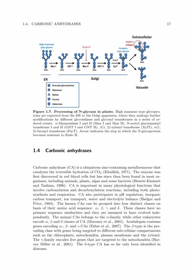
Upon arrival at the Golgi apparatus, plant N-glycans can be further modifiedinto complex-type N-glycans during the transport of the glycoprotein fromcis, through medial to trans cisternae of the Golgi. First, the α-mannosidaseI removes one to four α1,2-mannose residues, resulting in Man5GlcNAc2(Figure 1.7). Then N-acetylglucosaminyl transferase I (GNT I) transfersan N-acetylglucosamine (GlcNAc) residue to the α1,3-mannoside branch ofMan5GlcNAc2, to yield GlcNAcMan5GlcNAc2. Two additional mannosesare then removed by α-mannosidase II and another GlcNAc is transferred tothe α1,6-mannoside branch by GNT II, resulting in GlcNAc2Man3GlcNAc2.Further action of Golgi localized glucosyl transferases results in plant spe-cific N-glycans. Transfer of β(1, 2)-xylose to the β-mannose and a(1,3)-fucoseto the proximal GlcNAc core seem to be independent events occurring inthe medial and trans cisternae of the Golgi. Later, additional modificationof the complex-type glycan by transfer of fucose and galactose residues byβ1,3-galactosyl transferase and α1,4-fucosyl transferase to the terminal Glc-NAc residues might take place, resulting in antennae with Galβ1-3(Fucα1-4)GlcNAc sequences. These structures are also known as Lewis a antigensand found on the cell surface of mammalian cells and involved in cell-cellrecognition and cell adhesion processes (Lerouge et al., 1998; Rayon et al.,1998). Additional modification of the N-linked glycan can take place duringthe transport to or in the final destination, such as for vacuoles (Lerouge etal., 1998; Rayon et al., 1998).
The reason for the occurrence of such plant-specific N-glycans is not known.A mutant allele of Arabidopsis, defective in GNT I and unable to producecomplex-type N-glycans, did not show any obvious phenotype when grownunder standard conditions (von Schaewen et al., 1993). However, mutationof this enzyme in mammalian cells are deleterious (Ioffe and Stanley, 1994).
1.4. CARBONIC ANHYDRASES 17
Asn Asn Asn Asn Asn Asn
Asn
Asn
Man I GNT I Man II GNT II XylT
FucT
ER Golgi
Vacuole
ExtracellularLewis aantigen
N-acetyl glucosamine
Mannose
Xylose
Fucose
Galactose
High-mannosetype glycan Complex
type glycan
Endo H
Figure 1.7. Processing of N-glycans in plants. High mannose type glycopro-teins are exported from the ER to the Golgi apparatus, where they undergo furthermodifications by different glycosidases and glycosyl transferases in a series of or-dered events. α-Mannosidase I and II (Man I and Man II), N-acetyl glucosaminyltransferase I and II (GNT I and GNT II), β(1, 2)-xylosyl transferase (XylT), α(1,3)-fucosyl transferase (FucT). Arrow indicates the step in which the N-glycoproteinbecomes resistant to Endo H.
1.4 Carbonic anhydrases
Carbonic anhydrase (CA) is a ubiquitous zinc-containing metalloenzyme thatcatalyzes the reversible hydration of CO2 (Khalifah, 1971). The enzyme wasfirst discovered in red blood cells but has since then been found in most or-ganisms, including animals, plants, algae and some bacteria (Hewett-Emmettand Tashian, 1996). CA is important in many physiological functions thatinvolve carboxylation and decarboxylation reactions, including both photo-synthesis and respiration. CA also participates in pH regulation, inorganiccarbon transport, ion transport, water and electrolyte balance (Badger andPrice, 1994). The known CAs can be grouped into four distinct classes onbasis of their amino acid sequence: α, β, γ and δ. These classes have noprimary sequence similarities and they are assumed to have evolved inde-pendently. The animal CAs belongs to the α-family, while other eukaryotesencode α, β and δ classes of CA (Moroney et al., 2001). Arabidopsis containsgenes encoding α-, β- and γ-CAs (Fabre et al., 2007). The β-type is the pre-vailing class with genes being targeted to different sub-cellular compartmentssuch as the chloroplasts, mitochondria, plasma membrane and the cytosol.The γ-family encodes five genes that are targeted to the mitochondria (Har-vey Millar et al., 2001). The δ-type CA has so far only been identified indiatoms.

18 CHAPTER 1. BACKGROUND
The requirement of a CA activity for photosynthesis, as well as for any bio-logical system, is obvious. In the plant chloroplast, the non-catalyzed inter-conversion of CO2 and HCO−
3 is considered to be 104 times slower than thebiological flux needed for CO2 fixation by Rubisco (Badger and Price, 1994).If interconversion between these two species is important for the supply ofCO2 to the active site of Rubisco, then CA activity would be required toenable effective photosynthesis in the chloroplast stroma. Despite this impor-tant function, little progress has been made in fully elucidating the role ofCAs in C3 photosynthesis.
1.4.1 The α-CAs in Arabidopsis
At least eight genes encoding α-type CAs are present in Arabidopsis thaliana(AtαCA1-8). Although the essential amino acids are present in the pre-dicted gene products from all eight CA genes (Fabre et al., 2007), sug-gesting that they are functional isozymes, expressed sequence tags (ESTs)have only been reported to The Arabidopsis Information Resource (TAIR,www.arabidopsis.org) for five of them (CA1, CA2, CA3, CA5 and CA8, asof November 2009) This indicates that CA4, CA6 and CA7 could be pseu-dogenes or expressed at very low levels or under specific conditions. CA1(CAH1) was found in all organs except root, while CA2 was only expressedin stem and root and CA3 restricted to flowers and siliques (Fabre et al.,2007). CA3 has previously been found in mature pollen in proteomic analysis(Holmes-Davis et al., 2005; Noir et al., 2005). In addition, a cDNA for CA7was isolated from an expanded Arabidopsis library (Yamada et al., 2003) andCA4 was identified in the thylakoid membranes in a mass-spectrometric pro-teomic approach (Friso et al., 2004; Sun et al., 2009). Sequence analysis showsthat CA2 is lacking N-terminal targeting information, presumably encoding acytoplasmic variant of the protein, while the remaining α-CAs have predictedSPs for co-translational insertion into the ER lumen.

Chapter 2
Results achieved
In this chapter, results and conclusions from the paper and the manuscriptsof the thesis are outlined and discussed in an attempt to summarize thedifferent parts into one consecutive story. At the time of the initiation ofthis PhD project, a carbonic anhydrase, CAH1, had been found in the Ara-bidopsis thaliana chloroplast. What made CAH1 peculiar compared to otherchloroplast proteins was the presence of N-linked glycans on the protein. N-glycosylation is only known to occur in the endomembrane system, and noroute for proteins between the endomembrane system and the chloroplast wasknown at that time. In paper I this finding was reported, that a chloroplaststroma localized protein, instead of following the canonical route through theToc-Tic system in the chloroplast envelope, was trafficking via the ER andGolgi to the chloroplast. Additionally, we could show that the protein arrivingat the chloroplast was N-glycosylated. In an attempt to genetically examinecomponents of the trafficking mechanism reported in paper I, manuscriptII shows that proteins known for their involvement in canonical ER-to-Golgivesicle trafficking also are involved in transport of CAH1. In addition, themanuscript describes an improved method for transient co-expression of mul-tiple genes in plant cells. We also try to emphasize the potential of thisoptimized method and how it can be used in trafficking studies of CAH1and/or other proteins. Manuscript III focuses on the importance of post-translational modifications of CAH1, and the effect of these modificationson fundamental processes such as folding, trafficking, and protein function-ality and/or activity. The results from this study encourage us to presenta hypothesis to why a protein trafficking pathway from the endomembranesystem to the chloroplast exists in the plant cell. Finally, manuscript IVpresents data obtained from Arabidopsis plants with disrupted CAH1 geneexpression, which clearly indicate the relevance of the activity of this CA inthe photosynthetic performance and the chloroplast function of a C3 plant.
2.1 Paper I
CAH1 was identified in a proteomic screening for chloroplast localized CAs inthe plant model Arabidopsis thaliana. Chloroplast localization of the proteinwas suggested from subcellular fractionation of leaf material (Paper I, Figure1d). Neither CAH1 gene transcript, nor the CAH1 protein itself, was detected
19

20 CHAPTER 2. RESULTS ACHIEVED
in roots, indicating that the protein is only present in above ground organs,presumably concentrated in leaf tissues. Electron microscopy of immunogoldlabelled cell sections confirmed that the CAH1 protein was localized in thestroma, the soluble interior of the chloroplast (Paper I, Figure 1c).
2.1.1 CAH1 is lacking N-terminal transit peptide fortargeting to the chloroplast
Analysis of the primary sequence (amino acid sequence) can supply impor-tant information about a protein. For example, conserved domains of enzymeclasses can be found, specific sites for post-translational modifications can bepredicted and the secondary structure for different stretches of the polypep-tide sequence can be deduced. Even the complete 3D structure of the proteincan be modelled if structure of a related protein has been resolved. Addition-ally, comparison to the vast genome and protein sequence information nowavailable from many organisms can give valuable clues about the function orlocalization of the protein.
One of the tools available for analysis is the algorithm known as TargetP(Emanuelsson et al., 2000), a neural network-based service for large-scaleprediction of subcellular location. Targeting information is often present atthe N-terminus of polypeptides, e.g. targeting to the secretory pathway, themitochondria and, in plant cells, the chloroplasts. The overall success ratefor analysis of protein targeting to these organelles is as high as 85%, inwhich proteins destined to the ER are most successfully predicted (95%) andchloroplast proteins being most difficult (69%). Analysis of the 284 amino acidlong polypeptide of CAH1 strongly suggested that the protein was targetedto the ER, a prediction in total disagreement with the experimentally verifiedsubcellular location of the protein. Additionally, TargetP proposed a cleavagesite for the ER SP at amino acid 24 and 25 (ADA-QT, Paper I, Figure 1a).Although prediction of chloroplast located proteins is difficult, further analysisusing Predotar (Small et al., 2004) confirmed suggested ER targeting.
Comparison to other CAs identified the region spanning from amino acid 35to 262 as the carbonic anhydrase domain (PD000865) (Servant et al., 2002),and confirmed that the CAH1 polypeptide possessed an N-terminal exten-sion not necessarily important for CA activity. While this analysis only wasbased on sequence analyses, it was important to determine whether the CAH1precursor protein could be chloroplast imported, indicating false ER SP pre-diction. In vitro uptake studies were performed with isolated pea chloroplastsand dog pancreas microsomes to see if the CAH1 precursor was competent forimport to any of the two cellular compartments. Expression of the protein inthe presence of pea chloroplasts, often used in this type of analyses, showedthat neither the CAH1 precursor, nor the polypeptide lacking the predictedER SP, was imported (Paper I, Figure S1e). Instead, full length CAH1 was

2.1. PAPER I 21
efficiently translocated into dog pancreas microsomes (Paper I, Figure 2c).The polypeptide was additionally processed by a signal peptidase in the ERlumen, indicating that the CAH1 precursor protein indeed possessed a func-tional ER SP despite its chloroplast localization in planta. In addition, theprotein accumulated in the microsomes with a similar weight as the nativestromal protein (around 38 kDa) which is substantially larger than the ex-pected mass of the polypeptide backbone alone (32.7 or 30.0 kDa, with orwithout SP, respectively). This difference in mass suggested that the matureCAH1 harboured some kind of post-translational modification.
At this point our experimental data were pointing in two different directions.Location of the protein appeared to be within the chloroplast while targetinginformation strongly indicated a protein heading for the endomembrane sys-tem, and not the chloroplast. The only way for us to put these observationstogether required a (novel) route for proteins from the secretory system tothe chloroplast (Figure 2.1).
NUCLEOUS
CHLOROPLAST
GOLGIAPPARATUS
ENDOPLASMICRETICULUM
Sar1RabD2a
Arf1
CYTOSOL
Standard precursor
N-glycosylatedplastid protein
Toccomplex
Ticcomplex
Unknowntranslocator
COPII
COPI
Figure 2.1. New glycoprotein pathway to the chloroplast via the en-domembrane system. CAH1 is translocated into the ER, where the protein ac-quires N-linked glycans. The N-glycans are processed as the protein travels from theER to the Golgi. CAH1 is then transported, presumably via Golgi derived vesicles,to the chloroplast where the vesicles fuse with the outer envelope membrane. CAH1finally crosses the envelope membrane by an unknown mechanism and reaches thestroma.
As mentioned earlier, engulfment of a Gram-negative prokaryotic cyanobac-terium with two membranes, by a eukaryotic cell surrounded by a plasmamembrane, intuitively would have resulted in an endosymbiotic organelle with
22 CHAPTER 2. RESULTS ACHIEVED
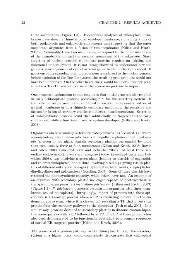
three membranes (Figure 1.3). Biochemical analyses of chloroplast mem-branes have shown a chimeric outer envelope membrane, containing a mix ofboth prokaryotic and eukaryotic components and suggesting that the outermembrane originates from a fusion of two membranes (Kilian and Kroth,2003). Presumably these two membranes correspond to the outer membraneof the cyanobacterium and the vacuolar membrane of the eukaryote. Sincetargeting of nuclear encoded chloroplast proteins requires an existing andfunctional import system, it is not straightforward to understand how thegenomic rearrangement of cyanobacterial genes to the nucleus proceeded. Ifgenes encoding cyanobacterial proteins were transferred to the nuclear genomebefore evolution of the Toc-Tic system, the resulting gene products would nothave been imported. On the other hand, there would be no evolutionary pres-sure for a Toc-Tic system to arise if there were no proteins to import.
One proposed explanation to this enigma is that initial gene transfer resultedin early "chloroplast" proteins possessing SPs for the secretory system. Ifthe outer envelope membrane contained eukaryotic components, either ata third membrane or at a chimeric secondary membrane, the receptors andfactors for fusion of secretory vesicles could exist in such membrane. Secretionof endosymbiotic proteins could then additionally be targeted to the earlychloroplast, while a functional Toc-Tic system developed (Kilian and Kroth,2003).
Organisms where secondary or tertiary endosymbiosis has occurred, i.e. wherea non-photosynthetic eukaryotic host cell engulfed a photosynthetic eukary-ote (a green or red alga), contain secondary plastids surrounded by morethan two, usually three or four, membranes (Kilian and Kroth, 2003; Ravenand Allen, 2003; Sanchez-Puerta and Delwiche, 2008). At least three sec-ondary endosymbiotic events are recognized today (Sanchez-Puerta and Del-wiche, 2008), two involving a green algae (leading to plastids of euglenoidsand chlorarachniophytes) and a third involving a red alga giving rise to plas-tids of different eukaryotic lineages (haptophytes, heterokonts, cryptophytes,dinoflagellates and apicomplexa) (Keeling, 2009). Some of these plastids haveretained the photosynthetic capacity, while others have not. An example ofan organism with secondary plastid no longer capable of photosynthesis isthe apicomplexan parasite Plasmodium falciparum (Kilian and Kroth, 2003)(Figure 1.3). P. falciparum possesses cytoplasmic organelles with three mem-branes (called apicoplasts). Intriguingly, import of proteins into these api-coplasts is a two-step process where a SP is mediating import into the en-domembrane system, where it is cleaved off, revealing a TP that diverts theprotein from the secretory pathway to the apicoplast (Foth et al., 2003). In asimilar way, proteins destined to secondary plastids in diatoms contain bipar-tite pre-sequences with a SP followed by a TP. The SP of these proteins hasalso been demonstrated to be functionally equivalent to precursor sequencesof normal ER-targeted proteins (Kilian and Kroth, 2005).
The presence of a protein pathway to the chloroplast through the secretorysystem in a higher plant would conclusively demonstrate that chloroplast

2.1. PAPER I 23
protein import is not exclusively dependent on a functional Toc-Tic system. Inaddition, such a finding could suggest that sorting of proteins to the evolvingchloroplast initially might have occurred via the secretory system (Reyes-Prieto et al., 2007), in a similar way as for secondary plastids of diatoms andP. falciparum.
2.1.2 CAH1 contains an N-terminal signal peptide for theER, but is localized to the chloroplast
To verify chloroplast targeting of the protein by a method independent ofthe use of CAH1 antibodies, a translational fusion of CAH1 and GFP (greenfluorescent protein) was constructed. GFP was fused C-terminally to CAH1 toavoid interfering with the ER precursor sequence of CAH1. As expected fromimmunogold and subcellular fractionation experiments, the protein localizedto the chloroplast as seen by confocal microscopy (Paper I, Figure 1e). Incontrast, addition of an ER retention signal (KDEL) to the C-terminus ofthe CAH1-GFP construct resulted in fusion protein being localized to the ER(Paper I, Figure 2a and b). The KDEL tail was added to ensure that fusionprotein translocated into the ER lumen would be retained and not furthertrafficked. No GFP signal was detected in the chloroplast, excluding thepossibility that the protein was simultaneously sent to ER and chloroplast bydual targeting, a phenomenon that had been reported in previous publications(Levitan et al., 2005). This result concluded that CAH1 indeed containedactive and functional precursor sequences for the ER an ER only.
CAH1 is presumed to be a low-abundance protein in Arabidopsis, and effortsto deduce the exact signal peptidase cleavage site by N-terminal sequencingof the native protein failed. Instead, another approach was tested in whichthe gene construct for the ER-retained GFP fusion protein was stably trans-formed and expressed in Tobacco BY2 cell suspension culture. Accumulationof the protein in these cells proved to be high, and ER-retained protein har-vested from the total extract and purified by a single step of anion exchangechromatography could be N-terminally sequenced. Since processing in the ERis highly conserved, the cleavage site in the native protein could be assumedto be identical to the ER retained polypeptide (ADAQ-T), notably only oneresidue from the site predicted by SignalP (Bendtsen et al., 2004).
2.1.3 Chloroplast localized CAH1 is N-glycosylated in theER
A common post-translational modification taking place in the ER of eukary-otic cells is N-glycosylation. N-glycosylation results in sugar complexes an-chored to specific sites of polypeptides (Asn-X-Ser/Thr, where X can be any

24 CHAPTER 2. RESULTS ACHIEVED
amino acid but Pro). CAH1 has five such sites, of which four are predictedto be decorated with N-linked glycans (www.cbs.dtu.dk/services/NetNGlyc).Each N-glycan has a mass of about 1.5-2 kDa, depending on the numberand type of sugar molecules present, resulting in a theoretical increase ofCAH1 weight by 6-10 kDa, well in accordance to the 8 kDa difference seenbetween SP processed protein and mature stroma CAH1 (30.0 and 38 kDa,respectively) (Paper I, Figure 2c, lane 4 and Figure 3b). Inhibition of N-glycosylation during uptake studies into dog pancreas microsomes confirmedthat the protein was glycosylated in vitro and that the glycosylated polypep-tide migrated with a similar weight as the native protein, strongly suggestingthat the chloroplast located protein was glycosylated.
Targeting of CAH1 to the ER was shown both in vitro and in vivo usinguptake studies into microsomes and confocal microscopy of KDEL taggedprotein, respectively, but this did not say anything about further traffickingof the protein from the ER to the chloroplast. Translocation of the proteinout of the ER, followed by Toc-Tic mediated translocation, seemed unlikelysince this would require unfolding of the protein for passage through theenvelope translocon. Also, the presence of bulky N-glycans would certainlyaffect transport through Toc-Tic. Other scenarios could mimic the targetingof proteins to the secondary plastids. In order to avoid passage throughvacuoles, the two most likely alternatives would be direct targeting from theER to the chloroplast, or via the Golgi apparatus.
The most predominant forms of lipids in the chloroplasts are galactolipids.While assembly of galactolipids takes place in the chloroplast envelope, thegalactolipid precursors (diacylglyrecol moieties) of many plants species (in-cluding Arabidopsis) originate from two different compartments, the plastid(prokaryotic pathway) or the ER (eukaryotic pathway). In the prokaryoticpathway diacylglycerol is assembled directly from fatty acids synthesised inchloroplasts and incorporated into chloroplast galactolipids. In the eukary-otic pathway the fatty acids are transported to the ER where diacylglycerolis assembled and later returned to the chloroplast envelope for galactolipidsynthesis (Benning et al., 2006; Kelly and Dormann, 2004; Xu et al., 2008).Obviously, such events require some kind of interaction between the ER andthe chloroplast. Not only have contact sites between ER and plastids been re-ported, so-called plastid associated membranes (PLAMs) (Hanson and Köh-ler, 2001; Kunst and Samuels, 2003), but protein-protein interactions havealso been suggested (Andersson et al., 2007) and recently a protein respon-sible for mediating lipid transfer between the ER and the outer envelopemembrane was suggested (Xu et al., 2008). Considering reported interactionsbetween plastids and the ER, a direct transfer from the ER to the chloroplastemerged as an attractive explanation for transport of CAH1.

2.1. PAPER I 25
2.1.4 Attachment of complex type glycans to CAH1 suggesta route to the chloroplast via the Golgi apparatus
One way to study targeting of glycoproteins is to analyse the type of N-linked glycan attached to the protein. Maturation of N-glycans along thesecretory pathway has previously been described and the subcellular actionof several glucosidases and glucosyl transferases determined (Figure 1.7). Forexample, glucosidases I and II remove the glucose residues of the oligosac-charide precursor Glc3Man9GlcNAc2 in the ER (Lerouge et al., 1998), re-sulting in a structure known as high mannose-type N-glycans. These N-glycans can be further modified in the Golgi, resulting in complex-type N-glycans. Plant complex-type glycans are characterized by the presence ofα(1,3)-fucose and/or a β(1,2)-xylose residues, respectively, linked to the prox-imal N-acetyl-glucosamine (GlcNAc) residue of the chitobiose core and the β-mannose residue of the core, and by the presence of β(1,2)-GlcNAc residueslinked to the α-mannose units (Lerouge et al., 1998; Rayon et al., 1998).
By analyzing the type of N-linked glycans attached to the CAH1 protein wecould deduce whether the protein was targeted directly to the chloroplastfrom the ER, or if CAH1 was trafficking via the Golgi. Immunoprecipitation(IP) of CAH1-GFP fusion protein and subsequent Western blot analysis usingantibodies against α(1,3)-fucose (Paper I, Figure 3a) clearly showed presenceof complex type N-glycans anchored to the chloroplast localized polypeptide.This finding that complex type glycans are attached to CAH1-GFP provedthat the protein was trafficking through the Golgi apparatus on the way to thechloroplast. The presence of α(1,3)-fucose residues was not detected in theER-retained CAH1-GFP-KDEL, indicating that the KDEL-tagged variantwas not reaching the Golgi and that the antibody binding was specific.
To provide further evidence that the native protein was following the sameroute as the GFP-tagged polypeptide, proteins from Arabidopsis stroma wasseparated by 2D-gel electrophoresis. Western blot analysis using antibodiesagainst CAH1, α(1,3)-fucose or β(1,2)-xylose, together detected a series ofspots that appeared to originate from the same polypeptide, indicating thatthe native protein also harboured complex type N-glycans (Paper I, FigureS2). Attachment of complex type N-glycans, containing both α(1,3)-fucoseand β(1,2)-xylose residues, to native CAH1 was later confirmed by enzymatictreatments. Endo H, which only removes high mannose type glycans, did notchange the migration pattern of CAH1. In addition, CAH1 protein isolatedfrom wild type (wt) Arabidopsis was resistant to peptide-N-glycosidase F(PNGase F), an enzyme that cannot cleave N-glycans containing α(1,3)-fucoseresidues. However, CAH1 isolated from the mur1 mutant was sensitive toPNGase (Paper I, Figure 3c). The mur1 mutant is unable to synthesizefucose (Bonin et al., 1997) therefore its endogenous N-glycoproteins do containcomplex type N-glycans without α(1,3)-fucose residues which will be sensitiveto PNGase F.

26 CHAPTER 2. RESULTS ACHIEVED
2.1.5 Trafficking of CAH1 is blocked by Brefeldin A
The presence of complex-type N-glycans anchored to the CAH1 polypeptideindicated that this protein is trafficking from the ER via the Golgi to thechloroplast. To test whether the protein could be blocked using established in-hibitors of the trafficking pathways through the endomembrane system, proto-plasts expressing chloroplast targeted CAH1-GFP were treated with brefeldinA (BFA). BFA is an antibiotic produced by fungal organisms that interfereswith the function of the GTPase Arf1 in COPI coat recruitment, eventuallyleading to an integration of the ER and Golgi apparatus (Baluska et al.,2002; Batoko et al., 2000; Brandizzi et al., 2002; daSilva et al., 2004; Xu andScheres, 2005).
Addition of BFA caused an accumulation of the fluorescently tagged proteinin large aggregates, similar to the effect of the BFA treatment on trafficking ofwell-known secretory proteins (Lee et al., 2002). When the BFA was washedaway, normal ER and Golgi structures could be reformed whereby the proteinwas released and finally detected in the chloroplast (Paper I, Figure 4a-e).Addition of the protein biosynthesis inhibitor cycloheximide (CHX) whenBFA was washed away ensured that the same protein seen blocked upon BFAtreatment was later on detected in the chloroplasts.
2.2 Manuscript II
To continue our study of the mechanisms for transport of CAH1 betweenthe ER and the Golgi, we wished to explore the requirement for specificGTPases involved in vesicle trafficking. Although BFA arrested CAH1-GFPin aggregate-like structures, BFA is known to have different effects in differentspecies, and even in different tissues within a species (Robinson et al., 2008).In addition, BFA at high concentrations is likely to induce secondary effects,and the effect of BFA on trafficking of CAH1 should be seen only as a firstindication of COPII mediated vesicle transport between the ER and Golgi.
Involvement of three individual GTPases was tested: Sar1, RabD2a, andArf1, all of them playing important roles in vesicle formation or docking atthe ER/Golgi interface. Single site mutagenesis of these GTPases has resultedin arrested and non-functional enzymes. RabD2a is a small GTPase involvedin targeting and fusion of ER-derived COPII vesicles at the Golgi surface.Dominant negative variants of the protein, where an N121I (Asn121 to Ile)substitution was introduced in the GTP binding motif, was created. Thisvariant was shown to have inhibited trafficking of secreted and Golgi targetedproteins out from the ER (Batoko et al., 2000; Jurgens, 2004; Pinheiro et al.,2009). On the contrary, Sar1 and Arf1 are directly involved in the formation ofCOPII and COPI vesicles, respectively (Jurgens, 2004; Takeuchi et al., 2002).

2.2. MANUSCRIPT II 27
Two mutant isoforms of Arf1 were tested: Arf1(Q71L) and Arf1(T31N), bothshown to affect ER-to-Golgi trafficking and to relocate Golgi markers to theER (Takeuchi et al., 2002). The Q71L (Gln71 to Leu) mutant shows reducedGTPase activity, therefore acting as a constitutively activated mutant inter-fering with sorting of membrane proteins into Golgi-derived COPI vesicles.The T31N (Thr31 to Asn) mutant instead has low affinity for GTP, acting asa dominant-negative mutant that blocks formation of COPI vesicles (Xu andScheres, 2005). The mutant version H74L (His74 to Leu) of the Sar1 smallGTPase protein results in a dominant mutated variant which is insensitiveto its GTPase-activating factor and thus it is fixed as a GTP-bound form.This mutant variant changes the localization of cis-Golgi marker to the ERas well as blocks trafficking of a vacuolar protein out of the ER (Takeuchi etal., 2000). These mutant proteins are believed to be dominant by titratingout factors needed for normal GTPase activity, and therefore out competethe function of the native proteins.
To study the effect of these dominant mutant GTPases on trafficking of CAH1,we wished to use an experimental system in which expression of the mutantproteins could be controlled by an inducible system. Arf1 expression, inducedby the onset of a heat shock promoter (Xu and Scheres, 2005), in four-week-old plants grown on soil was tested. Unfortunately induction in leaves wasvery low, which in combination with the preparation time needed for goodchloroplast isolations, convinced us that a different experimental system hadto be found. In addition, disrupted protein biosynthesis by CHX indicatedthat turnover of the CAH1 protein was low. If these GTPases affected traffick-ing, low turnover could result in a concealed inhibitory effect, where proteinalready located at the chloroplast would remain at nearly wt levels and po-tentially mask blocked chloroplast targeting by the mutant GTPases.
In conclusion, a system that could be transiently induced was desired sinceconstant expression of these mutants likely would be lethal for the plant. Atthe same time, the experimental setup had to ensure that only CAH1 ex-pressed following induction of the mutant GTPase was analyzed. Last butnot the least, the CAH1 that was being studied must originate from the samecell as that where the mutant GTPase was expressed. The last requirement isa problem when using transient expression techniques, such as transfection ofprotoplasts, where only a fraction of the cells are transfected while the major-ity are behaving as wt cells. One way to solve this was to use a fluorescentlytagged mutant protein, and exclusively analyze cells exhibiting fluorescence.Unfortunately, previous studies on some GTPase proteins have reported thatthe proteins might become inactive when a tag is present (Samalova et al.,2006).
To summarize, we were looking for an experimental system fulfilling thesecriteria:
1. Enabling transient expression of the mutant protein (GTPase) in theabsence of any kind of tagging system.

28 CHAPTER 2. RESULTS ACHIEVED
2. Expression of the mutant protein must take place in the same cell as theCAH1 protein that is being analyzed.
3. The CAH1 protein that is being analyzed must have been expressed atthe same time or following expression of the mutant GTPase.
2.2.1 Development of an optimized 2A peptideco-expression system
To fulfil these three criteria, we decided to optimize the use of a short peptidecalled 2A peptide. The 2A peptide is a 16-20 amino acid long peptide that isused by some RNA viruses and enables the synthesis of several gene products(proteins) from a single transcript without requiring extra-ribosomal factors(Donnelly et al., 2001; Halpin et al., 1999; Szymczak et al., 2004). Duringtranslation of the transcript, the 2A peptide causes a premature release of thepolypeptide N-terminus of the 2A peptide without stopping translation of thetranscript. The process is hereafter termed "cleavage", although no proteaseis involved. In short, several individual proteins can be produced from onetranscript consisting of several genes joined by the sequence encoding the2A peptide. The 2A peptide system in combination with an epitope taggedversion of CAH1 would fulfil all three criteria. Protoplast transfection withthis construct would transiently express the studied GTPase, and expressionof the GTPase would only take place in the same cell and at the same timeas epitope tagged CAH1 (Figure 2.2).
The 2A peptide had previously been tested in plant cells, but efficient cleav-age of the 2A polyprotein was questioned. A recent publication indicatedthat less than 50% of the polyprotein was actually cleaved (Samalova et al.,2006). As trafficking of CAH1 and function of the GTPase required individ-ual polypeptides, 2A peptide cleavage efficiency had to be optimized beforethe effect of the mutant GTPases could be assessed. Initially, two factorspotentially influencing cleavage efficiency were tested.
1. Cytosolic versus membrane-bound ribosomal translation.2. Primary sequence N-terminal of the 2A peptide.
A fluorescent marker protein (CFP), transcriptionally fused to RabD2a viathe 2A peptide (CFP-2A-RabD2a), constituted the starting point used in thedevelopment of an efficient 2A expression system. CFP facilitated visualiza-tion in vivo and cleavage efficiency could be quantified by Western blottingusing commercially available antibodies. Antibodies detecting RabD2a GT-Pase, kindly provided by Dr. Ian Moore (Oxford University, UK) were usedto verify that separation of the polypeptides was due to 2A peptide cleavage,instead of protease degradation of non-cleaved polyprotein, and that the sep-arated GTPase was stable after separation from the 2A peptide polyprotein.

2.2. MANUSCRIPT II 29
Gene 1 2A Gene 2 mRNA
Protein2
Protein1 +
Golgi-targeted GFP 2A wt GTPase
Golgi-targeted GFP 2A mut GTPase
translation
wtGFP +
mutGFP +
Figure 2.2. Principle of 2A peptide co-expression system. Two gene se-quences, transcriptionally fused but separated by the sequence encoding the 2Apeptide, are translated into two individual proteins. Separation of the two polypep-tides occurs at the ribosome. The 2A peptide remains attached to the N-terminalpolypeptide. Co-expression of the Golgi marker NAGGFP with mutant GTPase (inthis figure Sar1) blocks the protein in the ER, indicating functionality of the 2Apeptide derived proteins.
2A peptide constructs translated by ER bound ribosomes results in increasedcleavage efficiency Expression by cytosolic ribosomes resulted in low cleav-age efficiency (about 20%) (Manuscript II, Figure 2h), in accordance withprevious expression assays of 2A peptide constructs in plant cells (Samalovaet al., 2006). Directing translation to the ER membrane using the ER SPof CAH1 (SPCFP-2A-RabD2a) or the Golgi marker NAG (NAGCFP-2A-RabD2a, Grebe2003) increased cleavage efficiency considerably (to 55-70%).In addition, insertion of the β-glucuronidase (GUS) protein between CFPand the 2A peptide (CFPGUS-2A-Rabd2a) to alter the N-terminal sequenceof the 2A peptide surprisingly increased the cleavage efficiency even more(to over 90%), indicating that several (unexpected) factors influence cleavageefficiency of 2A polyprotein (data not shown).
To demonstrate that cleavage efficiency was not affected by differences in sta-bility of the 2A polyprotein or separated polypeptides, transfected protoplastswere incubated with CHX and the ratio between non-cleaved and cleaved 2Apolyprotein at different time points was estimated. Rapid degradation ofnon-cleaved 2A polyprotein, due to instability or aggregate formation, wouldresult in low levels of non-cleaved polyprotein and produce an overestimationof the 2A peptide cleavage efficiency. Importantly, the ratio of non-cleavedpolyprotein to cleaved polypeptide did not alter upon CHX treatment, indi-cating that the estimated cleavage efficiencies were likely to be correct (datanot shown).
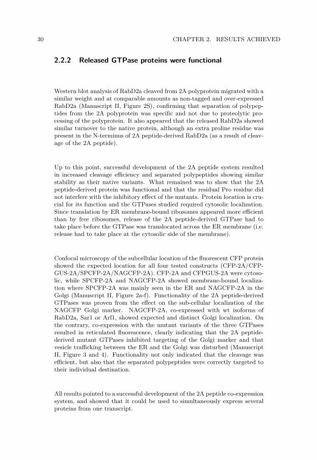
30 CHAPTER 2. RESULTS ACHIEVED
2.2.2 Released GTPase proteins were functional
Western blot analysis of RabD2a cleaved from 2A polyprotein migrated with asimilar weight and at comparable amounts as non-tagged and over-expressedRabD2a (Manuscript II, Figure 2S), confirming that separation of polypep-tides from the 2A polyprotein was specific and not due to proteolytic pro-cessing of the polyprotein. It also appeared that the released RabD2a showedsimilar turnover to the native protein, although an extra proline residue waspresent in the N-terminus of 2A peptide-derived RabD2a (as a result of cleav-age of the 2A peptide).
Up to this point, successful development of the 2A peptide system resultedin increased cleavage efficiency and separated polypeptides showing similarstability as their native variants. What remained was to show that the 2Apeptide-derived protein was functional and that the residual Pro residue didnot interfere with the inhibitory effect of the mutants. Protein location is cru-cial for its function and the GTPases studied required cytosolic localization.Since translation by ER membrane-bound ribosomes appeared more efficientthan by free ribosomes, release of the 2A peptide-derived GTPase had totake place before the GTPase was translocated across the ER membrane (i.e.release had to take place at the cytosolic side of the membrane).
Confocal microscopy of the subcellular location of the fluorescent CFP proteinshowed the expected location for all four tested constructs (CFP-2A/CFP-GUS-2A/SPCFP-2A/NAGCFP-2A). CFP-2A and CFPGUS-2A were cytoso-lic, while SPCFP-2A and NAGCFP-2A showed membrane-bound localiza-tion where SPCFP-2A was mainly seen in the ER and NAGCFP-2A in theGolgi (Manuscript II, Figure 2a-f). Functionality of the 2A peptide-derivedGTPases was proven from the effect on the sub-cellular localization of theNAGCFP Golgi marker. NAGCFP-2A, co-expressed with wt isoforms ofRabD2a, Sar1 or Arf1, showed expected and distinct Golgi localization. Onthe contrary, co-expression with the mutant variants of the three GTPasesresulted in reticulated fluorescence, clearly indicating that the 2A peptide-derived mutant GTPases inhibited targeting of the Golgi marker and thatvesicle trafficking between the ER and the Golgi was disturbed (ManuscriptII, Figure 3 and 4). Functionality not only indicated that the cleavage wasefficient, but also that the separated polypeptides were correctly targeted totheir individual destination.
All results pointed to a successful development of the 2A peptide co-expressionsystem, and showed that it could be used to simultaneously express severalproteins from one transcript.

2.2. MANUSCRIPT II 31
2.2.3 Endo H resistance assays can reveal information abouttrafficking
Trafficking of N-glycoproteins from the ER to the Golgi results in maturationand processing of the N-linked glycan, converting the glycoprotein from EndoH sensitive to Endo H resistant glycoform. This has been proven to be a fast,easy and efficient assay to study trafficking of N-glycoproteins from the ER(Downing et al., 2006; Hong et al., 2008). As long as the protein is retainedin the ER, the protein harbours N-linked glycans sensitive to Endo H, whileER-export and trafficking to the Golgi exposes the protein to mannosidase II,inferring resistance to Endo H (Figure 1.7). Separation of the polypeptide bySDS-PAGE after Endo H treatment can give important information about theattached N-glycan, and also about the subcellular localization and traffickingof the protein.
We decided to combine the Endo H assay with our optimized 2A peptide co-expression system to answer whether these GTPases are involved in CAH1trafficking. The fluorescent marker protein was replaced by an HA-taggedvariant of wt CAH1 (HA-CAH1, Manuscript II, Figure 1), enabling simulta-neous expression of HA-CAH1 with wt and mutant variants of RabD2a, Sar1or Arf1. Stable expression of HA-CAH1 in suspension cultured cells con-firmed that the HA-tagged protein was targeted to the chloroplast and thatthe protein was N-glycosylated (Manuscript II, Table I and Figure 6). Sincecleavage efficiency was higher when translation was performed by ER mem-brane bound ribosomes, and HA-CAH1 pre-protein possesses a SP for theER, the HA-CAH1 protein was inserted N-terminal of the 2A peptide (gener-ating HA-CAH1-2A-GTPase) (Manuscript II, Figure 1). Cleavage efficiencywas high (about 85%), confirming that translation by ER bound ribosomesin our system results in high cleavage efficiency.
In an attempt to quantify the effect of the different variants of the mutantGTPases on the trafficking of HA-CAH1 protein, the ratio of Endo H resistantto Endo H sensitive HA-CAH1-2A was calculated. A clear mutant phenotypewas revealed from co-expression of mutant Rab2a and Sar1 (Manuscript II,Figure 5), as anticipated from their direct involvement in ER-to-Golgi traf-ficking. Arf1, involved in retrograde trafficking from the Golgi to the ER,resulted in a less strong phenotype. While there seemed to be an effect ofthe GDP-locked variant, no significant effect could be deduced from GTP-locked Arf1. Interestingly, a similar effect was presented in a recent publica-tion where trafficking of rice α-amylase isoform I-1 (AmyI-1) to the plastidin onion epidermal cells was evaluated in the presence of mutant Sar1 andArf1 variants (Kitajima et al., 2009). They could show that mutant Sar1strongly inhibited arrival of the AmyI-1 protein to the plastid, while Arf1 hada smaller inhibitory effect. Also in their system, the GDP-locked Arf1 variantgave clearer mutant phenotype than the GTP-locked isoform (Kitajima et al.,2009).

32 CHAPTER 2. RESULTS ACHIEVED
The paper by the Japanese group (Kitajima et al., 2009) reported about thetrafficking mechanism of an N-glycosylated protein to the plastid in mono-cot systems (rice, Oryza sativa, and onion, Allium cepa). Their results werein good agreement with our Endo H assay, where involvement of RabD2aand Sar1 in trafficking of CAH1 was obvious. In a previous study, theypublished data regarding another rice plastid enzyme, a nucleotide pyrophos-phate/phosphodiesterase 1 (NPP1). This protein was also targeted to theplastid through the endomembrane system. Moreover, the mature form ofNPP1 was N-glycosylated. Interestingly, and to us unexpectedly, the plastidlocalized proteins NPP1 and AmyI-1 seemed to harbour high mannose-type N-glycans, as revealed by binding of two glycoproteins to the lectin concanavalinA (ConA) and their susceptibility to Endo H (Asatsuma et al., 2005; Nanjoet al., 2006), indicating either a direct transport of these two proteins fromthe ER to the plastid, or that the N-linked glycans were not further processedin the Golgi. The occurrence of a rice-specific glycosylation pattern can beruled out since the monocot N-glycosylation machinery has been shown to beidentical to the one found in Brassicacea family of dicot plants (Léonard etal., 2004). Closer evaluation of the NPP1 plastid localization indicated that,in addition to uniform internal plastid labelling, numerous distinct dots ofNPP1-GFP, seemingly located at the plastid envelope, were visible (Nanjo etal. 2006, Figure 4). In our eyes, these dots strongly resembled ER-chloroplastcontact sites, PLAMs, previously reported (Andersson et al., 2007). Basedon this, one could speculate about a direct transfer of NPP1 from the en-domembrane system to the plastid by a similar mechanism as the transportof lipid precursors from the ER to the plastid (Benning et al., 2006; Kellyand Dormann, 2004; Xu et al., 2008).
In an attempt to study targeting of NPP1 in our protoplast system, riceNPP1 was fused to GFP and transfected into Arabidopsis protoplasts. Thefusion protein was expressed at significant levels. In addition, it was sensitiveto Endo H, indicating that the polypeptide acquired high mannose-type N-glycans as when expressed in the native rice system. Despite these facts, wecould not verify plastid targeting in Arabidopsis protoplasts. Instead NPP1-GFP appeared to accumulate in the ER (data not shown), indicating that riceNPP1 is not targeted to chloroplasts in a dicotyledon plant such as Arabidopsisthaliana, or that targeting is rice or monocot specific.
2.2.4 Potential areas of application for the 2A peptideco-expression system
One drawback with transient expression techniques, such as protoplast trans-fection, is that when a visible phenotype is lacking, analysis of your sampleis based on a mixture of transfected and non-transfected cells (mutant andwt cells). If either the effect one aims to study, or the transfection efficiencyis very low (or a combination of both), the resulting phenotype might be

2.2. MANUSCRIPT II 33
+
+ - + -+
16 h
+
STEP 1
STEP 2
STEP 3
DNA PROTOPLASTS
FACS sorting ++
+
--
-
+
+
--
+
- - - --
16 h
DNA PROTOPLASTS
Transfection
BiP GFP
STEP 2
STEP 3
T TNT NT
BiP GFPTransformed
+ +- -
GFP vector Empty vector
TotalExtracts
Sorted samples
Figure 2.3. Obtaining a homogenous mutant protoplast population usingFACS. Transfection of protoplasts results in a heterogeneous population where onlya fraction of the protoplasts are actually transfected. Analysis of the protoplasts aftertransfection (Step 2) shows that the transfected population (T), in contrast to thenon-transfected cells (NT), is expressing the transgene. While the protein extract instep 2 originates from a mix of transfected and non-transfected cells, further sortingof the transfected population based on GFP fluorescence results in a homogenousmutant population (Step 3). Western blotting using anti-BiP (a chaperone in theER) or anti-GFP anti shows that only the transfected population is expressing GFP(Step 2), and that GFP fluorescing protoplasts can be efficiently sorted (Step 3).

34 CHAPTER 2. RESULTS ACHIEVED
too small to be detected among the background generated by non-transfectedcells. In such cases it would be desirable to sort out mutant cells in orderto achieve a homogenous mutant population. To test whether the 2A systemoffered such possibilities, protoplasts were transfected with the Golgi labelledNAGCFP-2A-RabD2a construct and analyzed by flow cytometry, using afluorescence-activated cell sorter (FACS). A subpopulation corresponding toabout 10% of the intact protoplasts was defined as positively transfected basedon their level of CFP fluorescence. Protoplasts belonging to both the trans-fected and non-transfected group were sorted and examined by fluorescencemicroscopy. A clear CFP fluorescence could be detected in the positivelysorted protoplasts. In addition, Western blot analysis of both populationsrevealed a strong CFP signal in the extract from the positively sorted popu-lation, while no signal could be detected in extract from the negatively sortedprotoplasts (Figure 2.3).
In conclusion, the versatility of the 2A peptide co-expression system offersseveral possibilities for over-expression of proteins where direct tagging is notpossible. Co-expression with fluorescent markers can in such cases enableeasy visualization of cells in which the protein is being expressed. Also, theeffect derived from the over-expression of the protein can be studied in realtime using different microscopic techniques, such as confocal microscopy. Fur-thermore, sorting of transfected cells using FACS, can allow obtaining of ahomogenous mutant population expressing your favourite protein in less than24 h.
2.3 Manuscript III
While paper I and manuscript II focused on localization and trafficking ofCAH1 to the chloroplast, one obvious question remained to be considered -why CAH1 is targeted to the chloroplast via the secretory pathway instead ofthe Toc-Tic translocation system from the cytosol as for most other chloro-plast proteins. We were also lacking important biochemical information aboutthe protein, in particular that related to post-translational modifications. Inpaper I we reported that the protein is N-glycosylated. N-glycosylation isexclusively initiated in the ER and constitutes a logical starting point forfurther analysis of the protein. Over-expression of epitope-tagged versionsof CAH1 in different systems -BY2 Tobacco and Arabidopsis cell suspensioncultures, as well as in Arabidopsis and Tobacco plants- always resulted inits accumulation at rather low amounts, hampering the purification of CAH1protein to substantial quantities. So, instead of a mass spectrometric ap-proach, we decided to do mutagenesis of the protein. A collection of aminoacids, mainly representing targets for N-glycosylation, were point mutatedand the resulting mutated protein versions expressed in protoplasts.

2.3. MANUSCRIPT III 35
2.3.1 CAH1 harbours four or five N-glycans
First we wanted to elucidate the number and positions of N-glycans anchoredto the protein. As seen in paper I, theoretical and native molecular masses ofCAH1 differed by approximately 8 kDa, indicating that around 4-5 N-glycanscould be decorating the CAH1 protein (supposing about 1,5-2 kDa/N-glycan).All five sites were point mutated, both individually and in combination, re-sulting in single, double, triple, and quadruple mutants in addition to a non-glycosylated variant (Manuscript III, Table 1). All mutant proteins possessedan HA-epitope tag to enable the use of the highly specific anti-HA antibodyand to avoid detection of native protein (as background).
It appeared as if the vast majority of the HA-CAH1 molecules were harbour-ing four or five N-glycans, and that partial occupation of glycosylation sitefour (Asn194) resulted in these two isoforms (Manuscript III, Figure 2 b andc). This result was in agreement with the in vitro import study into micro-somes in paper I, where two dominating bands were seen (Paper I, Figure2c). In addition, we detected glycoforms containing either high mannose- andcomplex type N-glycans of both 4- and 5-glycan HA-CAH1, explaining thefour-band-pattern seen when the over-expressed HA-CAH1 from protoplastswas separated in SDS-PAGE (Manuscript III, Figure 3 a-c). Interestingly, twoisoforms of the native protein, exhibiting slightly different molecular masses,were also frequently detected in leaf extracts, presumably indicating thatCAH1 can also accumulate as two glycoforms in planta. None of these twoglycoforms could be deglycosylated by Endo H, indicating that both harbourcomplex type N-glycans (data not shown). Moreover, these results suggestthat partial occupancy of N-glycosylation site four is an intrinsic characteris-tic of the CAH1 protein, and not a secondary effect of the over-expression ofthe tagged isoform. While the heavier isoform was prevailing in some proteinpreparations, the opposite could been seen in other preparations. The rea-son for this behaviour is unknown to us, but could depend on some kind ofregulation of the protein. N-glycosylation site four (Asn194) is adjacent to acysteine residue (Cys191). Corresponding cysteine residue in human α-typeCA IV has been reported to form part of an intramolecular disulphide bridge(Waheed et al., 1996). If a similar cysteine bond would appear in CAH1, thepresence of N-linked glycan at Asn194 could interfere with bridge formationand result in synthesis of an unstable or non-functional CAH1, alternativelyproducing an enzyme with altered activity.
The presence of both high mannose and complex type N-glycans seemed tooriginate from over-expression of the HA-CAH1 protein, perhaps due to sat-uration of the CAH1 targeting machinery. It was recently shown that proteintrafficking from the Golgi apparatus to the plastid in onion epidermal cellswas tightly regulated (Kitajima et al., 2009). Inhibition of protein biosynthe-sis by CHX reduced the fraction of high mannose-isoforms and suggested thatglycoproteins harbouring high mannose-type N-glycans originated from newlysynthesized HA-CAH1 molecules waiting to be trafficked from the ER to the

36 CHAPTER 2. RESULTS ACHIEVED
Golgi, or from misfolded polypeptides destined for degradation by the pro-teasome (Manuscript III, Supplemental Figure 11). Indeed, immunogold la-belling and electron microscopy revealed that a small portion of the HA-CAH1protein was associated with the endomembrane system, though the majorityof the HA-CAH1 molecules were located to the chloroplast (Manuscript III,Supplemental Figure 2). Subcellular fractionation confirmed that the Endo Hsensitive glycoforms were originating from microsome fractions, while Endo Hresistant HA-CAH1 were present in the soluble, stroma containing, fraction(Manuscript III, Figure 3 d-f).
2.3.2 A disulphide bridge is important for folding andER-export of CAH1
Disulphide bridges between cysteine residues are important for protein stabil-ity as well as for regulation of enzyme activity. The ER is a highly oxidizingenvironment for proteins, and several ER chaperones assist in formation ofsuch bonds (Hebert and Molinari, 2007; Jensen, 2006). Sequence analysisof CAH1 revealed four cysteine residues in the SP processed polypeptide,of which the first and the third (Cys27 and Cys191) were positioned closeto each other in the 3D-structure modelled protein, indicating that a sim-ilar disulphide bridge could exist in CAH1 as in human CAIV (Waheed etal., 1996) (Manuscript III, Supplemental Figure 7). Separation of the nativeprotein under reducing and non-reducing conditions demonstrated alteredmigration pattern and that CAH1 was sensitive to reducing agents, such asβ-mercaptoethanol (β-ME) (Manuscript III, Figure 5). Mutating all fourcysteine residues individually generated two isoforms apparently resistant to2-ME. As expected, they corresponded to Cys27 and Cys191 and suggesteda disulphide bridge between these residues (Manuscript III, Figure 6 a). For-mation of these bridges in the ER is a crucial part of the folding process.If a protein cannot fold correctly it will be retained in the ER, bound to achaperone (BiP) and then translocated over the ER membrane for degrada-tion by the proteasome in the cytosol (Hebert and Molinari, 2007; Pimpl etal., 2006). Retention of a protein in the ER accordingly also prevents pro-cessing of its N-glycans in the Golgi, and the protein accumulates as Endo Hsensitive glycoprotein. Therefore, an Endo H assay, similar to the one usedin manuscript II to study effect on trafficking upon co-expression of mu-tant GTPases, could be employed in monitoring misfolding of the cysteineresidue mutants. Indeed, mutating Cys27 and Cys191 resulted in a proteinwith significantly higher proportion of Endo H sensitive glycoforms comparedto the wt protein, or isoforms where any of the other two cysteine residues(Cys68 and Cys229) were mutated (Manuscript III, Figure 6 d and e). Therequirement of a disulphide bond between Cys27 and Cys191 for folding ofER-export competent protein was finally confirmed by the demonstrated as-sociation of the ER-chaperone BiP with proteins mutated at any of these sites(Manuscript III, Figure 6 b and c).

2.3. MANUSCRIPT III 37
2.3.3 Non-glycosylated CAH1 forms aggregates
To our knowledge, chloroplasts lack N-glycosylation machinery. A functionalrequirement of N-linked glycans at CAH1 could explain why the protein istrafficked through the endomembrane system to the chloroplast. Similar tothe requirement of disulphide bridges for correct structure of certain proteins,N-glycosylation has also been shown to be required for folding (Mitra et al.,2006; Parodi, 2000). Expression of the non-glycosylated mutant and separa-tion of the resulting polypeptide by SDS-PAGE under non-reducing conditionsresulted in the presence of an additional band around 65 kDa. This additionalband presumably corresponded to a dimer of the non-glycosylated protein dueto the formation of non-native inter-molecular disulphide bridges (ManuscriptIII, Figure 4 a). In addition, several high molecular mass forms were detectedby the HA-antibodies, indicating that aggregates of the protein were formedwhen no glycans could be attached to the HA-CAH1 polypeptide. Similarto the disulphide bridge mutants, misfolding of the non-glycosylated proteinwas verified by its increased association with BiP (Manuscript III, Figure 4 band c).
Lack of N-glycosylation or a correct disulphide bridge in CAH1 results in de-creased CA activity Activity of CAs can be estimated by measuring the rateby which carbon dioxide in CO2 (g) saturated water is converted to bicarbon-ate in a solution containing the CA enzyme. This inter-conversion lowers thepH of the solution and can be recorded using a fast and sensitive pH electrode.Measurements are often performed with purified proteins expressed in bacte-ria, but since bacteria (as chloroplasts) lack the N-glycosylation machinery,such expression would generate non-glycosylated CAH1. To overcome this,activity was instead measured on protein extracts from transfected proto-plasts.
Expression of a cysteine mutant protein unable to form an intra-moleculardisulphide bridge, and non-glycosylated HA-CAH1, resulted in mutant pro-tein extracts with almost identical CA activity, significantly lower than theactivity measured in the wt extracts. Considering the rather low transfectionefficiency (about 5-10% as calculated from GFP transfected cells) and thedilution effect caused by having mostly non-transfected protoplast exhibitingtheir own CA activity, this experiment clearly indicated the importance of N-glycosylation and disulphide bridge formation for the assembly of a functionalCAH1 (Manuscript III, Figure 7).
2.3.4 Evolutionary aspects of CAH1
The biochemical characterization of CAH1 showed that N-linked glycans, aswell as disulphide bridge formation, were required for correct folding of theCAH1 protein. When not correctly folded, the protein appeared to be unable

38 CHAPTER 2. RESULTS ACHIEVED
to be exported from the ER and presumably was translocated out of theER and degraded. We propose that these features of CAH1 explain why theprotein is trafficked via the endomembrane system to the chloroplast.
The question that then arises is whether CAH1 is a protein with eukaryoticorigin that found its way to the chloroplast where it acquired a new functionwithin the endosymbiont, or whether CAH1 is a protein with cyanobacterialorigin whose gene was transferred to the eukaryotic genome and later wastransported back to the evolving chloroplast using the proposed, early route,for proteins (Bhattacharya et al., 2007; Reyes-Prieto et al., 2007). Blastingthe CAH1 protein sequence against the NCBI database (www.ncbi.nlm.nih.gov)and excluding Viridiplantae from the search algorithm, proteobacterium pro-teins rank at the top. Proteobacterium is considered to be the ancestor of themitochondrium (Cavalier-Smith, 2009), suggesting that CAH1 might have anorigin from outside the cyanobacteria.
A proteomic study of Arabidopsis chloroplasts (Kleffmann et al., 2004), pub-lished in 2004 (before the pathway of CAH1 was discovered), identified 690proteins. Unexpectedly for the authors, many of the identified proteins didnot possess targeting information for the canonical Toc-Tic system, as shownusing the TargetP prediction tool on the identified polypeptides (Emanuelssonet al., 2000). Subsequent to removal of envelope proteins, which often do nothave canonical chloroplast TP (as discussed earlier), and other obvious con-taminants, only 67% of the remaining proteins were predicted being targetedto the chloroplast. Out of the 318 identified proteins in the soluble fraction,24 (or 7.5%) contained a predicted SP for the ER (Kleffmann et al., 2004). Inanother recent study where data from all available plastid proteomic studieswas compiled, about 30% of the identified proteins did not possess canonicalchloroplast TP (Armbruster et al., 2009). These authors concluded that atleast two types of non-canonical chloroplast proteins exist: the inner-envelopetype and the ER-dependent type.
2.4 Manuscript IV
The specific function of CAH1 in the chloroplast stroma of Arabidopsis is atpresent unknown. For a long time it was assumed that the chloroplastic β-CA1, representing up to 5% of the soluble protein in the stroma, could play animportant role in optimizing CO2 availability for Rubisco in C3 plants (Badgerand Price, 1994). However, several studies using antisense strategies haveshown that specific reduction of the expression of β-CA1 do not cause obviousgrowth phenotype and had only minor effects on mesophyll CO2 transferand photosynthesis (Majeau et al., 1994; Price et al., 1994). In anotherrecent study, photosynthetic measurements on a few days old seedlings showeddecreased CO2 uptake rates in mutants lacking the β-CA1 protein, indicatingthat the protein might play a role in photosynthesis in the early development
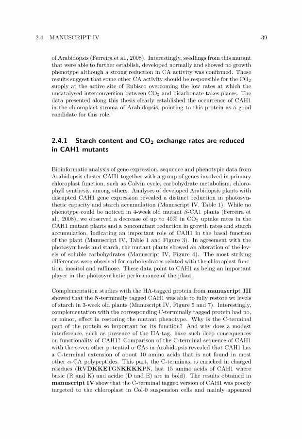
2.4. MANUSCRIPT IV 39
of Arabidopsis (Ferreira et al., 2008). Interestingly, seedlings from this mutantthat were able to further establish, developed normally and showed no growthphenotype although a strong reduction in CA activity was confirmed. Theseresults suggest that some other CA activity should be responsible for the CO2supply at the active site of Rubisco overcoming the low rates at which theuncatalysed interconversion between CO2 and bicarbonate takes places. Thedata presented along this thesis clearly established the occurrence of CAH1in the chloroplast stroma of Arabidopsis, pointing to this protein as a goodcandidate for this role.
2.4.1 Starch content and CO2 exchange rates are reducedin CAH1 mutants
Bioinformatic analysis of gene expression, sequence and phenotypic data fromArabidopsis cluster CAH1 together with a group of genes involved in primarychloroplast function, such as Calvin cycle, carbohydrate metabolism, chloro-phyll synthesis, among others. Analyses of developed Arabidopsis plants withdisrupted CAH1 gene expression revealed a distinct reduction in photosyn-thetic capacity and starch accumulation (Manuscript IV, Table 1). While nophenotype could be noticed in 4-week old mutant β-CA1 plants (Ferreira etal., 2008), we observed a decrease of up to 40% in CO2 uptake rates in theCAH1 mutant plants and a concomitant reduction in growth rates and starchaccumulation, indicating an important role of CAH1 in the basal functionof the plant (Manuscript IV, Table 1 and Figure 3). In agreement with thephotosynthesis and starch, the mutant plants showed an alteration of the lev-els of soluble carbohydrates (Manuscript IV, Figure 4). The most strikingdifferences were observed for carbohydrates related with the chloroplast func-tion, inositol and raffinose. These data point to CAH1 as being an importantplayer in the photosynthetic performance of the plant.
Complementation studies with the HA-tagged protein from manuscript IIIshowed that the N-terminally tagged CAH1 was able to fully restore wt levelsof starch in 3-week old plants (Manuscript IV, Figure 5 and 7). Interestingly,complementation with the corresponding C-terminally tagged protein had no,or minor, effect in restoring the mutant phenotype. Why is the C-terminalpart of the protein so important for its function? And why does a modestinterference, such as presence of the HA-tag, have such deep consequenceson functionality of CAH1? Comparison of the C-terminal sequence of CAH1with the seven other potential α-CAs in Arabidopsis revealed that CAH1 hasa C-terminal extension of about 10 amino acids that is not found in mostother α-CA polypeptides. This part, the C-terminus, is enriched in chargedresidues (RVDKKETGNKKKKPN, last 15 amino acids of CAH1 wherebasic (R and K) and acidic (D and E) are in bold). The results obtained inmanuscript IV show that the C-terminal tagged version of CAH1 was poorlytargeted to the chloroplast in Col-0 suspension cells and mainly appeared

40 CHAPTER 2. RESULTS ACHIEVED
associated with the ER and, to a minor extent, with the Golgi apparatus(Manuscript IV, Figure 5). In addition, the tagging of the C-terminus wasnot only disturbing a signal potentially involved in transport of the proteinto the chloroplast and causing its retention in the ER, but also altered thebiochemical features of the protein (Manuscript IV, Figure 6 c and d). Thedata from the complementation analysis of the knockout CAH1 mutants withthe two tagged versions revealed relevant information concerning the role ofCAH1. The role of CAH1 seems to be directly linked to its localization inclose vicinity to the site of CO2 fixation by the Rubisco enzyme and not to theactivity of this protein along the route through the endomembrane system tothe plastid. From our studies we can conclude that CAH1 is trafficked fromthe endomembrane system to the chloroplast. In addition, the type of N-linked glycan attached to the protein suggests a pathway for CAH1 to thechloroplast via the Golgi. In the Golgi, CAH1 is sorted from the defaultsecretory pathway and arrives at the chloroplast using, presumably, a noveltype of vesicle transport (Figure 2.1). Vesicle fusion at the chloroplast willmost likely result in delivery of the polypeptide to the inter-membrane space.Since CAH1 becomes folded in the ER, the protein could not be translocatedvia the Tic-part of the Toc-Tic system, suggesting that occurrence of somekind of novel and specific transporter at the inner membrane must be involved.
We believe that the C-terminus of CAH1 is, in some way, involved in thistargeting, either as a sorting signal at the Golgi or in assisting transportover the inner envelope membrane; and that interference of the C-terminusresults in a mistargeted and therefore non-functional enzyme. Interestingly,some bacterial proteins are transported across the cytoplasmic membrane infolded conformation. Transport mediated by this system requires a twin-arginine (RR) consensus, therefore called the Tat-pathway (Twin-argininetranlocation). This pathway is also involved in the transport of some proteinsover the thylakoid membrane (Muller and Klosgen, 2005). Since the innerenvelope membrane of the chloroplast is believed to have a cyanobacterialorigin, a similar system could exist for translocation of folded proteins fromthe inter-membrane space to the stroma, and the C-terminus of CAH1 couldbe involved in mediating this step.
The inability of the C-terminally tagged protein to complement the CAH1 KOphenotype raises several questions about targeting, regulation and functionof CAH1 that will require extensive research to be answered.

Chapter 3
Conclusions
The work presented in this thesis shows two aspects that will change our viewof intracellular protein trafficking in plants. First of all, it reveals the pres-ence of a direct pathway from the endomembrane system to the chloroplast.Second, it also shows the occurrence of N-glycoproteins in an endosymbi-otic organelle such as the chloroplast. So far, targeting of nuclear-encodedpolypeptides to the chloroplast interior was believed to occur exclusively viathe Toc-Tic system, a translocon complex in the chloroplast envelope whereproteins which were translated in the cytosol are transported across the dou-ble membrane of the chloroplast in an extended conformation. In this thesis,an additional pathway for N-glycosylated proteins from the endomembranesystem to the chloroplast is presented.
The existence of proteins derived from the endomembrane system in thechloroplast had already been proposed, although conclusive evidence describ-ing this phenomenon was missing (Chen et al., 2004). In this work we showthat an α-type carbonic anhydrase (CAH1) in the model plant Arabidopsisthaliana is localized to the chloroplast stroma, although it is first directed tothe ER where N-linked glycans are acquired. The type of glycan attached toCAH1 suggested that the protein is trafficked through the Golgi apparatus onits way to the chloroplast. Shortly after our publication, other studies describ-ing a similar transport pathway for two different proteins to the chloroplastin rice were published. As with CAH1, these proteins were N-glycosylated inthe ER and their trafficking to the chloroplast was sensitive to exposure ofan ER-to-Golgi trafficking inhibitor. In contrast to CAH1, both rice proteinspresented a different type of N-linked glycan, suggesting a slightly different(possibly direct) route from the ER to the chloroplast. Our work, togetherwith these two reports, indicates that two individual pathways for targetingof N-glycoproteins to the chloroplast might co-exist in plants.
Development of a transient co-expression system enabled us to show thatsmall GTPases involved in canonical ER-to-Golgi vesicle transport were re-quired for trafficking of CAH1 to the chloroplast. A question that arose waswhy a small set of proteins that are targeted to the chloroplast follow thispathway, while the vast majority of the nuclear-encoded chloroplast proteinsare transported through the Toc-Tic complex? We characterized two typesof protein modifications taking place in the ER/Golgi: N-glycosylation anddisulphide bridge formation. CAH1 was found to be glycosylated at four orfive sites and to possess an intra-molecular disulphide bridge. Both types of
41

42 CHAPTER 3. CONCLUSIONS
protein modifications were required for correct folding of the CAH1 polypep-tide, since protein lacking any of these modifications formed aggregates inthe ER, unable to be targeted further. In addition, we also showed the im-portance of these post-translational modifications for producing a functionalCA.
Based on these findings we propose that the protein is not functional with-out endomembrane-associated post-translational modifications. As a conse-quence, a pathway to the chloroplast from the secretory system (where theprotein is modified to be functional) is required. Since the unfolding of theprotein, required for crossing the chloroplast envelope through the Toc-Tic,most certainly would result in a CAH1 with impaired structure and function,this alternative pathway should be independent of the Toc-Tic machinery.
The existence of such a pathway for proteins to the chloroplast implies thatthe activities of plastid N-glycoproteins, such as CAH1, are relevant for thefunction of the chloroplast. A genetic dissection using Arabidopsis plantswith disrupted CAH1 gene expression showed that the activity of this CAplays a pivotal role in the photosynthetic performance of the plant. Mutantplants exhibited lower photosynthetic capacity and reduced starch content.An allele with an especially strong mutant phenotype additionally showedretarded growth and altered levels of soluble carbohydrates. In agreementwith the mutant phenotype, bioinformatic analysis indicated that CAH1 isco-expressed with other chloroplast proteins involved in basic functions of thechloroplast such as photosynthesis and synthesis of photosynthetic pigments.
In conclusion, this thesis presents firm evidence for a pathway of proteinsfrom the endomembrane system to the chloroplast in Arabidopsis thaliana.We show that trafficking of proteins along this route depends on canonicalmechanisms, such as ER-directed translation and ER-to-Golgi vesicle trans-port. Study of the mechanisms involved in transport of CAH1 to the chloro-plast can reveal crucial information for our understanding about chloroplast,and plant cell evolution. Gene expression data, together with the phenotypeobserved in CAH1 mutant plants, indicate that the CAH1 protein plays acrucial role in the photosynthetic process. Activity measurement of over-expressed CAH1 showed that ER-associated post-translational modifications,such as N-glycosylation and disulphide bridge formation, are not only im-portant for folding and trafficking of the protein, but also for its function,possibly explaining why this pathway (still?) exist in plants.
3.1 Future perspectives
Several questions remain to be answered, both regarding the mechanism ofthis pathway to the chloroplast and the exact function of CAH1. For example,

3.1. FUTURE PERSPECTIVES 43
which signals target CAH1, and other glycoproteins (?), from the endomem-brane system to the chloroplast? Are there one or several pathways from thesecretory system to the chloroplast? What is the function of the C-terminalpart of the CAH1 protein? How is CAH1 crossing the inner membrane ofthe chloroplast envelope? Is there an active transporter for CAH1 across themembrane? In what way is CAH1 involved in the photosynthetic process?Which other proteins follow the same route as CAH1? What is the evolution-ary reason for having two (or more) protein pathways to the chloroplast?
What initially attracted my attention to this project was the emerging poten-tial of using plants as bio-factories for production of recombinant proteins, andspecifically how this pathway could be used for accumulation of chloroplast-localized recombinant glycoproteins. Many commercially interesting proteinsrequire N-linked glycosylation for their functionality, e.g. antibodies. Thechloroplasts (or plastids in general) have been shown to be able to accumu-late large amounts of foreign proteins without interfering with the functionof the organelle. Until now, the requirement of N-linked glycans has directedexpression and accumulation of the proteins to compartments such as the ERor the vacuole, or to be secreted. The possibility of combining expression ofN-glycosylated polypeptides in plants with targeting to and accumulation inplastids emerged to me as an appealing alternative to existing methods. Whatis clear to me now, after 5 years of (hard?) work, is that ideas which appearto be straightforward to implement are not always that easy to carry out inpractice, and that intense continued research in this field will be needed.


Chapter 4
Acknowledgement
Slim For believing in me from the beginning and for always supporting mein whatever crazy idea/experiment I have had, for making me continuewhen my belief in the project has been low, for letting me do part of thework outside Umeå, for all late-night changes of antibody solutions. . .
Arsenio For not taking your hand away from my project even when moving4000 km away, for letting me come to Madrid and do part of the workthere, for all the help with the thesis and manuscripts the last crazydays before Christmas. It would not have been possible without you.
Laszlo Bako For helping me maintaining my dear cell suspension culturesduring my frequent trips to Spain, for knowing everything about bio-chemistry and for becoming more than a colleague.
Markus Grebe For always being interested in my project, for bringing a lotof new knowledge to UPSC and for all visits to my office.
Daniel Ziggy, Diamyd-Danne, Herr Toffel. . . Kärt barn har många namn.Tack för alla luncher nere på stan eller IKSU, alla fika, för allt ovärderligtbörssnack och för att alltid lyckas inkludera USA’s handelsunderskott ivarje diskussion (oavsett tema). . .
Max För att vara en trogen kompis och hockeysupporter, för din mycketspeciella humor, för alla stunder på innebandyplanen. Även om minpublikationslista aldrig kommer att vara lika lång som din, så har jag ialla fall spikat Organisk kemi B. . . Så det så.
Rolleboll För alla konstiga mail som har gjort mig glad, för att kunnaBilbao’s bakgator utan och innan, för alla Guggenheim-besök. Dockbesviken på att du sticker från Spanien nu, förväntar mig att du flyttartillbaka snarast. . .
Cristian For being a really really nice friend, for always being optimistic, forall squash games and discussions in the sauna, for showing me Madrid.
Maribel and Markus For being so extremely nice. Please move down soon.Andreas För att alltid ställa upp, för att alltid kunna besvara datorrelat-
erade frågor, för att visa mig krångligast möjliga dataprogram för attsätta samman denna avhandling.
Claes För vår goda vänskap sedan 1-års ålder, för all hjälp när jag flyttathit och dit och för att alltid låtit mig bo hos dig närhelst det behövts.Förväntar mig att du kommer och besöker mig ofta.
45

46 CHAPTER 4. ACKNOWLEDGEMENT
Martin Frenkel För din tröstsockerkaka när AIK lyckades att misslyckasmed att ta klivet upp i elitserien våren 2004.
Juande For your laughter and for always losing against me in squash.Sergey and Grit For BBQs and fikas. It is a shame that I have to leave
just after getting to know you. You are welcome to Spain whenever. . .Innebandygänget Vem ska nu hacka på Daniel’s smalben?Tatiana For standing my taste of music, for always being nice and helpful
in the office. We all hope, want and pray that you will be back soon.Adele Many thanks for reading my thesis pre-printing and improving my
Swenglish, for not being as dangerous as you look and for being a reallynice person in the lab.
All past Slim group members Linda, Dan. . . that has made the lab a goodplace to work.
All past UPSC members that has made UPSC a fun place to be. Steve,Aurora, Olivier, Frank. . .
All UPSC It has been fun to be part of UPSC these years and I will missyou all.
All people outside UPSC that has helped me and been part of my lifeduring these years.
All my new friends in Madrid Flor for helping me to feel at home in thelab, Amaya and Andrea for collaborations, yoghurt-Ismael for having12.30 fika with me, Lars for proving to me that also Spanish people canlisten to real music, and many more. . .
Marisa and family For letting me stay at your place in Madrid and makingme have a second family.
Jose and Chelo For accepting me although I don’t know the difference be-tween "Nieve, Nuevo and Nueve" or "Silla and Casilla". I will improve,I promise!
Familjen in Rönninge För alla besök som jag kunnat göra i samband medSpanienvistelser, trots att det måste varit jobbigt.
Jonas För att vara en bra bror som mer än gärna korrekturläser under helger.Mina föräldrar För att vara så förstående.Cris FOR EVERYTHING!!
Chapter 5
References
Abdallah, F., Salamini, F., and Leister, D. (2000): A prediction of the sizeand evolutionary origin of the proteome of chloroplasts of Arabidopsis. TrendsPlant Sci 5, 141-2.
Andersson, M. X., Goksor, M., and Sandelius, A. S. (2007): Optical manip-ulation reveals strong attracting forces at membrane contact sites betweenendoplasmic reticulum and chloroplasts. J Biol Chem 282, 1170-4.
Armbruster, U., Hertle, A., Makarenko, E., Zühlke, J., Pribil, M., Dietz-mann, A., Schliebner, I., Aseeva, E., Fenino, E., Scharfenberg, M., Voigt, C.,and Leister, D. (2009): Chloroplast Proteins without Cleavable Transit Pep-tides: Rare Exceptions or a Major Constituent of the Chloroplast Proteome.Molecular Plant 2, 1325-1335.
Asatsuma, S., Sawada, C., Itoh, K., Okito, M., Kitajima, A., and Mitsui,T. (2005): Involvement of alpha-amylase I-1 in starch degradation in ricechloroplasts. Plant Cell Physiol 46, 858-69.
Badger, M. R., and Price, G. D. (1994): THE ROLE OF CARBONIC AN-HYDRASE IN PHOTOSYNTHESIS. Annu Rev Plant Physiol Plant Mol Biol45, 369-392.
Baluska, F., Hlavacka, A., Samaj, J., Palme, K., Robinson, D. G., Matoh, T.,McCurdy, D. W., Menzel, D., and Volkmann, D. (2002): F-Actin-DependentEndocytosis of Cell Wall Pectins in Meristematic Root Cells. Insights fromBrefeldin A-Induced Compartments. Plant Physiology 130, 422-431.
Barber, J. (2008): Photosynthetic generation of oxygen. Philos Trans R SocLond B Biol Sci 363, 2665-74.
Batoko, H., Zheng, H. Q., Hawes, C., and Moore, I. (2000): A Rab1 GTPaseis required for transport between the endoplasmic reticulum and Golgi appa-ratus and for normal Golgi movement in plants. Plant Cell 12, 2201-2217.
Bedard, J., and Jarvis, P. (2005): Recognition and envelope translocation ofchloroplast preproteins. J Exp Bot 56, 2287-320.
47

48 CHAPTER 5. REFERENCES
Bendtsen, J. D., Nielsen, H., von Heijne, G., and Brunak, S. (2004): Improvedprediction of signal peptides: SignalP 3.0. J. Mol. Biol. 340, 783-795.
Benning, C., Xu, C., and Awai, K. (2006): Non-vesicular and vesicular lipidtrafficking involving plastids. Curr Opin Plant Biol 9, 241-7.
Bhattacharya, D., Archibald, J. M., Weber, A. P., and Reyes-Prieto, A.(2007): How do endosymbionts become organelles? Understanding earlyevents in plastid evolution. Bioessays 29, 1239-46.
Björn, L., and Govindjee (2009): The evolution of photosynthesis and chloro-plasts. CURRENT SCIENCE 96, 1466-1474.
Bonin, C. P., Potter, I., Vanzin, G. F., and Reiter, W.-D. (1997): The MUR1gene of Arabidopsis thaliana encodes an isoform of GDP-D-mannose-4,6-dehydratase, catalyzing the first step in the de novo synthesis of GDP-L-fucose. Proc Natl Acad Sci U S A 94, 2085-2090.
Brandizzi, F., Snapp, E. L., Roberts, A. G., Lippincott-Schwartz, J., andHawes, C. (2002): Membrane Protein Transport between the EndoplasmicReticulum and the Golgi in Tobacco Leaves Is Energy Dependent but Cy-toskeleton Independent: Evidence from Selective Photobleaching. Plant Cell14, 1293-1309.
Cavalier-Smith, T. (2009): Predation and eukaryote cell origins: A coevolu-tionary perspective. The International Journal of Biochemistry Cell Biology41, 307-322.
Chen, M.-H., Huang, L.-F., Li, H.-M., Chen, Y.-R., and Yu, S.-M. (2004):Signal Peptide-Dependent Targeting of a Rice a-Amylase and Cargo Proteinsto Plastids and Extracellular Compartments of Plant Cells. Plant Physiology135, 1367-1377.
Cline, K., and Dabney-Smith, C. (2008): Plastid protein import and sorting:different paths to the same compartments. Curr Opin Plant Biol 11, 585-92.
Crofts, A. J., Leborgne-Castel, N., Pesca, M., Vitale, A., and Denecke, J.(1998): BiP and calreticulin form an abundant complex that is independentof endoplasmic reticulum stress. Plant Cell 10, 813-24.
daSilva, L. L., Snapp, E. L., Denecke, J., Lippincott-Schwartz, J., Hawes,C., and Brandizzi, F. (2004): Endoplasmic reticulum export sites and Golgibodies behave as single mobile secretory units in plant cells. Plant Cell 16,1753-71.
Donnelly, M. L. L., Luke, G., Mehrotra, A., Li, X. J., Hughes, L. E., Gani,D., and Ryan, M. D. (2001): Analysis of the aphthovirus 2A/2B polyprotein

49
’cleavage’ mechanism indicates not a proteolytic reaction, but a novel trans-lational effect: a putative ribosomal ’skip’. Journal of General Virology 82,1013-1025.
Downing, W. L., Galpin, J. D., Clemens, S., Lauzon, S. M., Samuels, A. L.,Pidkowich, M. S., Clarke, L. A., and Kermode, A. R. (2006): Synthesis ofenzymatically active human alpha-L-iduronidase in Arabidopsis cgl (complexglycan-deficient) seeds. Plant Biotechnol J 4, 169-81.
Emanuelsson, O., Nielsen, H., Brunak, S., and von Heijne, G. (2000): Pre-dicting subcellular localization of proteins based on their N-terminal aminoacid sequence. J Mol Biol 300, 1005-16.
Fabre, N., Reiter, I. M., Becuwe-Linka, N., Genty, B., and Rumeau, D. (2007):Characterization and expression analysis of genes encoding alpha and betacarbonic anhydrases in Arabidopsis. Plant Cell Environ 30, 617-29.
Faso, C., Boulaflous, A., and Brandizzi, F. (2009): The plant Golgi apparatus:Last 10 years of answered and open questions. FEBS Letters 583, 3752-3757.
Ferreira, F. J., Guo, C., and Coleman, J. R. (2008): Reduction of Plastid-Localized Carbonic Anhydrase Activity Results in Reduced Arabidopsis SeedlingSurvivorship. Plant Physiology 147, 585-594.
Foth, B. J., Ralph, S. A., Tonkin, C. J., Struck, N. S., Fraunholz, M., Roos,D. S., Cowman, A. F., and McFadden, G. I. (2003): Dissecting apicoplasttargeting in the malaria parasite Plasmodium falciparum. Science 299, 705-8.
Friso, G., Giacomelli, L., Ytterberg, A. J., Peltier, J. B., Rudella, A., Sun,Q., and Wijk, K. J. (2004): In-depth analysis of the thylakoid membraneproteome of Arabidopsis thaliana chloroplasts: new proteins, new functions,and a plastid proteome database. Plant Cell 16, 478-99.
Halpin, C., Cooke, S. E., Barakate, A., El Amrani, A., and Ryan, M. D.(1999): Self-processing 2A-polyproteins - a system for co-ordinate expressionof multiple proteins in transgenic plants. Plant Journal 17, 453-459.
Hammond, C., Braakman, I., and Helenius, A. (1994): Role of N-linkedoligosaccharide recognition, glucose trimming, and calnexin in glycoproteinfolding and quality control. Proc Natl Acad Sci U S A 91, 913-7.
Hanson, M. R., and Köhler, R. H. (2001): GFP imaging: methodology andapplication to investigate cellular compartmentation in plants. Journal ofExperimental Botany 52, 529-539.
Hanton, S. L., Matheson, L. A., and Brandizzi, F. (2006): Seeking a way out:export of proteins from the plant endoplasmic reticulum. TRENDS in PlantScience 11, 335-343.

50 CHAPTER 5. REFERENCES
Harvey Millar, A., Sweetlove, L. J., Giegé, P., and Leaver, C. J. (2001):Analysis of the Arabidopsis Mitochondrial Proteome. Plant Physiology 127,1711-1727.
Hawes, C., Osterrieder, A., Hummel, E., and Sparkes, I. (2008): The PlantER-Golgi Interface. Traffic 9, 1571-1580.
Hebert, D. N., and Molinari, M. (2007): In and out of the ER: protein folding,quality control, degradation, and related human diseases. Physiol Rev 87,1377-408.
Hewett-Emmett, D., and Tashian, R. E. (1996): Functional diversity, con-servation, and convergence in the evolution of the α-, β-, and γ-carbonicanhydrase gene families. Molecular Phylogenetics and Evolution 5, 50-77.
Holmes-Davis, R., Tanaka, C. K., Vensel, W. H., Hurkman, W. J., andMcCormick, S. (2005): Proteome mapping of mature pollen of Arabidopsisthaliana. Proteomics 5, 4864-84.
Hong, Z., Jin, H., Tzfira, T., and Li, J. (2008): Multiple mechanism-mediatedretention of a defective brassinosteroid receptor in the endoplasmic reticulumof Arabidopsis. Plant Cell 20, 3418-29.
Ioffe, E., and Stanley, P. (1994): Mice lacking N-acetylglucosaminyltransferaseI activity die at mid-gestation, revealing an essential role for complex or hybridN-linked carbohydrates. Proc Natl Acad Sci USA 91, 728-732.
Jarvis, P. (2008): Targeting of nucleus-encoded proteins to chloroplasts inplants. New Phytol 179, 257-85.
Jarvis, P., and Robinson, C. (2004): Mechanisms of protein import and rout-ing in chloroplasts. Curr Biol 14, R1064-77.
Jarvis, P., and Soll, J. (2002): Toc, tic, and chloroplast protein import.Biochim Biophys Acta 1590, 177-89.
Jensen, O. N. (2006): Interpreting the protein language using proteomics.Nat Rev Mol Cell Biol 7, 391-403.
Jin, H., Hong, Z., Su, W., and Li, J. (2009): A plant-specific calreticulin is akey retention factor for a defective brassinosteroid receptor in the endoplasmicreticulum. Proc Natl Acad Sci U S A 106, 13612-7.
Jin, H., Yan, Z., Nam, K. H., and Li, J. (2007): Allele-specific suppression ofa defective brassinosteroid receptor reveals a physiological role of UGGT inER quality control. Mol Cell 26, 821-30.

51
Jurgens, G. (2004): Membrane trafficking in plants. Annual Review of Celland Developmental Biology 20, 481-504.
Keeling, P. J. (2009): Chromalveolates and the Evolution of Plastids by Sec-ondary Endosymbiosis. J. Eukaryot. Microbiol. 56, 1-8.
Kelly, A. A., and Dormann, P. (2004): Green light for galactolipid trafficking.Curr Opin Plant Biol 7, 262-9.
Khalifah, R. G. (1971): The carbon dioxide hydration activity of carbonicanhydrase. I. Stop-flow kinetic studies on the native human isozymes B andC. J Biol Chem 246, 2561-2673.
Kilian, O., and Kroth, P. G. (2003): Evolution of Protein Targeting into"Complex" Plastids: The "Secretory Transport Hypothesis". Plant biol 5,350-358.
Kilian, O., and Kroth, P. G. (2005): Identification and characterization of anew conserved motif within the presequence of proteins targeted into complexdiatom plastids. Plant J 41, 175-83.
Kitajima, A., Asatsuma, S., Okada, H., Hamada, Y., Kaneko, K., Nanjo, Y.,Kawagoe, Y., Toyooka, K., Matsuoka, K., Takeuchi, M., Nakano, A., andMitsui, T. (2009): The Rice alpha-Amylase Glycoprotein Is Targeted fromthe Golgi Apparatus through the Secretory Pathway to the Plastids. PlantCell 21, 2844-58.
Kleffmann, T., Russenberger, D., von Zychlinski, A., Christopher, W., Sjolan-der, K., Gruissem, W., and Baginsky, S. (2004): The Arabidopsis thalianachloroplast proteome reveals pathway abundance and novel protein functions.Curr Biol 14, 354-62.
Kunst, L., and Samuels, A. L. (2003): Biosynthesis and secretion of plantcuticular wax. Progress in Lipid Research 42, 51-80. Lee, M. H., Min, M.K., Lee, Y. J., Jin, J. B., Shin, D. H., Kim, D. H., Lee, K. H., and Hwang,I. (2002): ADP-ribosylation factor 1 of arabidopsis plays a critical role in in-tracellular trafficking and maintenance of endoplasmic reticulum morphologyin Arabidopsis. Plant Physiology 129, 1507-1520.
Leister, D. (2003): Chloroplast research in the genomic age. Trends Genet19, 47-56.
Léonard, R., Kolarich, D., Paschinger, K., Altmann, F., and Wilson, I. (2004):A genetic and structural analysis of the N-glycosylation capabilities. PlantMol Biol 55, 631-644.
Leonard, R., Strasser, R., and Altmann, F. (2009): Plant glycosidases actingon protein-linked oligosaccharides. Phytochemistry 70, 318-24.

52 CHAPTER 5. REFERENCES
Lerouge, P., Cabanes-Macheteau, M., Rayon, C., Fischette-Laine, A. C., Go-mord, V., and Faye, L. (1998): N-glycoprotein biosynthesis in plants: recentdevelopments and future trends. Plant Mol Biol 38, 31-48.
Levitan, A., Trebitsh, T., Kiss, V., Pereg, Y., Dangoor, I., and Danon, A.(2005): Dual targeting of the protein disulfide isomerase RB60 to the chloro-plast and the endoplasmic reticulum. Proc Natl Acad Sci U S A 102, 6225-6230.
Li, J., Zhao-Hui, C., Batoux, M., Nekrasov, V., Roux, M., Chinchilla, D.,Zipfel, C., and Jones, J. D. (2009): Specific ER quality control componentsrequired for biogenesis of the plant innate immune receptor EFR. Proc NatlAcad Sci U S A 106, 15973-8.
Majeau, N., Arnoldo, M. A., and Coleman, J. R. (1994): Modification ofcarbonic anhydrase activity by antisense and over-expression constructs intransgenic tobacco. Plant Molecular Biology 25, 377-385.
Martin, W., Rujan, T., Richly, E., Hansen, A., Cornelsen, S., Lins, T., Leister,D., Stoebe, B., Hasegawa, M., and Penny, D. (2002): Evolutionary analysisof Arabidopsis, cyanobacterial, and chloroplast genomes reveals plastid phy-logeny and thousands of cyanobacterial genes in the nucleus. Proc Natl AcadSci U S A 99, 12246-51.
Matheson, L. A., Hanton, S. L., and Brandizzi, F. (2006): Traffic between theplant endoplasmic reticulum and Golgi apparatus: to the Golgi and beyond.Current Opinion in Plant Biology 9, 1-9.
McFadden, G. I. (2001): Chloroplast origin and integration. Plant Physiol125, 50-3.
Miras, S., Salvi, D., Ferro, M., Grunwald, D., Garin, J., Joyard, J., and Rol-land, N. (2002): Non-canonical transit peptide for import into the chloroplast.J Biol Chem 277, 47770-8.
Mitra, N., Sinha, S., Ramya, T. N., and Surolia, A. (2006): N-linked oligosac-charides as outfitters for glycoprotein folding, form and function. TrendsBiochem Sci 31, 156-63.
Moroney, J. V., Bartlett, S. G., and Samuelsson, G. (2001): Carbonic anhy-drases in plants and algae. Plant Cell Environ 24, 141-153.
Muller, M., and Klosgen, R. B. (2005): The Tat pathway in bacteria andchloroplasts (review). Mol Membr Biol 22, 113-21.
Nada, A., and Soll, J. (2004): Inner envelope protein 32 is imported intochloroplasts by a novel pathway. J Cell Sci 117, 3975-82.

53
Nanjo, Y., Oka, H., Ikarashi, N., Kaneko, K., Kitajima, A., Mitsui, T.,Munoz, F. J., Rodriguez-Lopez, M., Baroja-Fernandez, E., and Pozueta-Romero, J. (2006): Rice plastidial N-glycosylated nucleotide pyrophosphatase/phosphodiesterase is transported from the ER-golgi to the chloroplast throughthe secretory pathway. Plant Cell 18, 2582-92.
Navazio, L., Baldan, B., Mariani, P., Gerwig, G. J., and Vliegenthart, J. F.(1996): Primary structure of the N-linked carbohydrate chains of Calreticulinfrom spinach leaves. Glycoconj J 13, 977-83.
Noir, S., Brautigam, A., Colby, T., Schmidt, J., and Panstruga, R. (2005): Areference map of the Arabidopsis thaliana mature pollen proteome. BiochemBiophys Res Commun 337, 1257-66.
Parodi, A. J. (2000): Role of N-oligosaccharide endoplasmic reticulum pro-cessing reactions in glycoprotein folding and degradation. Biochem J 348 Pt1, 1-13.
Pimpl, P., Taylor, J. P., Snowden, C., Hillmer, S., Robinson, D. G., andDenecke, J. (2006): Golgi-mediated vacuolar sorting of the endoplasmic retic-ulum chaperone BiP may play an active role in quality control within thesecretory pathway. Plant Cell 18, 198-211.
Pinheiro, H., Samalova, M., Geldner, G., Chory, J., Martinez, A., and Moore,I. (2009): Genetic evidence that the higher plant Rab-D1 and Rab-D2 GT-Pases exhibit distinct but overlapping interactions in the early secretory path-way. Journal of Cell Science 122, 3749-3758.
Price, G. D., yon Caemmerer, S., Evans, J. R., Yu, J.-W., Lloyd, J., Oja,V., Kell, P., Harrison, K., Gallagher, A., and Badger, M. R. (1994): Spe-cific reduction of chloroplast carbonic anhydrase activity by antisense RNAin transgenic tobacco plants has a minor effect on photosynthetic CO2 assim-ilation. Planta 193, 331-340.
Raven, J. A., and Allen, J. F. (2003): Genomics and chloroplast evolution:what did cyanobacteria do for plants? Genome Biol 4, 209.
Rayon, C., Lerouge, P., and Faye, L. (1998): The protein N-glycosylation inplants. Journal of Experimental Botany 49, 1463-1472.
Reyes-Prieto, A., Weber, A. P., and Bhattacharya, D. (2007): The origin andestablishment of the plastid in algae and plants. Annu Rev Genet 41, 147-68.
Robinson, D. G., Langhans, M., Saint-Jore-Dupas, C., and Hawes, C. (2008):BFA effects are tissue and not just plant specific. Trends in Plant Science 13,405-408.

54 CHAPTER 5. REFERENCES
Rujan, T., and Martin, W. (2001): How many genes in Arabidopsis come fromcyanobacteria? An estimate from 386 protein phylogenies. Trends Genet 17,113-20.
Runions, J., Brach, T., Kuhner, S., and Hawes, C. (2006): Photoactivation ofGFP reveals protein dynamics within the endoplasmic reticulum membrane.J Exp Bot 57, 43-50.
Samalova, M., Fricker, M., and Moore, I. (2006): Ratiometric fluorescence-imaging assays of plant membrane traffic using polyproteins. Traffic 7, 1701-1723.
Sanchez-Puerta, M. V., and Delwiche, C. F. (2008): A HYPOTHESIS FORPLASTID EVOLUTION IN CHROMALVEOLATES. J. Phycol. 44, 1097-1107.
Servant, F., Bru, C., Carrère, S., Courcelle, E., Gouzy, J., Peyruc, D., andKahn, D. (2002): ProDom: Automated clustering of homologous domains.Briefings in Bioinformatics 3, 246-251.
Sjödin, A. (2007): Populus transcriptomics - from noise to biology. ISBN:978-91-7264-442-7. Department of Plant Physiology, Umeå University, Umeå.
Small, I., Peeters, N., Legeai, F., and Lurin, C. (2004): Predotar: A tool forrapidly screening proteomes for N-terminal targeting sequences. Proteomics4, 1581-1590.
Soussilane, P., D’Alessio, C., Paccalet, T., Fitchette, A. C., Parodi, A. J.,Williamson, R., Plasson, C., Faye, L., and Gomord, V. (2009): N-glycantrimming by glucosidase II is essential for Arabidopsis development. Glyco-conj J 26, 597-607.
Sparkes, I. A., Frigerio, L., Tolley, N., and Hawes, C. (2009): The plantendoplasmic reticulum: a cell-wide web. Biochem J 423, 145-55.
Staehelin, L. A. (1997): The plant ER: a dynamic organelle composed of alarge number of discrete functional domains. Plant J 11, 1151-65.
Stefano, G., Renne, L., Chatre, L., Hanton, S. L., Moreau, P., Hawes, C., andBrandizzi, F. (2006): In tobacco leaf epidermal cells, the integrity of proteinexport from the endoplasmic reticulum and of ER export sites depends onactive COPI machinery. The Plant Journal 46, 95-110.
Stengel, A., Benz, J. P., Buchanan, B. B., Soll, J., and Bolter, B. (2009): Pre-protein Import into Chloroplasts via the Toc and Tic Complexes Is Regulatedby Redox Signals in Pisum sativum. Mol Plant 2, 1181-97.

55
Sun, Q., Zybailov, B., Majeran, W., Friso, G., Olinares, P. D., and van Wijk,K. J. (2009): PPDB, the Plant Proteomics Database at Cornell. NucleicAcids Res 37, D969-74.
Szymczak, A. L., Workman, C. J., Wang, Y., Vignali, K. M., Dilioglou, S.,Vanin, E. F., and Vignali, D. A. A. (2004): Correction of multi-gene deficiencyin vivo using a single ’self-cleaving’ 2A peptide-based retroviral vector. NatureBiotechnology 22, 589-594.
Takeuchi, M., Ueda, T., Sato, K., Abe, H., Nagata, T., and Nakano, A. (2000):A dominant negative mutant of sar1 GTPase inhibits protein transport fromthe endoplasmic reticulum to the Golgi apparatus in tobacco and Arabidopsiscultured cells. Plant J 23, 517-25.
Takeuchi, M., Ueda, T., Yahara, N., and Nakano, A. (2002): Arf1 GTPaseplays roles in the protein traffic between the endoplasmic reticulum and theGolgi apparatus in tobacco and Arabidopsis cultured cells. Plant J 31, 499-515.
Waheed, A., Okuyama, T., Heyduk, T., and Sly, W. S. (1996): Carbonicanhydrase IV: purification of a secretory form of the recombinant humanenzyme and identification of the positions and importance of its disulfidebonds. Arch Biochem Biophys 333, 432-8.
Waters, M. T., and Langdale, J. A. (2009): The making of a chloroplast.Embo J 28, 2861-73.
Vitale, A., and Ceriotti, A. (2004): Protein Quality Control Mechanisms andProtein Storage in the Endoplasmic Reticulum. A Conflict of Interests? PlantPhysiology 136, 3420-3426.
von Schaewen, A., Sturm, A., O’Neill, J., and Chrispeels, M. J. (1993): Iso-lation of a mutant Arabidopsis plant that lacks N-acetyl glucosaminyl trans-ferase I and is unable to synthesize Golgi-modified complex N-linked glycans.Plant Physiol 102, 1109-18.
Xu, C., Fan, J., Cornish, A. J., and Benning, C. (2008): Lipid traffickingbetween the endoplasmic reticulum and the plastid in Arabidopsis requiresthe extraplastidic TGD4 protein. Plant Cell 20, 2190-204.
Xu, J., and Scheres, B. (2005): Dissection of Arabidopsis ADP-RIBOSYLATIONFACTOR 1 function in epidermal cell polarity. Plant Cell 17, 525-536.
Yamada, K., Lim, J., Dale, J. M., Chen, H., Shinn, P., Palm, C. J., South-wick, A. M., Wu, H. C., Kim, C., Nguyen, M., Pham, P., Cheuk, R., Karlin-Newmann, G., Liu, S. X., Lam, B., Sakano, H., Wu, T., Yu, G., Miranda, M.,Quach, H. L., Tripp, M., Chang, C. H., Lee, J. M., Toriumi, M., Chan, M. M.,

56 CHAPTER 5. REFERENCES
Tang, C. C., Onodera, C. S., Deng, J. M., Akiyama, K., Ansari, Y., Arakawa,T., Banh, J., Banno, F., Bowser, L., Brooks, S., Carninci, P., Chao, Q., Choy,N., Enju, A., Goldsmith, A. D., Gurjal, M., Hansen, N. F., Hayashizaki, Y.,Johnson-Hopson, C., Hsuan, V. W., Iida, K., Karnes, M., Khan, S., Koe-sema, E., Ishida, J., Jiang, P. X., Jones, T., Kawai, J., Kamiya, A., Meyers,C., Nakajima, M., Narusaka, M., Seki, M., Sakurai, T., Satou, M., Tamse,R., Vaysberg, M., Wallender, E. K., Wong, C., Yamamura, Y., Yuan, S., Shi-nozaki, K., Davis, R. W., Theologis, A., and Ecker, J. R. (2003): Empiricalanalysis of transcriptional activity in the Arabidopsis genome. Science 302,842-6.