Embed Size (px)
Citation preview

1
Target volume delineation –primary head and neck sites
Punita LalDepartment of Radiotherapy,
Sanjay Gandhi Postgraduate Institute of Medical Sciences, Lucknow

2
Road Map of each site
• Anatomy – different subsites• Disease visualization on CECT scan• Target delineation – primary GTV, CTV,
PTV• Nodal delineation with each primary site

3
Oral cavity – subsites
Hard Palate
Mylohyoid (cut) Hyoglossus
Stylohyoid
Hyoid Bone
PalatoglossusPalatopharyngeus
Stylopharyngeus
Genioglossus
Styloglossus
Geniohyoid
Anterior TongueBuccalMucosa
• Lip• Buccal mucosa• Anterior tongue• Floor of mouth• Alveolus• Hard palate• RMT

4
Normal Oral Radiological Anatomy
Hard palate
Cheek Tongue
Imaging – CECT axial scans; MRI complimentary, better soft tissue resolution

5
Oral lesions- Gross Target
GTV- clinical
Gross Target volume – Gross palpable or radiological disease
GTV- radiological
We now need to expand and draw the CTV!

6
Primary oral target delineation
Primary CTV = Original GTV + 1.5 cm margin depending upon anatomical barriers
CTV & PTV expansion in CC, AP, ML directions
CTV to PTV expansion – 3-5mm. No editing
GTVCTVPTV
Anatomical barrier maybe edited from CTVBone Air

7
In general, CTV is an anatomy-clinical concept
• Tumor site • Size/ stage• Differentiation• Morphology
Therapeutic CTV -T: high riskProphylactic CTV-P: Low risk
8
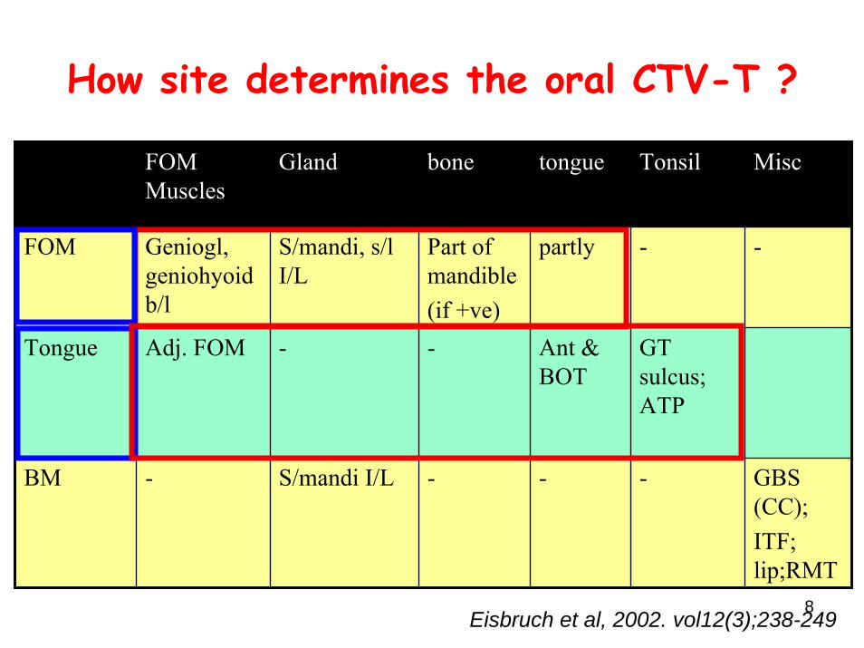
How site determines the oral CTV-T ?
FOM Muscles
Gland bone tongue Tonsil
partly -
GT sulcus; ATP
-
Ant & BOT
-
Misc
FOM Geniogl, geniohyoidb/l
S/mandi, s/lI/L
Part of mandible(if +ve)
-
Tongue Adj. FOM - -
BM - S/mandi I/L - GBS (CC);ITF; lip;RMT
Eisbruch et al, 2002. vol12(3);238-249

9
Prophylactic CTV delineation of LN oral cancers
• Ipsilateral level I-III suffice in a lateralized N0 lesion eg. Buccal mucosa
• Midline crossing tumors – include both sides I-III• Anterior tongue lesions- include level IV• Level II LN + - include ipsilateral level V also• Bilateral LN –Treat each site according to N stage• Hard fixed LN –may need to include the adjacent area

10
Oropharynx - subsites
PalatopharyngeusPalatoglossus
Sup. Constrictor
Tensor veli palatini Levator veli palatini
Base of tongueLateral and posterior pharyngeal wall
Soft palate
Tonsil
• Base tongue• Tonsil• Soft palate• Lateral Pharyngeal wall

11
Oropharynx – Normal Radiological anatomy
Base Tongue
Tonsil

12
How does the tumor spread (CTV-T) in oropharynx?
Harari, 2004
13
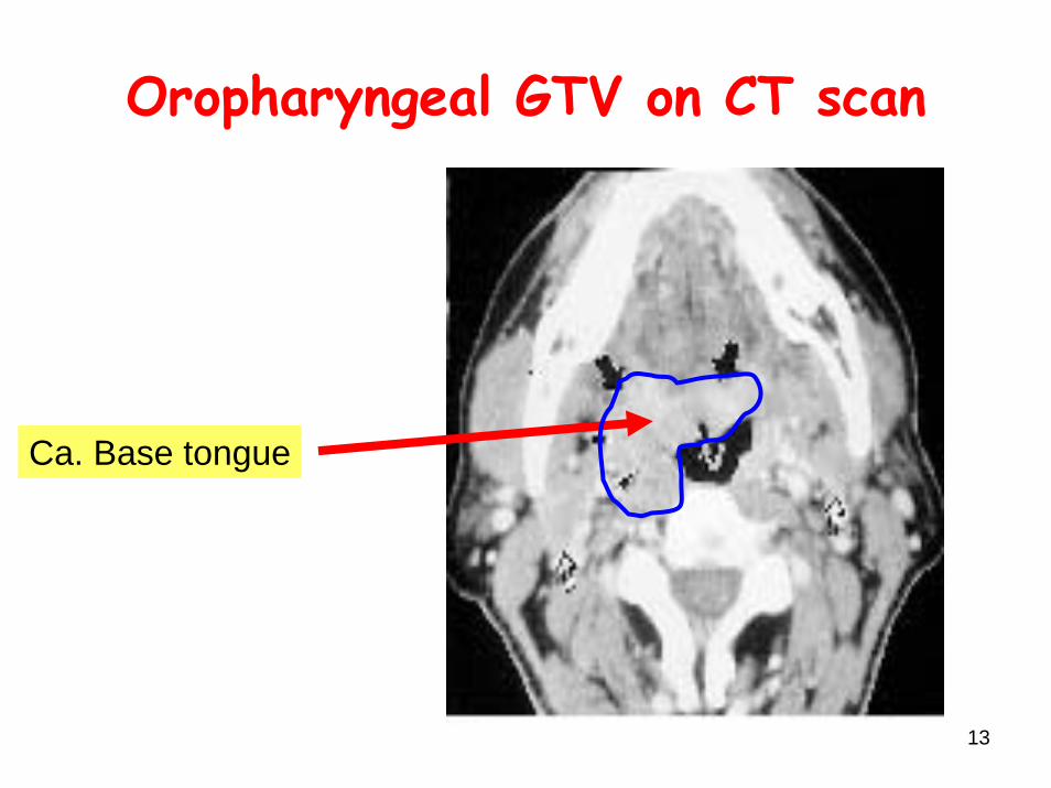
Oropharyngeal GTV on CT scan
Ca. Base tongue
14
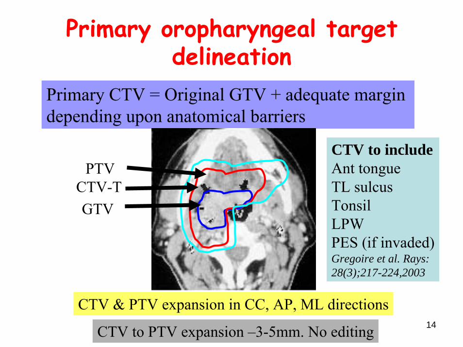
Primary oropharyngeal target delineation
Primary CTV = Original GTV + adequate margin depending upon anatomical barriers
CTV & PTV expansion in CC, AP, ML directions
CTV to PTV expansion –3-5mm. No editing
CTV to includeAnt tongueTL sulcusTonsilLPWPES (if invaded)Gregoire et al. Rays:28(3);217-224,2003
GTVCTV-T
PTV

15
An example
Dasine, 2004
16
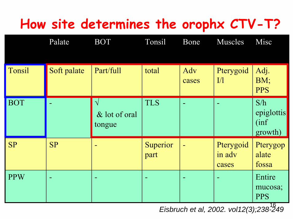
How site determines the orophx CTV-T?Palate BOT Tonsil Bone Muscles
Adv cases
PterygoidI/l
-
SP SP - Superior part
- Pterygoidin adv cases
Pterygopalatefossa
-
-
-
Misc
Tonsil Soft palate Part/full total Adj. BM; PPS
BOT - √& lot of oral tongue
TLS S/h epiglottis (infgrowth)
PPW - - - Entire mucosa; PPS
Eisbruch et al, 2002. vol12(3);238-249

17
Prophylactic CTV delineation of LN –oropharyngeal lesions
• Bilateral neck to be include Ib to IV(ipsilateral) and II-IV (contralateral)
• Include Retropharyngeal LN in LN + and Postpharyngeal wall tumors.
• Level II LN + - include ipsilateral level V also• Bilateral LN –Treat each site according to N stage• Hard fixed LN –may need to include the adjacent area

18
Larynx and HypopharynxSite Subsite Structure
EpiglottisA E FoldArytenoidFalse cord
Glottis True cord Subglottis
Pyriform SinusPostcricoid
Hypopharynx
Post Phx wall
Larynx Supraglottis

19
Epiglottis
Normal radiological anatomy – Larynx and hypopharynx region
Hyoid Sub mandibular
Vallecula
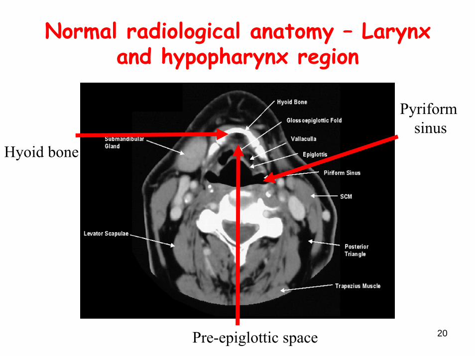
20
Hyoid bone
Normal radiological anatomy – Larynx and hypopharynx region
Pyriformsinus
Pre-epiglottic space

21
AE fold
Normal radiological anatomy – Larynx and hypopharynx region
Thyroid cartilage
PFS

22
False cord
Normal radiological anatomy – Larynx and hypopharynx region
Arytenoid
Cricoid cartilage
Crico-arejoint

23
Pyriform sinus
Normal radiological anatomy – Larynx and hypopharynx region
Vestibule Para glotticspace
Posterior Commissure
24
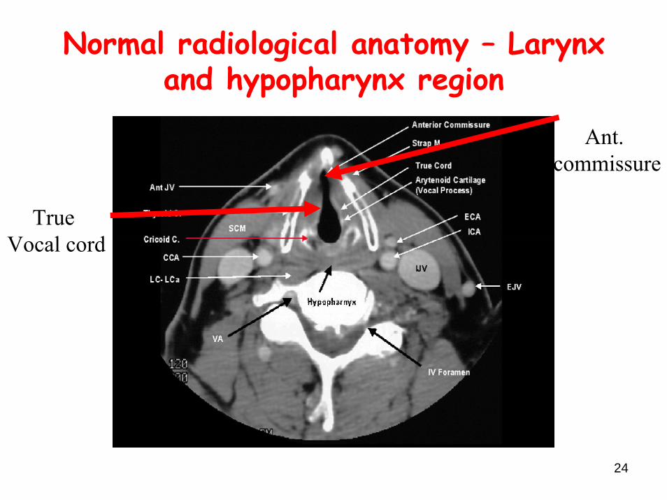
True Vocal cord
Normal radiological anatomy – Larynx and hypopharynx region
Ant. commissure

25
Cricoid
Normal radiological anatomy – Larynx and hypopharynx region
Cricopharyngeal region

26
Thyroid gland
Normal radiological anatomy – Larynx and hypopharynx region
TracheaCricoidcartlage
CCA IJV

27
Normal radiological anatomy – Larynx and hypopharynx region
TracheaThyroidgland
Esophagus
28
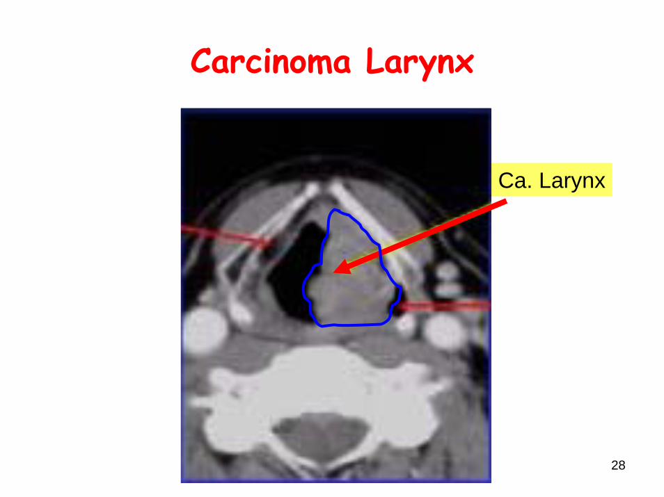
Carcinoma Larynx
Ca. Larynx

29
GTV-CTV-PTV in carcinoma larynx
Ca. LarynxPTV
CTV
GTV

30
Primary target delineation -larynx
Primary CTV = Original GTV + adequate margin depending upon anatomical barriers
CTV to include – PES, PGS, AEF, Vallecula (if suprohyoid ds)Hyoid (if complete PES),Pre Lx Ms & fat (if involved),Longus capitis (if
involved)Gregoire et al. Rays:28(3);217-224, 2003CTV & PTV expansion in CC, AP, ML directions
CTV to PTV expansion –3-5mm. No editing
Anatomical barrier maybe edited from CTVBone Air

31
Prophylactic CTV delineation of LN –laryngeal primary (except T1N0)
• Include Level II-IV bilaterally • In subglottic extension include level VI• Level II LN + - include ipsilateral level V also• Bilateral LN –Treat each site according to N stage• Hard fixed LN –may need to include the adjacent
area

32
Hypopharyngeal cancer- as seen on CT scan
Ca. Pyriform
Ca. Postcricoid

33
Primary target delineation-hypopharynx
Primary CTV = Original GTV + adequate margin depending upon anatomical barriers
CTV & PTV expansion - CC, AP, ML
CTV to PTV expansion –3- 5mm. No editing
CTV to includePGSAEFThyroid cartilage (if PGS)Cricoid (if arytenoid)PPW (if Lat/Post wall)VC (if PGS/PC)Gregoire et al. Rays:28(3);217-224,2003

34
How site determines the adv Larynx-hypophx CTV ?
Larynx PFS vallecula Space & Ms
Cartilage
PES, PGS
Thyroid
--
Misc
Larynx √ √ √ Tracheostomy
PFS I/L hemiLx; PPW
- - Submucosal; I/L Thyroid
Eisbruch et al, 2002. vol12(3);238-249

35
Prophylactic CTV delineation of LN –hypopharyngeal primary
• Include Level II-IV bilaterally • In PFS and esophageal extension include level VI• Include Retropharyngeal LN in LN + and
Postpharyngeal wall tumors.• Level II LN + - include ipsilateral level V also• Bilateral LN –Treat each site according to N stage• Hard fixed LN –may need to include the adjacent
area
36
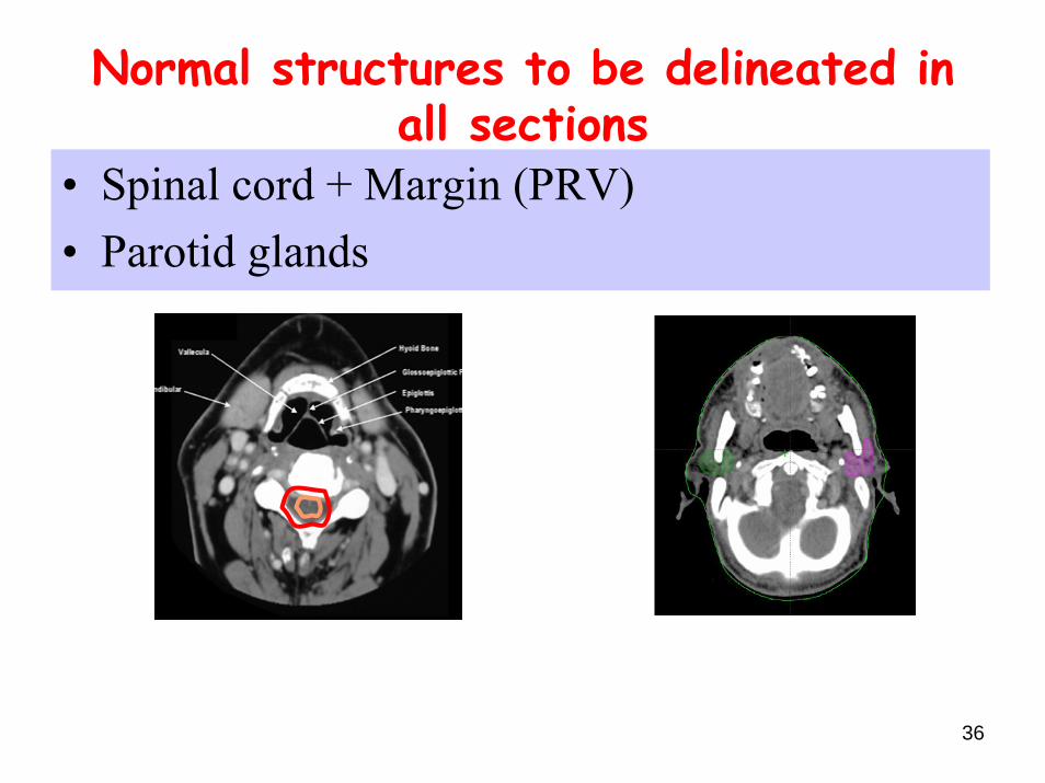
Normal structures to be delineated in all sections
• Spinal cord + Margin (PRV)• Parotid glands
37
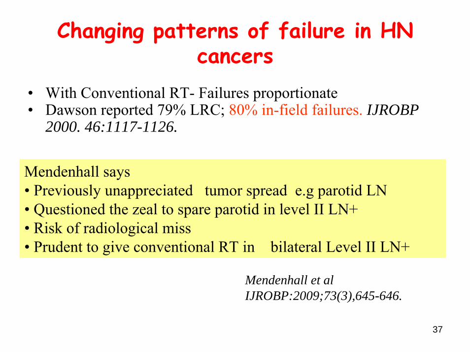
Changing patterns of failure in HN cancers
• With Conventional RT- Failures proportionate• Dawson reported 79% LRC; 80% in-field failures. IJROBP
2000. 46:1117-1126.
Mendenhall says• Previously unappreciated tumor spread e.g parotid LN • Questioned the zeal to spare parotid in level II LN+• Risk of radiological miss • Prudent to give conventional RT in bilateral Level II LN+
Mendenhall et alIJROBP:2009;73(3),645-646.

38
Conclusions
• Knowledge of anatomy, radiological anatomy (normal and abnormal (GTV)) required.
• Delineation is an important link in treatment.
• CTV- determined by patterns of spread and is bounded by natural barriers i.e. bone & air. It should be guided by conventional treatment portals.
• PTV- set up errors of HN site(s) at your centre.

39
Conclusions
• Worst complication is tumor recurrence• Generous target delineation, high quality imaging
and understanding patterns of failure• Need for consensus for primary targets facilitating
consistent selection & delineation
Lee et al, IJROBP, 2004.57:49-60