Embed Size (px)
Citation preview
Tangeretin has anti-asthmatic effects via regulatingPI3K and Notch signaling and modulating Th1/Th2/Th17 cytokine balance in neonatal asthmatic mice
L.-L. Liu1, F.-H. Li1, Y. Zhang1, X.-F. Zhang2 and J. Yang1
1Children’s Medical Center, Qilu Hospital of Shandong University, Jinan, Shandong, China2Department of Pathology, Shandong University of Medicine, Jinan, Shandong, China
Abstract
Asthma is a chronic allergic disease characterized by airway inflammation, airway hyper-responsiveness (AHR), and mucushypersecretion. T-lymphocytes are involved in the pathogenesis of asthma, mediating airway inflammatory reactions by secret-ing cytokines. The phosphoinositide 3-kinase (PI3K) and Notch signaling pathways are associated with Tcell signaling,proliferation, and differentiation, and are important in the progression of asthma. Thus, compounds that can modulate Tcellproliferation and function may be of clinical value. Here, we assessed the effects of tangeretin, a plant-derived flavonoid,in experimental asthma. BALB/c mice at postnatal day (P) 12 were challenged with ovalbumin (OVA). Separate groups of mice(n=18/group) were administered tangeretin at 25 or 50 mg/kg body weight by oral gavage. Dexamethasone was used as apositive control. Tangeretin treatment reduced inflammatory cell infiltration in bronchoalveolar lavage fluid (BALF) and alsorestored the normal histology of lung tissues. OVA-specific IgE levels in serum and BALF were reduced. AHR, as determined byairway resistance and lung compliance, was normalized. Flow cytometry analyses revealed a reduced Th17 cell population.Tangeretin reduced the levels of Th2 and Th17 cytokines and raised IFN-g levels. PI3K signaling was inhibited. The expressionsof the Notch 1 receptor and its ligands Jagged 1 and 2 were downregulated by tangeretin. Our findings support the possible useof tangeretin for treating allergic asthma.
Key words: Asthma; Inflammation; Notch signaling; PI3K; Tangeretin; T-helper cells
Introduction
Asthma, an airway inflammatory disease, is common,chronic, and increasing in prevalence. It is associated withan extensive array of symptoms that include mucus hyper-secretion, airway hyper-responsiveness (AHR), and airwayremodeling (1). T lymphocytes play major roles in airwayinflammation and remodeling through cytokines (2). Th2cytokines (IL-4, IL-5, IL-13) induce allergen-specificimmunoglobulin (Ig) E production and inflammatory medi-ator release from mast cells (2). IFN-g, secreted by Th1cells, suppresses Th2 immune responses. Class switch-ing is induced by IL-4 in IgG1 and IgE, whereas IFN-g isassociated with IgG2 a class switching (3). Thus, Th1/Th2cytokine stability is an important measure in the assess-ment of asthma (3).
Th17 cells, a subset of T lymphocytes, have beenreported to be involved in the pathogenesis of severalimmune-mediated disorders and also in asthma (4). Th17cells release several cytokines, including IL-17, IL-6, TNF-a,and IL-22. IL-17 induces eosinophil infiltration and the
recruitment of macrophages (5) in asthma models. Studieshave shown an increase in Th17 cells in airway inflammationin asthma (6). Thus, regulation of Th17 cells could be ofbenefit in asthma therapy.
Phosphoinositide 3-kinase (PI3K) is a major enzymeinvolved in cell proliferation, differentiation, and cell signal-ing. On activation, PI3K activates its effector protein Akt,that in turn affects several down-stream targets as mammaliantarget of rapamycin (mTOR) and glycogen synthase kinase3b (GSK3b). PI3K/Akt pathway is negatively regulated byphosphatase and tensin homolog (PTEN) (7). PI3K iswidely distributed in lung tissue and plays an importantrole in T cell signaling (8). In particular, PI3K activation inT cells promotes survival (9) and cell cycle progression (10),modulates differentiation and controls the acquisition ofeffector and memory phenotypes (11). The pathway criticallyinfluences the inflammatory response of the airway (12).Thus, inhibitors of PI3K/AKT/mTOR pathway can interferewith T cell activation and function, and would be beneficial
Correspondence: J. Yang: <[email protected]>
Received November 2, 2016 | Accepted May 12, 2017
Braz J Med Biol Res | doi: 10.1590/1414-431X20175991
Brazilian Journal of Medical and Biological Research (2017) 50(8): e5991, http://dx.doi.org/10.1590/1414-431X20175991ISSN 1414-431X Research Article 1/13
in inflammatory disorders. Herrero-Sánchez et al. (13) reportedthat the targeted inhibition of the PI3K/Akt pathway effectivelysuppressed T cell activation and prevented graft-versus-host disease development.
The Notch signaling pathway is also involved in T cellactivation and differentiation (14). Dysregulation of Notchsignaling may induce human disorders such as asthma.Pharmacological inhibitors of Notch signaling can regulateTh1 and Th2 cell-mediated inflammatory responses, reduc-ing allergic pulmonary inflammation (15). Notch signalingregulates retinoic acid-related orphan receptor gt, an impor-tant transcription factor in Th17 differentiation (16). Thus,inhibition of Notch signaling may be beneficial to treatasthma, and compounds that regulate PI3K signaling maybe of therapeutic value.
In this study, we assessed the anti-asthmatic effects oftangeretin, an O-polymethoxylated flavone found in tanger-ine and other citrus fruits. Tangeretin exhibits significantanti-cancer, anti-oxidant and anti-proliferative, activities (17).Studies have reported that tangeretin inhibits inflamma-tory responses by inhibiting NF-kB activation and pro-inflammatory cytokines as interleukins (IL-1b) and tumornecrosis factor (TNF-a) (6,17,18). In this study, we assessedthe ability of tangeretin to modulate PI3K/Akt and Notchsignaling and Th2/Th1 and Th17 cytokine levels in experi-mentally induced asthma in neonatal mice, consideringthe various biological activities of tangeretin.
Material and Methods
Chemicals and antibodiesOvalbumin (OVA), methacholine, dexamethasone (DEX),
tangeretin, and modified Wright-Giemsa stain were fromSigma-Aldrich (USA). ELISA (Enzyme-linked immunosor-bent assay) kits for IL-4, IL-5, and IL-13 were obtainedfrom Biolegend (USA). Antibodies against cyclinA, cyclinD1,Akt, p-Akt, GSK-3b, p- GSK-3b, phosphatise, and PTENwere from Cell Signaling Technology (USA). Mammaliantarget of rapamycin (mTORc1), Jagged 1, Jagged 2, Notch 1,Notch receptor intracellular domain (NICD), and p27kip1antibodies were from Santa Cruz Biotechnology (USA).FITC-labelled anti-human CD4, phycoerythrin (PE), andanti-mouse IL-17A were from eBioscience Co. (USA). Allother analytical-grade chemicals were from Sigma-Aldrichunless noted otherwise.
Experimental animalsBALB/c mice were obtained from Shandong University
Experimental Animal Centre (China) and were maintainedin sterile cages in a controlled environment (24±1°C,40–80% humidity) with a 12/12 h day/night cycle and wereprovided with water and food ad libitum. The animals werecarefully monitored for the day of birth of pups, which wasnoted as postnatal day 0 (P0). This study was approvedby the Ethical Committee of the Shandong University(SD2015031428). Study animals were maintained and
handled according to international regulatory guidelinesfor the maintenance of experimental laboratory animals (19).
Experimental designP12 mice (n=18/group) were used. Separate groups
of mice were challenged with OVA after sensitization, asdescribed by Bao et al. (20). Mice on days 0 and 14 weresensitized with ip injections of OVA (20 mg) and 4 mgAl(OH)3 suspended in saline (0.1 mL). Furthermore, theywere tested with 1% OVA aerosol for 30 min on days22–24. Treatment groups were administered tangeretin(25 or 50 mg/kg) orally regularly from days 0 to 14 and onthe days of OVA administration, tangeretin was given 2 hprior to each OVA challenge. Tangeretin at the specifieddoses was dissolved in 1 mL saline and administeredorally via gavage. The doses were determined fromprevious studies in our laboratory. An equal volume ofsaline was used as control. For a positive control, DEX(2 mg/kg) was injected 1 h prior to OVA administration.
Collection of bronchoalveolar lavage fluidMice (n=6/group) were sacrificed 24 h following the
last OVA challenge using an overdose of pentobarbital(50 mg/kg) and a tracheotomy was performed. Bronchoalveolarlavage fluid (BALF) was collected after instillation of ice-cold PBS (0.5 mL) into a lung; with three successiveaspirations, a total volume of 1.5 mL was collected viatracheal cannulation. BALF samples were centrifuged(252 g, 10 min, 4°C) and the supernatants were storedat –80°C to analyze cytokines. The cell pellet was resus-pended in saline (100 mL) and stained with modifiedWright-Giemsa stain for 8 min on glass slides. Then theslides were assessed for differential cell counts at � 40magnification.
Determination of cytokine levels in BALFLevels of cytokines (IL-4, IL-5, IL-6, IL-13, IL-17, and
IFN-g) in the BALF were determined by ELISA accordingto the manufacturers’ protocols. For IL-6, IL-17, and IFN-g,the kits were from R&D Systems (USA).
Determination of OVA-specific IgE levels in serum andBALF
In serum and BALF, OVA-specific IgE levels weredetermined as described by Jain et al. (21). Serum wasseparated from the whole blood that was collected fromOVA-induced mice. Briefly, a 96-well microtiter platecoated with OVA (10 mg/mL) was treated with BALF orsera followed by treatment with rat anti-mouse IgE (biotin-conjugated; Pharmingen, USA). The absorbance wasread at 405 nm after the addition of avidin-horseradishperoxidase (HRP) solution to each well.
Isolation of CD4+ T cellsFrom mice of the different experimental groups, spleens
were excised at 24 h after the last OVA challenge.
Braz J Med Biol Res | doi: 10.1590/1414-431X20175991
Anti-asthmatic effects of tangeretin in mice 2/13
The tissues were placed in Gibcos RPMI 1640 (ThermoFisher Scientific, USA) and a single-cell suspension of thespleen was prepared. The cells were divided into tubesand washed in phosphate-buffered saline (PBS). CD4+ Tcellswere isolated by using Mouse CD4 T Lymphocyte Enrich-ment Set - DM (BD Biosciences, USA) according to themanufacturer’s protocol. The two-step negative enrichmentmethod uses the Biotinylated Mouse CD4 T LymphocyteEnrichment Cocktail that stains erythrocytes and mostleukocytes except the CD4 T lymphocytes. The positiveand negative fractions were analyzed using flow cytometry(FACSCalibur instrument with CellQuest software; BDBiosciences).
Flow cytometry analysis of Th17 cellsThe spleen cell suspensions were prepared as mentioned
above. To analyze the Th17 cell population, cells wereincubated with fluorescein isothiocyanate (FITC)-conjugatedanti-human CD4 for 30 min at 4°C. After surface staining,the cells were fixed, permeabilized, and stained with PEconjugated-anti-mouse 1L-17A and subjected to flowcytometry (FACSCalibur instrument with CellQuest soft-ware; BD Biosciences).
Measurement of airway hyper-responsivenessAHR was determined using aerosolized methacho-
line at different concentrations using Buxco’s modularand invasive system (Buxco Electronics Inc., USA).Variation in airway resistance (RI) and lung compliance(Cdyn) after reaction with various concentrations ofmethacholine were measured as described by Pichavantet al. (22). Briefly, the mice (n=6/group) were anes-thetized, tracheotomized, cannulated, and kept within aventilated body plethysmograph chamber. The animalswere monitored closely for a steady baseline airwaypressure (o5% variation over 2.5 min) and upon reach-ing that pressure, they were administered aerosolizedPBS or increasing concentrations of methacholine(3.25, 6.25, 12.5, and 25 mg/mL) using a jet nebulizer.Observed RI and Cdyn values are reported as percent-ages relative to the particular basal values in responseto PBS (23).
Histological examinationUsing 10% formalin, lung tissues harvested from mice
not used for BALF collection and other analysis (n=6/group) were fixed and then embedded in paraffin wax;5 mm sections were cut and stained with hematoxylin andeosin (H&E). Peribronchial cell counts were performedaccording to Duan et al. (24) to assess the degree ofleucocyte infiltration. Cell counts were based on a five-point scoring system: 0: no cells, 1: a few cells, 2: a ring ofcells 1 cell layer deep, 3: a ring of cells 2–4 cell layersdeep, and 4: a ring of cells 44 cell layers deep. Toanalyze mucus production, periodic acid-fluorescenceSchiff stain was used, according to Bao et al. (20). The
mucin granules showed red fluorescence at an excitationwavelength of 380–580 nm and were observed at 600–650 nm using a confocal microscope (TCS SP5, LeicaMicrosystems, USA).
mRNA levels of cyclinA, cyclinD1, and p27kip1Cell cycle is tightly regulated by intricate intra-cellular
reactions. Cyclin A, cyclin D1 and p27kip1 are some ofthe chief regulatory molecules of the cell cycle (25).We assessed T-cell proliferation levels. Using RT-PCRcyclinA, cyclinD1, and p27kip1 mRNA levels were deter-mined. From CD4+ T lymphocytes, total RNA wasisolated using Trizol (Invitrogen, USA). By incubationwith reverse transcriptase at 37°C for 1 h, the RNA(1 mg) was reverse transcribed to cDNA. RT-PCRwas carried out using specific primers for rat cyclinA,cyclinD1, and p27kip1, as follows: cyclinD1 forwardprimer 50-CCTGGATGCTGGAGGTCTG-30 and reverse50-AGGGTGGGTTGGAAATGAAC-30, cyclinA forwardprimer 50-GGGCACCTCGAGGCATTC-30 and reverse50-CCTATTACCCGTCGAGTCTTGAG-30, p27kip1 forwardprimer 50-AGCCTACGCTCCGACTGTTTG-30 and reverse50-CCTCCTGCCACTCGTATCTGC-30. Relative expres-sion was normalized with GADPH (forward 50-TCTCCTCTGACTTCAACAGCGAC-30 and reverse 50-CCCTGTTGCTGTAGCCAAATTC-30). PCR was carried as describedpreviously (25).
Western blot analysisIsolated CD4+ T cells were incubated in lysis buffer with
protease inhibitors (0.5 M EDTA, 5 M NaCl, 10% NonidetP-40, 0.1 M phenylmethylsulfonyl fluoride, 0.1 M EGTA,1 M sodium fluoride, 1 M HEPES, 2 mg/mL aprotinin, 0.2 Msodium orthovanadate, and 2 mg/mL leupeptin). The con-centrations of proteins were determined using a proteinassay kit (Bio-Rad Laboratories, USA). The isolatedproteins were separated electrophoretically by SDS PAGE(12%). The separated proteins were blotted onto nitrocel-lulose membranes. Then the membranes were blockedwith 5% non-fat dry milk and were incubated with primaryantibodies (against cyclinA, cyclinD1, GSK-3b, p-GSK-3b,Akt, p-Akt, mTORc1, PTEN, Notch 1, Jagged 1, Jagged 2,NICD, and p27kip 1) overnight at 4°C and further incu-bated at room temperature for 1 h with peroxidase-conjugatedsecondary antibodies. The bands were visualized andanalyzed with a chemiluminescence system (AmershamBioscience, UK). The band intensities were normalized tothose of b-actin.
Statistical analysisThe results are reported as means±SD, from 6 dif-
ferent experiments. Data were evaluated using one-wayanalysis of variance (ANOVA) followed by Duncan’smultiple range test (DMRT) using the SPSS software(ver. 21.0, IBM, USA). P valueso0.05 were considered toindicate statistical significance.
Braz J Med Biol Res | doi: 10.1590/1414-431X20175991
Anti-asthmatic effects of tangeretin in mice 3/13
Results
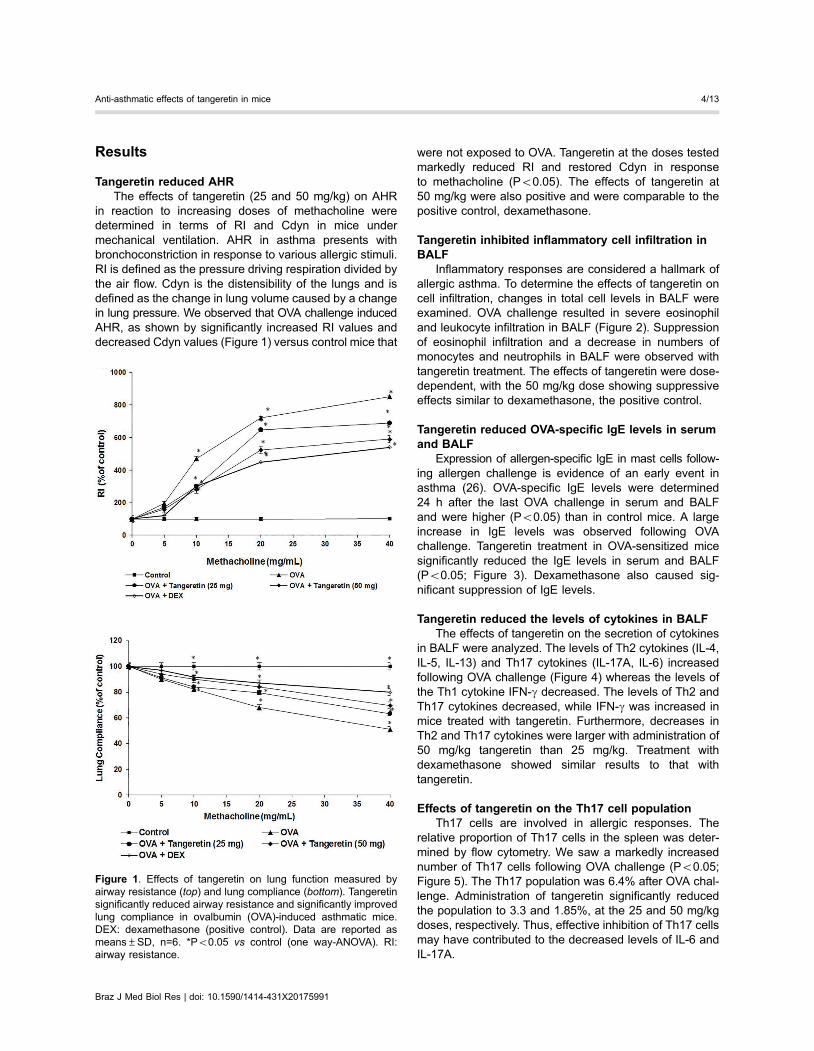
Tangeretin reduced AHRThe effects of tangeretin (25 and 50 mg/kg) on AHR
in reaction to increasing doses of methacholine weredetermined in terms of RI and Cdyn in mice undermechanical ventilation. AHR in asthma presents withbronchoconstriction in response to various allergic stimuli.RI is defined as the pressure driving respiration divided bythe air flow. Cdyn is the distensibility of the lungs and isdefined as the change in lung volume caused by a changein lung pressure. We observed that OVA challenge inducedAHR, as shown by significantly increased RI values anddecreased Cdyn values (Figure 1) versus control mice that
were not exposed to OVA. Tangeretin at the doses testedmarkedly reduced RI and restored Cdyn in responseto methacholine (Po0.05). The effects of tangeretin at50 mg/kg were also positive and were comparable to thepositive control, dexamethasone.
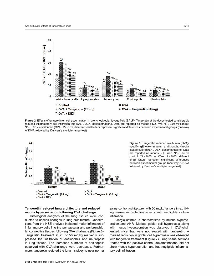
Tangeretin inhibited inflammatory cell infiltration inBALF
Inflammatory responses are considered a hallmark ofallergic asthma. To determine the effects of tangeretin oncell infiltration, changes in total cell levels in BALF wereexamined. OVA challenge resulted in severe eosinophiland leukocyte infiltration in BALF (Figure 2). Suppressionof eosinophil infiltration and a decrease in numbers ofmonocytes and neutrophils in BALF were observed withtangeretin treatment. The effects of tangeretin were dose-dependent, with the 50 mg/kg dose showing suppressiveeffects similar to dexamethasone, the positive control.
Tangeretin reduced OVA-specific IgE levels in serumand BALF
Expression of allergen-specific IgE in mast cells follow-ing allergen challenge is evidence of an early event inasthma (26). OVA-specific IgE levels were determined24 h after the last OVA challenge in serum and BALFand were higher (Po0.05) than in control mice. A largeincrease in IgE levels was observed following OVAchallenge. Tangeretin treatment in OVA-sensitized micesignificantly reduced the IgE levels in serum and BALF(Po0.05; Figure 3). Dexamethasone also caused sig-nificant suppression of IgE levels.
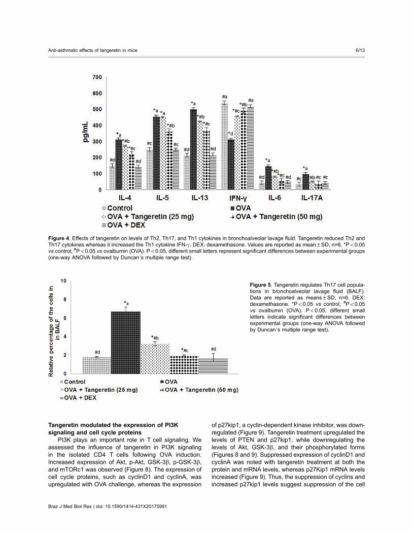
Tangeretin reduced the levels of cytokines in BALFThe effects of tangeretin on the secretion of cytokines
in BALF were analyzed. The levels of Th2 cytokines (IL-4,IL-5, IL-13) and Th17 cytokines (IL-17A, IL-6) increasedfollowing OVA challenge (Figure 4) whereas the levels ofthe Th1 cytokine IFN-g decreased. The levels of Th2 andTh17 cytokines decreased, while IFN-g was increased inmice treated with tangeretin. Furthermore, decreases inTh2 and Th17 cytokines were larger with administration of50 mg/kg tangeretin than 25 mg/kg. Treatment withdexamethasone showed similar results to that withtangeretin.
Effects of tangeretin on the Th17 cell populationTh17 cells are involved in allergic responses. The
relative proportion of Th17 cells in the spleen was deter-mined by flow cytometry. We saw a markedly increasednumber of Th17 cells following OVA challenge (Po0.05;Figure 5). The Th17 population was 6.4% after OVA chal-lenge. Administration of tangeretin significantly reducedthe population to 3.3 and 1.85%, at the 25 and 50 mg/kgdoses, respectively. Thus, effective inhibition of Th17 cellsmay have contributed to the decreased levels of IL-6 andIL-17A.
Figure 1. Effects of tangeretin on lung function measured byairway resistance (top) and lung compliance (bottom). Tangeretinsignificantly reduced airway resistance and significantly improvedlung compliance in ovalbumin (OVA)-induced asthmatic mice.DEX: dexamethasone (positive control). Data are reported asmeans±SD, n=6. *Po0.05 vs control (one way-ANOVA). RI:airway resistance.
Braz J Med Biol Res | doi: 10.1590/1414-431X20175991
Anti-asthmatic effects of tangeretin in mice 4/13
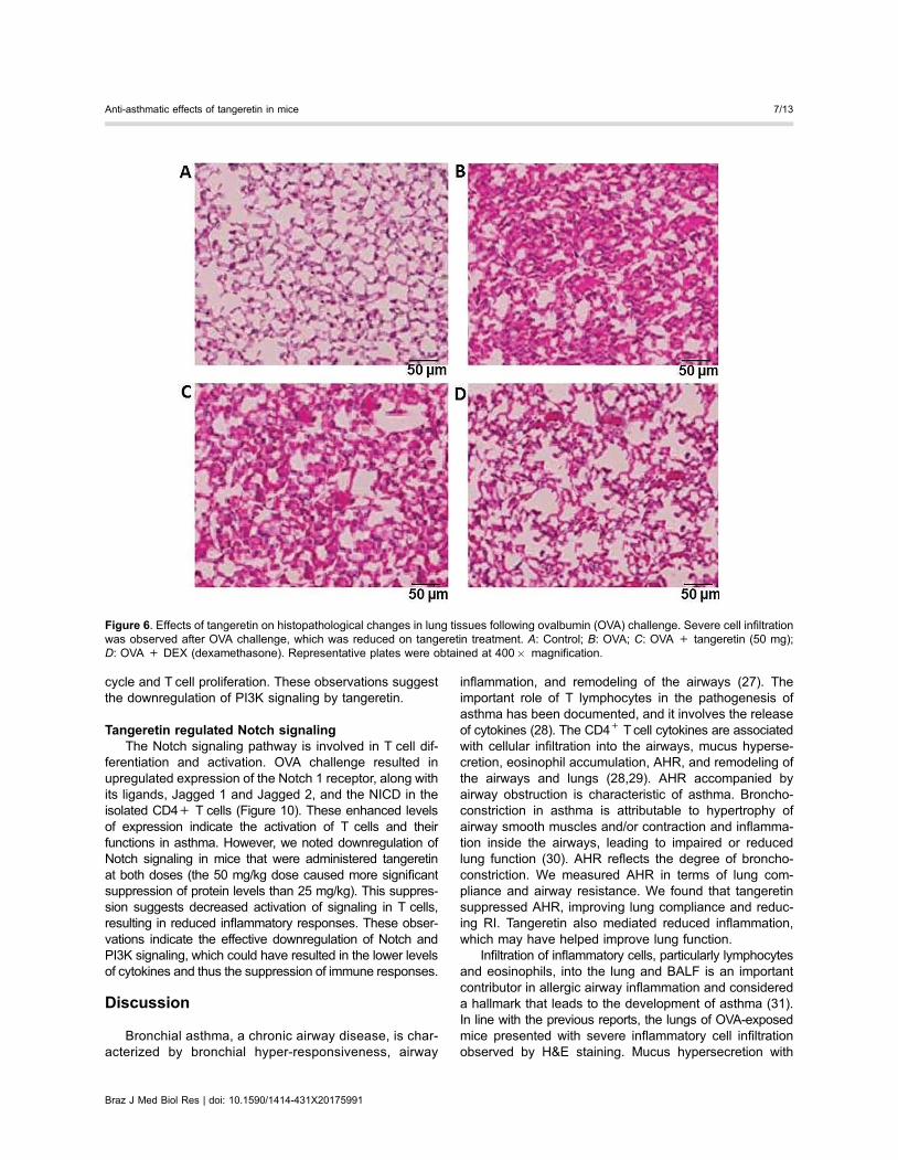
Tangeretin restored lung architecture and reducedmucus hypersecretion following OVA challenge
Histological analyses of the lung tissues were con-ducted to assess changes in lung architecture. Observa-tions from the H&E analysis indicated major infiltration ofinflammatory cells into the perivascular and peribronchio-lar connective tissues following OVA challenge (Figure 6).Tangeretin treatment at 25 or 50 mg/kg markedly sup-pressed the infiltration of eosinophils and neutrophilsin lung tissues. The increased numbers of eosinophilsobserved with OVA challenge were decreased. Further-more, tangeretin restored the lung histology to near normal
saline control architecture, with 50 mg/kg tangeretin exhibit-ing maximum protective effects with negligible cellularinfiltration.
Allergic asthma is characterized by mucus hyperse-cretion and AHR. Marked goblet cell hyperplasia alongwith mucus hypersecretion was observed in OVA-chal-lenged mice that were not treated with tangeretin. Amarked reduction in goblet cell hyperplasia was observedwith tangeretin treatment (Figure 7). Lung tissue sectionstreated with the positive control, dexamethasone, did notshow mucus hypersecretion and had negligible inflamma-tory cell infiltration.
Figure 2. Effects of tangeretin on cell accumulation in bronchoalveolar lavage fluid (BALF). Tangeretin at the doses tested considerablyreduced inflammatory cell infiltration into BALF. DEX: dexamethasone. Data are reported as means±SD, n=6. *Po0.05 vs control;#Po0.05 vs ovalbumin (OVA). Po0.05, different small letters represent significant differences between experimental groups (one-wayANOVA followed by Duncan’s multiple range test).
Figure 3. Tangeretin reduced ovalbumin (OVA)-specific IgE levels in serum and bronchoalveolarlavage fluid (BALF). DEX: dexamethasone. Dataare reported as means±SD, n=6. *Po0.05 vscontrol; #Po0.05 vs OVA. Po0.05, differentsmall letters represent significant differencesbetween experimental groups (one-way ANOVAfollowed by Duncan’s multiple range test).
Braz J Med Biol Res | doi: 10.1590/1414-431X20175991
Anti-asthmatic effects of tangeretin in mice 5/13
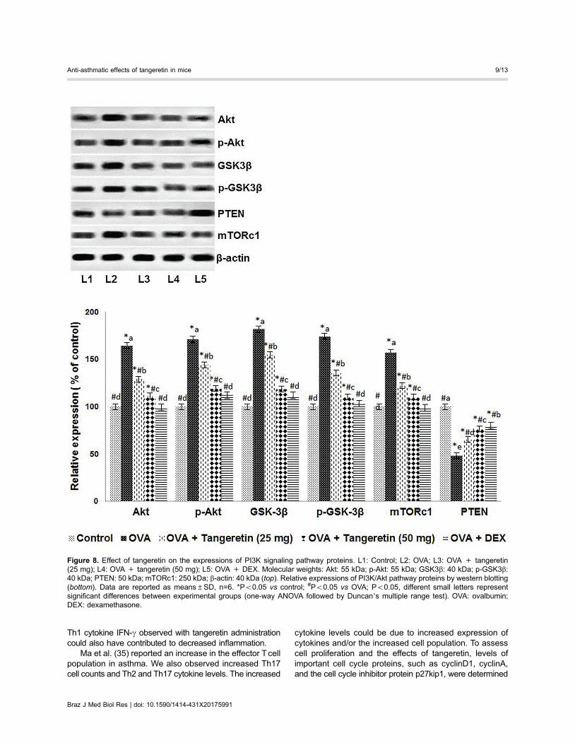
Tangeretin modulated the expression of PI3Ksignaling and cell cycle proteins
PI3K plays an important role in T cell signaling. Weassessed the influence of tangeretin in PI3K signalingin the isolated CD4 T cells following OVA induction.Increased expression of Akt, p-Akt, GSK-3b, p-GSK-3b,and mTORc1 was observed (Figure 8). The expression ofcell cycle proteins, such as cyclinD1 and cyclinA, wasupregulated with OVA challenge, whereas the expression
of p27kip1, a cyclin-dependent kinase inhibitor, was down-regulated (Figure 9). Tangeretin treatment upregulated thelevels of PTEN and p27kip1, while downregulating thelevels of Akt, GSK-3b, and their phosphorylated forms(Figures 8 and 9). Suppressed expression of cyclinD1 andcyclinA was noted with tangeretin treatment at both theprotein and mRNA levels, whereas p27Kip1 mRNA levelsincreased (Figure 9). Thus, the suppression of cyclins andincreased p27kip1 levels suggest suppression of the cell
Figure 4. Effects of tangeretin on levels of Th2, Th17, and Th1 cytokines in bronchoalveolar lavage fluid. Tangeretin reduced Th2 andTh17 cytokines whereas it increased the Th1 cytokine IFN-g. DEX: dexamethasone. Values are reported as mean±SD, n=6. *Po0.05vs control; #Po0.05 vs ovalbumin (OVA). Po0.05, different small letters represent significant differences between experimental groups(one-way ANOVA followed by Duncan’s multiple range test).
Figure 5. Tangeretin regulates Th17 cell popula-tions in bronchoalveolar lavage fluid (BALF).Data are reported as means±SD, n=6. DEX:dexamethasone. *Po0.05 vs control; #Po0.05vs ovalbumin (OVA). Po0.05, different smallletters indicate significant differences betweenexperimental groups (one-way ANOVA followedby Duncan’s multiple range test).
Braz J Med Biol Res | doi: 10.1590/1414-431X20175991
Anti-asthmatic effects of tangeretin in mice 6/13
cycle and T cell proliferation. These observations suggestthe downregulation of PI3K signaling by tangeretin.
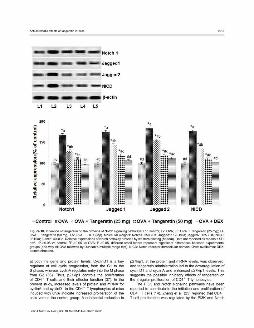
Tangeretin regulated Notch signalingThe Notch signaling pathway is involved in T cell dif-
ferentiation and activation. OVA challenge resulted inupregulated expression of the Notch 1 receptor, along withits ligands, Jagged 1 and Jagged 2, and the NICD in theisolated CD4+ T cells (Figure 10). These enhanced levelsof expression indicate the activation of T cells and theirfunctions in asthma. However, we noted downregulation ofNotch signaling in mice that were administered tangeretinat both doses (the 50 mg/kg dose caused more significantsuppression of protein levels than 25 mg/kg). This suppres-sion suggests decreased activation of signaling in T cells,resulting in reduced inflammatory responses. These obser-vations indicate the effective downregulation of Notch andPI3K signaling, which could have resulted in the lower levelsof cytokines and thus the suppression of immune responses.
Discussion
Bronchial asthma, a chronic airway disease, is char-acterized by bronchial hyper-responsiveness, airway
inflammation, and remodeling of the airways (27). Theimportant role of T lymphocytes in the pathogenesis ofasthma has been documented, and it involves the releaseof cytokines (28). The CD4+ Tcell cytokines are associatedwith cellular infiltration into the airways, mucus hyperse-cretion, eosinophil accumulation, AHR, and remodeling ofthe airways and lungs (28,29). AHR accompanied byairway obstruction is characteristic of asthma. Broncho-constriction in asthma is attributable to hypertrophy ofairway smooth muscles and/or contraction and inflamma-tion inside the airways, leading to impaired or reducedlung function (30). AHR reflects the degree of broncho-constriction. We measured AHR in terms of lung com-pliance and airway resistance. We found that tangeretinsuppressed AHR, improving lung compliance and reduc-ing RI. Tangeretin also mediated reduced inflammation,which may have helped improve lung function.
Infiltration of inflammatory cells, particularly lymphocytesand eosinophils, into the lung and BALF is an importantcontributor in allergic airway inflammation and considereda hallmark that leads to the development of asthma (31).In line with the previous reports, the lungs of OVA-exposedmice presented with severe inflammatory cell infiltrationobserved by H&E staining. Mucus hypersecretion with
Figure 6. Effects of tangeretin on histopathological changes in lung tissues following ovalbumin (OVA) challenge. Severe cell infiltrationwas observed after OVA challenge, which was reduced on tangeretin treatment. A: Control; B: OVA; C: OVA + tangeretin (50 mg);D: OVA + DEX (dexamethasone). Representative plates were obtained at 400� magnification.
Braz J Med Biol Res | doi: 10.1590/1414-431X20175991
Anti-asthmatic effects of tangeretin in mice 7/13
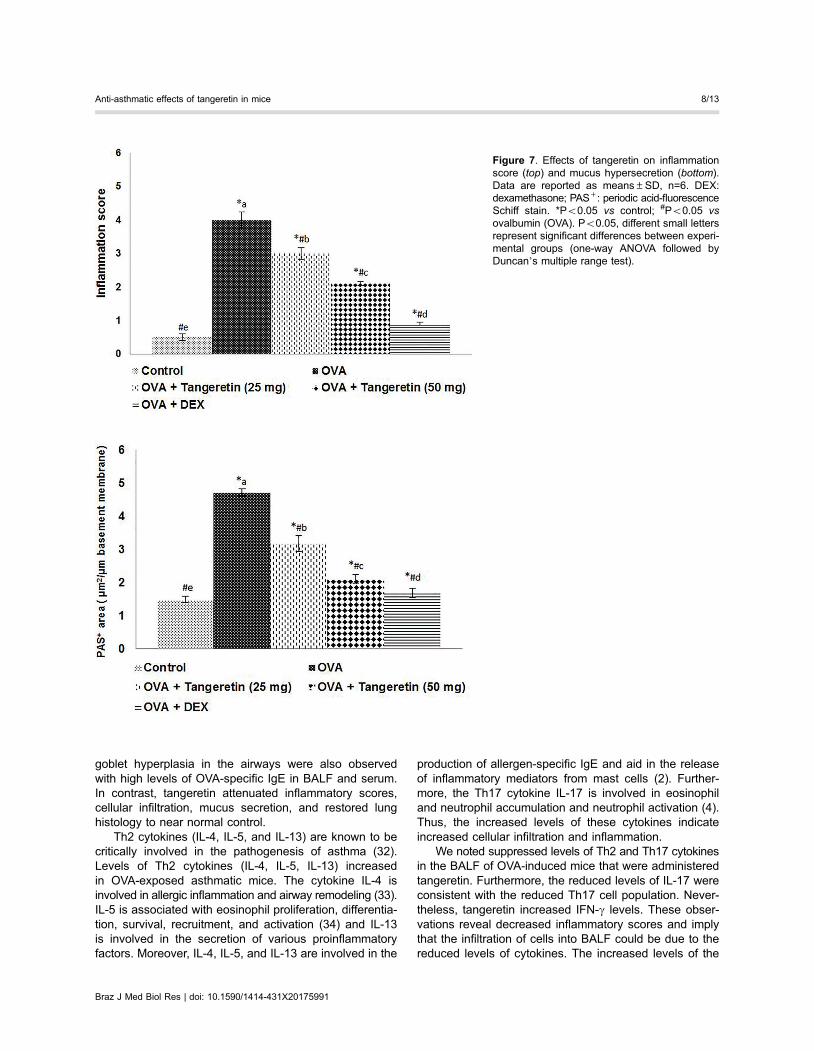
goblet hyperplasia in the airways were also observedwith high levels of OVA-specific IgE in BALF and serum.In contrast, tangeretin attenuated inflammatory scores,cellular infiltration, mucus secretion, and restored lunghistology to near normal control.
Th2 cytokines (IL-4, IL-5, and IL-13) are known to becritically involved in the pathogenesis of asthma (32).Levels of Th2 cytokines (IL-4, IL-5, IL-13) increasedin OVA-exposed asthmatic mice. The cytokine IL-4 isinvolved in allergic inflammation and airway remodeling (33).IL-5 is associated with eosinophil proliferation, differentia-tion, survival, recruitment, and activation (34) and IL-13is involved in the secretion of various proinflammatoryfactors. Moreover, IL-4, IL-5, and IL-13 are involved in the
production of allergen-specific IgE and aid in the releaseof inflammatory mediators from mast cells (2). Further-more, the Th17 cytokine IL-17 is involved in eosinophiland neutrophil accumulation and neutrophil activation (4).Thus, the increased levels of these cytokines indicateincreased cellular infiltration and inflammation.
We noted suppressed levels of Th2 and Th17 cytokinesin the BALF of OVA-induced mice that were administeredtangeretin. Furthermore, the reduced levels of IL-17 wereconsistent with the reduced Th17 cell population. Never-theless, tangeretin increased IFN-g levels. These obser-vations reveal decreased inflammatory scores and implythat the infiltration of cells into BALF could be due to thereduced levels of cytokines. The increased levels of the
Figure 7. Effects of tangeretin on inflammationscore (top) and mucus hypersecretion (bottom).Data are reported as means±SD, n=6. DEX:dexamethasone; PAS+: periodic acid-fluorescenceSchiff stain. *Po0.05 vs control; #Po0.05 vsovalbumin (OVA). Po0.05, different small lettersrepresent significant differences between experi-mental groups (one-way ANOVA followed byDuncan’s multiple range test).
Braz J Med Biol Res | doi: 10.1590/1414-431X20175991
Anti-asthmatic effects of tangeretin in mice 8/13
Th1 cytokine IFN-g observed with tangeretin administrationcould also have contributed to decreased inflammation.
Ma et al. (35) reported an increase in the effector T cellpopulation in asthma. We also observed increased Th17cell counts and Th2 and Th17 cytokine levels. The increased
cytokine levels could be due to increased expression ofcytokines and/or the increased cell population. To assesscell proliferation and the effects of tangeretin, levels ofimportant cell cycle proteins, such as cyclinD1, cyclinA,and the cell cycle inhibitor protein p27kip1, were determined
Figure 8. Effect of tangeretin on the expressions of PI3K signaling pathway proteins. L1: Control; L2: OVA; L3: OVA + tangeretin(25 mg); L4: OVA + tangeretin (50 mg); L5: OVA + DEX. Molecular weights: Akt: 55 kDa; p-Akt: 55 kDa; GSK3b: 40 kDa; p-GSK3b:40 kDa; PTEN: 50 kDa; mTORc1: 250 kDa; b-actin: 40 kDa (top). Relative expressions of PI3K/Akt pathway proteins by western blotting(bottom). Data are reported as means±SD, n=6. *Po0.05 vs control; #Po0.05 vs OVA; Po0.05, different small letters representsignificant differences between experimental groups (one-way ANOVA followed by Duncan’s multiple range test). OVA: ovalbumin;DEX: dexamethasone.
Braz J Med Biol Res | doi: 10.1590/1414-431X20175991
Anti-asthmatic effects of tangeretin in mice 9/13
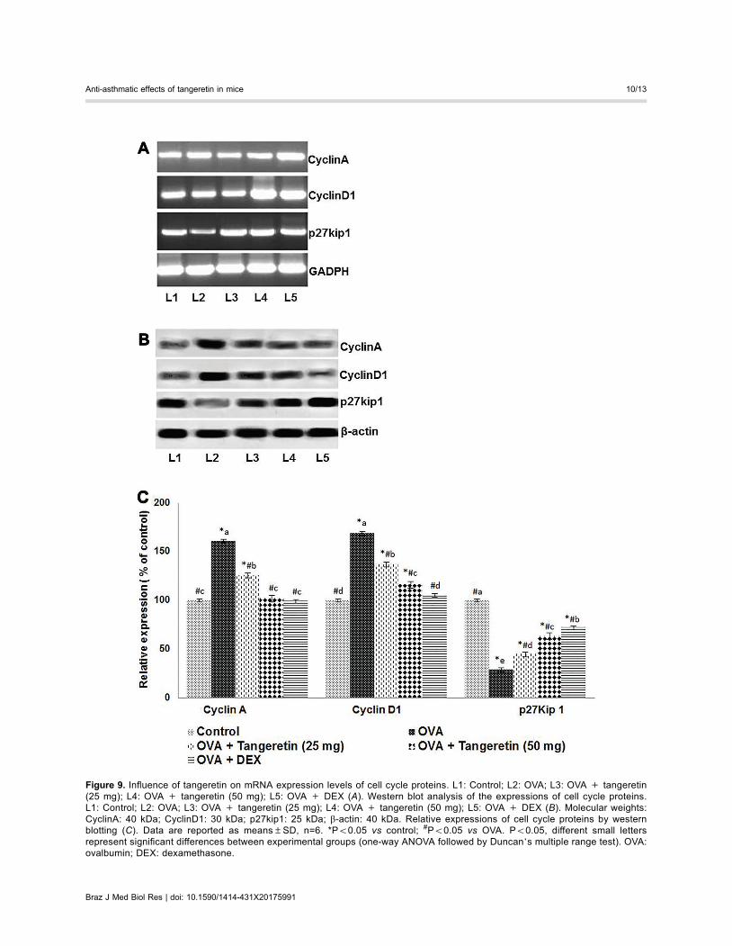
Figure 9. Influence of tangeretin on mRNA expression levels of cell cycle proteins. L1: Control; L2: OVA; L3: OVA + tangeretin(25 mg); L4: OVA + tangeretin (50 mg); L5: OVA + DEX (A). Western blot analysis of the expressions of cell cycle proteins.L1: Control; L2: OVA; L3: OVA + tangeretin (25 mg); L4: OVA + tangeretin (50 mg); L5: OVA + DEX (B). Molecular weights:CyclinA: 40 kDa; CyclinD1: 30 kDa; p27kip1: 25 kDa; b-actin: 40 kDa. Relative expressions of cell cycle proteins by westernblotting (C). Data are reported as means±SD, n=6. *Po0.05 vs control; #Po0.05 vs OVA. Po0.05, different small lettersrepresent significant differences between experimental groups (one-way ANOVA followed by Duncan’s multiple range test). OVA:ovalbumin; DEX: dexamethasone.
Braz J Med Biol Res | doi: 10.1590/1414-431X20175991
Anti-asthmatic effects of tangeretin in mice 10/13
at both the gene and protein levels. CyclinD1 is a keyregulator of cell cycle progression, from the G1 to theS phase, whereas cyclinA regulates entry into the M phasefrom G2 (36). Thus, p27kip1 controls the proliferationof CD4+ T cells and their effector function (37). In thepresent study, increased levels of protein and mRNA forcyclinA and cyclinD1 in the CD4+ T lymphocytes of miceinduced with OVA indicate increased proliferation of thecells versus the control group. A substantial reduction in
p27kip1, at the protein and mRNA levels, was observed,and tangeretin administration led to the downregulation ofcyclinD1 and cyclinA and enhanced p27kip1 levels. Thissuggests the possible inhibitory effects of tangeretin onthe irregular proliferation of CD4+ T lymphocytes.
The PI3K and Notch signaling pathways have beenreported to contribute to the initiation and proliferation ofCD4+ T cells (14). Zhang et al. (25) reported that CD4+
T cell proliferation was regulated by the PI3K and Notch
Figure 10. Influence of tangeretin on the proteins of Notch signaling pathways. L1: Control; L2: OVA; L3: OVA+ tangeretin (25 mg); L4:OVA + tangeretin (50 mg); L5: OVA + DEX (top). Molecular weights: Notch1: 250 kDa; Jagged1: 120 kDa; Jagged2: 125 kDa; NICD:93 kDa; b-actin: 40 kDa. Relative expressions of Notch pathway proteins by western blotting (bottom). Data are reported as means±SD,n=6. *Po0.05 vs control; #Po0.05 vs OVA; Po0.05, different small letters represent significant differences between experimentalgroups (one-way ANOVA followed by Duncan’s multiple range test). NICD: Notch receptor intracellular domain; OVA: ovalbumin; DEX:dexamethasone.
Braz J Med Biol Res | doi: 10.1590/1414-431X20175991
Anti-asthmatic effects of tangeretin in mice 11/13

signaling pathways. Consistent with previous reports,we observed increased expression of the Notch1 receptorand its ligands, Jagged 1 and Jagged 2 (25,38). NICDexpression also increased. Notch signaling is initiatedwhen Notch receptors bind their ligands, leading tocleavage of NICD, which is then translocated to thenucleus and initiates the transcription of downstreamtarget genes (38). Upregulation of PI3K pathway proteinswas also observed in T cells isolated from OVA-alone-induced mice. Nevertheless, significant down-regulationof Notch signaling and PI3K/Akt signaling by tangeretinsuggests reduced cell proliferation and survival.
Previous studies have reported that tangeretin exhibitsanti-inflammatory effects (14,15,39). Tangeretin was foundto inhibit lipopolysaccharide (LPS)-induced nitric oxide, tumornecrosis factor alpha, IL-6, and IL-1b in LPS-stimulatedmicroglia (39). Jang et al. (40) reported that flavonoidstangeretin and nobiletin effectively inhibited NF-kB activationand histamine action following histamine-stimulation in mice.
The present study illustrates the potent anti-asthmatic andanti-inflammatory efficiency of tangeretin via reducing inflam-matory cytokines and modulating major pathways of inflam-matory responses. However, how tangeretin causes theseeffects and the underlying molecular events need to beexplored further.
Collectively, our results indicated the effectiveness oftangeretin in reducing AHR, likely by regulating Th1, Th2,and Th17 cytokine levels and Notch and PI3K signaling.Further experiments with tangeretin could provide abetter understanding of the molecular events involved.Thus, tangeretin is a possible candidate for treatment ofasthma.
Acknowledgments
This study was supported by Foundation for Out-standing Young Scientist in Shandong Province (No:BS2011YY018).
References
1. Galli SJ, Tsai M, Piliponsky AM. The development of allergicinflammation. Nature 2008; 454: 445–454, doi: 10.1038/nature07204.
2. Mosmann TR, Moore KW. The role of IL-10 in crossregulation of TH1 and TH2 responses. Immunol Today1991; 12: A49–A53, doi: 10.1016/S0167-5699(05)80015-5.
3. Brewer JM, Conacher M, Hunter CA, Mohrs M, BrombacherF, Alexander J. Aluminium hydroxide adjuvant initiatesstrong antigen-specific Th2 responses in the absence ofIL-4- or IL-13-mediated signalling. J Immunol 1999; 163:6448–6454.
4. Li J, Zhang B. Apigenin protects ovalbumin-induced asthmathrough the regulation of Th17 cells. Fitoterapia 2013; 91:298–304, doi: 10.1016/j.fitote.2013.09.009.
5. Sergejeva S, Ivanov S, Lotvall J, Linden A. IL-17 as arecruitment and survival factor for airway macrophages inallergic airway inflammation. Am J Respir Cell Mol Biol2005; 33: 248–253, doi: 10.1165/rcmb.2004-0213OC.
6. Zhao Y, Yang J, Gao YD, Guo W. Th17 immunity in patientswith allergic asthma. Int Arch Allergy Immunol 2010; 151:297–307, doi: 10.1159/000250438.
7. Zhu Q, Yang J, Han S, Liu J, Holzbeierlein J, Thrasher JB,et al. Suppression of glycogen synthase kinase 3 activityreduces tumor growth of prostate cancer in vivo. Prostate2011; 71: 835–845, doi: 10.1002/pros.21300.
8. Okkenhaug K, Patton DT, Bilancio A, Garcon F, Rowan WC,Vanhaesebroeck B. The p110 delta isoform of phosphoino-sitide 3-kinase controls clonal expansion and differentiationof Th cells. J Immunol 2006; 177: 5122–5128, doi: 10.4049/jimmunol.177.8.5122.
9. Wu LX, La Rose J, Chen L, Neale C, Mak T, Okkenhaug K,et al. CD28 regulates the translation of Bcl-xL via thephosphatidylinositol 3-kinase/mammalian target of rapamy-cin pathway. J Immunol 2005; 174: 180–194, doi: 10.4049/jimmunol.174.1.180.
10. Appleman LJ, van Puijenbroek AA, Shu KM, Nadler LM,Boussiotis VA. CD28 costimulation mediates down-regulation
of p27kip1 and cell cycle progression by activation of thePI3K/PKB signaling pathway in primary human T cells.J Immunol 2002; 168: 2729–2736, doi: 10.4049/jimmunol.168.6.2729.
11. Araki K, Youngblood B, Ahmed R. The role of mTOR inmemory CD8 T-cell differentiation. Immunol Rev 2010; 235:234–243, doi: 10.1111/j.0105-2896.2010.00898.x.
12. Takeda M, Ito W, Tanabe M, Ueki S, Kihara J, Kato H, et al.The pathophysiological roles of PI3Ks and therapeuticpotential of selective inhibitors in allergic inflammation.Int Arch Allergy Immunol 2010; 152: 90–95, doi: 10.1159/000312132.
13. Herrero-Sánchez MC, Rodríguez-Serrano C, Almeida J,San Segundo L, Inogés S, Santos-Briz, et al. Targeting ofPI3K/AKT/mTOR pathway to inhibit T cell activation andprevent graft-versus-host disease development. J HematolOncol 2016; 9: 113, doi: 10.1186/s13045-016-0343-5.
14. Laky K, Fowlkes BJ. Notch signaling in CD4 and CD8 T celldevelopment. Curr Opin Immunol 2008; 20: 197–202,doi: 10.1016/j.coi.2008.03.004.
15. Kang JH, Kim BS, Uhm TG, Lee SH, Lee GR, Park CS, et al.g-secretase inhibitor reduces allergic pulmonary inflamma-tion by modulating Th1 and Th2 responses. Am J Resp CritCare Med 2009; 179: 875–882, doi: 10.1164/rccm.200806-893OC.
16. Ito T, Connett JM, Kunkel SL, Matsukawa A. Notch systemin the linkage of innate and adaptive immunity. J LeukocyteBiol 2012; 92: 59–65, doi: 10.1189/jlb.1011529.
17. Shu Z, Yang B, Zhao H, Xu B, Jiao W, Wang Q, et al.Tangeretin exerts anti-neuroinflammatory effects via NF-kBmodulation in lipopolysaccharide-stimulated microglial cells.Int Immunopharmacol 2014; 19: 275–282, doi: 10.1016/j.intimp.2014.01.011.
18. Sundaram R, Shanthi P, Sachdanandam P. Tangeretin,a polymethoxylated flavone, modulates lipid homeostasisand decreases oxidative stress by inhibiting NF-kB activa-tion and proinflammatory cytokines in cardiac tissue of
Braz J Med Biol Res | doi: 10.1590/1414-431X20175991
Anti-asthmatic effects of tangeretin in mice 12/13
streptozotocin-induced diabetic rats. J Funct Foods 2015;16: 315–333, doi: 10.1016/j.jff.2015.03.024.
19. Garber JC. Committee for the Update of the Guide for theCare and Use of Laboratory Animals. Guide for the Careand Use of Laboratory Animals. 8th edn. National Academyof Sciences; 2011.
20. Bao Z, Guan SP, Cheng C, Wu S, Wong SH, Kemeny DM,et al. A novel anti-inflammatory role for andrographolide inasthma via inhibition of the NF-kB pathway. Am J Respir CritCare Med 2009; 179: 657–665, doi: 10.1164/rccm.200809-1516OC.
21. Jain VV, Kitagaki K, Businga T, Hussain I, George C,O’shaughnessy P, et al. CpG-oligodeoxynucleotides inhibitairway remodeling in a murine model of chronic asthma.J Allergy Clin Immunol 2002; 110: 867e-872e, doi: 10.1067/mai.2002.129371.
22. Pichavant M, Goya S, Hamelmann E, Gelfand EW, UmetsuDT. Animal models of airway sensitization. Curr ProtocImmunol 2007; 15-18, doi: 10.1002/0471142735.im1518s79.
23. Glaab T, Daser A, Braun A, Neuhaus-Steinmetz U, Fabel H,Alarie Y, et al. Tidal Mid expiratory flow as a measure ofairway hyperresponsiveness in allergic mice. Am J PhysiolLung Cell Mol Physiol 2001; 280: L565-573, PMID:11159041.
24. Duan W, Chan JH, Wong CH. Anti-inflammatory effectsof mitogen activated protein kinase inhibitor U0126 in anasthma mouse model. J Immunol 2004; 172: 7053-7059,doi: 10.4049/jimmunol.172.11.7053.
25. Zhang W, Nie Y, Chong L, Cai X, Zhang H, Lin B, et al.PI3K and Notch signal pathways co-ordinately regulate theactivation and proliferation of T lymphocytes in asthma. LifeSci 2013; 92: 890-895, doi: 10.1016/j.lfs.2013.03.005.
26. Li J, Zhang B. Apigenin protects ovalbumin-induced asthmathrough the regulation of Th17 cells. Fitoterapia 2013; 91:298-304, doi: 10.1016/j.fitote.2013.09.009.
27. Holgate ST, Arshad HS, Roberts GC, Howarth PH, ThurnerP, Davies DE. A new look at the pathogenesis of asthma.Clin Sci (Lond) 2010; 118: 439-450, doi: 10.1042/CS20090474.
28. Busse WW, Lemanske Jr RF. Asthma. N Engl J Med 2001;334: 350-362, doi: 10.1056/NEJM200102013440507.
29. Kips JC, Tournoy KG, Pauwels RA. New anti-asthmatherapies: suppression of the effect of interleukin (IL)-4and IL-5. Eur Respir J 2001; 17: 499-506, PMID:11405532.
30. Wenzel SE. Asthma: defining of the persistent adult pheno-types. Lancet 2006; 368: 804-813, doi: 10.1016/S0140-6736(06)69290-8.
31. Umetsu DT, McIntire JJ, Akbari O, Macaubas C, DeKruyffRH. Asthma: an epidemic of dysregulated immunity. NatureImmunol 2002; 3: 715-720, doi: 10.1038/ni0802-715.
32. Huang X, Tang L, Wang F, Song G. Astragaloside IVattenuates allergic inflammation by regulationTh1/Th2 cyto-kine and enhancement CD4+CD25+Foxp3 T cells inoval-bumin-induced asthma. Immunobiology 2014; 219: 565-571,doi: 10.1016/j.imbio.2014.03.005.
33. Jie Z, Jin M, Cai Y, Bai C, Shen Y, Yuan Z, et al. The effectsof Th2 cytokines on the expression of ADAM33 in allergen-induced chronic airway inflammation. Respir Physiol Neu-robiol 2009; 168: 289-294, doi: 10.1016/j.resp.2009.07.019.
34. Simon D, Braathen LR, Simon HU. Eosinophils and atopicdermatitis. Allergy 2004; 59: 561-570, doi: 10.1111/j.1398-9995.2004.00476.x.
35. Ma C, Ma Z, Fu Q, Ma S. Curcumin attenuates allergicairway inflammation by regulation of CD4+CD25+ regula-tory T cells (Tregs)/Th17 balance in ovalbumin-sensitizedmice. Fitoterapia 2013; 87: 57-64, doi: 10.1016/j.fitote.2013.02.014.
36. Blagosklonny MV, Pardee AB. The restriction point of thecell cycle. Cell Cycle 2002; 1: 103-110, PMID:12429916,doi: 10.4161/cc.1.2.108.
37. Rowell RA, Walsh MC, Wells AD. Opposing roles for thecyclin-dependent kinase inhibitor p27kip1 in the control ofCD4+ T cell proliferation and effector function. J Immunol2005; 174: 3359-3368, doi: 10.4049/jimmunol.174.6.3359.
38. Zhang W, Zhang X, Sheng A, Weng C, Zhu T, Zhao W, et al.g-Secretase inhibitor alleviates acute airway inflammationof allergic asthma in mice by downregulating Th17 celldifferentiation. Mediators Inflamm 2015; 2015: 258168,doi: 10.1155/2015/258168.
39. Lee YY, Lee EJ, Park JS, Jang SE, Kim DH, Kim HS.Anti-inflammatory and antioxidant mechanism of tangeretinin activated microglia. J Neuroimmune Pharmacol 2016; 11:294-305, doi: 10.1007/s11481-016-9657-x.
40. Jang SE, Ryu KR, Park SH, Chung S, Teruya Y, Han MJ,et al. Nobiletin and tangeretin ameliorate scratching beha-vior in mice by inhibiting the action of histamine and theactivation of NF-kB, AP-1 and p38. Int Immunopharmacol2013; 17: 502-507, doi: 10.1016/j.intimp.2013.07.012.
Braz J Med Biol Res | doi: 10.1590/1414-431X20175991
Anti-asthmatic effects of tangeretin in mice 13/13