Embed Size (px)
Citation preview
University of Wollongong University of Wollongong
Research Online Research Online
Faculty of Engineering and Information Sciences - Papers: Part B
Faculty of Engineering and Information Sciences
2017
Tailoring the wettability and mechanical properties of electrospun Tailoring the wettability and mechanical properties of electrospun
poly(L-lactic acid)-poly(glycerol sebacate) core-shell membranes poly(L-lactic acid)-poly(glycerol sebacate) core-shell membranes
for biomedical applications for biomedical applications
Yi Yan University of Wollongong
Vitor Sencadas University of Wollongong, [email protected]
Tiantian Jin University of Wollongong, [email protected]
Xu-Feng Huang University of Wollongong, [email protected]
Jun Chen University of Wollongong, [email protected]
See next page for additional authors
Follow this and additional works at: https://ro.uow.edu.au/eispapers1
Part of the Engineering Commons, and the Science and Technology Studies Commons
Recommended Citation Recommended Citation Yan, Yi; Sencadas, Vitor; Jin, Tiantian; Huang, Xu-Feng; Chen, Jun; Wei, Dongbin; and Jiang, Zhengyi, "Tailoring the wettability and mechanical properties of electrospun poly(L-lactic acid)-poly(glycerol sebacate) core-shell membranes for biomedical applications" (2017). Faculty of Engineering and Information Sciences - Papers: Part B. 616. https://ro.uow.edu.au/eispapers1/616
Research Online is the open access institutional repository for the University of Wollongong. For further information contact the UOW Library: [email protected]
Tailoring the wettability and mechanical properties of electrospun poly(L-lactic Tailoring the wettability and mechanical properties of electrospun poly(L-lactic acid)-poly(glycerol sebacate) core-shell membranes for biomedical applications acid)-poly(glycerol sebacate) core-shell membranes for biomedical applications
Abstract Abstract Tissue and biomedical engineering fields are in constant mutation and in searching for innovative processing techniques capable to tailor the material properties. In this work, poly(l-lactic acid) (PLLA) and elastomeric poly(glycerol sebacate) (PGS) were dissolved in the same solvents and electrospun together, in a single needle system. A core-shell structure where the hydrophilic PGS was placed onto the surface of the hydrophobic PLLA fibre was obtained for elastomeric concentrations up to 25 wt%. It was found that the PLLA:PGS blends are immiscible and the blends present the melting temperatures of the individual polymers. Moreover, their surface properties were deeply influenced by the presence of the PGS, and a superhydrophilic membrane was obtained, after PGS curing at 120 °C for 48 h. When the concentration of PGS is up to 25 wt%, the blend’s Young modulus decreases from ∼35.9 ± 7.1 to 7.5 ± 1.4 MPa and a twofold improvement in the sample stretchability was observed, compared with the pristine PLLA electrospun samples. Finally, in vitro hypothalamus A59 nerve cell culture shows that the core-shell electrospun samples enhanced cell adhesion and proliferation, suggesting that these developed materials have great potentials for nerve regeneration and biomedical engineering applications.
Disciplines Disciplines Engineering | Science and Technology Studies
Publication Details Publication Details Yan, Y., Sencadas, V., Jin, T., Huang, X., Chen, J., Wei, D. & Jiang, Z. (2017). Tailoring the wettability and mechanical properties of electrospun poly(L-lactic acid)-poly(glycerol sebacate) core-shell membranes for biomedical applications. Journal of Colloid and Interface Science, 508 87-94.
Authors Authors Yi Yan, Vitor Sencadas, Tiantian Jin, Xu-Feng Huang, Jun Chen, Dongbin Wei, and Zhengyi Jiang
This journal article is available at Research Online: https://ro.uow.edu.au/eispapers1/616

https://doi.org/10.1016/j.jcis.2017.08.033
1
Tailoring the wettability and mechanical properties of electrospun poly(L-
lactic acid)-poly(glycerol sebacate) core-shell membranes for biomedical
applications
Yi Yana, Vitor Sencadasa,b,*, Tiantian Jinc, Xufeng Huangc, Jun Chend, Dongbin Weia,e,
Zhengyi Jianga,*
*Corresponding authors e-mail:
Vitor Sencadas, [email protected],
Zhengyi Jiang, [email protected],
aSchool of Mechanical, Materials, Mechatronic and Biomedical Engineering, University of
Wollongong, Wollongong, NSW 2522, Australia
bARC Center of Excellence for Electromaterials Science, University of Wollongong, 2522
NSW, Australia
cIllawarra Health and Medical Research Institute, University of Wollongong, Wollongong,
NSW 2522, Australia
dIntelligent Polymer Research Institute, University of Wollongong, Innovation Campus,
Squires Way, Fairy Meadow, NSW 2519, Australia
eSchool of Electrical, Mechanical and Mechatronic Systems, University of Technology
Sydney, Ultimo NSW 2007, Australia
Abstract: Tissue and biomedical engineering fields are in constant mutation and in searching
for innovative processing techniques capable to tailor the material properties. In this work,
https://doi.org/10.1016/j.jcis.2017.08.033
2
poly(L-lactic acid) (PLLA) and elastomeric poly(glycerol sebacate) (PGS) were dissolved in
the same solvents and electrospun together, in a single needle system. A core-shell structure
where the hydrophilic PGS was placed onto the surface of the hydrophobic PLLA fibre was
obtained for elastomeric concentrations up to 25 wt%. It was found that the PLLA:PGS
blends are immiscible and the blends present the melting temperatures of the individual
polymers. Moreover, their surface properties were deeply influenced by the presence of the
PGS, and a superhydrophilic membrane was obtained, after PGS curing at 120 ˚C for 48 h.
When the concentration of PGS is up to 25 wt%, the blend’s Young modulus decreases from
~35.9 ± 7.1 to 7.5 ± 1.4 MPa and a two-fold improvement in the sample stretchability was
observed, compared with the pristine PLLA electrospun samples. Finally, in vitro
hypothalamus A59 nerve cell culture shows that the core-shell samples produced enhanced
cell adhesion and proliferation, suggesting that these developed materials have great
potentials for nerve regeneration and biomedical engineering.
Keywords: Electrospun membranes, core-shell electrospun materials, superhydrophilic,
nerve tissue engineering
1. Introduction
Polymer membranes and scaffolds are widely used in tissue engineering, drug delivery,
wound dressing and other biomedical applications [1]. Different techniques are used to
produce fibrous membranes, e.g. drawing, template synthesis, phase separation and self-
assembly [2, 3]. Nevertheless, electrospinning seems to be the ideal processing method since
it is cost-effective and allows the production of one-by-one continuous micro and nanofibers
from various polymers, and it can be scaled to industrial production [2, 3].

https://doi.org/10.1016/j.jcis.2017.08.033
3
Poly(glycerol sebacate) (PGS), is a ductile and biodegradable elastomer usually obtained by
poly-condensation of glycerol and sebacic acid [4, 5]. The mechanical properties and
biodegradation kinetics are deeply influenced by the temperature and time of the synthesis
and curing process used. It was reported that the PGS Young modulus increases with the
temperature and/or time of curing [6]. Previous studies reported that the curing of the PGS
pre-polymer was performed at temperatures in the range of 90 – 150 ˚C, and at low pressure,
for 24 up to 96 h [6].
Electrospinning PGS pre-polymer does not lead to the production of polymeric fibres due to
their low molecular weight [6, 7], and has a glass transition temperature below room
temperature, which promotes flow to the polymer chains, ultimately giving origin to a
polymer film [8]. Moreover, cross-linked PGS is insoluble in organic solvents [6, 8], and is
not possible to produce a suitable solution for electrospinning.
Core-shell electrospinning can use an electrospinnable polymer as a carrier to produce
electrospun fibres of non-electrospinnable materials. Due to the high temperatures used for
the PGS curing process, the carrier material needs to be thermally stable and with a melting
transition above the temperature used for the PGS curing.
Different strategies are being developed to produce PGS electrospun membranes, based in the
blend of the PGS pre-polymer blend with a suitable carrier, that can form fibres and
facilitates the fibre formation. Jeffries et al. [8] blended PGS pre-polymer with poly(vinyl
alcohol) (PVA) in a common 1,1,1,3,3,3-hexafluoroisopropanol (HFIP) solvent, and the
electrospun sample was submitted to a cross-linking procedure, followed by removal of the
carrier polymer. PVA:PGS overall fibre diameter was around 2.8 µm, with high extensions of
fibre fusion, especially between their contact points.
Poly(methyl methacrylate) (PMMA) [9] and gelatin [10] were also used as carrier materials
to form electrospun PGS fibres membranes. When PMMA was used, the average fibre
https://doi.org/10.1016/j.jcis.2017.08.033
4
diameter of the electrospun membranes was ~ 200 nm, with a strong hydrophobic behaviour,
which was probably due to the presence of higher amounts of PMMA on the surface of the
fibres [9]. When gelatin was used as a carrier polymer for the PGS pre-polymer, an increase
of the average diameter of electrospun fibre and hydrophilicity was observed [10].
Poly (L-lactic acid) (PLLA) was also used as a carrier polymer to produce the PGS
electrospun fibre membranes. Xu et al. [11] demonstrated that core-shell electrospinning was
a feasible technique to immobilise the PGS pre-polymer on the surface of the PLLA fibres. In
their work, they electrospun the PLLA in the outer layer of the needle and the PGS
prepolymer was fed onto the shell of the needle, immobilising the PGS prepolymer in the
core of the produced PLLA fibres. After cross-linking at 130 ˚C for three days, fibres with an
average diameter of 10 µm were obtained, and the membranes produced presented high
extensions of fibre fusion.
The objective of this work is to provide a novel, reliable and reproducible method to produce
electrospun PLLA:PGS membranes, where the PGS elastomer is immobilised on the surface
of the PLLA fibre. The PGS prepolymer was blended with the PLLA solution and
electrospun together in a single needle system, followed by PGS cross-linking. The effect on
fibre average diameter of the PGS pre-polymer concentration in the electrospinning solution
was systematically assessed. Moreover, the influences of the PGS layer on the fibre
membranes wettability, thermal and mechanical properties were studied. Finally, biological
assays demonstrated that the core-shell samples produced have great potentials for nerve
regeneration and biomedical engineering.
2. Experimental
2.1. Materials: Medical degree poly(L-lactic acid) (PLLA, Purasorb PL18, Mw= 217 – 225
kDa, from Corbion Netherlands), glycerol, sebacic acid, N,N-dimethylformamide (DMF),

https://doi.org/10.1016/j.jcis.2017.08.033
5
dichloromethane (DMC), hexane and formamide (from Sigma-Aldrich Australia) were used
as received.
2.2. PGS synthesis: For PGS prepolymer preparation, equimolar glycerol and sebacic acid
were mixed in the flask at 120 °C under argon for 24 h, and used without further purification
during the materials processing. PGS prepolymer was cured in the oven (Shel Lab, Model
1401D) at 120 °C under vacuum, for 48 h to prepare cross-linked PGS films.
2.3. PLLA and PGS:PLLA electrospun membranes: PLLA pellets were dissolved in a mixed
solvent of DMF and DMC (3/7 v/v) to achieve a solution with a final polymer concentration
of 10 wt%. PGS:PLLA solutions were prepared by adding the desired amounts of PGS
prepolymer (0, 25, 40 and 50 wt%) to the PLLA solution prepared according to the method
described above. The PLLA and PGS:PLLA were dissolved at room temperature with the
help of a magnetic stirrer (Velp, Model MST) until complete dissolution. After, the solution
was transferred to a commercial 10 mL glass syringe fitted with a steel needle (inner diameter
500 µm). Electrospinning system was set at 1.67 kV/cm with a high voltage power supply
(Gamma High Voltage). A syringe pump (KDScientific) fed the polymer solutions into the
needle tip at the rate of 0.5 mL/h. Randomly aligned PGS:PLLA fibres were collected in
ground collecting plate 15 cm away from the needle tip. All experiments were conducted at
21 ± 2 ºC and a relative humidity of 43 ± 5%.
PGS cross-linking was performed as per the method described above for the PGS film.
Briefly, the samples were placed inside a vacuum oven at 120 °C, for 48 h. The same
procedure was also performed to a neat PLLA to understand the effect of the curing
temperature.
https://doi.org/10.1016/j.jcis.2017.08.033
6
2.4. Materials characterisations: Surface morphology of the samples was analysed by
scanning electron microscopy (JEOL-6490LV), after deposition of a thin layer of platinum
(10 nm) on the sample surface (Dynavac Sputter Coater). For sample cross-section analysis,
the material was immersed in the liquid nitrogen for around 10 min for thorough cooling,
followed by breaking material, and then attached to the cross-section holder for platinum
coating, followed by morphology observation using JEOL JSM-7500.
Fibres diameters were measured using ImageJ [12] based on the surface morphology SEM
images with at least 100 fibres included for each sample.
Thermal gravimetric analysis (TGA) was carried out using TA instruments TGA (Q500).
Samples of around 10 mg were prepared and heated from room temperature to 600 °C at
20 °C/min. Differential scanning calorimetry (DSC) experiments were conducted by TA
instruments (Q100). Samples of around 5 mg were placed inside 50 µL aluminium pans, and
heated between – 50 and 200 °C at a heating rate of 10 °C/min. All experiments (TGA and
DSC) were performed under a nitrogen atmosphere.
The PGS:PLLA electrospun samples and PGS film, were cut into dog-bone shape of 2 mm
wide and 18 mm for the gauge length. Quasi-static tensile tests were conducted by Shimadzu
EZ-L mechanical tester using the 2 N load cell (for PGS) and 10 N load cell (for fibres) with
the cross-head speed of 1 mm/min at room temperature. At least three measurements were
conducted for each sample.
2.5. Samples wettability: Water contact angle was measured using goniometer (DataPhysics
OCA 20 contact angle system from Germany) to study the surface properties of the fibrous
samples. At least five measurements were done for each sample.

https://doi.org/10.1016/j.jcis.2017.08.033
7
2.6. In vitro biocompatibility: All the fibrous samples were cut into round shape with a
diameter of 6.4 mm. To remove the possibly left unreacted monomers, the samples were
immersed in ethanol for around 2 h, followed by washing twice with phosphate-buffered
saline (PBS, pH = 7.4) and then mechanically stirred in PBS for 12 h. Samples were sterilised
with ultra violet light (𝜆 = 254 𝑛𝑛) for 20 min each side before cell seeding. Adult mouse
hypothalamus neurons A59 cell line (mHypoA-59, Cellutions biosystems) was used to
examine the in vitro biocompatibility. The samples were put on the bottom of the wells of 96
well plate and 1000 cells were seeded for each well. Cells were incubated in 100 µL
Dulbecco’s modified Eagle medium (DMEM) (D5796, Sigma-Aldrich) at 37 °C and 5% CO2
for 5 days with the medium refreshed on day 2 and day 4. The cell culture wells with cells
only were used as control.
Methotrexate (MTX) assay was performed on day 1, day 3 and day 5. 20 µL MTX solution
was added into each well and the culture plate was incubated at the same condition for 2 h.
Supernatants were collected to detect absorbance value via using Spectromax plus 384 plate
reader. Cell viability of the samples after 1, 3 and 5 days was obtained by comparing the
optical density values of the sample groups to control group.
The outgrowth and morphology of the cells were observed by JEOL-6490LV. On day 1, day
3 and day 5, cells were fixed with 4% paraformaldehyde in Dulbecco's PBS for 20 min at
room temperature and then the samples were dehydrated by immersion in a series of ethanol
solutions (50, 75, 90, 95 and 100%), dried at room temperature and coated with a platinum
thin film of 10 nm on the surface for SEM observation.
2.7. Statistics and Data Analysis: Statistical analysis was performed by a Kruskal-Wallis test
with a Dunn`s post hoc test, using GraphPad Prism 6.01 (GraphPad Software, Inc.).
Statistical differences were assigned to the experimental groups with p<0.05.
https://doi.org/10.1016/j.jcis.2017.08.033
8
3. Results and discussion
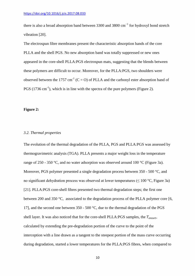
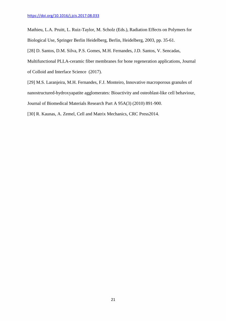
3.1. Morphology and structural analysis
In this work, PLLA polymer and PGS prepolymer were mixed in the same solution and
electrospun together. PLLA electrospun fibre mats showed a random distribution and
cylindrical, smooth fibres, in the absence of bead defects, and the thermal annealing at
120 °C, and during 48h (simulating the PGS curing procedure) does not influence the fibre
morphology (Figure 1a). Moreover, when 25 wt% PGS prepolymer is added to the polymer
solution and electrospun together, smooth fibres without the presence of bead defects are
obtained (Figure 1b). However, when the prepolymer concentration increases above 25 wt%,
spots of prepolymer randomly distributed within the fibre membrane where observed,
increasing their size with the increase of the amount of prepolymer added to the electrospun
solution (Figure 1c and d).
The PGS curing procedure at 120 °C didn’t alter the fibre morphology, or removed the PGS
prepolymer spots from the membrane. On the other hand, for the sample with 25 wt% PGS, it
was observed that the PGS polymer was dragged by the PLLA polymer during
electrospinning, and after curing, the elastomeric material was placed on outside, and the
PLLA was on the core of the electrospun fibre, creating a core-shell structure, in a single
electrospinning step (Figure 1e). Usually, core-shell fibre area obtained by feeding two
independent solutions through a core-shell needle, where one polymer is fed in the core of the
needle and the other is fed on the outside chamber of the needle, and both solutions, only met
on the edge of the needle [13, 14].
The processing method developed to electrospun the PGS:PLLA polymer, creates a core-
shell structure in a continuous manner without clogging the needle tip and avoids the search
for solvent compatible for electrospinning two different solutions [14].
https://doi.org/10.1016/j.jcis.2017.08.033
9
The influence of the amount of PGS prepolymer in the final electrospun fibre diameter was
measured over 100 fibres from different SEM images with the help of Image J. It was
observed that the pristine PLLA electrospun fibres had an average diameter of 746 ± 274 nm,
and with the incorporation of PGS prepolymer, a reduction in the fibre average diameter
down to 332 ± 103 nm was observed for the sample with 50 wt% PGS (Figure 1f). This
reduction in the fibre diameter shows that the PGS prepolymer probably affects charge
density of the solutions. PGS prepolymer can be ionised and carry more charges, stretching
the polymer droplet even further, resulting in a decrease of the average fibre diameter of the
core-shell structure. Similar effect was observed for mixture of polymers with ceramic fillers,
e.g. PLLA with glass reinforced hydroxyapatite [15], or for PLLA zeolite nanocomposite
samples [16]. In comparison with other works, the average diameter of the electrospun core-
shell fibres obtained in this work, was remarkably thinner. Previous works reported a fibre
average diameter between ~2 and 8 µm for PGS electrospun membranes produced with PCL
[17] or PVA [8] as carrier polymers.
Figure 1:
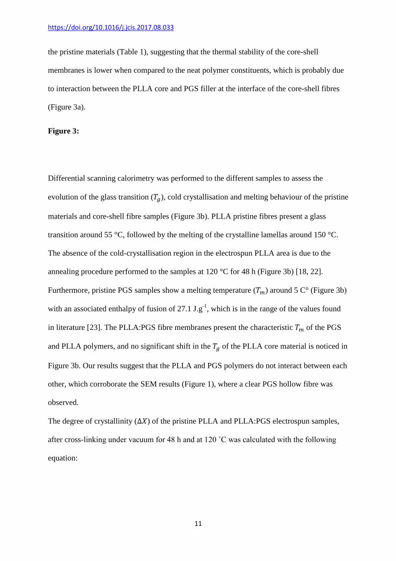
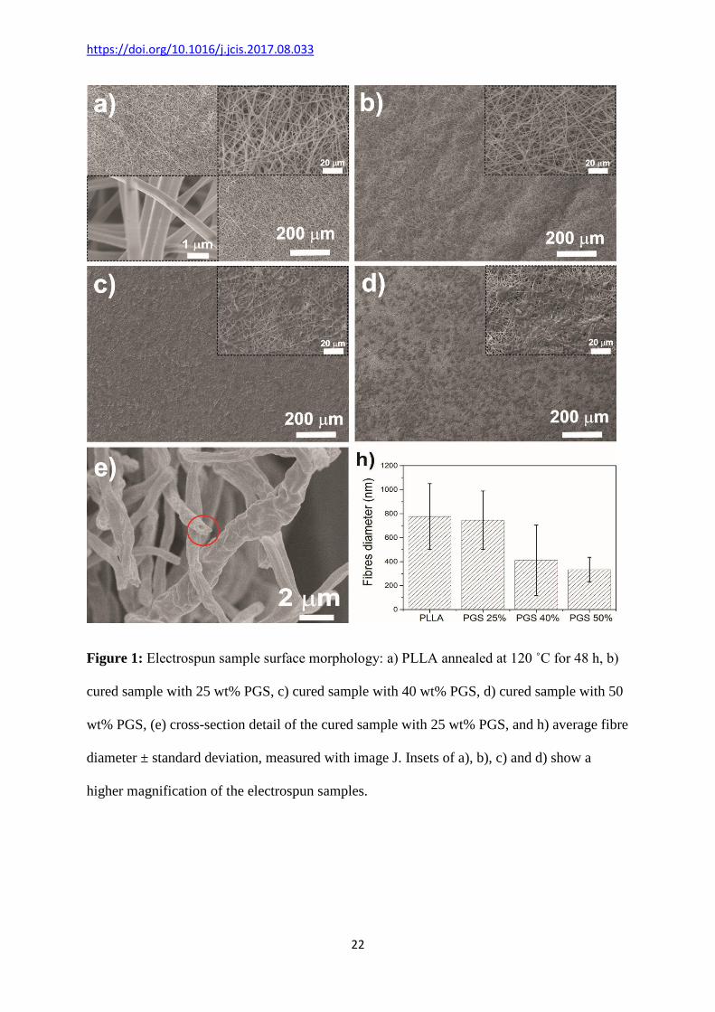
The effect of the PLLA:PGS electrospinning processing parameters can be monitored by
Fourier transform infrared (FTIR) spectroscopy in Attenuated Total Reflectance (ATR).
PLLA absorption band at 1757 cm-1 can be assigned to the C = O, attributed to the
amorphous regions of the polymer chains, while the characteristic absorption band for the
amorphous 𝛼 and 𝛼′ was observed at 1183 cm-1 (𝜐𝑎𝑎(𝐶 − 𝑂 − 𝐶) + 𝑟𝑎(𝐶𝐻3)) and 1092 cm-1
(𝜐𝑎(𝐶 − 𝑂 − 𝐶)), respectively [18]. PGS elastomer presents a characteristic absorption band
at 1736 cm-1, associated to the stretching of the carbonyl ester group (C = O) and the
absorption band at 1185 cm-1 is due to the C - O in-plane bending vibration [19]. Moreover,
https://doi.org/10.1016/j.jcis.2017.08.033
10
there is also a broad absorption band between 3300 and 3800 cm−1 for hydroxyl bond stretch
vibration [20].
The electrospun fibre membranes present the characteristic absorption bands of the core
PLLA and the shell PGS. No new absorption band was totally suppressed or new ones
appeared in the core-shell PLLA:PGS electrospun mats, suggesting that the blends between
these polymers are difficult to occur. Moreover, for the PLLA:PGS, two shoulders were
observed between the 1757 cm-1 (C = O) of PLLA and the carbonyl ester absorption band of
PGS (1736 cm-1), which is in line with the spectra of the pure polymers (Figure 2).
Figure 2:
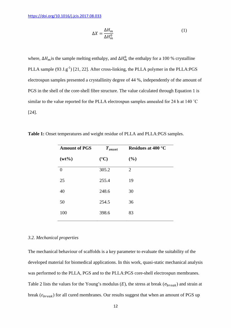
3.2. Thermal properties
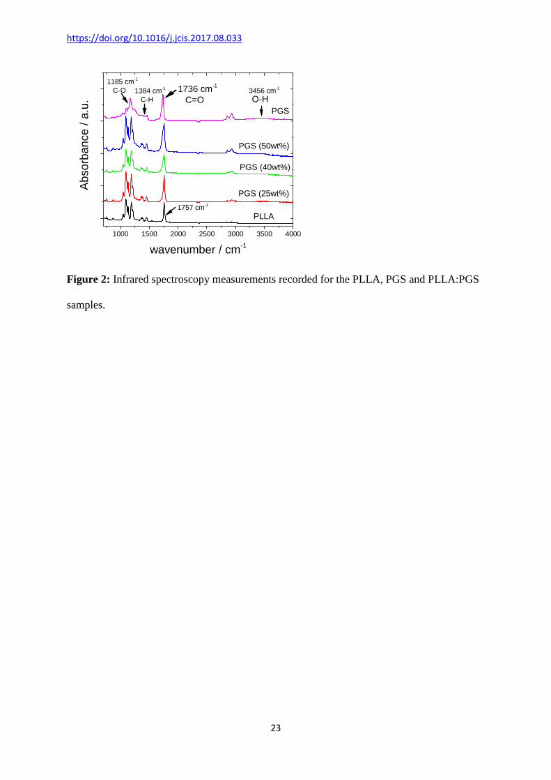
The evolution of the thermal degradation of the PLLA, PGS and PLLA:PGS was assessed by
thermogravimetric analysis (TGA). PLLA presents a major weight loss in the temperature
range of 250 - 350 °C, and no water adsorption was observed around 100 °C (Figure 3a).
Moreover, PGS polymer presented a single degradation process between 350 - 500 °C, and
no significant dehydration process was observed at lower temperatures (≤ 100 °C, Figure 3a)
[21]. PLLA:PGS core-shell fibres presented two thermal degradation steps; the first one
between 200 and 350 °C, associated to the degradation process of the PLLA polymer core [6,
17], and the second one between 350 - 500 °C, due to the thermal degradation of the PGS
shell layer. It was also noticed that for the core-shell PLLA:PGS samples, the 𝑇𝑜𝑜𝑎𝑜𝑜,
calculated by extending the pre-degradation portion of the curve to the point of the
interception with a line drawn as a tangent to the steepest portion of the mass curve occurring
during degradation, started a lower temperatures for the PLLA:PGS fibres, when compared to
https://doi.org/10.1016/j.jcis.2017.08.033
11
the pristine materials (Table 1), suggesting that the thermal stability of the core-shell
membranes is lower when compared to the neat polymer constituents, which is probably due
to interaction between the PLLA core and PGS filler at the interface of the core-shell fibres
(Figure 3a).
Figure 3:
Differential scanning calorimetry was performed to the different samples to assess the
evolution of the glass transition (𝑇𝑔), cold crystallisation and melting behaviour of the pristine
materials and core-shell fibre samples (Figure 3b). PLLA pristine fibres present a glass
transition around 55 °C, followed by the melting of the crystalline lamellas around 150 °C.
The absence of the cold-crystallisation region in the electrospun PLLA area is due to the
annealing procedure performed to the samples at 120 °C for 48 h (Figure 3b) [18, 22].
Furthermore, pristine PGS samples show a melting temperature (𝑇𝑚) around 5 C° (Figure 3b)
with an associated enthalpy of fusion of 27.1 J.g-1, which is in the range of the values found
in literature [23]. The PLLA:PGS fibre membranes present the characteristic 𝑇𝑚 of the PGS
and PLLA polymers, and no significant shift in the 𝑇𝑔 of the PLLA core material is noticed in
Figure 3b. Our results suggest that the PLLA and PGS polymers do not interact between each
other, which corroborate the SEM results (Figure 1), where a clear PGS hollow fibre was
observed.
The degree of crystallinity (∆𝑋) of the pristine PLLA and PLLA:PGS electrospun samples,
after cross-linking under vacuum for 48 h and at 120 ˚C was calculated with the following
equation:
https://doi.org/10.1016/j.jcis.2017.08.033
12
∆𝑋 =∆𝐻𝑚∆𝐻𝑚0
(1)
where, ∆𝐻𝑚is the sample melting enthalpy, and ∆𝐻𝑚0 the enthalpy for a 100 % crystalline
PLLA sample (93 J.g-1) [21, 22]. After cross-linking, the PLLA polymer in the PLLA:PGS
electrospun samples presented a crystallinity degree of 44 %, independently of the amount of
PGS in the shell of the core-shell fibre structure. The value calculated through Equation 1 is
similar to the value reported for the PLLA electrospun samples annealed for 24 h at 140 ˚C
[24].
Table 1: Onset temperatures and weight residue of PLLA and PLLA:PGS samples.
Amount of PGS
(wt%)
𝑻𝒐𝒐𝒐𝒐𝒐
(°C)
Residues at 400 °C
(%)
0 305.2 2
25 255.4 19
40 248.6 30
50 254.5 36
100 398.6 83
3.2. Mechanical properties
The mechanical behaviour of scaffolds is a key parameter to evaluate the suitability of the
developed material for biomedical applications. In this work, quasi-static mechanical analysis
was performed to the PLLA, PGS and to the PLLA:PGS core-shell electrospun membranes.
Table 2 lists the values for the Young’s modulus (𝐸), the stress at break (𝜎𝑏𝑏𝑜𝑎𝑏) and strain at
break (𝜀𝑏𝑏𝑜𝑎𝑏) for all cured membranes. Our results suggest that when an amount of PGS up
https://doi.org/10.1016/j.jcis.2017.08.033
13
to 40 wt% is added to the electrospinning solution, the ductility of the polymeric blend is
improved compared to the pristine PLLA electrospun fibres. For instance, the cured
PLLA:PGS membranes with 25 and 40 wt% show an approximately 2-fold increase of the
strain at break when compared to the pristine PLLA. Moreover, the incorporation of the PGS
into the PLLA:PGS membranes leads to a decrease of the 𝜎𝑏𝑏𝑜𝑎𝑏 when 25 wt% PGS is
incorporated into the polymer solution, however, for higher PGS concentrations, there is an
increase of the stress at break, probably due to the reduction of the pore size and the presence
of the PGS agglomerates, present not only on the surface of the material but also through the
bulk of the membrane (Figure 1). Frydrych et al. [25] found that the amount of PGS added to
the PLLA:PGS scaffold influences the polymeric blend mechanical behaviour. In their work,
they found that the incorporation of higher amounts of PGS, led to a reduction of the pore
diameter, and consequently to an increase of the mechanical stiffness and reduction of the
strain at break of the prepared scaffolds.
Table 2: Mechanical properties of electrospun fibres and PGS.
Amount of
PGS
(wt%)
𝝈𝒃𝒃𝒐𝒃𝒃 (MPa) 𝜺𝒃𝒃𝒐𝒃𝒃
(%)
𝑬
(MPa)
0 2.6 ± 0.3 29 ± 6 35.9 ± 7.1
25 1.1 ± 0.2 66 ± 8 7.2 ± 1.4
40 6.8 ± 0.2 52 ± 9 379.0 ± 29.9
50 6.7 ± 1.1 13 ± 5 336.2 ±25.8
100 0.18 ± 0.01 219 ± 8 0.08 ± 0.01
https://doi.org/10.1016/j.jcis.2017.08.033
14
3.4. Wettability properties
Membrane surface properties and wettability are important features to assess the potential of
the developed membranes for biomedical applications, because they play a vital role in cell
proliferation and adhesion [26]. The average water contact angle (WCA) measured for the
PLLA electrospun fibres was 125 ± 2°, revealing a strong hydrophobic behaviour, probably
due to the overall membrane roughness [15, 22]. Moreover, the PGS polymer film showed to
have a hydrophilic behaviour, with a WCA of 42 ± 3°. Interestingly, for all PLLA:PGS
electrospun samples, the water drop placed on top of the membrane was not stable and the
membrane was capable of uptake of almost instantaneously the liquid. The results reveal that
the incorporation of PGS onto the shell of the PLLA:PGS fibres improves the wettability of
the electrospun membranes significantly, and no WCA measurements and drop penetration
test are possible to perform on this superhydrophilic membranes. Similar results were
reported for poly(caprolactone)-PGS electrospun fibres, and the superhydrophilic behaviour
was attributed to the presence of the hydroxyl groups attached to the backbone of the PGS
polymer, and due to the penetration and capillary effect of the porous membrane [20].
3.5. In vitro biocompatibility of PLLA:PGS fibrous blends
Biomaterial-cell interaction is affected by material roughness, surface free energy,
hydrophilicity, among other material properties [27]. Thus, for tissue engineering and
biomedical applications, it is highly desirable to have hydrophilic surfaces [22, 27]. In the
present work, it was observed that the immobilisation of the PGS polymer onto the surface of
the PLLA fibre gives origin to superhydrophilic membranes, capable to almost
instantaneously uptake water.
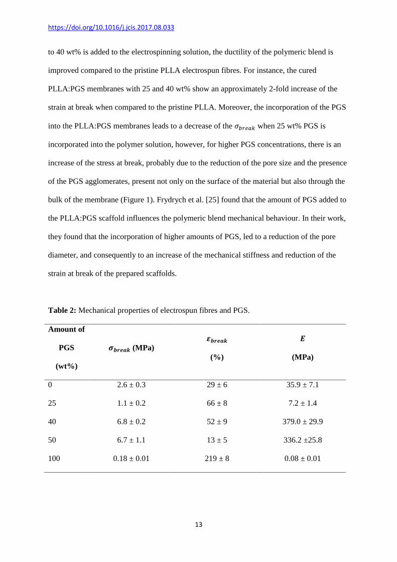
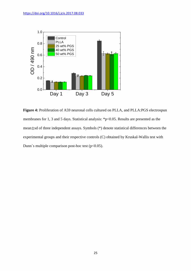
A59 neuronal cell viability in PLLA and PLLA:PGS fibrous membranes was evaluated in
vitro by using MTX assay, which gives an indication of cell metabolic activity. Cells were

https://doi.org/10.1016/j.jcis.2017.08.033
15
seeded on the surface of the fibrous samples, and grown for for one, three and five days, after
which proliferation was assessed by measuring the metabolic activity of the cells with an
MTX assay (Figure 4). After 1one and three days of incubation, the A59 neuronal cells
cultured on the PLLA and PLLA:PGS fibrous mats showed a proliferation profile to the one
obtained with the control, where the cells were cultured in a standard polystyrene (TCPS)
wells. After five days of incubation, proliferation of cells grown on PLLA and PLLA:PGS
electrospun membranes was slightly lower when compared to the control (Figure 4).
Nevertheless, no significant differences were detected between the different PLLA and
PLLA:PGS samples, which suggests that the presence of the hydrophilic PGS shell is suitable
material for biomedical applications.
Figure 4:
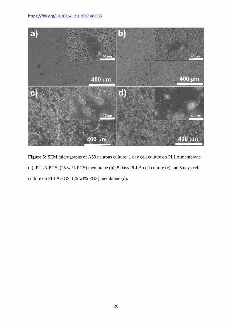
The micrographs recorded with SEM, showed spread cells on the membrane surface (Figure
5), displaying the characteristic flat phenotype, with extended cytoplasm, and appearing to
interact and associate with surrounding fibres (Figure 5) [28, 29]. Even at day 1, the
morphology of the cells was dominated by cells in advanced stage of adhesion (Figure 5). It
was possible to observe that cells were spread on the surface of the fibres, adopting a
polygonal morphology, and adhered to the surface with lamellipodia and filopodia extensions.
It was observed that in the case of the PLLA:PGS fibres, the cells spread out and elongated to
form long processes of neurite varicosities, tried to interact with the electrospun fibres and
communicate with neighbour cells through the nanofibers (Figure 5), suggesting that the
developed membranes have the ability to induce the differentiation of A59 cells into neuron-
like cells, even in the absence of growth factor or chemical treatments.
https://doi.org/10.1016/j.jcis.2017.08.033
16
In the first few hours after culture, cells often attempt to explore the surroundings by
dynamically extruding and retracting thin filopodia protrusions in arbitrary directions. As the
cell begins to flatten, on the surface of the fibrous membranes, wider lamellipodia protrusions
at the leading edge of the cell starts to appear and actively propel the cell front forward [30].
After 5 days, cells actively proliferated, and the establishment of intercellular contacts were
identified within PLLA and PLLA:PGS electrospun membranes (Figure 5).
Figure 5:
4. Conclusions
PLLA and PLLA:PGS fibre structure was obtained by a single needle electrospinning system,
which is versatile, reproducible and can be readily upscale, without compromising the
membrane characteristics. It was found that the incorporation of PGS amounts up to 25 wt%,
individual and defect free fibres were obtained, where the PGS shell wraps the PLLA core,
while for higher amounts of PGS pre-polymer present in the electrospun solution, randomly
dispersed agglomerates of PGS film was observed in the membrane samples. Further, the
blends between the PGS pre-polymer and the PLLA are immiscible and a melting transition
can be observed at ~5 ˚C for the PGS shell, and at ~150 ˚C corresponding to the PLLA fibre
core melting. When PGS is added with a concentration up to 25 wt%, the blend’s Young
modulus decrease from ~35.9 ± 7.1 MPa (pristine PLLA) down to 7.5 ± 1.4 MPa (core-shell
membrane), and a two-fold improvement in the sample stretchability was observed, when
compared with the pristine PLLA electrospun samples. Moreover, by incorporating the
PGS polymer on the surface of the core-shell PLLA:PGS fibres, the wettability of the
materials is enhanced, and a superhydrophilic material was achieved for the sample with
https://doi.org/10.1016/j.jcis.2017.08.033
17
small amount of PGS. Finally, in vitro hypothalamus A59 nerve cell culture shows that the
core-shell samples produced enhanced cell adhesion and proliferation, suggesting that these
materials have great potentials for nerve regeneration and biomedical engineering.
Acknowledgements
The authors acknowledge financial support from China Scholarship Council (CSC), UPA
from University of Wollongong and Australian Research Council (ARC) for Future
Fellowship (Z.Y. Jiang). The authors acknowledge use of facilities within the UOW Electron
Microscopy Centre and the Australian National Nanofabrication Facility – Materials node for
equipment use. The authors acknowledge Mr. Jiangshan Zhang and Dr. Tony Romeo for
SEM imaging, Ms. Hongqin Wang, Dr. Xiaoqi Chen, Dr. Qiang Fu and Ms. Zehra Boz for
their help of cell culture. V. Sencadas thanks support from the COST Actions MP1206:
“Electrospun nano-fibres for bio inspired composite materials and innovative industrial
applications” and MP1301: “New Generation Biomimetic and Customized Implants for Bone
Engineering”.
References
[1] U. Boudriot, R. Dersch, A. Greiner, J.H. Wendorff, Electrospinning Approaches Toward
Scaffold Engineering—A Brief Overview, Artificial Organs 30(10) (2006) 785-792.
[2] Z.-M. Huang, Y.Z. Zhang, M. Kotaki, S. Ramakrishna, A review on polymer nanofibers
by electrospinning and their applications in nanocomposites, Composites Science and
Technology 63(15) (2003) 2223-2253.
[3] J.L. Ifkovits, R.F. Padera, J.A. Burdick, Biodegradable and radically polymerized
elastomers with enhanced processing capabilities, Biomed Mater 3(3) (2008).

https://doi.org/10.1016/j.jcis.2017.08.033
18
[4] Y. Yan, V. Sencadas, J. Zhang, G. Zu, D. Wei, Z. Jiang, Processing, characterisation and
electromechanical behaviour of elastomeric multiwall carbon nanotubes-poly (glycerol
sebacate) nanocomposites for piezoresistive sensors applications, Composites Science and
Technology 142 (2017) 163-170.
[5] Y. Wang, G.A. Ameer, B.J. Sheppard, R. Langer, A tough biodegradable elastomer,
Nature biotechnology 20(6) (2002) 602-6.
[6] R. Rai, M. Tallawi, A. Grigore, A.R. Boccaccini, Synthesis, properties and biomedical
applications of poly(glycerol sebacate) (PGS): A review, Progress in Polymer Science 37(8)
(2012) 1051-1078.
[7] K.G. Akhilesh, N. Mehdi, S. Shilpa, K. Ali, Anisotropic poly (glycerol sebacate)-poly ( ϵ
-caprolactone) electrospun fibers promote endothelial cell guidance, Biofabrication 7(1)
(2015) 015001.
[8] E.M. Jeffries, R.A. Allen, J. Gao, M. Pesce, Y. Wang, Highly elastic and suturable
electrospun poly(glycerol sebacate) fibrous scaffolds, Acta Biomaterialia 18 (2015) 30-39.
[9] J. Hu, D. Kai, H. Ye, L. Tian, X. Ding, S. Ramakrishna, X.J. Loh, Electrospinning of
poly(glycerol sebacate)-based nanofibers for nerve tissue engineering, Materials Science and
Engineering: C 70, Part 2 (2017) 1089-1094.
[10] J.L. Ifkovits, J.J. Devlin, G. Eng, T.P. Martens, G. Vunjak-Novakovic, J.A. Burdick,
Biodegradable Fibrous Scaffolds with Tunable Properties Formed from Photo-Cross-
Linkable Poly(glycerol sebacate), ACS Applied Materials & Interfaces 1(9) (2009) 1878-
1886.
[11] B. Xu, Y. Li, X. Fang, G.A. Thouas, W.D. Cook, D.F. Newgreen, Q. Chen,
Mechanically tissue-like elastomeric polymers and their potential as a vehicle to deliver
functional cardiomyocytes, Journal of the Mechanical Behavior of Biomedical Materials 28
(2013) 354-365.
https://doi.org/10.1016/j.jcis.2017.08.033
19
[12] C.A. Schneider, W.S. Rasband, K.W. Eliceiri, NIH Image to ImageJ: 25 years of image
analysis, Nat Meth 9(7) (2012) 671-675.
[13] B. Xu, B. Rollo, L.A. Stamp, D. Zhang, X. Fang, D.F. Newgreen, Q. Chen, Non-linear
elasticity of core/shell spun PGS/PLLA fibres and their effect on cell proliferation,
Biomaterials 34(27) (2013) 6306-6317.
[14] S. Ramakrishna, K. Fujijhara, W.-E. Teo, T.-C. Lim, Z. Ma, An Introduction to
Electrospinning and Nanofibers, World Scientific2005.
[15] D. Santos, C.O. Correia, D.M. Silva, P.S. Gomes, M.H. Fernandes, J.D. Santos, V.
Sencadas, Incorporation of glass-reinforced hydroxyapatite microparticles into poly(lactic
acid) electrospun fibre mats for biomedical applications, Materials Science and Engineering:
C 75 (2017) 1184-1190.
[16] H. Zhou, K.-W. Kim, E. Giannelis, Y.L. Joo, Nanofibers from Polylactic Acid
Nanocomposites: Effect of Nanoclays on Molecular Structures, in: Darrell H. Reneker, H.
Fong (Eds.), Polymeric Nanofibers, American Chemical Society2006, pp. 217-230.
[17] A.K. Gaharwar, A. Patel, A. Dolatshahi-Pirouz, H. Zhang, K. Rangarajan, G. Iviglia, S.-
R. Shin, M.A. Hussain, A. Khademhosseini, Elastomeric nanocomposite scaffolds made from
poly(glycerol sebacate) chemically crosslinked with carbon nanotubes, Biomaterials Science
(2015).
[18] R. Clarisse, S. Vitor, C. Carlos Miguel, R. José Luís Gómez, L.-M. Senentxu, Tailoring
the morphology and crystallinity of poly(L-lactide acid) electrospun membranes, Science and
Technology of Advanced Materials 12(1) (2011) 015001.
[19] H.M. Aydin, K. Salimi, M. Yilmaz, M. Turk, Z.M.O. Rzayev, E. Pişkin, Synthesis and
characterization of poly(glycerol-co-sebacate-co-ε-caprolactone) elastomers, Journal of
Tissue Engineering and Regenerative Medicine 10(1) (2016) E14-E22.

https://doi.org/10.1016/j.jcis.2017.08.033
20
[20] S. Salehi, M. Fathi, S.H. Javanmard, T. Bahners, J.S. Gutmann, S. Ergün, K.P. Steuhl,
T.A. Fuchsluger, Generation of PGS/PCL Blend Nanofibrous Scaffolds Mimicking Corneal
Stroma Structure, Macromolecular Materials and Engineering 299(4) (2014) 455-469.
[21] V. Sencadas, C.M. Costa, G. Botelho, C. Caparrós, C. Ribeiro, J.L. Gómez-Ribelles, S.
Lanceros-Mendez, Thermal Properties of Electrospun Poly(Lactic Acid) Membranes, Journal
of Macromolecular Science, Part B 51(3) (2012) 411-424.
[22] T.A.M. Valente, D.M. Silva, P.S. Gomes, M.H. Fernandes, J.D. Santos, V. Sencadas,
Effect of Sterilization Methods on Electrospun Poly(lactic acid) (PLA) Fiber Alignment for
Biomedical Applications, ACS Applied Materials & Interfaces 8(5) (2016) 3241-3249.
[23] I.H. Jaafar, M.M. Ammar, S.S. Jedlicka, R.A. Pearson, J.P. Coulter, Spectroscopic
evaluation, thermal, and thermomechanical characterization of poly(glycerol-sebacate) with
variations in curing temperatures and durations, Journal of Materials Science 45(9) (2010)
2525-2529.
[24] J.C. Dias, C. Ribeiro, V. Sencadas, G. Botelho, J.L.G. Ribelles, S. Lanceros-Mendez,
Influence of fiber diameter and crystallinity on the stability of electrospun poly(l-lactic acid)
membranes to hydrolytic degradation, Polymer Testing 31(6) (2012) 770-776.
[25] M. Frydrych, B. Chen, Large three-dimensional poly(glycerol sebacate)-based scaffolds
- a freeze-drying preparation approach, Journal of Materials Chemistry B 1(48) (2013) 6650-
6661.
[26] M. Raul, C. André da, S. Vitor, G.-A. Carmen, M.C. Carlos, P. Jorge, G. Andreia, L.-M.
Senentxu, R.-C. José Carlos, C. Margarida, Electrospun silk-elastin-like fibre mats for tissue
engineering applications, Biomedical Materials 8(6) (2013) 065009.
[27] B. Gupta, N. Anjum, Plasma and Radiation-Induced Graft Modification of Polymers for
Biomedical Applications, in: H. Kausch, N. Anjum, Y. Chevolot, B. Gupta, D. Léonard, H.J.
https://doi.org/10.1016/j.jcis.2017.08.033
21
Mathieu, L.A. Pruitt, L. Ruiz-Taylor, M. Scholz (Eds.), Radiation Effects on Polymers for
Biological Use, Springer Berlin Heidelberg, Berlin, Heidelberg, 2003, pp. 35-61.
[28] D. Santos, D.M. Silva, P.S. Gomes, M.H. Fernandes, J.D. Santos, V. Sencadas,
Multifunctional PLLA-ceramic fiber membranes for bone regeneration applications, Journal
of Colloid and Interface Science (2017).
[29] M.S. Laranjeira, M.H. Fernandes, F.J. Monteiro, Innovative macroporous granules of
nanostructured-hydroxyapatite agglomerates: Bioactivity and osteoblast-like cell behaviour,
Journal of Biomedical Materials Research Part A 95A(3) (2010) 891-900.
[30] R. Kaunas, A. Zemel, Cell and Matrix Mechanics, CRC Press2014.
https://doi.org/10.1016/j.jcis.2017.08.033
22
Figure 1: Electrospun sample surface morphology: a) PLLA annealed at 120 ˚C for 48 h, b)
cured sample with 25 wt% PGS, c) cured sample with 40 wt% PGS, d) cured sample with 50
wt% PGS, (e) cross-section detail of the cured sample with 25 wt% PGS, and h) average fibre
diameter ± standard deviation, measured with image J. Insets of a), b), c) and d) show a
higher magnification of the electrospun samples.
https://doi.org/10.1016/j.jcis.2017.08.033
23
1000 1500 2000 2500 3000 3500 4000
1384 cm-1
C-H O-H1736 cm-1
C=O
PGS (50wt%)
PGS (40wt%)
PGS (25wt%)
PGS
Abso
rban
ce /
a.u.
wavenumber / cm-1
PLLA
3456 cm-1
1757 cm-1
1185 cm-1
C-O
Figure 2: Infrared spectroscopy measurements recorded for the PLLA, PGS and PLLA:PGS
samples.
https://doi.org/10.1016/j.jcis.2017.08.033
24
0 100 200 300 400 500 6000
20
40
60
80
100
Wei
ght (
%)
Temperature (oC)
C0
C25
C40
C50
C100
a
0 100 200 300 400 500 6000
20
40
60
80
100
Wei
ght (
%)
Temperature (oC)
C0
C25
C40
C50
C100
a
-50 0 50 100 150 200
50 wt% PGS
100 wt% PGS
40 wt% PGS
25 wt% PGS
Q /
mW
Temperature / °C
b
.PLLA
5 m
W
-50 0 50 100 150 200
50 wt% PGS
100 wt% PGS
40 wt% PGS
25 wt% PGS
Q /
mW
Temperature / °C
b
.PLLA
5 m
W
Figure 3: a) Thermogravimetric behavior, and b) differential scanning calorimetry recorded
for the PLLA, PGS and PLLA:PGS core-shell fibres.
https://doi.org/10.1016/j.jcis.2017.08.033
25
Day 1 Day 3 Day 50.0
0.2
0.4
0.6
0.8
1.0
*
*
OD
/ 49
0 nm
Control PLLA 25 wt% PGS 40 wt% PGS 50 wt% PGS
**
Figure 4: Proliferation of A59 neuronal cells cultured on PLLA, and PLLA:PGS electrospun
membranes for 1, 3 and 5 days. Statistical analysis: *p<0.05. Results are presented as the
mean±sd of three independent assays. Symbols (*) denote statistical differences between the
experimental groups and their respective controls (C) obtained by Kruskal-Wallis test with
Dunn`s multiple comparison post-hoc test (p<0.05).
https://doi.org/10.1016/j.jcis.2017.08.033
26
Figure 5: SEM micrographs of A59 neurons culture: 1 day cell culture on PLLA membrane
(a); PLLA:PGS (25 wt% PGS) membrane (b); 5 days PLLA cell culture (c) and 5 days cell
culture on PLLA:PGS (25 wt% PGS) membrane (d).