Embed Size (px)
Citation preview
Applied Veterinary Bacteriology and Mycology: Identification of aerobic and facultative anaerobic bacteria
Chapter 4: The identification of Bacillus species with special reference to Bacillus anthracis
Applied Veterinary Bacteriology and Mycology: Identification of aerobic and facultative anaerobic bacteria Chapter 4: The identification of Bacillus species with special reference to Bacillus anthracis
Author: Dr. Valerius de Vos
Licensed under a Creative Commons Attribution license.
TABLE OF CONTENTSINTRODUCTION......................................................................................................................................... 2
IDENTIFICATION........................................................................................................................................ 2
Table 4.1: Identification of Bacillus species..................................................................................4
Table 4.2: Secondary characteristics in the identification of Bacillus spp. in Morphological Group 1........................................................................................................................................ 7
Table 4.3: Biochemical characteristics of Morphological Group 1................................................7
Table 4.4: Identification of Bacillus species..................................................................................8
Table 4.5: Suggested tests to differentiate B. anthracis from B. cereus (numbers positive to numbers tested from Brown et al, 1958).......................................................................................9
APPENDIX 1............................................................................................................................................. 16
1 | P a g e
Applied Veterinary Bacteriology and Mycology: Identification of aerobic and facultative anaerobic bacteria
Chapter 4: The identification of Bacillus species with special reference to Bacillus anthracis
INTRODUCTIONMost species in the genus Bacillus are large, aerobic of facultatively anaerobic, Gram-positive, endospore-producing rods. Spore-producing bacteria embrace a large number of bacterial species with a great diversity of properties. Most of them are contaminants or saprophytes with the ability to adapt to a wide range of environmental conditions. In spite of the fact that some have attained pathogenic status for humans and animals, it remains the general rule in diagnostic laboratories to dismiss aerobic spore-bearers as contaminants. There is evidence that while Bacillus anthracis, B. cereus, B.licheniformis and B. subtilis have attained the status of potential pathogens, other species within this genus may also be incriminated if studied more closely (Gilbert et al, 1983; Gordon, 1981; Parry et a1, 1983; Tuazon et al, 1979). Especially B. cereus, but also B. licheniformis and B. subtilis, have been associated with a wide range of infections, such as bacteraemia and septicaemia, wound and respiratory infections, ophthalmia, peritonitis, gastroenteritis, kidney and urinary tract infections, endocarditis, meningitis and bovine rnastitis. B. anthracis however, stands out in this group of bacteria, causing anthrax in humans and animals. Anthrax is a peracute, acute or subacute, highly contagious disease of domestic and wild animals and humans caused by the bacterium Bacillus anthracis. In most species of animals it is characterized terminally by the development of a rapidly fatal septicaemia, resulting, in sudden death. The principal lesions are those of widespread oedema, haemorrhage and necrosis.
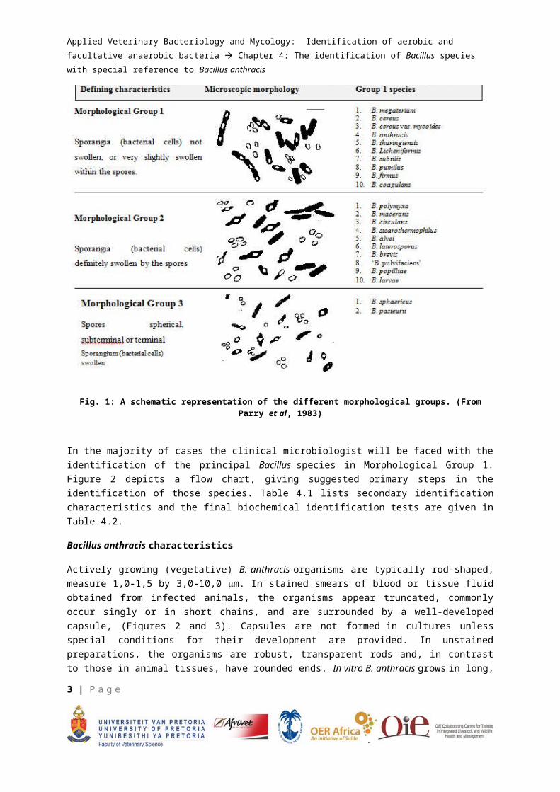
IDENTIFICATION Smith et al (1946, 1952) found that the genus Bacillus, or aerobic spore-bearers, can be divided into 3 groups on the basis of the shape of the spore and swelling or absence of swelling of the sporangium by the spore. Morphological Group 1 is defined by the absence of sporangial swelling and possession of ellipsoidal spores and includes all of the known pathogens of this genus. Morphological Group 2 includes species whose sporangia are swollen by oval spores, and Morphological Group 3 includes species that produce round spores. (Figure 1)
2 | P a g e
Applied Veterinary Bacteriology and Mycology: Identification of aerobic and facultative anaerobic bacteria
Chapter 4: The identification of Bacillus species with special reference to Bacillus anthracis
Fig. 1: A schematic representation of the different morphological groups. (From Parry et al, 1983)
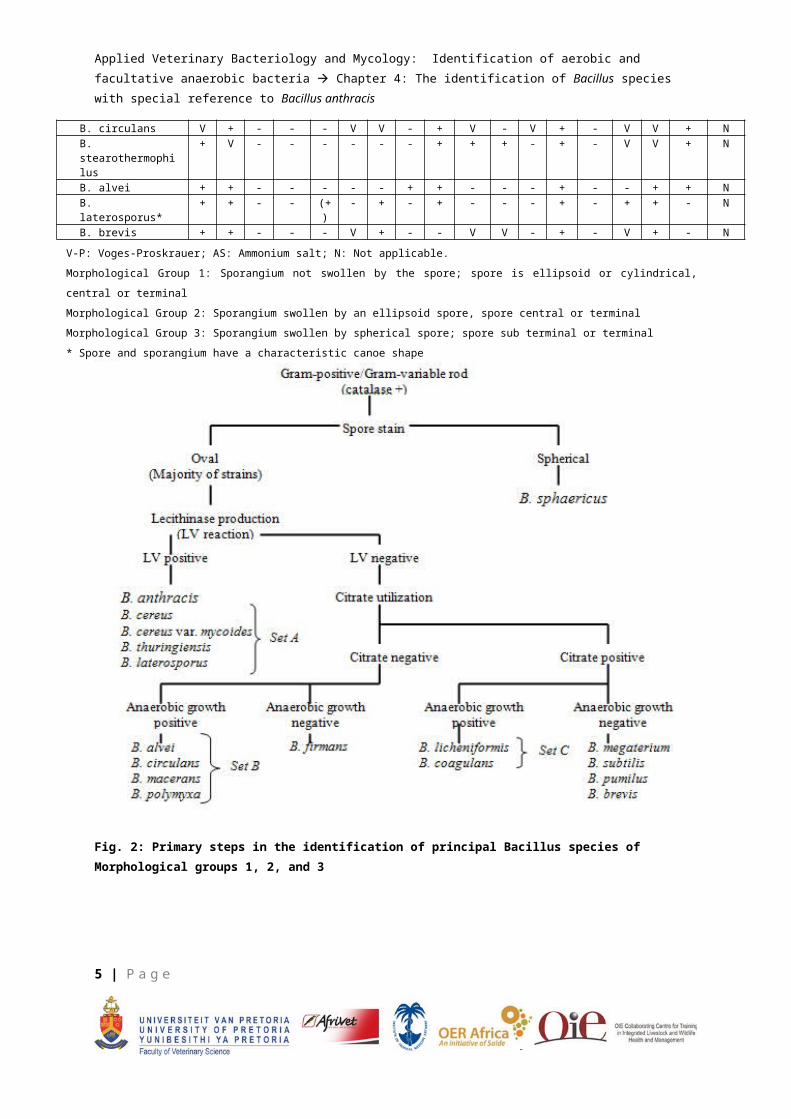
In the majority of cases the clinical microbiologist will be faced with the identification of the principal Bacillus species in Morphological Group 1. Figure 2 depicts a flow chart, giving suggested primary steps in the identification of those species. Table 4.1 lists secondary identification characteristics and the final biochemical identification tests are given in Table 4.2.
Bacillus anthracis characteristics
Actively growing (vegetative) B. anthracis organisms are typically rod-shaped, measure 1,0-1,5 by 3,0-10,0 m. In stained smears of blood or tissue fluid obtained from infected animals, the organisms appear truncated, commonly occur singly or in short chains, and are surrounded by a well-developed capsule, (Figures 2 and 3). Capsules are not formed in cultures unless special conditions for their development are provided. In unstained preparations, the organisms are robust, transparent rods and, in contrast to those in animal tissues, have rounded ends. In vitro B. anthracis grows in long, undulant chains composed of many cells which resemble the segments of a bamboo pole.
The spores, which are never found in the living animal, are ellipsoidal or oval, and are formed equatorially without causing a swelling of the sporangium. Spores develop under suitable, environmental conditions and are liberated by lysis of the bacilli. They germinate by polar rupture.
3 | P a g e
Applied Veterinary Bacteriology and Mycology: Identification of aerobic and facultative anaerobic bacteria
Chapter 4: The identification of Bacillus species with special reference to Bacillus anthracis
Sporulation in cultures on the surface of solid media, commences at about the end of logarithmic growth, is far advanced by 24 hours, and is usually complete by 48 hours. The shape, wall thickness and size of the spores relative to the sporangium, are important criteria in the taxonomy of the genus Bacillus and are of considerable assistance in distinguishing B. anthracis from other members of the genus.
Bacillus anthracis belongs to Morphological Group 1 (absence of sporangial swelling, and ellipsoidal spores). Other Bacillus spp., such as B. megaterium, B. cereus, B. cereus var. mycoides, B. thuringiensis, B. licheniformis, B. subtilis however, also possess these characteristics; consequently other methods must be used to differentiate them (Table 4.3. Figure 2)
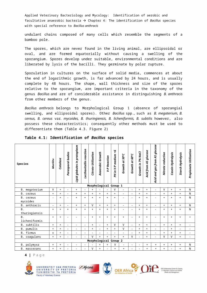
Table 4.1: Identification of Bacillus species
Species
Mot
ility
Cat
alas
e
Para
spor
al b
odie
s
Lipi
d gl
obul
es in
pro
topl
asm
Lect
hovi
telli
n re
actio
n
Citr
ate
Util
izat
ion
Ana
erob
ic G
row
th
V-P
Rea
ctio
n
PH in
V-P
med
ium
<6
Gro
wth
at 5
0°C
Gro
wth
at 6
0°C
Gro
wth
in 7
% N
aCl
Aci
d fr
om A
S gl
ucos
e
Aci
d an
d ga
s fr
om A
S gl
ucos
e
Nitr
ate
redu
ctio
n
Cas
ein
Hyd
roly
sis
Star
ch H
ydro
lysi
s
Prop
iona
te U
tiliz
atio
n
Morphological Group 1B. megaterium V + - + - + - - V - - + + - V + + NB. cereus + + - + + + + + + - - + + - + + + NB. cereus mycoides - + - + + + + + + - - + + - + + + NB. anthracis - + - + + V + + + - - + + - + + + NB. thuringiensis + + + + + + + + + - - + + - + + + NB. licheniformis + + - - - + + + + + - + + - + + + +B. subtilis + + - - - + - + V V - + + - + + + -B. pumulis + + - - - + - + + V - + + - - + - -B. firmus v + - - - - - - - - - + + - + + + -B. coagulans + + - - - V + + + + V - + - V V + -
Morphological Group 2B. polymyxa + + - - - - + + V - - - + + + + + NB. mascerans + + - - - V + - + + - - + + + - + NB. circulans V + - - - V V - + V - V + - V V + NB. stearothermophilus
+ V - - - - - - + + + - + - V V + N
B. alvei + + - - - - - + + - - - + - - + + NB. laterosporus* + + - - (+) - + - + - - - + - + + - NB. brevis + + - - - V + - - V V - + - V + - N
V-P: Voges-Proskrauer; AS: Ammonium salt; N: Not applicable.
Morphological Group 1: Sporangium not swollen by the spore; spore is ellipsoid or cylindrical, central or terminal
Morphological Group 2: Sporangium swollen by an ellipsoid spore, spore central or terminal
Morphological Group 3: Sporangium swollen by spherical spore; spore sub terminal or terminal
* Spore and sporangium have a characteristic canoe shape
4 | P a g e
Applied Veterinary Bacteriology and Mycology: Identification of aerobic and facultative anaerobic bacteria
Chapter 4: The identification of Bacillus species with special reference to Bacillus anthracis
Fig. 2: Primary steps in the identification of principal Bacillus species of Morphological groups 1, 2, and 3
5 | P a g e
Applied Veterinary Bacteriology and Mycology: Identification of aerobic and facultative anaerobic bacteria
Chapter 4: The identification of Bacillus species with special reference to Bacillus anthracis
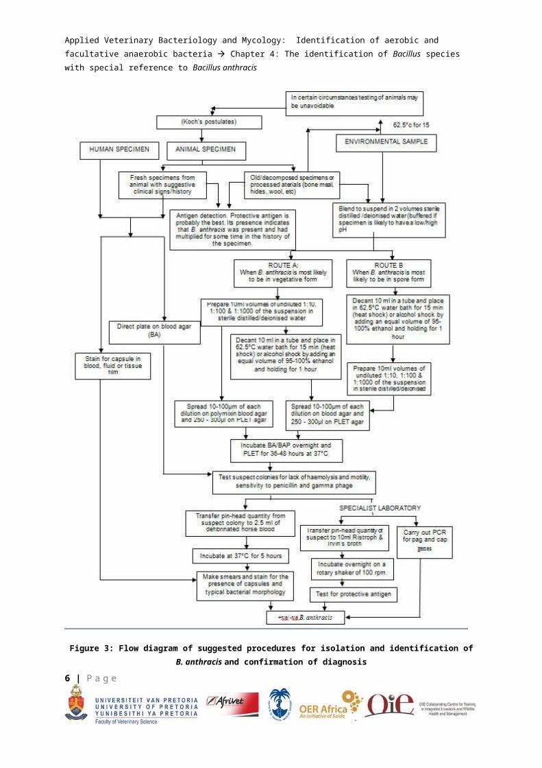
Figure 3: Flow diagram of suggested procedures for isolation and identification of B. anthracis and confirmation of diagnosis
6 | P a g e
Applied Veterinary Bacteriology and Mycology: Identification of aerobic and facultative anaerobic bacteria
Chapter 4: The identification of Bacillus species with special reference to Bacillus anthracis
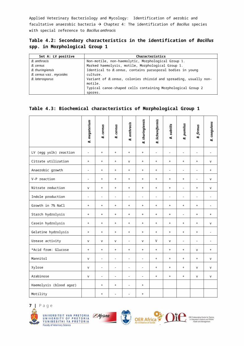
Table 4.2: Secondary characteristics in the identification of Bacillus spp. in Morphological Group 1
Set A: LV positive CharacteristicsB. anthracisB. cereusB. thuringiensisB. cereus var. mycoidesB. laterosporus
Non-motile, non-haemolytic, Morphological Group 1.Marked haemolysis, motile, Morphological Group 1.Identical to B. cereus, contains parasporal bodies in young culture.Variant of B. cereus, colonies rhizoid and spreading, usually non-motile.Typical canoe-shaped cells containing Morphological Group 2 spores.
Table 4.3: Biochemical characteristics of Morphological Group 1B
. meg
ater
ium
B. c
ereu
s
B. c
ereu
s
var .
myc
oide
s
B. a
nthr
acis
B. t
hurin
gien
sis
B. l
iche
nifo
rmis
B. s
ubtil
is
B. p
umilu
s
B. f
irmus
B. c
oagu
lans
LV (egg yolk) reaction - + + + + - - - - -
Citrate utilization + + + v + + + + + v
Anaerobic growth - + + + + + - - - +
V-P reaction - + + + + + + + - v
Nitrate reduction v + + + + + + - + v
Indole production - - - - - - - - - -
Growth in 7% NaCl + + + + + + + + + -
Starch hydrolysis + + + + + + + - + +
Casein hydrolysis + + + + + + + + + v
Gelatine hydrolysis + + + + + + + + + -
Urease activity v v v - v V v - - -
*Acid from: Glucose + + + + + + + + v +
Mannitol v - - - - + + + + v
Xylose v - - - - + + + v v
Arabinose v - - - - + + + v v
Haemolysis (blood agar) + + - +
Motility + - - +
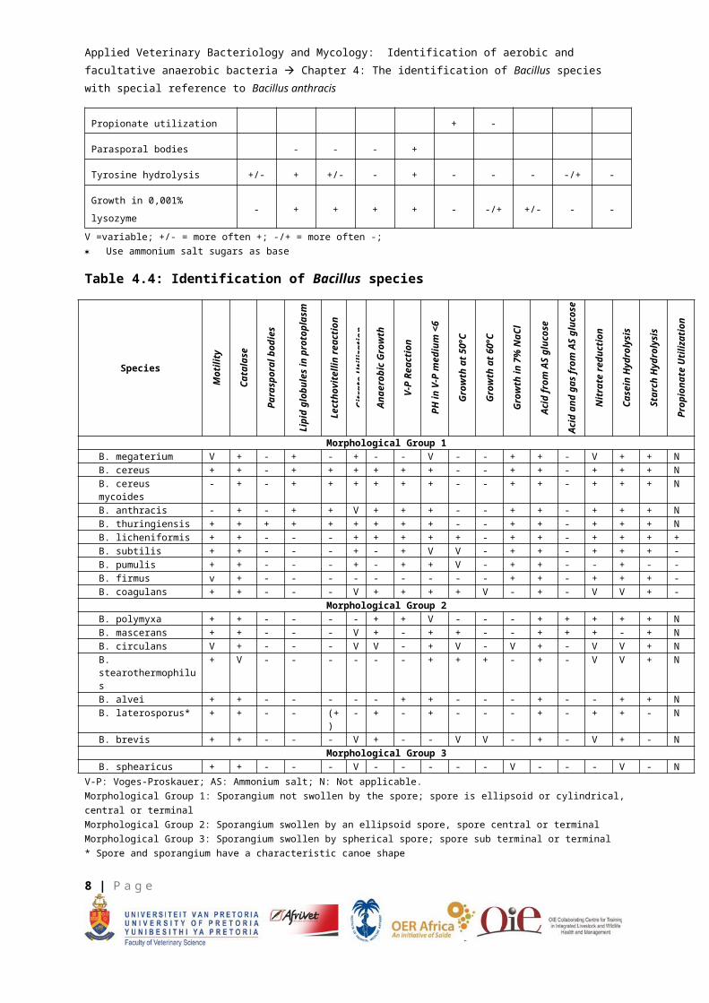
Propionate utilization + -
Parasporal bodies - - - +
Tyrosine hydrolysis +/- + +/- - + - - - -/+ -
7 | P a g e
Applied Veterinary Bacteriology and Mycology: Identification of aerobic and facultative anaerobic bacteria
Chapter 4: The identification of Bacillus species with special reference to Bacillus anthracis
Growth in 0,001% lysozyme - + + + + - -/+ +/- - -
V =variable; +/- = more often +; -/+ = more often -; Use ammonium salt sugars as base
Table 4.4: Identification of Bacillus species
Species
Mot
ility
Cat
alas
e
Para
spor
al b
odie
s
Lipi
d gl
obul
es in
pro
topl
asm
Lect
hovi
telli
n re
actio
n
Citr
ate
Util
izat
ion
Ana
erob
ic G
row
th
V-P
Rea
ctio
n
PH in
V-P
med
ium
<6
Gro
wth
at 5
0°C
Gro
wth
at 6
0°C
Gro
wth
in 7
% N
aCl
Aci
d fr
om A
S gl
ucos
e
Aci
d an
d ga
s fr
om A
S gl
ucos
e
Nitr
ate
redu
ctio
n
Cas
ein
Hyd
roly
sis
Star
ch H
ydro
lysi
s
Prop
iona
te U
tiliz
atio
n
Morphological Group 1B. megaterium V + - + - + - - V - - + + - V + + NB. cereus + + - + + + + + + - - + + - + + + NB. cereus mycoides - + - + + + + + + - - + + - + + + NB. anthracis - + - + + V + + + - - + + - + + + NB. thuringiensis + + + + + + + + + - - + + - + + + NB. licheniformis + + - - - + + + + + - + + - + + + +B. subtilis + + - - - + - + V V - + + - + + + -B. pumulis + + - - - + - + + V - + + - - + - -B. firmus v + - - - - - - - - - + + - + + + -B. coagulans + + - - - V + + + + V - + - V V + -
Morphological Group 2B. polymyxa + + - - - - + + V - - - + + + + + NB. mascerans + + - - - V + - + + - - + + + - + NB. circulans V + - - - V V - + V - V + - V V + NB. stearothermophilus + V - - - - - - + + + - + - V V + NB. alvei + + - - - - - + + - - - + - - + + NB. laterosporus* + + - - (+) - + - + - - - + - + + - NB. brevis + + - - - V + - - V V - + - V + - N
Morphological Group 3B. sphearicus + + - - - V - - - - - V - - - V - N
V-P: Voges-Proskauer; AS: Ammonium salt; N: Not applicable.Morphological Group 1: Sporangium not swollen by the spore; spore is ellipsoid or cylindrical, central or terminalMorphological Group 2: Sporangium swollen by an ellipsoid spore, spore central or terminalMorphological Group 3: Sporangium swollen by spherical spore; spore sub terminal or terminal* Spore and sporangium have a characteristic canoe shape
Bacillus anthracis is generally Gram-positive, but this attribute is often lost with age. Gram's staining of smears of the organism grown in culture should therefore be carried out as soon as possible, usually within 24 hours of the commencement of growth. The capsule, although also Gram-positive, is more easily decolourized than is the body of the young bacilli, with the result that individual Gram-positive bacilli may be enveloped by a Gram-negative capsule. Several Bacillus spp., including B. anthracis, B. subtilis, B. licheniformis and B. megaterium under appropriate growth conditions, produce carbohydrate capsules containing varying amounts of the polypeptide, poly-D-glutamic acid. The organisms are readily stained by the usual stains (Parry et al, 1983). In contrast to the other polysaccharide capsule-producing bacilli, the capsule of B. anthracis consists predominantly of poly-D-glutamate and only shows up well with Wright's and Giemsa stains, polychrome methylene blue (M'Fayden reaction stain), and 0,1% toluidine blue in a 1% aqueous solution of alcohol, or by the application of immunofluorescence techniques. Of the techniques described, the McFayden and
8 | P a g e
Applied Veterinary Bacteriology and Mycology: Identification of aerobic and facultative anaerobic bacteria
Chapter 4: The identification of Bacillus species with special reference to Bacillus anthracis
Giemsa staining methods are preferred by most laboratories. Demonstration of the capsule by staining blood smears helps with the confirmation of the diagnosis of anthrax. Other Bacillus spp., such as B. subtilis, B, licheniformis and B. megaterium also have capsules that contain polypeptides which impart similar staining characteristics. These species are however, unlikely to be encountered in specimens of blood or tissues from animals or humans with suspected anthrax. Although the spores of B. anthracis can be stained by the usual spore stains, Schaeffer and Fulton's malachite green technique is recommended (Parry et al, 1983). The use of phase contrast microscopy is also helpful in the examination of spores.
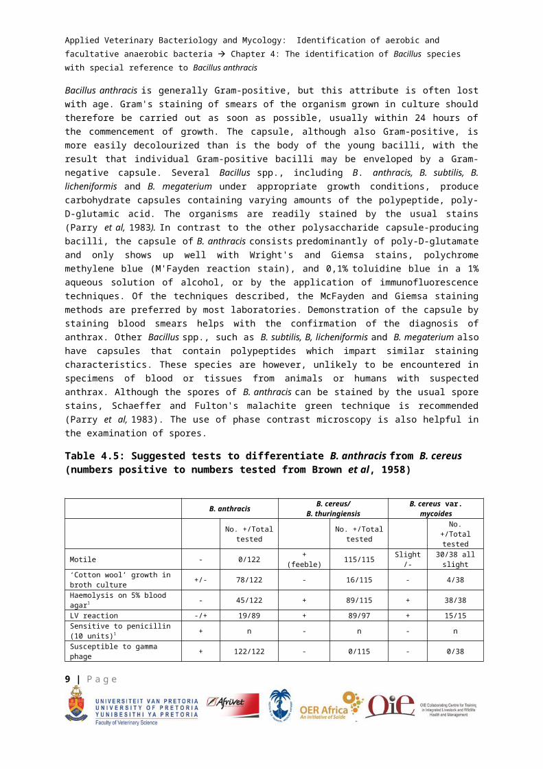
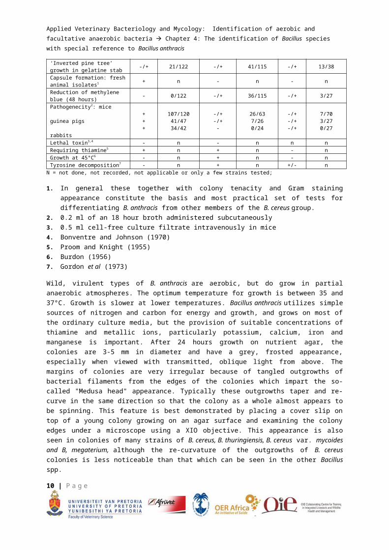
Table 4.5: Suggested tests to differentiate B. anthracis from B. cereus (numbers positive to numbers tested from Brown et al, 1958)
B. anthracis B. cereus/B. thuringiensis B. cereus var. mycoides
No. +/Total tested
No. +/Total tested
No. +/Total tested
Motile - 0/122 + (feeble) 115/115 Slight/- 30/38 all slight
‘Cotton wool’ growth in broth culture +/- 78/122 - 16/115 - 4/38
Haemolysis on 5% blood agar1 - 45/122 + 89/115 + 38/38LV reaction -/+ 19/89 + 89/97 + 15/15Sensitive to penicillin (10 units)1 + n - n - nSusceptible to gamma phage + 122/122 - 0/115 - 0/38‘Inverted pine tree’ growth in gelatine stab -/+ 21/122 -/+ 41/115 -/+ 13/38
Capsule formation: fresh animal isolates1 + n - n - n
Reduction of methylene blue (48 hours) - 0/122 -/+ 36/115 -/+ 3/27
Pathogenecity2: mice guinea pigs rabbits
+++
107/12041/4734/42
-/+-/+-
26/637/260/24
-/+-/+-/+
7/703/270/27
Lethal toxin3,4 - n - n n nRequiring thiamine5 + n + n - nGrowth at 45°C6 - n + n - nTyrosine decomposition7 - n + n +/- n
N = not done, not recorded, not applicable or only a few strains tested;
1. In general these together with colony tenacity and Gram staining appearance constitute the basis and most practical set of tests for differentiating B. anthracis from other members of the B. cereus group.
2. 0.2 ml of an 18 hour broth administered subcutaneously3. 0.5 ml cell-free culture filtrate intravenously in mice4. Bonventre and Johnson (1970)5. Proom and Knight (1955)6. Burdon (1956)7. Gordon et al (1973)
Wild, virulent types of B. anthracis are aerobic, but do grow in partial anaerobic atmospheres. The optimum temperature for growth is between 35 and 37°C. Growth is slower at lower temperatures. Bacillus anthracis utilizes simple sources of nitrogen and carbon for energy and growth, and grows on most of the ordinary culture media, but the provision of suitable concentrations of thiamine and
9 | P a g e
Applied Veterinary Bacteriology and Mycology: Identification of aerobic and facultative anaerobic bacteria
Chapter 4: The identification of Bacillus species with special reference to Bacillus anthracis
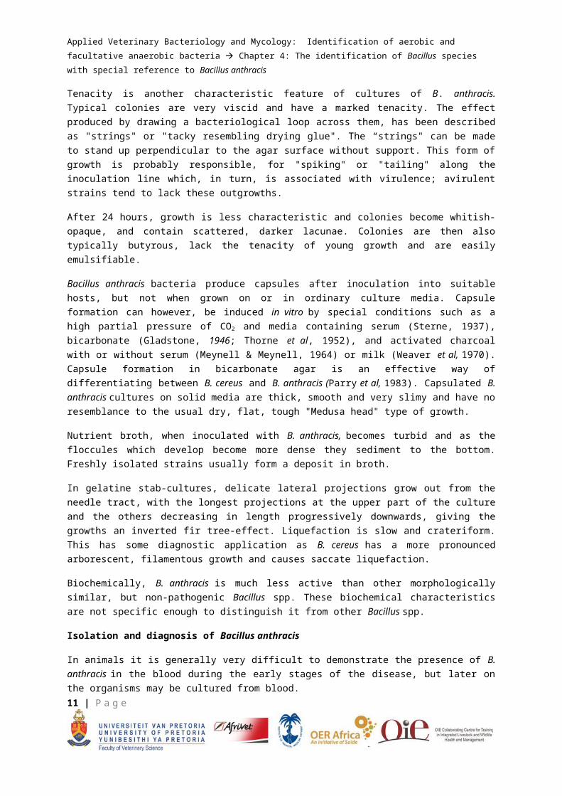
metallic ions, particularly potassium, calcium, iron and manganese is important. After 24 hours growth on nutrient agar, the colonies are 3-5 mm in diameter and have a grey, frosted appearance, especially when viewed with transmitted, oblique light from above. The margins of colonies are very irregular because of tangled outgrowths of bacterial filaments from the edges of the colonies which impart the so-called "Medusa head" appearance. Typically these outgrowths taper and re-curve in the same direction so that the colony as a whole almost appears to be spinning. This feature is best demonstrated by placing a cover slip on top of a young colony growing on an agar surface and examining the colony edges under a microscope using a XIO objective. This appearance is also seen in colonies of many strains of B. cereus, B. thuringiensis, B. cereus var. mycoides and B, megaterium, although the re-curvature of the outgrowths of B. cereus colonies is less noticeable than that which can be seen in the other Bacillus spp.
Tenacity is another characteristic feature of cultures of B. anthracis. Typical colonies are very viscid and have a marked tenacity. The effect produced by drawing a bacteriological loop across them, has been described as "strings" or "tacky resembling drying glue". The “strings" can be made to stand up perpendicular to the agar surface without support. This form of growth is probably responsible, for "spiking" or "tailing" along the inoculation line which, in turn, is associated with virulence; avirulent strains tend to lack these outgrowths.
After 24 hours, growth is less characteristic and colonies become whitish-opaque, and contain scattered, darker lacunae. Colonies are then also typically butyrous, lack the tenacity of young growth and are easily emulsifiable.
Bacillus anthracis bacteria produce capsules after inoculation into suitable hosts, but not when grown on or in ordinary culture media. Capsule formation can however, be induced in vitro by special conditions such as a high partial pressure of CO2 and media containing serum (Sterne, 1937), bicarbonate (Gladstone, 1946; Thorne et al, 1952), and activated charcoal with or without serum (Meynell & Meynell, 1964) or milk (Weaver et al, 1970). Capsule formation in bicarbonate agar is an effective way of differentiating between B. cereus and B. anthracis (Parry et al, 1983). Capsulated B. anthracis cultures on solid media are thick, smooth and very slimy and have no resemblance to the usual dry, flat, tough "Medusa head" type of growth.
Nutrient broth, when inoculated with B. anthracis, becomes turbid and as the floccules which develop become more dense they sediment to the bottom. Freshly isolated strains usually form a deposit in broth.
In gelatine stab-cultures, delicate lateral projections grow out from the needle tract, with the longest projections at the upper part of the culture and the others decreasing in length progressively downwards, giving the growths an inverted fir tree-effect. Liquefaction is slow and crateriform. This has some diagnostic application as B. cereus has a more pronounced arborescent, filamentous growth and causes saccate liquefaction.
Biochemically, B. anthracis is much less active than other morphologically similar, but non-pathogenic Bacillus spp. These biochemical characteristics are not specific enough to distinguish it from other Bacillus spp.
10 | P a g e
Applied Veterinary Bacteriology and Mycology: Identification of aerobic and facultative anaerobic bacteria
Chapter 4: The identification of Bacillus species with special reference to Bacillus anthracis
Isolation and diagnosis of Bacillus anthracis
In animals it is generally very difficult to demonstrate the presence of B. anthracis in the blood during the early stages of the disease, but later on the organisms may be cultured from blood.
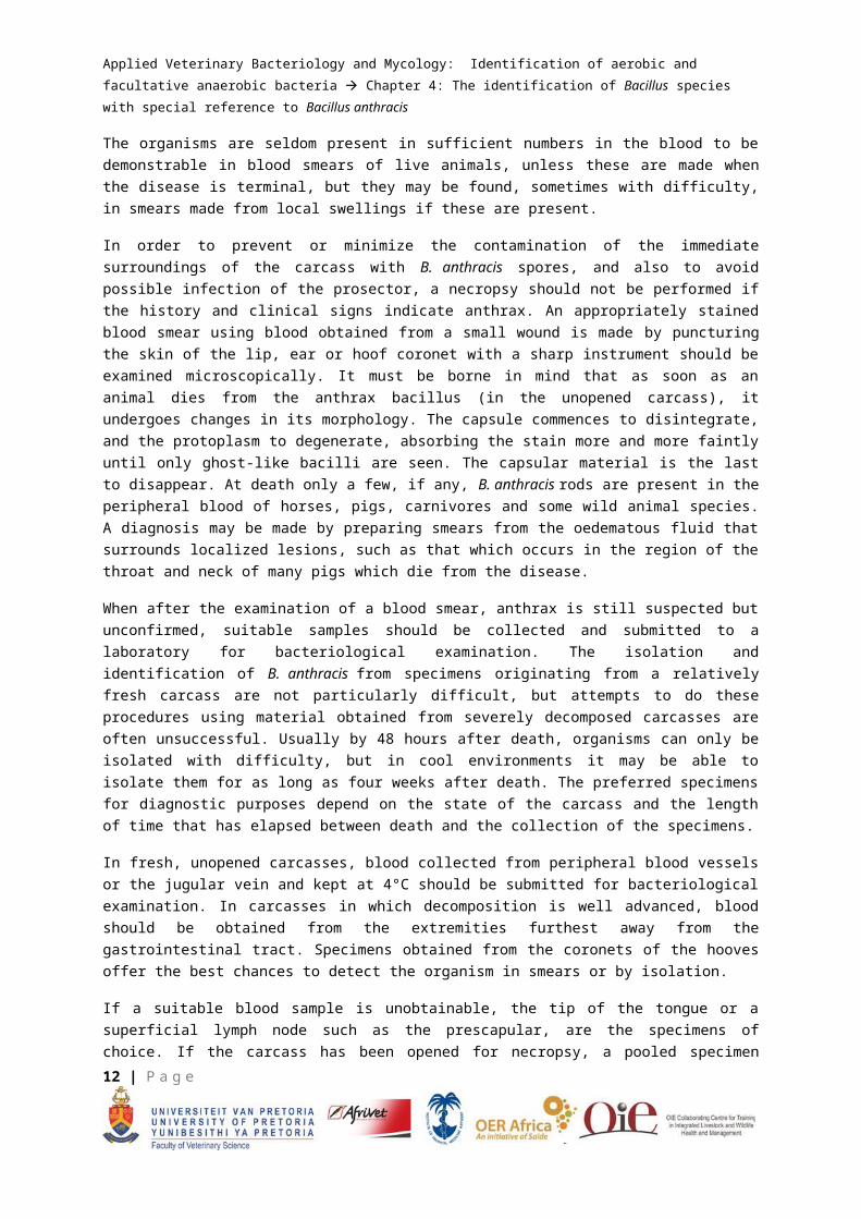
The organisms are seldom present in sufficient numbers in the blood to be demonstrable in blood smears of live animals, unless these are made when the disease is terminal, but they may be found, sometimes with difficulty, in smears made from local swellings if these are present.
In order to prevent or minimize the contamination of the immediate surroundings of the carcass with B. anthracis spores, and also to avoid possible infection of the prosector, a necropsy should not be performed if the history and clinical signs indicate anthrax. An appropriately stained blood smear using blood obtained from a small wound is made by puncturing the skin of the lip, ear or hoof coronet with a sharp instrument should be examined microscopically. It must be borne in mind that as soon as an animal dies from the anthrax bacillus (in the unopened carcass), it undergoes changes in its morphology. The capsule commences to disintegrate, and the protoplasm to degenerate, absorbing the stain more and more faintly until only ghost-like bacilli are seen. The capsular material is the last to disappear. At death only a few, if any, B. anthracis rods are present in the peripheral blood of horses, pigs, carnivores and some wild animal species. A diagnosis may be made by preparing smears from the oedematous fluid that surrounds localized lesions, such as that which occurs in the region of the throat and neck of many pigs which die from the disease.
When after the examination of a blood smear, anthrax is still suspected but unconfirmed, suitable samples should be collected and submitted to a laboratory for bacteriological examination. The isolation and identification of B. anthracis from specimens originating from a relatively fresh carcass are not particularly difficult, but attempts to do these procedures using material obtained from severely decomposed carcasses are often unsuccessful. Usually by 48 hours after death, organisms can only be isolated with difficulty, but in cool environments it may be able to isolate them for as long as four weeks after death. The preferred specimens for diagnostic purposes depend on the state of the carcass and the length of time that has elapsed between death and the collection of the specimens.
In fresh, unopened carcasses, blood collected from peripheral blood vessels or the jugular vein and kept at 4°C should be submitted for bacteriological examination. In carcasses in which decomposition is well advanced, blood should be obtained from the extremities furthest away from the gastrointestinal tract. Specimens obtained from the coronets of the hooves offer the best chances to detect the organism in smears or by isolation.
If a suitable blood sample is unobtainable, the tip of the tongue or a superficial lymph node such as the prescapular, are the specimens of choice. If the carcass has been opened for necropsy, a pooled specimen taken from the spleen and several lymph nodes is preferred. When carcasses are dehydrated and putrefaction is advanced, samples should be collected from areas which might have been contaminated with blood at an earlier stage where sporulation of B. anthracis could have taken place, such as at natural body openings or parts of the body mutilated by scavengers. Even when bones are all that remains of a carcass, bacteria may be more readily isolated from specimens of the
11 | P a g e
Applied Veterinary Bacteriology and Mycology: Identification of aerobic and facultative anaerobic bacteria
Chapter 4: The identification of Bacillus species with special reference to Bacillus anthracis
bones forming the eye sockets, mandible and ischium than from those taken randomly. Soil from below the carcass may also be submitted for culturing,
Bacillus anthracis grows readily on artificial culture, and when isolation is attempted from uncontaminated fresh specimens, nutrient agar can be used for this purpose, but best results are, in general, obtained on media containing serum or blood. Severely contaminated material, such as samples of soil or bone-meal, should be cultured on selective media of which there are several available. PLFT medium is suitable for the purpose of isolating B. anthracis spores in soil, even when they are present in concentrations as low as 3 spores per gram. Seeded plates should be incubated for 12 to 24 hours at 37°C and thereafter examined under a stereo microscope with transmitted light from the side and above. Colonies suspected of being B. anthracis on the grounds of colonial morphology and tenacity, are lifted with a bacteriological needle and inoculated on a 5% blood agar plate. If no haemolysis occurs within 24 hours at 37°C and the organisms conform to Morphological Group 1 bacilli, further tests are required to differentiate B. anthracis from the other members of this group.
Additional tests in laboratory animals are considered essential if B. anthracis is to be conclusively identified. In this respect, no universal standardized procedures have been formulated. It is generally recommended that 0,2 ml of a broth suspension should be inoculated subcutaneously into mice or guinea pigs, intramuscularly into guinea pigs, or intraperitoneally into mice. The intraperitoneal injection in mice of 1 to 5 x 10⁵ B. anthracis spores is favoured.
An API Bacillus system (API Laboratory Products, Ltd) has been developed to aid in the identification of Bacillus spp. and to facilitate in the identification of both typical and atypical strains of B. anthracis. Separation of slightly virulent and avirulent strains of B. anthracis from closely related Bacillus spp. is based on API and phage-sensitivity tests.
Lysis by bacteriophage (gamma) is a highly specific differential test for B. anthracis and is popular as a diagnostic aid in laboratories dealing with anthrax.
When B. anthracis is grown in the presence of low concentrations of penicillin, the bacilli swell and filaments of them appear as chains of spores or of round cellular forms referred to as "strings of pearls". This phenomenon, which was first described by Jensen and Kleemeyer, is specific for B. anthracis, although exceptions do occur as some strains of B. anthracis do not grow under these circumstances. Because of the specificity and relative ease at which it can be performed, this is a useful diagnostic test for the identification of B. anthracis.
Generally, serological and immunofluorescent diagnostic methods are unreliable for the diagnosis of anthrax. Direct and indirect immunofluorescence assays, immunoradiometric assay and the enzyme-linked immunosorbent assay may be developed in future for determining the serological relatedness of B. anthracis and other Bacillus spp. An enzyme immunoassay and the production of monoclonal antibodies against the protective antigen of B. anthracis have also been considered for diagnostic purposes, and specialist laboratories use the PCR technique with great success.
For practical purposes a battery of tests may be required to confirm a diagnosis of anthrax.
12 | P a g e
Applied Veterinary Bacteriology and Mycology: Identification of aerobic and facultative anaerobic bacteria
Chapter 4: The identification of Bacillus species with special reference to Bacillus anthracis
Specialized media, reagents and procedures
Only specialized media and reagents needed for isolation, identification, diagnosis and confirmation of anthrax are provided.
Staining
1. Giemsa
Mature, at least two weeks on shelf.
Dilute the stain 1:10 and insert the slide for 30 minutes to 1 hour. Wash with distilled water, dry and examine. This stain is the best stain to be used when blood films are made from carcasses that have been for dead a while.
2. Polychrome methylene blue (McFayden stain reaction)
Prepare a saturated solution of methylene blue in 95% ethanol by mixing ± 0.5g of the dye in 50 ml of the alcohol. Add 30ml of this to 100ml of a 0.01% KOH solution in distilled water. Add K2CO3 to a final concentration of I%.
Allow to stand in half-filled bottles stoppered with cotton-wool plugs. The bottles should be shaken periodically for fuller aeration. Oxidation ("ripening") takes several months.
Make thin smears and air dry. Fix by dipping in absolute or 95% methanol or ethanol for 30-60 seconds and re-dry. Put a large drop of polychrome rnethylene blue on the smear and spread to cover all parts of the smear. Leave for 30-60 seconds. Wash with water, blot and dry. Under oil immersion (100X) the capsule is seen clearly (pink) surrounding the blue-black, often square-ended bacilli. Although the McFayden stain reaction gives the best results in fresh cases, it was found that during putrefaction the capsule loses its affinity for methyIene blue
3. CAM's Quick stain
It can be used with great success, but mainly on fresh cases.
"PLET" Selective medium
PLET (Knisely, 1966) is the best selective agar currently available for isolation of B. anthracis from animal or environmental specimens contaminated with other organisms, including other Bacillus species.
"Difco" heart infusion agar (or Difco heart infusion broth with agar base) is made up according to the manufacturer's instructions. EDTA (0.3g/1) and thallium acetate (0.04g/1) are added before autoclaving. After autoclaving, the agar is cooled to 50°C and polymyxin (30 000 units/1) and lysozyme (300 000 units/1) are added. After swirling the, agar is poured into Petri dishes.
13 | P a g e
Applied Veterinary Bacteriology and Mycology: Identification of aerobic and facultative anaerobic bacteria
Chapter 4: The identification of Bacillus species with special reference to Bacillus anthracis
Polymixin blood agar
This is useful for testing an unheated suspension of old, decomposed or processed animal or environmental specimens and reduces or prevents growth of many Gram-negative species.
Polymixin.B sulphate should be added to a level of 100 000 units/litre of medium to the cooled blood agar base at the same time as adding the blood.
Blood culture
Capsule formation can be demonstrated by transferring a pin-head quantity of growth from a suspect colony to approximately 2,5ml defibrinated blood in a sterile test tube or small bottle and incubating 5 hours to overnight. A smear is made from the blood, stained and examined microscopically. Defibrinated horse blood seems to be the best.
Laboratory animal tests
In view of concerns about animal welfare, and of the increasing reliability and sophistication of alternative in vitro methods, the use of animals for isolation or confirmation of identity of B. anthracis can and should generally be avoided. There are however, still occasions where it is used, such as animals that were treated before the specimen was taken and where environmental samples contain sporostatic substances.
Confirmation of identity or of virulence can be done by injecting light suspensions (approximately
10 000 colony-forming units/ml) into mice (0.05-0,1 ml subcutaneously) or guinea pigs (0,1-0.2 ml intramuscularly). Virulent B. anthracis will kill the animals in about 42 - 48 hours. Blood smear examination should reveal large numbers of capsulated bacilli.
If soil or environmental samples are used the animals should be inoculated the day before with subcutaneous doses of mixed gas gangrene antisera and anti-tetanus sera. If unavailable, the material to be exacmined should first be heated at 62.5°C for 15 min.
"String of Pearls’ test
Prepare a solution of sodium benzyl penicillin in sterile distilled water to contain 50 units/ml. Add 1 ml of this to 100 ml of melted blood agar base and pour 25 ml into Petri dishes. Allow to set. Using a scalpel blade, cut a block approximately 1.5 cm square from the penicillin agar plate and place it on a microscope slide. Put the slide in a Petri dish together with a small piece of moistened cotton wool. Make a line with a suitable marker along the length of a clean cover slip and about 5 mm from one edge. Touch a loop to the edge of a young vigorous growing colony of the suspect culture and streak it along the centre of the agar block on the slide. Place the cover slip so that the line (see above) is face down along the streak of the culture. The line then acts as focusing and location guides. Put the lid on the Petri dish and place in a 37°C incubator. After 2 hours, place the slide on a microscope stage. Focus on the line with the X10 objective and bring the high dry or oil immersion lens into use to look for the ‘string of pearls’
14 | P a g e
Applied Veterinary Bacteriology and Mycology: Identification of aerobic and facultative anaerobic bacteria
Chapter 4: The identification of Bacillus species with special reference to Bacillus anthracis
Gamma phage
Inoculate a blood agar plate or other suitable medium evenly over its entire surface with a loopful of the test culture. If necessary, allow the plate to dry for a few minutes. Place a loopful or small drop of phage suspension in the centre of the plate and incubate at 37°C. The phage inhibition should be readily apparent at 6-8 hours of incubation, although it can be read overnight.
Motility
Various tests for motility are available. The two most reliable methods are the hanging drop method and growth in semi-solid agar (in a Craigie tube).
15 | P a g e
Applied Veterinary Bacteriology and Mycology: Identification of aerobic and facultative anaerobic bacteria
Chapter 4: The identification of Bacillus species with special reference to Bacillus anthracis
APPENDIX 1Propagation and concentration of the gamma phage
Please note that a few isolates will be gamma-phage negative and a few B. cereus isolates with be gamma-phage positive. Thus it should be used in a panel of tests.
i) Spread a blood agar plate with the Sterne’s vaccine strain of B. anthracis.ii) Inoculate 10ml of nutrient broth with growth from the blood agar plate and incubate for 4 hours
(until just cloudy), then refrigerate.iii) Spread 100µl of the culture from ii) and spread onto three blood agar plates and incubate at 37°C
for 30 to 60 minutes.iv) Spread 100µl of the gamma phage suspension over the plates and incubate overnight at 37°C.v) Harvest the phage-lyzed growth on the blood agar plate into 5 ml nutrient broth followed by a
second wash in 5 ml nutrient broth. Incubate overnight at 37°C.vi) Filter using a 0.45µm filter and retain the filtrate.vii) Repeat steps iii) to, vi) once more to increase the concentration of gamma phage.viii) Inoculate 100 ml of brain heart infusion broth with 2.5 ml of the culture from ii) and incubate on a
rotary shaker at 37°C until just turbid.ix) Add 20 ml of the filtrate from vii) and incubate overnight at 37°C.x) Filter. The resultant filtrate should be checked for sterility and titrated in ten-fold dilutions to
determine the concentration of the phage. Running the test in triplicate, 20µl of diluted filtrate is placed on lawns of the B. anthracis culture. For the best results 108 – 109 plaque-forming units per ml should be obtained.
16 | P a g e