Embed Size (px)
Citation preview
SYSTEMIC PATHOLOGY
Pathology of Muscle
Lecture 2
Paul Hanna Winter 2018

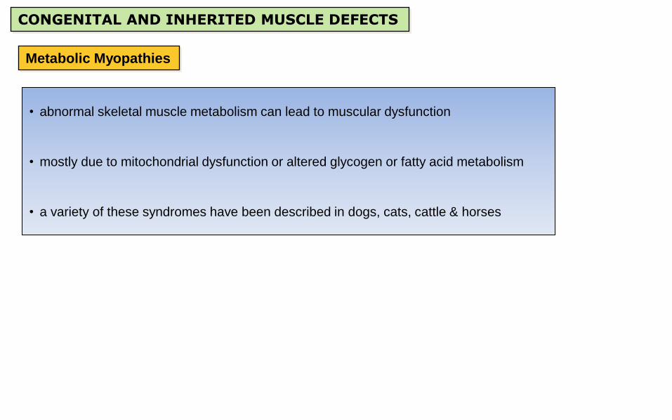
CONGENITAL AND INHERITED MUSCLE DEFECTS
• there are a wide variety of congenital and/or hereditary muscle conditions in animals
Jubb, Kennedy & Palmer's Pathology of Domestic Animals, 6th Edition. [For Information Only]

CONGENITAL AND INHERITED MUSCLE DEFECTS
Arthrogryposis (= congenital articular rigidity)
• relatively common in aborted fetuses / stillborns
• can be caused by myogenic or neurogenic disorders
• often due to lack of innervation during gestation causing muscular hypoplasia
• neurologic defects are usually related to failure of neural tube closure (dysraphism), +/- spina bifida.
• also seen with other cord defects (eg syringomyelia, hydromyelia)
• CNS defects can be genetic OR associated with in utero exposure to toxins (eg wild lupine) or in utero
viral infections (eg bluetongue and border disease)

Arthrogryposis
The musculature of the limbs is poorly developed; with all muscles shortened many joints are fixed / rigid and the limbs in various combinations of flexion and
extension. Also note the yellow color of the wool which is due to meconium staining and is consistent with fetal anoxia due to dystocia (ie the fixed limbs often
lead to difficult / prolonged deliveries.

Fig 3-38 Newborn calf with arthrogryposis. Jubb, Kennedy & Palmer's Pathology of Domestic Animals, 6th ed.
Animals affected with arthrogryposis often have spinal
cord defects (myelodysplasia)
Fig a - note the cleft (dysraphism / myeloschisis) in the spinal
cord (arrows)
Fig b - note the abnormal cavitations (syringomyelia) in the
cord (arrowheads)
Arthrogryposis
CONGENITAL AND INHERITED MUSCLE DEFECTS
Metabolic Myopathies
• abnormal skeletal muscle metabolism can lead to muscular dysfunction
• mostly due to mitochondrial dysfunction or altered glycogen or fatty acid metabolism
• a variety of these syndromes have been described in dogs, cats, cattle & horses

Equine Polysaccharide Storage Myopathy (PSSM)
• a glycogen storage myopathy seen in many horse breeds
• AD with variable expression; defects in CHO metabolism (eg GYS1)
• asymptomatic to progressive weakness / lameness / muscle atrophy to exertional rhabdomyolysis
• histo: accumulation of polysaccharides and degeneration / necrosis in type 2 fibers
Fig 3-66 (Maxie) A. Normal glycogen staining pattern of horse muscle. B. Multiple
myofibers with peripheral aggregates of densely stained glycogen in an Arabian horse
with polysaccharide storage myopathy.
Fig 15-35 (Zachary) Equine PSSM. C. Severe form, showing
PAS positive inclusions which are not digested by amylase
and are characteristic of complex polysaccharide
CONGENITAL AND INHERITED MUSCLE DEFECTS
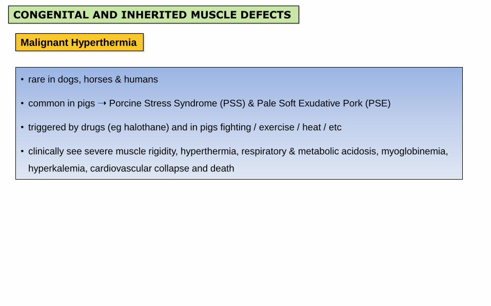
Malignant Hyperthermia
• rare in dogs, horses & humans
• common in pigs Porcine Stress Syndrome (PSS) & Pale Soft Exudative Pork (PSE)
• triggered by drugs (eg halothane) and in pigs fighting / exercise / heat / etc
• clinically see severe muscle rigidity, hyperthermia, respiratory & metabolic acidosis, myoglobinemia,
hyperkalemia, cardiovascular collapse and death
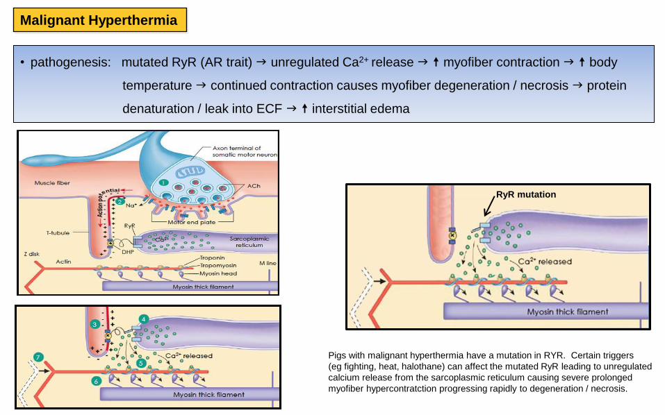
Malignant Hyperthermia
• pathogenesis: mutated RyR (AR trait) unregulated Ca2+ release myofiber contraction body
temperature continued contraction causes myofiber degeneration / necrosis protein
denaturation / leak into ECF interstitial edema
Pigs with malignant hyperthermia have a mutation in RYR. Certain triggers
(eg fighting, heat, halothane) can affect the mutated RyR leading to unregulated
calcium release from the sarcoplasmic reticulum causing severe prolonged
myofiber hypercontratction progressing rapidly to degeneration / necrosis.
RyR mutation

Malignant Hyperthermia
Gross pathology:
• muscles with higher proportions of type 2 fibers (back / hindlimbs) are pale and wet
• also severe pulmonary edema and hydropericardium / hydrothroax / ascites
Markedly pale muscles with abundant edema in fascia and
exuding from cut surfaces
Marked pallor of the epaxial (eg longissimus) muscles

http://www.fao.org/DOCREP/003/X6909E/x6909e04.htm http://www.animalagriculture.org/Proceedings/2003%20Proc/Moeller.htm
Pale soft exudative (PSE) pork
normal pork chop
Varying degrees of pallor in cuts of pork; also have soft (putty-like) texture and accumulation of fluid in packing tray (lower right)

Malignant Hyperthermia
• Histopathology: acute myofiber degeneration / necrosis & interstitial edema
Fragmented fibers (asterisk) and retraction cap/cup (arrow) indicative of acute myofiber degeneration / necrosis
*

CONGENITAL AND INHERITED MUSCLE DEFECTS
Splayleg in piglets
www.youtube.com/watch?v=H4lEEYf2q14
Splay Leg Tape
• self-adhesive, anti-splay tape to counteract splay leg
www.kerbl.com
CONGENITAL AND INHERITED MUSCLE DEFECTS
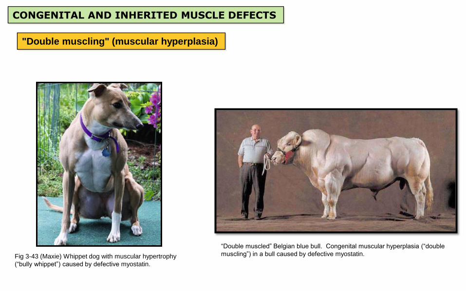
"Double muscling" (muscular hyperplasia)
Fig 3-43 (Maxie) Whippet dog with muscular hypertrophy
(“bully whippet”) caused by defective myostatin.
“Double muscled” Belgian blue bull. Congenital muscular hyperplasia (“double
muscling”) in a bull caused by defective myostatin.

CONGENITAL AND INHERITED MUSCLE DEFECTS
Muscular steatosis
Bovine, skeletal muscle with prominent fatty infiltration
(also called “steatosis”). Usually subclinical and
typically a problem only in meat inspection.
Bovine, skeletal muscle, muscular steatosis (fatty infiltration). Replacement
of muscle tissue with mature adipose tissue.

CONGENITAL AND INHERITED MUSCLE DEFECTS
Muscular Dystrophy
Fig 27-10 (Robbin’s) Relationship between the cell membrane
(sarcolemma) and the sarcolemmal associated proteins. Dystrophin, an
intracellular protein, forms an interface between the cytoskeletal proteins
and a group of transmembrane proteins, the dystroglycans and the
sarcoglycans. These transmembrane proteins have interactions with the
extracellular matrix, including the laminin proteins. Dystrophin also
interacts with dystrobrevin and the syntrophins, which form a link with
neuronal type nitric oxide synthetase (nNOS) and caveolin. Mutations in
dystrophin are associated with the X-linked muscular dystrophies;
mutations in caveolin and the sarcoglycan proteins with the limb-girdle
muscular dystrophies, which can be autosomal dominant or recessive
disorders; and mutations in the α2-laminin (merosin) with autosomal
recessive congenital muscular dystrophy.
[For Information Only]

CONGENITAL AND INHERITED MUSCLE DEFECTS
Myotonic and Spastic syndromes
The normal action potential in the muscle membrane (illustrated in the above diagram) primarily involves sodium and potassium fluctuations. So defects in the
sodium channel can alter the resting muscle membrane potential which can lead to increased muscle action potentials (with muscle fasciculations and spasms)
&/or complete depolarization (hypotonia / collapse). Chloride channel mutations can also affect membrane excitability (eg exaggerated response to stimulation).
[For Information Only]
• myotonia is defined clinically as a temporary inability of skeletal muscle to relax after voluntary contraction
• most myotonias result from muscle membrane electrical abnormalities, esp ion channel defects for
regulation of sodium or chloride (also called “channelopathies)
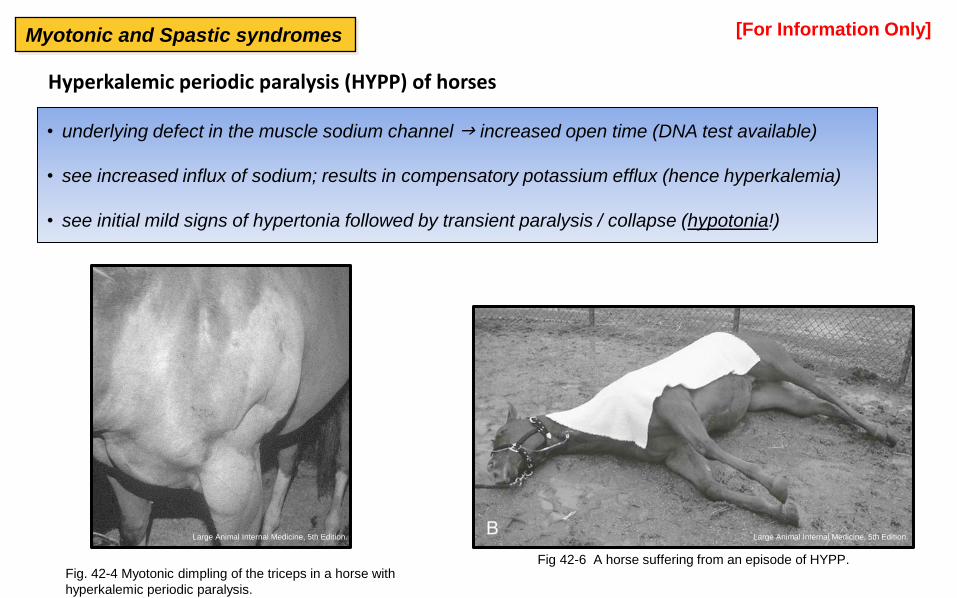
Hyperkalemic periodic paralysis (HYPP) of horses
• underlying defect in the muscle sodium channel increased open time (DNA test available)
• see increased influx of sodium; results in compensatory potassium efflux (hence hyperkalemia)
• see initial mild signs of hypertonia followed by transient paralysis / collapse (hypotonia!)
Fig 42-6 A horse suffering from an episode of HYPP. Fig. 42-4 Myotonic dimpling of the triceps in a horse with
hyperkalemic periodic paralysis.
[For Information Only]
Large Animal Internal Medicine, 5th Edition. Large Animal Internal Medicine, 5th Edition.
Myotonic and Spastic syndromes

• spastic syndromes are also associated with tightening of the muscles, causing stiff and awkward movement
Fig 12-42 A Holstein calf with spastic paresis. Notice that
although the calf is standing still she keeps the left pelvic limb
extended caudally. Rebhun's Diseases of Dairy Cattle, 2nd Ed
Scottie Cramp is an AR trait. Following excitement or exercise
see a progressive increase in muscle tone causing lumbar
kyphosis and decreased flexion of the pelvic limbs; +/- falling
over. A deficiency of serotonin activity in the spinal cord gray
matter has been implicated as the underlying cause
[For Information Only] Myotonic and Spastic syndromes
CONGENITAL AND INHERITED MUSCLE DEFECTS
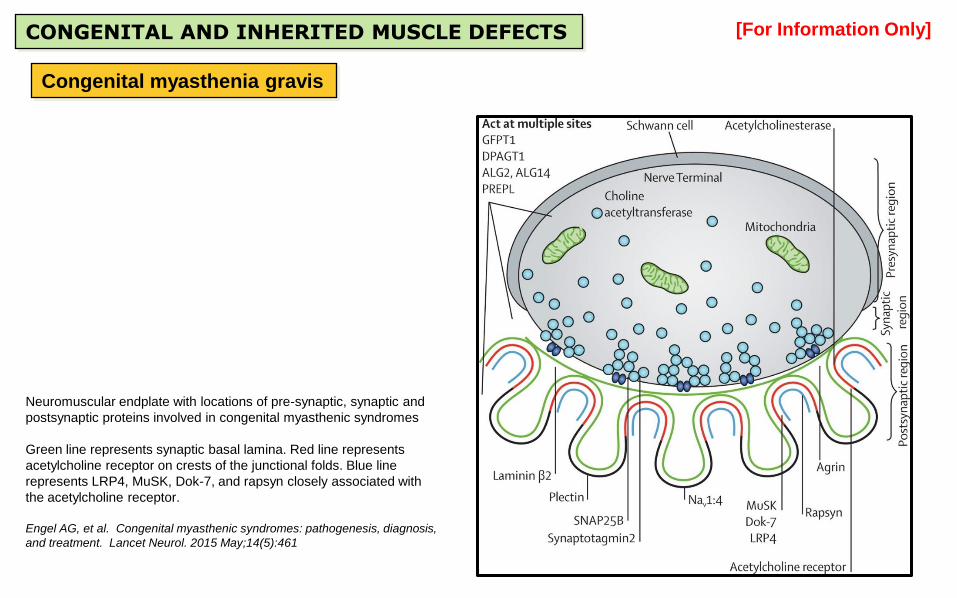
Congenital myasthenia gravis
Neuromuscular endplate with locations of pre-synaptic, synaptic and
postsynaptic proteins involved in congenital myasthenic syndromes
Green line represents synaptic basal lamina. Red line represents
acetylcholine receptor on crests of the junctional folds. Blue line
represents LRP4, MuSK, Dok-7, and rapsyn closely associated with
the acetylcholine receptor.
Engel AG, et al. Congenital myasthenic syndromes: pathogenesis, diagnosis,
and treatment. Lancet Neurol. 2015 May;14(5):461
[For Information Only]

TRAUMATIC / CIRCULATORY DISTURBANCES OF MUSCLE
Downer syndrome
• in large animals following prolonged recumbency (disease / anesthesia) the weight of the body
compresses the muscles inadequate vascular perfusion pressure (+/- thrombosis)
• ~ 6-12 hours get ischemic necrosis of pectoral or limb muscles that are tucked under the body
• muscle necrosis from Downer syndrome then becomes a reason the animal can’t rise

Downer syndrome
Serial transverse sections of a leg from a cow with a history of being
down for several weeks. Dark areas of necrosis and hemorrhage
(asterisks) involving several muscle groups. Necrosis is due to physical
trauma, compressive ischemia and thrombosis (see next slide for close-up).
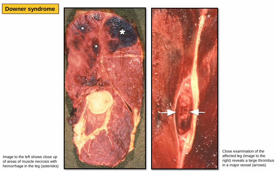
Downer syndrome
Close examination of the
affected leg (image to the
right) reveals a large thrombus
in a major vessel (arrows)
Image to the left shows close up
of areas of muscle necrosis with
hemorrhage in the leg (asterisks)

Postanesthetic myopathy in horses
Fig 11-11 A horse with postanesthetic myopathy of the right
gluteal muscles. The severe swelling is notable. The horse
recovered fully with supportive care. Equine Internal Medicine, 3rd ed
• seen in 3-6% horses undergoing prolonged recumbency
• pathogenesis: compressive pressure due to large weight of animal during recumbence &/or muscle hypoxia
from systemic hypotension associated with the general anesthetic
• ranges from muscle swelling with lameness to paresis with renal failure / shock

Fasciotomy of the lower limb in a
human to treat compartment syndrome.
Compartment syndrome
• characterized by degeneration / necrosis in muscles surrounded by heavy aponeurosis
• pathogenesis: muscular swelling / expansion associated with injury in a non-expandable compartment
results in vascular compression ischemia and infarction of the muscle

Compartment syndrome
Deep pectoral myopathy
• frequently seen in poultry, esp heavily muscled breeds of turkeys & chickens
• disease is preceded by a brief but vigorous period of wing flapping few hrs later localized area of
degeneration and necrosis in the deep breast (supracoracoid) muscle
Greenish color of the supracoracoid pectoral muscle due to degeneration and necrosis (arrow) . Photo to right is close up.

Ischemic necrosis supracoracoid muscle (cooked specimen): Dark appearance of necrotic muscle found in this cooked chicken (asterix). Lesions appear
a few hours after exercise and because they are deeply “buried in the pectoral muscles they are sometimes not grossly detected until "supper time.”
Compartment syndrome
Deep pectoral myopathy

NUTRITIONAL MYOPATHY (WHITE MUSCLE DISEASE)
• in older vet literature was mistakenly called nutritional muscular dystrophy
• associated with dietary deficiency of Sel &/or Vit E
• other factors exercise, environment, other nutrition factors / toxicants
• relatively common; esp young, rapidly growing, well-conditioned sheep, cattle, pigs
• in pigs it may be independent or coexist with other Sel / Vit E deficiency diseases
• less common in foals & goats & other species.

NUTRITIONAL MYOPATHY (WMD)
• pathogenesis: inadequate amounts of radical-scavenging Sel / Vit E lipid peroxidation of membranes
influx of Ca2+ into sarcoplasm hypercontraction & activation of proteases myofiber
degeneration / necrosis and often calcification
• with cell membrane damage intracellular enzymes (eg CK) leak into ECF / serum
• muscles with many type 1 fibers more severely affected (diaphragm, intercostals, tongue)

Fig. 15-40 (Zachary) Nutritional myopathy (white muscle disease), skeletal muscles of the caudal thigh, sagittal section, calf. In this early stage, affected muscles have yellow and white
streaks, often in a patchy distribution. These streaks are areas of necrotic myofibers. Later as the necrotic myofibers calcify, white streaks (chalky texture, mineralization) are visible grossly.
Gross Pathology:
• muscle pallor; can be subtle in mild cases or inapparent in peracute cases
• in severe cases, muscles are pale with chalky white areas or streaks
NUTRITIONAL MYOPATHY (WMD)
Close-up of affected skeletal muscle in a calf show prominent pale streaking
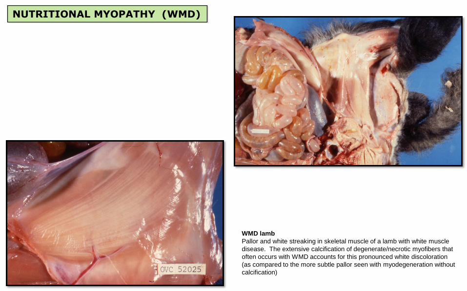
WMD lamb
Pallor and white streaking in skeletal muscle of a lamb with white muscle
disease. The extensive calcification of degenerate/necrotic myofibers that
often occurs with WMD accounts for this pronounced white discoloration
(as compared to the more subtle pallor seen with myodegeneration without
calcification)
NUTRITIONAL MYOPATHY (WMD)

WMD Foal. Extremely pale muscle (W) from a few days old foal that
died of WMD. Note the normal color of equine muscle (N).
WMD Pig
The musculature of this pig with WMD was slightly pale. However, because
normal porcine muscle is often pale, histopathology was required to confirm the
muscle degeneration / necrosis in this case.
Pigs can develop other diseases with Vit E / Sel deficiency (eg mulberry heart
disease & hepatosis dietetica) which can occur independently or together.
NUTRITIONAL MYOPATHY (WMD)

Remember to inspect the heart. Calf heart (right) showing patchy areas of chalky white
discoloration in the left ventricular myocardium.
NUTRITIONAL MYOPATHY (WMD)

Fragmented myofibers with swelling, hyalinization and loss of striations in some areas
(asterisks) and extensive blue-purple granularity, representing calcification (arrows), in
other areas. H&E
*
*
Special staining is often used to confirm calcification. In this figure the
calcified myofibers stain dark with Von Kossa stain.
Histopathology:
NUTRITIONAL MYOPATHY (WMD)