Embed Size (px)
Citation preview
www.scireproject.com Version 5.0
Syringomyelia Following Spinal Cord Injury
Robert Teasell MD FRCPC
Amanda McIntyre MSc Spencer Thompson BSc Shannon Janzen MSc
Swati Mehta MA Keith Sequeira MD FRCPC Michael Boyd MD FRCSC
Key Points
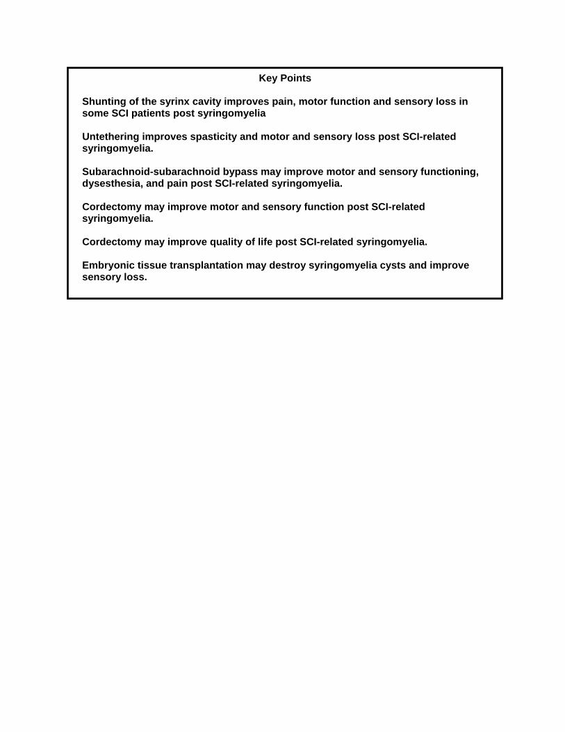
Shunting of the syrinx cavity improves pain, motor function and sensory loss in some SCI patients post syringomyelia
Untethering improves spasticity and motor and sensory loss post SCI-related syringomyelia.
Subarachnoid-subarachnoid bypass may improve motor and sensory functioning, dysesthesia, and pain post SCI-related syringomyelia.
Cordectomy may improve motor and sensory function post SCI-related syringomyelia. Cordectomy may improve quality of life post SCI-related syringomyelia.
Embryonic tissue transplantation may destroy syringomyelia cysts and improve sensory loss.
This review has been prepared based on the scientific and professional information available in 2013. The SCIRE information (print, CD or web site www.scireproject.com) is provided for informational and educational purposes only. If you have or suspect you have a health problem, you should consult your health care provider. The SCIRE editors, contributors and supporting partners shall not be liable for any damages, claims, liabilities, costs or obligations arising from the use or misuse of this material. McIntyre A, Thompson S, Janzen S, Mehta S, Sequeira K, Boyd M, Teasell RW (2014). Syringomyelia Following Spinal Cord Injury. In Eng JJ, Teasell RW, Miller WC, Wolfe DL, Townson AF, Hsieh JTC, Connolly SJ, Noonan VK, Loh E, McIntyre A, editors. Spinal Cord Injury Rehabilitation Evidence. Version 5.0: p 1-14. www.scireproject.com
Table of Contents
Abbreviations ................................................................................................................. i 1.0 Introduction ............................................................................................................. 1
1.1 Epidemiology ......................................................................................................... 1 1.2 Pathophysiology .................................................................................................... 1 1.3 Clinical Presentation and Natural History .............................................................. 2
2.0 Diagnosis and Monitoring ...................................................................................... 2 2.1 Magnetic Resonance Imaging ............................................................................... 2 2.2 Myelography Enhanced Computed Tomography ................................................... 2 2.3 Virtual Endocopy by Computer Tomography ......................................................... 2 2.4 Ultrasonography .................................................................................................... 3 2.5 Intraoperative Somatosensory Evoked Potentials ................................................. 3
3.0 Management ............................................................................................................ 3 3.1 Shunting ................................................................................................................ 3 3.2 Untethering ............................................................................................................ 7 3.3 Subarachnoid–Subarachnoid Bypass .................................................................... 9 3.4 Cordectomy ......................................................................................................... 10 3.5 Neural Tissue Transplantation ............................................................................. 11
4.0 Summary ................................................................................................................ 12 References ................................................................................................................... 13
i
Abbreviations CSF Cerebrospinal Fluid CT-myelography Myelography-enhanced computed tomography EQ European Quality of Life MRI Magnetic Resonance Imaging SCI Spinal Cord Injury SF-36 Short Form-36 S-S Bypass Subarachnoid-subarachnoid bypass SSEP Intraoperative somatosensory evoked potentials VE Virtual Endoscopy
1
Syringomyelia Following Spinal Cord Injury 1.0 Introduction
Post-traumatic syringomyelia is a term used to describe the formation of an intramedullary cyst filled with cerebrospinal fluid (CSF) within the spinal cord (Brodbelt & Stoodley, 2003). Though uncommon, its impact can be devastating following spinal cord injury (SCI). It can be seen as early as two months after injury, or many years later (Vernon, Silver, & Symon, 1983). For example, Ko et al. (2012) reported that syringomyelia developed within 5 years after injury in 7.3% of patients.
1.1 Epidemiology
Syringomyelia occurs in approximately 2% of individuals with SCI (Klekamp & Samii 2002). Syringomyelia found at autopsy has a 22% higher incidence than those presenting clinically (Vannemreddy et al. 2002). No relationship has been reported between the level of SCI and the likelihood of developing syringomyelia. Incidence rates are similar in individuals with either tetraplegia or paraplegia (Brodbelt & Stoodley 2003; Klekamp & Samii 2002; Ko et al. 2012). However, an increased risk of post-traumatic syringomyelia has been reported in complete SCI individuals (Vannemreddy et al. 2002; Kramer & Levine 1997). Lyons et al. (1987) reported that the development of syringomyelia occurred after a shorter interval in individuals with incomplete SCI than those with complete SCI; although, other studies have found no such relationship (El Masry & Biyani 1996; Ko et al. 2012; Vernon et al. 1982).
1.2 Pathophysiology
The pathophysiology of syringomyelia following SCI is not completely understood. The most supported theory is William’s “Cranial-Spinal Pressure Dissociation Theory” which involves formation of the cavity and its enlargement and extension (Williams et al. 1981). Initially, at the site of SCI, a cavity forms after liquefaction of cord tissue or hematoma (Biyani & El Masry 1994; Williams et al. 1981). The liquefaction and cyst formation at the site has been linked to microinfarcts and the release of cellular enzymes (Williams et al. 1981; Kao & Chang 1977). Cyst formation results in partial obstruction of cerebral spinal fluid movement, creating a pressure gradient between the intracranial space and spinal space (Sharma et al. 2006). The second phase, enlargement and extension, is the result of this pressure gradient. It has been linked to two mechanisms affecting fluid dynamics, ‘slosh’ and ‘suck’ (Biyani & El Masry 1994; Williams et al. 1981). The ‘slosh’ and ‘suck’ effects are due to increased epidural venous flow triggered by everyday activities such as coughing and sneezing (Williams 1992). Specifically, the ‘slosh’ is due to increased epidural venous pressure and CSF movement. This pressure causes areas of structural weakness in the cord leading to proximal and distal extension of the syrinx. The second mechanism ‘suck’ is the result of a partial subarachnoid block. As the fluid is initially forced up due to increased epidural venous pressure, it returns slowly creating a pressure gradient across the partial subarachnoid block with negative pressure caudal to it (Biyani & El Masry 1994). This contributes to the syrinx formation and progression.
Figure 1. Cerebral spinal fluid filled cord (Elliot, 2008a)
2
1.3 Clinical Presentation and Natural History
The classic symptoms of syringomyelia (i.e., suspended sensory loss, segmental weakness and burning) are often not present in individuals with SCI. Many individuals may lack symptoms in general or present with nonspecific symptoms that may be attributed to other complications of SCI such as spasticity, autonomic dysreflexia or neuropathic pain. Most commonly, symptoms include radicular pain, gait ataxia, sensory disturbance, dysesthesias and motor weakness (Brodbelt & Stoodley 2003; Klekamp & Samii 2002; Kramer & Levine 1997; Lyons et al. 1987). As syringomyelia progresses, reduction in sensation and increased spasticity may be seen (Carroll & Brackenridge 2005). Progression is usually slow in most patients, with the clinical presentation remaining static for many years (Mariani et al. 1991).
2.0 Diagnosis and Monitoring
2.1 Magnetic Resonance Imaging
Magnetic resonance imaging (MRI) is currently the diagnostic test of choice for syringomyelia as it is able to detect fluid movement, syrinxes, and other abnormalities (Brodbelt & Stoodley 2003). MRI also has an important role in planning and monitoring outcomes of treatments. T1-weighted (T1W) and T2-weighted (T2W) images can be obtained from a MRI and allow for image contrast between different types of tissue (Enzmann 1991). Phase contrast with MRI can identify obstruction of the subarachnoid space and demonstrate normalization of CSF flow following surgery (as seen in Figure 2; Brodbelt & Stoodley 2003). MRI limited in its ability to differentiate posttraumatic syringomyelia from myelomalacia or tumor-associated syringomyelia (Biyani & El Masry 1994).
2.2 Myelography Enhanced Computed Tomography
Myelography-enhanced computed tomography (CT-myelography) of the spinal cord is a significant improvement over plain CT in the diagnosis of syringomyelia. Images from plain CT are considered unreliable due to imaging distortions of the surrounding bone (Klekamp & Samii 2002). CT-myelography is able to show swelling and fixation of the cord and localized CSF flow obstruction (Dworkin & Staas 1985; Klekamp & Samii 2002). As water soluble contrast accumulates in the cyst, CT-myelography can show the syrinx itself (Aubin et al. 1981). However, 10-50% of syrinxes may still be missed using this tool; therefore, MRI remains the diagnostic tool of choice (Brodbelt & Stoodley 2003).
2.3 Virtual Endocopy by Computer Tomography Improvements in CT technology have resulted in better 3-dimensional imaging. Advanced computer graphics hardware and software have enabled the visualization of organs both inside and out. Virtual endoscopy (VE) is “a realistic 3D intraluminal simulation of tubular structures
Figure 2. A) Abnormal cerebral spinal fluid due to syringomyelia. B) Normal cerebral spinal fluid flow post-surgery.
3
that is generated by post-processing of CT data sets” (Kotani et al. 2012, p. E752). Using this technology, surgeons can noninvasively explore the spinal canal in all directions.VE can provide information regarding the extent of stenosis, which plain CT cannot provide. It is useful for diagnosis, preoperative planning, and postoperative assessing.
2.4 Ultrasonography
Ultrasonography is usually confined to the intraoperative setting. It is useful in localizing syrinxes, determining the safest place to open the dura, and facilitating optimal shunt placement during surgery (Brodbelt & Stoodley 2003).
2.5 Intraoperative Somatosensory Evoked Potentials
Since the spinal cord is usually already significantly damaged, intraoperative somatosensory evoked potentials are of limited value in assessing for syringomyelia. However, it may be used during surgery to prevent neurological damage if action potentials can be obtained.
3.0 Management
Medications can be used to manage presenting symptoms but they do not treat the syringomyelia itself. Surgery is only recommended for patients with neurological deterioration or intractable pain (El Masry & Biyani 1996; Klekamp & Samii 2002), although its use is commonly reported in the treatment of syringomyelia in SCI patients.
3.1 Shunting
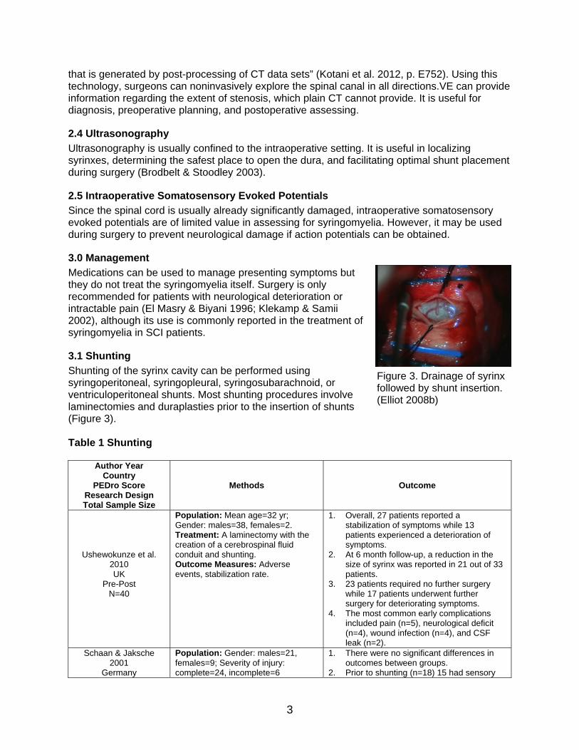
Shunting of the syrinx cavity can be performed using syringoperitoneal, syringopleural, syringosubarachnoid, or ventriculoperitoneal shunts. Most shunting procedures involve laminectomies and duraplasties prior to the insertion of shunts (Figure 3). Table 1 Shunting
Author Year Country
PEDro Score Research Design Total Sample Size
Methods Outcome
Ushewokunze et al. 2010 UK
Pre-Post N=40
Population: Mean age=32 yr; Gender: males=38, females=2. Treatment: A laminectomy with the creation of a cerebrospinal fluid conduit and shunting. Outcome Measures: Adverse events, stabilization rate.
1. Overall, 27 patients reported a stabilization of symptoms while 13 patients experienced a deterioration of symptoms.
2. At 6 month follow-up, a reduction in the size of syrinx was reported in 21 out of 33 patients.
3. 23 patients required no further surgery while 17 patients underwent further surgery for deteriorating symptoms.
4. The most common early complications included pain (n=5), neurological deficit (n=4), wound infection (n=4), and CSF leak (n=2).
Schaan & Jaksche 2001
Germany
Population: Gender: males=21, females=9; Severity of injury: complete=24, incomplete=6
1. There were no significant differences in outcomes between groups.
2. Prior to shunting (n=18) 15 had sensory
Figure 3. Drainage of syrinx followed by shunt insertion. (Elliot 2008b)
4
Author Year Country
PEDro Score Research Design Total Sample Size
Methods Outcome
Cohort N=30
Treatment: Patients with syringomyelia were divided into 3 groups: Group 1 (n=18) received single or multiple shunting procedures; Group 2 (n=5) received shunting procedures before surgical creation of a pseudomeningocele; Group 3 (n=7) was treated only with the surgical pseudomeningocele. Outcome Measures: Sensory and motor deficit, pain, and syringobulbia.
deficits, 13 had motor deficits, 14 had pain and 2 had syringobulbia. Post-surgery: a. sensory deficits improved in 5,
deteriorated in 2, and were unchanged in 8 patients.
b. motor deficits improved in 5, deteriorated in 2 and were unchanged in 4 patients.
c. pain improved in 5, deteriorated in 4, and was unchanged in 5 patients.
d. syringobulbia improved in 1 and deteriorated in the second patient.
3. Prior to shunting and pseudomeningocele (n=5) 5 had sensory and motor deficits, 1 had pain and 1 had syringobulbia. Post-surgery: a. sensory deficits improved in 4 and
deteriorated in 1 patient. b. motor deficits improved in 3,
deteriorated in 1 and was unchanged in 1 patient.
c. pain and syringobulbia improved in both patients.
4. Prior to pseudomeningocele only (n=7) 7 patients had sensory deficits, 6 had motor deficits, 3 had pain, and 1 had syringobulbia. Post-surgery: a. Sensory deficits improved in 6 and
were unchanged in 1 patient. b. Motor deficits improved in 2,
deteriorated in 1, and were unchanged in 3 patients.
c. pain and syringobulbia improved in all patients.
Falci et al. 1999 USA
Case Series N=59
Population: Mean age=26 yr; Gender: males=49, females=10; Severity of injury: AIS A=90%, B=2%, C=5%, D=3%. Treatment: All patients underwent spinal untethering and if a spinal cyst was present a lumbo-peritoneal shunt tube was placed along the length of the cyst. Outcome Measures: Pinprick, motor and light touch scores, MRI findings, and somatosensory evoked potentials.
1. Participants with no previous surgery showed a significant increase in light touch (+2.38), pinprick (+3.88) and motor scores (+1.47) post-surgery.
2. Participants who had previous surgery had a decrease in touch, pinprick and motor score, although it was minimal (0.7, 0.8, and 0.5, respectively).
3. At two weeks post-surgery, MRI showed decreased cyst size or complete collapse.
4. Somatosensory evoked potentials were improved in amplitude compared to baseline; latency of 2 milliseconds or greater was observed in 27 patients.
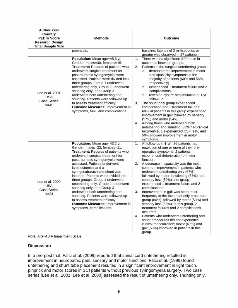
Lee et al. 2001 USA
Case Series N=45
Population: Mean age=45.6 yr; Gender: males=30, females=15. Treatment: Records of patients who underwent surgical treatment for posttraumatic syringomyelia were assessed. Patients were divided into three groups: Group 1 underwent untethering only, Group 2 underwent shunting only, and Group 3
1. There was no significant difference in outcomes between groups.
2. Patients in the surgical untethering group: a. demonstrated improvement in motor
and spasticity symptoms in the majority of patients (60% and 58%, respectively).
b. experienced 1 treatment failure and 2 complications.
5
Author Year Country
PEDro Score Research Design Total Sample Size
Methods Outcome
underwent both untethering and shunting. Patients were followed up to assess treatment efficacy. Outcome Measures: Improvement in symptoms, MRI, and complications.
c. revealed cyst re-accumulation at 1 yr follow-up.
3. The shunt only group experienced 1 complication and 3 treatment failures; 60% of patients in this group experienced improvement in gait followed by sensory (57%) and motor (54%).
4. Among those who underwent both untethering and shunting, 33% had clinical recurrence, 1 experienced CSF leak, and 50% showed improvement in motor symptoms.
Lee et al. 2000 USA
Case Series N=34
Population: Mean age=43.2 yr; Gender: males=23, females=11. Treatment: Records of patients who underwent surgical treatment for posttraumatic syringomyelia were assessed. Patients underwent laminectomies and a syringosubarachnoid shunt was inserted. Patients were divided into three groups: Group 1 underwent untethering only, Group 2 underwent shunting only, and Group 3 underwent both untethering and shunting. Patients were followed up to assess treatment efficacy. Outcome Measures: Improvement in symptoms, and complications.
1. At follow-up (>1 yr), 26 patients resolution of one or more of the presenting symptoms was achieved post operatively; 2 patients experienced deterioration of motor function.
2. A decrease in spasticity was the most common improvement in patients who underwent untethering only (67%), followed by motor functioning (57%) and sensory loss (50%); this group experienced 1 treatment failure and 2 complications.
3. Improvement in gait was seen in the highest number of patients from the shunt only procedure group (60%), followed by motor (50%) and sensory loss (50%); in this group, 2 treatment failures and 2 complications occurred.
4. Patients who underwent untethering and shunt procedures did not experience clinical reoccurrence; motor (67%) and gait (50%) improved in patients in this group.
Hess & Foo 2001 USA
Case Series N=8
Population: Level of injury: T=2, C=5, L=1; Severity of injury: AIS A/B=7, C=1; Mean time post SCI=10 yr. Treatment: Charts of patients who received shunts were assessed. Outcome Measures: Outcome of shunting, and complications.
1. A significant reduction in pain was reported by >80% of patients post –surgery; improvement in strength (n=6) and sensory (n=2) was also reported.
2. At follow-up, 4 patients had shunt failure resulting in neurologic decline, while 2 developed a new syrinx.
3. In 1 patient a new cavity was found with MRI, while the original remained decompressed.
Hida et al. 1994 Japan
Case Series N=14
Population: Mean age=48 yr; Gender: males=10, females=4; Level of injury: C=5, T=5, L=4. Treatment: Charts of patients who underwent syringosubarachnoid (n=6), syringoperitoneal (n=4), and ventriculoperitoneal (n=1) shunts were assessed. Outcome Measures: Neurological, motor, sensory functioning, and shunt malfunction.
1. Neurological amelioration was obtained in all patients.
2. Of the 9 patients with motor function difficulty, 8 improved.
3. Sensory disturbance and relief of local pain or numbness improved in all patients.
4. Malfunction was reported in 3 of 4 syringoperitoneal shunts and in the one ventriculoperitoneal shunt.
Ronen et al. 1999 Population: Mean age=31.3 yr; 1. 4 out of 5 patients in the shunt surgery
6
Author Year Country
PEDro Score Research Design Total Sample Size
Methods Outcome
Israel Case Control
N=10
Gender: males=10, females=0; Level of injury: C=5, L=4; Severity of injury: incomplete=5, complete=5. Treatment: Charts of patients with syringomyelia were reviewed. Patients were divided into two groups: patients receiving rehabilitation only and patients receiving rehabilitation and shunting. Outcome Measures: Functional and neurological outcome.
and rehabilitation group showed functional and neurological deterioration; the fifth patient remained unchanged.
2. Patients in the rehabilitation only group remained unchanged except for one who showed significant functional improvement without any change in neurological status.
Note: AIS=ASIA Impairment Scale Discussion In all of the studies, no shunting procedure was found to be superior to another. Schaan and Jaksche (2001) in a cohort study assessed the efficacy of syringomyelia treatment in three groups of patients: Group 1 received various shunts only, Group 2 received shunting followed by surgical creation of a pseudomeningocele, and Group 3 was treated with the pseudomeningocele only. The study found improvement in sensory and motor deficits, pain and syringobulbia in all three groups. However, more patients experienced greater pain post-surgery in the shunting only group than the other two groups. Falci et al. (1999) demonstrated that untethering and shunt tube placement among individuals without prior surgery can significantly improve light touch, pinprick and motor scores. Two case series (Lee et al. 2001; Lee et al. 2000) further examined untethering and shunting treatment in individuals with syringomyelia. In the study by Lee et al. (2001), patients were divided into three groups: untethering only, shunting only, and untethering and shunting. Improvement in motor and sensory functioning was observed in all three groups. In the first group, untethering only, improvement in spasticity was more common; while the shunting only group found gait improvement to be the most common. Furthermore, shunting alone has been reported to significantly improve pain (Hess & Foo 2001; Hida et al. 1994), strength (Hess & Foo 2001), motor function (Hida et al. 1994) and sensation (Hess & Foo 2001; Hida et al. 1994) in patients with syringomyelia. Although, a high rate of shunt failure (50% and 36%) has been reported (Hess & Foo 2001 and Hida et al. 1994, respectively). One case control study (Ronen et al. 1999) reviewed charts of patients receiving either rehabilitation only or rehabilitation and shunting for syringomyelia. The study found 80% of patients in the shunting and rehabilitation group experienced functional and neurological deterioration, while patients in the rehabilitation group remained either unchanged or improved. One must be careful when drawing conclusions from such a study because allocation to either group was dictated by receiving the treatment, which presumably was given to those patients already deteriorating or those considered at higher risk of doing so. Ushewokunze et al. (2010) reported a reduction in syrinx size among 21/40 patients and a stabilization of symptoms among 27/40. However, symptoms deteriorated for 13 individuals. In 17 individuals, a second surgical procedure was required to improve deteriorating symptoms. Several adverse events were reported including pain, neurological deficit, infection and CSF leakage.
7
Conclusions There is level 2 evidence (from 1 cohort, 1 pre-post, and 5 case series studies; Ushewokenze et al. 2010; Schaan & Jaksche 2001; Falci et al. 1999; Lee et al. 2000; Lee et al. 2001; Hess &Foo 2001; Hida et al. 1994) that shunting improves pain, motor function and sensory loss in some SCI patients with syringomyelia; however, a high rate of shunt failure has been observed.
3.2 Untethering
Spinal cord tethering is commonly seen in patients with syringomyelia. A tethered spinal cord occurs when scar tissue forms and holds the spinal cord to its surrounding soft tissue membrane and dura. It has significant effects on spinal cord movement, CSF flow and extracellular fluid flow resulting in mobility issues and intramedullary pressure changes when exerting certain movements (Klekamp & Samii 2002). Untethering of the spinal cord is used to prevent or revise neurological or orthopedic sequelae. Table 2 Untethering
Author Year Country
PEDro Score Research Design Total Sample Size
Methods Outcome
Falci et al. 2009 USA
Pre-Post N= 362
Population: Mean age=40.5 yr; Severity of injury: AIS A=63.3%, B=9.9%, C=11.3%, D=14%, E=0.6%; Level of injury: C6=45%, C6-T1=23%, T1=32%; Treatment: Surgical treatment for spinal cord untethering. Outcome Measures: AIS sensory and motor scores, sensory and motor changes, and subjective report of changes post-surgery.
1. 60% of the patients found an improvement in spasticity, 77% found an improvement in hyperhidrosis and 47% reported an improvement in neuropathic pain.
2. Most patients (86.5%) required only one surgery.
3. Progressive myelopathy regarding sensory and motor functions was arrested for an average of 3.3-3.4 yr post-surgery.
4. 89% of patients reported an arrest in loss of sensory and/or motor function post-surgery.
5. Return of function was reported in 46% of the patients.
Falci et al. 1999 USA
Case Series N=59
Population: Mean age=26 yr; Gender: males=49, females=10; Severity of injury: AIS A=90%, B=2%, C=5%, D=3%. Treatment: All patients underwent spinal untethering and if a spinal cyst was present a lumbo-peritoneal shunt tube was placed along the length of the cyst. Outcome Measures: Pinprick, motor and light touch scores, MRI findings, and somatosensory evoked
1. Participants with no previous surgery showed a significant increase in light touch (+2.38), pinprick (+3.88) and motor scores (+1.47) post-surgery.
2. Participants who had previous surgery had a decrease in touch, pinprick and motor score, although it was minimal (0.7, 0.8, and 0.5, respectively).
3. At two weeks post-surgery, MRI showed decreased cyst size or complete collapse.
4. Somatosensory evoked potentials were improved in amplitude compared to
Shunting of the syrinx cavity improves pain, motor function and sensory loss in
some SCI patients post syringomyelia
8
Author Year Country
PEDro Score Research Design Total Sample Size
Methods Outcome
potentials. baseline; latency of 2 milliseconds or greater was observed in 27 patients.
Lee et al. 2001 USA
Case Series N=45
Population: Mean age=45.6 yr; Gender: males=30, females=15. Treatment: Records of patients who underwent surgical treatment for posttraumatic syringomyelia were assessed. Patients were divided into three groups: Group 1 underwent untethering only, Group 2 underwent shunting only, and Group 3 underwent both untethering and shunting. Patients were followed up to assess treatment efficacy. Outcome Measures: Improvement in symptoms, MRI, and complications.
1. There was no significant difference in outcomes between groups.
2. Patients in the surgical untethering group: a. demonstrated improvement in motor
and spasticity symptoms in the majority of patients (60% and 58%, respectively).
b. experienced 1 treatment failure and 2 complications.
c. revealed cyst re-accumulation at 1 yr follow-up.
3. The shunt only group experienced 1 complication and 3 treatment failures; 60% of patients in this group experienced improvement in gait followed by sensory (57%) and motor (54%).
4. Among those who underwent both untethering and shunting, 33% had clinical recurrence, 1 experienced CSF leak, and 50% showed improvement in motor symptoms.
Lee et al. 2000 USA
Case Series N=34
Population: Mean age=43.2 yr; Gender: males=23, females=11. Treatment: Records of patients who underwent surgical treatment for posttraumatic syringomyelia were assessed. Patients underwent laminectomies and a syringosubarachnoid shunt was inserted. Patients were divided into three groups: Group 1 underwent untethering only, Group 2 underwent shunting only, and Group 3 underwent both untethering and shunting. Patients were followed up to assess treatment efficacy. Outcome Measures: Improvement in symptoms, complications
1. At follow-up (>1 yr), 26 patients had resolution of one or more of their pre-operative symptoms; 2 patients experienced deterioration of motor function.
2. A decrease in spasticity was the most common improvement in patients who underwent untethering only (67%), followed by motor functioning (57%) and sensory loss (50%); this group experienced 1 treatment failure and 2 complications.
3. Improvement in gait was seen most frequently in the the shunt only procedure group (60%), followed by motor (50%) and sensory loss (50%); in this group, 2 treatment failures and 2 complications occurred.
4. Patients who underwent untethering and shunt procedures did not experience clinical reoccurrence; motor (67%) and gait (50%) improved in patients in this group.
Note: AIS=ASIA Impairment Scale Discussion In a pre-post trial, Falci et al. (2009) reported that spinal cord untethering resulted in improvement in neuropathic pain, sensory and motor functions. Falci et al. (1999) found untethering and shunt tube placement resulted in a significant improvement in light touch, pinprick and motor scores in SCI patients without previous syringomyelia surgery. Two case series (Lee et al. 2001; Lee et al. 2000) assessed the result of untethering only, shunting only,
9
and untethering and shunting in a group of SCI individuals with syringomyelia. Both studies found that more patients in the untethering only group had improved spasticity than patients in the other groups. Motor function and sensory loss improvement was common in all three groups. However, the shunting group only demonstrated more improvement in gait than the untethering group. Conclusions There is level 4 evidence (from 1 pre-post and 3 case series studies; Falci et al. 2009; Falci et al. 1999; Lee et al. 2000; Lee et al. 2001) that untethering improves motor and sensory loss. There is level 4 evidence (from 2 case series studies; Lee et al. 2000; Leet et al. 2001) that untethering improves spasticity in more patients with syringomyelia than shunting.
3.3 Subarachnoid–Subarachnoid Bypass
A new type of surgical technique for posttraumatic syringomyelia has been described in the literature, subarachnoid-subarachnoid bypass (S-S Bypass). Hayashi et al. (2013) hypothesized that reconstruction of the subarachnoid channels could re-establish CSF flow and therefore correct the underlying issue causing syrinx formation. In general, the S-S Bypass technique is accomplished by surgical laminectomy at the level of trauma, followed by a midline dural opening made under a microscope. One or two silicone tubes are inserted into the cephalic and caudal ends of the normal subarachnoid space; after a watertight dural closure, Bypass tubes are laid in the subdural space (Hayashi, 2013). A single pre-post study has assessed the effectiveness of S-S Bypass in 20 individuals (mean age=47.3 years, 19 males) with SCI-related syringomyelia (Hayashi et al. 2013). The mean time since SCI was 126 months (range 2-336 months) and they were followed up, on average, for 48.2 months (range 12-93 months). Post-surgery, 12 patients showed improvements, four remained stable, and four showed signs of deterioration. Three of the four patients who demonstrated deterioration underwent a shunt replacement; two improved and one remained unchanged. There was no significant correlation between AISA scores at baseline and follow-up. Finally, no patient experienced a CSF leak that needed treatment (Hayashi et al. 2013). The authors conclude that S-S Bypass is not only an effective method in treating syringomyelia but that it may be associated with better clinical results than those of other surgical interventions (e.g., shunts, cordectomy). Further investigation from multiple studies is required to make conclusions as to its clinical effectiveness. Conclusions There is level 4 evidence (from 1 pre-post study; Hayashi et al. 2013) that subarachnoid-subarachnoid bypass may improve motor and sensory functioning, dysesthesia, and pain post SCI-related syringomyelia.
Untethering improves spasticity and motor and sensory loss post SCI-related
syringomyelia.
10
3.4 Cordectomy
Cordectomy has been shown to be a useful procedure in the surgical treatment of syringomyelia. It can be used to manage spasticity, pain and improve neurological dysfunction. However, since it is an invasive procedure and irreversible, it is only considered when other options have been exhausted (Gautschi et al. 2011). Table 3 Cordectomy
Author Year Country
PEDro Score Research Design Total Sample Size
Methods Outcome
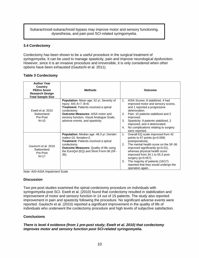
Ewelt et al. 2010 Switzerland
Pre-Post N=15
Population: Mean age: 52 yr, Severity of Injury: AIS A=7, B=8. Treatment: Patients received a spinal cordectomy. Outcome Measures: AISA motor and sensory function, Visual Analogue Scale, adverse events, and spasticity.
1. AISA Scores: 8 stabilized, 4 had improved motor and sensory scores, and 1 reported a progressive deterioration.
2. Pain: 10 patients stabilized and 2 improved.
3. Spasticity: 9 patients stabilized, 2 improved, and 4 deteriorated.
4. No complications relating to surgery were reported.
Gautschi et al. 2010 Switzerland
Pre-Post N=17
Population: Median age: 48.3 yr; Gender: males=15, females=2. Treatment: Patients received a spinal cordectomy. Outcome Measures: Quality of life using the EuroQol (EQ) and Short Form-36 (SF-36).
1. Overall EQ scale improved from 42 points to 67 points (p=0.006) postoperatively.
2. The mental health score on the SF-36 improved significantly (p=0.01), whereas physical health score improved from 34.1 to 55.3 post-surgery (p=0.057).
3. The majority of patients (16/17) reported that they would undergo the operation again.
Note: AIS=ASIA Impairment Scale Discussion Two pre-post studies examined the spinal cordectomy procedure on individuals with syringomyelia post SCI. Ewelt et al. (2010) found that cordectomy resulted in stabilization and improvement of motor and sensory function in 14 out of 15 patients. The study also reported improvement in pain and spasticity following the procedure. No significant adverse events were reported. Gautschi et al. (2010) reported a significant improvement in the quality of life of individuals who underwent the cordectomy procedure and high levels of subjective satisfaction. Conclusions There is level 4 evidence (from 1 pre-post study; Ewelt et al. 2010) that cordectomy improves motor and sensory function post SCI-related syringomyelia.
Subarachnoid-subarachnoid bypass may improve motor and sensory functioning,
dysesthesia, and pain post SCI-related syringomyelia.
11
There is level 4 evidence (from 1 pre-post study; Gautschi et al. 2010) that cordectomy improves quality of life of individuals post SCI-related syringomyelia.
3.5 Neural Tissue Transplantation
A novel treatment for syringomyelia involves transplantation of neural tissue alone or in conjunction with surgical unthethering or cyst shunting. Embryonic spinal cord grafts have been shown to help repair structure and function of the spinal cord in experimental studies with an 80-90% survival rate (Houle & Reier 1988; Reier et al. 1988; Akesson et al. 1998). These grafts are used to fill the syrinx cavity and minimize cystic deformations in patients with progressive posttraumatic syringomyelia (Falci et al. 1997). Two case reports (Falci et al. 1997; Wirth III et al. 2001), examined the use of embryonic tissue transplantation in the treatment of syringomyelia in patients post SCI. The studies involved untethering, cyst drainage and implantation of embryonic fetal tissue of SCI patients. Both studies reported collapse of cyst in the transplantation region and improvement in sensation. Improvement in spasticity was also observed, although it was short lived. Wirth III et al. (2001) demonstrated improvement in bladder functioning post-surgery, while Falci et al. (1997) showed significant improvement in deafferentation pain. At 7 month follow-up, MRI images showed no reoccurrence of the cyst in the transplantation region; however improvement of secondary complications were not maintained (Falci et al. 1997). Further investigation using studies with more subjects is required to make conclusions as to its clinical effectiveness. Conclusions There is level 5 evidence (from 2 case reports; Falci et al. 1997; Wirth III et al. 2001) that embryonic tissue transplantation along with drainage, untethering and shunting may obliterate syringomyelia cysts and improve sensory loss.
Embryonic tissue transplantation may destroy syringomyelia cysts and improve
sensory loss.
Cordectomy may improve motor and sensory function post SCI-related
syringomyelia.
Cordectomy may improve quality of life post SCI-related syringomyelia.
12
4.0 Summary
Syringomyelia is a term used to describe the formation of an intramedullary cyst filled with cerebrospinal fluid within the spinal cord. The pathophysiology of syringomyelia following SCI is not completely understood. Magnetic resonance imaging is currently the diagnostic test of choice for diagnosing syringomyelia and is able to detect fluid movement, syringomyelia and other abnormalities. There is level 2 evidence (from 1 cohort, 1 pre-post, and 5 case series studies; Ushewokenze et al. 2010; Schaan & Jaksche 2001; Falci et al. 1999; Lee et al. 2000; Lee et al. 2001; Hess &Foo 2001; Hida et al. 1994) that shunting improves pain, motor function and sensory loss in some SCI patients with syringomyelia; however, a high rate of shunt failure has been observed. There is level 4 evidence (from 1 pre-post and 3 case series studies; Falci et al. 2009; Falci et al. 1999; Lee et al. 2000; Lee et al. 2001) that untethering improves motor and sensory loss. There is level 4 evidence (from 2 case series studies; Lee et al. 2000; Leet et al. 2001) that untethering improves spasticity in more patients with syringomyelia than shunting. There is level 4 evidence (from 1 pre-post study; Hayashi et al. 2013) that subarachnoid-subarachnoid bypass may improve motor and sensory functioning, dysesthesia, and pain post SCI-related syringomyelia. There is level 4 evidence (from 1 pre-post study; Ewelt et al. 2010) that cordectomy improves motor and sensory function post SCI-related syringomyelia. There is level 4 evidence (from 1 pre-post study; Gautschi et al. 2010) that cordectomy improves quality of life of individuals post SCI-related syringomyelia. There is level 5 evidence (from 2 case reports; Falci et al. 1997; Wirth III et al. 2001) that embryonic tissue transplantation along with drainage, untethering and shunting may obliterate syringomyelia cysts and improve sensory loss.
13
References
Akesson E, Kjaeldgaard A, Seiger A. Human embryonic spinal cord grafts in adult rat spinal
cord cavities: survival, growth, and interactions with the host. Exp Neurol 1998;149:262-76. Aubin ML, Vignaud J, Jardin C, Bar, D Computed tomography in 75 clinical cases of
syringomyelia. Am J Neuroradiol 1981;2:199-204. Biyani A, El Masry, WS. Post-traumatic syringomyelia: A review of the literature. Paraplegia
1994;32:723-31. Brodbelt AR, Stoodley, MA. Post-traumatic syringomyelia: A review. J Clin Neurosci
2003;10:401-8. Carroll AM, Brackenridge P. Post-traumatic syringomyelia: A review of the cases presenting in
a regional spinal injuries unit in the North East of England over a 5-year period. Spine 2005;30:1206-10.
Dworkin GE, Staas WE, Jr. Posttraumatic syringomyelia. Arch Phys Med Rehabil 1985;66:329-31.
El Masry WS, Biyani A. Incidence, management, and outcome of post-traumatic syringomyelia. J Neurol Neurosurg Psychiatry 1996;60:141-6.
Elliot NS. 2008a. Fluid filled syrinx in the spinal cord [Photograph]. Retrieved April 12, 2010 from: http://www2.warwick.ac.uk/alumni/services/eportfolios/esrebb.
Elliot NSJ. 2008b. Syrinx drainage and shunt insertion [Photograph]. Retrieved April 12, 2010 from: http://www2.warwick.ac.uk/alumni/services/eportfolios/esrebb.
Enzmann D. Imaging in syringomyelia. In U. Batzdorf (Eds.), Syringomyelia: Current concepts in diagnosis and treatment (pp. 116-39). Baltimore: Williams & Wilkins. 1991.
Ewelt C, Stalder S, Steiger HJ, Hildebrant G, Heilbronner R. Impact of cordectomy as a treatment option for posttraumatic and non-posttraumatic syringomyelia with tethered cord syndrome and myelopathy. J Neurosurg Spine 2010;13:193-9.
Falci S, Holtz A, Akesson E, Azizi M, Ertzgaard P, Hultling C. Obliteration of a posttraumatic spinal cord cyst with solid human embryonic spinal cord grafts: First clinical attempt. J Neurotrauma 1997;14:875-84.
Falci SP, Indeck C, Lammertse DP. Posttraumatic spinal cord tethering and syringomyelia: Surgical treatment and long term outcome. J Neurosurg Spine 2009;11:445-60.
Falci SP, Lammertse DP, Best L, Starnes CA, Prenger EC, Stavros AT. Surgical treatment of posttraumatic cystic and tethered spinal cords. J Spinal Cord Med 1999;22:173-81.
Gautschi OP, Seule MA, Cadosch D, Gores M, Ewelt C, Hildebrandt G, Heilbronner R. Health related quality of life following spinal cordectomy for syringomyelia. Acta Neurochir 2011;153:575-9.
Hayashi T, Ueta T, Kubo M, Maeda T, Shiba K. Subarachnoid-subarachnoid bypass: A new surgical technique for posttraumatic Syringomyelia. Clin Art J Neurosurg Spine 2013;18:382-7.
Hess MJ, Foo D. Shunting for syringomyelia in patients with spinal cord injuries: Self-reported, long-term effects in 8 patients. Arch Phys Med Rehabil 2001;82:1633-6.
Hida K, Iwasaki Y, Imamura H, Abe H. Posttraumatic syringomyelia: Its characteristic magnetic resonance imaging findings and surgical management. Neurosurg 1994;35:886-91.
Houle JD, Reier PJ. Transplantation of fetal spinal cord tissue into the chronically injured adult rat spinal cord. J Comp Neurol 1988;269:535-47.
Kao C, Chang, L. The mechanism of spinal cord cavitation following spinal cord transection. Part I. A correlated histo-chemical study. J Neurosurg 1977;46:197-209.
Klekamp J, Samii M. Syringomyelia associated with diseases of the spinal canal. Syringomyelia: Diagnosis and Treatment 2002;111-93.
14
Ko HY, Kim W, Kim SY, Shin MJ, Cha YS, Chang JH, et al. Factors associated with early onset post-traumatic syringomyelia. Spinal Cord 2012;50:695-98.
Kotani T, Nagaya S, Sonoda M, Akazawa T, Lumawig JMT, Nemoto T, et al. Virtual endoscopic imaging of the spine. Spine 2012;37:752-6.
Kramer KM, Levine AM. Posttraumatic syringomyelia: A review of 21 cases. Clin Ortho Related Res 1997;334:190-9.
Lee TT, Alameda GJ, Camilo E, Green BA. Surgical treatment of post-traumatic myelopathy associated with syringomyelia. Spine 2001;26:119-28.
Lee TT, Alameda GJ, Gromelski EB, Green BA. Outcome after surgical treatment of progressive posttraumatic cystic myelopathy. J Neurosurg 2000;92:149-54.
Lyons BM, Brown DJ, Calvert JM, Woodward, JM, Wriedt, CH. The diagnosis and management of post traumatic syringomyelia. Paraplegia 1987;25:340-50.
Mariani C, Cislaghi MG, Barbieri S, Filizzolo F, Di PF, Farina E. The natural history and results of surgery in 50 cases of syringomyelia. J Neurol 1991;238:433-8.
Reier PJ, Houle, JD, Jakeman L, Winialski D, Tessler A. Transplantation of fetal spinal cord tissue into acute and chronic hemisection and contusion lesions of the adult rat spinal cord. Prog Brain Res 1988;78:173-9.
Ronen J, Catz A, Spasser R, Gepstein R. The treatment dilemma in post-traumatic syringomyelia. Disabil Rehabil 1999;21:455-7.
Schaan M, Jaksche H. Comparison of different operative modalities in post-traumatic syringomyelia: preliminary report. Euro Spine J 2001;10:135-40.
Sharma M, Coppa N, Sandhu F. Syringomyelia: A review. Semin Spine Surg 2006;18:180-4. Ushewokunze SO, Gan YC, Phillips K, Thacker K, Flint G. Surgical treatment of post traumatic
syringomyelia. Spinal Cord 2010;48:710-3. Vannemreddy SS, Rowed DW, Bharatwal N. Posttraumatic syringomyelia: Predisposing
factors. Bri J Neurosurg 2002;16;276-83. Vernon JD, Silver JR, Ohry A. Post-traumatic syringomyelia. Paraplegia 1982;20:339-64. Vernon JD, Silver JR, Symon L. Post-traumatic syringomyelia: The results of surgery.
Paraplegia 1983;21;37-46. Williams B. Pathogenesis of post-traumatic syringomyelia. Brit J Neurosurg 1992;6:517-20. Williams B, Terry AF, Jones F, McSweeney T. Syringomyelia as a sequel to traumatic
paraplegia. Paraplegia 1981;19:67-80. Wirth III ED, Reier PJ, Fessler RG, Thompson FJ, Uthman B, Behrman A. Feasibility and safety
of neural tissue transplantation in patients with syringomyelia. J Neurotrauma 2001;18:911-29.