Embed Size (px)
Citation preview

Synthesis,characterization and antibacterial properties of zinc oxide nanoparticles
45
SYNTHESIS,CHARACTERIZATION AND ANTIBACTERIAL PROPERTIES OF ZINC OXIDE
NANOPARTICLES
2.1 Introduction
2.2 Experimental
2.3 Results and Discussion
2.4 Conclusion
Zinc oxide nanoparticles were prepared by reacting zinc chloride and sodium hydroxide in different mediums such as chitosan, poly vinyl alcohol, ethanol and starch. The materials were characterized by scanning electron microscopy (SEM), x-ray diffraction (XRD) studies, transmission electron microscopy (TEM) and thermogravimetric analysis (TGA). Elemental analysis was done by energy dispersive X-ray Analysis (EDAX). Antibacterial properties were studied using Bacillus aereus (gram positive) and Escherichia coli (gram negative) bacteria. The above studies were also carried on commercially available zinc oxide (CZO). Zinc oxide nanoparticles prepared using chitosan medium (NZO) showed lowest crystallite size in XRD. Morphology of the particle was changed by changing the reaction medium. XRD showed hexagonal wurtzite crystal structure of ZnO. TEM analysis showed particle size of the smallest particle of NZO is 35 nm and highest as 67 nm. Thermogravimetric studies (TGA) indicated improved thermal stability of NZO than CZO particles. Antibacterial studies showed excellent resistance of CZO and NZO to Bacillus aereus and Escherichia coli.
Con
tent
s

Chapter -2
46
2.1 Introduction
Nano zinc oxide has a wide range of applications in various fields due to
its unique and superior physical and chemical properties compared with bulk
ZnO. The large specific surface area, high pore volume, low toxicity,
nanostructured properties and low cost of nano ZnO [1] make it a promising
candidate particularly in catalysts [2], chemical absorbents [3], as polymer
additives [4], antiwear additives for oil lubricants [5] and advanced ceramics
[6]. Zinc oxide nanoparticles have optical, electrical and photochemical
activity [7], it can be used as photocatalysts [8], solar energy conversion cells
[9,10],Ultra Violet (UV) detectors and UV emitting devices [11], chemical
sensors sensitive to chemicals such as alcohol and benzene [12], and gas
sensing materials for many gases such as ammonia, hydrogen and ozone
[13-15]. Zinc oxide with direct band gap of about 3.37 eV at room temperature
is a good material suitable for generating UV light [16]. ZnO with exciton
binding energy of about 60 meV can ensure an efficient exciton emission at
room temperature and low excitation energy [17, 18]. Thin films and nanoscale
coating of ZnO particles on suitable substrates is important for its potential
applications as substrates for functional coatings, printing, UV inks, e-print,
optical communications (security papers), protection, portable energy, sensors,
photocatalytic wallpaper with antibacterial activity etc [19-33]. The powder is
widely used as an additive into numerous materials and products including
plastics, ceramics, glass, cement, rubber, lubricants, paints, ointments, adhesives,
sealants, pigments, source of Zn nutrient in foods, fire retardants etc[34]. The
films of ZnO, indium tin oxide and cadmium oxide have been investigated in
recently as transparent conducting oxide due to their good optical and
electrical properties in combination with large band gap, abundance in nature,
optical transmittance in visible region and non toxicity. Because the optical,
electrical and magnetic properties of ZnO are markedly influenced by its

Synthesis,characterization and antibacterial properties of zinc oxide nanoparticles
47
particle size, morphology and structure [35-37]. ZnO nanoparticles can be
obtained by physical and chemical routes. However, chemical methods are
more suitable for industrial scale production [35] due to its low efficiency and
cost in obtaining nanoparticles with uniform size and morphology [36].
Different techniques for preparing ZnO nanoparticles have been reported such
as, sol– gel technique [36], microemulsion synthesis [38], mechanochemical
processing [37], spray pyrolysis [39], thermal decomposition of organic
precursor [40], RF plasma synthesis [41], supercritical-water processing [42],
self-assembling [43], vapour transport process [44], sonochemical or
microwave-assisted synthesis [45], direct precipitation [46] homogeneous
precipitation [47],chemical co-precipitation [48,49], chemical vapour
deposition [50],thermal decomposition [51,52], hydrothermal synthesis [53,54],
solid-state reaction [55],spray pyrolysis [56], and microemulsion precipitation
[57-61]. Different ZnO nano structures such as nanospheres, nanorods,
nanowires, nanotubes and flower-like nanostructures are obtained from these
techniques. Variety of morphologies including prismatic forms [62],
bipyramidal dumbbell-like [63], ellipsoidal [64], spheres [65], nanorods [66, 67],
nanowires [67], whiskers [68], nanotubes [69], nanorings [70] and columnar
hexagonal shaped ZnO [71] have been investigated.
LI Yan et al reported the formation of nest like ZnO by solvothermal
synthesis [72].Tengfa Long et al studied the formation of ZnO nanorods and
nanodisks from zinc chloride aqueous solution [73]. Yoshie Ishikawa et al
prepared ZnO nanorods by pulsed laser ablation technique in water media at
high temperature [74]. Ameer Azam etal used low temperature synthesis using
micromechanical route [75].
The above methods require complex equipment and complicated
operations. So, it is a challenge to search for a simple route to prepare metal
Chapter -2
48
oxides nanoparticles with a high yield. We have used precipitation technique
for the preparation of zinc oxide nanoparticles. In this work ZnO was prepared
using zinc chloride and sodium hydroxide in different mediums such as
chitosan, ethanol, polyvinyl alcohol (PVA) and starch. Compared with the
above-mentioned methods, this method has four advantages: (1) it is very
cheap and facile because of not requiring any complex equipment and
complicated operations (2) the reaction is environmentally friendly since the
byproducts are chitosan and sodium chloride (3) the reaction is safe and quick
and (4) the high yield. Prepared ZnO was characterized by TEM, SEM, XRD
and TGA. Antibacterial properties of ZnO was studied using a gram positive
and gram negative bacteria.
2.2 Experimental 2.2.1 Materials
Chitosan samples were supplied by M/s. Indian Sea Foods, Cochin,
Kerala (Viscosity 304 cps) and degree of deacetylation is 80.79%. PP
homopolymer (REOL H200MA) was supplied by M/s. Reliance Industries
limited. PVA, starch, ethanol, NaOH and ZnCl2 was supplied by alpha
chemicals, Cochin.
2.2.2 Methods 2.2.2.1 Preparation of zinc oxide
Zinc oxide nanoparticles were prepared by reacting zinc chloride and
sodium hydroxide in different mediums such as chitosan, poly vinyl alcohol,
ethanol and starch. In this method zinc chloride (5g in 500 ml 1% acetic acid
in water) was added to chitosan (5g in 500ml 1% acetic acid in water) with
vigorous stirring using mechanical stirrer. This was allowed to react for 24
hours. During this period stabilization of the complex take place. Then sodium
hydroxide (25g in 500ml 1% acetic acid in water) was added drop wise from
Synthesis,characterization and antibacterial properties of zinc oxide nanoparticles
49
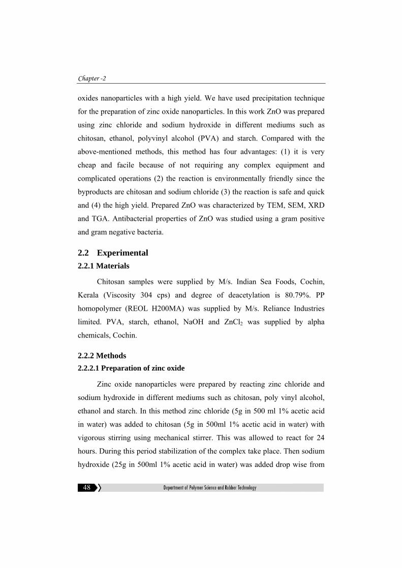
burette to the above solution with stirring using mechanical stirrer. The whole
mixture was allowed to digest for 12 hours at room temperature. This was to
obtain homogeneous diffusion of OH- and Cl- to the matrix. The precipitate
formed was washed several times with distilled water until complete removal
of sodium chloride and dried at 1000C. Then it was calcined at 5500C for four
hours. Figure 2.1 shows the schematic representation of preparation zinc oxide
nanoparticles in chitosan medium. The experiment was repeated using other
mediums such as poly vinyl alcohol, ethanol and starch.
Figure 2.1: Schematic representation of preparation of zinc oxide
Zinc chloride solution in acetic acid
Chitosan in acetic acid solution
Stirring
Zinc chloride/chitosan complex
Zinc hydroxide
Mechanical stirring
Sodium hydroxide in acetic acid solution
Zinc oxide
Calcination

Chapter -2
50
2.2.2.2 X-ray diffraction analysis
XRD is used to determine the size and shape of the unit cell for any
compound most easily using the diffraction of x-rays. X-ray diffraction studies
are used for the finger print characterization of crystalline materials and their
structure determination. Each crystalline solid has its unique characteristics.
Once the material has been identified, it is used to determine its structure, i.e.
how the atoms packed together in the crystalline state and what the interatomic
distances and angle are, etc.
XRD studies were done using Rigaku Geigerflex at wavelength CuKa=1.54 A0.
Particle size was calculated using Debye Sherrer equation:
CS = 0.9λ/βcosӨ ----------------------------------------------------------- (2.1)
where CS is the crystallite size, β is the full width at half-maximum
(FWHMhkl) of an hkl peak at 2Ө value, 2Ө is the scattering angle.
2.2.2.3 Scanning electron microscopy
Scanning electron microscopy is used to gather information about
topography, morphology, composition and micro structural information of
materials. In SEM, electrons are thermoionically emitted from a tungsten or
lanthanum hexa boride cathode and are accelerated towards an anode;
alternatively electrons can be emitted via field emission. The electron beam,
which has an energy ranging from a few hundred eV to 100keV, is focused
by one or two condenser lenses in to a beam. Characteristic X-rays are
emitted when the primary beam causes the ejection of inner shell electrons
from the sample and are used to determine the elemental composition of the
sample. The back scattered electrons emitted from the sample can be used
alone to form an image or in composition with the characteristic X-rays.
Synthesis,characterization and antibacterial properties of zinc oxide nanoparticles
51
These signals are monitored by photomultiplier tubes and magnified. An
image of the investigated microscopic region of the specimen is observed in
cathode ray tube and is photographed.
In this work, morphology of the ZnO was studied using scanning
electron microscope (JOEL model JSM 6390LV). The powder was mounted
on a metallic stub and an ultrathin coating of gold is deposited by low vacuum
sputter coating. This is done to prevent the accumulation of static electric
fields at the specimen due to the electron irradiation during imaging and to
improve contrast. The SEM can produce high resolution images of the sample
surface.
2.2.2.4 Transmission electron microscopy
In TEM, a beam of electrons is transmitted through an ultra thin
specimen, interacting with the specimen as it passes through. An image is
formed by the interaction of the electrons transmitted through the specimen.
The image is magnified and focused into a fluorescent screen, on a layer of
photographic film, or to be detected by a sensor such as a CCD camera.
TEM gives significantly higher resolution images than light microscopes,
due to the small de Broglie wavelength of electrons. This helps the user to
examine fine detail even as small as a single column of atoms, which is tens
of thousands times smaller than the smallest resolvable object in a light
microscope.
In this work, TEM studies were carried out on JEM-2200-FS, Field
Emission Transmission Electron Microscope, JEOL, Japan.
2.2.2.5 Energy Dispersive Analysis by X-rays
EDAX is a widely used technique to analyze the chemical composition
of a material under SEM by detecting the X-rays produced as the result of the

Chapter -2
52
electron beam interactions with the sample. When the sample is bombarded by
the electron beam, the electrons are ejected from atoms comprising the sample
surface. The resulting electron vacancies are filled by electrons from a higher
state and an x-ray is emitted to balance the energy difference. EDS x-ray
detector measures the relative abundance of X-rays against their energy. The
detector is typically a lithium-drifted silicon solid-state device. As the incident
x-ray hits the detector, creates a pulse of charge that is proportional to the
energy of x-ray. The pulse charge is converted to a pulse voltage by using a
charge sensitive preamplifier. The signal is pass to a multichannel analyzer
where the pulses are sorted by the tension. The energy determined by
measuring the voltage per incident x-ray is sent to a computer for display and
further data evaluation. The spectrum of x-ray energy versus counts is
measured to determine the elemental composition of the sample volume. Very
light elements such as hydrogen do not emit X-rays and are impossible to
detect because of the low-energy transitions occur. In this work elemental
composition of the ZnO was studied using scanning electron microscope
(JOEL model JSM 6390LV).
2.2.2.6 Thermogravimetric analysis
Thermogravimetric analysis (TGA) measures the weight change in a
material as a function of temperature and time, in a controlled environment.
This can be very useful to investigate the thermal stability of a material. It is
suitable for use with all types of solid materials, including organic and
inorganic materials. The material is heated to very high temperature so that
one of the components decomposes into a gas, which dissociates into the air.
If the compounds in the mixture remain are known, then the percentage by
mass can be determined by taking the weight of what is left in the mixture
and dividing it by the initial mass. From the mass of the original mixture and
the total mass of impurities liberating upon heating, the stoichiometric ratio

Synthesis,characterization and antibacterial properties of zinc oxide nanoparticles
53
can be used to calculate the percent mass of the substance in a sample. TGA
is commonly employed in research to determine the characteristics of
materials such as polymers, to determine degradation temperatures, absorbed
moisture content of materials, the level of inorganic and organic components
in materials and solvent residues. Thermogravimetric analyzer (TGA Q-50,
TA instruments) was used to study the effect of TiO2 on the thermal stability
of PP. Approximately 10 mg of the samples were heated at a rate of 200
C/min from ambient to 8000C.
2.2.2.7 Antibacterial properties
Bacterial nutrient medium (nutrient broth) was used to cultivate the gram
positive [Bacillus aereus (NCIM No:2155)] and gram negative bacteria
[Escherichia coli (NCIM No:2343)] respectively. Control nutrient broth as
such was inoculated with the bacteria. NZO and CZO was dispersed at a
concentration of 0.1mg/ml in the test media, prepared and commercially
available. The test organisms were inoculated in the tubes and they were
allowed to grow over night. Next day, the culture was serially diluted and
plated on to the nutrient agar plates. The colonies formed were counted the
next day. The result was expressed as CFU.
2.3 Results and Discussion 2.3.1 X-ray Diffraction
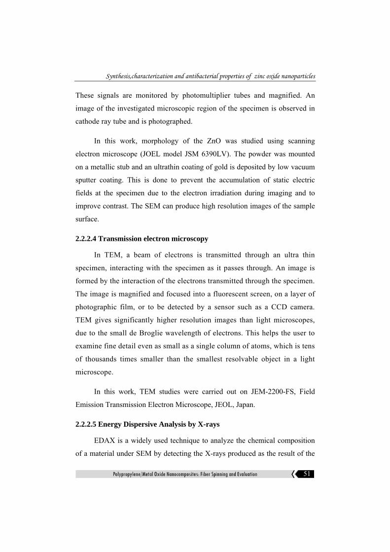
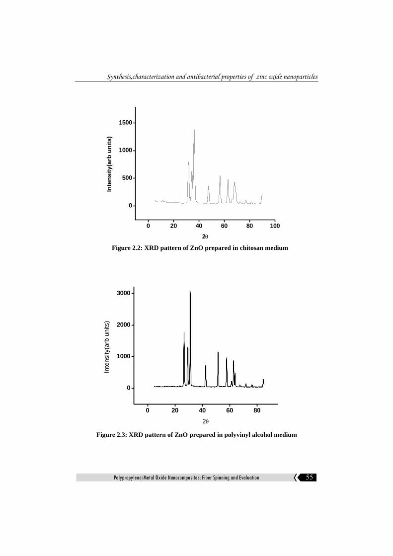
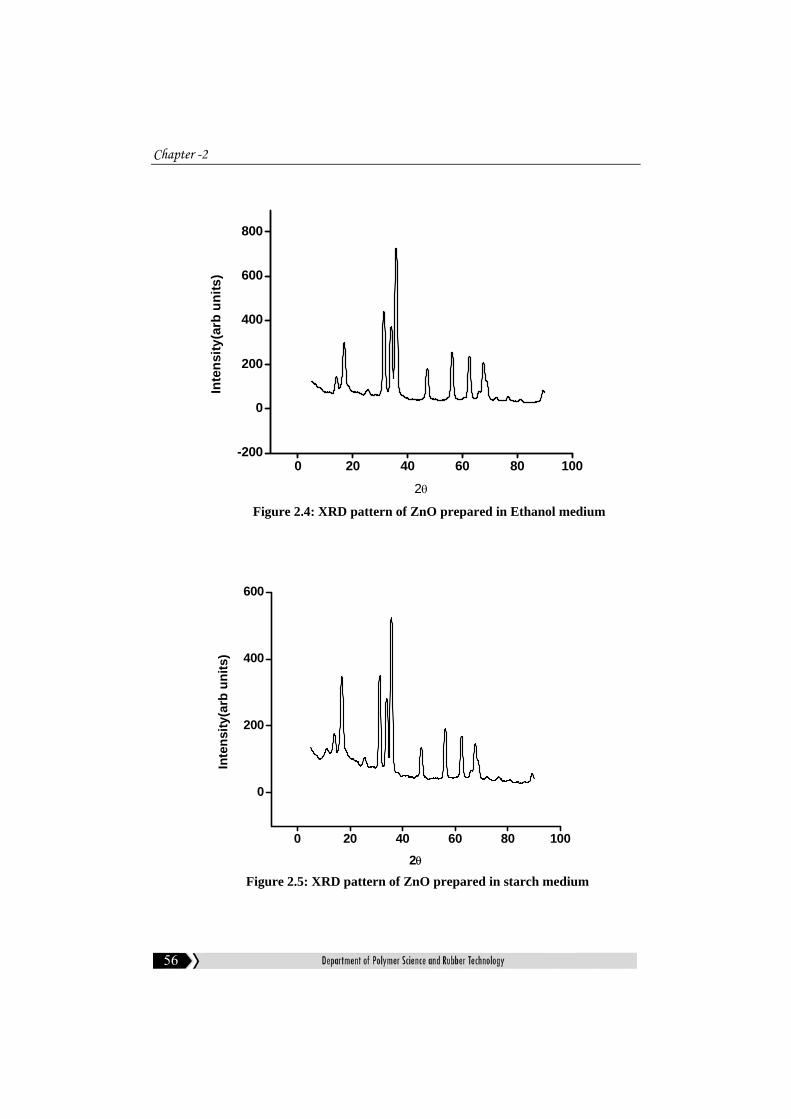
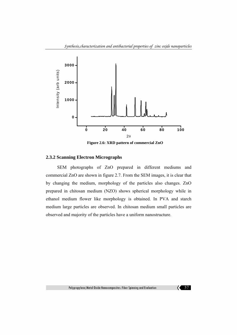
The XRD pattern of ZnO prepared in different mediums such as
chitosan, PVA, starch and ethanol is given in figures 2.2-2.5 respectively.
Figure 2.6 represents the XRD of commercial zinc oxide. These figures
show the characteristics peaks of hexagonal crystal structure. The peaks
obtained are correspond to (100), (002), (101), (102), (110), (103), (112),
(201), (004), (202), (104) planes. The (101) plane is most prominent.
Hexagonal wurtzite structure of ZnO belongs to the space group P63mc1

Chapter -2
54
and lattice parameters a = 3.2539 Å and c = 5.2098 Å. The crystallite size
of ZnO calculated using Debye Sherrer equation is given in table 2.1.
Least crystallite size is obtained for ZnO prepared in chitosan medium
(NZO). The influence of chitosan matrix is important in forming the
nanoparticles of the zinc oxide. The stability of the complex formed
between the particle and the surrounding polymer matrix and the proper
diffusion of the reacting species are the most important criteria for the
formation of nanoparticles.
Table 2.1: Crystallite size of ZnO prepared in different mediums
Medium Crystallite size(nm)
Chitosan 13.4
Polyvinyl alcohol 28.29
Starch 25.01
Ethanol 21.4
Commercial ZnO 29.2
Synthesis,characterization and antibacterial properties of zinc oxide nanoparticles
55
0 20 40 60 80 100
0
500
1000
1500
Inte
nsity
(arb
uni
ts)
2θ
Figure 2.2: XRD pattern of ZnO prepared in chitosan medium
0 20 40 60 80
0
1000
2000
3000
Inte
nsity
(arb
uni
ts)
2θ
Figure 2.3: XRD pattern of ZnO prepared in polyvinyl alcohol medium
Chapter -2
56
0 20 40 60 80 100-200
0
200
400
600
800In
tens
ity(a
rb u
nits
)
2θ
Figure 2.4: XRD pattern of ZnO prepared in Ethanol medium
0 20 40 60 80 100
0
200
400
600
Inte
nsity
(arb
uni
ts)
2θ Figure 2.5: XRD pattern of ZnO prepared in starch medium
Synthesis,characterization and antibacterial properties of zinc oxide nanoparticles
57
0 20 40 60 80 100
0
1000
2000
3000
Inte
nsity
(a
rb u
nits
)
2θ
Figure 2.6: XRD pattern of commercial ZnO
2.3.2 Scanning Electron Micrographs
SEM photographs of ZnO prepared in different mediums and
commercial ZnO are shown in figure 2.7. From the SEM images, it is clear that
by changing the medium, morphology of the particles also changes. ZnO
prepared in chitosan medium (NZO) shows spherical morphology while in
ethanol medium flower like morphology is obtained. In PVA and starch
medium large particles are observed. In chitosan medium small particles are
observed and majority of the particles have a uniform nanostructure.

Chapter -2
58
(a) (b)
(c) (d)
(e) (f)
Figure 2.7: Scanning electron micrographs of ZnO prepared in (a) starch (b) chitosan (c) PVA (d) ethanol (e) chitosan (10,000 magnification) (f) commercial ZnO
Synthesis,characterization and antibacterial properties of zinc oxide nanoparticles
59
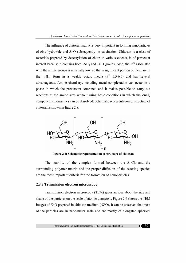
The influence of chitosan matrix is very important in forming nanoparticles
of zinc hydroxide and ZnO subsequently on calcination. Chitosan is a class of
materials prepared by deacetylation of chitin to various extents, is of particular
interest because it contains both -NH2 and –OH groups. Also, the PKa associated
with the amine groups is unusually low, so that a significant portion of them are in
the –NH2 form in a weakly acidic media (PH 5.5-6.5) and has several
advantageous. Amine chemistry, including metal complexation can occur in a
phase in which the precursors combined and it makes possible to carry out
reactions at the amine sites without using basic conditions in which the ZnCl2
components themselves can be dissolved. Schematic representation of structure of
chitosan is shown in figure 2.8.
Figure 2.8: Schematic representation of structure of chitosan
The stability of the complex formed between the ZnCl2 and the
surrounding polymer matrix and the proper diffusion of the reacting species
are the most important criteria for the formation of nanoparticles.
2.3.3 Trnsmission electron microscopy
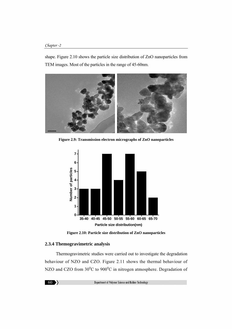
Transmission electron microscopy (TEM) gives an idea about the size and
shape of the particles on the scale of atomic diameters. Figure 2.9 shows the TEM
images of ZnO prepared in chitosan medium (NZO). It can be observed that most
of the particles are in nano-meter scale and are mostly of elongated spherical
Chapter -2
60
shape. Figure 2.10 shows the particle size distribution of ZnO nanoparticles from
TEM images. Most of the particles in the range of 45-60nm.
Figure 2.9: Transmission electron micrographs of ZnO nanoparticles
35-40 40-45 45-50 50-55 55-60 60-65 65-700
1
2
3
4
5
6
7
Num
ber o
f par
ticle
s
Particle size distribution(nm)
Figure 2.10: Particle size distribution of ZnO nanoparticles
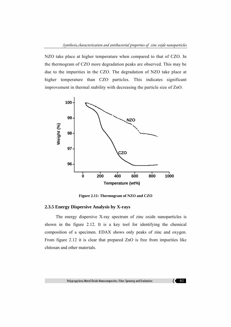
2.3.4 Themogravimetric analysis
Thermogravimetric studies were carried out to investigate the degradation
behaviour of NZO and CZO. Figure 2.11 shows the thermal behaviour of
NZO and CZO from 300C to 9000C in nitrogen atmosphere. Degradation of
Synthesis,characterization and antibacterial properties of zinc oxide nanoparticles
61
NZO take place at higher temperature when compared to that of CZO. In
the thermogram of CZO more degradation peaks are observed. This may be
due to the impurities in the CZO. The degradation of NZO take place at
higher temperature than CZO particles. This indicates significant
improvement in thermal stability with decreasing the particle size of ZnO.
0 200 400 600 800 1000
96
97
98
99
100
Wei
ght (
%)
Temperature (wt%)
NZO
CZO
Figure 2.11: Thermogram of NZO and CZO
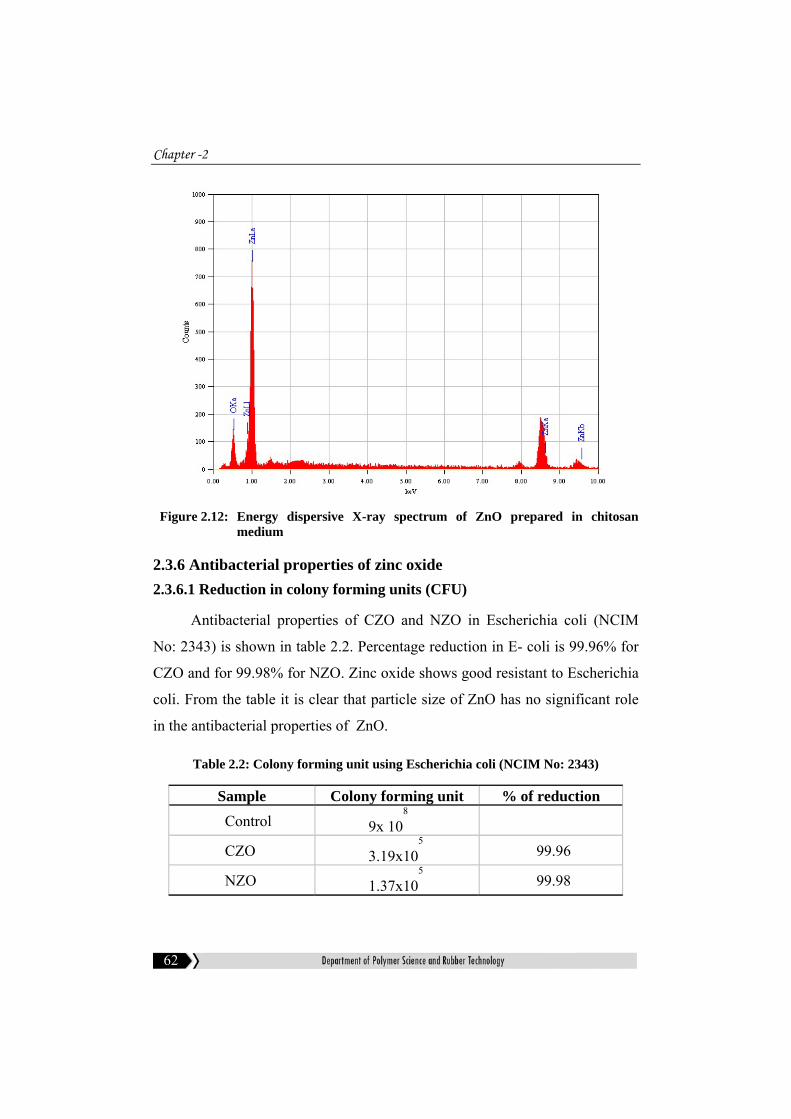
2.3.5 Energy Dispersive Analysis by X-rays
The energy dispersive X-ray spectrum of zinc oxide nanoparticles is
shown in the figure 2.12. It is a key tool for identifying the chemical
composition of a specimen. EDAX shows only peaks of zinc and oxygen.
From figure 2.12 it is clear that prepared ZnO is free from impurities like
chitosan and other materials.
Chapter -2
62
Figure 2.12: Energy dispersive X-ray spectrum of ZnO prepared in chitosan medium
2.3.6 Antibacterial properties of zinc oxide 2.3.6.1 Reduction in colony forming units (CFU)
Antibacterial properties of CZO and NZO in Escherichia coli (NCIM
No: 2343) is shown in table 2.2. Percentage reduction in E- coli is 99.96% for
CZO and for 99.98% for NZO. Zinc oxide shows good resistant to Escherichia
coli. From the table it is clear that particle size of ZnO has no significant role
in the antibacterial properties of ZnO.
Table 2.2: Colony forming unit using Escherichia coli (NCIM No: 2343)
Sample Colony forming unit % of reduction Control 9x 10
8
CZO 3.19x10⁵
99.96
NZO 1.37x10⁵
99.98

Synthesis,characterization and antibacterial properties of zinc oxide nanoparticles
63
Table 2.3 shows antibacterial properties of CZO and NZO in to Bacillus
aereus (NCIM No: 2155). Zinc oxide shows good resistant to Bacillus aereus
irrespective of the particle size.
Table 2.3: Colony forming unit using Bacillus aereus (NCIM No:2155)
Sample Colony forming unit % of reduction Control 75x 10
6
CZO 1.34x10⁶ 98.66
NZO 8x10⁵ 98.94
2.4 Conclusion
Zinc oxide nanoparticles were prepared by precipitation method in
different mediums. X-ray diffraction analysis indicates least crystallite size is
obtained when chitosan is used as the medium. From X-ray diffraction analysis
it is clear that all ZnO particles are in hexagonal wurtzite crystal structure.
Scanning electron micrographs show formation of spherical particles of ZnO
prepared in chitosan medium. Flower like morphology is observed for ZnO
prepared in ethanol medium. EDAX shows the absence of side products or
impurities after calcination. TGA analysis shows the better thermal stability of
NZO compared to that of CZO. TEM studies showed that the particle size of
smallest particle of NZO is 22nm. CZO and NZO show excellent resistance to
Bacillus aereus and Escherichia coli. Reduction in particle size has no role in
the antibacterial properties.

Chapter -2
64
References
[1] Franklin N M, Rogers N J, Apte S C, Batley G E, Gadd G E, Casey P S, Environ Sci Tech,2007, 41, 8487.
[2] Jung H, Choi H, Appl Catal B, 2006,66, 288 .
[3] Sayyadnejad M A, Ghaffarian H R, Saeidi M, Int J Environ Sci Tech, 2008,5,565.
[4] Carrion F G, Sanes J, Bermudez M D, Wear, 2007, 262, 1504.
[5] Hernandez Battez H, Gonzalez R, Viesca J L, Fernandez J E, Dlaz Fernandez J M, Machado A, Chou R, Riba J, Wear, 2008,265, 422.
[6] Rao K J, Mahesh K, Kumar S, Bull. Mater. Sci., 2005, 28, 19.
[7] Kumpika T, Thongsuwan W, Singjai P, Thin solid Films, 2008,516, 5640.
[8] Park S B, Kang Y C, J. Aerosol Sci., 1997, 28, S473.
[9] Rao A R , Dutta V, Nanotechnology, 2008, 19, 445712.
[10] Wei D, Unalan H E, Han D, Zhang Q, Niu L, Amaratunga G, Ryhanen T, Nanotechnology,2008, 19, 424006.
[11] Carrey J, Carrere H, Khan M L, Chaudret B, Marie X, Respaud M, Semicond. Sci. Technol.,2008,23,025003.
[12] Ge C, Xie C, Hu M, Gui Y, Bai Z, Zeng D, Mater. Sci. Eng. B, 2007,141, 43.
[13] Rout C S, Hegde M, Govindaraj A, Rao C N R, Nanotechnology, 2007,18, 205504.
[14] Rout C S, Raju A R, Govindaraj A, Rao C N R, Solid State Commun, 2006,138, 136.
[15] Kenanakis G, Vernardou D, Koudoumas E, Kiriakidis G, Katsarakis N, Sens. Actuators B, 2007, 124,187.
[16] Singhal M, Chhabra V, Kang P, Shah D O, Mater. Res. Bull., 1997, 32, 239.
[17] Feldmann C, Adv. Fundam. Mater., 2003,13,101.

Synthesis,characterization and antibacterial properties of zinc oxide nanoparticles
65
[18] Kitano M, Shiojiri M, Powder Technol., 1997,93, 267.
[19] Ozgur U, Alivov Y I, Liu C, Teke A, Reshchikov M A, Dogan S, Avrutin V, Cho S J, Morkoc H, J. Appl. Phys., 2005, 98, 041301.
[20] Yamamoto O, Int. J. Inorg. Mater., 2001, 3, 643.
[21] Yamamoto O, Komatsu M, Sawa J, Nakagawa Z E, J. Mater. Sci : Mater. Med., 2004, 15, 847.
[22] Sawai J, Shoji S, Igarashi H, Hashimoto A, Kokugan T, Shimizu M, Kojima H, Ferment J, Bioeng., 1998, 86, 521.
[23] Pearton S J, Norton D P, Ip K, Heo Y W , Steiner T, J. Vac. Sci. Technol.B, 2004, 22, 932.
[24] Brayner R, Ferrari Iliou R, Brivois N, Djediat S, Benedetti M F, Fievet F, Nano Lett., 2006, 6, 866.
[25] Jing L Q, Sun X J, Shang J, Cai W M, Xu Z L, Du Y G, Fu H G, Sol. Energy Mater. Sol. Cells, 2003, 79, 133.
[26] Shen W F, Zhao Y , Zhang C B, Thin Solid Films, 2005, 483, 382.
[27] Bae S H, Lee S Y, Jin B J,Im S, Appl. Surf. Sci., 2001, 169, 525.
[28] Niesen T P, De Guire M R, J. Electroceram., 2001, 6, 169.
[29] Bachari E M, Ben Amor S, Baud G, Jacquet M, Mater. Sci. Eng. B, 2001, 79, 165.
[30] Ibanez R U, Barrado J R R, Martin F, Brucker F, Leinen D, Surf. Coat. Technol., 2004, 675,188.
[31] Golego N, Studenikin S A, Cocivera M, J. Electrochem. Soc., 2000, 147, 1592.
[32] Chaudhuri S, Bhattacharyya D, Maity A B, A K Pal, Surf. Coat. Adv. Mater., 1997, 246, 181.
[33] Bahnemann D, Sol. Energy, 2004, 77, 445.
[34] Anne Aimable, Maria Teresa Buscaglia, Vincenzo Buscaglia, Paul Bowen, Journal of the European Ceramic Society, 2010, 30, 591.
Chapter -2
66
[35] Fotou G P, Pratsinis S E, Chem. Eng. Commum., 1996,151, 251.
[36] Mondelaers D, Vanhoyland G,Van den Rul H, Haen J D,Van Bael M K, Mullens J, Van Poucke L C, Mater. Res. Bull., 2002, 37, 901.
[37] Tsuzuki T, McCormick P G, Scripta Mater., 2001, 44, 1731.
[38] Singhal M, Chhabra V, Kang P, Shah D O, Mater. Res. Bull., 1997, 32 , 239.
[39] Okuyama K, Lenggoro I W, Chem. Eng. Sci., 2003, 58, 537.
[40] Rataboul F, Nayral C, Casanove M J, Maisonnat A, Chaudret B, J. Organomet. Chem. 2002, 307, 643644 .
[41] Sato T, Tanigaki T, Suzuki H, Saito Y, Kido O, Kimura Y, Kaito C, Takeda A, Kaneko S, J. Cryst. Growth, 2003, 313, 255.
[42] Viswanathan R, Lilly G D, Gale W F, Gupta R B, Ind. Eng. Chem. Res., 2003, 42 ,5535.
[43] Koh Y W, Lin M, Tan C K, Foo Y L, Loh K P, J. Phys.Chem. B, 2004,108,11419.
[44] Yu W D, Li X M, Gao X D, Cryst. Growth Des., 2005, 5 , 151.
[45] Hu X L, Zhu Y J, Wang S W, Mater. Chem. Phys., 2004,421.
[46] Wang J M, Gao L, Inorg. Chem. Commun., 2003, 6, 877.
[47] Kim J H, Choi W C, Kim H Y, Kang Y, Park Y K, Powder Technol.,2005, 153, 166.
[48] Wang L N, Muhammed M, J. Mater. Chem., 1999, 9, 2871.
[49] Rodriguez-Paez J E, Caballero A C, Villegas M, Moure C, Duran P, Fernandez J F, J. Eur. Ceram. Soc., 2001, 21, 925.
[50] Purica M, Budianu E, Rusu E, Danila M, Gaurila R, Thin Solid Films,2002, 403, 485.
[51] Audebrand N, Auffredic J P, Louer D, Chem. Mater., 1998,10, 2450.
[52] Yang Y, Chen H, Zhao B, Bao X, J. Cryst. Growth, 2004,263, 447.
Synthesis,characterization and antibacterial properties of zinc oxide nanoparticles
67
[53] Liu B, Zeng H C, J. Am. Chem. Soc. , 2003, 125, 4430.
[54] Lu C H, Yeh C H, Ceram. Int., 2000, 26, 351.
[55] Zhu Y, Zhou Y, Appl. Phys. A, 2008, 92, 275.
[56] Tani T, Madler L, Pratsinis S E, J. Nanopart. Res., 20020, 4, 337.
[57] Hingorani S, Pillai V, Kumar P, Multani M S, D. O. Shah D O, Mat. Res. Bull., 1993, 28, 1303.
[58] Hingorani S, Shah D O, J. Mater. Res., 1995, 10, 461.
[59] Shingal M, Chhabra V, Kang P, Shah D O, Mat. Res. Bull., 1997,32, 239.
[60] Lim B P, Wang J, Ng S C, Chew C H, Gan L M, Ceram. Int., 1998, 24, 205.
[61] Inoguchi M, Suzuki K, Kageyama K, Takagi H, Sakabe Y, J. Am. Ceram. Soc., 2008, 91, 3850.
[62] Li W J, Shi E W, Zhong W Z and Yin Z , J. Cryst. Growth, 1999, 203, 186.
[63] Wang B G, Shi E W, Zhong W Z , Cryst. Res. Technol., 1998, 33, 937.
[64] Lu C H , Yeh C H , Ceram. Int., 2000, 26, 351.
[65] Neves M C, Trindade T, Timmons A M B, Pedrosa de Jesus J D, Mater. Res. Bull. 2001, 36, 1099.
[66] Pal U and Santiago P J. Phys. Chem. B,2005, 109, 15317.
[67] Vayssieres L , Adv. Mater.,2003, 15, 464
[68] Hu J Q, Li Q,Wong N B, Lee C S and Lee S T , Chem. Mater.,2002, 14, 1216.
[69] Sun Y, Fuge G M, Fox N A, Riley D J and Ashfold M N R,Adv. Mater. 2005, 17, 2477.
[70] Gui Z, Liu J, Wang Z Z, Song L, Hu Y, Fan W C and Chen D Y , J. Phys. Chem. B, 2005, 109, 1113.
[71] Ni Y H, Wei X W, Ma X and Hong J M, J. Cryst. Growth, 2005, 283, 48.
[72] LI Yan, FENG Hui-yun, ZHANG Nan, LIU Chuan-sheng, Trans. nonferrous Met.Soc. China, 2010, 20, 119.

Chapter -2
68
[73] Tengfa Long, Shu Yin , Kouta Takabatake ,Peilin Zhnag ,Tsugio Sato, Nanoscale Res Lett 2009, 4,247.
[74] Yoshie Ishikawa, Yoshiki Shimizu, Takeshi Sasaki, Naoto Koshizaki , Journal of Colloid and Interface Science,2006, 300, 612.
[75] Ameer Azam, Faheem Ahmed, Nishat Arshi, Chaman M, Naqvi A H, International Journal of Theoretical & Applied Sciences, 2009, 12.
….. …..